THE DEAD PARTS OF CELL 3 rd meeting 1

Welcome message from author
This document is posted to help you gain knowledge. Please leave a comment to let me know what you think about it! Share it to your friends and learn new things together.
Transcript
8/3/2019 03-The Dead Parts of Cell
http://slidepdf.com/reader/full/03-the-dead-parts-of-cell 1/22
THE DEAD PARTS OF CELL
3rd
meeting
1

8/3/2019 03-The Dead Parts of Cell
http://slidepdf.com/reader/full/03-the-dead-parts-of-cell 2/22
BASE COMPETENCE
THE STUDENTS UNDERSTAND STRUCTURE
AND FUNCTION OF THE PLANT CELL PARTS
INDICATOR
The students can identify many dead partsof cell from the picture of plant cell
The students can mention the function of
many dead parts of cell
2

8/3/2019 03-The Dead Parts of Cell
http://slidepdf.com/reader/full/03-the-dead-parts-of-cell 3/22
THE DEAD PARTS OF CELL
CONSIST OF:
VACUOLESERGASTIC SUBSTANCES
CELL WALL
3

8/3/2019 03-The Dead Parts of Cell
http://slidepdf.com/reader/full/03-the-dead-parts-of-cell 4/22
VACUOLES
• Occupy more than 90% of the volume of most matureplant cells
• Vacuoles surrounded by a membrane called tonoplast
• It contains various organic and inorganic substances,
such as sugars, proteins, organic acids, phosphatides,tannins, flavonoid pigments and calcium oxalate
• Some substances in the vacuole may occur in solid formand may even be crystalline
• In meristematic cells possess many minute vacuoles
• In mature cells the vacuoles enlarge form large centralvacuole
4
8/3/2019 03-The Dead Parts of Cell
http://slidepdf.com/reader/full/03-the-dead-parts-of-cell 5/22
INITIATION OF VACUOLES
• From pre-existing vacuoles which multiply byfission, and after cell division each daughtercell receive a number of vacuoles
• By a de novo process, by attraction of waterto a certain localized region in the cytoplasmand the formation of membrane around it
•
From Golghi vesicles• By dilatation of ER cisternae or vesicles
derived from the ER
5
8/3/2019 03-The Dead Parts of Cell
http://slidepdf.com/reader/full/03-the-dead-parts-of-cell 6/22
THE FUNCTION OF VACUOLES
The function of vacuoles are regulation thewater and solute content of the cell, i.e. inosmoregulation, in storage and in digestion
Vacuoles contain digestive enzymes thatcapable of breaking down the cytoplasmiccomponent and metabolites
The hydrolytic activity of vacuoles resemblesthat of lissomes of animal cells
6

8/3/2019 03-The Dead Parts of Cell
http://slidepdf.com/reader/full/03-the-dead-parts-of-cell 7/22
ERGASTIC SUBSTANCES
Reserve and waste materials produced bythe cell are called ergastic substances
The kind of ergastic substances, consistof:
1. Starch
2. Proteins
3. Oil, fats and waxes
4. Crystals and silica bodies
5. Tannins
6. Pigmentation7

8/3/2019 03-The Dead Parts of Cell
http://slidepdf.com/reader/full/03-the-dead-parts-of-cell 8/22
STARCH
a carbohydrate composed of long chainmolecules.
It appears in the form of grains
Starch grains are first form in chloroplasts
In the other case the starch is broken downand moves as sugar to storage tissueswhere it is resynthesized in amyloplast
8

8/3/2019 03-The Dead Parts of Cell
http://slidepdf.com/reader/full/03-the-dead-parts-of-cell 9/22
STARCH
• Starch grains commonlyshow layering around apoint termed hillum
•The hillum may becentrally situated or it maybe eccentric
•The layer termed lamella•In the starch grains of
cereals the number oflayer corresponds to thenumber of days duringwhich the grain growths
9
8/3/2019 03-The Dead Parts of Cell
http://slidepdf.com/reader/full/03-the-dead-parts-of-cell 10/22
PROTEINS
Amorphous protein is found in theoutermost endosperm layer, the aleuronlayer, of the caryopsis of cereals
Protein in the form of cuboidal crystalloid
is found in the cells of the peripheralparenchyma of potato tuber and in the fruitparenchyma of Capsicum
Crystalline and amorphous protein arefound together in aleurone grains in theendosperms and embryos of many seeds
10

8/3/2019 03-The Dead Parts of Cell
http://slidepdf.com/reader/full/03-the-dead-parts-of-cell 11/22
Endosperm of seed
Zea mays
11

8/3/2019 03-The Dead Parts of Cell
http://slidepdf.com/reader/full/03-the-dead-parts-of-cell 12/22
OIL, FATS AND WAXES
Oil and fats are important reserve materialsin plants
Most commonly present in seeds and fruits
Fats and oils may be produced by elaioplastor spherosomes
Lipid compounds others than fats, oils andwaxes
12
8/3/2019 03-The Dead Parts of Cell
http://slidepdf.com/reader/full/03-the-dead-parts-of-cell 13/22
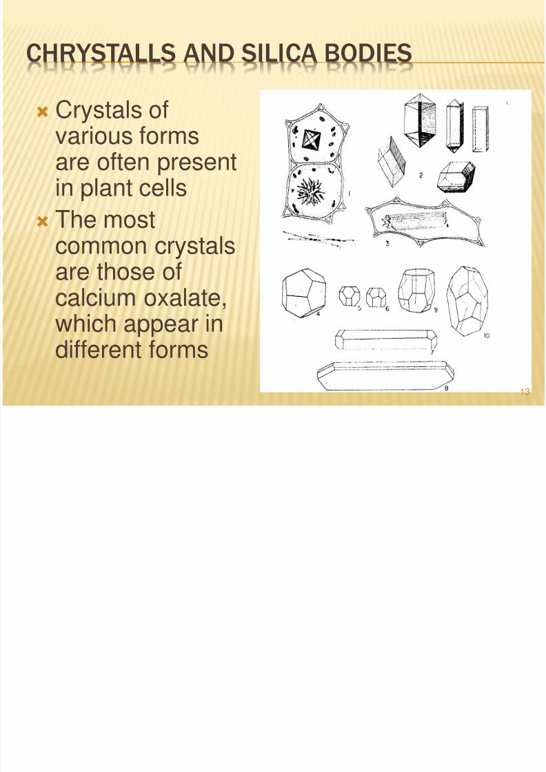
CHRYSTALLS AND SILICA BODIES
Crystals ofvarious formsare often present
in plant cells The most
common crystalsare those ofcalcium oxalate,which appear indifferent forms
13
8/3/2019 03-The Dead Parts of Cell
http://slidepdf.com/reader/full/03-the-dead-parts-of-cell 14/22
TANNINS
They are a heterogeneous group of phenolderivates
In microscopically section they usually appears asgranular masses or bodies colored yellow, red or
brown Tannin can be found in leaves, periderm, vascular
tissue, unripe fruits, seed coats, and in tissues ofpathogenic growth
Tannins may be found in the vacuoles or in theform of droplet in the cytoplasm
Tannins are used commercially especially in theindustry of the tanning of animal skins to obtain
leather 14
8/3/2019 03-The Dead Parts of Cell
http://slidepdf.com/reader/full/03-the-dead-parts-of-cell 15/22
PIGMENTATION
The plant pigment usually found in theplastids and in the vacuoles
The green color is due to chlorophyll whichis found in the chloroplasts
Carotenoids is the yellow to red pigments,are also found but they are masked by thechlorophyll, they become noticeable whenthere is a little or no chlorophyll
15
8/3/2019 03-The Dead Parts of Cell
http://slidepdf.com/reader/full/03-the-dead-parts-of-cell 16/22
PIGMENTATION
Flavonoids, generally present in vacuoles ,water soluble, they found in flowers and fruits
Anthocyanins, give red, pink, lilac and bluecolor
In acid solution the color varies from orange-red to lilac
In basic solutions blue-colouredanthocyanins are form
16

8/3/2019 03-The Dead Parts of Cell
http://slidepdf.com/reader/full/03-the-dead-parts-of-cell 17/22
CELL WALL
The present of a wall in plant celldistinguishes them from animal cells
The cell wall growth when in contact with theprotoplast but out side of it
The cell wall consist of cellulose micro fibrils
The matrix of cell wall consist mainly ofpectin substances and hemicelluloses
17
8/3/2019 03-The Dead Parts of Cell
http://slidepdf.com/reader/full/03-the-dead-parts-of-cell 18/22
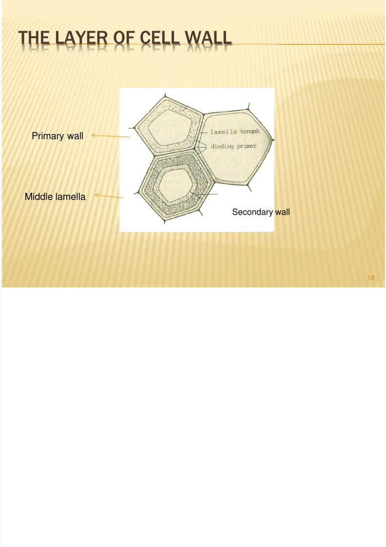
THE LAYER OF CELL WALL
Secondary wall
Primary wall
Middle lamella
18

8/3/2019 03-The Dead Parts of Cell
http://slidepdf.com/reader/full/03-the-dead-parts-of-cell 19/22
THE STRUCTURE OF CELL WALL
•The chain likecellulosemolecules are
regularly arrangein bundles.•Each bundle,which form anelementary fibril,consist of about40 molecules
19
8/3/2019 03-The Dead Parts of Cell
http://slidepdf.com/reader/full/03-the-dead-parts-of-cell 20/22
THE GROWTH OF CELL WALL
The growth of cell wall is a accomplished byintussusception or by apposition
According to Frey-Wyssling and Stecher (1951),suggested that the primary cell wall grows in a way thathas been termed mosaic growth . The fibrillar texture incertain wall areas become loosened as a result of turgorpressure and afterwards mended by deposition of newmicro fibrils in the gaps
Another theory termed multinet growth (Houwink and
Ruelofsen, 1954). According to this theory the thickeningof the primary wall is brought about by the separation ofthe crossed microfibrils and alteration in their orientationin the earliest formed lamellae , from being almosttransverse to almost longitudinal
20

8/3/2019 03-The Dead Parts of Cell
http://slidepdf.com/reader/full/03-the-dead-parts-of-cell 21/22
NOKTAH (PIT)
Certain portion of the cell wall that remainthin even as the secondary wall is formed,called noktah or pit
The pits are apparently areas through whichsubstances pass from cell to cell
Generally each pit has a complementary pit
exactly opposite it in the wall of theneighboring cell
21

8/3/2019 03-The Dead Parts of Cell
http://slidepdf.com/reader/full/03-the-dead-parts-of-cell 22/22
SURFACE OF PITS
22
Related Documents