Dr Jaffar Raza Syed Page 1 Acute Gingival Infections CLASSIFICATION a. Traumatic lesions of gingiva: • Physical injury • Chemical injury b. Viral infections: • Acute herpetic gingivostomatitis • Herpangina • Hand, foot and mouth diseases • Measles • Herpes varicella/zoster virus infections • Glandular fever c. Bacterial infections: • Necrotizing ulcerative gingivitis • Tuberculosis • Syphilis d. Fungal diseases: • Candidiasis e. Gingival abscess f. Aphthous ulceration g. Erythema multiforme h. Drug allergy

Welcome message from author
This document is posted to help you gain knowledge. Please leave a comment to let me know what you think about it! Share it to your friends and learn new things together.
Transcript
Dr Jaffar Raza Syed Page 1
Acute Gingival Infections
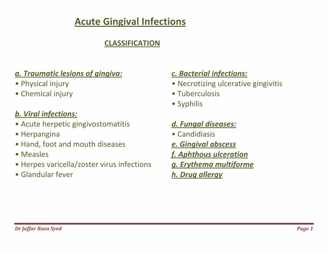
CLASSIFICATION a. Traumatic lesions of gingiva: • Physical injury • Chemical injury b. Viral infections: • Acute herpetic gingivostomatitis • Herpangina • Hand, foot and mouth diseases • Measles • Herpes varicella/zoster virus infections • Glandular fever
c. Bacterial infections: • Necrotizing ulcerative gingivitis • Tuberculosis • Syphilis d. Fungal diseases: • Candidiasis e. Gingival abscess f. Aphthous ulceration g. Erythema multiforme h. Drug allergy
Dr Jaffar Raza Syed
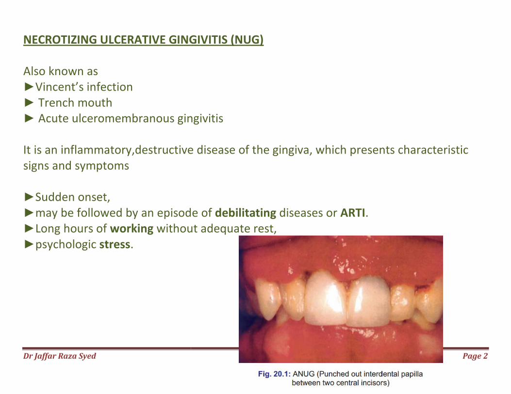
NECROTIZING ULCERATIVE GINGIVITIS (NUG) Also known as ►Vincent’s infection ► Trench mouth ► Acute ulceromembranous gingivitis It is an inflammatory,destructive disease of the gingiva, which presents characteristic signs and symptoms ►Sudden onset, ►may be followed by an episode of ►Long hours of working withou►psychologic stress.
NECROTIZING ULCERATIVE GINGIVITIS (NUG)
Acute ulceromembranous gingivitis
It is an inflammatory,destructive disease of the gingiva, which presents characteristic
may be followed by an episode of debilitating diseases or ARTI. without adequate rest,
Page 2
It is an inflammatory,destructive disease of the gingiva, which presents characteristic
Dr Jaffar Raza Syed Page 3
Signs and Symptoms ►Punched out, crater-like depressions at the crest of the interdental papillae, subsequently involving marginal gingiva and rarely attached gingiva ►grayish pseudomembranous slough ►gingival hemorrhage or pronounced bleeding on the slightest stimulation. ►Fetid odor and increased salivation. ►extremely sensitive to touch
Dr Jaffar Raza Syed Page 4
►constant radiating, gnawing pain that is intensified by eating spicy or hot foods and chewing ►metallic foul taste ►pasty saliva ►local lymphadenopathy ►elevation in temperature
Dr Jaffar Raza Syed
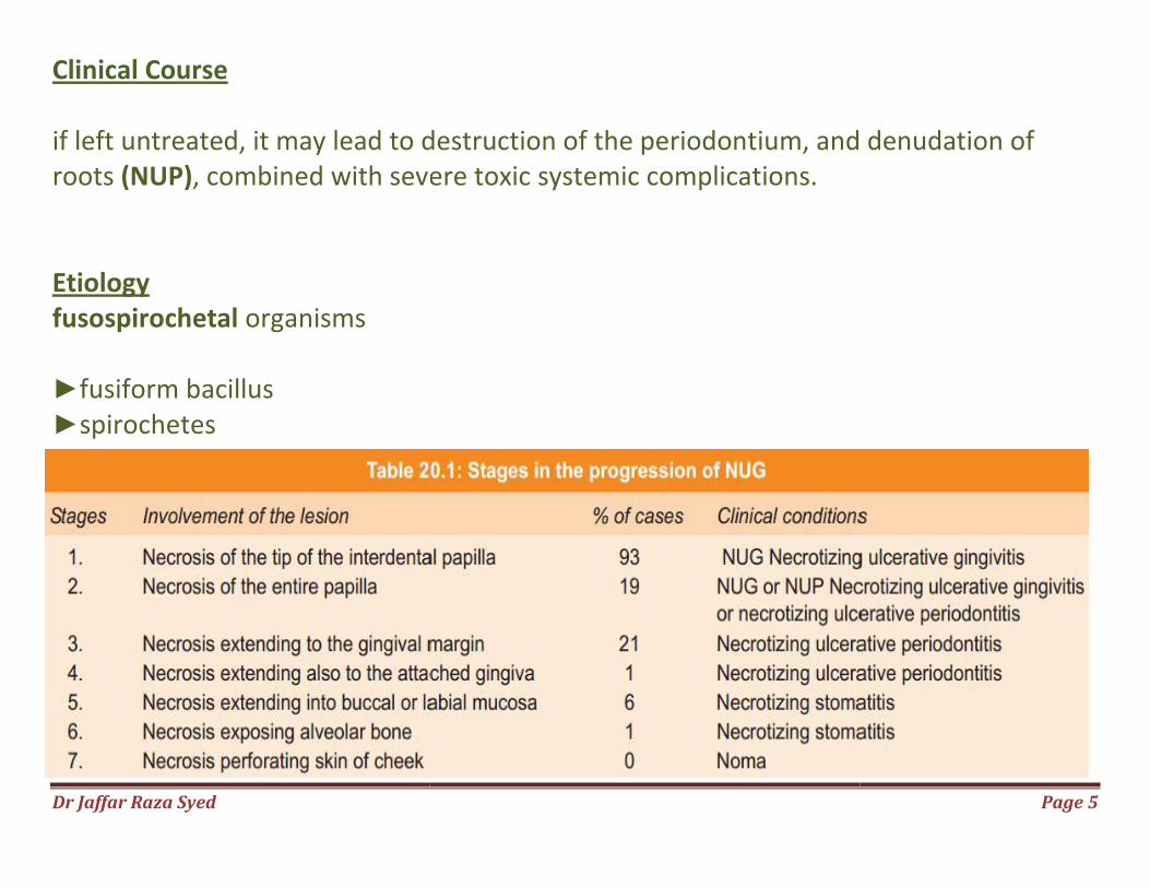
Clinical Course if left untreated, it may lead to destruction of the periodontium, and denudation of roots (NUP), combined with severe toxic Etiology fusospirochetal organisms ►fusiform bacillus ►spirochetes
if left untreated, it may lead to destruction of the periodontium, and denudation of , combined with severe toxic systemic complications.
Page 5
if left untreated, it may lead to destruction of the periodontium, and denudation of
Dr Jaffar Raza Syed Page 6
Local Predisposing Factors Most important predisposing factors are: i. Pre-existing gingivitis ii. Injury to the gingiva iii. Smoking Systemic Predisposing Factors ►Nutritional deficiency ►Debilitating diseases ►Psychosomatic factors activation of the hypothalamic pituitary adrenal axis ↑ cor�sol levels ↓ lymphocyte and polymorphonuclear leukocytes func�on
Dr Jaffar Raza Syed Page 7
Relationship of Bacteria to the Characteristic Lesions four zones 1. Zone I—Bacterial zone: It is the most superficial zone, consists of varied bacteria, including a few Spirochetes of the small, medium-sized and large types. 2. Zone II—Neutrophil-rich zone: Contains numerous leukocytes predominantly neutrophils with bacteria including spirochetes of various types. 3. Zone III—Necrotic zone: Consists of a dead tissue cells, remnants of connective tissue fragments, and numerous spirochetes. 4. Zone IV—Zone of spirochetal infiltration: Consists of a well preserved tissue infiltrated with spirochetes of intermediate and large-sized without other organisms.
Dr Jaffar Raza Syed Page 8
Treatment Treatment for Non-ambulatory Patients Day 1: a. gently removing the necrotic pseudomembrane with a pellet of cotton saturated with hydrogen peroxide (H2O2). b. Advised bed rest and rinse the mouth every 2 hours with a diluted 3 percent hydrogen peroxide (H2O2). c. Systemic antibiotics like penicillin or metronidazole can be prescribed.
Dr Jaffar Raza Syed Page 9
Day 2: After 24 hours, a bedside visit should be made. The treatment again includes gently swab the area with hydrogen peroxide, instructions of the previous day are repeated. Day 3: Most cases, the condition will be improved, start the treatment for ambulatory patients.
Dr Jaffar Raza Syed Page 10
Treatment for Ambulatory Patients First visit: ►topical anesthetic ►gently swabbed with a cotton pellet to remove pseudomembrane and non-attached surface debris. ►area is cleansed with warm water ►superficial calculus is removed with ultrasonic scalers. ►Antibiotics prescription ►Subgingival scaling and curettage are contraindicated Instructions to the patient 1. Avoid smoking and alcohol. 2. Rinse with 3 percent hydrogen peroxide and warm water for every two hours. 3. Confine toothbrushing to the removal of surface debris with a bland dentifrice, use of interdental aids and chlorhexidine mouth rinse are recommended.
Dr Jaffar Raza Syed Page 11
Second visit: ►Scalers and curettes are added to the instrumentarium. ►Shrinkage of the gingiva may expose previously covered calculus which is gently removed. ►Same instructions are reinforced. Third visit: ►Scaling and root planing are repeated, ►Plaque control instructions are given. ►Hydrogen peroxide rinses are discontinued. Fourth visit: ►Oral hygiene instructions are reinforced ►thorough scaling and root planing are performed.
Dr Jaffar Raza Syed Page 12
Fifth visit: ►Appointments are fixed for treatment of chronic gingivitis, periodontal pockets and pericoronal flaps, and for the elimination of all local irritants. ►Patient is placed on maintenance program. Further Treatment Considerations 1. Gingivoplasty. 2. Systemic antibiotics—only in patients with toxic systemic complications. 3. Supportive systemic treatment—copious fluid consumption and administration of analgesics and adequate bed rest. 4. Nutritional supplements—vitamin B/C supplements.
Dr Jaffar Raza Syed Page 13
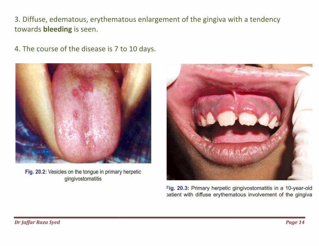
ACUTE HERPETIC GINGIVOSTOMATITIS (AHG) ►viral infection of the oral mucous membrane caused by HSV I and II ►occurs most frequently in infants and children younger than 6 years of age but is also seen in adults. Clinical Features 1. appears as a diffuse, shiny erythematous, involvement of the gingiva and the adjacent oral mucosa with varying degrees of edema and gingival bleeding. 2. In its initial stage it may appear as discrete, spherical, clusters of vesicles dispersed in different areas, e.g. labial and buccal mucosa, hard palate, pharynx and tongue. After approximately 24 hours the vesicles rupture and form painful shallow ulcers with scalloped borders and surrounding erythema.
Dr Jaffar Raza Syed
3. Diffuse, edematous, erythematous enlargement of the gingiva with a tendency towards bleeding is seen. 4. The course of the disease is 7 to 10 days.
3. Diffuse, edematous, erythematous enlargement of the gingiva with a tendency
4. The course of the disease is 7 to 10 days.
Page 14
3. Diffuse, edematous, erythematous enlargement of the gingiva with a tendency
Dr Jaffar Raza Syed
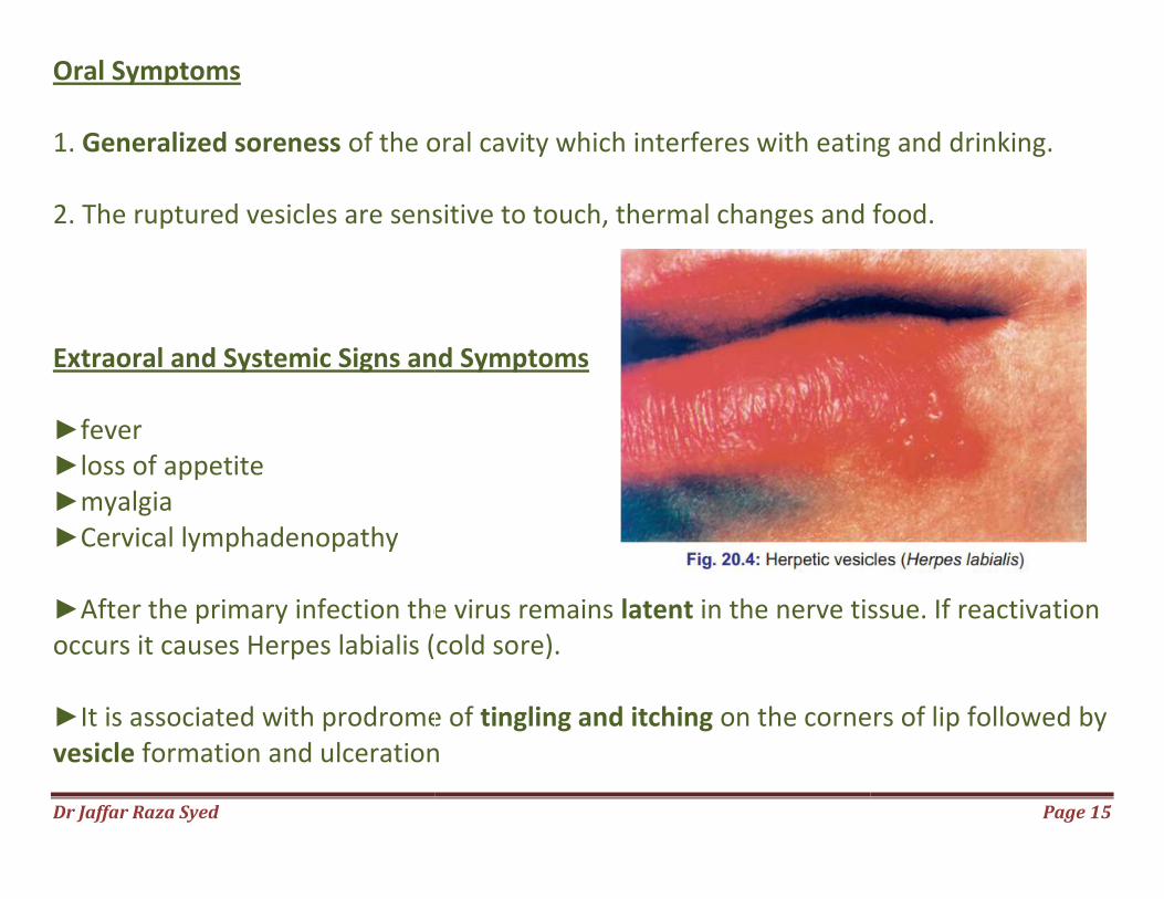
Oral Symptoms 1. Generalized soreness of the oral cavity which interferes with eating and drinking. 2. The ruptured vesicles are sensitive to touch, thermal changes and food. Extraoral and Systemic Signs and Symptoms ►fever ►loss of appetite ►myalgia ►Cervical lymphadenopathy ►After the primary infection the virus remains occurs it causes Herpes labialis (cold so ►It is associated with prodrome of vesicle formation and ulceration
of the oral cavity which interferes with eating and drinking.
2. The ruptured vesicles are sensitive to touch, thermal changes and food.
d Systemic Signs and Symptoms
After the primary infection the virus remains latent in the nerve tissue. If reactivation occurs it causes Herpes labialis (cold sore).
It is associated with prodrome of tingling and itching on the corners of lip followed by formation and ulceration
Page 15
of the oral cavity which interferes with eating and drinking.
2. The ruptured vesicles are sensitive to touch, thermal changes and food.
in the nerve tissue. If reactivation
on the corners of lip followed by
Dr Jaffar Raza Syed
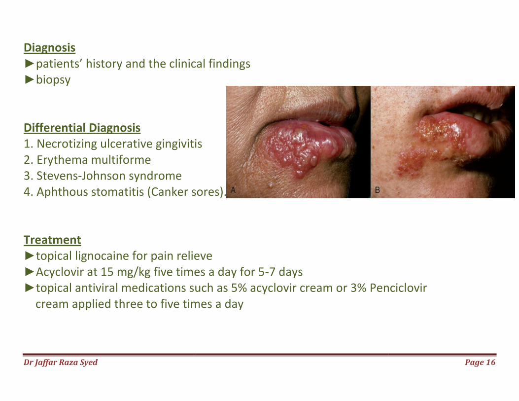
Diagnosis ►patients’ history and the clinical findings►biopsy Differential Diagnosis 1. Necrotizing ulcerative gingivitis2. Erythema multiforme 3. Stevens-Johnson syndrome 4. Aphthous stomatitis (Canker sores). Treatment ►topical lignocaine for pain relieve►Acyclovir at 15 mg/kg five times a day for 5►topical antiviral medications such as 5% acyclovir cream or 3% cream applied three to five times a day
patients’ history and the clinical findings
1. Necrotizing ulcerative gingivitis
4. Aphthous stomatitis (Canker sores).
topical lignocaine for pain relieve Acyclovir at 15 mg/kg five times a day for 5-7 days topical antiviral medications such as 5% acyclovir cream or 3% Penciclovir cream applied three to five times a day
Page 16
Penciclovir
Dr Jaffar Raza Syed
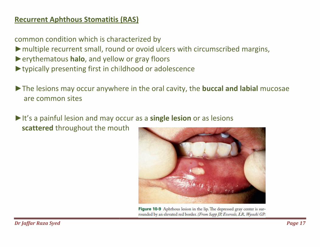
Recurrent Aphthous Stomatitis (RAS common condition which is characterized by ►multiple recurrent small, round or ovoid ulcers with circumscribed►erythematous halo, and yellow or gray floors►typically presenting first in childhood or adolescenc ►The lesions may occur anywhere in the oral cavity, the are common sites ►It’s a painful lesion and may occur as a scattered throughout the mout
Recurrent Aphthous Stomatitis (RAS)
common condition which is characterized by recurrent small, round or ovoid ulcers with circumscribed
, and yellow or gray floors typically presenting first in childhood or adolescence
The lesions may occur anywhere in the oral cavity, the buccal and labial
lesion and may occur as a single lesion or as lesions throughout the mouth
Page 17
recurrent small, round or ovoid ulcers with circumscribed margins,
buccal and labial mucosae
Dr Jaffar Raza Syed Page 18
Types Minor aphthae: ►Is the most common affecting about 80% of patients with RAS ►ulcers are round or oval usually <5 mm in diameter with a gray-white pseudomembrane and an erythematous halo. ►The ulcers heal within 10-14 days without scarring. Major aphthae: ►Is a rare severe form of Aphthous ulcer. ►Ulcers are oval and may exceed 1 cm in diameter. ►Ulcers persist for up to 6 weeks and often heal with scarring.
Dr Jaffar Raza Syed Page 19
Herpetiform aphthae: ►least common variety ►characterized by multiple recurrent crops of widespread small, painful ulcers. ►As many as 100 ulcers may be present at a given time, ►each measuring 2-3 mm in diameter.
Dr Jaffar Raza Syed Page 20
Etiology ►Unknown ►linked to RAS are genetic predisposition, ►Hematinic deficiencies, ►Immunologic abnormalities, ►stress, ►food allergy ►gastrointestinal disorders. ►Predisposing factors include hormonal disturbances, trauma, cessation of smoking and menstruation Treatment ►topical lignocaine ►Topical steroids like Triamcinolone and Clobetasol ►systemic steroids and Thalidomide to reduce the number of ulcers and recurrences.
Dr Jaffar Raza Syed
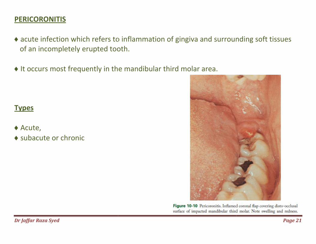
PERICORONITIS
acute infection which refers to inflammation of of an incompletely erupted tooth.
It occurs most frequently in the mandibular Types
Acute,
subacute or chronic
acute infection which refers to inflammation of gingiva and surrounding soft tissues erupted tooth.
It occurs most frequently in the mandibular third molar area.
Page 21
gingiva and surrounding soft tissues
Dr Jaffar Raza Syed Page 22
Signs and Symptoms
markedly red, edematous suppurating lesion that is extremely tender with radiating pain to the ear, throat and floor of the mouth
foul taste and inability to close the jaws.
swelling of the cheek
interferes with complete jaw closure
flap is traumatized by contact with the opposing jaw and inflammatory involvement is aggravated.
toxic systemic complications such as fever, leukocytosis and malaise
Dr Jaffar Raza Syed Page 23
Complications
Localized pericoronal abscess or cyst formation
may spread posteriorly into the oropharyngeal area and medially into the base of the tongue, making it difficult for the patient to swallow Peritonsillar abscess formation, cellulitis and Ludwig’s angina are the potential complications Treatment The treatment of pericoronitis depends on:
• Severity of the inflammation.
• The systemic complications, and
• The advisability of retaining the involved tooth
Dr Jaffar Raza Syed Page 24
First Visit
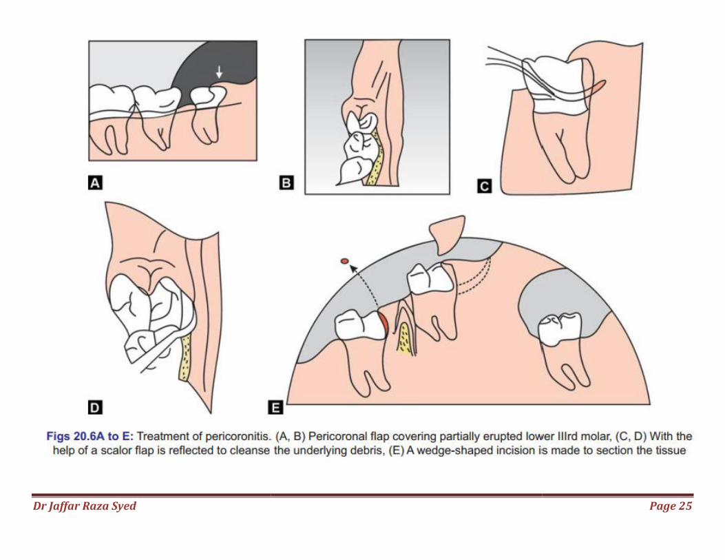
warm water flush + topical anesthetic agent
flap is reflected with a scaler and the underlying debris is also removed
hourly rinses instructions
copious fluid intake
systemic antibiotics
If the gingival flap is swollen and fluctuant an antero-posterior incision to establish drainage is made with a No. 15 bard parker blade
followed by insertion of 1/4th inch gauze wick
In the next visit, determination is made as to whether the tooth is to be retained or extracted
Dr Jaffar Raza Syed
Page 25
Related Documents