Podocin and MEC-2 bind cholesterol to regulate the activity of associated ion channels Tobias B. Huber*, Bernhard Schermer*, Roman Ulrich Mu ¨ ller*, Martin Ho ¨ hne*, Malte Bartram*, Andrea Calixto † , Henning Hagmann*, Christian Reinhardt* ‡ , Fabienne Koos*, Karl Kunzelmann § , Elena Shirokova ¶ , Dietmar Krautwurst ¶ , Christian Harteneck , Matias Simons*, Hermann Pavensta ¨dt ‡ , Dontscho Kerjaschki**, Christoph Thiele †† , Gerd Walz*, Martin Chalfie †‡‡ , and Thomas Benzing* §§ *Renal Division, University Hospital Freiburg, 79106 Freiburg, Germany; ‡ University Hospital Mu ¨ nster, D-48129 Mu ¨ nster, Germany; † Department of Biological Sciences, Columbia University, New York, NY 10027-6902; § Department of Physiology, University of Regensburg, 93053 Regensburg, Germany; ¶ Department of Molecular Genetics, German Institute of Human Nutrition, 14558 Nuthetal, Germany; Pharmacology, Charite ´ , University of Berlin, 14050 Berlin, Germany; **Department of Pathology, University of Vienna, A-1010 Vienna, Austria; and †† Max Planck Institute of Molecular Cell Biology and Genetics, 01307 Dresden, Germany This contribution is part of the special series of Inaugural Articles by members of the National Academy of Sciences elected on April 20, 2004. Contributed by Martin Chalfie, September 5, 2006 The prohibitin (PHB)-domain proteins are membrane proteins that regulate a variety of biological activities, including mechanosen- sation, osmotic homeostasis, and cell signaling, although the mechanism of this regulation is unknown. We have studied two members of this large protein family, MEC-2, which is needed for touch sensitivity in Caenorhabditis elegans, and Podocin, a protein involved in the function of the filtration barrier in the mammalian kidney, and find that both proteins bind cholesterol. This binding requires the PHB domain (including palmitoylation sites within it) and part of the N-terminally adjacent hydrophobic domain that attaches the proteins to the inner leaflet of the plasma membrane. By binding to MEC-2 and Podocin, cholesterol associates with ion-channel complexes to which these proteins bind: DEGENaC channels for MEC-2 and TRPC channels for Podocin. Both the MEC-2-dependent activation of mechanosensation and the Podocin-dependent activation of TRPC channels require choles- terol. Thus, MEC-2, Podocin, and probably many other PHB-domain proteins by binding to themselves, cholesterol, and target proteins regulate the formation and function of large protein– cholesterol supercomplexes in the plasma membrane. prohibitin-domain proteins TRP channels DEG/ENaC channels slit diaphragm mechanosensation T he prohibitin homology (PHB)-domain proteins constitute a family of 1,800 proteins (SMART database; http:smart. embl-heidelberg.de) (1) all of which share an 150-aa domain similar to that in the mitochondrial protein prohibitin (2). More than 340 of these proteins, many of which have an N-terminal adjacent hydrophobic region that places them on the inner leaf let of the lipid bilayer, have been identified in animals. These membrane-associated proteins regulate osmotic homeostasis, mechanosensation, and cell signaling (3–5). Several of the ani- mal PHB-domain proteins including flotillin, Podocin, prohib- itin, stomatin, UNC-1, UNC-24, and the UNC-24-like mamma- lian protein SLP-1 are found in cholesterol-rich membrane fractions derived from the plasma membrane (reviewed in ref. 2). In this article, we investigate the function of these proteins using two members of the family, MEC-2 from Caenorhabditis elegans and Podocin from mouse. MEC-2 (6) and Podocin (7) have a single, central hydrophobic domain that embeds these proteins in the inner leaflet of the plasma membrane with their N- and C-terminal tails facing the cytoplasm (Fig. 1a). Although the two proteins contain different N and C termini, they have hydrophobic regions that are 35% identical and 75% similar and PHB-domains that are 50% identical and 80% similar (Fig. 1b). The PHB domain is critical for the action of both proteins (8, 9). The role of these conserved domains and the function of PHB-domain proteins, however, are unclear. MEC-2 is part of a multiprotein-channel complex with the degenerinepithelial Na channel (DEGENaC) proteins MEC-4 and MEC-10 that transduces gentle touch (6, 10, 11). In touch-receptor neurons, this channel complex is localized to regular puncta along the neuronal process; MEC-2 has been shown to regulate the MEC-4MEC-10 ion channel in these puncta (9). However, the mechanism by which MEC-2 regulates ion-channel activity is elusive. Podocin localizes specifically to the slit diaphragm of the mammalian kidney. The slit-diaphragm is a specialized intercel- lular junction that connects adjacent foot processes of kidney podocytes and is part of the glomerular-filtration barrier (12). Slit diaphragm proteins induce signal transduction in podocytes, the visceral epithelial cells of the kidney glomerulus, that regulates cytoskeletal rearrangement and transcriptional activity (3). Like MEC-2, Podocin is part of a multiprotein complex containing the transmembrane proteins Neph1, Neph2, and Nephrin and the cytoplasmic adaptor protein CD2AP (3). Mutations in Podocin and the other proteins of this complex result in focal segmental glomerulosclerosis and congenital nephrotic syndrome, severe genetic kidney disorders in humans characterized by disruption of the filtration barrier (7, 13, 14). Recently, mutations in the transient receptor potential C chan- nel protein TRPC6 were also shown to cause focal segmental glomerulosclerosis (15, 16). In this article, we show that Podocin and MEC-2 are choles- terol-binding proteins and that cholesterol binding plays an important role in regulating the activity of ion channels to which these PHB-domain proteins bind. Podocin, as we show here, Author contributions: T.B.H., B.S., and R.U.M. contributed equally to this work. T.B.H., B.S., R.U.M., M.H., M.B., A.C., H.H., C.R., F.K., K.K., E.S., D. Krautwurst, C.H., M.S., D. Kerjaschki, G.W., M.C., and T.B. designed research; T.B.H., B.S., R.U.M., M.H., M.B., A.C., H.H., C.R., F.K., K.K., E.S., D. Krautwurst, C.H., M.S., D. Kerjaschki, M.C., and T.B. performed research; C.H., H.P., and C.T. contributed new reagentsanalytic tools; T.B.H., B.S., R.U.M., M.H., M.B., K.K., D. Krautwurst, H.P., D. Kerjaschki, C.T., G.W., M.C., and T.B. analyzed data; and D. Krautwurst, M.C., and T.B. wrote the paper. Conflict of interest statement: Columbia University has filed a provisional patent applica- tion based on this research. Abbreviations: MBCD, methyl--cyclodextrine; NMDG, n-methyl-D-glucamine; OAG, oleoyl-2-acetyl-sn-glycerol; PHB, prohibitin. ‡‡ To whom correspondence may be addressed at: Department of Biological Sciences, Columbia University, 1012 Fairchild Center, M.C. 2446 New York, NY 10027. E-mail: [email protected]. §§ To whom correspondence may be addressed at: Renal Division, University Hospital Freiburg, Hugstetterstrasse 55, 79106 Freiburg, Germany. E-mail: thomas.benzing@ uniklinik-freiburg.de. © 2006 by The National Academy of Sciences of the USA www.pnas.orgcgidoi10.1073pnas.0607465103 PNAS November 14, 2006 vol. 103 no. 46 17079 –17086 CELL BIOLOGY INAUGURAL ARTICLE

Welcome message from author
This document is posted to help you gain knowledge. Please leave a comment to let me know what you think about it! Share it to your friends and learn new things together.
Transcript

Podocin and MEC-2 bind cholesterol to regulatethe activity of associated ion channelsTobias B. Huber*, Bernhard Schermer*, Roman Ulrich Muller*, Martin Hohne*, Malte Bartram*, Andrea Calixto†,Henning Hagmann*, Christian Reinhardt*‡, Fabienne Koos*, Karl Kunzelmann§, Elena Shirokova¶, Dietmar Krautwurst¶,Christian Harteneck�, Matias Simons*, Hermann Pavenstadt‡, Dontscho Kerjaschki**, Christoph Thiele††, Gerd Walz*,Martin Chalfie†‡‡, and Thomas Benzing*§§
*Renal Division, University Hospital Freiburg, 79106 Freiburg, Germany; ‡University Hospital Munster, D-48129 Munster, Germany; †Department of BiologicalSciences, Columbia University, New York, NY 10027-6902; §Department of Physiology, University of Regensburg, 93053 Regensburg, Germany; ¶Departmentof Molecular Genetics, German Institute of Human Nutrition, 14558 Nuthetal, Germany; �Pharmacology, Charite, University of Berlin,14050 Berlin, Germany; **Department of Pathology, University of Vienna, A-1010 Vienna, Austria; and ††Max Planck Instituteof Molecular Cell Biology and Genetics, 01307 Dresden, Germany
This contribution is part of the special series of Inaugural Articles by members of the National Academy of Sciences elected on April 20, 2004.
Contributed by Martin Chalfie, September 5, 2006
The prohibitin (PHB)-domain proteins are membrane proteins thatregulate a variety of biological activities, including mechanosen-sation, osmotic homeostasis, and cell signaling, although themechanism of this regulation is unknown. We have studied twomembers of this large protein family, MEC-2, which is needed fortouch sensitivity in Caenorhabditis elegans, and Podocin, a proteininvolved in the function of the filtration barrier in the mammaliankidney, and find that both proteins bind cholesterol. This bindingrequires the PHB domain (including palmitoylation sites within it)and part of the N-terminally adjacent hydrophobic domain thatattaches the proteins to the inner leaflet of the plasma membrane.By binding to MEC-2 and Podocin, cholesterol associates withion-channel complexes to which these proteins bind: DEG�ENaCchannels for MEC-2 and TRPC channels for Podocin. Both theMEC-2-dependent activation of mechanosensation and thePodocin-dependent activation of TRPC channels require choles-terol. Thus, MEC-2, Podocin, and probably many other PHB-domainproteins by binding to themselves, cholesterol, and target proteinsregulate the formation and function of large protein–cholesterolsupercomplexes in the plasma membrane.
prohibitin-domain proteins � TRP channels � DEG/ENaC channels �slit diaphragm � mechanosensation
The prohibitin homology (PHB)-domain proteins constitute afamily of �1,800 proteins (SMART database; http:��smart.
embl-heidelberg.de) (1) all of which share an �150-aa domainsimilar to that in the mitochondrial protein prohibitin (2). Morethan 340 of these proteins, many of which have an N-terminaladjacent hydrophobic region that places them on the innerleaflet of the lipid bilayer, have been identified in animals. Thesemembrane-associated proteins regulate osmotic homeostasis,mechanosensation, and cell signaling (3–5). Several of the ani-mal PHB-domain proteins including flotillin, Podocin, prohib-itin, stomatin, UNC-1, UNC-24, and the UNC-24-like mamma-lian protein SLP-1 are found in cholesterol-rich membranefractions derived from the plasma membrane (reviewed inref. 2).
In this article, we investigate the function of these proteinsusing two members of the family, MEC-2 from Caenorhabditiselegans and Podocin from mouse. MEC-2 (6) and Podocin (7)have a single, central hydrophobic domain that embeds theseproteins in the inner leaflet of the plasma membrane with theirN- and C-terminal tails facing the cytoplasm (Fig. 1a). Althoughthe two proteins contain different N and C termini, they havehydrophobic regions that are 35% identical and 75% similar andPHB-domains that are 50% identical and 80% similar (Fig. 1b).The PHB domain is critical for the action of both proteins (8, 9).
The role of these conserved domains and the function ofPHB-domain proteins, however, are unclear.
MEC-2 is part of a multiprotein-channel complex with thedegenerin�epithelial Na� channel (DEG�ENaC) proteinsMEC-4 and MEC-10 that transduces gentle touch (6, 10, 11). Intouch-receptor neurons, this channel complex is localized toregular puncta along the neuronal process; MEC-2 has beenshown to regulate the MEC-4�MEC-10 ion channel in thesepuncta (9). However, the mechanism by which MEC-2 regulatesion-channel activity is elusive.
Podocin localizes specifically to the slit diaphragm of themammalian kidney. The slit-diaphragm is a specialized intercel-lular junction that connects adjacent foot processes of kidneypodocytes and is part of the glomerular-filtration barrier (12).Slit diaphragm proteins induce signal transduction in podocytes,the visceral epithelial cells of the kidney glomerulus, thatregulates cytoskeletal rearrangement and transcriptional activity(3). Like MEC-2, Podocin is part of a multiprotein complexcontaining the transmembrane proteins Neph1, Neph2, andNephrin and the cytoplasmic adaptor protein CD2AP (3).Mutations in Podocin and the other proteins of this complexresult in focal segmental glomerulosclerosis and congenitalnephrotic syndrome, severe genetic kidney disorders in humanscharacterized by disruption of the filtration barrier (7, 13, 14).Recently, mutations in the transient receptor potential C chan-nel protein TRPC6 were also shown to cause focal segmentalglomerulosclerosis (15, 16).
In this article, we show that Podocin and MEC-2 are choles-terol-binding proteins and that cholesterol binding plays animportant role in regulating the activity of ion channels to whichthese PHB-domain proteins bind. Podocin, as we show here,
Author contributions: T.B.H., B.S., and R.U.M. contributed equally to this work. T.B.H., B.S.,R.U.M., M.H., M.B., A.C., H.H., C.R., F.K., K.K., E.S., D. Krautwurst, C.H., M.S., D. Kerjaschki,G.W., M.C., and T.B. designed research; T.B.H., B.S., R.U.M., M.H., M.B., A.C., H.H., C.R., F.K.,K.K., E.S., D. Krautwurst, C.H., M.S., D. Kerjaschki, M.C., and T.B. performed research; C.H.,H.P., and C.T. contributed new reagents�analytic tools; T.B.H., B.S., R.U.M., M.H., M.B., K.K.,D. Krautwurst, H.P., D. Kerjaschki, C.T., G.W., M.C., and T.B. analyzed data; and D.Krautwurst, M.C., and T.B. wrote the paper.
Conflict of interest statement: Columbia University has filed a provisional patent applica-tion based on this research.
Abbreviations: MBCD, methyl-�-cyclodextrine; NMDG, n-methyl-D-glucamine; OAG,oleoyl-2-acetyl-sn-glycerol; PHB, prohibitin.
‡‡To whom correspondence may be addressed at: Department of Biological Sciences,Columbia University, 1012 Fairchild Center, M.C. 2446 New York, NY 10027. E-mail:[email protected].
§§To whom correspondence may be addressed at: Renal Division, University HospitalFreiburg, Hugstetterstrasse 55, 79106 Freiburg, Germany. E-mail: [email protected].
© 2006 by The National Academy of Sciences of the USA
www.pnas.org�cgi�doi�10.1073�pnas.0607465103 PNAS � November 14, 2006 � vol. 103 � no. 46 � 17079–17086
CELL
BIO
LOG
YIN
AU
GU
RAL
ART
ICLE

binds to, colocalizes at the slit diaphragm with, and regulates theactivity of TRPC6. Our results suggest that these proteins,similar to the proteins associated with MEC-2, may be part of amechanosensitive protein complex at the slit diaphragm ofpodocytes. In general, we propose that many of the PHB-domainproteins regulate membrane protein function by binding sterols,perhaps by altering their local lipid environment.
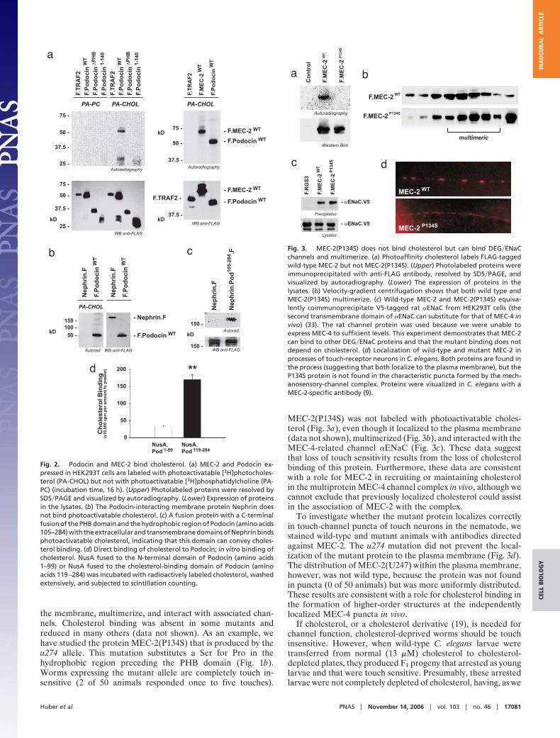
ResultsPodocin and MEC-2 Are Cholesterol-Binding Proteins. Podocin (8),MEC-2 (T.B.H., B.S., R.U.M., and T.B., unpublished data), andseveral other PHB-domain proteins are found in cholesterol-richmembrane fractions. To test whether Podocin and MEC-2 bindto cholesterol, we expressed both proteins in HEK293T cells andtested for binding of photoactivatable lipids (Fig. 2a). Thesederivatives attach to associated molecules when they are stim-ulated by UV light (17). Podocin and MEC-2 bound cholesterolbut not phosphatidylcholine (Fig. 2a). Cholesterol binding re-quired the PHB domain, because Podocin lacking this domain(Podocin�PHB) did not label. Binding to membrane proteins wasquite specific, for example, the Ig superfamily member andPodocin-interacting protein Nephrin was not labeled (Fig. 2b).These data were confirmed by using digitonin precipitation (Fig.7, which is published as supporting information on the PNASweb site).
Although a detailed description of the cholesterol-bindingsites on Podocin and MEC-2 is outside the scope of this article,we tested the importance of different regions of Podocin forcholesterol binding by fusing them to the extracellular andtransmembrane domains of Nephrin. A fusion containing thePHB domain bound cholesterol (data not shown), but more
efficient cholesterol binding was observed when the PHB do-main and the adjacent N-terminal hydrophobic domain wereincluded (Fig. 2c). To ensure that cholesterol labeling was theresult of direct binding and did not occur through passivestochastic attachment of cholesterol at the cell membrane, weproduced fragments of Podocin in Escherichia coli and testedtheir ability to bind [3H]cholesterol in vitro. The PHB domainwas sufficient for cholesterol binding, but binding was moreefficient when the polypeptide included the PHB domain and theN-terminal adjacent hydrophobic sequence (Fig. 2d). Bindingcould be competed with excess cold cholesterol (Fig. 8, which ispublished as supporting information on the PNAS web site).
We had previously shown that Podocin homooligomerizes andforms high-molecular-weight complexes by homophilic interac-tions that require the PHB domain (8). MEC-2 also homooli-gomerizes (see below) as do several other PHB-domain proteins(18). The size of the complexes suggested that they contain atleast 20–50 molecules (Fig. 9a, which is published as supportinginformation on the PNAS web site). Multimerization, however,does not require cholesterol binding. Limited cholesterol deple-tion with methyl-�-cyclodextrine (MBCD) of Podocin-expressing cells did not interfere with the formation of high-molecular-weight complexes (Fig. 9b). Thus, Podocin andMEC-2 bind cholesterol, themselves, and other proteins.
Touch Sensitivity Requires Sterol Binding by MEC-2 in C. elegans. Totest the in vivo importance of cholesterol binding, we made useof the requirement for MEC-2 in C. elegans touch sensitivity.Twenty-three mec-2 alleles causing touch insensitivity in C.elegans have missense mutations (9), and we screened most of theresulting proteins for their ability to bind cholesterol, localize to
a
b
Fig. 1. Structure and sequence of Podocin and MEC-2. (a) Membrane orientation of Podocin and MEC-2. The hydrophobic region (yellow) inserts into the innerleaflet of the plasma membrane, causing the remaining parts of the protein, including the PHB domain (red) to face the cytoplasm and the inner leaflet. Sitesof palmitate attachment are indicated by blue wavy lines. (b) Alignment of mouse Podocin and C. elegans MEC-2, showing the hydrophobic region (yellow), PHBdomain (red), palmitoylation sites (open blue triangles), and the site of the Pro-to-Ser mutation in the hydrophobic region that prevents cholesterol binding(filled blue triangle).
17080 � www.pnas.org�cgi�doi�10.1073�pnas.0607465103 Huber et al.
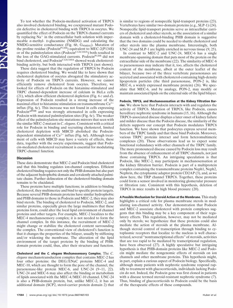
the membrane, multimerize, and interact with associated chan-nels. Cholesterol binding was absent in some mutants andreduced in many others (data not shown). As an example, wehave studied the protein MEC-2(P134S) that is produced by theu274 allele. This mutation substitutes a Ser for Pro in thehydrophobic region preceding the PHB domain (Fig. 1b).Worms expressing the mutant allele are completely touch in-sensitive (2 of 50 animals responded once to five touches).
MEC-2(P134S) was not labeled with photoactivatable choles-terol (Fig. 3a), even though it localized to the plasma membrane(data not shown), multimerized (Fig. 3b), and interacted with theMEC-4-related channel �ENaC (Fig. 3c). These data suggestthat loss of touch sensitivity results from the loss of cholesterolbinding of this protein. Furthermore, these data are consistentwith a role for MEC-2 in recruiting or maintaining cholesterolin the multiprotein MEC-4 channel complex in vivo, although wecannot exclude that previously localized cholesterol could assistin the association of MEC-2 with the complex.
To investigate whether the mutant protein localizes correctlyin touch-channel puncta of touch neurons in the nematode, westained wild-type and mutant animals with antibodies directedagainst MEC-2. The u274 mutation did not prevent the local-ization of the mutant protein to the plasma membrane (Fig. 3d).The distribution of MEC-2(U247) within the plasma membrane,however, was not wild type, because the protein was not foundin puncta (0 of 50 animals) but was more uniformly distributed.These results are consistent with a role for cholesterol binding inthe formation of higher-order structures at the independentlylocalized MEC-4 puncta in vivo.
If cholesterol, or a cholesterol derivative (19), is needed forchannel function, cholesterol-deprived worms should be touchinsensitive. However, when wild-type C. elegans larvae weretransferred from normal (13 �M) cholesterol to cholesterol-depleted plates, they produced F1 progeny that arrested as younglarvae and that were touch sensitive. Presumably, these arrestedlarvae were not completely depleted of cholesterol, having, as we
a
b c
d
Fig. 2. Podocin and MEC-2 bind cholesterol. (a) MEC-2 and Podocin ex-pressed in HEK293T cells are labeled with photoactivatable [3H]photocholes-terol (PA-CHOL) but not with photoactivatable [3H]phosphatidylcholine (PA-PC) (incubation time, 16 h). (Upper) Photolabeled proteins were resolved bySDS�PAGE and visualized by autoradiography. (Lower) Expression of proteinsin the lysates. (b) The Podocin-interacting membrane protein Nephrin doesnot bind photoactivatable cholesterol. (c) A fusion protein with a C-terminalfusion of the PHB domain and the hydrophobic region of Podocin (amino acids105–284) with the extracellular and transmembrane domains of Nephrin bindsphotoactivatable cholesterol, indicating that this domain can convey choles-terol binding. (d) Direct binding of cholesterol to Podocin; in vitro binding ofcholesterol. NusA fused to the N-terminal domain of Podocin (amino acids1–99) or NusA fused to the cholesterol-binding domain of Podocin (aminoacids 119–284) was incubated with radioactively labeled cholesterol, washedextensively, and subjected to scintillation counting.
a b
c d
Fig. 3. MEC-2(P134S) does not bind cholesterol but can bind DEG�ENaCchannels and multimerize. (a) Photoaffinity cholesterol labels FLAG-taggedwild-type MEC-2 but not MEC-2(P134S). (Upper) Photolabeled proteins wereimmunoprecipitated with anti-FLAG antibody, resolved by SDS�PAGE, andvisualized by autoradiography. (Lower) The expression of proteins in thelysates. (b) Velocity-gradient centrifugation shows that both wild type andMEC-2(P134S) multimerize. (c) Wild-type MEC-2 and MEC-2(P134S) equiva-lently coimmunoprecipitate V5-tagged rat �ENaC from HEK293T cells (thesecond transmembrane domain of �ENaC can substitute for that of MEC-4 invivo) (33). The rat channel protein was used because we were unable toexpress MEC-4 to sufficient levels. This experiment demonstrates that MEC-2can bind to other DEG�ENaC proteins and that the mutant binding does notdepend on cholesterol. (d) Localization of wild-type and mutant MEC-2 inprocesses of touch-receptor neurons in C. elegans. Both proteins are found inthe process (suggesting that both localize to the plasma membrane), but theP134S protein is not found in the characteristic puncta formed by the mech-anosensory-channel complex. Proteins were visualized in C. elegans with aMEC-2-specific antibody (9).
Huber et al. PNAS � November 14, 2006 � vol. 103 � no. 46 � 17081
CELL
BIO
LOG
YIN
AU
GU
RAL
ART
ICLE

show below, sufficient cholesterol for touch sensitivity but notenough for further development.
We demonstrated the need of cholesterol in C. elegans touchsensitivity in two ways. First, we generated a version of MEC-2with reduced cholesterol binding by mutating its predictedpalmitoylation sites. Substitution of Ala for Cys at amino acids140 and 174 resulted in the loss of palmitoylation (Fig. 4a) anda reduction of cholesterol binding (Fig. 4b). These alterations didnot affect overall protein levels, multimerization, or localizationto the plasma membrane (data not shown), but mec-2-null wormsexpressing the mec-2(C140�174A) gene conditionally dependedon cholesterol for touch sensitivity. These animals showedvirtually the same touch sensitivity as wild-type animals on plateswith normal amounts of cholesterol but reduced touch sensitivitywhen grown on cholesterol-free plates (Fig. 4 c and d). Thisdefect depended on the cholesterol concentration and could berescued by substituting lathosterol, ergosterol, and 7-dehydrocholesterol for cholesterol in the growth medium (Fig. 4c).Because touch sensitivity of wild-type animals was not affectedby this limited cholesterol depletion, the effects we see with thepalmitoylation mutant cannot be attributed to indirect effects onneuronal growth or development.
The second demonstration of cholesterol dependence wasseen when we lowered cholesterol levels further by transferringanimals grown on minimal (20 nM) cholesterol plates for threegenerations (we find that the animals arrest their developmentafter approximately four generations) to 0 nM cholesterol plates.Larvae placed on minimal or zero cholesterol never becameadults, but arrested in their development. The animals werenoticeably more debilitated (many could not move) under theseconditions. Nonetheless, wild-type animals that showed normalmovement had become relatively insensitive to touch, andanimals with the MEC-2 palmitoylation mutations were even lesssensitive to touch (Fig. 4d). These data show that touch sensi-tivity depends on sterols in vivo and suggest that sterols recruitedto the MEC-4 channel complex by MEC-2 are needed for itsfunction.
Podocin-Mediated Regulation of TRPC Channel Activity. We havefound that Podocin, like MEC-2, is associated with ion-channelsubunits at the glomerular-slit diaphragm of the kidney. Asdescribed above, mutations in the genes encoding Podocin andTRPC6 cause disruption of the kidney filter and focal segmentalglomerulosclerosis (15, 16). To test whether these proteins may
ab
Fig. 5. Podocin interacts and colocalizes with TRPC6. (a) Mouse TRPC6 coimmunoprecipitates with FLAG-tagged Podocin (F.Podocin) but not with a control protein(F.GFP). (Top) Coprecipitated TRPC6 channel after immunoprecipitation of Podocin or GFP. (Middle and Bottom) Expression of the proteins in the lysates. (b) TRPC6 islocated at the slit diaphragm (SD) of podocyte foot processes (FP) near the glomerular basement membrane (GBM). This localization mimics that of Podocin. Rat kidneyswere perfused with ice-cold PBS, fixed in situ, and subjected to immunogold electron microscopy. Arrows indicate the localization of gold particles in the electronmicrograph.
a b c d
Fig. 4. Cholesterol dependence of touch sensitivity in C. elegans. Substitution of Ala for Cys in two predicted palmitoylation sites results in the loss ofpalmitoylation (a) and a reduction of cholesterol binding (b). HEK293T cells were transfected with wild-type MEC-2 or MEC-2(C140�174A) and labeled with[3H]palmitic acid (a) or [3H]photoaffinity cholesterol (b). Equal expression of proteins in the lysates was confirmed on Western blots (data not shown). (c) Touchsensitivity in MEC-2(C140�174A) mutants (black bars) requires cholesterol or its derivatives. Responses of wild-type animals (white bars) are also shown and arenot affected by limited cholesterol depletion. mec-2-null worms were transformed with the mec-2(C140�174A) gene and grown on plates with defined sterolconcentrations before analysis of touch sensitivity. Depicted is the mean � SEM (number of animals tested is indicated. **, P � 0.001 as compared withmec-2(C140�174A) at high cholesterol). (d) Severe cholesterol depletion lowers the sensitivity of wild-type animals (white bars) and mec-2(C140�174A) mutants(black bars). Worms grown on plates containing 20 nM cholesterol for three generations were either maintained on 20 nM cholesterol for another generationor placed on cholesterol-free plates before analysis of response to gentle touch (number of animals tested is indicated; **, P � 0.001 as compared with wild-typeworms on 13 �M cholesterol; #, P � 0.001 as compared with mutants on 13 �M cholesterol).
17082 � www.pnas.org�cgi�doi�10.1073�pnas.0607465103 Huber et al.

functionally interact, we first coexpressed tagged versions of theproteins in HEK293T cells and tested for coimmunoprecipita-tion. Podocin coprecipitated with TRPC6, whereas a controlprotein did not (Fig. 5a). Similar to MEC-2, which does notinfluence targeting of the DEG�ENaC ion-channel complex (9),Podocin did not affect TRPC6 localization to the plasma mem-brane (data not shown). Podocytes express TRPC6 as well asseveral related TRPC channels (TRPC1, 3, and 4) (Fig. 10a,which is published as supporting information on the PNAS website). TRPC channels are thought to be heteromeric (20), so itwas no surprise that Podocin coprecipitated with these otherTRPC channels but not with a control protein (Fig. 10b).Consistent with another study (15), immunofluorescence stain-ing of rat kidney sections confirmed expression of TRPC6 inglomerular podocytes (data not shown). Using immunogoldelectron microscopy, we localized TRPC6 to the insertion site ofthe glomerular-slit diaphragm (Fig. 5b), the structure that
expresses Podocin (12). Although TRPC6 could be detected invarious compartments of the podocyte, immunoreactivity in thesecondary processes of the podocyte was clearly confined to theinsertion site of the slit diaphragm. Thus, Podocin colocalizeswith TRPC6 in vivo.
We next tested whether Podocin affected TRPC6 channelactivity by examining TRPC6 currents in Xenopus laevis oocyteswith and without Podocin. Expression of TRPC6 induced aninward Na� current in a Ca2�-free bath solution that was furtheraugmented by stimulation with the membrane-permeable dia-cylglycerol homologue 1-oleoyl-2-acetyl-sn-glycerol (OAG)(Fig.6a). This increase required the TRPC6 channel; it was not seenin water-injected oocytes. The OAG-induced currents weresignificantly augmented in oocytes coexpressing TRPC6 andPodocin, but were not increased in oocytes coexpressing TRPC6and Podocin�PHB (Fig. 6a). These data show that Podocininteracts with TRPC6 to regulate TRPC6 activity.
Fig. 6. Activation of TRPC-channel activity by Podocin. (a) Podocin, but not Podocin�PHB, enhances TRPC6 currents in Xenopus oocytes stimulated with 10 �MOAG. Expression of TRPC6 induces an inward Na� current in a Ca2�-free bath solution that is further augmented by stimulation with OAG. The OAG-inducedcurrents were significantly augmented in oocytes coexpressing TRPC6 and Podocin but were not increased in oocytes coexpressing TRPC6 and Podocin�PHB. (b)Podocin increases the effect of OAG (10 �M 1-oleoyl-2-acetyl-sn-glycerol; black bars) on NMDG-sensitive conductance (GNMDG) of TRPC6 channels in Xenopusoocytes, but mutant Podocins do not. Currents in control oocytes (white bars) were not affected. The Podocin mutants used were Podocin�PHB, PodocinP120S, andPodocinC126/160A (see Fig. 1). The number of oocytes examined is given in parentheses. *, P � 0.05 as compared with water-injected oocytes; #, P � 0.05 as comparedwith TRPC6 coexpressed with Podocin�PHB. (c) Wild-type, but not mutant, Podocin increases histamine-induced calcium influx (measured as a change influorescence, �F�F) in HeLa Cx43 cells. Cells were transiently mock transfected (filled circles) or transfected with DNA coding for wild-type Podocin (filledtriangles), Podocin�PHB (open triangles), and PodocinC126/160A (inverted filled triangles) and measured simultaneously in the same experiment by using a FLIPR.Data are means � SD from three to five independent experiments. Measurements are taken at the shoulder of the response (line in the inset, which shows thecalcium responses of the cells challenged with 10 �M histamine). Vertical scale, 104 arbitrary fluorescence units; horizontal scale, 2 min. (d) The stimulation byPodocin is abolished when cells are treated with MBCD to deplete cholesterol.
Huber et al. PNAS � November 14, 2006 � vol. 103 � no. 46 � 17083
CELL
BIO
LOG
YIN
AU
GU
RAL
ART
ICLE
To test whether the Podocin-mediated activation of TRPC6also involved cholesterol binding, we coexpressed mutant Podo-cin defective in cholesterol binding with TRPC6 in oocytes. Wequantified the effect of Podocin on the TRPC6 channel currentsby replacing Na� in the extracellular bath solution with imper-meable n-methyl- D-glucamine (NMDG) and calculating theNMDG-sensitive conductance (Fig. 6b, GNMDG). Mutation ofthe proline residue (PodocinP120S) equivalent to MEC-2(P134S)or of the palmitoylation sites (PodocinC126/160A) both resulted inthe loss of the OAG-stimulated currents. PodocinP120S did notbind cholesterol, and PodocinC126/160A showed weak cholesterol-binding activity, but both interacted with TRPC6 (not shown).
These data suggest that the regulation of TRPC6 by Podocinrequires cholesterol binding. We would like to have shown thatcholesterol depletion of oocytes abrogated the stimulatory ac-tivity of Podocin on TRPC6 currents. However, we cannotefficiently remove cholesterol from oocytes. Therefore, welooked for effects of Podocin on the histamine-stimulated andTRPC channel-dependent increase of calcium in HeLa cells(21), which allow efficient cholesterol depletion (Fig. 6 c and d).Expression of Podocin resulted in a strong increase of themaximal effect to histamine stimulation on transmembrane Ca2�
influx (Fig. 6c). This increase was not found in cells expressingPodocin�PHB and was strongly attenuated in cells expressingPodocin with mutated palmitoylation sites (Fig. 6c). The weakereffect of the palmitoylation site mutations mirrors that seen withthe similar MEC-2 mutant in C. elegans. Consistent with a criticalrole for Podocin in binding and recruiting cholesterol, limitedcholesterol depletion with MBCD abolished the Podocin-dependent stimulation of Ca2� influx (Fig. 6d). Although treat-ment of cells with MBCD may have a variety of effects, thesedata, together with the oocyte experiments, suggest that Podo-cin-mediated cholesterol recruitment is essential for modulatingTRPC-channel function.
DiscussionThese data demonstrate that MEC-2 and Podocin bind cholesteroland that this binding regulates ion-channel complexes. Efficientcholesterol binding requires not only the PHB domains but also partof the adjacent hydrophobic domain and covalently attached palmi-tate chains. Further delineation of the cholesterol-binding domainremains to be elucidated.
These proteins have multiple functions; in addition to bindingcholesterol, they multimerize and bind to specific protein targets.Because several PHB-domain proteins have similar hydrophobicand PHB domains to those in Podocin and MEC-2, they may alsobind sterols. The binding of cholesterol to Podocin, MEC-2, andsimilar proteins, especially given the large multimers that theseproteins form, could alter the local lipid environment of channelproteins and other targets. For example, MEC-2 localizes to theMEC-4 mechanosensory complex; it is not needed to form thechannel complex. In this instance, the recruitment of MEC-2might change or stabilize the cholesterol content surroundingthe complex. The conventional view of cholesterol’s function isthat it changes the properties of the bilayer, usually by stiffeningand�or widening the membrane. The alteration of the lipidenvironment of the target proteins by the binding of PHB-domain proteins could, thus, alter their structure and function.
Cholesterol and the C. elegans Mechanosensory Complex. The C.elegans mechanotransduction complex that contains MEC-2 hasfour other proteins: the DEG�ENaC proteins MEC-4 andMEC-10, which are thought to form the pore of the channel, theparaoxonase-like protein MEC-6, and UNC-24 (9–11, 22).UNC-24 and MEC-6 may also affect the binding or metabolismof lipids associated with the MEC-4�MEC-10 channel. UNC-24is also a PHB-domain protein, but, unlike MEC-2, it has anadditional domain (SCP2, sterol-carrier protein domain 2) that
is similar to regions of nonspecific lipid-transport proteins (23).Vertebrates have similar two-domain proteins (e.g., SLP-1) (24).Nonspecific lipid-transport proteins serve as intracellular carri-ers of cholesterol and other sterols, so the association of a similardomain with a cholesterol-binding PHB domain is suggestivethat the two domains could be needed to shuttle cholesterol andother sterols into the plasma membrane. Interestingly, bothUNC-24 and SLP-1 are highly enriched in nervous tissue (9, 25).
In contrast to MEC-2 and UNC-24, MEC-6 has a singlemembrane-spanning domain that puts most of the protein on theextracellular side of the membrane (22). The similarity of MEC-6to paraoxonases may indicate that it, too, affects the cholesterolcontent of the membrane, albeit at the outer leaflet of thebilayer, because two of the three vertebrate paraoxonases aresecreted and associated with cholesterol-containing high-densitylipoprotein particles (the third paraoxonase, PON-2 is, likeMEC-6, a widely expressed membrane protein) (26). We spec-ulate that MEC-6, and by analogy, PON-2, may modify ormaintain associated lipids on the external side of the lipid bilayer.
Podocin, TRPC6, and Mechanosensation at the Kidney Filtration Bar-rier. We show here that Podocin interacts with and regulates theactivity of TRPC6. Mutation of TRPC6, like Podocin, causeshereditary nephrotic syndrome in humans (15, 16). Although theTRPC6-associated disease displays a later onset of kidney failureand milder disease than the Podocin disease, the similarity of thedefects supports our concept that Podocin modulates TRPC6function. We have shown that podocytes express several mem-bers of the TRPC family and that these bind Podocin. Moreover,various TRPC proteins interact and form heteromultimericcomplexes (20). These observations lead us to expect somefunctional redundancy with other channels of the TRPC family.The more pronounced disease caused by Podocin loss may resultfrom the absence of enhancement of all TRPC channels, not justthose containing TRPC6. An intriguing speculation is thatPodocin, like MEC-2, may participate in mechanosensation atthe kidney filtration barrier. Podocin is part of a multiproteincomplex containing the transmembrane proteins Neph1, Neph2,Nephrin, the cytoplasmic adaptor protein CD2AP (3), and, as weshow here, the TRP channel TRPC6. Together, these proteinscould form a sensor involved in monitoring glomerular pressureor filtration rate. Consistent with this hypothesis, deletion ofTRPC6 in mice results in high blood pressure (20).
A Possible Mechanism for Steroid Action at the Membrane. This studyhighlights a critical role for plasma membrane sterols in mod-ulating ion-channel activity. Our demonstration that Podocinand MEC-2 associate cholesterol with protein complexes sug-gests that this binding may be a key component of their regu-latory effects. This regulation, however, may not be mediatedonly by sterols; we hypothesize that these proteins could alsomediate the action of steroids at the plasma membrane. Al-though steroid control of transcription through binding to cy-toplasmic receptors that localize to the nucleus is well charac-terized, several ‘‘nontranscriptional effects’’ of steroids, activitiesthat are too rapid to be mediated by transcriptional regulation,have been observed (27). A highly speculative but intriguinghypothesis is that PHB-domain proteins like MEC-2 and Podo-cin might mediate the nongenomic effects of steroids on ionchannels and other membrane proteins. This hypothesis might,in part, explain a curious aspect of Podocin biology. Specifically,although many patients with nephrotic syndrome respond rap-idly to treatment with glucocorticoids, individuals lacking Podo-cin do not. Indeed, the Podocin gene was first cloned in patientswith a disease termed steroid-resistant nephrotic syndrome (7).Thus, binding of glucocorticoids to Podocin could be the basisof the therapeutic effects of these compounds.
17084 � www.pnas.org�cgi�doi�10.1073�pnas.0607465103 Huber et al.

Materials and MethodsReagents and Plasmids. Mouse Podocin cDNA constructs havebeen described (8, 28). TRPC6 was cloned from a humanpodocyte cDNA library. MEC-2 cDNA was cloned from a C.elegans ORF AAA87552 (Open Biosystems, Huntsville, AL).Truncations and mutations of Podocin, MEC-2, and TRPC6were generated by standard cloning procedures. All other con-structs have been described (10, 28). Some experiments involvingMEC-2 had to be performed with �ENaC, a mammalian ENaCprotein, instead of MEC-4, because MEC-4 cDNA did notexpress well in HEK293T cells. Antibodies have been describedor were obtained from Sigma, (St. Louis, MO) (anti-FLAG M2),Alomone Labs (Jerusalem, Israel), and Chemicon (Temecula,CA) (anti-TRPC6), and Serotec, Toronto, ON, Canada) (anti-V5). Bacterial vectors for the expression of His-tagged recom-binant proteins fused to the C terminus of NusA were kindlyprovided by Gunter Stier (European Molecular Biology Labo-ratory, Heidelberg, Germany).
Cell Culture Studies. Most cell studies used HEK293T cells thatwere grown in DMEM as described (28). Cholesterol-depletedcells were prepared by growing cells in DMEM with pravastatin(8 �M) for 2 days and then MBCD (5 mM) for 30 min just beforethe experiment. Immunoprecipitations from HEK293T cellswere performed as described (28). Plamitate labeling, the dig-itonin precipitation assay (29), and photoaffinity labeling (17)were performed as described. Expression and purification ofrecombinant proteins has been described (30).
PHB protein multimerization was studied by velocity-gradientcentrifugation and blue native-gel electrophoresis (31). Forpreparation of Podocin multimeric complexes, HEK293T cellswere lysed in 1 ml of Mes-buffered saline (MBS) in the presenceof 1% Triton X-100 and centrifuged for 10 min at 1,000 � g at4°C. After centrifugation, the supernatant was collected, andSDS was added at a final concentration of 0.1% and incubatedfor 20 min on ice. Thereafter, the lysate was cleared by centrif-ugation for 15 min at 100,000 � g. Four milliliters of a discon-tinuous sucrose gradient (40–5%) was layered on top of a 60%sucrose cushion in an ultracentrifuge tube (Beckman, Fullerton,CA). One milliliter of the cell lysate was adjusted with 1 ml ofMBS, added on top of this gradient, and subjected to centrifu-gation for 16 h at 180,000 � g at 4°C in a Beckman SW-41 rotor.After centrifugation, 2 ml of the supernatant were discarded and8 fractions (500 �l each) were collected, starting from the top,and analyzed by SDS�PAGE.
In Vitro Cholesterol Interaction. Podocin truncations were clonedinto various bacterial expression vectors and tested for theexpression of soluble recombinant fusion proteins. Expression asHis-tagged proteins fused to the C terminus of NusA (vectorskindly provided by Gunter Stier) resulted in a large fraction ofsoluble recombinant Podocin protein that could be affinity-purified on Ni� columns. Purity of the preparation was con-firmed on Coomassie gels. For in vitro cholesterol-interactionassays, 2–20 �g of affinity-purified Podocin protein was boundto 30 �l of Ni� beads and incubated with 0.1 �Ci (1 Ci � 37 GBq)of [3H]cholesterol (Amersham, Piscataway, NJ) complexed withlow amounts of MBCD. After binding for 10 min at 37°C, beadswere washed extensively and counted in a scintillation counter.To confirm equal loading of the beads, aliquots of the boundprotein were run on Coomassie gels. Competition experiments
were performed with 1 �g of affinity-purified Podocin protein,varying amounts of [3H]cholesterol, and an �100-fold excess ofcold cholesterol.
C. elegans Experiments. C. elegans strains were cultured at 20°C,assayed for touch sensitivity, and prepared for immunofluores-cence as described (9, 10). Media for growth on limiting, or nocholesterol, or on other sterols were prepared from chloroform-extracted reagents as described by Matyash et al. (32).
Oocyte Electrophysiology. X. laevis oocytes were isolated fromadult frogs (Kahler, Hamburg, Germany), dispersed and defol-liculated by a 45-min treatment with collagenase (type A;Boehringer, Ingelheim, Germany), rinsed, and kept at 18°C inND96 buffer: 96 mmol�liter NaCl, mmol�liter KCI 2, 1.8 mmol�liter CaCl2, 1 mmol�liter MgCl2, 5 mmol�liter Hepes, 2.5 mmol�liter sodium pyruvate, pH 7.55), supplemented with theophylline(0.5 mmol�l) and gentamycin (5 mg�l). cRNAs (1–10 ng) forTRPC6, Podocin and Podocin-�PHB were transcribed in vitrofrom cDNAs by using the T7 promoter and polymerase (Pro-mega, Madison, WI) and injected into oocytes after dissolving in47 nl of double-distilled water (Nanoliter Injector; World Pre-cision Instruments, Berlin, Germany). Water-injected oocytesserved as controls. Two to four days after injection, oocytes wereimpaled with two electrodes (Clark Brothers Instrument, ShelbyTownship, MI) that had resistances of �1 M� when filled with2.7 mol�liter KCI. By using two bath electrodes and a virtual-ground headstage, the voltage drop across Rserial was effectivelyzero. Membrane currents were measured by voltage clamping ofthe oocytes (oocyte clamp amplifier OC725C; Warner Instru-ments, Hamden, CT) in intervals from 60 to �40 mV, each 1 s.Conductances were calculated according to Ohm’s law. Na�
conductances were determined by replacing Na� by N-methyl-D-glucamine (GNMDG) in a Ca2�-free bath solution, before andafter stimulation with 10 �M dioctanoyl glycerol (Sigma-Aldrich, Munich, Germany). During the whole experiment, thebath was continuously perfused at a rate of 5–10 ml�min. Allexperiments were conducted at room temperature (22°C).
Ca2�-FLIPR Assay. HeLa-Cx43 cells were loaded with 4 �MFLUO-4�AM and 0.04% Pluronic F-127 (both from MolecularProbes, Eugene, OR) in HBS but with 20 mM Hepes and 2.5 mMprobenecid, and assays were performed as described (21).
Statistical Analysis. Data are expressed as mean � SEM of nexperiments. Statistical evaluation was performed by usingStudent’s t test or ANOVA for repeated measures, followed bya Bonferroni test as posttest (SigmaPlot; Jandel Scientific,San Rafael, CA, and Instat2, GraphPad Software, San Diego,CA). Values of P � 0.05 were considered to be statisticallysignificant.
Note Added in Proof. Spassova et al. (34) find that TRPC6 channels canbe mechanically gated, a finding that supports our hypothesis thatTRPC6 acts as a mechanosensor in the mammalian kidney.
We thank Christina Engel, Stefanie Keller, Petra Damisch, CharlotteMeyer, and John Byun for technical assistance; Gunter Stier for pro-viding plasmids; and members of the Benzing and Chalfie laboratoriesfor helpful discussions. This study was supported by the DeutscheForschungsgemeinschaft (T.B.H., T.B., and G.W.) and the NationalInstitutes of Health (M.C.).
1. Ponting CP, Schultz J, Milpetz F, Bork P (1999) Nucleic Acids Res 27:229–232.2. Morrow IC, Parton RG (2005) Traffic 6:725–740.3. Benzing T (2004) J Am Soc Nephrol 15:1382–1391.4. Ernstrom GG, Chalfie M (2002) Annu Rev Genet 36:411–453.5. Stewart GW (2004) Curr Opin Hematol 11:244–250.6. Huang M, Gu G, Ferguson EL, Chalfie M (1995) Nature 378:292–295.
7. Boute N, Gribouval O, Roselli S, Benessy F, Lee H, Fuchshuber A, Dahan K,Gubler MC, Niaudet P, Antignac C (2000) Nat Genet 24:349–354.
8. Huber TB, Simons M, Hartleben B, Sernetz L, Schmidts M, Gundlach E,Saleem MA, Walz G, Benzing T (2003) Hum Mol Genet 12:3397–3405.
9. Zhang S, Arnadottir J, Keller C, Caldwell GA, Yao CA, Chalfie M (2004) CurrBiol 14:1888–1896.
Huber et al. PNAS � November 14, 2006 � vol. 103 � no. 46 � 17085
CELL
BIO
LOG
YIN
AU
GU
RAL
ART
ICLE

10. Goodman MB, Ernstrom GG, Chelur DS, O’Hagan R, Yao CA, Chalfie M(2002) Nature 415:1039–1042.
11. O’Hagan R, Chalfie M, Goodman MB (2005) Nat Neurosci 8:43–50.12. Roselli S, Gribouval O, Boute N, Sich M, Benessy F, Attie T, Gubler MC,
Antignac C (2002) Am J Pathol 160:131–139.13. Kestila M, Lenkkeri U, Mannikko M, Lamerdin J, McCready P, Putaala H,
Ruotsalainen V, Morita T, Nissinen M, Herva R, et al. (1998) Mol Cell1:575–582.
14. Shih NY, Li J, Karpitskii V, Nguyen A, Dustin ML, Kanagawa O, Miner JH,Shaw AS (1999) Science 286:312–315.
15. Reiser J, Polu KR, Moller CC, Kenlan P, Altintas MM, Wei C, Faul C, HerbertS, Villegas I, Avila-Casado C, et al. (2005) Nat Genet 37:739–744.
16. Winn MP, Conlon PJ, Lynn KL, Farrington MK, Creazzo T, Hawkins AF,Daskalakis N, Kwan SY, Ebersviller S, Burchette JL, et al. (2005) Science308:1801–1804
17. Thiele C, Hannah MJ, Fahrenholz F, Huttner WB (2000) Nat Cell Biol2:42–49.
18. Umlauf E, Mairhofer M, Prohaska R (2006) J Biol Chem 281:23349–23356.
19. Chitwood DJ (1999) Crit Rev Biochem Mol Biol 34:273–284.20. Freichel M, Vennekens R, Olausson J, Stolz S, Philipp SE, Weissgerber P,
Flockerzi V (2005) J Physiol 567:59–66.
21. Shirokova E, Schmiedeberg K, Bedner P, Niessen H, Willecke K, Raguse JD,Meyerhof W, Krautwurst D (2005) J Biol Chem 280:11807–11815.
22. Chelur DS, Ernstrom GG, Goodman MB, Yao CA, Chen L, O’Hagan R,Chalfie M (2002) Nature 420:669–673.
23. Barnes TM, Jin Y, Horvitz HR, Ruvkun G, Hekimi S (1996) J Neurochem 67:46–57.24. Wang Y, Morrow JS (2000) J Biol Chem 275:8062–8071.25. Seidel G, Prohaska R (1998) Gene 225:23–29.26. Getz GS, Reardon CA (2004) Curr Opin Lipidol 15:261–267.27. Losel RM, Falkenstein E, Feuring M, Schultz A, Tillmann HC, Rossol-
Haseroth K, Wehling M (2003) Physiol Rev 83:965–1016.28. Huber TB, Kottgen M, Schilling B, Walz G, Benzing T (2001) J Biol Chem
276:41543–41546.29. Charrin S, Manie S, Thiele C, Billard M, Gerlier D, Boucheix C, Rubinstein
E (2003) Eur J Immunol 33:2479–2489.30. Benzing T, Brandes R, Sellin L, Schermer B, Lecker S, Walz G, Kim E (1999)
Nat Med 5:913–918.31. Schagger H, von Jagow G (1991) Anal Biochem 199:223–231.32. Matyash V, Entchev EV, Mende F, Wilsch-Brauninger M, Thiele C, Schmidt
AW, Knolker HJ, Ward S, Kurzchalia TV (2004) PLoS Biol 2:e280.33. Hong K, Driscoll M (1994) Nature 367:470–473.34. Spassova MA, Hewavitharana T, Xu W, Soboloff J, Gill DL (2006) Proc Natl
Acad Sci USA 103:16586–16591.
17086 � www.pnas.org�cgi�doi�10.1073�pnas.0607465103 Huber et al.
Related Documents