UNIVERSIDADE FEDERAL DO RIO GRANDE DO SUL INSTITUTO DE CIÊNCIAS BÁSICAS DA SAÚDE DEPARTAMENTO DE BIOQUÍMICA PROGRAMA DE PÓS – GRADUAÇÃO EM CIÊNCIAS BIOLÓGICAS: BIOQUÍMICA AVALIAÇÃO DA HOMEOSTASE ENERGÉTICA EM VÁRIOS TECIDOS E HISTOPATOLOGIA CEREBRAL EM CAMUNDONGOS NOCAUTE PARA A ENZIMA GLUTARIL-COA DESIDROGENASE Alexandre Umpierrez Amaral ORIENTADOR: Prof. Dr. Moacir Wajner Tese de Doutorado apresentada ao Programa de Pós-Graduação em Ciências Biológicas - Bioquímica da Universidade Federal do Rio Grande do Sul como requisito parcial à obtenção do grau de Doutor em Bioquímica. Porto Alegre, 2014

Welcome message from author
This document is posted to help you gain knowledge. Please leave a comment to let me know what you think about it! Share it to your friends and learn new things together.
Transcript
UNIVERSIDADE FEDERAL DO RIO GRANDE DO SUL
INSTITUTO DE CIÊNCIAS BÁSICAS DA SAÚDE
DEPARTAMENTO DE BIOQUÍMICA
PROGRAMA DE PÓS – GRADUAÇÃO EM CIÊNCIAS BIOLÓGICAS:
BIOQUÍMICA
AVALIAÇÃO DA HOMEOSTASE ENERGÉTICA EM VÁRIOS
TECIDOS E HISTOPATOLOGIA CEREBRAL EM CAMUNDONGOS
NOCAUTE PARA A ENZIMA GLUTARIL-COA DESIDROGENASE
Alexandre Umpierrez Amaral
ORIENTADOR: Prof. Dr. Moacir Wajner
Tese de Doutorado apresentada ao Programa de Pós-Graduação em
Ciências Biológicas - Bioquímica da Universidade Federal do Rio Grande do
Sul como requisito parcial à obtenção do grau de Doutor em Bioquímica.
Porto Alegre, 2014

I
Aos meus pais,
Anselmo e Nilza, mais uma vez.

II
“A morte não é nada, mas viver vencido
e sem glória é morrer todos os dias”
(Napoleão Bonaparte)
III
AGRADECIMENTOS
À Universidade Federal do Rio Grande do Sul e ao Departamento de Bioquímica, representado pelos professores e funcionários, por fornecerem todo o suporte necessário para o desenvolvimento desta tese. Ao meu orientador, Professor Moacir Wajner, pela orientação e por me transmitir conhecimento, experiência e sabedoria para me tornar um bom cientista. Aos professores mais experientes do grupo de Erros Inatos do Metabolismo, Ângela, Clóvis e Dutra, por estarem sempre dispostos a ajudar.
Ao Professor Guilhian, pela amizade e conselhos imprescindíveis para a realização deste trabalho, e por ser um grande companheiro.
À Bianca, por estar sempre disposta a me ajudar em todas as atividades, experimentos ou conselhos, sempre quando preciso. Estou certo que nunca consigo retribuir à altura.
À Cristiane, minha bolsista incansável que foi responsável por grande parte do trabalho árduo desta tese.
Ao César e ao Mateus Struecker, grandes companheiros em todos os momentos e sempre dispostos a uma boa discussão, seja futebol, política ou ciência.
A todos os demais colegas de laboratório 38 e 27, participantes diretos ou não deste trabalho, pela boa convivência de sempre.
Aos meus mais antigos amigos, que, apesar de saberem muito pouco ou quase nada sobre este trabalho, me proporcionam amizade, diversão e companheirismo imprescindíveis.
À Cris, por todo o amor, compreensão, companheirismo, enfim por existir e por me aguentar.
À família da Cris, pessoas fantásticas que sempre me acolheram e trataram como um dos seus.
À minha família, pela ótima convivência e respeito de sempre, tanto nos bons como nos maus momentos.
Aos meus avós, vivos ou já falecidos, pelo exemplo de vida que sempre foram para mim.
Aos meus irmãos, pela amizade, dignidade e respeito que sempre tivemos um pelo outro.
Aos meus pais, pelo amor, educação, dedicação e esforços inacreditáveis que me conduziram e conduzirão a todas minhas conquistas, e pelos quais serei eternamente grato.
IV
SUMÁRIO
PARTE I – Introdução e Objetivos................................................................................. 1
RESUMO........................................................................................................................... 2
ABSTRACT........................................................................................................................ 3
LISTA DE ABREVIATURAS.............................................................................................. 4
I.1. INTRODUÇÃO............................................................................................................ 5
I.1.1. Erros inatos do metabolismo................................................................................ 5
I.1.2. Acidemias orgânicas............................................................................................ 5
I.1.3. Acidemia glutárica tipo I (AG I)........................................................................... 7
I.1.3.1. Achados clínicos ..................................................................................... 9
I.1.3.2. Diagnóstico .............................................................................................. 10
I.1.3.3. Achados neuropatológicos ...................................................................... 11
I.1.3.4. Tratamento .............................................................................................. 12
I.1.3.5. Modelos animais de acidemia glutárica tipo I (AG I) ............................... 13
I.1.3.6. Fisiopatologia .......................................................................................... 14
I.1.4. Metabolismo energético cerebral ........................................................................ 17
I.1.5. Ciclo do ácido cítrico (CAC), fosforilação oxidativa, cadeia transportadora de
elétrons e parâmetros respiratórios....................................................................
17
I.1.6. Creatina quinase (CK) ....................................................................................... 22
I.1.7. Na+, K+-ATPase ................................................................................................. 23
I.1.8. Metabolismo energético e doenças neurodegenerativas .................................. 25
I.2. OBJETIVOS…………………………………………..........…………………………..…... 26
I.2.1. Objetivo geral..................................................................................................... 26
V
I.2.2. Objetivos específicos…………………………………………………......……….... 26
PARTE II - Artigos Científicos .........................................……………………………….. 28
Capítulo I ........................................................................................................................ 29
Capítulo II ....................................................................................................................... 36
Capítulo III ...................................................................................................................... 45
PARTE III - Discussão e Conclusões ........................................................................... 80
III.1. DISCUSSÃO............................................................................................................ 81
III.2. CONCLUSÕES........................................................................................................ 92
III.3. PERSPECTIVAS..................................................................................................... 93
REFERÊNCIAS BIBLIOGRÁFICAS................................................................................ 95
LISTA DE FIGURAS........................................................................................................ 111
1
PARTE I
Introdução e Objetivos
2
RESUMO Estudamos a homeostase energética no cérebro (córtex cerebral, estriado e hipocampo) e tecidos periféricos (coração e músculo esquelético) de camundongos selvagens (WT) e nocaute para a enzima glutaril-CoA desidrogenase (Gcdh-/-), modelo animal genético para estudo da acidemia glutárica tipo I (AG I), com 15 e 30 dias de vida. Esses animais também foram submetidos a uma sobrecarga de lisina através de uma injeção intraperitoneal (8 µmol/g) desse aminoácido ou de uma dieta rica em lisina (4,7 %) por 60 horas. Os parâmetros da homeostase energética analisados foram as atividades dos complexos I-III, II, II-III e IV da cadeia respiratória, das enzimas do ciclo do ácido cítrico (CAC) citrato sintase (CS), aconitase, isocitrato desidrogenase (IDH), α-cetoglutarato desidrogenase, sucinato desidrogenase e malato desidrogenase, da creatina quinase (CK) e da Na+, K+ - ATPase, bem como a liberação de lactato, os parâmetros respiratórios mitocondriais estados 3 e 4, razão de controle respiratório e o estado desacoplado, além do potencial de membrana mitocondrial na presença ou ausência de Ca2+. Estudos histológicos também foram conduzidos no córtex cerebral e estriado dos camundongos WT e Gcdh-/- de 30, 60 e 90 dias de vida submetidos por um pequeno (60 horas) ou longo (30 dias) período com dieta com alta concentração de lisina (4,7 %). Verificamos leves alterações nas atividades dos complexos da cadeia respiratória no cérebro, coração e músculo esquelético dos animais Gcdh-/- quando comparados aos WT com 15 e 30 dias de vida. Além disso, demonstramos uma diminuição significativa das atividades da CS e IDH em preparações mitocondriais de estriado de camundongos Gcdh-/- submetidos a uma sobrecarga de lisina associada a um pequeno aumento na liberação de lactato. No entanto, não encontramos alterações nos parâmetros respiratórios e no potencial de membrana em mitocôndrias de estriado dos camundongos Gcdh-/-quando comparados aos WT. Por outro lado, as atividades da Na+, K+-ATPase (cérebro) e CK (cérebro e músculo esquelético) foram significativamente menores em camundongos Gcdh-/- com 15 dias de vida quando submetidos a uma injeção intraperitoneal de lisina. Além disso, encontramos uma redução na atividade da Na+, K+-ATPase associada com uma diminuição da sua expressão em córtex cerebral, mas não em estriado e hipocampo, de camundongos Gcdh-/- com 30 dias de vida submetidos ou não a uma dieta rica em lisina. Finalmente, a análise histológica revelou a presença de vacúolos no córtex cerebral dos camundongos Gcdh-/- com 60 e 90 dias de vida, bem como no estriado dos animais Gcdh-/- com 90 dias de vida que foram alimentados com uma dieta rica em lisina por 30 dias. Concluindo, presumimos que uma redução das atividades da Na+, K+-ATPase e CK possa contribuir para o dano neurológico encontrado nos camundongos Gcdh-/- e possivelmente nos pacientes com AG I.
3
ABSTRACT
We studied energy homeostasis in the brain (cerebral cortex, striatum and hippocampus) and peripheral tissues (heart and skeletal muscle) from 15 and 30-day-old wild type (WT) and glutaryl-CoA dehydrogenase deficient (Gcdh-/-) mice, which is a genetic animal model to study glutaric academia type I (GA I). These animals were also submitted to lysine overload through an intraperitoneal injection (8 µmol/g) of this amino acid or supplementing the mice with a high lysine (4.7 %) diet for 60 hours. The energy homeostasis parameters evaluated were the activities of the respiratory chain complexes I-III, II, II-III and IV, of the citric acid cycle (CAC) enzymes citrate synthase (CS), aconitase, isocitrate dehydrogenase (IDH), α-ketoglutarate dehydrogenase, succinate dehydrogenase and malate dehydrogenase, creatine kinase (CK) and Na+, K+ - ATPase, as well as the lactate release, the mitochondrial respiratory parameters states 3 and 4, respiratory control ratio and uncoupled state, besides the mitochondrial membrane potential in the presence or absence of Ca2+. Histological studies were also conducted in the cerebral cortex and striatum from 30, 60 and 90-day-old WT and Gcdh-/- mice submitted for a short (60 hours) or long (30 days) period to a high lysine (4.7 %) diet. We verified mild alterations in the respiratory chain activity in the brain, heart and skeletal muscle from Gcdh-/- animals when compared to 15 and 30-day-old WT mice. Furthermore, we demonstrated a reduction in the activities of CS and IDH in striatum mitochondrial preparations from Gcdh-/- mice submitted to a lysine overload associated with a mild increase of lactate release. However, we did not find alterations in the respiratory parameters and membrane potential in striatum mitochondria from Gcdh-/- mice when compared to WT. On the other hand, the activities of Na+, K+-ATPase (brain) and CK (brain and skeletal muscle) were significantly reduced in 15-day-old Gcdh-/- mice when received an intraperitoneal injection of lysine. Moreover, a reduction in the Na+, K+-ATPase activity associated with a diminution of its expression was observed in the cerebral cortex, but not in striatum and hippocampus, from 30-day-old Gcdh-/- mice submitted or not to a high lysine diet. Finally, the histological analyses revealed the presence of vacuoles in the cerebral cortex from 60 and 90-day-old Gcdh-/- mice, as well as in the striatum from 90-day-old Gcdh-/- animals that were fed a high Lys chow for 30 days. In conclusion, we presume that a reduction in the activities of Na+, K+-ATPase and CK may contribute to the brain damage found in Gcdh-/- mice and possibly in GA I patients.
4
LISTA DE ABREVIATURAS
AG I – acidemia glutárica tipo I;
AG – ácido glutárico;
ACO – aconitase;
ADP - adenosina-5’-difosfato;
ATP - adenosina-5’-trifosfato;
α-CGDH - α-cetoglutarato desidrogenase;
CAC – ciclo do ácido cítrico;
CS – citrato sintase;
CK – creatina quinase;
FAD - flavina adenina dinucleotídeo;
FADH2 - flavina adenina dinucleotídeo reduzido;
GABA - ácido gama-aminobutírico;
GCDH – glutaril-CoA desidrogenase;
Gcdh-/- - camundongos nocautes para glutaril-CoA desidrogenase;
HE – hematoxilina-eosina;
3HG – ácido 3-hidróxi-glutárico;
IDH – isocitrato desidrogenase;
MDH – malato desidrogenase;
NAD+ - adenina dinucleotídeo;
NADH - nicotinamida adenina dinucleotídeo reduzido;
NADPH – nicotinamida adenina dinucleotídeo fosfato reduzido;
NMDA – N-metil-D-aspartato;
RCR – razão de controle respiratório;
SDH – sucinato desidrogenase;
SNC – sistema nervoso central.
5
I.1. INTRODUÇÃO
I.1.1. Erros inatos do metabolismo
Erros inatos do metabolismo são distúrbios hereditários, majoritariamente
de herança autossômica recessiva, cuja característica bioquímica principal é a
deficiência ou ausência da atividade de uma enzima específica de uma rota
metabólica. Além das enzimas, outras proteínas com função alterada como
proteínas de transporte e proteínas estruturais, imunoglobulinas, hormônios, entre
outras, podem estar afetadas nos erros inatos do metabolismo. O resultado da
deficiência de uma atividade enzimática leva a um bloqueio da rota metabólica
levando ao acúmulo de substâncias tóxicas nos tecidos e líquidos corporais ou à
falta de substâncias essenciais, muitas vezes, acarretando prejuízo no
desenvolvimento mental e/ou físico dos indivíduos afetados (Scriver, 2001). Além
disso, rotas alternativas também originam outras substâncias tóxicas (Bickel,
1987). Até o momento, foram descritos mais de 600 erros inatos do metabolismo,
a maioria deles envolvendo processos de síntese, degradação, transporte e
armazenamento de moléculas no organismo (Scriver, 2001). Embora
individualmente raras, essas doenças afetam aproximadamente 1 a cada 500-
1000 recém nascidos vivos (Baric et al., 2001).
I.1.2. Acidemias orgânicas
As acidemias ou acidúrias orgânicas constituem um grupo de erros inatos
do metabolismo bioquimicamente caracterizados pelo acúmulo de um ou mais
ácidos orgânicos (carboxílicos) nos líquidos biológicos e tecidos dos pacientes
6
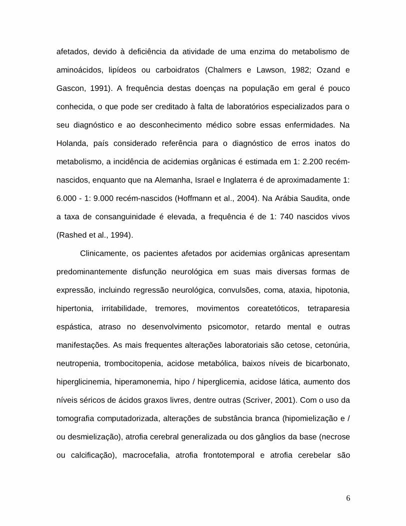
afetados, devido à deficiência da atividade de uma enzima do metabolismo de
aminoácidos, lipídeos ou carboidratos (Chalmers e Lawson, 1982; Ozand e
Gascon, 1991). A frequência destas doenças na população em geral é pouco
conhecida, o que pode ser creditado à falta de laboratórios especializados para o
seu diagnóstico e ao desconhecimento médico sobre essas enfermidades. Na
Holanda, país considerado referência para o diagnóstico de erros inatos do
metabolismo, a incidência de acidemias orgânicas é estimada em 1: 2.200 recém-
nascidos, enquanto que na Alemanha, Israel e Inglaterra é de aproximadamente 1:
6.000 - 1: 9.000 recém-nascidos (Hoffmann et al., 2004). Na Arábia Saudita, onde
a taxa de consanguinidade é elevada, a frequência é de 1: 740 nascidos vivos
(Rashed et al., 1994).
Clinicamente, os pacientes afetados por acidemias orgânicas apresentam
predominantemente disfunção neurológica em suas mais diversas formas de
expressão, incluindo regressão neurológica, convulsões, coma, ataxia, hipotonia,
hipertonia, irritabilidade, tremores, movimentos coreatetóticos, tetraparesia
espástica, atraso no desenvolvimento psicomotor, retardo mental e outras
manifestações. As mais frequentes alterações laboratoriais são cetose, cetonúria,
neutropenia, trombocitopenia, acidose metabólica, baixos níveis de bicarbonato,
hiperglicinemia, hiperamonemia, hipo / hiperglicemia, acidose lática, aumento dos
níveis séricos de ácidos graxos livres, dentre outras (Scriver, 2001). Com o uso da
tomografia computadorizada, alterações de substância branca (hipomielização e /
ou desmielização), atrofia cerebral generalizada ou dos gânglios da base (necrose
ou calcificação), macrocefalia, atrofia frontotemporal e atrofia cerebelar são
7
encontradas na maioria dos pacientes afetados por essas doenças (Mayatepek et
al., 1996).
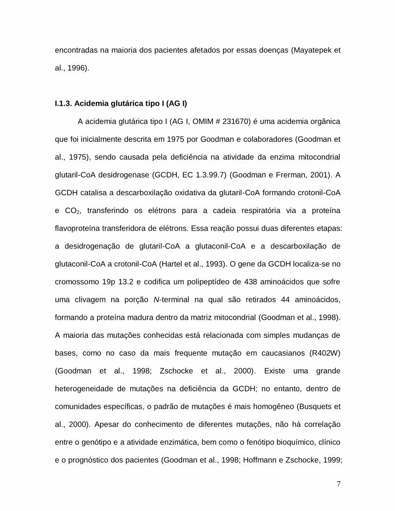
I.1.3. Acidemia glutárica tipo I (AG I)
A acidemia glutárica tipo I (AG I, OMIM # 231670) é uma acidemia orgânica
que foi inicialmente descrita em 1975 por Goodman e colaboradores (Goodman et
al., 1975), sendo causada pela deficiência na atividade da enzima mitocondrial
glutaril-CoA desidrogenase (GCDH, EC 1.3.99.7) (Goodman e Frerman, 2001). A
GCDH catalisa a descarboxilação oxidativa da glutaril-CoA formando crotonil-CoA
e CO2, transferindo os elétrons para a cadeia respiratória via a proteína
flavoproteína transferidora de elétrons. Essa reação possui duas diferentes etapas:
a desidrogenação de glutaril-CoA a glutaconil-CoA e a descarboxilação de
glutaconil-CoA a crotonil-CoA (Hartel et al., 1993). O gene da GCDH localiza-se no
cromossomo 19p 13.2 e codifica um polipeptídeo de 438 aminoácidos que sofre
uma clivagem na porção N-terminal na qual são retirados 44 aminoácidos,
formando a proteína madura dentro da matriz mitocondrial (Goodman et al., 1998).
A maioria das mutações conhecidas está relacionada com simples mudanças de
bases, como no caso da mais frequente mutação em caucasianos (R402W)
(Goodman et al., 1998; Zschocke et al., 2000). Existe uma grande
heterogeneidade de mutações na deficiência da GCDH; no entanto, dentro de
comunidades específicas, o padrão de mutações é mais homogêneo (Busquets et
al., 2000). Apesar do conhecimento de diferentes mutações, não há correlação
entre o genótipo e a atividade enzimática, bem como o fenótipo bioquímico, clínico
e o prognóstico dos pacientes (Goodman et al., 1998; Hoffmann e Zschocke, 1999;
8
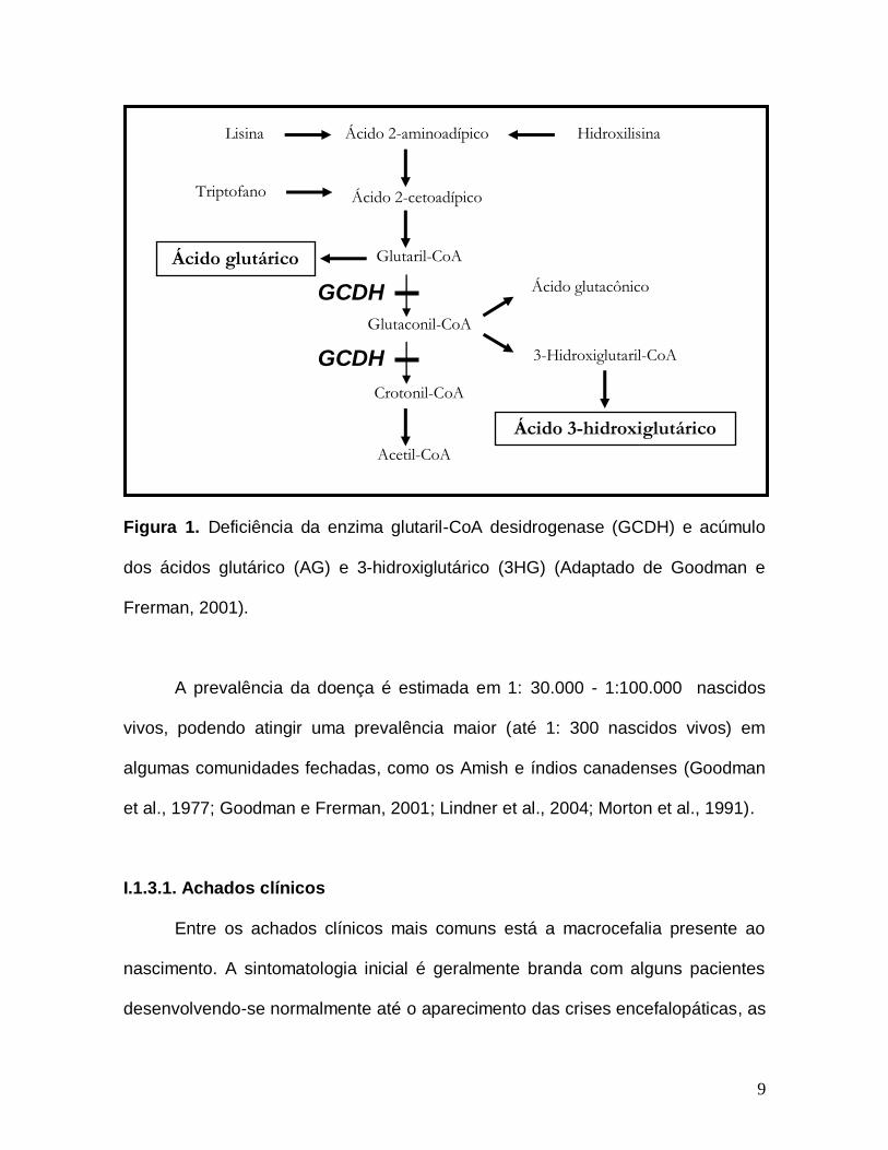
Kolker et al., 2006). Com o bloqueio da atividade enzimática, formam-se rotas
metabólicas alternativas que culminam na presença de concentrações elevadas
dos ácidos glutárico (AG), 3-hidroxiglutárico (3HG) e, algumas vezes, glutacônico
nos tecidos e líquidos biológicos (plasma, urina e líquor) dos indivíduos afetados
(Goodman et al., 1977; Goodman e Frerman, 2001) (Figura 1).
As concentrações plasmáticas destes ácidos variam entre 5 e 400 μmol/L
(Hoffmann et al., 1991; Merinero et al., 1995), mas as cerebrais podem atingir
500–5000 µmol/L para o AG e 40–200 µmol/L para o 3HG (Funk et al., 2005;
Sauer et al., 2006). Tais diferenças podem ser explicadas pelo fato de que o AG e
o 3HG serem produzidos nas células neurais e que a barreira hematoencefálica é
pouco permeável a esses ácidos orgânicos, ocasionando o acúmulo dessas
substâncias no sistema nervoso central (SNC), o que constitui num fator de risco
na neurodegeneração característica dos pacientes afetados (Hoffmann et al., 1993;
Kolker et al., 2006; Sauer et al., 2006).
9
Figura 1. Deficiência da enzima glutaril-CoA desidrogenase (GCDH) e acúmulo
dos ácidos glutárico (AG) e 3-hidroxiglutárico (3HG) (Adaptado de Goodman e
Frerman, 2001).
A prevalência da doença é estimada em 1: 30.000 - 1:100.000 nascidos
vivos, podendo atingir uma prevalência maior (até 1: 300 nascidos vivos) em
algumas comunidades fechadas, como os Amish e índios canadenses (Goodman
et al., 1977; Goodman e Frerman, 2001; Lindner et al., 2004; Morton et al., 1991).
I.1.3.1. Achados clínicos
Entre os achados clínicos mais comuns está a macrocefalia presente ao
nascimento. A sintomatologia inicial é geralmente branda com alguns pacientes
desenvolvendo-se normalmente até o aparecimento das crises encefalopáticas, as
Lisina
Triptofano
Hidroxilisina Ácido 2-aminoadípico
Ácido 2-cetoadípico
Glutaril-CoA
Glutaconil-CoA
Crotonil-CoA
Acetil-CoA
Ácido glutárico
Ácido glutacônico
3-Hidroxiglutaril-CoA
Ácido 3-hidroxiglutárico
GCDH
GCDH
10
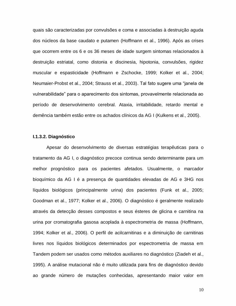
quais são caracterizadas por convulsões e coma e associadas à destruição aguda
dos núcleos da base caudato e putamen (Hoffmann et al., 1996). Após as crises
que ocorrem entre os 6 e os 36 meses de idade surgem sintomas relacionados à
destruição estriatal, como distonia e discinesia, hipotonia, convulsões, rigidez
muscular e espasticidade (Hoffmann e Zschocke, 1999; Kolker et al., 2004;
Neumaier-Probst et al., 2004; Strauss et al., 2003). Tal fato sugere uma “janela de
vulnerabilidade” para o aparecimento dos sintomas, provavelmente relacionada ao
período de desenvolvimento cerebral. Ataxia, irritabilidade, retardo mental e
demência também estão entre os achados clínicos da AG I (Kulkens et al., 2005).
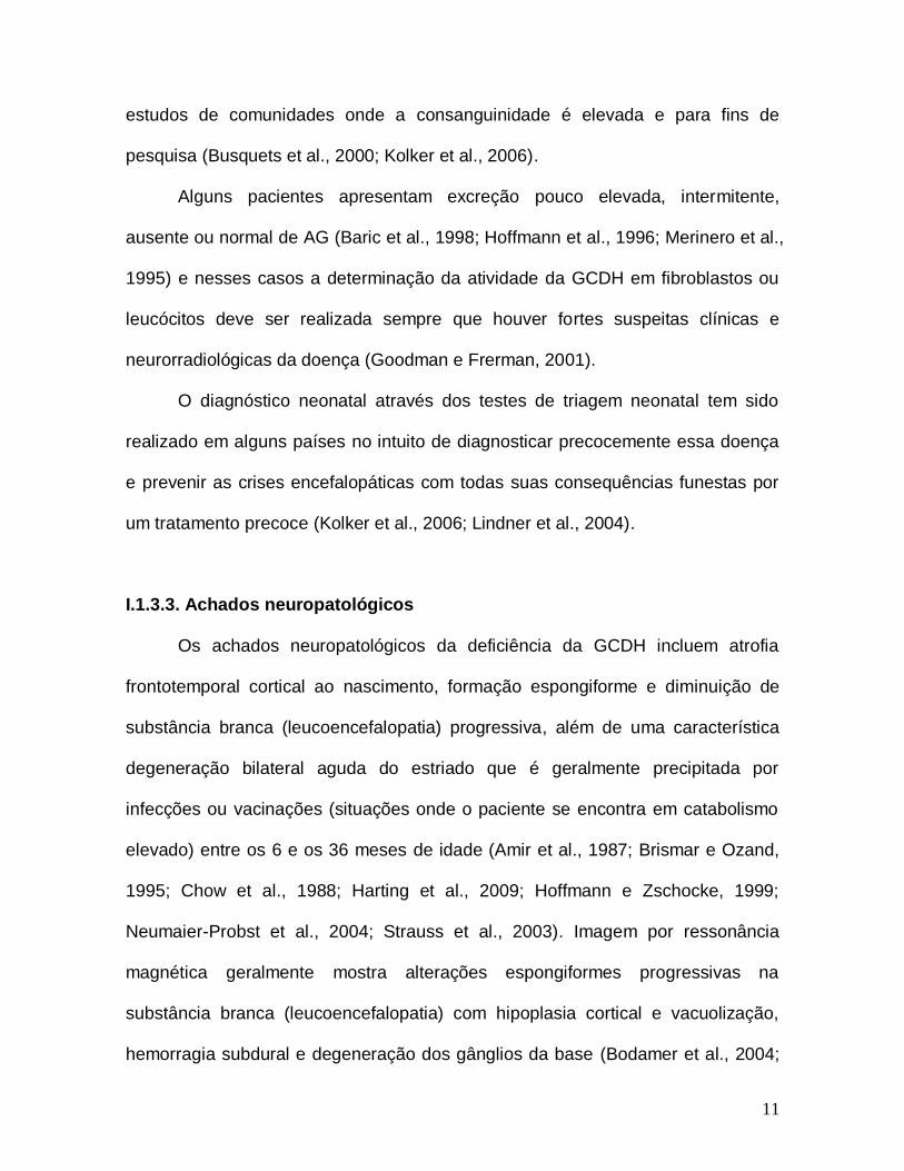
I.1.3.2. Diagnóstico
Apesar do desenvolvimento de diversas estratégias terapêuticas para o
tratamento da AG I, o diagnóstico precoce continua sendo determinante para um
melhor prognóstico para os pacientes afetados. Usualmente, o marcador
bioquímico da AG I é a presença de quantidades elevadas de AG e 3HG nos
líquidos biológicos (principalmente urina) dos pacientes (Funk et al., 2005;
Goodman et al., 1977; Kolker et al., 2006). O diagnóstico é geralmente realizado
através da detecção desses compostos e seus ésteres de glicina e carnitina na
urina por cromatografia gasosa acoplada à espectrometria de massa (Hoffmann,
1994; Kolker et al., 2006). O perfil de acilcarnitinas e a diminuição de carnitinas
livres nos líquidos biológicos determinados por espectrometria de massa em
Tandem podem ser usados como métodos auxiliares no diagnóstico (Ziadeh et al.,
1995). A análise mutacional não é muito utilizada para fins de diagnóstico devido
ao grande número de mutações conhecidas, apresentando maior valor em
11
estudos de comunidades onde a consanguinidade é elevada e para fins de
pesquisa (Busquets et al., 2000; Kolker et al., 2006).
Alguns pacientes apresentam excreção pouco elevada, intermitente,
ausente ou normal de AG (Baric et al., 1998; Hoffmann et al., 1996; Merinero et al.,
1995) e nesses casos a determinação da atividade da GCDH em fibroblastos ou
leucócitos deve ser realizada sempre que houver fortes suspeitas clínicas e
neurorradiológicas da doença (Goodman e Frerman, 2001).
O diagnóstico neonatal através dos testes de triagem neonatal tem sido
realizado em alguns países no intuito de diagnosticar precocemente essa doença
e prevenir as crises encefalopáticas com todas suas consequências funestas por
um tratamento precoce (Kolker et al., 2006; Lindner et al., 2004).
I.1.3.3. Achados neuropatológicos
Os achados neuropatológicos da deficiência da GCDH incluem atrofia
frontotemporal cortical ao nascimento, formação espongiforme e diminuição de
substância branca (leucoencefalopatia) progressiva, além de uma característica
degeneração bilateral aguda do estriado que é geralmente precipitada por
infecções ou vacinações (situações onde o paciente se encontra em catabolismo
elevado) entre os 6 e os 36 meses de idade (Amir et al., 1987; Brismar e Ozand,
1995; Chow et al., 1988; Harting et al., 2009; Hoffmann e Zschocke, 1999;
Neumaier-Probst et al., 2004; Strauss et al., 2003). Imagem por ressonância
magnética geralmente mostra alterações espongiformes progressivas na
substância branca (leucoencefalopatia) com hipoplasia cortical e vacuolização,
hemorragia subdural e degeneração dos gânglios da base (Bodamer et al., 2004;
12
Goodman et al., 1977; Harting et al., 2009; Hoffmann e Zschocke, 1999;
Neumaier-Probst et al., 2004; Nunes et al., 2013; Perez-Duenas et al., 2009;
Strauss e Morton, 2003).
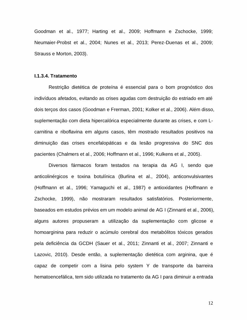
I.1.3.4. Tratamento
Restrição dietética de proteína é essencial para o bom prognóstico dos
indivíduos afetados, evitando as crises agudas com destruição do estriado em até
dois terços dos casos (Goodman e Frerman, 2001; Kolker et al., 2006). Além disso,
suplementação com dieta hipercalórica especialmente durante as crises, e com L-
carnitina e riboflavina em alguns casos, têm mostrado resultados positivos na
diminuição das crises encefalopáticas e da lesão progressiva do SNC dos
pacientes (Chalmers et al., 2006; Hoffmann et al., 1996; Kulkens et al., 2005).
Diversos fármacos foram testados na terapia da AG I, sendo que
anticolinérgicos e toxina botulínica (Burlina et al., 2004), anticonvulsivantes
(Hoffmann et al., 1996; Yamaguchi et al., 1987) e antioxidantes (Hoffmann e
Zschocke, 1999), não mostraram resultados satisfatórios. Posteriormente,
baseados em estudos prévios em um modelo animal de AG I (Zinnanti et al., 2006),
alguns autores propuseram a utilização da suplementação com glicose e
homoarginina para reduzir o acúmulo cerebral dos metabólitos tóxicos gerados
pela deficiência da GCDH (Sauer et al., 2011; Zinnanti et al., 2007; Zinnanti e
Lazovic, 2010). Desde então, a suplementação dietética com arginina, que é
capaz de competir com a lisina pelo system Y de transporte da barreira
hematoencefálica, tem sido utilizada no tratamento da AG I para diminuir a entrada
13
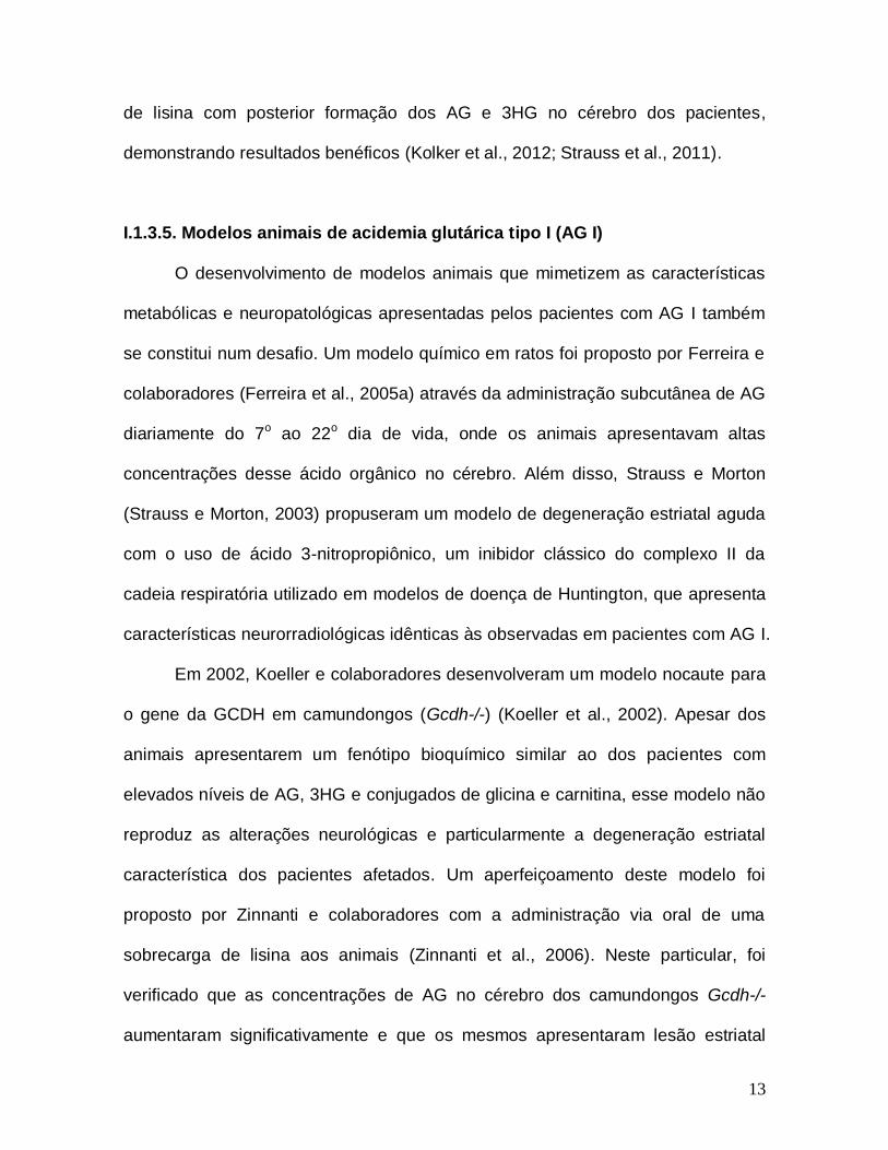
de lisina com posterior formação dos AG e 3HG no cérebro dos pacientes,
demonstrando resultados benéficos (Kolker et al., 2012; Strauss et al., 2011).
I.1.3.5. Modelos animais de acidemia glutárica tipo I (AG I)
O desenvolvimento de modelos animais que mimetizem as características
metabólicas e neuropatológicas apresentadas pelos pacientes com AG I também
se constitui num desafio. Um modelo químico em ratos foi proposto por Ferreira e
colaboradores (Ferreira et al., 2005a) através da administração subcutânea de AG
diariamente do 7o ao 22o dia de vida, onde os animais apresentavam altas
concentrações desse ácido orgânico no cérebro. Além disso, Strauss e Morton
(Strauss e Morton, 2003) propuseram um modelo de degeneração estriatal aguda
com o uso de ácido 3-nitropropiônico, um inibidor clássico do complexo II da
cadeia respiratória utilizado em modelos de doença de Huntington, que apresenta
características neurorradiológicas idênticas às observadas em pacientes com AG I.
Em 2002, Koeller e colaboradores desenvolveram um modelo nocaute para
o gene da GCDH em camundongos (Gcdh-/-) (Koeller et al., 2002). Apesar dos
animais apresentarem um fenótipo bioquímico similar ao dos pacientes com
elevados níveis de AG, 3HG e conjugados de glicina e carnitina, esse modelo não
reproduz as alterações neurológicas e particularmente a degeneração estriatal
característica dos pacientes afetados. Um aperfeiçoamento deste modelo foi
proposto por Zinnanti e colaboradores com a administração via oral de uma
sobrecarga de lisina aos animais (Zinnanti et al., 2006). Neste particular, foi
verificado que as concentrações de AG no cérebro dos camundongos Gcdh-/-
aumentaram significativamente e que os mesmos apresentaram lesão estriatal
14
semelhante aos pacientes afetados pela AG I, além de provocar a perda de
seletividade da barreira hematoencefálica.
I.1.3.6. Fisiopatologia
Nos últimos anos, distintos mecanismos foram propostos para explicar a
fisiopatogenia do dano cerebral da AG I. O fato de alguns pacientes excretarem
altas concentrações de lactato, 3-hidroxibutirato, acetoacetato e ácidos
dicarboxílicos, sugere que uma disfunção mitocondrial exerce um importante papel
na neuropatologia dos pacientes acometidos pela AG I (Floret et al., 1979;
Gregersen e Brandt, 1979). Neste sentido, foi demonstrado que o AG e 3HG in
vitro alteram de maneira moderada a atividade de alguns complexos da cadeia
respiratória, os níveis de fosfocreatina, a produção de CO2, a atividade da creatina
quinase (CK) e os níveis de ATP em cérebro e culturas de neurônios de ratos (Das
et al., 2003; Ferreira et al., 2005b; Ferreira et al., 2007a; Kolker et al., 2002a;
Kolker et al., 2002b; Latini et al., 2005a; Silva et al., 2000; Ullrich et al., 1999).
Além disso, estudos in vivo em músculo esquelético e cérebro de ratos tratados
cronicamente com AG mostraram inibições moderadas nas atividades dos
complexos I-III, II, II-III e da enzima CK (Ferreira et al., 2005a; Ferreira et al.,
2007b). Por outro lado, uma inibição da enzima Na+, K+ - ATPase pelo AG foi
relatada em córtex cerebral de ratos in vitro (Kolker et al., 2002b), assim como em
cérebro de ratos tratados cronicamente ou através de uma injeção intraestriatal
desse ácido orgânico (Fighera et al., 2006; Rodrigues et al., 2013). Sauer e
colaboradores também descreveram que o glutaril-CoA, diferentemente do AG e
3HG, foi capaz de inibir de maneira não-competitiva a enzima -cetoglutarato
15
desidrogenase (-CGDH) purificada (Sauer et al., 2005). Finalmente, a presença
de disfunção mitocondrial foi detectada em cultura de astrócitos de ratos tratados
com AG ou 3HG (Olivera et al., 2008).
Vários outros estudos in vivo e in vitro demonstraram efeitos deletérios de
AG e 3HG, incluindo excitotoxicidade e estresse oxidativo. Neste contexto,
estudos in vitro mostraram que tanto o AG (de Oliveira Marques et al., 2003) como
o 3HG (Latini et al., 2002; Latini et al., 2005b) aumentam a lipoperoxidação e
diminuiem as defesas antioxidantes e os níveis de glutationa reduzida em cérebro
de ratos. A produção de espécies reativas de oxigênio na presença de 3HG
também foi evidenciada em culturas de neurônios de telencéfalos de embriões de
pinto (Kolker et al., 2001). Além disso, foi demonstrada que a administração aguda
e crônica de AG aumenta a lipoperoxidação e diminuíram as defesas antioxidantes
em diferentes estruturas cerebrais, fígado e eritrócitos de ratos (Latini et al., 2007).
A excitotoxicidade também é um mecanismo muito relacionado com a
fisiopatogenia da AG I. Inicialmente, foi demonstrado um comprometimento da
neurotransmissão GABAérgica causada pelo metabólitos acumulados na AG I
(Leibel et al., 1980; Stokke et al., 1976; Wajner et al., 2004). Além disso, vários
trabalhos sugerem que a neurotoxicidade da AG I possa ocorrer devido à
interação dos AG e 3HG com receptores e transportadores glutamatérgicos em
culturas de células e cérebro de ratos (Bjugstad et al., 2001; de Mello et al., 2001;
Flott-Rahmel et al., 1997; Kolker et al., 1999, 2000; Kolker et al., 2002a; Kolker et
al., 2002b; Porciuncula et al., 2000; Porciuncula et al., 2004; Rosa et al., 2004).
16
Outros trabalhos sugerem que uma disfunção endotelial com perda da
integridade da barreira hematoencefálica esteja envolvida na lesão neurológica
característica dos pacientes com AG I e outras acidemias orgânicas (Muhlhausen
et al., 2006; Strauss e Morton, 2003; Zinnanti et al., 2006). Neste contexto, foi
demonstrado recentemente um aumento na permeabilidade da barreira
hematoencefálica em cérebro de camundongos Gcdh-/- (Zinnanti et al., 2014).
Além disso, metabólitos da via das quinureninas, uma das rotas de catabolismo do
triptofano, associados com outras substâncias acumuladas na AG I, podem
também estar envolvidos na neurodegeneração dessa doença (Heyes, 1987;
Lehnert e Sass, 2005; Varadkar e Surtees, 2004).
Enfatize-se que todos os resultados citados acima foram obtidos através de
estudos in vitro e in vivo em ratos selvagens ou em culturas de células obtidas de
animais com atividade normal da GCDH.
Estudos recentes com animais Gcdh-/- demonstraram que o transporte de
sucinato dos astrócitos para os neurônios, via o transportador de dicarboxilatos, foi
inibido pelos AG e 3HG em culturas primárias de astrócitos e neurônios de
camundongos Gcdh-/-, possivelmente comprometendo a reposição de
intermediários do ciclo do ácido cítrico (CAC) para os neurônios e,
consequentemente, prejudicando principalmente a produção dos
neurotransmissores glutamato e GABA, assim como de ATP (Lamp et al., 2011).
Neste contexto, Zinnanti e colaboradores encontraram uma diminuição nos níveis
de ATP, fosfocreatina, α-cetoglutarato, CoA, glutamato e GABA, bem como um
aumento de acetil-CoA, em cérebro de camundongos Gcdh-/ (Zinnanti et al., 2007).
Além disso, foi recentemente verificado a indução de estresse oxidativo em
17
cérebro de camundongos Gcdh-/- submetidos a uma sobrecarga de lisina
(Seminotti et al., 2012; Seminotti et al., 2013).
No entanto, apesar da intensa investigação, as causas da susceptibilidade
frontotemporal cortical durante a gestação e da janela de vulnerabilidade estriatal
durante os primeiros anos de vida, assim como o papel da disfunção mitocondrial,
permanecem obscuras na patogênese da AG I.
I.1.4. Metabolismo energético cerebral
O cérebro é um dos órgãos mais ativos metabolicamente, mas possui
reservas energéticas extremamente pequenas em relação a sua alta taxa
metabólica (Attwell e Laughlin, 2001; Dickinson, 1996).
A glicose é o principal composto energético do cérebro (Erecinska et al.,
2004). Em condições normais, o metabolismo energético nos tecidos neurais é
mantido, quase que exclusivamente, pelo metabolismo oxidativo da glicose
(Mergenthaler et al., 2013). A oxidação da glicose no cérebro ocorre mais
rapidamente do que em outros órgãos como fígado, coração ou rins. Em contraste
com outros tecidos, o cérebro não necessita de insulina para captar e oxidar a
glicose. Entretanto, durante o estado de jejum, os corpos cetônicos podem
substituir mais de 50% das necessidades energéticas cerebrais (Dickinson, 1996).
A oxidação da glicose através da via glicolítica forma piruvato, que é
convertido a CO2 e H2O no CAC e na cadeia transportadora de elétrons. O
acoplamento entre a cadeia transportadora de elétrons e a fosforilação oxidativa
gera grande parte do ATP necessário ao cérebro (Erecinska et al., 2004).
18
I.1.5. Ciclo do ácido cítrico (CAC), fosforilação oxidativa, cadeia
transportadora de elétrons e parâmetros respiratórios
O CAC é a via comum de oxidação dos glicídeos, aminoácidos e ácidos
graxos utilizando enzimas como citrato sintase (CS), aconitase (ACO), isocitrato
desidrogenase (IDH), α-CGDH, sucinato desidrogenase (SDH) e malato
desidrogenase (MDH). Enfatizamos que a IDH3 é a isoforma mitocondrial e
dependente de NAD em camundongos, possuindo três diferentes subunidades
onde a α é a catalítica (Kim et al., 1999). O metabolismo energético cerebral se
mostra essencialmente aeróbico, sendo a glicose o principal substrato utilizado
(Clark et al., 1993), entrando no ciclo sob a forma de acetil-CoA que é então
oxidado completamente a CO2. As reações anapleróticas que alimentam o ciclo
fornecendo diretamente seus intermediários, também fornecem substratos para as
reações de oxidação no cérebro.
Quando não há hipóxia, a fosforilação oxidativa é o processo mitocondrial
pelo qual o O2 é reduzido a H2O por elétrons doados pelo NADH e FADH2 que
fluem por vários pares de redução-oxidação (cadeia respiratória), ocorrendo
concomitantemente a produção de ATP a partir de ADP e Pi (Nelson e Cox, 2008).
As mitocôndrias são corpúsculos envoltos por uma membrana externa, facilmente
permeável a pequenas moléculas e íons, e por uma membrana interna,
impermeável à maioria das moléculas e íons, incluindo prótons (Nelson e Cox,
2008). O fluxo de elétrons a partir de NADH e FADH2 até o O2 (aceptor final de
elétrons) se dá através de complexos enzimáticos ancorados na membrana
mitocondrial interna com centros redox com afinidade crescente por elétrons. Essa
transferência de elétrons é impulsionada por um crescente potencial redox
19
existente entre os equivalentes reduzidos (NADH e o FADH2), os complexos
enzimáticos da cadeia transportadora de elétrons e o O2, que é o aceptor final
dessa cadeia de reações de oxidação.
A cadeia respiratória é composta por vários complexos enzimáticos e uma
coenzima lipossolúvel, a coenzima Q ou ubiquinona. O complexo I conhecido
como NADH desidrogenase ou NADH: ubiquinona oxidorredutase transfere os
elétrons do NADH para a ubiquinona. O complexo II reduz a ubiquinona com
elétrons do FADH2 provenientes da oxidação do sucinato a fumarato no CAC. O
complexo III citocromo bc1 ou ubiquinona-citocromo c oxidoredutase catalisa a
redução do citocromo c a partir da ubiquinona reduzida. Na parte final da cadeia
de transporte de elétrons, o complexo IV (citocromo c oxidase) catalisa a
transferência de elétrons de moléculas reduzidas de citocromo c para O2,
formando H2O.
O fluxo de elétrons através dos complexos da cadeia respiratória é
acompanhado pelo bombeamento de prótons da matriz mitocondrial para o
espaço intermembrana, através dos complexos I, III e IV, gerando um potencial de
membrana. Assim, cria-se um gradiente eletroquímico transmembrana que pode
ser utilizado por um quinto complexo proteico, a ATP sintase, para a síntese de
ATP. Dessa forma, a oxidação de substratos energéticos está acoplada ao
processo de fosforilação do ADP, ou seja, quando o potencial de membrana é
dissipado pelo fluxo de prótons a favor do gradiente eletroquímico, a energia
liberada é utilizada pela ATP sintase que atua como uma bomba de prótons ATP-
dependente (Nelson e Cox, 2008).
20
Desse modo, a respiração mitocondrial pode ser estimada através da
medida do consumo de O2. Apesar do fato de que essa medida determina
diretamente apenas a velocidade de uma única reação (transferência final de
elétrons para O2), muitas informações sobre outros processos mitocondriais
podem ser obtidos simplesmente pela adaptação das condições de incubação.
Vários passos podem ser investigados, incluindo o transporte de substratos
através da membrana mitocondrial, a atividade das desidrogenases, a atividade
dos complexos da cadeia respiratória, o transporte de nucleotídeos de adenina
pela membrana mitocondrial, a atividade da ATP sintase e a permeabilidade da
membrana mitocondrial a prótons (Nicholls e Ferguson, 2001). Experimentalmente,
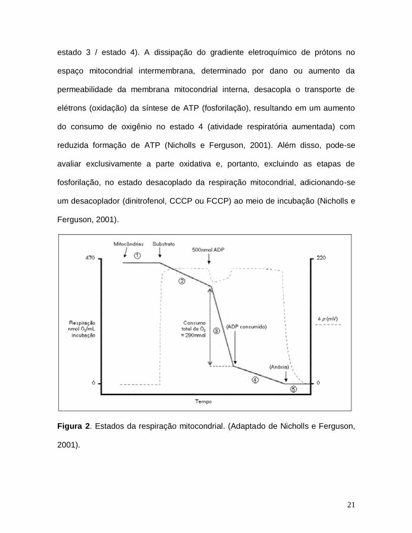
pode-se dividir a respiração mitocondrial em 5 estágios, conforme ilustra a figura 2.
No entanto, apenas os parâmetros estados 3 e 4 são comumente utilizados. O
estado 3 representa o consumo de oxigênio quando as mitocôndrias, em um meio
contendo substrato oxidável, são expostas a ADP, estimulando o consumo de O2
e produzindo ATP (estado fosforilante). O estado 4 reflete o consumo de O2 após
as mitocôndrias já terem depletado todo o ADP disponível ou podendo ser
estimulado por oligomicina A (inibidor da ATP sintase), reduzindo a taxa da
respiração (estado não-fosforilante) (Nicholls e Ferguson, 2001). Para que a ATP
sintase esteja ativa, são necessários dois fatores: disponibilidade de ADP e
potencial de membrana suficientemente alto (Nelson e Cox, 2008). Neste contexto,
o acoplamento da respiração mitocondrial é definido como a capacidade da
mitocôndria gerar energia (ATP) quando exposta ao ADP, ou seja, unir (acoplar)
os processos de oxidação e de fosforilação, o que pode ser avaliado
experimentalmente pela medida da razão de controle respiratório (RCR; razão do
21
estado 3 / estado 4). A dissipação do gradiente eletroquímico de prótons no
espaço mitocondrial intermembrana, determinado por dano ou aumento da
permeabilidade da membrana mitocondrial interna, desacopla o transporte de
elétrons (oxidação) da síntese de ATP (fosforilação), resultando em um aumento
do consumo de oxigênio no estado 4 (atividade respiratória aumentada) com
reduzida formação de ATP (Nicholls e Ferguson, 2001). Além disso, pode-se
avaliar exclusivamente a parte oxidativa e, portanto, excluindo as etapas de
fosforilação, no estado desacoplado da respiração mitocondrial, adicionando-se
um desacoplador (dinitrofenol, CCCP ou FCCP) ao meio de incubação (Nicholls e
Ferguson, 2001).
Figura 2. Estados da respiração mitocondrial. (Adaptado de Nicholls e Ferguson,
2001).
22
Além da regeneração do ATP, que é a sua principal função, a mitocôndria
desempenha outras funções importantes. Esta organela é a principal fonte de
espécies reativas de oxigênio e de defesas antioxidantes nas células (Cadenas e
Davies, 2000), gerando ânions superóxido (O2●-) no espaço intermembrana pelo
vazamento de elétrons que se combinam com o oxigênio molecular,
principalmente no complexo III, em um processo que é dependente de potencial
de membrana na matriz (Han et al., 2001). Além disso, a mitocôndria participa
ativamente da homeostase celular de Ca2+ (Nicholls e Akerman, 1982) e está
envolvida em diversos processos que levam à morte celular por apoptose,
incluindo liberação de citocromo c (Liu et al., 1996). É possível medir
experimentalmente, além da respiração mitocondrial, o potencial de membrana, o
inchamento, a produção de peróxido de hidrogênio, a capacidade de retenção de
Ca2+ e o conteúdo de NAD(P)H mitocondrial (Maciel et al., 2004; Saito e Castilho,
2010).
I.1.6. Creatina quinase (CK)
A CK é a enzima responsável pelo processo reversível de fosforilação da
creatina formando fosfocreatina, e desfosforilação da fosfocreatina formando
creatina.
Tecidos que possuem uma alta demanda de energia, como músculo
esquelético, cardíaco e cérebro, apresentam uma maior concentração de CK,
porque esta enzima regenera o ATP, que é muito consumido nesses tecidos
(Wyss et al., 1992). Atualmente são conhecidas cinco isoformas da CK. Três são
encontradas no citosol e duas na mitocôndria (CKmi). As isoformas citosólicas
23
formam dímeros, chamados MM-CK, MB-CK e BB-CK, compostos por dois tipos
de subunidades (monômeros): o monômero M (tipo muscular) e o monômero B
(tipo cerebral). A isoforma MM-CK é encontrada predominantemente em músculo
esquelético adulto e no músculo cardíaco, a BB-CK está presente principalmente
em tecidos neurais, e a MB-CK é somente encontrada em coração (Boehm et al.,
1996; Wyss et al., 1992).
A atividade da CK e as concentrações de creatina são importantes para o
tamponamento energético e para a transferência de energia dos sítios de
produção de ATP para os sítios de consumo que utilizam as ATPases, evitando
assim grandes variações nos níveis celulares de ATP e ADP do metabolismo
celular (O'Gorman et al., 1996; Wyss et al., 1992). Alterações na atividade da CK
são associadas com vários estados patológicos. A diminuição da atividade da CK
no coração está relacionada à cardiomiopatias e falência cardíaca, além de
pacientes com miopatias mitocondriais apresentarem mitocôndrias inchadas
devido à inibição da CKmi (O'Gorman et al., 1996).
I.1.7. Na+, K+-ATPase
A enzima Na+, K+-ATPase é uma proteína transmembrana constituída
principalmente por dois tipos de subunidades: a subunidade de 110kDa, que
contém os sítios catalíticos e de ligação de íons, e a subunidade β, que é uma
glicoproteína de 55kDa essencial para a atividade da enzima, mas de função não
totalmente esclarecida, formando uma estrutura dimérica (β)2 (Morth et al.,
2007). A subunidade catalítica da Na+, K+-ATPase se apresenta com 3 isoformas
24
em mamíferos, sendo que a 1 está expressa em todo o animal, a 2 em cérebro
(principalmente em astrócitos), músculo esquelético, coração e adipócitos,
enquanto que a 3 principalmente em neurônios (Blanco e Mercer, 1998; Chen et
al., 2013; Kawakami e Ikeda, 2006; Zhang et al., 2013).
A função dessa enzima é translocar os cátions Na+ e K+ através da
membrana plasmática contra seus respectivos gradientes de concentração,
utilizando a energia fornecida pela hidrólise de ATP. A enzima transporta
simultaneamente 3 íons Na+ para fora e 2 íons K+ para dentro da célula. A saída
de Na+ capacita as células animais a controlar osmoticamente seu conteúdo
hídrico (Aperia, 2007; Jorgensen et al., 2003). Visto que três cargas positivas são
transportadas para o meio extracelular e somente duas são transportadas para o
meio intracelular, o fluxo de íons Na+ e K+ produz um gradiente eletroquímico
através da membrana celular (Kaplan, 2002). Esse gradiente é usado como fonte
de energia para a despolarização e repolarização do potencial de membrana, para
a manutenção e regulação do volume celular, para transporte ativo secundário
dependente de íons Na+, transporte de glicose, de aminoácidos, de
neurotransmissores e outros íons (Geering, 1990). Alteração nos mecanismos que
mantêm o equilíbrio entre a taxa de Na+ e K+ intra e extracelular pode causar
graves consequências para as células do SNC (Erecinska et al., 2004), tendo sido
associadas a estados de neurodegeneração como nas doenças de Alzheimer,
Parkinson e esclerose lateral amiotrófica (Bagh et al., 2008; Dickey et al., 2005;
Ellis et al., 2003; Vignini et al., 2007).
25
I.1.8. Metabolismo energético e doenças neurodegenerativas
Numerosas hipóteses têm sido propostas para explicar a fisiopatologia de
doenças neurodegenerativas mais comuns, como as doenças de Alzheimer e
Parkinson sem, no entanto, obter até o momento uma explicação satisfatória para
o dano cerebral dessas doenças. Entretanto, acredita-se que alterações do
metabolismo energético, indução de estresse oxidativo e neurotoxicidade mediada
por receptores glutamatérgicos do tipo NMDA, ou, possivelmente, um somatório
desses fatores possam estar envolvidos na neurodegeneração (Camins et al.,
2008; Nakamura e Lipton, 2011; Rose e Henneberry, 1994). Uma das hipóteses é
de que alterações na cadeia transportadora de elétrons seria o evento etiológico
primário na maioria dessas doenças (Chaturvedi e Flint Beal, 2013; Parker et al.,
1990; Swerdlow et al., 1998).
O cérebro é altamente dependente de energia para seu funcionamento
normal e a mitocôndria é a organela intracelular que mantém os suprimentos de
energia para o cérebro. Uma alteração funcional nessa organela pode levar,
portanto, a consequências patológicas aos neurônios e astrócitos (Beal, 1995)
(Bowling e Beal, 1995; Davis et al., 1995). Assim, numerosas evidências
relacionam doenças neurodegenerativas a um comprometimento do metabolismo
energético. Estudos anteriores demonstraram uma diminuição na atividade do
complexo I da cadeia respiratória em cérebros postmortem de pacientes
portadores de doença de Parkinson (Gautier et al., 2013; Janetzky et al., 1994;
Parker et al., 2008). Também há relatos de defeitos nas atividades dos complexos
26
II e III da cadeia respiratória e da enzima -CGDH nessa doença (Mizuno et al.,
1990; Shen et al., 2000).
No que diz respeito a mais comum dentre as doenças neurodegenerativas,
a doença de Alzheimer, já foi relatado uma redução na atividade do complexo IV
da cadeia respiratória (Bobba et al., 2013; Maurer et al., 2000). Estudos em
cérebros postmortem de pacientes portadores dessa doença demonstraram uma
diminuição nas atividades do complexo enzimático da piruvato desidrogenase e da
-CGDH (Gibson et al., 1988; Gibson et al., 2012; Mastrogiacomo et al., 1993;
Perry et al., 1980).
I.2. OBJETIVOS
I.2.1. Objetivo geral
O objetivo do presente trabalho foi o de investigar importantes parâmetros
da homeostase energética celular em tecidos cerebrais (córtex cerebral, estriado e
hipocampo) e periféricos (coração e músculo esquelético), bem como alterações
histológicas no cérebro de camundongos Gcdh-/- e selvagens (WT) submetidos a
uma dieta normal ou a uma sobrecarga de lisina (injeção intraperitoneal ou dieta
rica em lisina), visando a uma melhor compreensão da fisiopatologia do dano
tecidual na AG I.
I.2.2. Objetivos específicos
1. Investigar o efeito de uma sobrecarga aguda de lisina (injeção intraperitoneal
27
de 8 µmol/g de lisina) sobre as atividades dos complexos I-III, II, II-III e IV da
cadeia respiratória, da CK e da Na+, K+ - ATPase em cérebro, coração e
músculo esquelético de camundongos Gcdh-/- e WT com 15 dias de vida.
2. Investigar o efeito de uma sobrecarga de lisina na dieta (4,7 %) por 60 horas
sobre as atividades dos complexos I-III, II, II-III e IV da cadeia respiratória, da
CK e da Na+, K+ - ATPase, assim como sobre os parâmetros respiratórios
(estados 3 e 4, RCR e estado desacoplado) em tecidos cerebrais (cérebro
total, córtex cerebral, estriado e hipocampo) de camundongos Gcdh-/- e WT
com 30 dias de vida.
3. Investigar o efeito de uma sobrecarga de lisina na dieta (4,7 %) por 60 horas
sobre as atividades das enzimas do CAC CS, ACO, IDH, α-CGDH, SDH e
MDH, assim como a liberação de lactato, os parâmetros respiratórios (estados
3 e 4, RCR e estado desacoplado) e o potencial de membrana mitocondrial na
presença e ausência de Ca2+ em preparações mitocondriais de córtex cerebral
e estriado de camundongos Gcdh-/- e WT com 30 dias de vida.
4. Investigar alterações histológicas em córtex cerebral e estriado de
camundongos Gcdh-/- e WT com 30, 60 e 90 dias de vida sob o efeito de uma
sobrecarga de lisina na dieta (4,7 %) por períodos variáveis (60 horas e 30
dias).
28
PARTE II
Artigos Científicos
29
Capítulo I
Marked reduction of Na+, K+-ATPase and creatine kinase activities induced
by acute lysine administration in glutaryl-CoA dehydrogenase deficient mice
Alexandre U Amaral, Cristiane Cecatto, Bianca Seminotti, Ângela Zanatta,
Carolina G Fernandes, Estela B Busanello, Luisa M Braga, César A Ribeiro, Diogo
O de Souza, Michael Woontner, David M Koeller, Stephen Goodman, Moacir
Wajner.
Artigo científico publicado no periódico
Molecular Genetics and Metabolism
Molecular Genetics and Metabolism 107 (2012) 81–86
Contents lists available at SciVerse ScienceDirect
Molecular Genetics and Metabolism
j ourna l homepage: www.e lsev ie r .com/ locate /ymgme
Marked reduction of Na+, K+-ATPase and creatine kinase activities induced by acutelysine administration in glutaryl-CoA dehydrogenase deficient mice
Alexandre Umpierrez Amaral a, Cristiane Cecatto a, Bianca Seminotti a, Ângela Zanatta a,Carolina Gonçalves Fernandes a, Estela Natacha Brandt Busanello a, Luisa Macedo Braga b,César Augusto João Ribeiro a, Diogo Onofre Gomes de Souza a, Michael Woontner c, David M. Koeller d,Stephen Goodman c, Moacir Wajner a,e,⁎a Departamento de Bioquímica, Instituto de Ciências Básicas da Saúde, Universidade Federal do Rio Grande do Sul, Porto Alegre, RS, Brazilb Fundação Estadual de Produção e Pesquisa em Saúde, Porto Alegre, RS, Brazilc School Medicine University of Colorado Denver, Aurora, USA;d Departments of Pediatrics, Molecular and Medical Genetics, Oregon Health & Science University, Portland, OR, USAe Serviço de Genética Médica, Hospital de Clínicas de Porto Alegre, Porto Alegre, RS, Brazil
Abbreviations: CK, creatine kinase; DCIP, dichlordiaminetetracetic acid; GA, glutaric acid; GA I, glutaric aciddehydrogenase; Gcdh−/−, glutaryl-CoA dehydrogenase deyglutaric acid; HEPES, N-[2-hydroxyethyl]piperazine-N′-[2α-ketoglutarate dehydrogenase; KO, knockout; Lys, lysine;age for the Social Sciences; TCA, tricarboxylic acid; WT, wild⁎ Corresponding author at: Departamento de Bioq
Básicas da Saúde, Universidade Federal de Rio GrandeNo. 2600 ‐ Anexo, CEP 90035‐003, Porto Alegre, RS, Brfax: +55 51 3308 5535.
E-mail address: [email protected] (M. Wajner).
1096-7192/$ – see front matter © 2012 Elsevier Inc. Alldoi:10.1016/j.ymgme.2012.04.015
a b s t r a c t
a r t i c l e i n f oArticle history:Received 10 March 2012Received in revised form 17 April 2012Accepted 17 April 2012Available online 24 April 2012
Keywords:Glutaric acidemia type IGlutaryl-CoA dehydrogenase deficient miceBrainBioenergeticsNa+, K+-ATPase activityCreatine kinase activity
Glutaric acidemia type I (GA I) is an inherited neurometabolic disorder caused by a severe deficiency of themitochondrial glutaryl-CoA dehydrogenase activity leading to accumulation of predominantly glutaric (GA)and 3-hydroxyglutaric (3HGA) acids in the brain and other tissues. Affected patients usually present withhypotonia and brain damage and acute encephalopathic episodes whose pathophysiology is not yet fullyestablished. In this study we investigated important parameters of cellular bioenergetics in brain, heartand skeletal muscle from 15-day-old glutaryl-CoA dehydrogenase deficient mice (Gcdh−/−) submitted to asingle intra-peritoneal injection of saline (Sal) or lysine (Lys — 8 μmol/g) as compared to wild type (WT)mice. We evaluated the activities of the respiratory chain complexes II, II-III and IV, α-ketoglutarate dehydro-genase (α-KGDH), creatine kinase (CK) and synaptic Na+, K+-ATPase. No differences of all evaluated param-eters were detected in the Gcdh−/− relatively to the WT mice injected at baseline (Sal). Furthermore, mildincreases of the activities of some respiratory chain complexes (II-III and IV) were observed in heart and skel-etal muscle of Gcdh−/− andWTmice after Lys administration. However, the most marked effects provoked byLys administration were marked decreases of the activities of Na+, K+-ATPase in brain and CK in brain andskeletal muscle of Gcdh−/− mice. In contrast, brain α-KGDH activity was not altered in WT and Gcdh−/−
injected with Sal or Lys. Our results demonstrate that reduction of Na+, K+-ATPase and CK activities mayplay an important role in the pathogenesis of the neurodegenerative changes in GA I.
© 2012 Elsevier Inc. All rights reserved.
1. Introduction
Glutaryl-CoA dehydrogenase (GCDH) deficiency or glutaricacidemia type I (GA I) (McKusick 231670) is an autosomal recessiveneurometabolic disease biochemically characterized by the
oindophenol; EDTA, ethylene-emia type I; GCDH, glutaryl-CoAficient mice; 3HGA, 3-hydrox--ethane-sulfonic acid]; α-KGDH,Sal, saline; SPSS, Statistical Pack-type.uímica, Instituto de Ciênciasdo Sul. Rua Ramiro Barcelos
azil. Tel.: +55 51 3308 5571;
rights reserved.
accumulation of glutaric acid (GA), and to a lesser extent 3-hydroxyglutaric acid (3HGA) and glutaconic acid in body fluidsand tissues [1–3]. Clinically, the disease is characterized bymacrocephaly with frontotemporal atrophy at birth and by markeddystonia and diskinesia, following encephalopathic episodes trig-gered by catabolic events, such as infections, fever and fasting,when the accumulating metabolites can reach millimolar concentra-tions [1,4,5]. However, progressive neurological symptoms withmental developmental delay and hypotonia without apparentacute episodes may also occur in a considerable number of patients[5–9]. Neuroradiological imaging shows, besides basal gangliadegeneration, widened Sylvian fissures, cortical atrophy withfrontotemporal volume loss, delayed myelination, ventriculomegalyand subdural hemorrhages [1,5,6,8,10,11]
Although the pathogenesis of the brain damage in GA I is not fullyestablished, accumulating evidence from in vitro and in vivo experi-ments performed in brain tissue and cultivated neural cells from
Alexandre
Typewritten Text
Alexandre
Typewritten Text
30
Alexandre
Sticky Note
MigrationConfirmed set by Alexandre
Alexandre
Sticky Note
MigrationNone set by Alexandre
82 AU. Amaral et al. / Molecular Genetics and Metabolism 107 (2012) 81–86
rodents and chick suggest that excitotoxicity [6,12–21], oxidativestress [22–31] and cellular bioenergetic dysfunction [2,32–39] are in-volved in the brain damage of GA I patients. It is emphasized thatthese studies were carried out in animal tissues with normal GCDHactivity.
A knockout (KO) model of GA I was developed in mice byreplacing the GCDH gene with an in-frame beta-galactosidase cas-sette in order to investigate pathomechanisms underlying brain dam-age in this disorder [40]. Glutaryl-CoA dehydrogenase deficient(Gcdh−/−) mice displayed vacuolization in the frontal cortex, butdid not develop striatal damage typical of the human disease evenwhen submitted to metabolic or infectious stress. It was subsequentlyfound that exposure of these animals to high protein or lysine (Lys)intake resulted in striatal damage, besides neuronal loss, myelin dis-ruption and gliosis mostly in the striatum and deep cortex [41].
Considering that various works have emphasized the role of bio-energetics dysfunction on the prominent neurologic findings of GA I[2,42] and that the toxic organic acids accumulating in this disorderare likely to induce secondary pathological changes in the brain[39,41], in the present study we further investigated the role of cellu-lar bioenergetics alterations in the pathophysiology of GA I. We eval-uated central components of mitochondrial energy production,transfer and utilization, by measuring the activities of the respiratorychain complexes II, II-III and IV, α-ketoglutarate dehydrogenase(α-KGDH), creatine kinase (CK) and Na+,K+-ATPase in brain, heartand skeletal muscle from 15-day-old Gcdh−/− mice on standardmouse chow, and after acute Lys administration in order to clarifywhether disturbance of cellular bioenergetics is involved in the path-ogenesis and more specifically in the brain damage of GA.
2. Materials and methods
2.1. Chemicals
All chemicals were of analytical grade and purchased from Sigma(St. Louis, MO, USA) unless otherwise stated. Solutions were preparedon the day of the experiments and the pH was adjusted to 7.2–7.4with the appropriate buffers for each technique.
2.2. Animals
Fifteen-day-old Gcdh−/− and wild type (WT) mice littermatecontrols, both of 129SvEv background [40], were used in the experi-ments. We used 15-day-old animals because this age correspondsto very young animals, matching to humans of approximately3–4 years of life. In addition, prior studies by Zinnanti et al. haveshown that when fed a high lysine diet, 4-week-old Gcdh−/− mice ac-cumulate high levels of glutaric acid and develop an acute brain inju-ry, whereas 8-week old mice accumulate less glutaric acid and do notsuffer from acute brain injury [39,41]. This age dependent sensitivityhas been interpreted to indicate that the blood brain barrier is morepermeable to Lys in younger animals, resulting in higher levels ofGA and 3HGA being formed in the brain. The mice were generatedfrom heterozygous and maintained at Unidade de ExperimentaçãoAnimal (UEA) of the Hospital de Clínicas de Porto Alegre (HCPA).The animals were maintained on a 12:12 h light/dark cycle (lightson 07.00–19.00 h) in air conditioned constant temperature (22±1 °C) colony room, with free access to water and 20% (w/w) proteincommercial chow (SUPRA, Porto Alegre, RS, Brazil).
2.3. Ethical statement
This study was performed in strict accordance with the Principlesof Laboratory Animal Care, National Institute of Health of UnitedStates of America, NIH, publication No. 85‐23, revised in 1996 and ap-proved by the Ethical Committee for the Care and Use of Laboratory
Animals of HCPA. All efforts were also made to use the minimal num-ber of animals necessary to produce reliable scientific data.
2.4. Lysine (Lys) administration
The WT and Gcdh−/− animals were administered with a single in-traperitoneal injection of saline (Sal) or Lys (8 μmol/g) solution inorder to investigate whether an acute Lys overload could disturbbionergetics in a model of GCDH deficiency. We have previouslyshown that this dose of lysine results in a significant increase in GAand 3HGA and induces an acute oxidative stress in the brains ofGcdh−/− mice [52]. Enzyme activities were measured in tissues col-lected 4 h after lysine injection. We observed that approximately20% of Lys-treated Gcdh−/− mice became less active within 4 h of in-jection, and these mice were not used for analysis. On the other hand,we also observed in a distinct set of Gcdh−/− mice from the samebackground that approximately the same percentage of animals be-came hypoactive and died 12 h after Lys injection. These animalswere also not used in the assays, so that the survival rate of themice used for the biochemical analyses was 100%.
2.5. Tissue preparation
The mice were anesthetized with a mixture of ketamine(90 mg/kg) and xilazine (10 mg/kg) and intracardiacally perfusedduring 5 min with Sal solution. After perfusion, brain, heart and skel-etal muscle were rapidly removed and placed on a Petri dish on ice.
For the determination of respiratory chain complexes, α-KGDHand total CK activities, the olfactory bulb, pons, medulla, and cerebel-lum were discarded, and the forebrain, as well as the heart and skel-etal muscle, were used. The tissues were homogenized in 19 vol(1:20, w/v) of SETH buffer, pH 7.4 (250 mM sucrose, 2.0 mM EDTA,10 mM Trizma base and 50 UI mL−1 heparin). Homogenates werecentrifuged at 800×g for 10 min at 4 °C to discard nuclei and cell de-bris. The pellet was discarded and the supernatant, a suspension ofmixed and preserved organelles, including mitochondria, was sepa-rated and used to measure these parameters.
For the determination of Na+, K+-ATPase activity, the forebrainwas homogenized in 10 volumes of 0.32 mM sucrose solution con-taining 5.0 mM HEPES and 1.0 mM EDTA. Synaptic plasma mem-branes were then prepared according to the method of Jones andMatus (1974) [43] using a discontinuous sucrose density gradientconsisting of successive layers of 0.3, 0.8 and 1.0 mM. After centrifu-gation at 69,000×g for 2 h, the fraction at the 0.8–1.0 mM sucroseinterface was taken as the synaptic membrane enzyme preparation.
2.6. Spectrophotometric analysis of the respiratory chain complexes I–IVactivities
The activities of the various complexes of the respiratory chainwere measured in the presence of approximately 30 μg of protein.Succinate-2,6-dichloroindophenol (DCIP)-oxidoreductase (complexII) and succinate:cytochrome c oxidoreductase (complex II–III) activ-ities were determined according to Fischer et al. (1985) [44]. The cy-tochrome c oxidase (complex IV) was assayed according to Rustin etal. (1994) [45]. The activities of the respiratory chain complexes werecalculated as nmol.min−1.mg protein−1.
2.7. Spectrophotometric analysis of creatine kinase (CK) activity
CK activity was measured in total homogenates according toHughes (1962) [46] with slight modifications. Briefly, the reaction mix-ture consisted of 50 mM Tris buffer, pH 7.5, containing 7.0 mM phos-phocreatine, 7.5 mM MgSO4, and 0.5–1.0 μg protein in a final volumeof 0.1 mL. The reaction was then started by addition of 4.0 mMADP and stopped after 10 min by addition of 0.02 mL of 50 mM
Alexandre
Typewritten Text
31
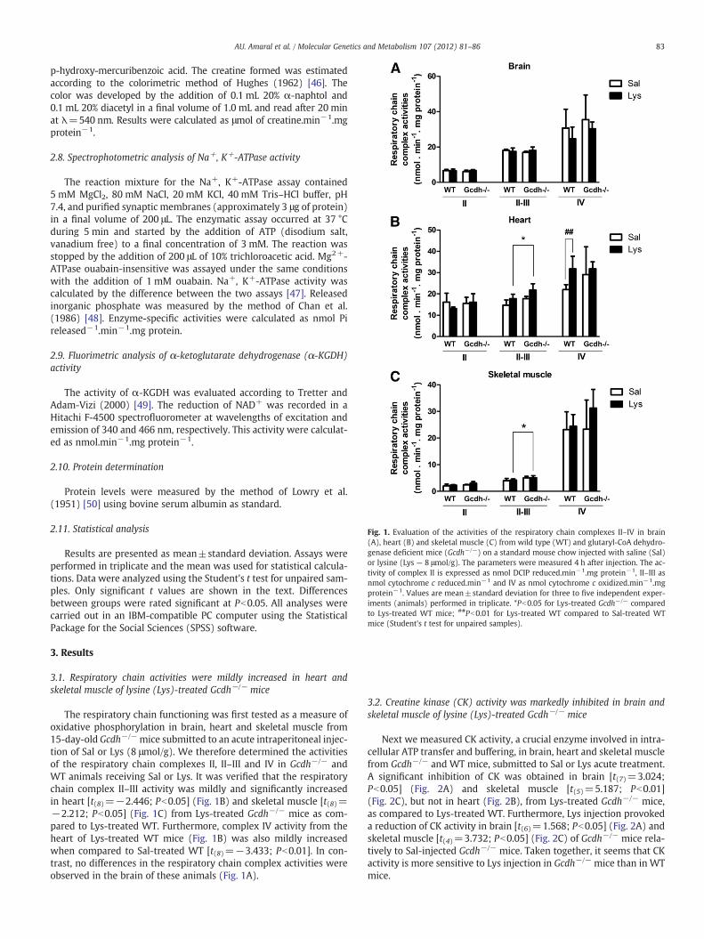
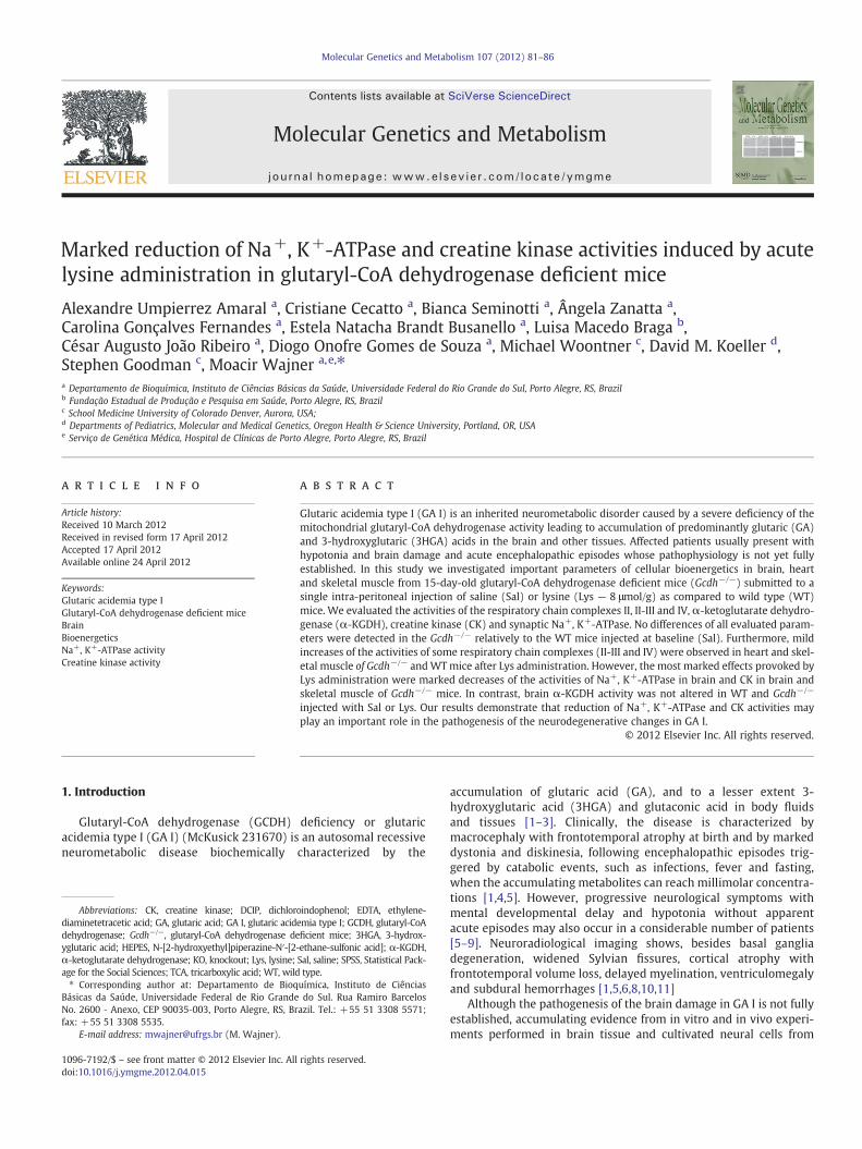
Fig. 1. Evaluation of the activities of the respiratory chain complexes II–IV in brain(A), heart (B) and skeletal muscle (C) from wild type (WT) and glutaryl-CoA dehydro-genase deficient mice (Gcdh−/−) on a standard mouse chow injected with saline (Sal)or lysine (Lys — 8 μmol/g). The parameters were measured 4 h after injection. The ac-tivity of complex II is expressed as nmol DCIP reduced.min−1.mg protein−1, II–III asnmol cytochrome c reduced.min−1 and IV as nmol cytochrome c oxidized.min−1.mgprotein−1. Values are mean±standard deviation for three to five independent exper-iments (animals) performed in triplicate. *Pb0.05 for Lys-treated Gcdh−/− comparedto Lys-treated WT mice; ##Pb0.01 for Lys-treated WT compared to Sal-treated WTmice (Student's t test for unpaired samples).
83AU. Amaral et al. / Molecular Genetics and Metabolism 107 (2012) 81–86
p-hydroxy-mercuribenzoic acid. The creatine formed was estimatedaccording to the colorimetric method of Hughes (1962) [46]. Thecolor was developed by the addition of 0.1 mL 20% α-naphtol and0.1 mL 20% diacetyl in a final volume of 1.0 mL and read after 20 minat λ=540 nm. Results were calculated as μmol of creatine.min−1.mgprotein−1.
2.8. Spectrophotometric analysis of Na+, K+-ATPase activity
The reaction mixture for the Na+, K+-ATPase assay contained5 mM MgCl2, 80 mM NaCl, 20 mM KCl, 40 mM Tris–HCl buffer, pH7.4, and purified synaptic membranes (approximately 3 μg of protein)in a final volume of 200 μL. The enzymatic assay occurred at 37 °Cduring 5 min and started by the addition of ATP (disodium salt,vanadium free) to a final concentration of 3 mM. The reaction wasstopped by the addition of 200 μL of 10% trichloroacetic acid. Mg2+-ATPase ouabain-insensitive was assayed under the same conditionswith the addition of 1 mM ouabain. Na+, K+-ATPase activity wascalculated by the difference between the two assays [47]. Releasedinorganic phosphate was measured by the method of Chan et al.(1986) [48]. Enzyme-specific activities were calculated as nmol Pireleased−1.min−1.mg protein.
2.9. Fluorimetric analysis of α-ketoglutarate dehydrogenase (α-KGDH)activity
The activity of α-KGDH was evaluated according to Tretter andAdam-Vizi (2000) [49]. The reduction of NAD+ was recorded in aHitachi F-4500 spectrofluorometer at wavelengths of excitation andemission of 340 and 466 nm, respectively. This activity were calculat-ed as nmol.min−1.mg protein−1.
2.10. Protein determination
Protein levels were measured by the method of Lowry et al.(1951) [50] using bovine serum albumin as standard.
2.11. Statistical analysis
Results are presented as mean±standard deviation. Assays wereperformed in triplicate and the mean was used for statistical calcula-tions. Data were analyzed using the Student's t test for unpaired sam-ples. Only significant t values are shown in the text. Differencesbetween groups were rated significant at Pb0.05. All analyses werecarried out in an IBM-compatible PC computer using the StatisticalPackage for the Social Sciences (SPSS) software.
3. Results
3.1. Respiratory chain activities were mildly increased in heart andskeletal muscle of lysine (Lys)-treated Gcdh−/− mice
The respiratory chain functioning was first tested as a measure ofoxidative phosphorylation in brain, heart and skeletal muscle from15-day-old Gcdh−/− mice submitted to an acute intraperitoneal injec-tion of Sal or Lys (8 μmol/g). We therefore determined the activitiesof the respiratory chain complexes II, II–III and IV in Gcdh−/− andWT animals receiving Sal or Lys. It was verified that the respiratorychain complex II–III activity was mildly and significantly increasedin heart [t(8)=−2.446; Pb0.05] (Fig. 1B) and skeletal muscle [t(8)=−2.212; Pb0.05] (Fig. 1C) from Lys-treated Gcdh−/− mice as com-pared to Lys-treated WT. Furthermore, complex IV activity from theheart of Lys-treated WT mice (Fig. 1B) was also mildly increasedwhen compared to Sal-treated WT [t(8)=−3.433; Pb0.01]. In con-trast, no differences in the respiratory chain complex activities wereobserved in the brain of these animals (Fig. 1A).
3.2. Creatine kinase (CK) activity was markedly inhibited in brain andskeletal muscle of lysine (Lys)-treated Gcdh−/− mice
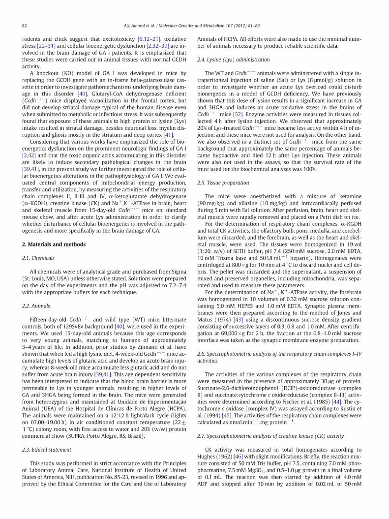
Next we measured CK activity, a crucial enzyme involved in intra-cellular ATP transfer and buffering, in brain, heart and skeletal musclefrom Gcdh−/− and WT mice, submitted to Sal or Lys acute treatment.A significant inhibition of CK was obtained in brain [t(7)=3.024;Pb0.05] (Fig. 2A) and skeletal muscle [t(5)=5.187; Pb0.01](Fig. 2C), but not in heart (Fig. 2B), from Lys-treated Gcdh−/− mice,as compared to Lys-treated WT. Furthermore, Lys injection provokeda reduction of CK activity in brain [t(6)=1.568; Pb0.05] (Fig. 2A) andskeletal muscle [t(4)=3.732; Pb0.05] (Fig. 2C) of Gcdh−/− mice rela-tively to Sal-injected Gcdh−/− mice. Taken together, it seems that CKactivity is more sensitive to Lys injection in Gcdh−/− mice than in WTmice.
Alexandre
Typewritten Text
32
Fig. 2. Evaluation of creatine kinase (CK) activity in brain (A), heart (B) and skeletalmuscle (C) from wild type (WT) and glutaryl-CoA dehydrogenase deficient mice(Gcdh−/−) on a standard mouse chow injected with saline (Sal) or lysine (Lys —
8 μmol/g). This parameter was measured 4 h after the injection. Values are mean±standard deviation for three to five independent experiments (animals) performedin triplicate and are expressed as μmol creatine.min−1.mg protein−1. *Pb0.05,**Pb0.01 for Lys-treated Gcdh−/− compared to Lys-treated WT mice; #Pb0.05 forLys-treated Gcdh−/− compared to Sal-treated Gcdh−/− mice (Student's t test for un-paired samples).
Fig. 3. Evaluation of Na+, K+-ATPase activity in brain from wild type (WT) and glutaryl-CoA dehydrogenase deficient mice (Gcdh−/−) on a standard mouse chow injected withsaline (Sal) or lysine (Lys — 8 μmol/g). This parameter was measured 4 h after the injec-tion. Values are mean±standard deviation for three to five independent experiments(animals) performed in triplicate and are expressed as nmol Pi.min−1.mg protein−1.*Pb0.05 for Lys-treated Gcdh−/− compared to Lys-treated WT mice; #Pb0.05 for Lys-treated Gcdh−/− compared to Sal-treated Gcdh−/− mice (Student's t test for unpairedsamples).
Table 1α-Ketoglutarate dehydrogenase (α-KGDH) activity in rat brain from wild type (WT)and glutaryl-CoA dehydrogenase deficient mice (Gcdh−/−) on a standard mouse
84 AU. Amaral et al. / Molecular Genetics and Metabolism 107 (2012) 81–86
3.3. Na+, K+-ATPase activity was significantly inhibited in brain of lysine(Lys)-treated Gcdh−/− mice
It was also observed that synaptic membrane Na+, K+-ATPaseactivity, which is important for neurotransmission, was markedly re-duced in brain from Lys-treated Gcdh−/− mice as compared toLys-treated WT [t(7)=2.370; Pb0.05] and Sal-treated Gcdh−/− mice[t(5)=2.351; Pb0.05] (Fig. 3). The data suggest that this enzyme ac-tivity is also more responsive to Lys treatment in Gcdh−/− mice ascompared to normal mice.
chow injected with saline (Sal) or lysine (Lys — 8 μmol/g).
α-KGDH activity
Sal Lys
WT 15.15±2.69 17.67±5.07Gcdh−/− 16.33±3.41 14.75±3.42
Values are mean±standard deviation of three to five independent experiments (animals)performed in triplicate and are expressed as nmol NADH.min−1.mg.protein−1. Nosignificant differences were detected between the various groups (Student's t test forunpaired samples).
3.4. α-Ketoglutarate dehydrogenase (α-KGDH) activity was not alteredin Gcdh−/− mice
Finally, we verified that α-KGDH activity, which is a key and arate-controlling enzyme of the tricarboxylic acid (TCA) cycle, didnot change in Gcdh−/− andWTmice submitted to Sal or Lys acute ad-ministration (Table 1).
4. Discussion
Impairment of cellular bioenergetics has been proposed as an im-portant pathomechanism of brain damage in GA I patients[2,6,36,39,51], but this is still being debated. However, this presump-tion was based on experiments performed on fresh cerebral cortexand striatum and on cell cultures from rat and chick brain embryowith normal GCDH activity, which makes the pathophysiologicalrelevance of these works uncertain.
Therefore, the aim of the present investigation was to evaluate im-portant parameters of mitochondrial metabolism regarding energyproduction, transfer and utilization namely the activities of the respi-ratory chain complexes, α-KGDH, CK and Na+,K+, ATPase in brain,skeletal muscle and heart of Gcdh−/− mice on a normal chow (0.9%Lys) and after Lys administration.
First, we observed no significant differences in the activity of re-spiratory chain enzymes, α-KGDH, CK, or Na+, K+-ATPase inmitochondrial-enriched tissues (brain, heart and skeletal muscle) be-tween Gcdh−/− and WT mice fed with a standard mouse chow (Sal).This is in line with a study showing no alterations of the respiratorychain activities in brain, as well as in liver, skeletal and heart muscleof Gcdh−/− mice when compared to WT animals [36]. Next, we eval-uated whether Gcdh−/− mice are more sensitive than WT mice toacute Lys overload by analyzing the same parameters of mitochondri-al homeostasis.
Mild increases of the activities of some respiratory chain com-plexes (II–III and IV) in heart and skeletal muscle of Gcdh−/− andWT mice were observed after Lys administration. Considering thatenhanced activity of the respiratory chain was previously reportedin skeletal muscle from rats treated with GA [38], it is possible thatLys administration induced elevation of GA and 3HGA concentrationsultimately leading to mild elevation of these activities in cardiac and
Alexandre
Typewritten Text
33
85AU. Amaral et al. / Molecular Genetics and Metabolism 107 (2012) 81–86
skeletal muscle of the animals. In this particular, we have recently ob-served that Lys injection leads to elevation of brain GA concentrationsin Gcdh−/− mice [52].
We also found that the activity of endogenous brain α-KGDH, arate-controlling enzyme of the TCA cycle, was not altered at baseline(Sal) in Gcdh−/− mice, or following Lys injection. These data are ap-parently in conflict with previous in vitro results showing thatglutaryl-CoA strongly inhibits α-KGDH from porcine heart [36]. How-ever, we must consider that the observed inhibitory effect wasachieved in vitro and on a purified commercial α-KGDH preparationobtained from a distinct species and tissue [36]. We cannot also ruleout the possibility that lower glutaryl-CoA concentrations werereached in brain of Gcdh−/− mice after Lys injection as compared tothe doses of glutaryl-CoA tested (0.25–2.0 mM) by Sauer and collab-orators (2005) [36].
A novel and interesting finding of the present investigation wasthat CK activity was markedly diminished in brain and skeletal mus-cle from Lys-treated Gcdh−/− mice, suggesting that intracellular ATPtransfer and buffering is compromised in the Gcdh−/− mice that re-ceived Lys administration. Consistent with this observation, Zinnantiand colleagues found markedly decreased concentrations of phos-phocreatine in the brains of Gcdh−/− mice on a high lysine diet [39].Our results are also in accord with other studies showing an inhibi-tion of CK by GA, which was prevented by glutathione (GSH), in ratmidbrain in vitro [33] and in vivo in skeletal muscle of ratschronically-treated with GA [38]. Considering that CK plays an impor-tant role in cellular energy homeostasis, this observation suggeststhat the severe brain injury and hypotonia presented by GA I patientsmay in part be the result of decreased activity of this enzyme.
We also observed that Na+, K+-ATPase activity was also stronglyinhibited in brain from Lys-treated Gcdh−/− mice. This enzyme isnecessary to maintain neuronal excitability and cellular volumecontrol through the generation and maintenance of the membrane po-tential by the active transport of sodium and potassium ions in the cen-tral nervous system. It is present at high concentrations in the brain,consuming about 40–50% of the ATP generated in this tissue, highlight-ing its importance for normal brain functioning. Indeed, reduction ofthis activity is related to neuronal damage in rat and human brain[53,54]. Furthermore, excitotoxicity and epilepsy have been related toa diminution of Na+, K+-ATPase activity [54,55]. Our present data, al-lied to previous works showing that GA and 3HGA inhibit in vitrothis enzyme in primary neuronal cultures from chick embryo telen-cephalon and in rat brain [14,27], as well as alter glutamate uptakeand induces glutamate receptor activation [13,15,17,19,20,27,56], rein-force the hypothesis that excitotoxicity may represent an importantmechanism of brain damage in GA I.
Regarding to the mechanisms by which CK and Na+, K+-ATPasewere found to be inhibited in Gcdh−/− mice after Lys overload, ithas been extensively reported that these activities are very suscepti-ble to free radical attack [53,57–63]. Furthermore, considering thatprevious studies demonstrated that the major metabolitesaccumulating in GA I provoke oxidative damage in the brain[23,25,26,28,36], we may presume that increased oxidative stressmay underlie the inhibitions of CK and Na+, K+-ATPase activities inthe Gcdh−/− animals submitted to Lys overload. This conclusion isin accordance with a recent publication showing that oxidative stressis elicited in the brain of Gcdh−/− after Lys supplementation [52].
Otherwise, it could be tentatively postulated that the results of thepresent study were achieved due to Lys accumulation. This is unlikely,since Lys administration did not change the mitochondrial parame-ters of homeostasis evaluated in WT mice. Therefore, we postulatethat the observed effects were probably caused by the increase in tis-sue concentrations of GA and/or 3HGA, which take place only inGcdh−/− mice [52].
Previous studies of the effect of GA and 3HGA on parameters ofenergy metabolism have generally reported in vitro effects of the
addition of supraphysiologic levels of these metabolites to WT rodenttissues [32,33,35,37,64]. In contrast, our data were obtained in aknock out model of GA I submitted to a metabolic stress with Lysoverload in which the concentrations of the major accumulating me-tabolites, especially GA, are similar to those observed in glutaricacidemia patients [40,52]. Consequently, it is likely that the dataobtained in the present work better mimic the in vivo conditions inhuman GA I patients.
In conclusion, we report for the first time that acute Lys supple-mentation to developing Gcdh−/− mice provokes marked reductionin the activities of the important enzyme activities Na+, K+-ATPase(brain) and CK (brain and skeletal muscle), as well as mild increasesin the activities of some complexes of the respiratory chain (heart andskeletal muscle). Our present data showing impairment of bioener-getics in the brain and skeletal muscle of Gcdh−/− mice followingLys administration may possibly explain the clinical observationsthat therapies aimed at reducing brain Lys uptake are effective fortreatment of GA I. Finally, considering that CK and Na+, K+-ATPaseactivities are crucial for normal energy transfer and for the mainte-nance of membrane potential necessary for neurotransmission,respectively, these results indicate that disruption of intracellularenergy transfer and neurotransmission may represent mechanismsresponsible for the brain damage and neurologic abnormalitiesobserved in patients affected by GA I.
5. Conclusions
Acute lysine overload in Gcdh−/− mice provokes a disturbance ofcellular bioenergetics through inhibition of creatine kinase (brainand skeletal muscle) and Na+, K+-ATPase (brain) activities.
Acknowledgments
We are grateful for the financial support of CNPq, PROPESq/UFRGS, FAPERGS, PRONEX, FINEP Rede Instituto Brasileiro de Neuro-ciência (IBN-Net) # 01.06.0842-00, Instituto Nacional de Ciência eTecnologia-Excitotoxicidade e Neuroproteção (INCT-EN).
References
[1] S.I. Goodman, M.D. Norenberg, R.H. Shikes, D.J. Breslich, P.G. Moe, Glutaricaciduria: biochemical and morphologic considerations, J. Pediatr. 90 (1977)746–750.
[2] K.A. Strauss, D.H. Morton, Type I glutaric aciduria, part 2: a model of acute striatalnecrosis, Am. J. Med. Genet. C Semin. Med. Genet. 121C (2003) 53–70.
[3] C.B. Funk, A.N. Prasad, P. Frosk, S. Sauer, S. Kolker, C.R. Greenberg, M.R. Del Bigio,Neuropathological, biochemical and molecular findings in a glutaric acidemiatype 1 cohort, Brain 128 (2005) 711–722.
[4] G.F. Hoffmann, J. Zschocke, Glutaric aciduria type I: from clinical, biochemical andmolecular diversity to successful therapy, J. Inherit. Metab. Dis. 22 (1999)381–391.
[5] S. Kolker, S.W. Sauer, J.G. Okun, G.F. Hoffmann, D.M. Koeller, Lysine intake andneurotoxicity in glutaric aciduria type I: towards a rationale for therapy? Brain129 (2006) e54.
[6] S. Kolker, D.M. Koeller, S. Sauer, F. Horster, M.A. Schwab, G.F. Hoffmann, K. Ullrich,J.G. Okun, Excitotoxicity and bioenergetics in glutaryl-CoA dehydrogenase defi-ciency, J. Inherit. Metab. Dis. 27 (2004) 805–812.
[7] K.A. Strauss, J. Lazovic, M. Wintermark, D.H. Morton, Multimodal imaging ofstriatal degeneration in Amish patients with glutaryl-CoA dehydrogenase defi-ciency, Brain 130 (2007) 1905–1920.
[8] I. Harting, E. Neumaier-Probst, A. Seitz, E.M. Maier, B. Assmann, I. Baric, M.Troncoso, C. Muhlhausen, J. Zschocke, N.P. Boy, G.F. Hoffmann, S.F. Garbade, S.Kolker, Dynamic changes of striatal and extrastriatal abnormalities in glutaricaciduria type I, Brain 132 (2009) 1764–1782.
[9] J. Heringer, S.P. Boy, R. Ensenauer, B. Assmann, J. Zschocke, I. Harting, T. Lucke,E.M. Maier, C. Muhlhausen, G. Haege, G.F. Hoffmann, P. Burgard, S. Kolker, Useof guidelines improves the neurological outcome in glutaric aciduria type I,Ann. Neurol. 68 (2010) 743–752.
[10] J. Brismar, P.T. Ozand, CT and MR of the brain in glutaric acidemia type I: a reviewof 59 published cases and a report of 5 new patients AJNR, Am. J. Neuroradiol. 16(1995) 675–683.
[11] S. Kimura, M. Hara, A. Nezu, H. Osaka, S. Yamazaki, K. Saitoh, Two cases of glutaricaciduria type 1: clinical and neuropathological findings, J. Neurol. Sci. 123 (1994)38–43.
Alexandre
Typewritten Text
34
86 AU. Amaral et al. / Molecular Genetics and Metabolism 107 (2012) 81–86
[12] S. Kolker, B. Ahlemeyer, J. Krieglstein, G.F. Hoffmann, 3-Hydroxyglutaric andglutaric acids are neurotoxic through NMDA receptors in vitro, J. Inherit. Metab.Dis. 22 (1999) 259–262.
[13] L.O. Porciuncula, A. Dal-Pizzol Jr., A.S. Coitinho, T. Emanuelli, D.O. Souza, M.Wajner, Inhibition of synaptosomal [3H]glutamate uptake and [3H]glutamatebinding to plasma membranes from brain of young rats by glutaric acid in vitro,J. Neurol. Sci. 173 (2000) 93–96.
[14] S. Kolker, G. Kohr, B. Ahlemeyer, J.G. Okun, V. Pawlak, F. Horster, E. Mayatepek, J.Krieglstein, G.F. Hoffmann, Ca(2+) and Na(+) dependence of3-hydroxyglutarate-induced excitotoxicity in primary neuronal cultures fromchick embryo telencephalons, Pediatr. Res. 52 (2002) 199–206.
[15] S. Kolker, J.G. Okun, B. Ahlemeyer, A.T. Wyse, F. Horster, M. Wajner, D.Kohlmuller, E. Mayatepek, J. Krieglstein, G.F. Hoffmann, Chronic treatment withglutaric acid induces partial tolerance to excitotoxicity in neuronal culturesfrom chick embryo telencephalons, J. Neurosci. Res. 68 (2002) 424–431.
[16] L.O. Porciuncula, T. Emanuelli, R.G. Tavares, C. Schwarzbold, M.E. Frizzo, D.O.Souza, M. Wajner, Glutaric acid stimulates glutamate binding and astrocytic up-take and inhibits vesicular glutamate uptake in forebrain from young rats,Neurochem. Int. 45 (2004) 1075–1086.
[17] R.B. Rosa, C. Schwarzbold, K.B. Dalcin, G.C. Ghisleni, C.A. Ribeiro, M.B. Moretto, M.E.Frizzo, G.F. Hoffmann, D.O. Souza, M. Wajner, Evidence that 3-hydroxyglutaric acidinteracts with NMDA receptors in synaptic plasmamembranes from cerebral cortexof young rats, Neurochem. Int. 45 (2004) 1087–1094.
[18] M. Wajner, S. Kolker, D.O. Souza, G.F. Hoffmann, C.F. de Mello, Modulation of glut-amatergic and GABAergic neurotransmission in glutaryl-CoA dehydrogenase de-ficiency, J. Inherit. Metab. Dis. 27 (2004) 825–828.
[19] K.B. Dalcin, R.B. Rosa, A.L. Schmidt, J.S. Winter, G. Leipnitz, C.S. Dutra-Filho, C.M.Wannmacher, L.O. Porciuncula, D.O. Souza, M. Wajner, Age and brain structuralrelated effects of glutaric and 3-hydroxyglutaric acids on glutamate binding toplasma membranes during rat brain development, Cell. Mol. Neurobiol. 27(2007) 805–818.
[20] R.B. Rosa, K.B. Dalcin, A.L. Schmidt, D. Gerhardt, C.A. Ribeiro, G.C. Ferreira, P.F.Schuck, A.T. Wyse, L.O. Porciuncula, S. Wofchuk, C.G. Salbego, D.O. Souza, M.Wajner, Evidence that glutaric acid reduces glutamate uptake by cerebral cortexof infant rats, Life Sci. 81 (2007) 1668–1676.
[21] D.V. Magni, A.F. Furian, M.S. Oliveira, M.A. Souza, F. Lunardi, J. Ferreira, C.F. Mello,L.F. Royes, M.R. Fighera, Kinetic characterization of l-[(3)H]glutamate uptake in-hibition and increase oxidative damage induced by glutaric acid in striatal synap-tosomes of rats, Int. J. Dev. Neurosci. 27 (2009) 65–72.
[22] S. Kolker, B. Ahlemeyer, R. Huhne, E. Mayatepek, J. Krieglstein, G.F. Hoffmann, Po-tentiation of 3-hydroxyglutarate neurotoxicity following induction of astrocyticiNOS in neonatal rat hippocampal cultures, Eur. J. Neurosci. 13 (2001) 2115–2122.
[23] S. Kolker, B. Ahlemeyer, J. Krieglstein, G.F. Hoffmann, Contribution of reactive ox-ygen species to 3-hydroxyglutarate neurotoxicity in primary neuronal culturesfrom chick embryo telencephalons, Pediatr. Res. 50 (2001) 76–82.
[24] F. de Oliveira Marques, M.E. Hagen, C.D. Pederzolli, A.M. Sgaravatti, K. Durigon,C.G. Testa, C.M. Wannmacher, A.T. de Souza Wyse, M. Wajner, C.S. Dutra-Filho,Glutaric acid induces oxidative stress in brain of young rats, Brain Res. 964(2003) 153–158.
[25] A. Latini, R. Borba Rosa, K. Scussiato, S. Llesuy, A. Bello-Klein, M. Wajner,3-Hydroxyglutaric acid induces oxidative stress and decreases the antioxidantdefenses in cerebral cortex of young rats, Brain Res. 956 (2002) 367–373.
[26] A. Latini, K. Scussiato, G. Leipnitz, C.S. Dutra-Filho, M. Wajner, Promotion of oxida-tive stress by 3-hydroxyglutaric acid in rat striatum, J. Inherit. Metab. Dis. 28(2005) 57–67.
[27] M.R. Fighera, L.F. Royes, A.F. Furian, M.S. Oliveira, N.G. Fiorenza, R. Frussa-Filho,J.C. Petry, R.C. Coelho, C.F. Mello, GM1 ganglioside prevents seizures, Na+,K+‐ATPase activity inhibition and oxidative stress induced by glutaric acidand pentylenetetrazole, Neurobiol. Dis. 22 (2006) 611–623.
[28] A. Latini, G.C. Ferreira, K. Scussiato, P.F. Schuck, A.F. Solano, C.S. Dutra-Filho, C.R.Vargas, M. Wajner, Induction of oxidative stress by chronic and acute glutaricacid administration to rats, Cell. Mol. Neurobiol. 27 (2007) 423–438.
[29] D.V. Magni, M.S. Oliveira, A.F. Furian, N.G. Fiorenza, M.R. Fighera, J. Ferreira, C.F.Mello, L.F. Royes, Creatine decreases convulsions and neurochemical alterationsinduced by glutaric acid in rats, Brain Res. 1185 (2007) 336–345.
[30] S. Olivera, A. Fernandez, A. Latini, J.C. Rosillo, G. Casanova, M.Wajner, P. Cassina, L.Barbeito, Astrocytic proliferation and mitochondrial dysfunction induced by ac-cumulated glutaric acidemia I (GAI) metabolites: possible implications for GAIpathogenesis, Neurobiol. Dis. 32 (2008) 528–534.
[31] S. Olivera-Bravo, A. Fernandez, M.N. Sarlabos, J.C. Rosillo, G. Casanova, M. Jimenez, L.Barbeito, Neonatal astrocyte damage is sufficient to trigger progressivestriatal degeneration in a ratmodel of glutaric acidemia-I, PLoS ONE 6 (2011) e20831.
[32] C.G. Silva, A.R. Silva, C. Ruschel, C. Helegda, A.T. Wyse, C.M. Wannmacher, C.S.Dutra-Filho, M. Wajner, Inhibition of energy production in vitro by glutaric acidin cerebral cortex of young rats, Metab. Brain Dis. 15 (2000) 123–131.
[33] G. da, C. Ferreira, C.M. Viegas, P.F. Schuck, A. Latini, C.S. Dutra-Filho, A.T. Wyse, C.M.Wannmacher, C.R. Vargas, M. Wajner, Glutaric acid moderatelycompromises energymetabolism in rat brain, Int. J. Dev. Neurosci. 23 (2005) 687–693.
[34] G. da, C. Ferreira, C.M. Viegas, P.F. Schuck, A. Tonin, C.A. Ribeiro, M. Coelho Dde, T.Dalla-Costa, A. Latini, A.T. Wyse, C.M. Wannmacher, C.R. Vargas, M. Wajner,Glutaric acid administration impairs energy metabolism in midbrain and skeletalmuscle of young rats, Neurochem. Res. 30 (2005) 1123–1131.
[35] A. Latini, M. Rodriguez, R. Borba Rosa, K. Scussiato, G. Leipnitz, D. Reis de Assis, G.da Costa Ferreira, C. Funchal, M.C. Jacques-Silva, L. Buzin, R. Giugliani, A. Cassina,R. Radi, M. Wajner, 3-Hydroxyglutaric acid moderately impairs energy metabo-lism in brain of young rats, Neuroscience 135 (2005) 111–120.
[36] S.W. Sauer, J.G. Okun, M.A. Schwab, L.R. Crnic, G.F. Hoffmann, S.I. Goodman, D.M.Koeller, S. Kolker, Bioenergetics in glutaryl-coenzyme A dehydrogenase deficien-cy: a role for glutaryl-coenzyme A, J. Biol. Chem. 280 (2005) 21830–21836.
[37] G.C. Ferreira, A. Tonin, P.F. Schuck, C.M. Viegas, P.C. Ceolato, A. Latini, M.L. Perry,A.T. Wyse, C.S. Dutra-Filho, C.M. Wannmacher, C.R. Vargas, M. Wajner, Evidencefor a synergistic action of glutaric and 3-hydroxyglutaric acids disturbing ratbrain energy metabolism, Int. J. Dev. Neurosci. 25 (2007) 391–398.
[38] G. da, C. Ferreira, P.F. Schuck, C.M. Viegas, A. Tonin, A. Latini, C.S. Dutra-Filho, A.T.Wyse, C.M. Wannmacher, C.R. Vargas, M. Wajner, Energy metabolism is com-promised in skeletal muscle of rats chronically-treated with glutaric acid,Metab. Brain Dis. 22 (2007) 111–123.
[39] W.J. Zinnanti, J. Lazovic, C. Housman, K. LaNoue, J.P. O'Callaghan, I. Simpson, M.Woontner, S.I. Goodman, J.R. Connor, R.E. Jacobs, K.C. Cheng, Mechanism ofage-dependent susceptibility and novel treatment strategy in glutaric acidemiatype I, J. Clin. Invest. 117 (2007) 3258–3270.
[40] D.M. Koeller, M. Woontner, L.S. Crnic, B. Kleinschmidt-DeMasters, J. Stephens, E.L.Hunt, S.I. Goodman, Biochemical, pathologic and behavioral analysis of a mousemodel of glutaric acidemia type I, Hum. Mol. Genet. 11 (2002) 347–357.
[41] W.J. Zinnanti, J. Lazovic, E.B. Wolpert, D.A. Antonetti, M.B. Smith, J.R. Connor, M.Woontner, S.I. Goodman, K.C. Cheng, A diet-induced mouse model for glutaricaciduria type I, Brain 129 (2006) 899–910.
[42] S.W. Sauer, Biochemistry and bioenergetics of glutaryl-CoA dehydrogenasedeficiency, J. Inherit. Metab. Dis. 30 (2007) 673–680.
[43] D.H. Jones, A.I. Matus, Isolation of synaptic plasma membrane from brain by com-bined flotation–sedimentation density gradient centrifugation, Biochim. Biophys.Acta 356 (1974) 276–287.
[44] J.C. Fischer, W. Ruitenbeek, J.A. Berden, J.M.F. Trijbels, J.H. Veerkamp, A.M.Stadhouders, R.C.A. Sengers, A.J.M. Janssen, Differential investigation of the ca-pacity of succinate oxidation in human skeletal muscle, Clin. Chim. Acta 153(1985) 23–36.
[45] P. Rustin, D. Chretien, T. Bourgeron, B. Gerard, A. Rotig, J.M. Saudubray, A.Munnich, Biochemical and molecular investigations in respiratory chain deficien-cies, Clin. Chim. Acta 228 (1994) 35–51.
[46] B.P. Hughes, A method for estimation of serum creatine kinase and its use in com-paring creatine kinase and aldolase activity in normal and pathological sera, Clin.Chim. Acta 7 (1962) 597 -&.
[47] S. Tsakiris, G. Deliconstantinos, Influence of phosphatidylserine on (Na+,K+)-stimulated ATPase and acetylcholinesterase activities of dog brain synapto-somal plasma membranes, Biochem. J. 220 (1984) 301–307.
[48] K.M. Chan, D. Delfert, K.D. Junger, A direct colorimetric assay for Ca2+‐stimulat-ed atpase activity, Anal. Biochem. 157 (1986) 375–380.
[49] L. Tretter, V. Adam-Vizi, Inhibition of Krebs cycle enzymes by hydrogen peroxide:a key role of [alpha]-ketoglutarate dehydrogenase in limiting NADH productionunder oxidative stress, J. Neurosci. 20 (2000) 8972–8979.
[50] O.H. Lowry, N.J. Rosebrough, A.L. Farr, R.J. Randall, Protein measurement with theFolin phenol reagent, J. Biol. Chem. 193 (1951) 265–275.
[51] C.B. Funk, A.N. Prasad, M.R. Del Bigio, Preliminary attempts to establish a ratmodel of striatal injury in glutaric acidaemia type I, J. Inherit. Metab. Dis. 27(2004) 819–824.
[52] B. Seminotti, M.S. da Rosa, C.G. Fernandes, A.U. Amaral, L.M. Braga, G. Leipnitz,D.O.G. de Souza, M. Woontner, D.M. Koeller, S. Goodman, M. Wajner, Inductionof oxidative stress in brain of glutaryl-CoA dehydrogenase deficient mice byacute lysine administration, Mol. Genet. Metab. 106 (2012) 31–38.
[53] G.J. Lees, Contributory mechanisms in the causation of neurodegenerative disor-ders, Neuroscience 54 (1993) 287–322.
[54] M.A. Cousin, D.G. Nicholls, J.M. Pocock, Modulation of ion gradients and gluta-mate release in cultured cerebellar granule cells by ouabain, J. Neurochem. 64(1995) 2097–2104.
[55] G.J. Lees,W. Leong, The sodium-potassiumATPase inhibitor ouabain is neurotoxici inthe rat substantia nigra and striatum, Neurosci. Lett. 188 (1995) 113–116.
[56] O. Stokke, S.I. Goodman, P.G. Moe, Inhibition of brain glutamate decarboxylase byglutarate, glutaconate, and beta-hydroxyglutarate: explanation of the symptomsin glutaric aciduria? Clin. Chim. Acta 66 (1976) 411–415.
[57] S.O. Burmistrov, O.P. Mashek, A.M. Kotin, The action of acute alcoholic intoxica-tion on the antioxidant system and creatine kinase activity in the brain of rat em-bryos, Eksp. Klin. Farmakol. 55 (1992) 54–56.
[58] H. Wolosker, R. Panizzutti, S. Engelender, Inhibition of creatine kinase byS-nitrosoglutathione, FEBS Lett. 392 (1996) 274–276.
[59] M.A. Arstall, C. Bailey, W.L. Gross, M. Bak, J.L. Balligand, R.A. Kelly, ReversibleS-nitrosation of creatine kinase by nitric oxide in adult rat ventricular myocytes,J. Mol. Cell. Cardiol. 30 (1998) 979–988.
[60] E.A. Konorev, N. Hogg, B. Kalyanaraman, Rapid and irreversible inhibition of cre-atine kinase by peroxynitrite, FEBS Lett. 427 (1998) 171–174.
[61] O. Stachowiak, M. Dolder, T. Wallimann, C. Richter, Mitochondrial creatine kinaseis a prime target of peroxynitrite-induced modification and inactivation, J. Biol.Chem. 273 (1998) 16694–16699.
[62] T. Wallimann, M. Dolder, U. Schlattner, M. Eder, T. Hornemann, E. O'Gorman, A.Ruck, D. Brdiczka, Some new aspects of creatine kinase (CK): compartmentation,structure, function and regulation for cellular and mitochondrial bioenergeticsand physiology, Biofactors 8 (1998) 229–234.
[63] E. Kurella, M. Kukley, O. Tyulina, D. Dobrota, M. Matejovicova, V. Mezesova, A.Boldyrev, Kinetic parameters of Na/K-ATPase modified by free radicals in vitroand in vivo, Ann. N.Y. Acad. Sci. 834 (1997) 661–665.
[64] M. Wajner, S.I. Goodman, Disruption of mitochondrial homeostasis in organicacidurias: insights from human and animal studies, J. Bioenerg. Biomembr. 43(2011) 31–38.
Alexandre
Typewritten Text
35
36
Capítulo II
Reduction of Na+, K+-ATPase activity and expression in cerebral cortex of
glutaryl-CoA dehydrogenase deficient mice: a possible mechanism for brain
injury in glutaric aciduria type I
Alexandre U Amaral, Bianca Seminotti, Cristiane Cecatto, Carolina G Fernandes,
Estela B Busanello, Ângela Zanatta, Luiza W Kist, Maurício R Bogo, Diogo O de
Souza, Michael Woontner, Stephen Goodman, David M Koeller, Moacir Wajner.
Artigo científico publicado no periódico
Molecular Genetics and Metabolism
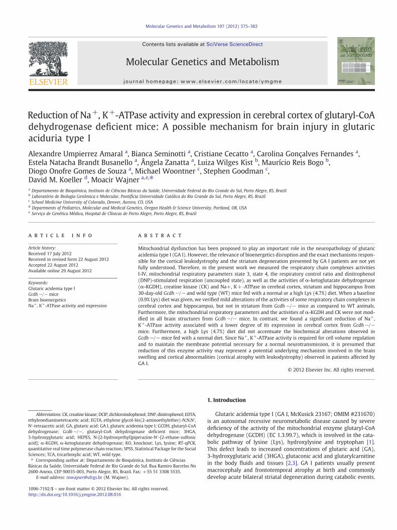
Molecular Genetics and Metabolism 107 (2012) 375–382
Contents lists available at SciVerse ScienceDirect
Molecular Genetics and Metabolism
j ourna l homepage: www.e lsev ie r .com/ locate /ymgme
Reduction of Na+, K+-ATPase activity and expression in cerebral cortex of glutaryl-CoAdehydrogenase deficient mice: A possible mechanism for brain injury in glutaricaciduria type I
Alexandre Umpierrez Amaral a, Bianca Seminotti a, Cristiane Cecatto a, Carolina Gonçalves Fernandes a,Estela Natacha Brandt Busanello a, Ângela Zanatta a, Luiza Wilges Kist b, Maurício Reis Bogo b,Diogo Onofre Gomes de Souza a, Michael Woontner c, Stephen Goodman c,David M. Koeller d, Moacir Wajner a,e,⁎a Departamento de Bioquímica, Instituto de Ciências Básicas da Saúde, Universidade Federal do Rio Grande do Sul, Porto Alegre, RS, Brazilb Laboratório de Biologia Genômica e Molecular, Pontifícia Universidade Católica do Rio Grande do Sul, Porto Alegre, RS, Brazilc School Medicine University of Colorado, Denver, Aurora, CO, USAd Departments of Pediatrics, Molecular and Medical Genetics, Oregon Health & Science University, Portland, OR, USAe Serviço de Genética Médica, Hospital de Clínicas de Porto Alegre, Porto Alegre, RS, Brazil
Abbreviations:CK, creatine kinase; DCIP, dichloroindophethylenediaminetetracetic acid; EGTA, ethylene glycol-bN′-tetraacetic acid; GA, glutaric acid; GA I, glutaric acidemdehydrogenase; Gcdh−/−, glutaryl-CoA dehydrogen3-hydroxyglutaric acid; HEPES, N-[2-hydroxyethyl]pipacid]; α-KGDH, α-ketoglutarate dehydrogenase; KO, knquantitative real time polymerase chain reaction; SPSS, SSciences; TCA, tricarboxylic acid; WT, wild type.⁎ Corresponding author at: Departamento de Bioquím
Básicas da Saúde, Universidade Federal de Rio Grande d2600‐Anexo, CEP 90035‐003, Porto Alegre, RS, Brazil. Fa
E-mail address: [email protected] (M. Wajner).
1096-7192/$ – see front matter © 2012 Elsevier Inc. Allhttp://dx.doi.org/10.1016/j.ymgme.2012.08.016
a b s t r a c t
a r t i c l e i n f oArticle history:Received 17 July 2012Received in revised form 22 August 2012Accepted 22 August 2012Available online 29 August 2012
Keywords:Glutaric acidemia type IGcdh−/− miceBrain bioenergeticsNa+, K+-ATPase activity and expression
Mitochondrial dysfunction has been proposed to play an important role in the neuropathology of glutaricacidemia type I (GA I). However, the relevance of bioenergetics disruption and the exact mechanisms respon-sible for the cortical leukodystrophy and the striatum degeneration presented by GA I patients are not yetfully understood. Therefore, in the present work we measured the respiratory chain complexes activitiesI-IV, mitochondrial respiratory parameters state 3, state 4, the respiratory control ratio and dinitrophenol(DNP)-stimulated respiration (uncoupled state), as well as the activities of α-ketoglutarate dehydrogenase(α-KGDH), creatine kinase (CK) and Na+, K+‐ATPase in cerebral cortex, striatum and hippocampus from30-day-old Gcdh−/− and wild type (WT) mice fed with a normal or a high Lys (4.7%) diet. When a baseline(0.9% Lys) diet was given, we verified mild alterations of the activities of some respiratory chain complexes incerebral cortex and hippocampus, but not in striatum from Gcdh−/− mice as compared to WT animals.Furthermore, the mitochondrial respiratory parameters and the activities of α-KGDH and CK were not mod-ified in all brain structures from Gcdh−/− mice. In contrast, we found a significant reduction of Na+,K+-ATPase activity associated with a lower degree of its expression in cerebral cortex from Gcdh−/−mice. Furthermore, a high Lys (4.7%) diet did not accentuate the biochemical alterations observed inGcdh−/− mice fed with a normal diet. Since Na+, K+-ATPase activity is required for cell volume regulationand to maintain the membrane potential necessary for a normal neurotransmission, it is presumed thatreduction of this enzyme activity may represent a potential underlying mechanism involved in the brainswelling and cortical abnormalities (cortical atrophy with leukodystrophy) observed in patients affected byGA I.
© 2012 Elsevier Inc. All rights reserved.
enol; DNP, dinitrophenol; EDTA,is(2-aminoethylether)-N,N,N′,ia type I; GCDH, glutaryl-CoAase deficient mice; 3HGA,erazine-N′-[2-ethane-sulfonicockout; Lys, lysine; RT-qPCR,tatistical Package for the Social
ica, Instituto de Ciênciaso Sul. Rua Ramiro Barcelos Nox: +55 51 3308 5535.
rights reserved.
1. Introduction
Glutaric acidemia type I (GA I, McKusick 23167; OMIM #231670)is an autosomal recessive neurometabolic disease caused by severedeficiency of the activity of the mitochondrial enzyme glutaryl-CoAdehydrogenase (GCDH) (EC 1.3.99.7), which is involved in the cata-bolic pathway of lysine (Lys), hydroxylysine and tryptophan [1].This defect leads to increased concentrations of glutaric acid (GA),3-hydroxyglutaric acid (3HGA), glutaconic acid and glutarylcarnitinein the body fluids and tissues [2,3]. GA I patients usually presentmacrocephaly and frontotemporal atrophy at birth and commonlydevelop acute bilateral striatal degeneration during catabolic events.
Alexandre
Typewritten Text
37

376 A.U. Amaral et al. / Molecular Genetics and Metabolism 107 (2012) 375–382
Progressive cortical leukodystrophy and striatal lesions without docu-mented acute metabolic events with encephalopathy are also found in10 to 20% of patients with neurological symptoms [4–7].
A great body of data have suggested that excitotoxicity [8–18], ox-idative stress [19–28] and mitochondrial dysfunction [29–37] are in-volved in the brain injury of GA I. However, the relevance of energyhomeostasis disruption in the brain damage in this disease is notyet established. Although some investigators proposed that thispathomechanism is crucial to explain the development of neurologi-cal symptoms and especially striatum degeneration in GA I patients[30,34], experimental studies revealed that GA and 3HGA causedonly mild alterations of mitochondrial homeostasis in the brain[29,31–33,35,36].
Recently a knockout (KO) GA I model was developed in mice byreplacing the glutaryl-CoA dehydrogenase (GCDH) gene with anin-frame beta-galactosidase cassette [38]. Glutaryl-CoA dehydroge-nase deficient mice (Gcdh−/−) presented increased cerebral, bloodand urine GA and 3HGA levels and displayed vacuolization in thefrontal cortex (spongiform leukoencephalopathy). However, theanimals did not develop striatal damage typical of the human diseaseeven when submitted to metabolic or infectious stress. This modelwas later improved by exposing these mice to high protein or Lysintake, which provoked neuronal loss, myelin disruption and gliosismostly in the striatum and deep cortex [37,39]. Oral Lys overload toweaning (4-week-old) Gcdh−/− mice resulted in a predominantincrease of brain Lys and GA concentrations after 48 h of Lys expo-sure. It was also seen a simultaneous decrease of Lys and increase ofbrain GA levels, indicating GA formation from Lys in the brain.These investigators suggested that the cortical and particularlystriatal lesions developed in the Gcdh−/− animals submitted to Lysoverload during 48–72 h were probably due to the increase of brainGA concentrations [37,39].
The major aim of the present study was to evaluate importantparameters of bioenergetics, such as the activities of the respiratorychain complexes I–III, II, II–III and IV, the respiratory parametersstates 3 and 4 respiration, the respiratory control ratio (RCR) anddinitrophenol (DNP)-stimulated respiration (uncoupled state), aswell as the key and regulatory activities of α-ketoglutarate dehydro-genase (α-KGDH), creatine kinase (CK) and Na+,K+-ATPase in cere-bral cortex, striatum and hippocampus from 30-day-old Gcdh−/−and WT mice under a baseline diet (0.9% Lys) or a high Lys (4.7%)dietary intake. We also evaluated the expression of the catalyticsubunits of Na+,K+-ATPase in cerebral cortex of Gcdh−/− and WTmice.
2. Materials and methods
2.1. Chemicals
All chemicals were of analytical grade and purchased from Sigma(St. Louis, MO, USA) unless otherwise stated. Solutions were preparedon the day of the experiments and the pH was adjusted to 7.2–7.4with the appropriate buffers for each technique.
2.2. Animals
Gcdh−/− andWTmice littermate controls, both of 129SvEv back-ground [38], were generated from heterozygous and maintained atthe Unidade Experimental Animal, Hospital de Clínicas de PortoAlegre (Porto Alegre, Brazil). The animals were maintained on a12:12 h light/dark cycle (lights on 07.00–19.00 h) in air conditionedconstant temperature (22±1 °C) colony room, with free access towater and a normal chow diet containing 20% (w/w) protein and0.9% Lys (NUVILAB). Thirty-day-old WT and Gcdh−/− mice from F1and F2 generations were used in all experiments. A group of WT
and Gcdh−/− animals were submitted to a 20% (w/w) protein dietcontaining 4.7% Lys.
2.3. Ethical statement
This study was performed in strict accordance with the Guide forthe Care and Use of Laboratory Animals (Eighth edition, 2001) andapproved by the Ethical Committee for the Care and Use of LaboratoryAnimals of Hospital de Clínicas de Porto Alegre. All efforts were alsomade to use the minimal number of animals necessary to producereliable scientific data.
2.4. Tissue preparation
Themicewere anesthetizedwith amixture of ketamine (90 mg/kg)and xilazine (10 mg/kg) and intracardially perfused during 5 min withsaline solution. After perfusion, the brain was rapidly removed andplaced on a Petri dish on ice.
For the determination of the respiratory chain complexes, α-KGDHand total CK activities, the olfactory bulb, pons,medulla, and cerebellumwere discarded, and the cerebral cortex, striatum and hippocampusdissected and weighed. The tissues were homogenized in 19 volumes(1:20, w/v) of SETH buffer, pH 7.4 (250 mM sucrose, 2.0 mM EDTA,10 mM Trizma base and 50 UI mL−1 heparin). Homogenates werecentrifuged at 800 ×g for 10 min at 4 °C to discard nuclei and celldebris. The pellet was discarded and the supernatant, a suspension ofmixed and preserved organelles, including mitochondria, was separat-ed and used to measure these parameters.
For the determination of Na+, K+-ATPase activity, the cerebralcortex, striatum and hippocampus were homogenized in 10 volumesof 0.32 mM sucrose solution containing 5.0 mM HEPES and 1.0 mMEDTA. Synaptic plasma membranes were then prepared accordingto the method of Jones and Matus [40] using a discontinuous sucrosedensity gradient consisting of successive layers of 0.3, 0.8 and1.0 mM. After centrifugation at 69,000 ×g for 2 h, the fraction at the0.8–1.0 mM sucrose interface was taken as the synaptic membraneenzyme preparation.
The expression of the catalytic subunits of Na+, K+-ATPase α1, α2and α3 was determined in cerebral cortex of Gcdh−/− and WT mice.This structure was dissected and immediately frozen in the presenceof Trizol® for isolation of total RNA.
Determination of the respiratory parameters was carried out inisolated mitochondrial preparations from forebrain. The olfactorybulb, pons, medulla, and cerebellum were discarded and forebrainmitochondria were isolated from rat brain as previously described[41]. The final pellet was gently washed and resuspended in isolationbuffer devoid of EGTA, at an approximate protein concentration of20 mg mL−1. This preparation results in a mixture of synaptosomaland non-synaptosomal mitochondria similar to the general braincomposition.
2.5. Spectrophotometric analysis of the respiratory chain complexesI-IV activities
The activity of NADH:cytochrome c oxidoreductase (complexesI–III) was assayed according to the method described by Schapiraet al. [42]. The activities of succinate-2,6-dichloroindophenol (DCIP)-oxidoreductase (complex II) and succinate:cytochrome c oxidoreduc-tase (complex II-III) were determined according to Fischer et al. [43].Cytochrome c oxidase (complex IV) activity was assayed according toRustin et al. [44]. The activities of the respiratory chain complexeswere calculated as nmol min−1. mg protein−1 and expressed as per-centage of controls.
Alexandre
Typewritten Text
38
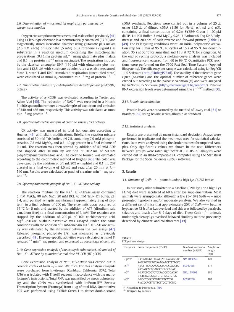
Table 1PCR primers design.
Enzymes Primer sequences (5′–3′) GenBank accessionnumber (mRNA)
Ampliconlength(bp)
Hprt1a F-CTCATGGACTGATTATGGACAGGACR-GCAGGTCAGCAAAGAACTTATAGCC
NM_013556 123
α1b F-CCTTTGACAAGACGTCAGCCACCTGR-CCATCACGGAGCCGCAGCAGAC
BC042435 177
α2b F-CATCTCCGTGTCTAAGCGGGACACR-CTCTGGGGACTGTCTTCCCTCTCG
NM_178405 186
α3b F-GGGTGGCCCTGTCCCACATCGR-AGCCACTTTCTTGTTCCGTTCTCG
BC037206 182
a According to Pernot et al. [49].b Designed by authors.
377A.U. Amaral et al. / Molecular Genetics and Metabolism 107 (2012) 375–382
2.6. Determination of mitochondrial respiratory parameters byoxygen consumption
Oxygen consumption ratewasmeasured as described previously [41]using a Clark-type electrode in a thermostatically controlled (37 °C) andmagnetically stirred incubation chamber using glutamate plus malate(2.5 mM each) or succinate (5 mM) plus rotenone (2 μg/mL) assubstrates in a reaction medium containing the mitochondrialpreparations (0.75 mg protein mL−1 using glutamate plus malateand 0.5 mg protein mL−1 using succinate). The respiration inducedby the classical uncoupler DNP (150 μM with glutamate plus ma-late and 112.5 μM with succinate as substrate) was also measured.State 3, state 4 and DNP-stimulated respiration (uncoupled state)were calculated as nmol O2 consumed min−1 mg of protein−1.
2.7. Fluorimetric analysis of α-ketoglutarate dehydrogenase (α-KGDH)activity
The activity of α-KGDH was evaluated according to Tretter andAdam-Vizi [45]. The reduction of NAD+ was recorded in a HitachiF-4500 spectrofluorometer at wavelengths of excitation and emissionof 340 and 466 nm, respectively. This activity was calculated as nmolmin−1 mg protein−1.
2.8. Spectrophotometric analysis of creatine kinase (CK) activity
CK activity was measured in total homogenates according toHughes [46] with slight modifications. Briefly, the reaction mixtureconsisted of 50 mM Tris buffer, pH 7.5, containing 7.0 mM phospho-creatine, 7.5 mM MgSO4, and 0.5–1.0 μg protein in a final volume of0.1 mL. The reaction was then started by addition of 4.0 mM ADPand stopped after 10 min by addition of 0.02 mL of 50 mMp-hydroxy-mercuribenzoic acid. The creatine formed was estimatedaccording to the colorimetric method of Hughes [46]. The color wasdeveloped by the addition of 0.1 mL 20% α-naphtol and 0.1 mL 20%diacetyl in a final volume of 1.0 mL and read after 20 min at λ=540 nm. Results were calculated as μmol of creatine. min−1 mg pro-tein−1.
2.9. Spectrophotometric analysis of Na+, K+-ATPase activity
The reaction mixture for the Na+, K+-ATPase assay contained5 mM MgCl2, 80 mM NaCl, 20 mM KCl, 40 mM Tris–HCl buffer, pH7.4, and purified synaptic membranes (approximately 3 μg of pro-tein) in a final volume of 200 μL. The enzymatic assay occurred at37 °C for 5 min and started by the addition of ATP (disodium salt,vanadium free) to a final concentration of 3 mM. The reaction wasstopped by the addition of 200 μL of 10% trichloroacetic acid.Mg2+-ATPase ouabain-insensitive was assayed under the sameconditions with the addition of 1 mM ouabain. Na+, K+-ATPase activ-ity was calculated by the difference between the two assays [47].Released inorganic phosphate (Pi) was measured as previouslydescribed [48]. Enzyme-specific activities were calculated as nmol Pireleased−1 min−1mg protein and expressed as percentage of controls.
2.10. Gene expression analysis of the catalytic subunits α1, α2 and α3 ofNa+, K+-ATPase by quantitative real time RT-PCR (RT-qPCR)
Gene expression analysis of Na+, K+-ATPase was carried out incerebral cortex of Gcdh−/− and WT mice. For this analysis reagentswere purchased from Invitrogen (Carlsbad, California, USA). TotalRNA was isolated with Trizol® reagent in accordance with the manu-facturer's instructions. Total RNA was quantified by spectrophotome-try and the cDNA was synthesized with ImProm-II™ ReverseTranscription System (Promega) from 1 μg of total RNA. QuantitativePCR was performed using SYBR® Green I to detect double-strand
cDNA synthesis. Reactions were carried out in a volume of 25 μLusing 12.5 μL of diluted cDNA (1:50 for Hprt1, α1, α2 and α3),containing a final concentration of 0.2× SYBR® Green I, 100 μMdNTP, 1× PCR Buffer, 3 mM MgCl2, 0.25 U Platinum® Taq DNA Poly-merase and 200 nM of each reverse and forward primers (Table 1)[49]. The PCR cycling conditions were: an initial polymerase activa-tion step for 5 min at 95 °C, 40 cycles of 15 s at 95 °C for denatur-ation, 35 s at 60 °C for annealing and 15 s at 72 °C for elongation. Atthe end of cycling protocol, a melting-curve analysis was includedand fluorescence measured from 60 to 99 °C. Quantitative PCR reac-tions were performed on the 7500 Fast Real-Time System (AppliedBiosystems). The efficiency per sample was calculated using LinRegPCR11.0 Software (http://LinRegPCR.nl). The stability of the reference geneHprt1 (M-value) and the optimal number of reference genes werecarried out according to the pairwise variation (V) and were analyzedby GeNorm 3.5 Software (http://medgen.ugent.be/genorm/). RelativeRNA expression levels were determined using the 2−ΔΔCTmethod [50].
2.11. Protein determination
Protein levels were measured by the method of Lowry et al. [51] orBradford [52] using bovine serum albumin as standard.
2.12. Statistical analysis
Results are presented as mean±standard deviation. Assays wereperformed in triplicate and the mean was used for statistical calcula-tions. Data were analyzed using the Student's t test for unpaired sam-ples. Only significant t values are shown in the text. Differencesbetween groups were rated significant at Pb0.05. All analyses werecarried out in an IBM-compatible PC computer using the StatisticalPackage for the Social Sciences (SPSS) software.
3. Results
3.1. Outcome of Gcdh−/− animals under a high Lys (4.7%) intake
In our study mice submitted to a baseline (0.9% Lys) or a high Lys(4.7%) diet were sacrificed at 60 h after Lys supplementation. Mostanimals were asymptomatic although a few (5–10%) Gcdh−/− micepresented hypotonia and/or moderate paralysis. We also verified ina different set of mice that approximately 20% of Gcdh−/− becamehypoactive 72 h after Lys overload and this was followed by paralysis,seizures and death after 5–7 days of diet. These Gcdh−/− animalsunder high dietary Lys overload behaved similarly to those previouslydescribed by Zinnanti and collaborators [39].
Alexandre
Typewritten Text
39
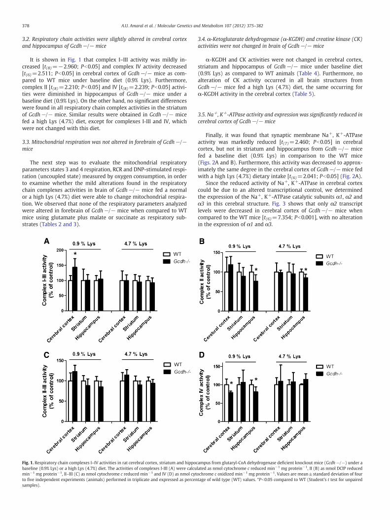
378 A.U. Amaral et al. / Molecular Genetics and Metabolism 107 (2012) 375–382
3.2. Respiratory chain activities were slightly altered in cerebral cortexand hippocampus of Gcdh−/− mice
It is shown in Fig. 1 that complex I–III activity was mildly in-creased [t(8)=−2.960; Pb0.05] and complex IV activity decreased[t(6)=2.511; Pb0.05] in cerebral cortex of Gcdh−/− mice as com-pared to WT mice under baseline diet (0.9% Lys). Furthermore,complex II [t(8)=2.210; Pb0.05] and IV [t(8)=2.239; Pb0.05] activi-ties were diminished in hippocampus of Gcdh−/− mice under abaseline diet (0.9% Lys). On the other hand, no significant differenceswere found in all respiratory chain complex activities in the striatumof Gcdh−/− mice. Similar results were obtained in Gcdh−/− micefed a high Lys (4.7%) diet, except for complexes I-III and IV, whichwere not changed with this diet.
3.3. Mitochondrial respiration was not altered in forebrain of Gcdh−/−mice
The next step was to evaluate the mitochondrial respiratoryparameters states 3 and 4 respiration, RCR and DNP-stimulated respi-ration (uncoupled state) measured by oxygen consumption, in orderto examine whether the mild alterations found in the respiratorychain complexes activities in brain of Gcdh−/− mice fed a normalor a high Lys (4.7%) diet were able to change mitochondrial respira-tion. We observed that none of the respiratory parameters analyzedwere altered in forebrain of Gcdh−/− mice when compared to WTmice using glutamate plus malate or succinate as respiratory sub-strates (Tables 2 and 3).
Fig. 1. Respiratory chain complexes I–IV activities in rat cerebral cortex, striatum and hippocbaseline (0.9% Lys) or a high Lys (4.7%) diet. The activities of complexes I-III (A) were calcumin−1 mg protein−1, II–III (C) as nmol cytochrome c reduced min−1 and IV (D) as nmol cytto five independent experiments (animals) performed in triplicate and expressed as percensamples).
3.4. α-Ketoglutarate dehydrogenase (α-KGDH) and creatine kinase (CK)activities were not changed in brain of Gcdh−/− mice
α-KGDH and CK activities were not changed in cerebral cortex,striatum and hippocampus of Gcdh−/− mice under baseline diet(0.9% Lys) as compared to WT animals (Table 4). Furthermore, noalteration of CK activity occurred in all brain structures fromGcdh−/− mice fed a high Lys (4.7%) diet, the same occurring forα-KGDH activity in the cerebral cortex (Table 5).
3.5. Na+, K+-ATPase activity and expression was significantly reduced incerebral cortex of Gcdh−/− mice
Finally, it was found that synaptic membrane Na+, K+-ATPaseactivity was markedly reduced [t(7)=2.460; Pb0.05] in cerebralcortex, but not in striatum and hippocampus from Gcdh−/− micefed a baseline diet (0.9% Lys) in comparison to the WT mice(Figs. 2A and B). Furthermore, this activity was decreased to approx-imately the same degree in the cerebral cortex of Gcdh−/− mice fedwith a high Lys (4.7%) dietary intake [t(6)=2.041; Pb0.05] (Fig. 2A).
Since the reduced activity of Na+, K+-ATPase in cerebral cortexcould be due to an altered transcriptional control, we determinedthe expression of the Na+, K+-ATPase catalytic subunits α1, α2 andα3 in this cerebral structure. Fig. 3 shows that only α2 transcriptlevels were decreased in cerebral cortex of Gcdh−/− mice whencompared to the WT mice [t(6)=7.354; Pb0.001], with no alterationin the expression of α1 and α3.
ampus from glutaryl-CoA dehydrogenase deficient knockout mice (Gcdh−/−) under alated as nmol cytochrome c reduced min−1 mg protein−1, II (B) as nmol DCIP reducedochrome c oxidized min−1 mg protein−1. Values are mean±standard deviation of fourtage of wild type (WT) values. *Pb0.05 compared to WT (Student's t test for unpaired
Alexandre
Typewritten Text
40
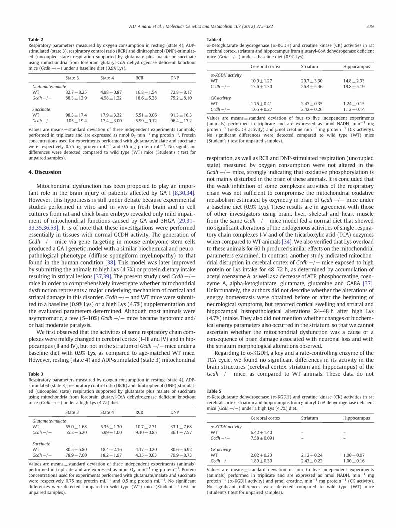
Table 2Respiratory parameters measured by oxygen consumption in resting (state 4), ADP-stimulated (state 3), respiratory control ratio (RCR) and dinitrophenol (DNP)-stimulat-ed (uncoupled state) respiration supported by glutamate plus malate or succinateusing mitochondria from forebrain glutaryl-CoA dehydrogenase deficient knockoutmice (Gcdh−/−) under a baseline diet (0.9% Lys).
State 3 State 4 RCR DNP
Glutamate/malateWT 82.7±8.25 4.98±0.87 16.8±1.54 72.8±8.17Gcdh−/− 88.3±12.9 4.98±1.22 18.6±5.28 75.2±8.10
SuccinateWT 98.3±17.4 17.9±3.32 5.51±0.06 91.3±16.3Gcdh−/− 105±19.4 17.4±3.00 5.99±0.12 96.4±17.2
Values are means±standard deviation of three independent experiments (animals)performed in triplicate and are expressed as nmol O2 min−1 mg protein−1. Proteinconcentrations used for experiments performed with glutamate/malate and succinatewere respectively 0.75 mg protein mL−1 and 0.5 mg protein mL−1. No significantdifferences were detected compared to wild type (WT) mice (Student's t test forunpaired samples).
Table 4α-Ketoglutarate dehydrogenase (α-KGDH) and creatine kinase (CK) activities in ratcerebral cortex, striatum and hippocampus from glutaryl-CoA dehydrogenase deficientmice (Gcdh−/−) under a baseline diet (0.9% Lys).
Cerebral cortex Striatum Hippocampus
α-KGDH activityWT 10.9±1.27 20.7±3.30 14.8±2.33Gcdh−/− 13.6±1.30 26.4±5.46 19.8±5.19
CK activityWT 1.75±0.41 2.47±0.35 1.24±0.15Gcdh−/− 1.65±0.27 2.42±0.26 1.12±0.14
Values are means±standard deviation of four to five independent experiments(animals) performed in triplicate and are expressed as nmol NADH. min−1 mgprotein−1 (α-KGDH activity) and μmol creatine min−1 mg protein−1 (CK activity).No significant differences were detected compared to wild type (WT) mice(Student's t test for unpaired samples).
379A.U. Amaral et al. / Molecular Genetics and Metabolism 107 (2012) 375–382
4. Discussion
Mitochondrial dysfunction has been proposed to play an impor-tant role in the brain injury of patients affected by GA I [8,30,34].However, this hypothesis is still under debate because experimentalstudies performed in vitro and in vivo in fresh brain and in cellcultures from rat and chick brain embryo revealed only mild impair-ment of mitochondrial functions caused by GA and 3HGA [29,31–33,35,36,53]. It is of note that these investigations were performedessentially in tissues with normal GCDH activity. The generation ofGcdh−/− mice via gene targeting in mouse embryonic stem cellsproduced a GA I genetic model with a similar biochemical and neuro-pathological phenotype (diffuse spongiform myelinopathy) to thatfound in the human condition [38]. This model was later improvedby submitting the animals to high Lys (4.7%) or protein dietary intakeresulting in striatal lesions [37,39]. The present study used Gcdh−/−mice in order to comprehensively investigate whether mitochondrialdysfunction represents a major underlying mechanism of cortical andstriatal damage in this disorder. Gcdh−/− andWTmice were submit-ted to a baseline (0.9% Lys) or a high Lys (4.7%) supplementation andthe evaluated parameters determined. Although most animals wereasymptomatic, a few (5–10%) Gcdh−/− mice became hypotonic and/or had moderate paralysis.
We first observed that the activities of some respiratory chain com-plexes were mildly changed in cerebral cortex (I–III and IV) and in hip-pocampus (II and IV), but not in the striatum of Gcdh−/−mice under abaseline diet with 0.9% Lys, as compared to age-matched WT mice.However, resting (state 4) and ADP-stimulated (state 3) mitochondrial
Table 3Respiratory parameters measured by oxygen consumption in resting (state 4), ADP-stimulated (state 3), respiratory control ratio (RCR) and dinitrophenol (DNP)-stimulat-ed (uncoupled state) respiration supported by glutamate plus malate or succinateusing mitochondria from forebrain glutaryl-CoA dehydrogenase deficient knockoutmice (Gcdh−/−) under a high Lys (4.7%) diet.
State 3 State 4 RCR DNP
Glutamate/malateWT 55.0±1.68 5.35±1.30 10.7±2.71 33.1±7.68Gcdh−/− 55.2±6.20 5.99±1.00 9.30±0.85 36.1±7.57
SuccinateWT 80.5±5.80 18.4±2.16 4.37±0.20 80.6±6.92Gcdh−/− 78.9±7.60 18.2±1.97 4.35±0.03 79.9±8.73
Values are means±standard deviation of three independent experiments (animals)performed in triplicate and are expressed as nmol O2. min−1 mg protein−1. Proteinconcentrations used for experiments performed with glutamate/malate and succinatewere respectively 0.75 mg protein mL−1 and 0.5 mg protein mL−1. No significantdifferences were detected compared to wild type (WT) mice (Student's t test forunpaired samples).
respiration, as well as RCR and DNP-stimulated respiration (uncoupledstate) measured by oxygen consumption were not altered in theGcdh−/− mice, strongly indicating that oxidative phosphorylation isnot mainly disturbed in the brain of these animals. It is concluded thatthe weak inhibition of some complexes activities of the respiratorychain was not sufficient to compromise the mitochondrial oxidativemetabolism estimated by oxymetry in brain of Gcdh−/− mice undera baseline diet (0.9% Lys). These results are in agreement with thoseof other investigators using brain, liver, skeletal and heart musclefrom the same Gcdh−/− mice model fed a normal diet that showedno significant alterations of the endogenous activities of single respira-tory chain complexes I-V and of the tricarboxylic acid (TCA) enzymeswhen compared to WT animals [34]. We also verified that Lys overloadto these animals for 60 h produced similar effects on themitochondrialparameters examined. In contrast, another study indicated mitochon-drial disruption in cerebral cortex of Gcdh−/− mice exposed to highprotein or Lys intake for 48–72 h, as determined by accumulation ofacetyl coenzyme A, aswell as a decrease of ATP, phosphocreatine, coen-zyme A, alpha-ketoglutarate, glutamate, glutamine and GABA [37].Unfortunately, the authors did not describe whether the alterations ofenergy homeostasis were obtained before or after the beginning ofneurological symptoms, but reported cortical swelling and striatal andhippocampal histopathological alterations 24–48 h after high Lys(4.7%) intake. They also did not mention whether changes of biochem-ical energy parameters also occurred in the striatum, so that we cannotascertain whether the mitochondrial dysfunction was a cause or aconsequence of brain damage associated with neuronal loss and withthe striatum morphological alterations observed.
Regarding to α-KGDH, a key and a rate-controlling enzyme of theTCA cycle, we found no significant differences in its activity in thebrain structures (cerebral cortex, striatum and hippocampus) of theGcdh−/− mice, as compared to WT animals. These data do not
Table 5α-Ketoglutarate dehydrogenase (α-KGDH) and creatine kinase (CK) activities in ratcerebral cortex, striatum and hippocampus from glutaryl-CoA dehydrogenase deficientmice (Gcdh−/−) under a high Lys (4.7%) diet.
Cerebral cortex Striatum Hippocampus
α-KGDH activityWT 6.42±1.40 – –
Gcdh−/− 7.58±0.091 – –
CK activityWT 2.02±0.23 2.12±0.24 1.00±0.07Gcdh−/− 1.89±0.30 2.43±0.22 1.00±0.16
Values are means±standard deviation of four to five independent experiments(animals) performed in triplicate and are expressed as nmol NADH. min−1 mgprotein−1 (α-KGDH activity) and μmol creatine. min−1 mg protein−1 (CK activity).No significant differences were detected compared to wild type (WT) mice(Student's t test for unpaired samples).
Alexandre
Typewritten Text
41
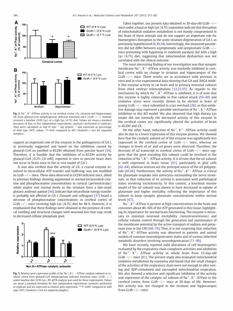
Fig. 2. Na+, K+-ATPase activity in rat cerebral cortex (A), striatum and hippocampus(B) from glutaryl-CoA dehydrogenase deficient knockout mice (Gcdh−/−). Animalsreceived a baseline (0.9% Lys) or a high Lys (4.7%) diet. Values are mean±standarddeviation of four to five independent experiments (animals) performed in triplicate.They were calculated as mol Pi min−1 mg protein−1 and expressed as percentageof wild type (WT) values. *Pb0.05 compared to WT (Student's t test for unpairedsamples).
380 A.U. Amaral et al. / Molecular Genetics and Metabolism 107 (2012) 375–382
support an important role of this enzyme in the pathogenesis of GA I,as previously suggested and based on the inhibition caused byglutaryl-CoA on purified α-KGDH obtained from porcine heart [34].Therefore, it is feasible that the inhibition of α-KGDH activity byglutaryl-CoA (0.25–2.0 mM) reported in vitro in porcine heart doesnot occur in brain mice in the in vivo model of GA I.
It was also verified that the activity of CK, a crucial enzyme in-volved in intracellular ATP transfer and buffering, was not modifiedin Gcdh−/−mice. These data observed in GCDH deficient mice, alliedto previous findings showing augmented or normal intracerebral crea-tine and phosphocreatine concentrations in cortical (periventricular)white matter and normal levels in the striatum from a late-onsetglutaric acidemic patient [54], indicate that intracellular energy transferis probably not affected in GA I. Zinnanti and colleagues [37] found adecrease of phosphocreatine concentrations in cerebral cortex ofGcdh−/− mice receiving high Lys (4.7%) diet for 48 h. However, it isemphasized that these findings were obtained in the presence of corti-cal swelling and structural changes with neuronal loss that may resultin decreased cellular phosphate pool.
Fig. 3. Relative gene expression profile of the Na+,K+−ATPase catalytic subunits in ce-rebral cortex from glutaryl-CoA dehydrogenase deficient knockout mice (Gcdh−/−)under baseline diet (0.9% Lys). RT-qPCR analysis was used for these experiments. Valuesare mean±standard deviation for four independent experiments (animals) performedin triplicate and are expressed as relative gene expression. ***Pb0.001 compared to wildtype (WT) (Student's t test for unpaired samples).
Taken together, our present data obtained in 30-day-old Gcdh−/−mice under a basal or high Lys (4.7%) exposition indicate that disruptionof mitochondrial oxidative metabolism is not mostly compromised inthe brain of these animals and do not support an important role forbioenergetics disruption in the acute striatum degeneration of GA I, aspreviously hypothesized [8,30,34]. Interestingly, themeasured parame-ters did not differ between asymptomatic and symptomatic Gcdh−/−mice presenting with hypotonia or moderate paralysis fed with a highLys (4.7%) diet, suggesting that mitochondrial dysfunction was notcorrelated with the clinical outcome.
The most interesting finding of our investigation was that synapticmembrane Na+, K+-ATPase activity was markedly inhibited in cere-bral cortex with no change in striatum and hippocampus of theGcdh−/− mice. These results are in accordance with previous invitro and in vivo experimental data showing that GA and 3HGA inhib-it this enzyme activity in rat brain and in primary neuronal culturesfrom chick embryo telencephalons [12,23,55]. As regards to themechanism by which Na+, K+-ATPase is inhibited, it is of note thatthis enzyme is highly vulnerable to free radical attack [56–60] andoxidative stress were recently shown to be elicited in brain ofyoung Gcdh−/−mice submitted to a Lys overload [28], so that oxida-tive damage may represent a possible mechanism of Na+, K+-ATPaseinhibition in this KO model. We also observed that high dietary Lysintake did not intensify the decreased activity of this enzyme inthe cerebral cortex nor significantly altered the activities of brainα-KGDH and CK.
On the other hand, reduction of Na+, K+- ATPase activity couldalso be due to a lower expression of this enzyme protein. We showedhere that the catalytic subunit α2 of this enzyme was significantly lessexpressed in the cerebral cortex of Gcdh−/− mice, whereas nochanges in levels of α1 and α3 genes were observed. Therefore, thedecrease of α2 transcript in cerebral cortex of Gcdh−/− mice sug-gests that the gene encoding this subunit could be involved in thereduction of Na+, K+- ATPase activity. It is of note that the α2 subunitis well expressed in brain tissue [61], particularly in glial cells[62–64], whereas neurons are the principal source of the α3 polypep-tide [65,66]. Furthermore, the activity of Na+, K+- ATPase is criticalfor glutamate reuptake into astrocytes surrounding the nerve termi-nals, so that reduction of its activity is associated with pathologicalstates involving excitotoxicity. In this context, a knockout murinemodel of the α2 subunit was shown to have decreased re-uptake ofglutamate and higher mortality, reflecting the importance of thissubunit to keep synaptic glutamate concentrations within normallevels [67].
Na+, K+-ATPase is present at high concentrations in the brain andconsumes about 40–50% of the ATP generated in this tissue, highlight-ing its importance for normal brain functioning. The enzyme is neces-sary to maintain neuronal excitability (neurotransmission) andcellular volume control through the generation and maintenance ofthe membrane potential by the active transport of sodium and potas-sium ions in the CNS [68–70]. Thus, it is not surprising that reductionof Na+, K+-ATPase activity was observed in patients and animalmodels of common neurodegenerative states and of various inheritedmetabolic disorders involving neurodegeneration [71–80].
We have recently reported mild alterations of cell bioenergeticsevaluated by the respiratory chain complexes activities and inhibitionof Na+, K+- ATPase activity in whole brain from 15-day-oldGcdh−/− mice [81]. The present study also evaluated mitochondrialoxidative metabolism by oxymetry and found that the small changesof the activities of the respiratory chain were not enough to alter rest-ing and ADP-stimulated and uncoupled mitochondrial respiration.We also showed a selective and significant inhibition of the activityand expression of the catalytic α2 subunit of Na+, K+-ATPase in thecerebral cortex, from Gcdh−/− mice at 30 days of life. However,this activity was not changed in the striatum and hippocampusfrom Gcdh−/− mice.
Alexandre
Typewritten Text
42
381A.U. Amaral et al. / Molecular Genetics and Metabolism 107 (2012) 375–382
In summary, the present findings demonstrate for the first time amarked inhibition of synaptic Na+, K+-ATPase activity and expres-sion in the cerebral cortex of young Gcdh−/− mice. Since Na+,K+-ATPase activity is crucial for normal brain development and func-tion, it is conceivable that reduction of this activity may be relevant toexplain at least in part the cortical swelling observed in Gcdh−/−mice [37] and the focal edema of affected patients [82]. A persistentdecrease of this activitymight also contribute in the chronic progressivechanges with leukoencephalopathy and cortical atrophy observed inglutaric acidemic patients [4,7]. Finally, the present study does notsupport an important role of bioenergetics dysfunction in the striatumdamage in GA I since Gcdh−/− mice under baseline (0.9% Lys) orhigh Lys (4.7%) intake did not show any significant alteration ofmitochondrial homeostasis in this cerebral structure. We cannot how-ever exclude the possibility that other pathomechanisms of brain dam-age occur in this disorder, including oxidative stress and excitotoxicity.The later mechanismmay be triggered or accentuated by the inhibitionof Na+, K+- ATPase activity causing an impairment of glutamate reup-take by astrocytes leaving more of this excitatory neurotransmitterin the synaptic cleft. The presence of cysts resembling lesions causedby excitotoxicity in the cerebral cortex of glutaric acidemic patientssupports this hypothesis [2].
5. Conclusions
The activity and α2 transcript levels of synaptic Na+, K+- ATPaseare significantly reduced in cerebral cortex of Gcdh−/− mice. It ispresumed that decrease of this crucial enzyme activity may representa relevant pathomechanism of the cortical abnormalities observed inGA I.
Conflicts of interest statement
There are no conflicts of interest between the authors.
Acknowledgments
We are grateful to the financial support of CNPq, PROPESq/UFRGS,FAPERGS, PRONEX, FINEP Rede Instituto Brasileiro de Neurociência(IBN-Net) # 01.06.0842-00, Instituto Nacional de Ciência e Tecnologia-Excitotoxicidade e Neuroproteção (INCT-EN) and Instituto Nacional deCiência e Tecnologia-Translacional em Medicina (INCT-TM).
References
[1] S. Goodman, F. Frerman, Organic Acidemias due to Defects in Lysine Oxidation:2-Ketoadipic Academia and Glutaric Acidemia, in: C.R. Scriver, A.L. Beaudet,W.S. Sly, D. Valle (Eds.), The Metabolic and Molecular Bases of Inherited Disease,McGraw-Hill Inc., New York, 2001, pp. 2195–2204.
[2] S.I. Goodman, M.D. Norenberg, R.H. Shikes, D.J. Breslich, P.G. Moe, Glutaricaciduria: biochemical and morphologic considerations, J. Pediatr. 90 (1977)746–750.
[3] S. Kolker, S.F. Garbade, C.R. Greenberg, J.V. Leonard, J.M. Saudubray, A. Ribes, H.S.Kalkanoglu, A.M. Lund, B. Merinero, M. Wajner, M. Troncoso, M. Williams, J.H.Walter, J. Campistol, M. Marti-Herrero, M. Caswill, A.B. Burlina, F. Lagler, E.M.Maier, B. Schwahn, A. Tokatli, A. Dursun, T. Coskun, R.A. Chalmers, D.M. Koeller,J. Zschocke, E. Christensen, P. Burgard, G.F. Hoffmann, Natural history, outcome,and treatment efficacy in children and adults with glutaryl-CoA dehydrogenasedeficiency, Pediatr. Res. 59 (2006) 840–847.
[4] C.B. Funk, A.N. Prasad, P. Frosk, S. Sauer, S. Kolker, C.R. Greenberg, M.R. Del Bigio,Neuropathological, biochemical and molecular findings in a glutaric acidemiatype 1 cohort, Brain 128 (2005) 711–722.
[5] S. Kulkens, I. Harting, S. Sauer, J. Zschocke, G.F. Hoffmann, S. Gruber, O.A.Bodamer, S. Kolker, Late-onset neurologic disease in glutaryl-CoA dehydrogenasedeficiency, Neurology 64 (2005) 2142–2144.
[6] K.A. Strauss, J. Lazovic, M. Wintermark, D.H. Morton, Multimodal imaging ofstriatal degeneration in Amish patients with glutaryl-CoA dehydrogenasedeficiency, Brain 130 (2007) 1905–1920.
[7] I. Harting, E. Neumaier-Probst, A. Seitz, E.M. Maier, B. Assmann, I. Baric, M.Troncoso, C. Muhlhausen, J. Zschocke, N.P. Boy, G.F. Hoffmann, S.F. Garbade, S.Kolker, Dynamic changes of striatal and extrastriatal abnormalities in glutaricaciduria type I, Brain 132 (2009) 1764–1782.
[8] S. Kolker, D.M. Koeller, S. Sauer, F. Horster, M.A. Schwab, G.F. Hoffmann, K. Ullrich,J.G. Okun, Excitotoxicity and bioenergetics in glutaryl-CoA dehydrogenase defi-ciency, J. Inherit. Metab. Dis. 27 (2004) 805–812.
[9] S. Kolker, G. Kohr, B. Ahlemeyer, J.G. Okun, V. Pawlak, F. Horster, E.Mayatepek, J. Krieglstein, G.F. Hoffmann, Ca(2+) and Na(+) dependenceof 3-hydroxyglutarate-induced excitotoxicity in primary neuronal culturesfrom chick embryo telencephalons, Pediatr. Res. 52 (2002) 199–206.
[10] S. Kolker, B. Ahlemeyer, J. Krieglstein, G.F. Hoffmann, 3-Hydroxyglutaric andglutaric acids are neurotoxic through NMDA receptors in vitro, J. Inherit. Metab.Dis. 22 (1999) 259–262.
[11] L.O. Porciuncula, A. Dal-Pizzol Jr., A.S. Coitinho, T. Emanuelli, D.O. Souza, M.Wajner, Inhibition of synaptosomal [3H]glutamate uptake and [3H]glutamatebinding to plasma membranes from brain of young rats by glutaric acid in vitro,J. Neurol. Sci. 173 (2000) 93–96.
[12] S. Kolker, J.G. Okun, B. Ahlemeyer, A.T. Wyse, F. Horster, M. Wajner, D.Kohlmuller, E. Mayatepek, J. Krieglstein, G.F. Hoffmann, Chronic treatment withglutaric acid induces partial tolerance to excitotoxicity in neuronal culturesfrom chick embryo telencephalons, J. Neurosci. Res. 68 (2002) 424–431.
[13] L.O. Porciuncula, T. Emanuelli, R.G. Tavares, C. Schwarzbold, M.E. Frizzo, D.O.Souza, M. Wajner, Glutaric acid stimulates glutamate binding and astrocytic up-take and inhibits vesicular glutamate uptake in forebrain from young rats,Neurochem. Int. 45 (2004) 1075–1086.
[14] R.B. Rosa, C. Schwarzbold, K.B. Dalcin, G.C. Ghisleni, C.A. Ribeiro, M.B. Moretto, M.E.Frizzo, G.F. Hoffmann, D.O. Souza, M. Wajner, Evidence that 3-hydroxyglutaric acidinteracts with NMDA receptors in synaptic plasmamembranes from cerebral cortexof young rats, Neurochem. Int. 45 (2004) 1087–1094.
[15] M. Wajner, S. Kolker, D.O. Souza, G.F. Hoffmann, C.F. de Mello, Modulation ofglutamatergic and GABAergic neurotransmission in glutaryl-CoA dehydrogenasedeficiency, J. Inherit. Metab. Dis. 27 (2004) 825–828.
[16] K.B. Dalcin, R.B. Rosa, A.L. Schmidt, J.S. Winter, G. Leipnitz, C.S. Dutra-Filho, C.M.Wannmacher, L.O. Porciuncula, D.O. Souza, M. Wajner, Age and brain structuralrelated effects of glutaric and 3-hydroxyglutaric acids on glutamate binding toplasma membranes during rat brain development, Cell. Mol. Neurobiol. 27(2007) 805–818.
[17] R.B. Rosa, K.B. Dalcin, A.L. Schmidt, D. Gerhardt, C.A. Ribeiro, G.C. Ferreira, P.F.Schuck, A.T. Wyse, L.O. Porciuncula, S. Wofchuk, C.G. Salbego, D.O. Souza, M.Wajner, Evidence that glutaric acid reduces glutamate uptake by cerebral cortexof infant rats, Life Sci. 81 (2007) 1668–1676.
[18] D.V. Magni, A.F. Furian, M.S. Oliveira, M.A. Souza, F. Lunardi, J. Ferreira, C.F. Mello,L.F. Royes, M.R. Fighera, Kinetic characterization of l-[(3)H]glutamate uptakeinhibition and increase oxidative damage induced by glutaric acid in striatalsynaptosomes of rats, Int. J. Dev. Neurosci. 27 (2009) 65–72.
[19] S. Kolker, B. Ahlemeyer, J. Krieglstein, G.F. Hoffmann, Contribution of reactiveoxygen species to 3-hydroxyglutarate neurotoxicity in primary neuronal culturesfrom chick embryo telencephalons, Pediatr. Res. 50 (2001) 76–82.
[20] F. de Oliveira Marques, M.E. Hagen, C.D. Pederzolli, A.M. Sgaravatti, K. Durigon,C.G. Testa, C.M. Wannmacher, A.T. de Souza Wyse, M. Wajner, C.S. Dutra-Filho,Glutaric acid induces oxidative stress in brain of young rats, Brain Res. 964(2003) 153–158.
[21] A. Latini, R. Borba Rosa, K. Scussiato, S. Llesuy, A. Bello-Klein, M. Wajner,3-Hydroxyglutaric acid induces oxidative stress and decreases the antioxidantdefenses in cerebral cortex of young rats, Brain Res. 956 (2002) 367–373.
[22] A. Latini, K. Scussiato, G. Leipnitz, C.S. Dutra-Filho, M. Wajner, Promotion of oxida-tive stress by 3-hydroxyglutaric acid in rat striatum, J. Inherit. Metab. Dis. 28(2005) 57–67.
[23] M.R. Fighera, L.F. Royes, A.F. Furian, M.S. Oliveira, N.G. Fiorenza, R. Frussa-Filho, J.C.Petry, R.C. Coelho, C.F. Mello, GM1 ganglioside prevents seizures, Na+, K+‐ATPase ac-tivity inhibition and oxidative stress induced by glutaric acid and pentylenetetrazole,Neurobiol. Dis. 22 (2006) 611–623.
[24] A. Latini, G.C. Ferreira, K. Scussiato, P.F. Schuck, A.F. Solano, C.S. Dutra-Filho, C.R.Vargas, M. Wajner, Induction of oxidative stress by chronic and acute glutaricacid administration to rats, Cell. Mol. Neurobiol. 27 (2007) 423–438.
[25] D.V. Magni, M.S. Oliveira, A.F. Furian, N.G. Fiorenza, M.R. Fighera, J. Ferreira, C.F.Mello, L.F. Royes, Creatine decreases convulsions and neurochemical alterationsinduced by glutaric acid in rats, Brain Res. 1185 (2007) 336–345.
[26] S. Olivera, A. Fernandez, A. Latini, J.C. Rosillo, G. Casanova, M.Wajner, P. Cassina, L.Barbeito, Astrocytic proliferation and mitochondrial dysfunction induced byaccumulated glutaric acidemia I (GAI) metabolites: possible implications forGAI pathogenesis, Neurobiol. Dis. 32 (2008) 528–534.
[27] S. Olivera-Bravo, A. Fernandez, M.N. Sarlabos, J.C. Rosillo, G. Casanova, M. Jimenez,L. Barbeito, Neonatal astrocyte damage is sufficient to trigger progressive striataldegeneration in a rat model of glutaric acidemia-I, PLoS One 6 (2011) e20831.
[28] B. Seminotti, M.S. da Rosa, C.G. Fernandes, A.U. Amaral, L.M. Braga, G. Leipnitz,D.O. de Souza, M. Woontner, D.M. Koeller, S. Goodman, M. Wajner, Induction ofoxidative stress in brain of glutaryl-CoA dehydrogenase deficient mice by acutelysine administration, Mol. Genet. Metab. 106 (2012) 31–38.
[29] C.G. Silva, A.R. Silva, C. Ruschel, C. Helegda, A.T. Wyse, C.M. Wannmacher, C.S.Dutra-Filho, M. Wajner, Inhibition of energy production in vitro by glutaric acidin cerebral cortex of young rats, Metab. Brain Dis. 15 (2000) 123–131.
[30] K.A. Strauss, D.H. Morton, Type I glutaric aciduria, part 2: a model of acute striatalnecrosis, Am. J. Med. Genet. C Semin. Med. Genet. 121C (2003) 53–70.
[31] G.C. Ferreira, C.M. Viegas, P.F. Schuck, A. Latini, C.S. Dutra-Filho, A.T. Wyse, C.M.Wannmacher, C.R. Vargas, M. Wajner, Glutaric acid moderately compromisesenergy metabolism in rat brain, Int. J. Dev. Neurosci. 23 (2005) 687–693.
[32] G.C. Ferreira, C.M. Viegas, P.F. Schuck, A. Tonin, C.A. Ribeiro, M. Coelho Dde, T.Dalla-Costa, A. Latini, A.T. Wyse, C.M. Wannmacher, C.R. Vargas, M. Wajner,
Alexandre
Typewritten Text
43

382 A.U. Amaral et al. / Molecular Genetics and Metabolism 107 (2012) 375–382
Glutaric acid administration impairs energy metabolism in midbrain and skeletalmuscle of young rats, Neurochem. Res. 30 (2005) 1123–1131.
[33] A. Latini, M. Rodriguez, R. Borba Rosa, K. Scussiato, G. Leipnitz, D. Reis de Assis, G.da Costa Ferreira, C. Funchal, M.C. Jacques-Silva, L. Buzin, R. Giugliani, A. Cassina,R. Radi, M. Wajner, 3-Hydroxyglutaric acid moderately impairs energy metabo-lism in brain of young rats, Neuroscience 135 (2005) 111–120.
[34] S.W. Sauer, J.G. Okun, M.A. Schwab, L.R. Crnic, G.F. Hoffmann, S.I. Goodman, D.M.Koeller, S. Kolker, Bioenergetics in glutaryl-coenzyme A dehydrogenase deficien-cy: a role for glutaryl-coenzyme A, J. Biol. Chem. 280 (2005) 21830–21836.
[35] G.C. Ferreira, A. Tonin, P.F. Schuck, C.M. Viegas, P.C. Ceolato, A. Latini, M.L. Perry,A.T. Wyse, C.S. Dutra-Filho, C.M. Wannmacher, C.R. Vargas, M. Wajner, Evidencefor a synergistic action of glutaric and 3-hydroxyglutaric acids disturbing ratbrain energy metabolism, Int. J. Dev. Neurosci. 25 (2007) 391–398.
[36] G.C. Ferreira, P.F. Schuck, C.M. Viegas, A. Tonin, A. Latini, C.S. Dutra-Filho, A.T. Wyse,C.M. Wannmacher, C.R. Vargas, M. Wajner, Energy metabolism is compromised inskeletal muscle of rats chronically-treated with glutaric acid, Metab. Brain Dis. 22(2007) 111–123.
[37] W.J. Zinnanti, J. Lazovic, C. Housman, K. LaNoue, J.P. O'Callaghan, I. Simpson, M.Woontner, S.I. Goodman, J.R. Connor, R.E. Jacobs, K.C. Cheng, Mechanism ofage-dependent susceptibility and novel treatment strategy in glutaric acidemiatype I, J. Clin. Invest. 117 (2007) 3258–3270.
[38] D.M. Koeller, M. Woontner, L.S. Crnic, B. Kleinschmidt-DeMasters, J. Stephens, E.L.Hunt, S.I. Goodman, Biochemical, pathologic and behavioral analysis of a mousemodel of glutaric acidemia type I, Hum. Mol. Genet. 11 (2002) 347–357.
[39] W.J. Zinnanti, J. Lazovic, E.B. Wolpert, D.A. Antonetti, M.B. Smith, J.R. Connor, M.Woontner, S.I. Goodman, K.C. Cheng, A diet-induced mouse model for glutaricaciduria type I, Brain 129 (2006) 899–910.
[40] D.H. Jones, A.I. Matus, Isolation of synaptic plasma membrane from brain bycombined flotation-sedimentation density gradient centrifugation, Biochem.Biophys. Acta 356 (1974) 276–287.
[41] A.U. Amaral, G. Leipnitz, C.G. Fernandes, B. Seminotti, P.F. Schuck, M. Wajner,Alpha-ketoisocaproic acid and leucine provoke mitochondrial bioenergetic dys-function in rat brain, Brain Res. 1324 (2010) 75–84.
[42] A.H.V. Schapira, V.M. Mann, J.M. Cooper, D. Dexter, S.E. Daniel, P. Jenner, J.B. Clark,C.D. Marsden, Anatomic and disease specificity of NADH CoQ1 reductase (Com-plex I) deficiency in Parkinson's disease, J. Neurochem. 55 (1990) 2142–2145.
[43] J.C. Fischer, W. Ruitenbeek, J.A. Berden, J.M.F. Trijbels, J.H. Veerkamp, A.M.Stadhouders, R.C.A. Sengers, A.J.M. Janssen, Differential investigation of the capacityof succinate oxidation in human skeletal muscle, Clin. Chim. Acta 153 (1985) 23–36.
[44] P. Rustin, D. Chretien, T. Bourgeron, B. Gerard, A. Rotig, J.M. Saudubray, A.Munnich, Biochemical and molecular investigations in respiratory chain deficien-cies, Clin. Chim. Acta 228 (1994) 35–51.
[45] L. Tretter, V. Adam-Vizi, Inhibition of Krebs cycle enzymes by hydrogen peroxide:A key role of [alpha]-ketoglutarate dehydrogenase in limiting NADH productionunder oxidative stress, J. Neurosci. 20 (2000) 8972–8979.
[46] B.P. Hughes, A method for estimation of serum creatine kinase and its use in com-paring creatine kinase and aldolase activity in normal and pathological sera, Clin.Chim. Acta 7 (1962) 597–603.
[47] S. Tsakiris, G. Deliconstantinos, Influence of phosphatidylserine on (Na+,K+)-stimulated ATPase and acetylcholinesterase activities of dog brain synapto-somal plasma membranes, Biochem. J. 220 (1984) 301–307.
[48] K.M. Chan, D. Delfert, K.D. Junger, A direct colorimetric assay for Ca2+‐stimulatedatpase activity, Anal. Biochem. 157 (1986) 375–380.
[49] F. Pernot, F. Dorandeu, C. Beaup, A. Peinnequin, Selection of reference genes forreal-time quantitative reverse transcription-polymerase chain reaction in hippo-campal structure in a murine model of temporal lobe epilepsy with focal seizures,J. Neurosci. Res. 88 (2010) 1000–1008.
[50] K.J. Livak, T.D. Schmittgen, Analysis of relative gene expression data using real-timequantitative PCR and the 2(−Delta Delta C(T)) Method, Methods 25 (2001) 402–408.
[51] O.H. Lowry, N.J. Rosebrough, A.L. Farr, R.J. Randall, Protein measurement with theFolin phenol reagent, J. Biol. Chem. 193 (1951) 265–275.
[52] M.M. Bradford, A rapid and sensitive method for the quantitation of microgramquantities of protein utilizing the principle of protein-dye binding, Anal. Biochem.72 (1976) 248–254.
[53] K. Ullrich, B. Flott-Rahmel, P. Schluff, U. Musshoff, A. Das, T. Lucke, R. Steinfeld, E.Christensen, C. Jakobs, A. Ludolph, A. Neu, R. Roper, Glutaric aciduria type I:pathomechanisms of neurodegeneration, J. Inherit. Metab. Dis. 22 (1999) 392–403.
[54] O.A. Bodamer, S. Gruber, S. Stockler-Ipsiroglu, Nuclear magnetic resonance spec-troscopy in glutaryl-CoA dehydrogenase deficiency, J. Inherit. Metab. Dis. 27(2004) 877–883.
[55] C.F. de Mello, S. Kolker, B. Ahlemeyer, F.R. de Souza, M.R. Fighera, E.Mayatepek, J. Krieglstein, G.F. Hoffmann, M. Wajner, Intrastriatal administra-tion of 3-hydroxyglutaric acid induces convulsions and striatal lesions inrats, Brain Res. 916 (2001) 70–75.
[56] G.J. Lees, Contributory mechanisms in the causation of neurodegenerative disor-ders, Neuroscience 54 (1993) 287–322.
[57] E. Kurella, M. Kukley, O. Tyulina, D. Dobrota, M. Matejovicova, V. Mezesova, A.Boldyrev, Kinetic parameters of Na/K-ATPase modified by free radicals in vitroand in vivo, Ann. N. Y. Acad. Sci. 834 (1997) 661–665.
[58] M.I. Yousef, H.A. El-Hendy, F.M. El-Demerdash, E.I. Elagamy, Dietary zinc deficien-cy induced-changes in the activity of enzymes and the levels of free radicals,lipids and protein electrophoretic behavior in growing rats, Toxicology 175(2002) 223–234.
[59] K. Hitschke, R. Buhler, H.J. Apell, G. Stark, Inactivation of the Na, K-ATPase byradiation-induced free radicals. Evidence for a radical-chain mechanism, FEBSLett. 353 (1994) 297–300.
[60] P. Muriel, G. Sandoval, Nitric oxide and peroxynitrite anion modulate liver plasmamembrane fluidity and Na(+)/K(+)-ATPase activity, Nitric Oxide 4 (2000)333–342.
[61] K.M. McGrail, J.M. Phillips, K.J. Sweadner, Immunofluorescent localization of threeNa,K-ATPase isozymes in the rat central nervous system: both neurons and gliacan express more than one Na, K-ATPase, J. Neurosci. 11 (1991) 381–391.
[62] M.L. Brines, R.J. Robbins, Cell-type specific expression of Na+, K(+)-ATPasecatalytic subunits in cultured neurons and glia: evidence for polarized distribu-tion in neurons, Brain Res. 631 (1993) 1–11.
[63] R. Cameron, L. Klein, A.W. Shyjan, P. Rakic, R. Levenson, Neurons and astrogliaexpress distinct subsets of Na,K-ATPase alpha and beta subunits, Brain Res. Mol.Brain Res. 21 (1994) 333–343.
[64] D. Fink, P.E. Knapp, M. Mata, Differential expression of Na,K-ATPase isoforms inoligodendrocytes and astrocytes, Dev. Neurosci. 18 (1996) 319–326.
[65] K.J. Sweadner, Overlapping and diverse distribution of Na-K ATPase isozymes inneurons and glia, Can. J. Physiol. Pharmacol. 70 (Suppl.) (1992) S255–S259.
[66] R. Levenson, Isoforms of the Na,K-ATPase: family members in search of function,Rev. Physiol. Biochem. Pharmacol. 123 (1994) 1–45.
[67] K. Kawakami, K. Ikeda, Modulation of neural activities by Na, K-ATPase alpha 2 subunitthrough functional coupling with transporters, Cell. Mol. Biol. (Noisy-le-Grand) 52(2006) 92–96.
[68] M. Erecinska, I.A. Silver, Ions and energy in mammalian brain, Prog. Neurobiol. 43(1994) 37–71.
[69] M. Erecinska, S. Cherian, I.A. Silver, Energy metabolism in mammalian brainduring development, Prog. Neurobiol. 73 (2004) 397–445.
[70] E. Satoh, Y. Nakazato, On the mechanism of ouabain-induced release of acetylcho-line from synaptosomes, J. Neurochem. 58 (1992) 1038–1044.
[71] A. Vignini, L. Nanetti, C. Moroni, L. Tanase, M. Bartolini, S. Luzzi, L. Provinciali, L.Mazzanti, Modifications of platelet from Alzheimer disease patients: a possiblerelation between membrane properties and NO metabolites, Neurobiol. Aging28 (2007) 987–994.
[72] M.A. Cousin, D.G. Nicholls, J.M. Pocock, Modulation of ion gradients and gluta-mate release in cultured cerebellar granule cells by ouabain, J. Neurochem. 64(1995) 2097–2104.
[73] E.N. Busanello, C.M. Viegas, A.P. Moura, A.M. Tonin, M. Grings, C.R. Vargas, M.Wajner, In vitro evidence that phytanic acid compromises Na(+), K(+)-ATPaseactivity and the electron flow through the respiratory chain in brain cortexfrom young rats, Brain Res. 1352 (2010) 231–238.
[74] E.N. Busanello, C.M. Viegas, A.M. Tonin, M. Grings, A.P. Moura, A.B. de Oliveira, P.Eichler, M. Wajner, Neurochemical evidence that pristanic acid impairs energyproduction and inhibits synaptic Na(+), K(+)-ATPase activity in brain of youngrats, Neurochem. Res. 36 (2011) 1101–1107.
[75] B. Seminotti, C.G. Fernandes, G. Leipnitz, A.U. Amaral, A. Zanatta, M. Wajner,Neurochemical evidence that lysine inhibits synaptic Na+, K+‐ATPase activityand provokes oxidative damage in striatum of young rats in vivo, Neurochem.Res. 36 (2011) 205–214.
[76] A.P. Moura, C.A. Ribeiro, A. Zanatta, E.N. Busanello, A.M. Tonin, M. Wajner,3-Methylcrotonylglycine disrupts mitochondrial energy homeostasis and inhibitssynaptic Na(+), K (+)-ATPase activity in brain of young rats, Cell. Mol.Neurobiol. 32 (2012) 297–307.
[77] G.J. Lees,W. Leong, The sodium-potassiumATPase inhibitor ouabain is neurotoxici inthe rat substantia nigra and striatum, Neurosci. Lett. 188 (1995) 113–116.
[78] D.Z. Ellis, J. Rabe, K.J. Sweadner, Global loss of Na,K-ATPase and its nitricoxide-mediated regulation in a transgenic mouse model of amyotrophic lateralsclerosis, J. Neurosci. 23 (2003) 43–51.
[79] C.A. Dickey, M.N. Gordon, D.M. Wilcock, D.L. Herber, M.J. Freeman, D. Morgan,Dysregulation of Na+/K+ ATPase by amyloid in APP+PS1 transgenic mice,BMC Neurosci. 6 (2005) 7.
[80] M.B. Bagh, A.K. Maiti, S. Jana, K. Banerjee, A. Roy, S. Chakrabarti, Quinone andoxyradical scavenging properties of N-acetylcysteine prevent dopamine mediat-ed inhibition of Na+, K+‐ATPase and mitochondrial electron transport chainactivity in rat brain: implications in the neuroprotective therapy of Parkinson'sdisease, Free Radic. Res. 42 (2008) 574–581.
[81] A.U. Amaral, C. Cecatto, B. Seminotti, A. Zanatta, C.G. Fernandes, E.N. Busanello,L.M. Braga, C.A.J. Ribeiro, D.O. de Souza, M. Woontner, D.M. Koeller, S. Goodman,M.Wajner, Marked reduction of Na(+), K(+)-ATPase and creatine kinase activitiesinduced by acute lysine administration in glutaryl-CoA dehydrogenase deficientmice, Mol. Genet. Metab. 107 (2012) 81–86.
[82] B. Perez-Duenas, A. De La Osa, A. Capdevila, A. Navarro-Sastre, A. Leist, A. Ribes, A.Garcia-Cazorla, M. Serrano, M. Pineda, J. Campistol, Brain injury in glutaricaciduria type I: the value of functional techniques in magnetic resonance imaging,Eur. J. Paediatr. Neurol. 13 (2009) 534–540.
Alexandre
Typewritten Text
44
45
Capítulo III
Histological alterations and mild disruption of mitochondrial energy
homeostasis in striatum of glutaryl-CoA dehydrogenase deficient mice
submitted to lysine overload
Alexandre U Amaral, Cristiane Cecatto, Bianca Seminotti, César A Ribeiro,
Valeska L Lagranha, Carolina Coffi, Luis E Soares, Francine H de Oliveira,
Stephen Goodman, David M Koeller, Michael Woontner, Diogo O de Souza,
Moacir Wajner
Artigo científico submetido ao periódico
Life Sciences
46
Histological alterations and mild disruption of mitochondrial energy homeostasis
in striatum of glutaryl-CoA dehydrogenase deficient mice submitted to lysine
overload
Alexandre Umpierrez Amarala, Cristiane Cecattoa, Bianca Seminottia, César
Augusto Ribeiroa, Valeska Lizzi Lagranhaa, Carolina Coffia, Luis Eduardo Soaresa,
Francine Hehn de Oliveirab, , Stephen Goodmanc, David M. Koellerd , Diogo Onofre
Gomes de Souzaa, Michael Woontnerc and Moacir Wajnera,e
a - Departamento de Bioquímica, Instituto de Ciências Básicas da Saúde,
Universidade Federal do Rio Grande do Sul, Porto Alegre, RS, Brazil
b - Servico de Patologia, Hospital de Clinicas de Porto Alegre, RS, Brazil
c - School Medicine University of Colorado Denver, Aurora, United States;
d - Departments of Pediatrics, Molecular and Medical Genetics, Oregon Health &
Science University, Portland, OR, USA
e - Serviço de Genética Médica, Hospital de Clínicas de Porto Alegre, Porto
Alegre, RS, Brazil
* Corresponding Author: Moacir Wajner
Departamento de Bioquímica, Instituto de Ciências Básicas da Saúde,
Universidade Federal de Rio Grande do Sul. Rua Ramiro Barcelos N° 2600 -
Anexo, CEP 90035-003, Porto Alegre, RS - Brasil. Phone: +55 51 3308-5571, fax:
+55 51 3308-5535, e-mail: [email protected]
47
Abstract
Bioenergetics dysfunction has been postulated as an important pathomechanism
of brain damage in glutaric aciduria type I (GA I), but this is still under debate.
Therefore, the present work investigated a large spectrum of important parameters
of mitochondrial energy homeostasis, namely the activities of key enzymes of the
citric acid cycle, including citrate synthase (CS), aconitase, isocitrate
dehydrogenase (IDH), α-ketoglutarate dehydrogenase, succinate dehydrogenase
and malate dehydrogenase, lactate release, the respiratory parameters states 3
and 4, respiratory control ratio and CCCP-stimulated state and the membrane
potential (ΔΨm) in purified mitochondrial preparations from cerebral cortex and
striatum of adolescent glutaryl-CoA dehydrogenase deficient (Gcdh-/-) and wild
type mice fed a normal or a high lysine (Lys, 4.7 %) chow for 60 hours. Histological
analysis was also evaluated in the brain of these animals. A moderate reduction of
CS and IDH activities (20-30 %) and a very mild (10 %) increase of lactate release
were observed in striatum from Gcdh-/- animals submitted to a high Lys chow. In
contrast, the respiratory parameters and ΔΨm were not altered in these animals.
Histological analysis revealed the presence of a few vacuoles in the cerebral
cortex, but not in striatum from Gcdh-/- mice exposed for a short time to a normal
or a high Lys show. However, intense vacuolation was found in the cerebral cortex
of 60 and 90-day-old Gcdh-/- mice fed a baseline chow and in the striatum of
Gcdh-/- mice fed a high Lys chow for 30 days. Taken together, the present data
demonstrate very mild impairment of bioenergetics homeostasis in striatum from
adolescent Gcdh-/- mice under a short exposition to a high Lys chow and important
histological alterations in this cerebral structure when these animals were

48
submitted to this chow for a long period.
Keywords: glutaric acidemia type I; glutaric acid; Gcdh-/- mice; citric acid cycle;
mitochondrial homeostasis, brain histology
Conflicts of interest
There are no conflicts of interest between the authors.
49
1. Introduction
Accumulation of glutaric acid (GA), 3-hydroxyglutaric acid (3HGA) and
glutarylcarnitine in the body fluids and tissues are the biochemical hallmark of
patients affected by glutaric aciduria type I (GA I, McKusick 23167; OMIM
#231670) (Goodman et al., 1977; Kolker et al., 2006). This neurometabolic disease
is caused by a deficiency in the activity of the mitochondrial enzyme glutaryl-CoA
dehydrogenase (GCDH) (EC 1.3.99.7), which is involved in the catabolic pathway
of lysine (Lys) and tryptophan (Goodman and Frerman, 2001). Untreated patients
present with acute bilateral striatal degeneration that follows catabolic events
occurring from 6 months to four years of age. A chronic presentation of
demyelination of the central nervous system, resulting in progressive cortical
atrophy, may be seen as well. (Funk et al., 2005; Harting et al., 2009; Strauss et
al., 2007). Neurological symptoms including developmental delay, dystonia,
dyskinesia, hypotonia, seizures and spasticity start especially after the
encephalopathic crises commonly found in these patients (Hoffmann and
Zschocke, 1999; Neumaier-Probst et al., 2004).
Excitotoxicity (Dalcin et al., 2007; Kolker et al., 1999; Kolker et al., 2004;
Kolker et al., 2002a; Kolker et al., 2002b; Magni et al., 2009; Porciuncula et al.,
2000; Porciuncula et al., 2004; Rosa et al., 2007; Rosa et al., 2004; Wajner et al.,
2004), oxidative stress (de Oliveira Marques et al. 2003; Fighera et al., 2006; Latini
et al., 2002; Latini et al., 2007; Latini et al., 2005b; Magni et al., 2007; Seminotti et
al., 2013) and mitochondrial dysfunction (Ferreira et al., 2005a; Ferreira et al.,
2005b; Ferreira et al., 2007a; Ferreira et al., 2007b; Latini et al., 2005a; Olivera et
al., 2008; Sauer et al., 2005; Silva et al., 2000; Strauss and Morton, 2003; Zinnanti
50
et al., 2007) have been proposed to be involved in GA I neuropathology, but the
contribution of each pathomechanism to the brain injury of affected patients is still
under discussion and needs further investigation. Mild alterations in mitochondrial
homeostasis are provoked by GA and 3HGA in brain of rodents with normal GCDH
activity (Ferreira et al., 2005a; Ferreira et al., 2005b; Latini et al., 2005a).
A model for the study of GA I was developed in mice by replacing most of
the GCDH gene with an in-frame beta-galactosidase cassette (Koeller et al., 2002).
Glutaryl-CoA dehydrogenase deficient mice (Gcdh-/-) had increased cerebral,
blood and urine GA and 3HGA levels and displayed vacuolation in the frontal
cortex (spongiform leukoencephalopathy). However, the animals did not develop
striatal damage spontaneously, as is typical of the human disease. Exposing these
mice to a high protein or Lys chow provoked neuronal loss, myelin disruption, and
gliosis, mostly in the striatum and deep cortex (Zinnanti et al., 2007; Zinnanti et al.,
2006). Oral Lys overload in weaning (4-week-old) Gcdh-/- mice resulted in
increased brain Lys and GA concentrations, followed by a simultaneous decrease
of Lys and further increase of brain GA levels, indicating GA formation from Lys
catabolism in the brain. These investigators suggested that the cortical and striatal
lesions developed in the Gcdh-/- animals submitted to Lys overload were probably
due to the increase of brain GA concentrations (Zinnanti et al., 2007; Zinnanti et al.,
2006).
Our group recently demonstrated mild impairment of the respiratory chain
and reduced activities of Na+, K+ - ATPase and creatine kinase in brain and skeletal
muscle of 15 and 30-day-old Gcdh-/- mice (Amaral et al., 2012a; Amaral et al.,
2012b). The purpose of the present study was to evaluate a large spectrum of
51
parameters of energy homeostasis in mitochondria obtained from cerebral cortex
and striatum of Gcdh-/- mice under distinct metabolic conditions.
2. Materials and Methods
2.1. Animals and reagents
Gcdh-/- and wild type (WT) mice littermate controls, both of 129SvEv
background (Koeller et al., 2002), were generated from heterozygotes and
maintained at the Unidade Experimental Animal, Hospital de Clínicas de Porto
Alegre (Porto Alegre, Brazil). This study was performed in strict accordance with
the Guide for the Care and Use of Laboratory Animals (Eighth edition, 2001) and
approved by the Ethical Committee for the Care and Use of Laboratory Animals of
Hospital de Clínicas de Porto Alegre. All efforts were also made to use the minimal
number of animals necessary to produce reliable scientific data.
The animals were maintained on a 12:12 h light/dark cycle (lights on 07.00-
19.00 h) in air conditioned constant temperature (22 ± 1 ºC) colony room, with free
access to water and a normal chow containing 20% (w/w) protein (NUVILAB).
Mitochondrial preparations obtained from cerebral cortex and striatum of 30-day-
old WT and Gcdh-/- mice submitted to a normal Lys (0.9 %) or high Lys (4.7 %)
chow for 60 hours were used for the biochemical determinations. Histological
analysis were carried out in the brain from these animals and also in 60-day-old
WT and Gcdh-/- animals under a normal diet and in 90-day-old animals fed a
normal or a high Lys (4.7 %) chow for 30 days.
All chemicals were of analytical grade and purchased from Sigma (St Louis,
52
MO, USA) unless otherwise stated. Solutions were prepared on the day of the
experiments and the pH was adjusted to 7.2-7.4 with the buffers used in each
technique.
2.2. Tissue Preparation and general experimental conditions
Determination of the respiratory parameters, the ΔΨm and the activities of
citric acid cycle (CAC) enzymes were carried out in mitochondrial preparations
from cerebral cortex and striatum of 30-day-old WT and Gcdh-/- mice, as
previously described (Rosenthal et al., 1987) with minor modifications (Mirandola
et al., 2008). Digitonin was used to permeabilize synaptosomal plasma
membranes. The final pellet was gently washed and resuspended in isolation
buffer devoid of EGTA, at an approximate protein concentration of 6-8 mg . mL-1.
This preparation results in a mixture of synaptosomal and non-synaptosomal
mitochondria similar to the general brain composition.
The expression of the catalytic subunit of NAD-dependent isocitrate
dehydrogenase (Idh3α) was determined in striatum of 30-day-old Gcdh-/- and WT
mice. These structures were dissected and immediately frozen in the presence of
Trizol® for isolation of total RNA.
For the determination of lactate release, cerebral cortex and striatum from
30-day-old WT and Gcdh-/- mice were cut into two perpendicular directions to
produce 400 mM-wide prisms using a Mcllwain chopper.
2.3. Citric acid cycle (CAC) enzyme activities
Citrate synthase (CS) activity was measured according to Srere (1969), by
53
determining DTNB reduction at = 412 nm; aconitase (ACO) according to Morrison
(1954); isocitrate dehydrogenase (IDH) activity by the method of Plaut (1969); α-
ketoglutarate dehydrogenase (αKGDH) complex according to Lai and Cooper
(1986) and Tretter and Adam-Vizi (2000), with slight modifications (Amaral et al.,
2010); succinate dehydrogenase (SDH) as described by Fischer et al. (1985); and
malate dehydrogenase (MDH) activity according to Kitto (1969). The activities of
the CAC enzymes were calculated as nmol . min-1. mg protein-1.
2.4. Gene expression analysis of the catalytic subunit of NAD-dependent
isocitrate dehydrogenase (Idh3α) by quantitative real time (RT-qPCR)
Total RNA was isolated with Trizol® reagent purchased from Invitrogen
(Carlsbad, California, USA) in accordance with the manufacturer's instructions.
RNA concentrations were evaluated at 260 and 280 nm absorbances in a
NanoDrop 1000 (Thermo Scientific, San Jose, CA, USA). cDNA was synthesized
by reverse transcription (RT) using SuperScript® III First-Strand Synthesis
SuperMix (Invitrogen, Grand Island, NY, USA). RT reactions contained 1 μg of
RNA, 1 uL of oligo dT(20), 1 μL of annealing buffer, 10 μL of 2X First-Strand
Reaction Mix and 2 μL of SuperScript® III/RNaseOUT™ Enzyme Mix in a total
reaction volume of 20 μL. Reactions were performed for 5 min at 65°C, 50 min at
50°C and terminated with 5 min at 85°C. Subsequently, cDNA was kept at -20°C
until PCR quantitation. A 1:20 dilution of cDNA solution was prepared in water for
quantitative real-time polymerase chain reaction (RT-qPCR).
Messenger RNA (mRNA) expression was measured by RT-qPCR using
54
gene-specific TaqMan FAM/MGB inventoried assays (Applied Biosystems, Foster
City, CA, EUA) (Idh3α, assay number Mm00499674_m1). Expression of the targed
gene was normalized to the expression of endogenous control Hprt, a gene with
low expression variability in the central nervous system (Pernot et al., 2010), using
another TaqMan probe (assay number Mm00446968_m1). Reactions were carried
out in a Stratagene MX3000p qPCR System (Stratagene, GE Healthcare Life
Sciences, Piscataway, NJ, USA). The cDNA of each mouse (n=4) was used
separately in RT-qPCR reactions. Reactions were carried out in a total volume of
12 μL using 5 μL of cDNA solution, 0.5 μL of gene specific TaqMan assay, 1.5 μL of
water milli-Q and 5 μL of Master Mix (Applied Biosystems), containing ROX,
Amplitaq Gold DNA polymerase, AmpErase UNG, dATP, dCTP, dGTP, dUTP, and
MgCl2. The cycling program was 2 min at 50 °C, 10 min at 95 °C, followed by 40
cycles of 15 s at 95 °C and 1 min at 60 °C. Reactions were performed in duplicate.
Target transcripts’ relative expression levels were determined by the DDCt method
(Livak and Schmittgen, 2001), using WT mice at each time point as calibrators.
2.5. Lactate release
Cortical prisms were initially pre-incubated at 37 ºC for 15 min in Krebs-
Ringer bicarbonate buffer, pH 7.0, followed by the addition of 5 mM glucose. After
60 min incubation at 37 ºC in a metabolic shaker (90 oscillations min-1), two
volumes of 0.3 N perchloric acid were immediately added to the incubation
medium. The excess of perchloric acid was precipitated as a potassium salt by the
addition of one volume of 3 M potassium bicarbonate. After centrifugation for 5 min
55
at 800 x g, lactate was measured in the supernatant by the lactase-peroxidase
method (Shimojo et al., 1989). Results were expressed as mmol of lactate h-1 . g
tissue-1.
2.6. Determination of mitochondrial respiratory parameters by oxygen
consumption
Oxygen consumption rate was measured as described previously (Amaral et
al., 2010) using a Clark-type electrode in a thermostatically controlled (37ºC) and
magnetically stirred incubation chamber using pyruvate plus malate (2.5 mM each)
as substrates in a reaction medium containing the striatum mitochondrial
preparations (0.4 mg protein . mL-1) and 300 mM sucrose, 5 mM potassium
phosphate, 1 mM EGTA, 5 mM MOPS and 0.1 % BSA. We measured state 3 (ADP
stimulated), state 4 (oligomycin stimulated) and carbonyl cyanide m-chlorophenyl
hydrazone (CCCP)-stimulated respiration (uncoupled state), which were calculated
as nmol O2 consumed . min-1 . mg of protein-1. The RCR (state 3 / state 4) was also
determined. Only mitochondrial preparations with RCR higher than 4 were used in
the experiments.
2.7. Determination of mitochondrial membrane potential (ΔΨm)
The ΔΨm was estimated according to (Akerman and Wikstrom, 1976;
Figueira et al., 2012) on a temperature-controlled Hitachi F-4500
spectrofluorometer with magnetic stirring operating at excitation and emission of
495 and 586 nm, respectively, slit width of 10 nm, using 2.5 mM pyruvate plus 2.5
mM malate as substrates and supplemented with 5 μM safranine O. Striatal
56
mitochondrial preparations (0.4 mg protein . mL-1) were incubated at 37 oC in 5 mM
HEPES buffer, pH 7.2, containing 150 mM potassium chloride, 5 mM magnesium
chloride, 0.015 mM EGTA, 5 mM potassium phosphate, 0.01 % BSA and 1 μg . mL-
1 oligomycin A. CaCl2 (10 µM) was added 100 s afterwards. In the end of the
measurements maximal depolarization was induced by 1 µM CCCP. Data were
expressed as fluorescence arbitrary units (FAU).
2.8. Histological analysis
In order to evaluate morphological changes, we performed histological
analysis in cerebral cortex and striatum from WT and Gcdh-/- mice using
hematoxylin and eosin (HE) staining. For each group four mice were used. Image
analysis (100 and 400 x magnification) was done using Q Capture Pro Software
(Olympus). Whole brains from 30, 60 and 90-day- old WT and Gcdh-/- mice were
removed and postfixed in 10% formaldehyde buffered solution (pH 7.00 - 7.05) for
48 h at room temperature and processed for paraffin embedded sectioning.
Cerebral cortex and striatum were sectioned (three micrometers) on a Microtome
(MICROM HM 360) and slices were collected for HE staining.
2.9. Protein Determination
Protein levels were measured by the method of Bradford (Bradford, 1976)
using bovine serum albumin as standard.
2.10. Statistical analysis
Results are presented as mean ± standard deviation. Assays were
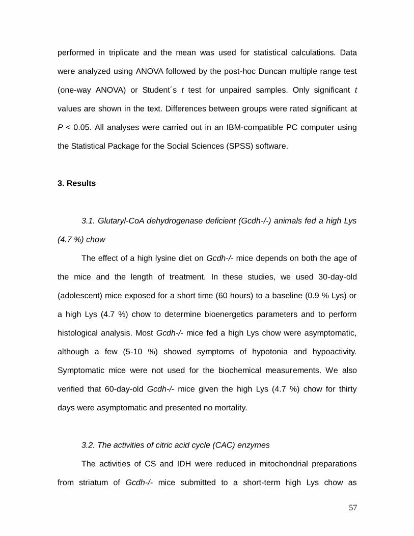
57
performed in triplicate and the mean was used for statistical calculations. Data
were analyzed using ANOVA followed by the post-hoc Duncan multiple range test
(one-way ANOVA) or Student´s t test for unpaired samples. Only significant t
values are shown in the text. Differences between groups were rated significant at
P < 0.05. All analyses were carried out in an IBM-compatible PC computer using
the Statistical Package for the Social Sciences (SPSS) software.
3. Results
3.1. Glutaryl-CoA dehydrogenase deficient (Gcdh-/-) animals fed a high Lys
(4.7 %) chow
The effect of a high lysine diet on Gcdh-/- mice depends on both the age of
the mice and the length of treatment. In these studies, we used 30-day-old
(adolescent) mice exposed for a short time (60 hours) to a baseline (0.9 % Lys) or
a high Lys (4.7 %) chow to determine bioenergetics parameters and to perform
histological analysis. Most Gcdh-/- mice fed a high Lys chow were asymptomatic,
although a few (5-10 %) showed symptoms of hypotonia and hypoactivity.
Symptomatic mice were not used for the biochemical measurements. We also
verified that 60-day-old Gcdh-/- mice given the high Lys (4.7 %) chow for thirty
days were asymptomatic and presented no mortality.
3.2. The activities of citric acid cycle (CAC) enzymes
The activities of CS and IDH were reduced in mitochondrial preparations
from striatum of Gcdh-/- mice submitted to a short-term high Lys chow as
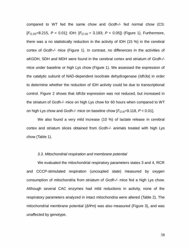
58
compared to WT fed the same chow and Gcdh-/- fed normal chow (CS:
[F(2,18)=8.215, P < 0.01]; IDH: [F(2,16) = 3.183; P < 0.05]) (Figure 1). Furthermore,
there was a no statistically reduction in the activity of IDH (15 %) in the cerebral
cortex of Gcdh-/- mice (Figure 1). In contrast, no differences in the activities of
αKGDH, SDH and MDH were found in the cerebral cortex and striatum of Gcdh-/-
mice under baseline or high Lys chow (Figure 1). We assessed the expression of
the catalytic subunit of NAD-dependent isocitrate dehydrogenase (Idh3α) in order
to determine whether the reduction of IDH activity could be due to transcriptional
control. Figure 2 shows that Idh3α expression was not reduced, but increased in
the striatum of Gcdh-/- mice on high Lys chow for 60 hours when compared to WT
on high Lys chow and Gcdh-/- mice on baseline chow [F(2,9)=9.118, P < 0.01].
We also found a very mild increase (10 %) of lactate release in cerebral
cortex and striatum slices obtained from Gcdh-/- animals treated with high Lys
chow (Table 1).
3.3. Mitochondrial respiration and membrane potential
We evaluated the mitochondrial respiratory parameters states 3 and 4, RCR
and CCCP-stimulated respiration (uncoupled state) measured by oxygen
consumption of mitochondria from striatum of Gcdh-/- mice fed a high Lys chow.
Although several CAC enzymes had mild reductions in activity, none of the
respiratory parameters analyzed in intact mitochondria were altered (Table 2). The
mitochondrial membrane potential (ΔΨm) was also measured (Figure 3), and was
unaffected by genotype.
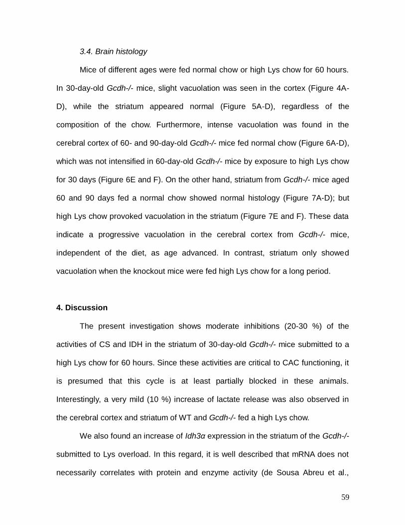
59
3.4. Brain histology
Mice of different ages were fed normal chow or high Lys chow for 60 hours.
In 30-day-old Gcdh-/- mice, slight vacuolation was seen in the cortex (Figure 4A-
D), while the striatum appeared normal (Figure 5A-D), regardless of the
composition of the chow. Furthermore, intense vacuolation was found in the
cerebral cortex of 60- and 90-day-old Gcdh-/- mice fed normal chow (Figure 6A-D),
which was not intensified in 60-day-old Gcdh-/- mice by exposure to high Lys chow
for 30 days (Figure 6E and F). On the other hand, striatum from Gcdh-/- mice aged
60 and 90 days fed a normal chow showed normal histology (Figure 7A-D); but
high Lys chow provoked vacuolation in the striatum (Figure 7E and F). These data
indicate a progressive vacuolation in the cerebral cortex from Gcdh-/- mice,
independent of the diet, as age advanced. In contrast, striatum only showed
vacuolation when the knockout mice were fed high Lys chow for a long period.
4. Discussion
The present investigation shows moderate inhibitions (20-30 %) of the
activities of CS and IDH in the striatum of 30-day-old Gcdh-/- mice submitted to a
high Lys chow for 60 hours. Since these activities are critical to CAC functioning, it
is presumed that this cycle is at least partially blocked in these animals.
Interestingly, a very mild (10 %) increase of lactate release was also observed in
the cerebral cortex and striatum of WT and Gcdh-/- fed a high Lys chow.
We also found an increase of Idh3α expression in the striatum of the Gcdh-/-
submitted to Lys overload. In this regard, it is well described that mRNA does not
necessarily correlates with protein and enzyme activity (de Sousa Abreu et al.,
60
2009; Griffin et al., 2002; Gygi et al., 1999; Tian et al., 2004), which could be
caused by posttranscriptional regulation and differences in mRNA and protein
turnover rates (Cox et al., 2005; Hack, 2004). In this case, the observed in vivo
increase of striatum Idh3α expression from Gcdh-/- mice exposed to a high Lys
chow may represent a compensatory mechanism in response to the persistent
reduction in this enzyme activity.
These results are possibly related to previous findings showing a decrease
in ATP, CoA, α-ketoglutarate, glutamate and GABA levels, as well as an increase of
acetyl-CoA in brain of 4-week-old Gcdh-/- submitted to a high Lys chow (Zinnanti et
al., 2007). Another work demonstrated that the succinate transport from astrocytic
to neuronal cells was compromised in primary astrocytic and neuronal culture of
Gcdh-/- mice, leading to the loss of CAC intermediates necessary for neuronal
functions (Lamp et al., 2011). Taken these data together, it is presumed that the
CAC activity is compromised in brain from Gcdh-/- mice under Lys overload.
In order to further evaluate bioenergetics in these animals, we tested the
resting (state 4) and ADP-stimulated (state 3) states, as well as RCR and CCCP-
stimulated respiration (uncoupled state) supported by pyruvate plus malate and
measured by oxygen consumption in striatum mitochondrial preparations from 30-
day-old WT and Gcdh-/- mice fed a high Lys diet for a short period. No significant
differences in these respiratory parameters were observed when comparing WT
and Gcdh-/- mice under Lys overload, indicating that these substrates were
sufficiently oxidized to support mitochondrial respiration. The observations of
normal respiratory parameters in Gcdh-/- mice were expected since only strong
inhibitions of the CAC and respiratory chain enzymes are necessary to significantly
61
alter these parameters (Brand and Nicholls, 2011).
ΔΨm was also not changed in striatum of Gcdh-/- fed a high Lys chow, even
when mitochondria were challenged by Ca2+, indicating that the mitochondrial
capacity to maintain the ion balance necessary to keep this potential, as well as
Ca2+ buffering were preserved in the striatum of Gcdh-/- mice.
We emphasize that our present data showing very mild alterations of
mitochondrial bioenergetics in striatum from 30-day-old Gcdh-/- mice occurred only
when these animals were exposed to a high Lys chow for 60 hours, mimicking the
human condition of catabolic stress leading to brain accumulation of GA and 3HGA
and simultaneous brain damage, especially in the striatum (Funk et al., 2005;
Harting et al., 2009; Hoffmann and Zschocke, 1999; Sauer et al., 2006). Therefore,
it is conceivable that Lys-induced effects were dependent on the increased brain
production of GA and 3HGA from Lys which easily cross the blood brain barrier in
Gcdh-/- mice (Zinnanti et al., 2007; Zinnanti et al., 2006).
In order to evaluate brain damage, we submitted Gcdh-/- mice to a short (60
hours) or a long (30 days) Lys (4.7 %) overload and performed histological analysis
in cerebral cortex and striatum. We verified a low degree of vacuolation in the
cerebral cortex, but not in the striatum of 30-day-old Gcdh-/- mice exposed to a
normal or a high Lys chow for 60 hours, as compared to WT mice. Furthermore, a
large number of vacuoles were verified in the cerebral cortex, but not in the
striatum, of 60- and 90-day-old Gcdh-/- mice fed a normal chow. Finally, when 60-
day-old Gcdh-/- mice were fed for 30 days a high Lys chow, we found intense
vacuolation in the striatum, but vacuolation in the cereral cortex did not change.
Our results are in agreement with previous findings showing minor microscopic
62
changes with vacuolation in the cerebral cortex of 4-week-old Gcdh-/- mice
exposed for a short time with high Lys (Zinnanti et al., 2006). The same
investigators observed severe histological alterations in the striatum and cerebral
cortex including neuronal loss and astrocyte activation in adult Gcdh-/- mice
exposed for a long time with a high Lys chow. These data, allied to a recent study
showing delayed onset of striatum degeneration caused by early GA treatment in
rat pups (Olivera-Bravo et al., 2011), suggest that striatum can also be
progressively damaged by the persistent increase of the accumulating metabolites
of GA I, in particular GA.
In conclusion, the present study demonstrated a very mild bioenergetics
dysfunction in the brain of adolescent Gcdh-/- mice exposed for a short time to a
high Lys chow. Furthermore, our histological findings clearly show important
striatum alterations in these animals only when submitted for a long period with Lys
overload, in contrast to the cerebral cortex where vacuolation was not increased by
this treatment. These data are in accordance with recent publications showing that,
apart from the progressive cortical injury, chronic striatum injury also takes place in
GA I patients (Neumaier-Probst et al., 2004). Therefore, we believe that
disturbance of bioenergetics possibly does not contribute to the chronically
progressive striatal and cortical damage found in Gcdh-/- mice. In case these data
can be extrapolated to the human condition, it is presumed that mitochondrial
dysfunction does not play a decisive role in the pathogenesis of the brain damage
in GA I. However, we cannot rule out the possibility that this pathomechanism,
acting synergistically with others such as oxidative stress (Latini et al., 2007;
Seminotti et al., 2013) and excitotoxicity (Kolker et al., 2002b; Wajner et al., 2004),
63
may underlie the neurological injury of this disorder.
Acknowledgments
We are grateful to the financial support of CNPq, PROPESq/UFRGS,
FAPERGS, PRONEX, FINEP Rede Instituto Brasileiro de Neurociência (IBN-Net) #
01.06.0842-00, Instituto Nacional de Ciência e Tecnologia-Excitotoxicidade e
Neuroproteção (INCT-EN).
REFERENCES
Akerman KE, Wikstrom MK. Safranine as a probe of the mitochondrial membrane potential. FEBS Lett. 1976;68:191-7.
Amaral AU, Cecatto C, Seminotti B, Zanatta A, Fernandes CG, Busanello EN, et al. Marked reduction of Na(+), K(+)-ATPase and creatine kinase activities induced by acute lysine administration in glutaryl-CoA dehydrogenase deficient mice. Mol Genet Metab. 2012a;107:81-6.
Amaral AU, Leipnitz G, Fernandes CG, Seminotti B, Schuck PF, Wajner M. Alpha-ketoisocaproic acid and leucine provoke mitochondrial bioenergetic dysfunction in rat brain. Brain Res. 2010;1324:75-84.
Amaral AU, Seminotti B, Cecatto C, Fernandes CG, Busanello EN, Zanatta A, et al. Reduction of Na+, K+-ATPase activity and expression in cerebral cortex of glutaryl-CoA dehydrogenase deficient mice: a possible mechanism for brain injury in glutaric aciduria type I. Mol Genet Metab. 2012b;107:375-82.
Bradford MM. A rapid and sensitive method for the quantitation of microgram quantities of protein utilizing the principle of protein-dye binding. Anal Biochem. 1976;72:248-54.
Brand MD, Nicholls DG. Assessing mitochondrial dysfunction in cells. Biochem J. 2011;435:297-312.
Cox B, Kislinger T, Emili A. Integrating gene and protein expression data: pattern analysis and profile mining. Methods. 2005;35:303-14.
Dalcin KB, Rosa RB, Schmidt AL, Winter JS, Leipnitz G, Dutra-Filho CS, et al. Age and brain structural related effects of glutaric and 3-hydroxyglutaric acids on glutamate binding to plasma membranes during rat brain development. Cell Mol Neurobiol. 2007;27:805-18.
de Oliveira Marques F, Hagen ME, Pederzolli CD, Sgaravatti AM, Durigon K, Testa CG, et al. Glutaric acid induces oxidative stress in brain of young rats. Brain Res. 2003;964:153-8.
de Sousa Abreu R, Penalva LO, Marcotte EM, Vogel C. Global signatures of protein and mRNA expression levels. Mol Biosyst. 2009;5:1512-26.
64
Ferreira, G.C., Tonin, A., Schuck, P.F., Viegas, C.M., Ceolato, P.C., Latini, A., Perry, M.L., Wyse, A.T., Dutra-Filho, C.S., Wannmacher, C.M., Vargas, C.R., Wajner, M., Evidence for a synergistic action of glutaric and 3-hydroxyglutaric acids disturbing rat brain energy metabolism. Int J Dev Neurosci 25, 391-398, 2007a.
Ferreira Gda, C., Schuck, P.F., Viegas, C.M., Tonin, A., Latini, A., Dutra-Filho, C.S., Wyse, A.T., Wannmacher, C.M., Vargas, C.R., Wajner, M., Energy metabolism is compromised in skeletal muscle of rats chronically-treated with glutaric acid. Metab Brain Dis 22, 111-123, 2007b.
Ferreira Gda, C., Viegas, C.M., Schuck, P.F., Tonin, A., Ribeiro, C.A., Coelho Dde, M., Dalla-Costa, T., Latini, A., Wyse, A.T., Wannmacher, C.M., Vargas, C.R., Wajner, M., Glutaric acid administration impairs energy metabolism in midbrain and skeletal muscle of young rats. Neurochem Res 30, 1123-1131, 2005a.
Ferreira, G.C, Viegas, C.M., Schuck, P.F., Latini, A., Dutra-Filho, C.S., Wyse, A.T., Wannmacher, C.M., Vargas, C.R., Wajner, M., Glutaric acid moderately compromises energy metabolism in rat brain. Int J Dev Neurosci 23, 687-693, 2005b.
Fighera MR, Royes LF, Furian AF, Oliveira MS, Fiorenza NG, Frussa-Filho R, et al. GM1 ganglioside prevents seizures, Na+,K+-ATPase activity inhibition and oxidative stress induced by glutaric acid and pentylenetetrazole. Neurobiol Dis. 2006;22:611-23.
Figueira TR, Melo DR, Vercesi AE, Castilho RF. Safranine as a fluorescent probe for the evaluation of mitochondrial membrane potential in isolated organelles and permeabilized cells. Methods Mol Biol. 2012;810:103-17.
Fischer JC, Ruitenbeek W, Berden JA, Trijbels JM, Veerkamp JH, Stadhouders AM, et al. Differential investigation of the capacity of succinate oxidation in human skeletal muscle. Clin Chim Acta. 1985;153:23-36.
Funk CB, Prasad AN, Frosk P, Sauer S, Kolker S, Greenberg CR, et al. Neuropathological, biochemical and molecular findings in a glutaric acidemia type 1 cohort. Brain. 2005;128:711-22.
Goodman S, Frerman F. Organic acidemias due to defects in lysine oxidation: 2-ketoadipic academia and glutaric acidemia. In: Scriver CR, Beaudet AL, Sly WS, Valle D, editors. The Metabolic and Molecular Bases of Inherited Disease. 8 ed. New York: McGraw-Hill Inc; 2001. p. 2195-204.
Goodman SI, Norenberg MD, Shikes RH, Breslich DJ, Moe PG. Glutaric aciduria: biochemical and morphologic considerations. J Pediatr. 1977;90:746-50.
Griffin TJ, Gygi SP, Ideker T, Rist B, Eng J, Hood L, et al. Complementary profiling of gene expression at the transcriptome and proteome levels in Saccharomyces cerevisiae. Mol Cell Proteomics. 2002;1:323-33.
Gygi SP, Rochon Y, Franza BR, Aebersold R. Correlation between protein and mRNA abundance in yeast. Mol Cell Biol. 1999;19:1720-30.
Hack CJ. Integrated transcriptome and proteome data: the challenges ahead. Brief Funct Genomic Proteomic. 2004;3:212-9.
Harting I, Neumaier-Probst E, Seitz A, Maier EM, Assmann B, Baric I, et al. Dynamic changes of striatal and extrastriatal abnormalities in glutaric aciduria type I. Brain. 2009;132:1764-82.
Hoffmann GF, Zschocke J. Glutaric aciduria type I: from clinical, biochemical and molecular diversity to successful therapy. J Inherit Metab Dis. 1999;22:381-91.

65
Kitto GB. Intra- and extramitochondrial malate dehydrogenase from chicken and tuna heart. Methods Enzymol. 1969;13:106-16.
Koeller DM, Woontner M, Crnic LS, Kleinschmidt-DeMasters B, Stephens J, Hunt EL, et al. Biochemical, pathologic and behavioral analysis of a mouse model of glutaric acidemia type I. Hum Mol Genet. 2002;11:347-57.
Kolker S, Ahlemeyer B, Krieglstein J, Hoffmann GF. 3-Hydroxyglutaric and glutaric acids are neurotoxic through NMDA receptors in vitro. J Inherit Metab Dis. 1999;22:259-62.
Kolker S, Garbade SF, Greenberg CR, Leonard JV, Saudubray JM, Ribes A, et al. Natural history, outcome, and treatment efficacy in children and adults with glutaryl-CoA dehydrogenase deficiency. Pediatr Res. 2006;59:840-7.
Kolker S, Koeller DM, Sauer S, Horster F, Schwab MA, Hoffmann GF, et al. Excitotoxicity and bioenergetics in glutaryl-CoA dehydrogenase deficiency. J Inherit Metab Dis. 2004;27:805-12.
Kolker S, Kohr G, Ahlemeyer B, Okun JG, Pawlak V, Horster F, et al. Ca(2+) and Na(+) dependence of 3-hydroxyglutarate-induced excitotoxicity in primary neuronal cultures from chick embryo telencephalons. Pediatr Res. 2002a;52:199-206.
Kolker S, Okun JG, Ahlemeyer B, Wyse AT, Horster F, Wajner M, et al. Chronic treatment with glutaric acid induces partial tolerance to excitotoxicity in neuronal cultures from chick embryo telencephalons. J Neurosci Res. 2002b;68:424-31.
Lai JC, Cooper AJ. Brain alpha-ketoglutarate dehydrogenase complex: kinetic properties, regional distribution, and effects of inhibitors. J Neurochem. 1986;47:1376-86.
Lamp J, Keyser B, Koeller DM, Ullrich K, Braulke T, Muhlhausen C. Glutaric aciduria type 1 metabolites impair the succinate transport from astrocytic to neuronal cells. J Biol Chem. 2011;286:17777-84.
Latini A, Borba Rosa R, Scussiato K, Llesuy S, Bello-Klein A, Wajner M. 3-Hydroxyglutaric acid induces oxidative stress and decreases the antioxidant defenses in cerebral cortex of young rats. Brain Res. 2002;956:367-73.
Latini A, Ferreira GC, Scussiato K, Schuck PF, Solano AF, Dutra-Filho CS, et al. Induction of oxidative stress by chronic and acute glutaric acid administration to rats. Cell Mol Neurobiol. 2007;27:423-38.
Latini A, Rodriguez M, Borba Rosa R, Scussiato K, Leipnitz G, Reis de Assis D, et al. 3-Hydroxyglutaric acid moderately impairs energy metabolism in brain of young rats. Neuroscience. 2005a;135:111-20.
Latini A, Scussiato K, Leipnitz G, Dutra-Filho CS, Wajner M. Promotion of oxidative stress by 3-hydroxyglutaric acid in rat striatum. J Inherit Metab Dis. 2005b;28:57-67.
Livak KJ, Schmittgen TD. Analysis of relative gene expression data using real-time quantitative PCR and the 2(-Delta Delta C(T)) Method. Methods. 2001;25:402-8.
Magni DV, Furian AF, Oliveira MS, Souza MA, Lunardi F, Ferreira J, et al. Kinetic characterization of l-[(3)H]glutamate uptake inhibition and increase oxidative damage induced by glutaric acid in striatal synaptosomes of rats. Int J Dev Neurosci. 2009;27:65-72.
66
Magni DV, Oliveira MS, Furian AF, Fiorenza NG, Fighera MR, Ferreira J, et al. Creatine decreases convulsions and neurochemical alterations induced by glutaric acid in rats. Brain Res. 2007;1185:336-45.
Mirandola SR, Melo DR, Schuck PF, Ferreira GC, Wajner M, Castilho RF. Methylmalonate inhibits succinate-supported oxygen consumption by interfering with mitochondrial succinate uptake. J Inherit Metab Dis. 2008;31:44-54.
Morrison JF. The activation of aconitase by ferrous ions and reducing agents. Biochem J. 1954;58:685-92.
Neumaier-Probst E, Harting I, Seitz A, Ding C, Kolker S. Neuroradiological findings in glutaric aciduria type I (glutaryl-CoA dehydrogenase deficiency). J Inherit Metab Dis. 2004;27:869-76.
Olivera-Bravo S, Fernandez A, Sarlabos MN, Rosillo JC, Casanova G, Jimenez M, et al. Neonatal astrocyte damage is sufficient to trigger progressive striatal degeneration in a rat model of glutaric acidemia-I. PLoS ONE. 2011;6:e20831.
Olivera S, Fernandez A, Latini A, Rosillo JC, Casanova G, Wajner M, et al. Astrocytic proliferation and mitochondrial dysfunction induced by accumulated glutaric acidemia I (GAI) metabolites: possible implications for GAI pathogenesis. Neurobiol Dis. 2008;32:528-34.
Pernot F, Dorandeu F, Beaup C, Peinnequin A. Selection of Reference Genes for Real-Time Quantitative Reverse Transcription-Polymerase Chain Reaction in Hippocampal Structure in a Murine Model of Temporal Lobe Epilepsy With Focal Seizures. J Neurosci Res. 2010;88:1000-8.
Plaut GWE. Isocitrate dehydrogenase from bovine heart. Methods Enzymol. 1969;13:34-42.
Porciuncula LO, Dal-Pizzol A, Jr., Coitinho AS, Emanuelli T, Souza DO, Wajner M. Inhibition of synaptosomal [3H]glutamate uptake and [3H]glutamate binding to plasma membranes from brain of young rats by glutaric acid in vitro. J Neurol Sci. 2000;173:93-6.
Porciuncula LO, Emanuelli T, Tavares RG, Schwarzbold C, Frizzo ME, Souza DO, et al. Glutaric acid stimulates glutamate binding and astrocytic uptake and inhibits vesicular glutamate uptake in forebrain from young rats. Neurochem Int. 2004;45:1075-86.
Rosa RB, Dalcin KB, Schmidt AL, Gerhardt D, Ribeiro CA, Ferreira GC, et al. Evidence that glutaric acid reduces glutamate uptake by cerebral cortex of infant rats. Life Sci. 2007;81:1668-76.
Rosa RB, Schwarzbold C, Dalcin KB, Ghisleni GC, Ribeiro CA, Moretto MB, et al. Evidence that 3-hydroxyglutaric acid interacts with NMDA receptors in synaptic plasma membranes from cerebral cortex of young rats. Neurochem Int. 2004;45:1087-94.
Rosenthal RE, Hamud F, Fiskum G, Varghese PJ, Sharpe S. Cerebral ischemia and reperfusion: prevention of brain mitochondrial injury by lidoflazine. J Cereb Blood Flow Metab. 1987;7:752-8.
Sauer SW, Okun JG, Fricker G, Mahringer A, Muller I, Crnic LR, et al. Intracerebral accumulation of glutaric and 3-hydroxyglutaric acids secondary to limited flux across the blood-brain barrier constitute a biochemical risk factor for neurodegeneration in glutaryl-CoA dehydrogenase deficiency. J Neurochem.
67
2006;97:899-910. Sauer SW, Okun JG, Schwab MA, Crnic LR, Hoffmann GF, Goodman SI, et
al. Bioenergetics in glutaryl-coenzyme A dehydrogenase deficiency: a role for glutaryl-coenzyme A. J Biol Chem. 2005;280:21830-6.
Seminotti B, Amaral AU, da Rosa MS, Fernandes CG, Leipnitz G, Olivera-Bravo S, et al. Disruption of brain redox homeostasis in glutaryl-CoA dehydrogenase deficient mice treated with high dietary lysine supplementation. Mol Genet Metab. 2013;108:30-9.
Shimojo N, Naka K, Nakajima C, Yoshikawa C, Okuda K, Okada K. Test-strip method for measuring lactate in whole blood. Clin Chem. 1989;35:1992-4.
Silva CG, Silva AR, Ruschel C, Helegda C, Wyse AT, Wannmacher CM, et al. Inhibition of energy production in vitro by glutaric acid in cerebral cortex of young rats. Metab Brain Dis. 2000;15:123-31.
Srere PA. Citrate synthase. Methods Enzymology. 1969;13:3-11. Strauss KA, Lazovic J, Wintermark M, Morton DH. Multimodal imaging of
striatal degeneration in Amish patients with glutaryl-CoA dehydrogenase deficiency. Brain. 2007;130:1905-20.
Strauss KA, Morton DH. Type I glutaric aciduria, part 2: a model of acute striatal necrosis. Am J Med Genet C Semin Med Genet. 2003;121C:53-70.
Tian Q, Stepaniants SB, Mao M, Weng L, Feetham MC, Doyle MJ, et al. Integrated genomic and proteomic analyses of gene expression in Mammalian cells. Mol Cell Proteomics. 2004;3:960-9.
Tretter L, Adam-Vizi V. Inhibition of Krebs cycle enzymes by hydrogen peroxide: A key role of [alpha]-ketoglutarate dehydrogenase in limiting NADH production under oxidative stress. J Neurosci. 2000;20:8972-9.
Wajner M, Kolker S, Souza DO, Hoffmann GF, de Mello CF. Modulation of glutamatergic and GABAergic neurotransmission in glutaryl-CoA dehydrogenase deficiency. J Inherit Metab Dis. 2004;27:825-8.
Zinnanti WJ, Lazovic J, Housman C, LaNoue K, O'Callaghan JP, Simpson I, et al. Mechanism of age-dependent susceptibility and novel treatment strategy in glutaric acidemia type I. J Clin Invest. 2007;117:3258-70.
Zinnanti WJ, Lazovic J, Wolpert EB, Antonetti DA, Smith MB, Connor JR, et al. A diet-induced mouse model for glutaric aciduria type I. Brain. 2006;129:899-910.
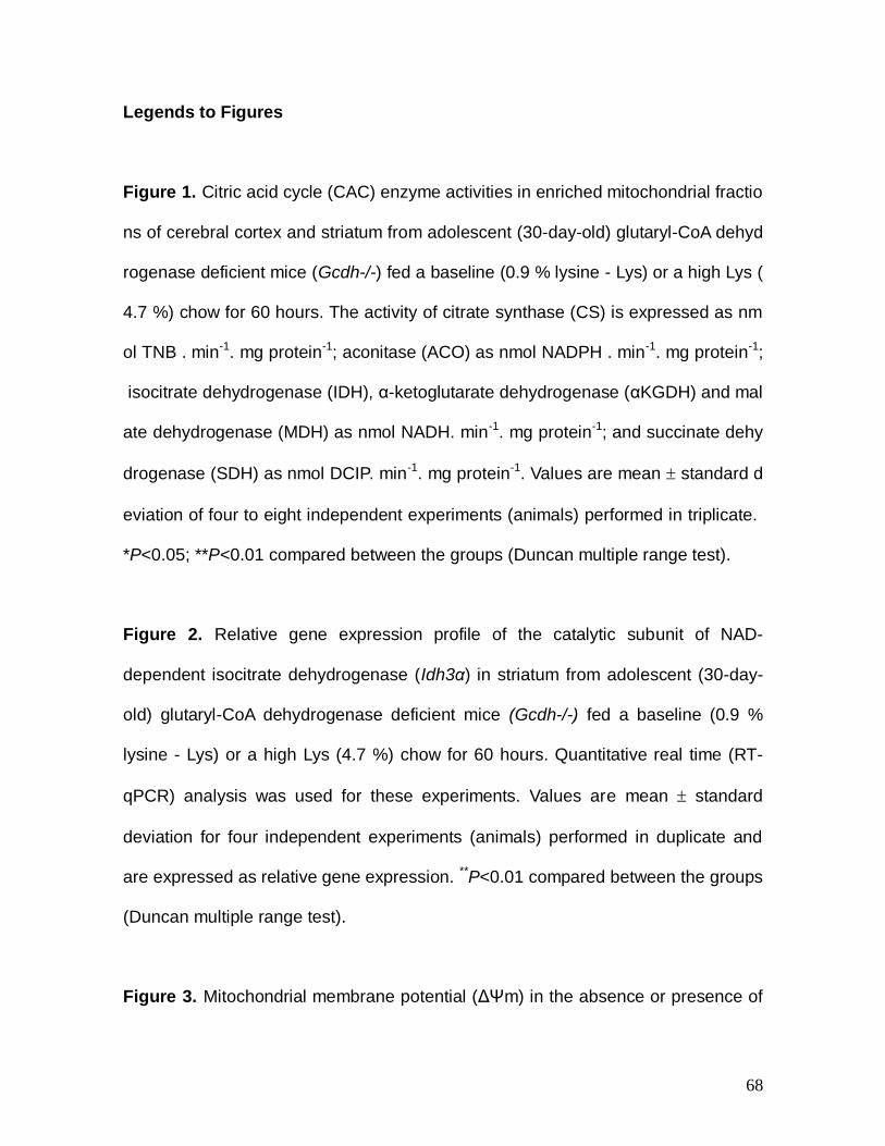
68
Legends to Figures
Figure 1. Citric acid cycle (CAC) enzyme activities in enriched mitochondrial fractio
ns of cerebral cortex and striatum from adolescent (30-day-old) glutaryl-CoA dehyd
rogenase deficient mice (Gcdh-/-) fed a baseline (0.9 % lysine - Lys) or a high Lys (
4.7 %) chow for 60 hours. The activity of citrate synthase (CS) is expressed as nm
ol TNB . min-1. mg protein-1; aconitase (ACO) as nmol NADPH . min-1. mg protein-1;
isocitrate dehydrogenase (IDH), α-ketoglutarate dehydrogenase (αKGDH) and mal
ate dehydrogenase (MDH) as nmol NADH. min-1. mg protein-1; and succinate dehy
drogenase (SDH) as nmol DCIP. min-1. mg protein-1. Values are mean standard d
eviation of four to eight independent experiments (animals) performed in triplicate.
*P<0.05; **P<0.01 compared between the groups (Duncan multiple range test).
Figure 2. Relative gene expression profile of the catalytic subunit of NAD-
dependent isocitrate dehydrogenase (Idh3α) in striatum from adolescent (30-day-
old) glutaryl-CoA dehydrogenase deficient mice (Gcdh-/-) fed a baseline (0.9 %
lysine - Lys) or a high Lys (4.7 %) chow for 60 hours. Quantitative real time (RT-
qPCR) analysis was used for these experiments. Values are mean standard
deviation for four independent experiments (animals) performed in duplicate and
are expressed as relative gene expression. **P<0.01 compared between the groups
(Duncan multiple range test).
Figure 3. Mitochondrial membrane potential (ΔΨm) in the absence or presence of
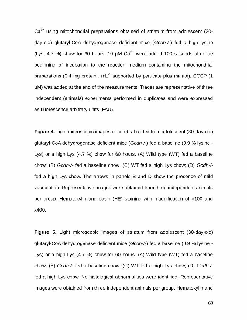
69
Ca2+ using mitochondrial preparations obtained of striatum from adolescent (30-
day-old) glutaryl-CoA dehydrogenase deficient mice (Gcdh-/-) fed a high lysine
(Lys; 4.7 %) chow for 60 hours. 10 µM Ca2+ were added 100 seconds after the
beginning of incubation to the reaction medium containing the mitochondrial
preparations (0.4 mg protein . mL-1 supported by pyruvate plus malate). CCCP (1
µM) was added at the end of the measurements. Traces are representative of three
independent (animals) experiments performed in duplicates and were expressed
as fluorescence arbitrary units (FAU).
Figure 4. Light microscopic images of cerebral cortex from adolescent (30-day-old)
glutaryl-CoA dehydrogenase deficient mice (Gcdh-/-) fed a baseline (0.9 % lysine -
Lys) or a high Lys (4.7 %) chow for 60 hours. (A) Wild type (WT) fed a baseline
chow; (B) Gcdh-/- fed a baseline chow; (C) WT fed a high Lys chow; (D) Gcdh-/-
fed a high Lys chow. The arrows in panels B and D show the presence of mild
vacuolation. Representative images were obtained from three independent animals
per group. Hematoxylin and eosin (HE) staining with magnification of ×100 and
x400.
Figure 5. Light microscopic images of striatum from adolescent (30-day-old)
glutaryl-CoA dehydrogenase deficient mice (Gcdh-/-) fed a baseline (0.9 % lysine -
Lys) or a high Lys (4.7 %) chow for 60 hours. (A) Wild type (WT) fed a baseline
chow; (B) Gcdh-/- fed a baseline chow; (C) WT fed a high Lys chow; (D) Gcdh-/-
fed a high Lys chow. No histological abnormalities were identified. Representative
images were obtained from three independent animals per group. Hematoxylin and
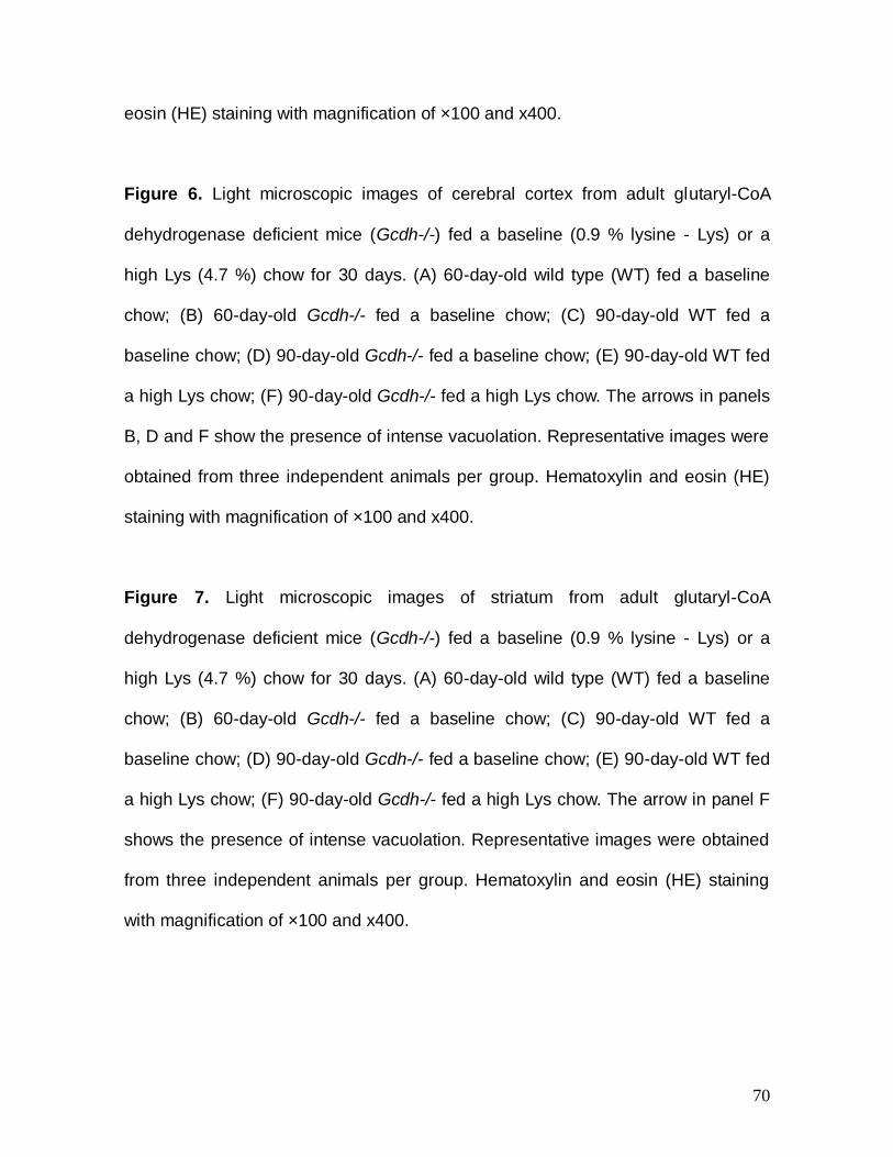
70
eosin (HE) staining with magnification of ×100 and x400.
Figure 6. Light microscopic images of cerebral cortex from adult glutaryl-CoA
dehydrogenase deficient mice (Gcdh-/-) fed a baseline (0.9 % lysine - Lys) or a
high Lys (4.7 %) chow for 30 days. (A) 60-day-old wild type (WT) fed a baseline
chow; (B) 60-day-old Gcdh-/- fed a baseline chow; (C) 90-day-old WT fed a
baseline chow; (D) 90-day-old Gcdh-/- fed a baseline chow; (E) 90-day-old WT fed
a high Lys chow; (F) 90-day-old Gcdh-/- fed a high Lys chow. The arrows in panels
B, D and F show the presence of intense vacuolation. Representative images were
obtained from three independent animals per group. Hematoxylin and eosin (HE)
staining with magnification of ×100 and x400.
Figure 7. Light microscopic images of striatum from adult glutaryl-CoA
dehydrogenase deficient mice (Gcdh-/-) fed a baseline (0.9 % lysine - Lys) or a
high Lys (4.7 %) chow for 30 days. (A) 60-day-old wild type (WT) fed a baseline
chow; (B) 60-day-old Gcdh-/- fed a baseline chow; (C) 90-day-old WT fed a
baseline chow; (D) 90-day-old Gcdh-/- fed a baseline chow; (E) 90-day-old WT fed
a high Lys chow; (F) 90-day-old Gcdh-/- fed a high Lys chow. The arrow in panel F
shows the presence of intense vacuolation. Representative images were obtained
from three independent animals per group. Hematoxylin and eosin (HE) staining
with magnification of ×100 and x400.
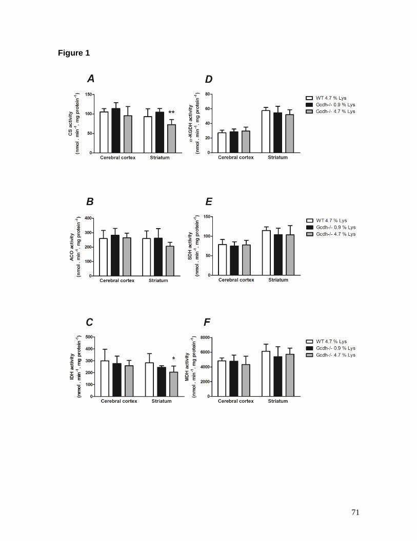
71
Figure 1
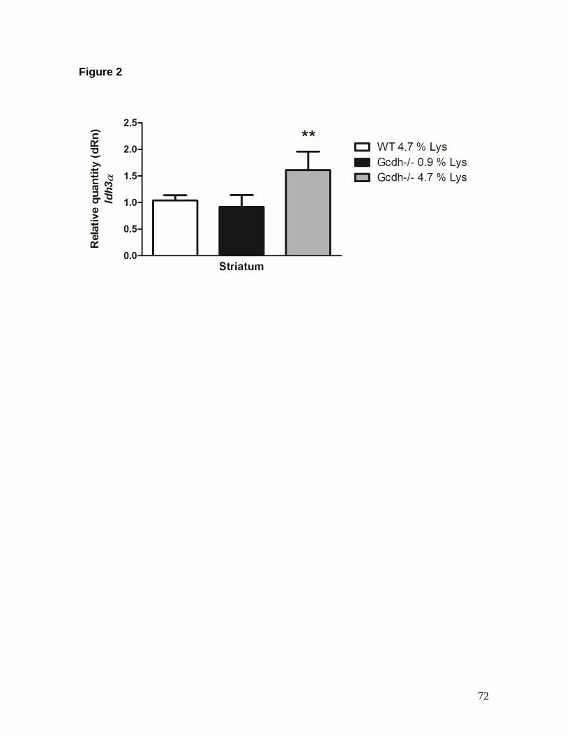
72
Figure 2
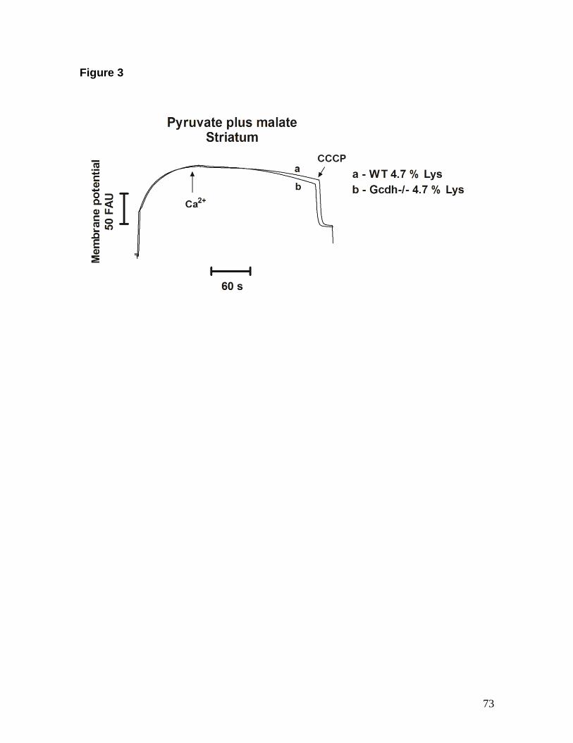
73
Figure 3
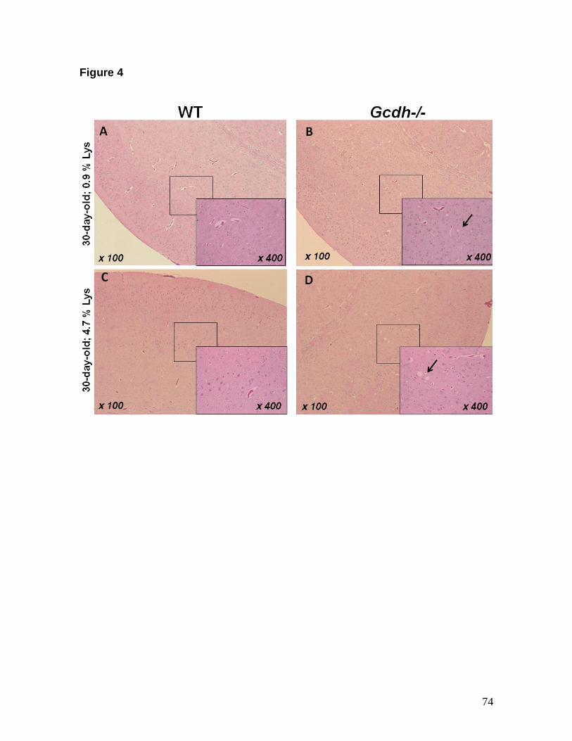
74
Figure 4
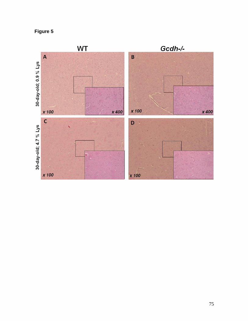
75
Figure 5
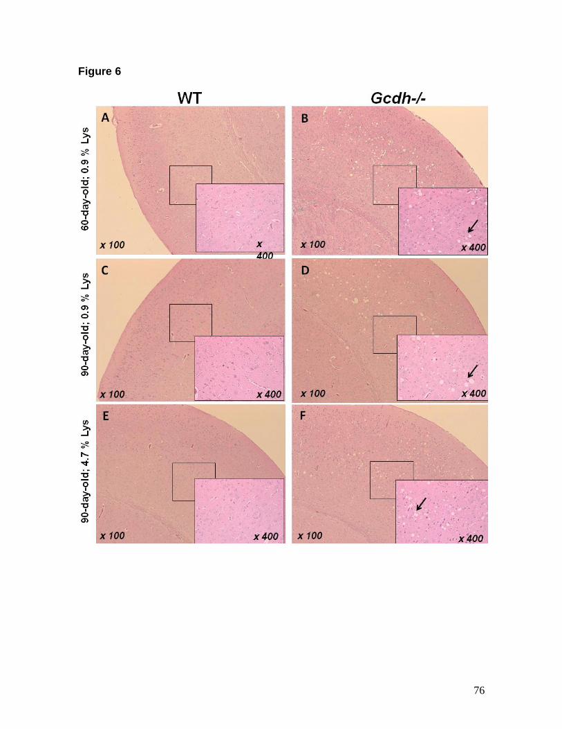
76
Figure 6
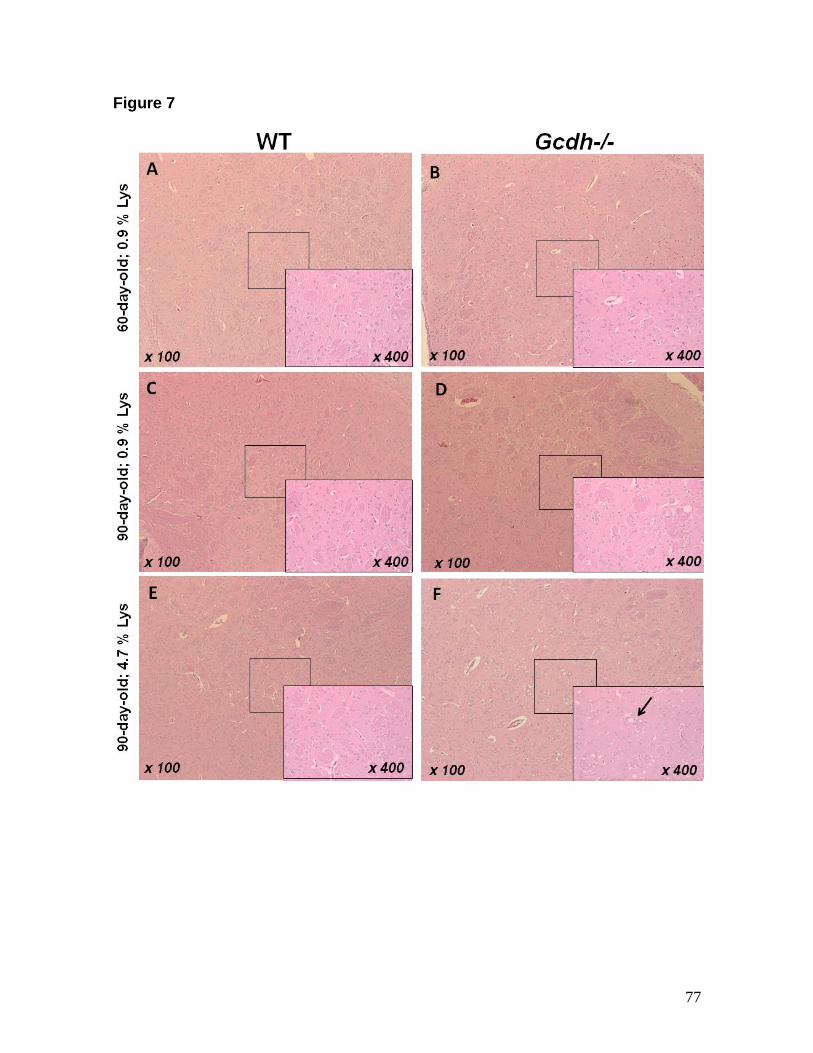
77
Figure 7
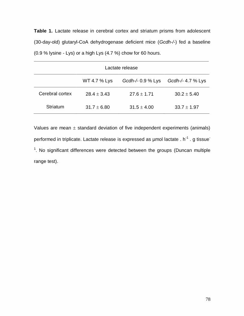
78
Table 1. Lactate release in cerebral cortex and striatum prisms from adolescent
(30-day-old) glutaryl-CoA dehydrogenase deficient mice (Gcdh-/-) fed a baseline
(0.9 % lysine - Lys) or a high Lys (4.7 %) chow for 60 hours.
Lactate release
WT 4.7 % Lys Gcdh-/- 0.9 % Lys Gcdh-/- 4.7 % Lys
Cerebral cortex 28.4 3.43 27.6 1.71 30.2 5.40
Striatum 31.7 6.80 31.5 4.00 33.7 1.97
Values are mean standard deviation of five independent experiments (animals)
performed in triplicate. Lactate release is expressed as µmol lactate . h-1 . g tissue-
1. No significant differences were detected between the groups (Duncan multiple
range test).
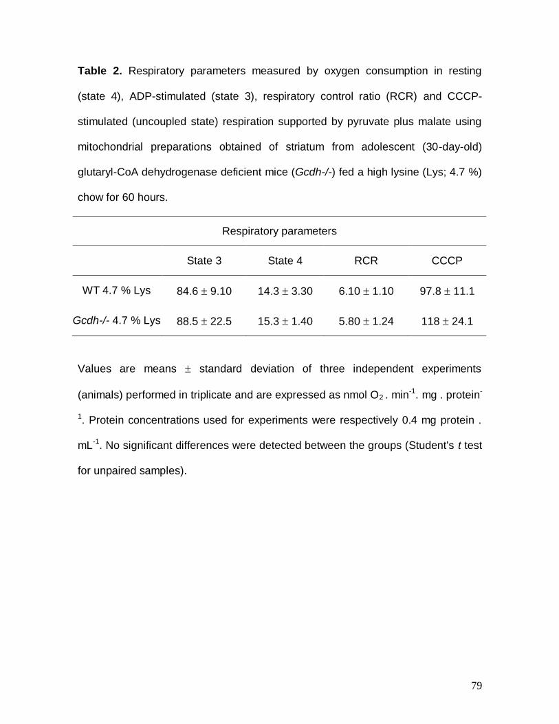
79
Table 2. Respiratory parameters measured by oxygen consumption in resting
(state 4), ADP-stimulated (state 3), respiratory control ratio (RCR) and CCCP-
stimulated (uncoupled state) respiration supported by pyruvate plus malate using
mitochondrial preparations obtained of striatum from adolescent (30-day-old)
glutaryl-CoA dehydrogenase deficient mice (Gcdh-/-) fed a high lysine (Lys; 4.7 %)
chow for 60 hours.
Respiratory parameters
State 3 State 4 RCR CCCP
WT 4.7 % Lys 84.6 9.10 14.3 3.30 6.10 1.10 97.8 11.1
Gcdh-/- 4.7 % Lys 88.5 22.5 15.3 1.40 5.80 1.24 118 24.1
Values are means standard deviation of three independent experiments
(animals) performed in triplicate and are expressed as nmol O2 . min-1. mg . protein-
1. Protein concentrations used for experiments were respectively 0.4 mg protein .
mL-1. No significant differences were detected between the groups (Student's t test
for unpaired samples).

80
PARTE III
Discussão e Conclusões
81
III.1. DISCUSSÃO
Pacientes com AG I apresentam caracteristicamente destruição estriatal
aguda que ocorre durante as crises encefalopáticas, bem como leucoencefalopatia
progressiva que abrange principalmente o córtex cerebral. Embora a etiopatogenia
do dano cerebral nessa doença não esteja bem esclarecida, várias hipóteses tem
sido levantadas para explicar as manifestações neurológicas dessa doença,
incluindo excitotoxicidade, estresse oxidativo, dano na barreira hematoencefálica e
alterações da produção de energia (Latini et al., 2007; Strauss e Morton, 2003;
Wajner et al., 2004). Neste particular, um comprometimento importante da
homeostase bioenergética tem sido proposto como um mecanismo fundamental
da injúria cerebral apresentada pelos pacientes acometidos por essa doença
(Kolker et al., 2004; Strauss e Morton, 2003). No entanto, essa hipótese ainda não
está comprovada, visto que esses estudos experimentais realizados in vitro e in
vivo em cérebro e cultura de células neurais demonstraram efeitos moderados dos
principais metabólitos acumulados nesta doença (AG e 3HG) sobre parâmetros da
função mitocondrial (Das et al., 2003; Ferreira et al., 2005a; Ferreira et al., 2005b;
Ferreira et al., 2007b; Kolker et al., 2002a; Kolker et al., 2002b; Latini et al., 2005a;
Silva et al., 2000; Ullrich et al., 1999). Além disso, quase todos os estudos prévios
foram feitos em tecidos de animais com atividade normal da enzima GCDH. O
desenvolvimento de camundongos geneticamente modificados com atividade nula
da GCDH (Gcdh-/-) reproduziu o fenótipo bioquímico e neuropatológico dos
pacientes, especialmente a leucoencefalopatia (Koeller et al., 2002), se tornando
um bom modelo animal para o estudo da AG I. Esse modelo foi mais tarde
aperfeiçoado pela suplementação de uma dieta rica em lisina (4,7 %) aos animais
82
Gcdh-/-, resultando em um aumento nas concentrações teciduais de AG e 3HG,
além de provocar o aparecimento de lesões no estriado desses animais,
semelhantes as que ocorrem em seres humanos afetados pela AG I (Zinnanti et
al., 2006).
A presente tese foi desenvolvida utilizando cérebro, músculo esquelético e
coração de camundongos WT e Gcdh-/- com 15 e 30 dias de vida no intuito de
investigar a homeostase energética nesses animais submetidos a uma sobrecarga
de lisina através de uma injeção aguda intraperitoneal de lisina (8 µmol/g) em
camundongos com 15 dias de vida, bem como por uma dieta rica em lisina (4,7 %)
por 60 horas para camundongos com 30 dias de vida. Foram avaliados os
seguintes parâmetros da bioenergética celular: atividades dos complexos I-III, II, II-
III e IV da cadeia respiratória, os parâmetros respiratórios (estados 3 e 4, RCR e o
estado desacoplado), atividades das enzimas do CAC CS, ACO, IDH, α-CGDH,
SDH e MDH, a liberação de lactato e o potencial de membrana mitocondrial com
ou sem a adição exógena de Ca2+, assim como as atividades da CK e da Na+, K+ -
ATPase. Estudos histológicos também foram feitos para determinar alterações na
arquitetura do córtex cerebral e estriado dos camundongos WT e Gcdh-/- em
diferentes idades (30 a 90 dias) submetidos por um curto (60 horas) ou longo (30
dias) período com dieta com alta concentração de lisina (4,7 %).
Determinamos inicialmente as atividades dos complexos da cadeia
respiratória e da α-CGDH em cérebro, coração e músculo esquelético de
camundongos WT e Gcdh-/- com 15 dias de vida submetidos a uma injeção
intraperitoneal de solução salina (NaCl 0,9 %) ou lisina (8 µmol/g). Também
investigamos a influência de uma dieta com alta concentração de lisina (4,7 %) por
83
60 horas em camundongos WT e Gcdh-/- com 30 dias de vida sobre as mesmas
medidas em córtex cerebral, estriado e hipocampo desses animais, bem como
sobre os parâmetros respiratórios estados 3 e 4, RCR e estado desacoplado. Com
relação aos animais Gcdh-/- com 15 dias de vida que receberam uma injeção
intraperitoneal de solução salina, não verificamos alteração em nenhum dos
parâmetros de bioenergética acima mencionados quando comparados aos
animais WT. Entretanto observamos um aumento na atividade do complexo II-III
em músculo esquelético e coração, mas não em cérebro total dos camundongos
Gcdh-/- submetidos à administração aguda de lisina.
Já nos camundongos Gcdh-/- de 30 dias de vida com dieta normal (0,9 %
de lisina), encontramos pequenas alterações na atividade de alguns complexos da
cadeia respiratória em córtex cerebral (I-III e IV) e hipocampo (II e IV). No entanto,
os parâmetros respiratórios (estados 3 e 4, RCR e estado desacoplado) não foram
alterados em mitocôndrias isoladas de cérebro total utilizando glutamato/malato ou
sucinato como substratos. Observamos também uma diminuição na atividade dos
complexos II e II-III no músculo esquelético desses animais. Nossos resultados
indicam que as pequenas alterações na atividade da cadeia transportadora de
elétrons não foram suficientes para comprometer o consumo de oxigênio no
estado não fosforilante (estado 4) e fosforilante (estado 3), bem como no estado
desacoplado no cérebro dos animais nocautes. Sabe-se que uma inibição
importante da atividade da cadeia transportadora de elétrons é necessária para
alterar a respiração mitocondrial (Brand e Nicholls, 2011). Finalmente, verificamos
também que uma sobrecarga de lisina na dieta não acentuou significativamente as
diferenças observadas.
84
Resultados anteriores mostraram que os complexos isolados da cadeia
respiratória não estão alterados em cérebro de camundongos Gcdh-/- submetidos
a uma dieta normal, o que reforça a hipótese de que não há um comprometimento
importante da cadeia respiratória nesses animais (Sauer et al., 2005). Esses
autores verificaram, no entanto, que o glutaril-CoA inibe significativamente a
atividade da α-CGDH purificada comercial. Em nossos ensaios realizados com
homogeneizados e com mitocôndrias purificadas de cérebro total, córtex cerebral,
estriado e hipocampo dos camundongos Gcdh-/- com 15 e 30 dias de vida
submetidos ou não a uma sobrecarga de lisina não identificamos qualquer inibição
desse complexo enzimático. Assim, é possível que em nosso modelo animal in
vivo concentrações intracerebrais de glutaril-CoA foram menores do que as
testadas por Sauer e colaboradores (2005) e necessárias para causar a inibição in
vitro observada por esses pesquisadores.
O próximo passo de nossa investigação foi o de verificar a função do CAC
através da determinação da atividade das enzimas CS, ACO, IDH, SDH e MDH,
bem como a liberação de lactato, os parâmetros respiratórios estados 3 e 4, RCR
e estado desacoplado, e o potencial de membrana em preparações mitocondriais
obtidas de córtex cerebral e estriado de camundongos Gcdh-/- de 30 dias de vida
submetidos a uma dieta padrão (0,9 % lisina) ou rica em lisina (4,7 %) por 60
horas.
Observamos uma diminuição moderada nas atividades da CS e IDH em
mitocôndrias de estriado de camundongos Gcdh-/- submetidos a uma dieta rica
em lisina. Considerando a importância dessas enzimas, podemos presumir que o
funcionamento do CAC está parcialmente prejudicado. Verificamos também um
85
pequeno aumento na liberação de lactato em córtex cerebral e estriado dos
camundongos Gcdh-/- suplementados com uma dieta rica em lisina.
Também encontramos um aumento acentuado na expressão gênica da
subunidade catalítica da IDH (Idh3α) no estriado de camundongos Gcdh-/-
expostos à dieta rica em lisina, não correlacionando com a atividade diminuída
dessa enzima que encontramos no estriado desses animais. Neste particular, está
descrito na literatura que a expressão do gene que codifica uma enzima não
necessariamente se correlaciona com a sua atividade (de Sousa Abreu et al.,
2009; Griffin et al., 2002; Gygi et al., 1999; Tian et al., 2004), a qual pode ser
dependente de alterações pós-transcricionais e muitas vezes existir uma diferença
na taxa de conversão de mRNA em proteína (Cox et al., 2005; Hack, 2004). Assim,
a expressão aumentada da Idh3α pode ocorrer como um mecanismo
compensatório em resposta à inibição da atividade da IDH.
Esses resultados estão possivelmente relacionados com um estudo prévio
que demonstrou uma diminuição nas concentrações de ATP, CoA, α-cetoglutarato,
glutamato e GABA, assim como com um aumento nos níveis de acetil-CoA, em
cérebro de camundongos Gcdh-/- submetidos a uma dieta com alta concentração
de lisina (Zinnanti et al., 2007). Um outro estudo também demonstrou um bloqueio
no transporte de sucinato dos astrócitos para os neurônios em culturas primárias
de animais Gcdh-/- (Lamp et al., 2011), impedindo a reposição de intermediários
do CAC para os neurônios.
Além disso, o fato de que a redução nas atividades da CS e IDH foi
encontrada quando os camundongos Gcdh-/- foram submetidos a uma sobrecarga
de lisina faz com que seja razoável associar esses efeitos ao aumento da
86
concentração de AG e 3HG que são derivados da lisina no cérebro desses
animais ( Zinnanti et al., 2006; Zinnanti et al., 2007).
No intuito de avaliar outros parâmetros da bioenergética mitocondrial no
estriado desses animais, medimos os estados 3 e 4, RCR e estado desacoplado
da respiração mitocondrial pelo consumo de oxigênio. Não verificamos alterações
significativas em nenhum dos parâmetros respiratórios quando foram comparadas
preparações mitocondriais obtidas de estriado de camundongos WT e Gcdh-/-
expostos a uma dieta rica em lisina e utilizando piruvato/malato como substratos
respiratórios. Esses resultados indicam que os substratos estão sendo
suficientemente oxidados no CAC para abastecer a respiração mitocondrial. No
entanto, como mencionado anteriormente, alterações leves na atividade de
enzimas do CAC e da cadeia respiratória não são suficientes para causar
alterações nos diferentes estados da respiração mitocondrial (Brand e Nicholls,
2011).
Observamos também que o potencial de membrana mitocondrial não se
alterou em preparações mitocondriais de estriado de camundongos Gcdh-/-
submetidos a uma sobrecarga de lisina, mesmo com suplementação exógena de
Ca2+. Esses dados sugerem que a função mitocondrial de formação e sustentação
do potencial de membrana e a capacidade de tamponamento de Ca2+ não foram
alteradas nesses animais.
Nosso estudo também demonstrou uma atividade reduzida da CK em
cérebro e músculo esquelético de camundongos Gcdh-/- com 15 dias de vida
injetados agudamente com lisina, indicando um comprometimento no processo de
transferência e tamponamento intracelular de ATP. Estudos anteriores mostraram
87
um efeito inibitório causado pelo AG in vitro na atividade da CK em cérebro
(Ferreira et al., 2005b) e in vivo em músculo esquelético de ratos (Ferreira et al.,
2007b). Essas observações corroboram com nossos achados presentes e
anteriores, demonstrando uma diminuição nos níveis de ATP e fosfocreatina em
camundongos Gcdh-/- expostos a uma sobrecarga dietética de lisina (Zinnanti et
al., 2007). Tendo em vista que a administração intraperitoneal de lisina resulta no
aumento das concentrações cerebrais de AG e 3HG no cérebro de camundongos
Gcdh-/- (Seminotti et al., 2012), é razoável postular que os efeitos encontrados em
nossa investigação sejam decorrentes do acúmulo tecidual desses ácidos
orgânicos. O significado desses achados é ainda desconhecido, mas
possivelmente pode estar ligado à disfunção neurológica e à hipotonia que
acomete os pacientes com AG I.
Um aspecto interessante de nossa investigação foi que não encontramos
inibição da CK no cérebro de animais Gcdh-/- de mais idade (30 dias) quando
submetidos à dieta rica em lisina por 60 horas. Neste particular, é possível que a
barreira hematoencefálica esteja menos permeável à passagem da lisina em
animais de 30 dias de vida com posterior conversão no cérebro em AG e 3HG
(Zinnanti et al., 2007).
Possivelmente, o achado mais importante do presente estudo foi uma
diminuição significativa da atividade da Na+, K+ - ATPase em membranas
sinápticas de cérebro dos camundongos Gcdh-/- com 15 e 30 dias de vida.
Verificamos ainda que essa inibição ocorreu no córtex cerebral, mas não em
estriado e hipocampo dos camundongos Gcdh-/- com 30 dias de vida. Esses
resultados corroboram com trabalhos anteriores que demonstram uma inibição
88
dessa atividade enzimática pelo AG in vitro (Kolker et al., 2002b) e in vivo (Fighera
et al., 2006; Rodrigues et al., 2013).
Com relação ao mecanismo pelo qual a CK e a Na+, K+ - ATPase estão
inibidas, sabe-se que essas enzimas são suscetíveis ao ataque oxidativo (Konorev
et al., 1998; Kurella et al., 1997; Lees, 1993; Stachowiak et al., 1998; Wallimann et
al., 1998). Neste contexto, trabalhos anteriores mostraram indução de estresse
oxidativo em cérebro de camundongos Gcdh-/- submetidos a uma sobrecarga de
lisina (Seminotti et al., 2012; Seminotti et al., 2013) e que o AG e 3HG induzem
estresse oxidativo in vitro (de Oliveira Marques et al., 2003; Kolker et al., 2001;
Latini et al., 2002; Latini et al., 2005b) e in vivo (Latini et al., 2007).
Alternativamente, a inibição da Na+, K+ - ATPase pode estar relacionada a
uma reduzida expressão gênica de alguma de suas subunidades. Neste particular,
encontramos uma diminuição na expressão da subunidade catalítica α2 da Na+, K+
- ATPase em córtex cerebral de animais Gcdh-/- com 30 dias de vida, sem
qualquer alteração nas isoformas α1 e α3. Esses resultados indicam que
possivelmente a diminuição da expressão gênica da isoforma α2 dessa
subunidade esteja relacionada com a inibição da atividade da Na+, K+ - ATPase. É
importante ressaltar que a isoforma α2 é bastante expressa em cérebro,
particularmente nos astrócitos (Brines e Robbins, 1993; Cameron et al., 1994;
Chen et al., 2013; Kawakami e Ikeda, 2006), e que a atividade da Na+, K+ -
ATPase nessas células auxilia na recaptação do glutamato liberado na fenda
sináptica (Rose et al., 2009). Neste contexto, um modelo de camundongos
nocaute para a subunidade α2 da Na+, K+ - ATPase revelou haver um prejuízo na
captação de glutamato, culminando numa alta mortalidade dos animais (Kawakami
89
e Ikeda, 2006). Dessa forma, nossos resultados mostrando uma redução na
expressão dessa isoforma e redução da atividade da Na+, K+ - ATPase pode
comprometer a captação do glutamato da fenda sináptica pelos astrócitos,
resultando em aumento extracelular desse neurotransmissor, levando a um
quadro de excitotoxicidade (Veldhuis et al., 2003).
Portanto, a inibição da atividade da Na+, K+ - ATPase cerebral pode levar à
excitotoxicidade secundária e ser um fator importante no dano cerebral
característico dos pacientes com AG I. Além disso, estudos anteriores mostram
uma possível interação dos AG e 3HG com receptores e transportadores
glutamatérgicos em culturas de células e cérebro de ratos (Bjugstad et al., 2001;
de Mello et al., 2001; Flott-Rahmel et al., 1997; Kolker et al., 1999; Kolker et al.,
2000; Kolker et al., 2002a; Kolker et al., 2002b; Porciuncula et al., 2000;
Porciuncula et al., 2004; Rosa et al., 2004; Wajner et al., 2004), o que indica que a
excitotoxicidade pode de fato representar um mecanismo patogênico na AG I.
Também não podemos desconsiderar o papel fundamental da Na+, K+ -
ATPase para o cérebro na manutenção do potencial de membrana da célula no
controle dos fluxos de sódio e potássio, mantendo a excitabilidade neuronal
(neurotransmissão) e controlando o volume celular. Aproximadamente 50 % do
ATP consumido pelo cérebro é gasto na atividade dessa enzima, o que implica em
sua importância para o funcionamento do SNC (Erecinska et al., 2004; Erecinska e
Silver, 1994; Satoh e Nakazato, 1992). Neste contexto, inibições da Na+, K+ -
ATPase tem sido associadas a diversas doenças neurodegenerativas e também
em doenças metabólicas hereditárias (Bagh et al., 2008; Busanello et al., 2011;
90
Cousin et al., 1995; Ellis et al., 2003; Lees e Leong, 1995; Moura et al., 2012;
Vignini et al., 2007).
A última etapa deste trabalho foi investigar anormalidades histológicas em
cérebro de camundongos Gcdh-/- com 30, 60 e 90 dias de vida submetidos a
diferentes períodos (60 horas ou 30 dias) com dieta rica em lisina. Observamos a
presença de alguns vacúolos no córtex cerebral, mas não no estriado de
camundongos Gcdh-/- com 30 dias de vida submetidos a uma dieta normal ou rica
em lisina por 60 horas. No entanto, animais Gcdh-/- com 60 e 90 dias de vida em
dieta normal apresentaram uma intensa vacuoalização no córtex cerebral. Além
disso, verificamos a presença de um grande número de vacúolos no estriado dos
camundongos Gcdh-/- com 90 dias de vida que foram alimentados por 30 dias
com uma dieta rica em lisina, enquanto que a vacuoalização cortical não foi
aumentada. Alguns autores demonstraram a presença de vacuolização em
estudos postmortem de cérebro de pacientes afetados pela AG I (Bergman et al.,
1989; Forstner et al., 1999; Goodman et al., 1977; Hoffmann e Zschocke, 1999;
Soffer et al., 1992).
Nossos resultados corroboram com um estudo anterior que descreve
pequenas alterações microscópicas no cérebro de camundongos Gcdh-/- jovens
expostos por um curto período (60 horas) a dieta com alta concentração de lisina,
bem como importantes alterações histológicas com uma pequena perda neuronal
e ativação astrocitária em córtex cerebral e estriado de camundongos Gcdh-/-
adultos tratados com sobrecarga de lisina na dieta por pelo menos 45 dias
(Zinnanti et al., 2006). Portanto, nossos dados indicam que o estriado dos
camundongos Gcdh-/- pode estar sendo progressivamente lesado pelo aumento
91
persistente dos metabólitos acumulados na AG I, particularmente o AG. Neste
sentido, um estudo recente observou uma degeneração estriatal tardia em ratos
tratados com AG no primeiro dia de vida (Olivera-Bravo et al., 2011).
Caso nossos resultados pudessem ser extrapolados para a condição
humana de AG I, acreditamos que as alterações leves da homeostase energética
mitocondrial aqui detectadas não desempenham um papel decisivo para o dano
neurológico severo que acomete principalmente o córtex cerebral e o estriado de
pacientes com AG I. Não podemos, no entanto, excluir que esse mecanismo,
associado a outros, como o estresse oxidativo e a excitotoxidade que são
deletérios para o SNC, possam contribuir para explicar a fisiopatogenia da injúria
cerebral na AG I. Por outro lado, considerando a importância da CK e Na+, K+-
ATPase para a transferência energética intracelular e manutenção do potencial de
membrana necessário para a neurotransmissão, respectivamente, inibições
dessas atividades enzimáticas podem representar mecanismos responsáveis pelo
dano cerebral, principalmente cortical, e pelas manifestações neurológicas
apresentadas pelos pacientes afetados pela AG I.
Finalmente, nossos achados histológicos claramente mostram que apenas
o estriado de camundongos Gcdh-/- adultos foi lesado após uma sobrecarga de
lisina por um longo período (30 dias), enquanto que as lesões encontradas no
córtex cerebral parecem não ser acentuadas por esse tratamento. Esses achados
estão de acordo com estudos neurorradiológicos feitos em pacientes com AG I,
demonstrando que lesão estriatal crônica também acomete pacientes com essa
doença (Neumaier-Probst et al., 2004).
92
III.2. CONCLUSÕES
Observamos pequenas alterações nos complexos da cadeia respiratória
em cérebro, músculo e coração de camundongos Gcdh-/- com 15 e 30 dias de
vida submetidos ou não a uma sobrecarga de lisina através de uma injeção
intraperitoneal desse aminoácido ou por uma sobrecarga de lisina na dieta;
entretanto essas alterações não foram suficientes para comprometer os
parâmetros respiratórios medidos pelo consumo de oxigênio (estado 3, estado 4,
RCR e estado desacoplado) em mitocôndrias de cérebro total de camundongos
Gcdh-/- com 30 dias de vida utilizando glutamato/malato ou sucinato como
substrato.
Verificamos uma redução moderada na atividade das enzimas CS e IDH
em mitocôndrias de estriado de camundongos Gcdh-/- com 30 dias de vida
submetidos a uma dieta rica em lisina associada com um pequeno aumento na
liberação de lactato.
A respiração mitocondrial avaliada pelos parâmetros respiratórios e o
potencial de membrana na presença ou ausência de Ca2+ permaneceram normais
em mitocôndrias de estriado de camundongos Gcdh-/- com 30 dias de vida
submetidos a uma dieta rica em lisina, utilizando piruvato/malato como substratos
respiratórios.
Verificamos uma atividade reduzida da CK em cérebro e músculo
esquelético de camundongos Gcdh-/- com 15 dias de vida injetados agudamente
com lisina, indicando um comprometimento no processo de transferência e
tamponamento intracelular de ATP.
93
Uma diminuição significativa da atividade da Na+, K+ - ATPase também foi
demonstrada em membranas sinápticas de cérebro dos camundongos Gcdh-/-
com 15 e 30 dias de vida. Verificamos ainda que essa inibição ocorreu no córtex
cerebral dos camundongos Gcdh-/- com 30 dias de vida e estava associada a uma
reduzida expressão da subunidade catalítica α2 da Na+, K+ - ATPase.
Finalmente, observamos a presença de vacuoalização no córtex cerebral
de camundongos Gcdh-/- que foi mais intensa em animais adultos (60 e 90 dias de
vida) e independentes de tratamento com lisina. Por outro lado, verificamos uma
intensa vacuoalização no estriado desses animais apenas quando camundongos
Gcdh-/- de 90 dias de vida foram alimentados por 30 dias com dieta rica em lisina.
Acreditamos que um comprometimento da homeostase energética
mitocondrial não contribua significativamente para o dano neurológico observado
nos animais Gcdh-/-.
Por outro lado, as inibições encontradas das atividades da CK e Na+, K+-
ATPase nos camundongos Gcdh-/- podem implicar que o dano cerebral observado
nesses animais e possivelmente nos pacientes com AG I, pode ser ao menos
parcialmente devido a essas alterações, levando-se em consideração a
importância dessas enzimas para a transferência energética intracelular e a
manutenção do potencial de membrana necessário para a neurotransmissão.
III.3. PERSPECTIVAS
Avaliar as atividades da cadeia respiratória, CK e Na+, K+-ATPase, bem
como os parâmetros respiratórios estado 3, estado 4, RCR e estado desacoplado
94
em cérebro (córtex cerebral e estriado) de camundongos WT e Gcdh-/- com 30, 60
e 90 dias de vida submetidos a longos períodos (30, 45 e 60 dias) com dieta rica
em lisina.
Avaliar o potencial de membrana, inchamento, conteúdo de NAD(P)H e a
capacidade de retenção de Ca2+ em preparações mitocondriais de cérebro (córtex
cerebral e estriado) de camundongos WT e Gcdh-/- com 30, 60 e 90 dias de vida,
na presença ou ausência de Ca2+, submetidos a longos períodos (30, 45 e 60 dias)
com dieta rica em lisina.
Fazer estudos de imunoistoquímica (anti-S100β, anti-GFAP, anti-NeuN) e
imunoblotting (anti-α-sinucleína, anti-sinaptofisina, anti-caspase-3) para melhor
avaliar as alterações histológicas observadas no cérebro dos camundongos Gcdh-
/- com 30, 60 e 90 dias de vida submetidos a curto (60 horas) e longos períodos
(30, 45 e 60 dias) com dieta rica em lisina.
Buscar a prevenção das alterações bioquímicas e histológicas
encontradas nos camundongos Gcdh-/- com substâncias neuroprotetoras
(gangliosídeo GM1, guanosina), antioxidantes (N-acetilcisteína e melatonina),
substratos energéticos (creatina) e moduladores do sistema glutamatérgico (MK-
801).

95
REFERÊNCIAS BIBLIOGRÁFICAS
Amir, N., el-Peleg, O., Shalev, R.S., Christensen, E., Glutaric aciduria type I: clinical heterogeneity and neuroradiologic features. Neurology 37, 1654-1657, 1987.
Aperia, A., New roles for an old enzyme: Na,K-ATPase emerges as an interesting drug target. J. Intern. Med. 261, 44-52, 2007.
Attwell, D., Laughlin, S.B., An energy budget for signaling in the grey matter of the brain. J. Cereb. Blood Flow Metab. 21, 1133-1145, 2001.
Bagh, M.B., Maiti, A.K., Jana, S., Banerjee, K., Roy, A., Chakrabarti, S., Quinone and oxyradical scavenging properties of N-acetylcysteine prevent dopamine mediated inhibition of Na+, K+-ATPase and mitochondrial electron transport chain activity in rat brain: implications in the neuroprotective therapy of Parkinson's disease. Free Radic. Res. 42, 574-581, 2008.
Baric, I., Fumic, K., Hoffmann, G.F., Inborn errors of metabolism at the turn of the millennium. Croatian Medical Journal 42, 379-383, 2001.
Baric, I., Zschocke, J., Christensen, E., Duran, M., Goodman, S.I., Leonard, J.V., Muller, E., Morton, D.H., Superti-Furga, A., Hoffmann, G.F., Diagnosis and management of glutaric aciduria type I. J Inherit Metab Dis 21, 326-340, 1998.
Beal, M., Aging, energy, and oxidative stress in neurodegenerative diseases. Ann Neurol 38, 357-366, 1995.
Bergman, I., Finegold, D., Gartner, J.C., Jr., Zitelli, B.J., Claassen, D., Scarano, J., Roe, C.R., Stanley, C., Goodman, S.I., Acute profound dystonia in infants with glutaric acidemia. Pediatrics 83, 228-234, 1989.
Bickel, H., Early diagnosis and treatment of inborn errors of metabolism. Enzyme 38, 14-26, 1987.
Bjugstad, K.B., Zawada, W.M., Goodman, S., Freed, C.R., IGF-1 and bFGF reduce glutaric acid and 3-hydroxyglutaric acid toxicity in striatal cultures. Journal of Inherited Metabolic Disease 24, 631-647, 2001.
Blanco, G., Mercer, R.W., Isozymes of the Na-K-ATPase: heterogeneity in structure, diversity in function. Am J Physiol 275, F633-650, 1998.
Bobba, A., Amadoro, G., Valenti, D., Corsetti, V., Lassandro, R., Atlante, A., Mitochondrial respiratory chain Complexes I and IV are impaired by beta-amyloid
96
via direct interaction and through Complex I-dependent ROS production, respectively. Mitochondrion 13, 298-311, 2013.
Bodamer, O.A., Gruber, S., Stockler-Ipsiroglu, S., Nuclear magnetic resonance spectroscopy in glutaryl-CoA dehydrogenase deficiency. J Inherit Metab Dis 27, 877-883, 2004.
Boehm, E., Radda, G., Tomlin, H., Clark, J., The utilisation of creatine and its analogues by cytosolic and mitochondrial creatine kinase. Biochim Biophys Acta 1274, 119-128, 1996.
Bowling, A., Beal, M., Bioenergetic and oxidative stress in neurodegenerative diseases. Life Sci 56, 1151-1171, 1995.
Brand, M.D., Nicholls, D.G., Assessing mitochondrial dysfunction in cells. Biochem J 435, 297-312, 2011.
Brines, M.L., Robbins, R.J., Cell-type specific expression of Na+, K(+)-ATPase catalytic subunits in cultured neurons and glia: evidence for polarized distribution in neurons. Brain Res 631, 1-11, 1993.
Brismar, J., Ozand, P.T., CT and MR of the brain in glutaric acidemia type I: a review of 59 published cases and a report of 5 new patients. AJNR: American Journal of Neuroradiology 16, 675-683, 1995.
Burlina, A.P., Zara, G., Hoffmann, G.F., Zschocke, J., Burlina, A.B., Management of movement disorders in glutaryl-CoA dehydrogenase deficiency: anticholinergic drugs and botulinum toxin as additional therapeutic options. J Inherit Metab Dis 27, 911-915, 2004.
Busanello, E.N., Viegas, C.M., Tonin, A.M., Grings, M., Moura, A.P., de Oliveira, A.B., Eichler, P., Wajner, M., Neurochemical evidence that pristanic acid impairs energy production and inhibits synaptic Na(+), K(+)-ATPase activity in brain of young rats. Neurochem Res 36, 1101-1107, 2011.
Busquets, C., Soriano, M., de Almeida, I.T., Garavaglia, B., Rimoldi, M., Rivera, I., Uziel, G., Cabral, A., Coll, M.J., Ribes, A., Mutation analysis of the GCDH gene in Italian and Portuguese patients with glutaric aciduria type I. Mol Genet Metab 71, 535-537, 2000.
Cadenas, E., Davies, K.J., Mitochondrial free radical generation, oxidative stress, and aging. Free Radical Biology and Medicine 29, 222-230, 2000.
Cameron, R., Klein, L., Shyjan, A.W., Rakic, P., Levenson, R., Neurons and astroglia express distinct subsets of Na,K-ATPase alpha and beta subunits. Brain Res Mol Brain Res 21, 333-343, 1994.
97
Camins, A., Pallas, M., Silvestre, J.S., Apoptotic mechanisms involved in neurodegenerative diseases: experimental and therapeutic approaches. Methods Find. Exp. Clin. Pharmacol. 30, 43-65, 2008.
Chalmers, R.A., Lawson, A.M., Organic acids in man. Analytical chemistry, biochemistry and diagnosis of the organic acidurias. Chapman & Hall, London. 1982.
Chalmers, R.A., Bain, M.D., Zschocke, J., Riboflavin-responsive glutaryl CoA dehydrogenase deficiency. Mol Genet Metab 88, 29-37, 2006.
Chaturvedi, R.K., Flint Beal, M., Mitochondrial diseases of the brain. Free Radic. Biol. Med. 63, 1-29, 2013.
Chen, X.L., Wee, N.L., Hiong, K.C., Ong, J.L., Chng, Y.R., Ching, B., Wong, W.P., Chew, S.F., Ip, Y.K., Properties and expression of Na+/K+-ATPase alpha-subunit isoforms in the brain of the swamp eel, Monopterus albus, which has unusually high brain ammonia tolerance. PLoS One 8, e84298, 2013.
Chow, C.W., Haan, E.A., Goodman, S.I., Anderson, R.M., Evans, W.A., Kleinschmidt-DeMasters, B.K., Wise, G., McGill, J.J., Danks, D.M., Neuropathology in glutaric acidaemia type 1. Acta Neuropathol 76, 590-594, 1988.
Clark, J., Bates, T., Cullingford, T., Land, J., Development of enzymes of energy metabolism in the neonatal mammalian brain. Dev Neurosci 15, 174-180, 1993.
Cousin, M.A., Nicholls, D.G., Pocock, J.M., Modulation of ion gradients and glutamate release in cultured cerebellar granule cells by ouabain. The Journal of Neurochemistry 64, 2097-2104, 1995.
Cox, B., Kislinger, T., Emili, A., Integrating gene and protein expression data: pattern analysis and profile mining. Methods 35, 303-314, 2005.
Das, A.M., Lucke, T., Ullrich, K., Glutaric aciduria I: creatine supplementation restores creatinephosphate levels in mixed cortex cells from rat incubated with 3-hydroxyglutarate. Mol Genet Metab 78, 108-111, 2003.
Davis, J., Hunnicutt, E.J., Chisholm, J., A mitochondrial bottleneck hypothesis of Alzheimer's disease. Mol Med Today 1, 240-247, 1995.
de Mello, C.F., Kolker, S., Ahlemeyer, B., de Souza, F.R., Fighera, M.R., Mayatepek, E., Krieglstein, J., Hoffmann, G.F., Wajner, M., Intrastriatal administration of 3-hydroxyglutaric acid induces convulsions and striatal lesions in rats. Brain Res 916, 70-75, 2001.
98
de Oliveira Marques, F., Hagen, M.E., Pederzolli, C.D., Sgaravatti, A.M., Durigon, K., Testa, C.G., Wannmacher, C.M., de Souza Wyse, A.T., Wajner, M., Dutra-Filho, C.S., Glutaric acid induces oxidative stress in brain of young rats. Brain Res 964, 153-158, 2003.
de Sousa Abreu, R., Penalva, L.O., Marcotte, E.M., Vogel, C., Global signatures of protein and mRNA expression levels. Mol Biosyst 5, 1512-1526, 2009.
Dickey, C.A., Gordon, M.N., Wilcock, D.M., Herber, D.L., Freeman, M.J., Morgan, D., Dysregulation of Na+/K+ ATPase by amyloid in APP+PS1 transgenic mice. BMC Neurosci. 6, 7, 2005.
Dickinson, C., Cerebral oxidative metabolism in hypertension. Clin Sci (Lond) 91, 539-550, 1996.
Ellis, D.Z., Rabe, J., Sweadner, K.J., Global loss of Na,K-ATPase and its nitric oxide-mediated regulation in a transgenic mouse model of amyotrophic lateral sclerosis. J. Neurosci. 23, 43-51, 2003.
Erecinska, M., Cherian, S., Silver, I.A., Energy metabolism in mammalian brian during development. Prog Neurobiol 73, 397-445, 2004.
Erecinska, M., Silver, I.A., Ions and energy in mammalian brain. Prog Neurobiol 43, 37-71, 1994.
Ferreira Gda, C., Viegas, C.M., Schuck, P.F., Tonin, A., Ribeiro, C.A., Coelho Dde, M., Dalla-Costa, T., Latini, A., Wyse, A.T., Wannmacher, C.M., Vargas, C.R., Wajner, M., Glutaric acid administration impairs energy metabolism in midbrain and skeletal muscle of young rats. Neurochem Res 30, 1123-1131, 2005a.
Ferreira, G.C, Viegas, C.M., Schuck, P.F., Latini, A., Dutra-Filho, C.S., Wyse, A.T., Wannmacher, C.M., Vargas, C.R., Wajner, M., Glutaric acid moderately compromises energy metabolism in rat brain. Int J Dev Neurosci 23, 687-693, 2005b.
Ferreira, G.C., Tonin, A., Schuck, P.F., Viegas, C.M., Ceolato, P.C., Latini, A., Perry, M.L., Wyse, A.T., Dutra-Filho, C.S., Wannmacher, C.M., Vargas, C.R., Wajner, M., Evidence for a synergistic action of glutaric and 3-hydroxyglutaric acids disturbing rat brain energy metabolism. Int J Dev Neurosci 25, 391-398, 2007a.
Ferreira Gda, C., Schuck, P.F., Viegas, C.M., Tonin, A., Latini, A., Dutra-Filho, C.S., Wyse, A.T., Wannmacher, C.M., Vargas, C.R., Wajner, M., Energy metabolism is compromised in skeletal muscle of rats chronically-treated with glutaric acid. Metab Brain Dis 22, 111-123, 2007b.
99
Fighera, M.R., Royes, L.F., Furian, A.F., Oliveira, M.S., Fiorenza, N.G., Frussa-Filho, R., Petry, J.C., Coelho, R.C., Mello, C.F., GM1 ganglioside prevents seizures, Na+,K+-ATPase activity inhibition and oxidative stress induced by glutaric acid and pentylenetetrazole. Neurobiol Dis 22, 611-623, 2006.
Floret, D., Divry, P., Dingeon, N., Monnet, P., [Glutaric aciduria. 1 new case]. Arch Fr Pediatr 36, 462-470, 1979.
Flott-Rahmel, B., Falter, C., Schluff, P., Fingerhut, R., Christensen, E., Jakobs, C., Musshoff, U., Fautek, J.D., Deufel, T., Ludolph, A., Ullrich, K., Nerve cell lesions caused by 3-hydroxyglutaric acid: a possible mechanism for neurodegeneration in glutaric acidaemia I. J Inherit Metab Dis 20, 387-390, 1997.
Forstner, R., Hoffmann, G.F., Gassner, I., Heideman, P., De Klerk, J.B., Lawrenz-Wolf, B., Doringer, E., Weiss-Wichert, P., Troger, J., Colombo, J.P., Plochl, E., Glutaric aciduria type I: ultrasonographic demonstration of early signs. Pediatr Radiol 29, 138-143, 1999.
Funk, C.B., Prasad, A.N., Frosk, P., Sauer, S., Kolker, S., Greenberg, C.R., Del Bigio, M.R., Neuropathological, biochemical and molecular findings in a glutaric acidemia type 1 cohort. Brain 128, 711-722, 2005.
Gautier, C.A., Corti, O., Brice, A., Mitochondrial dysfunctions in Parkinson's disease. Rev. Neurol. (Paris), 2013.
Geering, K., Subunit assembly and functional maturation of Na,K-ATPase. J Membr Biol 115, 109-121, 1990.
Gibson, G., Sheu, K., Blass, J., Baker, A., Carlson, K., Harding, B., Perrino, P., Reduced activities of thiamine-dependent enzymes in the brains and peripheral tissues of patients with Alzheimer's disease. Arch Neurol 45, 836-840, 1988.
Gibson, G.E., Chen, H.L., Xu, H., Qiu, L., Xu, Z., Denton, T.T., Shi, Q., Deficits in the mitochondrial enzyme alpha-ketoglutarate dehydrogenase lead to Alzheimer's disease-like calcium dysregulation. Neurobiol. Aging 33, 1121 e1113-1124, 2012.
Goodman, S.I., Markey, S.P., Moe, P.G., Miles, B.S., Teng, C.C., Glutaric aciduria; a "new" disorder of amino acid metabolism. Biochem Med 12, 12-21, 1975.
Goodman, S.I., Norenberg, M.D., Shikes, R.H., Breslich, D.J., Moe, P.G., Glutaric aciduria: biochemical and morphologic considerations. J Pediatr 90, 746-750, 1977.
Goodman, S.I., Stein, D.E., Schlesinger, S., Christensen, E., Schwartz, M., Greenberg, C.R., Elpeleg, O.N., Glutaryl-CoA dehydrogenase mutations in glutaric
100
acidemia (type I): review and report of thirty novel mutations. Hum Mutat 12, 141-144, 1998.
Goodman, S.I., Frerman, F., Organic acidemias due to defects in lysine oxidation: 2-ketoadipic academia and glutaric acidemia, 8 ed. McGraw-Hill, New York. 2001.
Gregersen, N., Brandt, N.J., Ketotic episodes in glutaryl-CoA dehydrogenase deficiency (glutaric aciduria). Pediatr Res 13, 977-981, 1979.
Griffin, T.J., Gygi, S.P., Ideker, T., Rist, B., Eng, J., Hood, L., Aebersold, R., Complementary profiling of gene expression at the transcriptome and proteome levels in Saccharomyces cerevisiae. Mol Cell Proteomics 1, 323-333, 2002.
Gygi, S.P., Rochon, Y., Franza, B.R., Aebersold, R., Correlation between protein and mRNA abundance in yeast. Mol Cell Biol 19, 1720-1730, 1999.
Hack, C.J., Integrated transcriptome and proteome data: the challenges ahead. Brief Funct Genomic Proteomic 3, 212-219, 2004.
Han, D., Williams, E., Cadenas, E., Mitochondrial respiratory chain-dependent generation of superoxide anion and its release into the intermembrane space. Biochemical Journal 353, 411-416, 2001.
Hartel, U., Eckel, E., Koch, J., Fuchs, G., Linder, D., Buckel, W., Purification of glutaryl-CoA dehydrogenase from Pseudomonas sp., an enzyme involved in the anaerobic degradation of benzoate. Arch Microbiol 159, 174-181, 1993.
Harting, I., Neumaier-Probst, E., Seitz, A., Maier, E.M., Assmann, B., Baric, I., Troncoso, M., Muhlhausen, C., Zschocke, J., Boy, N.P., Hoffmann, G.F., Garbade, S.F., Kolker, S., Dynamic changes of striatal and extrastriatal abnormalities in glutaric aciduria type I. Brain 132, 1764-1782, 2009.
Heyes, M.P., Hypothesis: a role for quinolinic acid in the neuropathology of glutaric aciduria type I. Can J Neurol Sci 14, 441-443, 1987.
Hoffmann, G.F., Trefz, F.K., Barth, P.G., Bohles, H.J., Biggemann, B., Bremer, H.J., Christensen, E., Frosch, M., Hanefeld, F., Hunneman, D.H., et al., Glutaryl-coenzyme A dehydrogenase deficiency: a distinct encephalopathy. Pediatrics 88, 1194-1203, 1991.
Hoffmann, G.F., Meier-Augenstein, W., Stockler, S., Surtees, R., Rating, D., Nyhan, W.L., Physiology and pathophysiology of organic acids in cerebrospinal fluid. Journal of Inherited Metabolic Disease 16, 648-669, 1993.
Hoffmann, G.F., Selective screening for inborn errors of metabolism--past, present and future. Eur J Pediatr 153, S2-8, 1994.
101
Hoffmann, G.F., Athanassopoulos, S., Burlina, A.B., Duran, M., de Klerk, J.B., Lehnert, W., Leonard, J.V., Monavari, A.A., Muller, E., Muntau, A.C., Naughten, E.R., Plecko-Starting, B., Superti-Furga, A., Zschocke, J., Christensen, E., Clinical course, early diagnosis, treatment, and prevention of disease in glutaryl-CoA dehydrogenase deficiency. Neuropediatrics 27, 115-123, 1996.
Hoffmann, G.F., Zschocke, J., Glutaric aciduria type I: from clinical, biochemical and molecular diversity to successful therapy. Journal of Inherited Metabolic Disease 22, 381-391, 1999.
Hoffmann, G.F., von Kries, R., Klose, D., Lindner, M., Schulze, A., Muntau, A.C., Roschinger, W., Liebl, B., Mayatepek, E., Roscher, A.A., Frequencies of inherited organic acidurias and disorders of mitochondrial fatty acid transport and oxidation in Germany. European Journal of Pediatrics 163, 76-80, 2004.
Janetzky, B., Hauck, S., Youdim, M., Riederer, P., Jellinger, K., Pantucek, F., Zöchling, R., Boissl, K., Reichmann, H., Unaltered aconitase activity, but decreased complex I activity in substantia nigra pars compacta of patients with Parkinson's disease. Neurosci Lett 169, 126-128, 1994.
Jorgensen, P.L., Hakansson, K.O., Karlish, S.J., Structure and mechanism of Na,K-ATPase: functional sites and their interactions. Annu. Rev. Physiol. 65, 817-849, 2003.
Kaplan, J.H., Biochemistry of Na,K-ATPase. Annu. Rev. Biochem. 71, 511-535, 2002.
Kawakami, K., Ikeda, K., Modulation of neural activities by Na, K-ATPase alpha 2 subunit through functional coupling with transporters. Cell Mol Biol (Noisy-le-grand) 52, 92-96, 2006.
Kim, Y.O., Koh, H.J., Kim, S.H., Jo, S.H., Huh, J.W., Jeong, K.S., Lee, I.J., Song, B.J., Huh, T.L., Identification and functional characterization of a novel, tissue-specific NAD(+)-dependent isocitrate dehydrogenase beta subunit isoform. J. Biol. Chem. 274, 36866-36875, 1999.
Koeller, D.M., Woontner, M., Crnic, L.S., Kleinschmidt-DeMasters, B., Stephens, J., Hunt, E.L., Goodman, S.I., Biochemical, pathologic and behavioral analysis of a mouse model of glutaric acidemia type I. Hum Mol Genet 11, 347-357, 2002.
Kolker, S., Ahlemeyer, B., Krieglstein, J., Hoffmann, G.F., 3-Hydroxyglutaric and glutaric acids are neurotoxic through NMDA receptors in vitro. J Inherit Metab Dis 22, 259-262, 1999.
Kolker, S., Ahlemeyer, B., Krieglstein, J., Hoffmann, G.F., Maturation-dependent neurotoxicity of 3-hydroxyglutaric and glutaric acids in vitro: a new
102
pathophysiologic approach to glutaryl-CoA dehydrogenase deficiency. Pediatr Res 47, 495-503, 2000.
Kolker, S., Ahlemeyer, B., Krieglstein, J., Hoffmann, G.F., Contribution of reactive oxygen species to 3-hydroxyglutarate neurotoxicity in primary neuronal cultures from chick embryo telencephalons. Pediatr Res 50, 76-82, 2001.
Kolker, S., Kohr, G., Ahlemeyer, B., Okun, J.G., Pawlak, V., Horster, F., Mayatepek, E., Krieglstein, J., Hoffmann, G.F., Ca(2+) and Na(+) dependence of 3-hydroxyglutarate-induced excitotoxicity in primary neuronal cultures from chick embryo telencephalons. Pediatr Res 52, 199-206, 2002a.
Kolker, S., Okun, J.G., Ahlemeyer, B., Wyse, A.T., Horster, F., Wajner, M., Kohlmuller, D., Mayatepek, E., Krieglstein, J., Hoffmann, G.F., Chronic treatment with glutaric acid induces partial tolerance to excitotoxicity in neuronal cultures from chick embryo telencephalons. J Neurosci Res 68, 424-431, 2002b.
Kolker, S., Koeller, D.M., Sauer, S., Horster, F., Schwab, M.A., Hoffmann, G.F., Ullrich, K., Okun, J.G., Excitotoxicity and bioenergetics in glutaryl-CoA dehydrogenase deficiency. J Inherit Metab Dis 27, 805-812, 2004.
Kolker, S., Garbade, S.F., Greenberg, C.R., Leonard, J.V., Saudubray, J.M., Ribes, A., Kalkanoglu, H.S., Lund, A.M., Merinero, B., Wajner, M., Troncoso, M., Williams, M., Walter, J.H., Campistol, J., Marti-Herrero, M., Caswill, M., Burlina, A.B., Lagler, F., Maier, E.M., Schwahn, B., Tokatli, A., Dursun, A., Coskun, T., Chalmers, R.A., Koeller, D.M., Zschocke, J., Christensen, E., Burgard, P., Hoffmann, G.F., Natural history, outcome, and treatment efficacy in children and adults with glutaryl-CoA dehydrogenase deficiency. Pediatr Res 59, 840-847, 2006.
Kolker, S., Boy, S.P., Heringer, J., Muller, E., Maier, E.M., Ensenauer, R., Muhlhausen, C., Schlune, A., Greenberg, C.R., Koeller, D.M., Hoffmann, G.F., Haege, G., Burgard, P., Complementary dietary treatment using lysine-free, arginine-fortified amino acid supplements in glutaric aciduria type I - A decade of experience. Mol Genet Metab 107, 72-80, 2012.
Konorev, E.A., Hogg, N., Kalyanaraman, B., Rapid and irreversible inhibition of creatine kinase by peroxynitrite. FEBS Lett. 427, 171-174, 1998.
Kulkens, S., Harting, I., Sauer, S., Zschocke, J., Hoffmann, G.F., Gruber, S., Bodamer, O.A., Kolker, S., Late-onset neurologic disease in glutaryl-CoA dehydrogenase deficiency. Neurology 64, 2142-2144, 2005.
Kurella, E., Kukley, M., Tyulina, O., Dobrota, D., Matejovicova, M., Mezesova, V., Boldyrev, A., Kinetic parameters of Na/K-ATPase modified by free radicals in vitro and in vivo. Ann N Y Acad Sci 834, 661-665, 1997.
103
Lamp, J., Keyser, B., Koeller, D.M., Ullrich, K., Braulke, T., Muhlhausen, C., Glutaric aciduria type 1 metabolites impair the succinate transport from astrocytic to neuronal cells. J Biol Chem 286, 17777-17784, 2011.
Latini, A., Borba Rosa, R., Scussiato, K., Llesuy, S., Bello-Klein, A., Wajner, M., 3-Hydroxyglutaric acid induces oxidative stress and decreases the antioxidant defenses in cerebral cortex of young rats. Brain Res 956, 367-373, 2002.
Latini, A., Rodriguez, M., Borba Rosa, R., Scussiato, K., Leipnitz, G., Reis de Assis, D., da Costa Ferreira, G., Funchal, C., Jacques-Silva, M.C., Buzin, L., Giugliani, R., Cassina, A., Radi, R., Wajner, M., 3-Hydroxyglutaric acid moderately impairs energy metabolism in brain of young rats. Neuroscience 135, 111-120, 2005a.
Latini, A., Scussiato, K., Leipnitz, G., Dutra-Filho, C.S., Wajner, M., Promotion of oxidative stress by 3-hydroxyglutaric acid in rat striatum. J Inherit Metab Dis 28, 57-67, 2005b.
Latini, A., Ferreira, G.C., Scussiato, K., Schuck, P.F., Solano, A.F., Dutra-Filho, C.S., Vargas, C.R., Wajner, M., Induction of oxidative stress by chronic and acute glutaric acid administration to rats. Cell Mol Neurobiol 27, 423-438, 2007.
Lees, G.J., Contributory mechanisms in the causation of neurodegenerative disorders. Neuroscience 54, 287-322, 1993.
Lees, G.J., Leong, W., The sodium-potassium ATPase inhibitor ouabain is neurotoxici in the rat substantia nigra and striatum. Neurosci. Lett. 188, 113-116, 1995.
Lehnert, W., Sass, J.O., Glutaconyl-CoA is the main toxic agent in glutaryl-CoA dehydrogenase deficiency (glutaric aciduria type I). Med Hypotheses 65, 330-333, 2005.
Leibel, R.L., Shih, V.E., Goodman, S.I., Bauman, M.L., McCabe, E.R., Zwerdling, R.G., Bergman, I., Costello, C., Glutaric acidemia: a metabolic disorder causing progressive choreoathetosis. Neurology 30, 1163-1168, 1980.
Lindner, M., Kolker, S., Schulze, A., Christensen, E., Greenberg, C.R., Hoffmann, G.F., Neonatal screening for glutaryl-CoA dehydrogenase deficiency. J Inherit Metab Dis 27, 851-859, 2004.
Liu, X., Kim, C.N., Yang, J., Jemmerson, R., Wang, X., Induction of apoptotic program in cell-free extracts: requirement for dATP and cytochrome c. Cell 86, 147-157, 1996.
104
Maciel, E.N., Kowaltowski, A.J., Schwalm, F.D., Rodrigues, J.M., Souza, D.O., Vercesi, A.E., Wajner, M., Castilho, R.F., Mitochondrial permeability transition in neuronal damage promoted by Ca2+ and respiratory chain complex II inhibition. The Journal of Neurochemistry 90, 1025-1035, 2004.
Mastrogiacomo, F., Bergeron, C., Kish, S., Brain alpha-ketoglutarate dehydrogenase complex activity in Alzheimer's disease. J Neurochem 61, 2007-2014, 1993.
Maurer, I., Zierz, S., Möller, H., A selective defect of cytochrome c oxidase is present in brain of Alzheimer disease patients. Neurobiol Aging 21, 455-462, 2000.
Mayatepek, E., Hoffmann, G.F., Baumgartner, R., Schulze, A., Jakobs, C., Trefz, F.K., Bremer, H.J., Atypical vitamin B12-unresponsive methylmalonic aciduria in sibship with severe progressive encephalomyelopathy: a new genetic disease? European Journal of Pediatrics 155, 398-403, 1996.
Mergenthaler, P., Lindauer, U., Dienel, G.A., Meisel, A., Sugar for the brain: the role of glucose in physiological and pathological brain function. Trends Neurosci. 36, 587-597, 2013.
Merinero, B., Perez-Cerda, C., Font, L.M., Garcia, M.J., Aparicio, M., Lorenzo, G., Martinez Pardo, M., Garzo, C., Martinez-Bermejo, A., Pascual Castroviejo, I., et al., Variable clinical and biochemical presentation of seven Spanish cases with glutaryl-CoA-dehydrogenase deficiency. Neuropediatrics 26, 238-242, 1995.
Mizuno, Y., Suzuki, K., Ohta, S., Postmortem changes in mitochondrial respiratory enzymes in brain and a preliminary observation in Parkinson's disease. J Neurol Sci 96, 49-57, 1990.
Morth, J.P., Pedersen, B.P., Toustrup-Jensen, M.S., Sorensen, T.L., Petersen, J., Andersen, J.P., Vilsen, B., Nissen, P., Crystal structure of the sodium-potassium pump. Nature 450, 1043-1049, 2007.
Morton, D.H., Bennett, M.J., Seargeant, L.E., Nichter, C.A., Kelley, R.I., Glutaric aciduria type I: a common cause of episodic encephalopathy and spastic paralysis in the Amish of Lancaster County, Pennsylvania. Am J Med Genet 41, 89-95, 1991.
Moura, A.P., Ribeiro, C.A., Zanatta, A., Busanello, E.N., Tonin, A.M., Wajner, M., 3-Methylcrotonylglycine disrupts mitochondrial energy homeostasis and inhibits synaptic Na(+),K (+)-ATPase activity in brain of young rats. Cell Mol Neurobiol 32, 297-307, 2012.
105
Muhlhausen, C., Ott, N., Chalajour, F., Tilki, D., Freudenberg, F., Shahhossini, M., Thiem, J., Ullrich, K., Braulke, T., Ergun, S., Endothelial effects of 3-hydroxyglutaric acid: implications for glutaric aciduria type I. Pediatr Res 59, 196-202, 2006.
Nakamura, T., Lipton, S.A., Redox modulation by S-nitrosylation contributes to protein misfolding, mitochondrial dynamics, and neuronal synaptic damage in neurodegenerative diseases. Cell Death Differ. 18, 1478-1486, 2011.
Nelson, D.L., Cox, M.M., Lehninger Principles of Biochemistry, 5th ed. W. H. Freeman. 2008.
Neumaier-Probst, E., Harting, I., Seitz, A., Ding, C., Kolker, S., Neuroradiological findings in glutaric aciduria type I (glutaryl-CoA dehydrogenase deficiency). J Inherit Metab Dis 27, 869-876, 2004.
Nicholls, D., Akerman, K., Mitochondrial calcium transport. Biochimica et Biophysica Acta 683, 57-88, 1982.
Nicholls, D.G., Ferguson, S.J., Bioenergetics, 4 ed. Academic Press, London. 2001.
Nunes, J., Loureiro, S., Carvalho, S., Pais, R.P., Alfaiate, C., Faria, A., Garcia, P., Diogo, L., Brain MRI findings as an important diagnostic clue in glutaric aciduria type 1. Neuroradiol J 26, 155-161, 2013.
O'Gorman, E., Beutner, G., Wallimann, T., Brdiczka, D., Differential effects of creatine depletion on the regulation of enzyme activities and on creatine-stimulated mitochondrial respiration in skeletal muscle, heart, and brain. Biochim Biophys Acta 1276, 161-170, 1996.
Olivera-Bravo, S., Fernandez, A., Sarlabos, M.N., Rosillo, J.C., Casanova, G., Jimenez, M., Barbeito, L., Neonatal astrocyte damage is sufficient to trigger progressive striatal degeneration in a rat model of glutaric acidemia-I. PLoS ONE 6, e20831, 2011.
Olivera, S., Fernandez, A., Latini, A., Rosillo, J.C., Casanova, G., Wajner, M., Cassina, P., Barbeito, L., Astrocytic proliferation and mitochondrial dysfunction induced by accumulated glutaric acidemia I (GAI) metabolites: possible implications for GAI pathogenesis. Neurobiol Dis 32, 528-534, 2008.
Ozand, P.T., Gascon, G.G., Organic acidurias: a review. Part 1. J Child Neurol 6, 196-219, 1991.
Parker, W.D., Jr., Parks, J.K., Swerdlow, R.H., Complex I deficiency in Parkinson's disease frontal cortex. Brain Res. 1189, 215-218, 2008.
106
Parker, W.J., Boyson, S., Luder, A., Parks, J., Evidence for a defect in NADH: ubiquinone oxidoreductase (complex I) in Huntington's disease. Neurology 40, 1231-1234, 1990.
Perez-Duenas, B., De La Osa, A., Capdevila, A., Navarro-Sastre, A., Leist, A., Ribes, A., Garcia-Cazorla, A., Serrano, M., Pineda, M., Campistol, J., Brain injury in glutaric aciduria type I: the value of functional techniques in magnetic resonance imaging. Eur J Paediatr Neurol 13, 534-540, 2009.
Perry, E., Perry, R., Tomlinson, B., Blessed, G., Gibson, P., Coenzyme A-acetylating enzymes in Alzheimer's disease: possible cholinergic 'compartment' of pyruvate dehydrogenase. Neurosci Lett 18, 105-110, 1980.
Porciuncula, L.O., Dal-Pizzol, A., Jr., Coitinho, A.S., Emanuelli, T., Souza, D.O., Wajner, M., Inhibition of synaptosomal [3H]glutamate uptake and [3H]glutamate binding to plasma membranes from brain of young rats by glutaric acid in vitro. J Neurol Sci 173, 93-96, 2000.
Porciuncula, L.O., Emanuelli, T., Tavares, R.G., Schwarzbold, C., Frizzo, M.E., Souza, D.O., Wajner, M., Glutaric acid stimulates glutamate binding and astrocytic uptake and inhibits vesicular glutamate uptake in forebrain from young rats. Neurochem Int 45, 1075-1086, 2004.
Rashed, M., Ozand, P.T., al Aqeel, A., Gascon, G.G., Experience of King Faisal Specialist Hospital and Research Center with Saudi organic acid disorders. Brain and Development 16 Suppl, 1-6, 1994.
Rodrigues, F.S., Souza, M.A., Magni, D.V., Ferreira, A.P., Mota, B.C., Cardoso, A.M., Paim, M., Xavier, L.L., Ferreira, J., Schetinger, M.R., Da Costa, J.C., Royes, L.F., Fighera, M.R., N-acetylcysteine prevents spatial memory impairment induced by chronic early postnatal glutaric Acid and lipopolysaccharide in rat pups. PLoS One 8, e78332, 2013.
Rosa, R.B., Schwarzbold, C., Dalcin, K.B., Ghisleni, G.C., Ribeiro, C.A., Moretto, M.B., Frizzo, M.E., Hoffmann, G.F., Souza, D.O., Wajner, M., Evidence that 3-hydroxyglutaric acid interacts with NMDA receptors in synaptic plasma membranes from cerebral cortex of young rats. Neurochem Int 45, 1087-1094, 2004.
Rose, C., Henneberry, R., Etiology of the neurodegenerative disorders: a critical analysis. Neurobiol Aging 15, 233-234, 1994.
Rose, E.M., Koo, J.C., Antflick, J.E., Ahmed, S.M., Angers, S., Hampson, D.R., Glutamate transporter coupling to Na,K-ATPase. J. Neurosci. 29, 8143-8155, 2009.
107
Saito, A., Castilho, R.F., Inhibitory effects of adenine nucleotides on brain mitochondrial permeability transition. Neurochem Res 35, 1667-1674, 2010.
Satoh, E., Nakazato, Y., On the mechanism of ouabain-induced release of acetylcholine from synaptosomes. The Journal of Neurochemistry 58, 1038-1044, 1992.
Sauer, S.W., Okun, J.G., Schwab, M.A., Crnic, L.R., Hoffmann, G.F., Goodman, S.I., Koeller, D.M., Kolker, S., Bioenergetics in glutaryl-coenzyme A dehydrogenase deficiency: a role for glutaryl-coenzyme A. J Biol Chem 280, 21830-21836, 2005.
Sauer, S.W., Okun, J.G., Fricker, G., Mahringer, A., Muller, I., Crnic, L.R., Muhlhausen, C., Hoffmann, G.F., Horster, F., Goodman, S.I., Harding, C.O., Koeller, D.M., Kolker, S., Intracerebral accumulation of glutaric and 3-hydroxyglutaric acids secondary to limited flux across the blood-brain barrier constitute a biochemical risk factor for neurodegeneration in glutaryl-CoA dehydrogenase deficiency. Journal of Neurochemistry 97, 899-910, 2006.
Sauer, S.W., Opp, S., Hoffmann, G.F., Koeller, D.M., Okun, J.G., Kolker, S., Therapeutic modulation of cerebral L-lysine metabolism in a mouse model for glutaric aciduria type I. Brain 134, 157-170, 2011.
Scriver, C.R., Beaudet, A.L., Sly, W.S., Valle, D., The metabolic and molecular bases of inherited disease, 8 ed. McGraw-Hill Inc, New York. 2001.
Seminotti, B., da Rosa, M.S., Fernandes, C.G., Amaral, A.U., Braga, L.M., Leipnitz, G., de Souza, D.O., Woontner, M., Koeller, D.M., Goodman, S., Wajner, M., Induction of oxidative stress in brain of glutaryl-CoA dehydrogenase deficient mice by acute lysine administration. Mol Genet Metab, 2012.
Seminotti, B., Amaral, A.U., da Rosa, M.S., Fernandes, C.G., Leipnitz, G., Olivera-Bravo, S., Barbeito, L., Ribeiro, C.A., de Souza, D.O., Woontner, M., Goodman, S.I., Koeller, D.M., Wajner, M., Disruption of brain redox homeostasis in glutaryl-CoA dehydrogenase deficient mice treated with high dietary lysine supplementation. Mol Genet Metab 108, 30-39, 2013.
Shen, X.M., Li, H., Dryhurst, G., Oxidative metabolites of 5-S-cysteinyldopamine inhibit the alpha-ketoglutarate dehydrogenase complex: possible relevance to the pathogenesis of Parkinson's disease. J. Neural Transm. 107, 959-978, 2000.
Silva, C.G., Silva, A.R., Ruschel, C., Helegda, C., Wyse, A.T., Wannmacher, C.M., Dutra-Filho, C.S., Wajner, M., Inhibition of energy production in vitro by glutaric acid in cerebral cortex of young rats. Metab Brain Dis 15, 123-131, 2000.
108
Soffer, D., Amir, N., Elpeleg, O.N., Gomori, J.M., Shalev, R.S., Gottschalk-Sabag, S., Striatal degeneration and spongy myelinopathy in glutaric acidemia. J Neurol Sci 107, 199-204, 1992.
Stachowiak, O., Dolder, M., Wallimann, T., Richter, C., Mitochondrial creatine kinase is a prime target of peroxynitrite-induced modification and inactivation. J. Biol. Chem. 273, 16694-16699, 1998.
Stokke, O., Goodman, S.I., Moe, P.G., Inhibition of brain glutamate decarboxylase by glutarate, glutaconate, and beta-hydroxyglutarate: explanation of the symptoms in glutaric aciduria? Clinica Chimica Acta 66, 411-415, 1976.
Strauss, K.A., Morton, D.H., Type I glutaric aciduria, part 2: a model of acute striatal necrosis. Am J Med Genet C Semin Med Genet 121C, 53-70, 2003.
Strauss, K.A., Puffenberger, E.G., Robinson, D.L., Morton, D.H., Type I glutaric aciduria, part 1: natural history of 77 patients. Am J Med Genet C Semin Med Genet 121C, 38-52, 2003.
Strauss, K.A., Brumbaugh, J., Duffy, A., Wardley, B., Robinson, D., Hendrickson, C., Tortorelli, S., Moser, A.B., Puffenberger, E.G., Rider, N.L., Morton, D.H., Safety, efficacy and physiological actions of a lysine-free, arginine-rich formula to treat glutaryl-CoA dehydrogenase deficiency: focus on cerebral amino acid influx. Mol Genet Metab 104, 93-106, 2011.
Swerdlow, R., Parks, J., Cassarino, D., Trimmer, P., Miller, S., Maguire, D., Sheehan, J., Maguire, R., Pattee, G., Juel, V., Phillips, L., Tuttle, J., Bennett, J.J., Davis, R., Parker, W.J., Mitochondria in sporadic amyotrophic lateral sclerosis. Exp Neurol 153, 135-142, 1998.
Tian, Q., Stepaniants, S.B., Mao, M., Weng, L., Feetham, M.C., Doyle, M.J., Yi, E.C., Dai, H., Thorsson, V., Eng, J., Goodlett, D., Berger, J.P., Gunter, B., Linseley, P.S., Stoughton, R.B., Aebersold, R., Collins, S.J., Hanlon, W.A., Hood, L.E., Integrated genomic and proteomic analyses of gene expression in Mammalian cells. Mol Cell Proteomics 3, 960-969, 2004.
Ullrich, K., Flott-Rahmel, B., Schluff, P., Musshoff, U., Das, A., Lucke, T., Steinfeld, R., Christensen, E., Jakobs, C., Ludolph, A., Neu, A., Roper, R., Glutaric aciduria type I: pathomechanisms of neurodegeneration. J Inherit Metab Dis 22, 392-403, 1999.
Varadkar, S., Surtees, R., Glutaric aciduria type I and kynurenine pathway metabolites: a modified hypothesis. J Inherit Metab Dis 27, 835-842, 2004.
109
Veldhuis, W.B., van der Stelt, M., Delmas, F., Gillet, B., Veldink, G.A., Vliegenthart, J.F., Nicolay, K., Bar, P.R., In vivo excitotoxicity induced by ouabain, a Na+/K+-ATPase inhibitor. J. Cereb. Blood Flow Metab. 23, 62-74, 2003.
Vignini, A., Nanetti, L., Moroni, C., Tanase, L., Bartolini, M., Luzzi, S., Provinciali, L., Mazzanti, L., Modifications of platelet from Alzheimer disease patients: a possible relation between membrane properties and NO metabolites. Neurobiol. Aging 28, 987-994, 2007.
Wajner, M., Kolker, S., Souza, D.O., Hoffmann, G.F., de Mello, C.F., Modulation of glutamatergic and GABAergic neurotransmission in glutaryl-CoA dehydrogenase deficiency. J Inherit Metab Dis 27, 825-828, 2004.
Wallimann, T., Dolder, M., Schlattner, U., Eder, M., Hornemann, T., O'Gorman, E., Ruck, A., Brdiczka, D., Some new aspects of creatine kinase (CK): compartmentation, structure, function and regulation for cellular and mitochondrial bioenergetics and physiology. Biofactors 8, 229-234, 1998.
Wyss, M., Smeitink, J., Wevers, R., Wallimann, T., Mitochondrial creatine kinase: a key enzyme of aerobic energy metabolism. Biochim Biophys Acta 1102, 119-166, 1992.
Yamaguchi, S., Orii, T., Yasuda, K., Kohno, Y., A case of glutaric aciduria type I with unique abnormalities in the cerebral CT findings. Tohoku J Exp Med 151, 293-299, 1987.
Zhang, L.N., Sun, Y.J., Pan, S., Li, J.X., Qu, Y.E., Li, Y., Wang, Y.L., Gao, Z.B., Na(+)-K(+)-ATPase, a potent neuroprotective modulator against Alzheimer disease. Fundam. Clin. Pharmacol. 27, 96-103, 2013.
Ziadeh, R., Hoffman, E.P., Finegold, D.N., Hoop, R.C., Brackett, J.C., Strauss, A.W., Naylor, E.W., Medium chain acyl-CoA dehydrogenase deficiency in Pennsylvania: neonatal screening shows high incidence and unexpected mutation frequencies. Pediatr Res 37, 675-678, 1995.
Zinnanti, W.J., Lazovic, J., Wolpert, E.B., Antonetti, D.A., Smith, M.B., Connor, J.R., Woontner, M., Goodman, S.I., Cheng, K.C., A diet-induced mouse model for glutaric aciduria type I. Brain 129, 899-910, 2006.
Zinnanti, W.J., Lazovic, J., Housman, C., LaNoue, K., O'Callaghan, J.P., Simpson, I., Woontner, M., Goodman, S.I., Connor, J.R., Jacobs, R.E., Cheng, K.C., Mechanism of age-dependent susceptibility and novel treatment strategy in glutaric acidemia type I. J Clin Invest 117, 3258-3270, 2007.
110
Zinnanti, W.J., Lazovic, J., Mouse model of encephalopathy and novel treatment strategies with substrate competition in glutaric aciduria type I. Mol Genet Metab 100 Suppl 1, S88-91, 2010.
Zinnanti, W.J., Lazovic, J., Housman, C., Antonetti, D.A., Koeller, D.M., Connor, J.R., Steinman, L., Mechanism of metabolic stroke and spontaneous cerebral hemorrhage in glutaric aciduria type I. Acta Neuropathol Commun 2, 13, 2014.
Zschocke, J., Quak, E., Guldberg, P., Hoffmann, G.F., Mutation analysis in glutaric aciduria type I. J Med Genet 37, 177-181, 2000.
111
LISTA DE FIGURAS
Figura 1. Deficiência da enzima glutaril-CoA desidrogenase (GCDH) e acúmulo
dos ácidos glutárico (AG) e 3-hidroxiglutárico (3HG) ..............................................9
Figura 2. Estados da respiração mitocondrial. (Adaptado de Nicholls e Ferguson,
2001) .................................................................................................................... 21
Related Documents