INDEX Chapter Content Page No 1. ABSRACT 5 2. Objectives and Introduction 6 3. Materials and Methods 17 4. Results and Discussions 39 5 Conclusion 44 6 References 46 1

Welcome message from author
This document is posted to help you gain knowledge. Please leave a comment to let me know what you think about it! Share it to your friends and learn new things together.
Transcript
INDEX
Chapter Content Page No
1. ABSRACT 5
2. Objectives and Introduction 6
3. Materials and Methods 17
4. Results and Discussions
39
5 Conclusion 44
6 References 46
1

ISOLATION OF MICROORGANISMS WITH LIPOLATIC ACTIVITY FROM DAIRY PRODUCTS
Abstract : Lipolytic bacteria are the heterogenous group of bacteria, which produce
lipases, which catalyze the hydrolysis of fats to fatty acids and glycerol.
Microorganisms, may be involved in the oxidation of fats, auto oxidation is
common. Many of the proteolytic bacteria are also lipolytic. The main source for
isolation of lipolytic microorganisms is butter and other dairy products. The
Microbiological quality of butter depends upon the quality of cream and sanitary
conditions used in the processing. Many psychrotrophic bacteria, molds and
yeasts are lipase producers. The main Lipolytic bacteria are pseudomonas
florescence, alcaligens, staphylococcus, seratia, micrococcus, bacillus,
clostridium, coliforms, enterococcus . The main mold is Geotrichum candidum and
yeast is candida species.
Lipases (lipolytic enzymes) are traditionally added to cows' milk to
produce cheese such as Feta, Romano, Kefalotyri, and Parmesan which are
traditionally made from goats' or sheeps' milk. That's because goats' and sheep's
milk, especially goats' milk, have more natural lipase than cows' milk.
Commercial lipases are commonly extracted from kid goats. Enzymes from the
bacteria showing high lipolytic activity may be commercialized
2

OBJECTIVES AND
INTRODUCTION
3

ISOLATION OF MICROORGANISMS WITH LIPOLYTIC
ACTIVITY
FROM DAIRY PRODUCTS (MILK)
OBJECTIVES: 1. Isolation of lipolytic microorganisms from Dairy products.
2. Screening of the isolates with high lipolytic activity. 3. Identification of the isolates with high lipolytic activity.
INTRODUCTION :
Milk contains about 87 percent water and 13 percent solids. The fat
portion of the milk contains fat soluble vitamins. The solids other than fat include
proteins, carbohydrates, water soluble vitamins, and minerals. These nutrients in
milk help make it nature’s most nearly perfect food.
Milk products contain high quality proteins. The whey proteins constitute
about 18 percent of the protein content of milk. Casein, a protein found only in
milk, contains all of the essential amino acids. It accounts for 82 percent of the
total proteins in milk and is used as a standard for evaluating protein of other
foods. Protein is needed to build and repair body tissues and to form antibodies
which circulate in the blood and help fight infection.
Milk contains the following nutrients: calcium, phosphorus, magnesium,
and potassium. The calcium found in milk is readily absorbed by the body.
Phosphorus plays a role in calcium absorption and utilization. Phosphorus is
needed in the proper ratio to calcium to form bone.
Milk and milk products like cheese, yogurt, and frozen dairy desserts are
the main source of calcium contributing about three-quarters of the calcium in the
food supply. Milk provides these two minerals in approximately the same ratio as
found in bone. Milk is also a significant source of riboflavin (vitamin B2) which
helps promote healthy skin and eyes, as well as vitamins A and D,B-12, and also
of riboflavin, calcium, phosphorus, magnesium, potassium, and zinc.Calcium is
important from a public-health perspective, because current calcium intakes by
many consumers are not sufficient for them to attain optimal peak bone mass and
4
to prevent age-related loss of bone, leading to osteoporosis. Bone mass peaks
around age 30, usually remains stable in the 30's, and commonly begins a decline
in the 40's that accelerates around age 50. Recent research also indicates that
adequate calcium intake is one key to achieving optimal blood pressure.
Milk is one of the widely consumed nutrient food and also it is an
excellent Culture medium for the growth and reproduction of micro
organisms. Microbial growth can be controlled by cooling the milk. Most micro-
organisms reproduce slowly in colder environments. Cooling milk also slows
chemical deterioration. The temperature of freshly drawn milk is about 38°C.
Bacteria multiply very rapidly in warm milk and milk sours rapidly if held at
these temperatures. If the milk is not cooled and is stored in the shade at an
average air temperature of 16°C, the temperature of the milk will only have fallen
to 28°C after 3 hours. Cooling the milk with running water will reduce the
temperature to 16°C after 1 hour. At this temperature bacterial growth will be
reduced and enzyme activity retarded. Thus, milk will preserved for longer
period if cooled.
Natural souring of milk may be advantageous: for example, in smallholder
butter-making, the acid developed assists in the extraction of fat during churning.
The low pH retards growth of lipolytic and proteolytic bacteria and therefore
protects the fat and protein in the milk. The acidity of the milk also inhibits the
growth of pathogens. It does not, however, retard the growth of moulds.
Naturally soured milk is used to make many products, e.g. irgo, yoghurt,
sour cream, ripened buttermilk and cheese. These products provide ways of
preserving milk and are also pleasant to consume. They are produced by the
action of fermentative bacteria on lactose and are more readily digested than fresh
milk.
The initial microflora of raw milk reflects directly microbial
contamination during production. The microflora in milk when it leaves the farm
is determined by the temperature to which it has been cooled and the temperature
at which it has been stored. The initial bacterial count of milk may range from less
than 1000 cells/ml to 106/ml. High counts (more than 105/ml) are evidence of
5

poor production hygiene. Rapid tests are available for estimating the bacterial
quality of milk.
The first and most universal change effected in milk is its souring. So
universal is this phenomenon that it is generally regarded as an inevitable change
which can not be avoided,The phenomenon is well understood. It is due to the
action of certain of the milk bacteria upon the milk sugar which converts it into
lactic acid, and this acid gives the sour taste and curdles the milk. After this acid
is produced in small quantity its presence proves deleterious to the growth of the
bacteria, and further bacterial growth is checked. After souring, therefore, the
milk for some time does not ordinarily undergo any further changes.
Milk souring has been commonly regarded as a single phenomenon, alike
in all cases. When it was first studied by bacteriologists it was thought to be due
in all cases to a single species of micro-organism which was discovered to be
commonly present and named Bacillus acidilactic. Such balanced diet milk
becomes contaminated with several types of micro organisms which originate
form soil , water, skin and the air of animals , utensils , from the milk handlers.
The number of species of bacteria which have been found to sour milk has
increased until something over a hundred are known to have this power.
These different species do not affect the milk in the same way. All
produce some acid, but they differ in the kind and the amount of acid, and
especially in the other changes which are effected at the same time that the milk is
soured, so that the resulting soured milk is quite variable. The spoilage of milk
due to production of heat resistance proteolytic enzymes degrades casein.
The spoilage of milk results in the production of many off- flavours
which are characterized as fruity , musty , bitter, rancid , putrid. Bacteria may
be classified according to their optimum temperature for growth and heat
resistance .
6

The bacteria encountered in milk are of the following 4 temperature types
1)Psychrophilic
2)Mesophilic
3)Thermophilic
4) Thermoduric
A) Many psychotropic bacteria are lipase producers. The main
Psychrotrophic bacteria are pseudomonas florescence, alcaligens, staphylococcus,
serratia, micrococcus, coliforms, enterococcus, Achromobacter, Vibrio,
Flavobacterium and Alcaligenes, They arc killed in the pasteurization process,
but are sometimes found in pasteurized milk.
B) The most important mesophilic bacteria are streptococci, lactobacilli and
coliforms, which produce acid and gas and off flavours. They are killed in the
pasteurization process
C) Thermophilic bacteria grow well at the temperature used in
pasteurization, specially when the low temperature holding method is followed.
Thermophilic bacteria develop best at 55-650C with minimum and maximum of
400C and 800C respectively. Most thermophilic forms are found in two genera,
Bacillus and Clostridium .
Ex: Bacillus stearothermophilus is an example of this type.
D) Thermoduric bacteria survive pasteurization in considerable numbers but
do not grow at pasteurization temperatures. Since they are not killed by
pasteurization, they may contaminate the containers. As a result of the faulty cleaning
of the containers, the subsequent batches of milk processed through the same
containers will become heavily contamined. Microbacterium lacticum, Micrococcus
luteus, Streptococcus thermophiles, Bacillus subtilis exemplify this category.The
main mold is Geotrichum candidum and yeast is candida species.
Sachharomycetaceae is the producer of lipases. Saccharomyces cerevisiae ,
Debaryomyces hasenii , Debaryomyces kleockeri and lipomyces starkeyi ,
cryptococcaceae , candida antarctica , C.deformans, C.rugosa and C.lipolytica
Candida parapsilosis, candida valida, Debramyces vanriji , Dedramyces hansenii,
7
Kluyveromyces marxianus , pichia burtonii , pichia kluyveri , Geotrichum
fermentans are producers of Lipases . The strain Hansenula anomala has the
highest lipolytic activity .
Copra and Cocca Butter may be spoiled by molds.During cold storage
after milk collection, psychrotrophic bacterial populations dominate the microflora,
and their extracellular enzymes, mainly proteases and lipases, contribute to the
spoilage of dairy products .
Milk pasteurization Pasteurization is the most common process used to destroy bacteria in
milk. In pasteurization, the milk is heated to a temperature sufficient to kill
pathogenic bacteria, but well below its boiling point. This also kills many non-
pathogenic organisms and thereby extends the storage stability of the milk.
Numerous time/temperature combinations are recommended but the
most usual is 72°C for 15 seconds followed by rapid cooling to below 10°C. This is
normally referred to as High Temperature Short Time (HTST) treatment. It is carried
out as a continuous process using a plate heat-exchanger to heat the milk and a
holding section to ensure that the milk is completely pasteurised.
Milk is normally pasteurized prior to sale as liquid milk. Pasteurisation
is used to reduce the microbial counts in milk for cheese-making, and cream is
pasteurised prior to tempering for butter-making in some factories.
Batch pasteurization is used where milk quantities are too small to
justify the use of a plate heat-exchanger. In batch pasteurisation, fixed quantities of
milk are heated to 63°C and held at this temperature for 30 minutes. The milk is then
cooled to 5°C and packed. The lower temperature used for batch pasteurisation
means that a longer time is required to complete the process—30 minutes at 63°C,
compared with 15 seconds a 72°C.
Effects of pasteurisation on milk Pasteurisation reduces the cream layer, since some of the fat globule
membrane constituents are denatured. This inhibits clustering of the fat globules and
consequently reduces the extent of creaming. However, pasteurisation does not
reduce the fat content of milk. Pasteurisation has little effect on the nutritive value of
milk. The major nutrients are not altered. There is some loss of vitamin C and B
8
group vitamins, but this is insignificant. The process kills many fermentative
organisms as well as pathogens. Micro-organisms that survive pasteurisation are
putrefactive. Although pasteurised milk has a storage stability of 2 to 3 days,
subsequent deterioration is cause by putrefactive organisms. Thus, pasteurised milk
will putrefy rather than develop acidity. In rural milk processing, many processes
depend on the development of acidity, and hence pasteurisation may not be
appropriate.
Milk sterilization In pasteurization, milk receives mild heat treatment to reduce the
number of bacteria present. In sterilisation, milk is subjected to severe heat treatment
that ensures almost complete destruction of the microbial population. The product is
then said to be commercially sterile. Time/temperature treatments of above 100°C for
15 to 40 minutes are used. The product has a longer shelf life than pasteurised milk.
Another method of sterilisation is ultra-heat treatment, or UHT. In this system, milk
is heated under pressure to about 140°C for 4 seconds. The product is virtually
sterile. However, it retains more of the properties of fresh milk than conventionally
sterilised milk.
Triglycerides are Tri- esters of glycerol and three fatty acids . They
are charceterized as either fats ( lipids that are solid at room temperature ) or
oils( lipids that are liquid at room temperature ), and are common components of
foods. Other types of lipids in foods include the fattyacid mono and di esters of
glycerol, termed mono glycerides and di glycerides , respectively . These are usually
generated as intermediates in the break down of fats and oils Tri glycerides are
Lipolytic bacteria are the heterogenous group of bacteria, which produce lipases,
which catalyze the hydrolysis of fats to fatty acids and glycerol. Triglycerides have
very low water solubilities , while the solubilities of mono and Di glycerides can
be greater . Hydrolysis of the ester bonds of the Tri , Di , and Mono glycerides
(lipolysis) Liberates free fatty acids ( FFA) . In food systems , such lipolysis is
usually catalyzed by enzymes , generally by the group of enzymes known as
lipases .
Lipases are defined as those enzymes capable of hydrolyzing the
carboxylic acid easterbonds of water – insoluble substrates . The biological role of
9
lipases is to initiate the metabolism of fats and oils by reducing them to readily
metabolized free fatty acids and glycerol .Humans readily detect the shorter chain
length fatty acids , up to about 10 carbons in length , by smell or taste . In some
cases, example : dairy products , it is often desirable that some or all sizes of
these shorter free fatty acids be released by lipolysis of endogenous lipid , or the
added during processing . They confer characteristic falvor or fragnence .
Fermented sausausages are also have lipolytic activity . Longer chain fatty acids ,
particularly those containing double bonds , will oxidize their lipolytic release from
a glyceride .
Some hydrolysis of the fats and oils in foods is non-microbial in
origin , the result of spontaneous lipid hydrolysis and the action of lipases that are
naturally present many food material. Fatty acid oxidation can also generate
undesirable favours . In some cases, oxidation and or the actions of endogeneous
lipases can play a larger role In spoilage than do microbial lipases . How ever ,
lipase production is a wide spread trait of bacteria , yeasts and molds. This ability
to produce lipase does not always result in lipolytic damage . Since the synthesis or
activity of the enzyme may be inhibited by components of the food or by the
conditions on incubation .
Lipases can be significant contributors to product deterioration . In
addition to lipases which act on triglycerides , micro organisms can produce other
lipid – hydrolyzing enzymes. Chief among these are the phospholipases , which
convert Phospho lipids , the primary components of cell membranes, to FFA ,
Lyso phosphatides , Mono and Di glycerides , Glycero phosphotides and simplar
materials . The presence of a Phospho lipase can stimulate the activity of a lipase .
For example, Phospholipase –C from Bacillus cereus or pseudomonas flurosence
enhances the lipolytic activities of both milk lipoprotein lipase and commercial
Rhizopus lipase . Micro organism that produce glycosidic enzymes can in
conjunction with bacterial proteases . Degrade milk membranes and then by expose
the milk lipids to lipases . Thus , the glycosidases can contribute indirectly to
lipolytic activity.
The glycosidases of pseudomonas fluroscences , in contrast to the
phosphorlipase-C and lipase produced by that organism are completely inactivated
10
by Milk pasteurization temperatures and therefore , would not be expected to play a
role in Protein traiting lipase activity in pasteurized or other wise similarly heated
samples. By the use of special plating media micro organisms that produce lipase
can be enumerated . Such enumartion as not usually performed on a protein basis.
Food manufactures and processors analyse for lipolytic micro organisms only when
a problem arises .
Determination of the number of lipolytic micro organisms Present in
a food sample can reveal the food processor whether the particular lipid – related
problem is microbial or non- microbial in origin . The fatty parts of food made up
of fats and oils. The fats and oils themselves are subject more often to chemical
than to microbial spoilage. Besides the fatty glycerides, natural fats and oils
usually contains small amounts of fatty acids and glycerol , other liquid alcohols
and sterols , Hydrocarons, proteins and Nitrogenous Compounds , phophatides ,
caroteinoid pigments . The chief types of spoilage result from hydrolysis ,
oxidation .
Flavour reversion : Flavor reversion is defined as the appearance of object is on able
flavors from less oxidation than is needed to produce rancidity .Oils that contain
lenolenic acid , fish oils , vegetable oils . Butter fat and Meat fats become “
tallowy” as the result of oxidation but butter fat is called rancid well only
hydrolysis of fatty acids and glycerol has taken place.
Some of the pigments produced by micro organisms are fat soluble
and therefore, can diffuse into fat , producing discoloration , ranging through
yellow, red, purple and brown . stamping-Ink discoloration of MEAT fat caused by
Yellow pigment micro cocci and bacilli . The fat- soluble pigment is an
Oxidation-Reduction indicator that changes from Yellow- green – blue and finally to
purple as Fat becomes more oxidized by the peroxides formed bythe bacteria ,
yellow , pink , red fat-soluble pigments may be produced by various bacteria,
yeasts and molds .Many of the proteolytic bacteria are also lipolytic. The main
source for isolation of lipolytic microorganisms is butter and other dairy
products.Yeasts and yeast like fungi are specific group of micro organisms on
various substrates . It is assumed that yeasts and yeast like fungi can adopt to
11
substrates rich in fat under conditions of anthropogenic impact . This
characteristic has become urgent due to utilization of industrial waste. The main
index of their activity is excreted lipolytic enzymes. Microorganisms, may be
involved in the oxidation of fats, auto oxidation is common. The Microbiological
quality of butter depends upon the quality of cream and sanitary conditions used in
the processing.
BIOCHEMICAL ACTIVITIES : :
If allowed to stand under condition that permit bacterial growth, raw
milk of a good sanitary quality will rapidly undergo a series of chemical changes.
The principal change is lactose fermentation to lactic acid. This change is brought
about by acid uric lactic organisms, especially Strepotococcus lactis and certain
lactobacilli. These include two distinct biochemical types, homo-and
heterofermentative. In homofermentation lactic acid is the major product of lactose
fermentation. Hetero fermentative organisms, however, produce lactic, acetic,
propionic, and some other acids, and some alcohols and gases such as CO2 and H2
Organisms continue to form lactic acid until the concentration of acid is itself too
great for the organisms to remain live.
Microbacteria, micrococci, coliforn18, etc. also ferment lactose to
lactic acid and other products. Many Clostriifiul1J species and, some yeasts such as
Torula lactic, and Torula cremoris ferment lactose with acid and gas production.
As the acidity continues to increase and reaches a pH of 4.7, it eventually causes a
precipitation of casein. Organisms capable of metabolizing lactic and other acids
develop especially acid uric, yeasts and moulds. The acidity of milk is diminished
and the alkaline products of protein decomposition such as amines, ammonia and the
like are produced.
This is accomplished by many species of the genera Bacillus, Clostridium,
Pseudomonas,Proteus and numerous other forms.
The action of microorganisms does not involve fat as readily as it does lactose and
protein. Lipolysis results from the action of lipase produced by bacteria such as
Pseudomonas, Achromobacter and by some yeasts and moulds. Fat is hydrolysed to
glycerol and fatty acids. Some of the fatty acids, for example, butyric and caproic
acid give milk products, distinctive and usually rancid, odours and flavours.
12
Several microorganisms also bring about certain objection able changes in the milk
which may not be deleterious to health. Rapines in milk is sometimes encountered.
The milk become ropy or slimy and may be pulled out into long threads. It is
produced by several organisms but the most important species is Alcaligenes
viscolactis. A rapid fermentation of lactose in milk is sometimes observed and is
known as stormy fermentation. This is brought about by Clostridium perfringens.
The curd become torn to shreds by the vigorous fermentation and gas production.
Several organisms have been isolated from milk which impart brilliant colours.
Pseudomonas syncyanea imparts blue colour, pseudomonas synxantha yellow
colour and Serratia marcescens red colour to the milk. From the review of the
literature it was Observed the importance of the analysis. In day to day human.
Bacterial types commonly associated with milk.
Pseudomonas SpoilageBrucella PathogenicEnterobacteriaceae Pathogenic and spoilage StaphylococciStaphylococcus aureus PathogenicStreptococcusS. agalactiae PathogenicS. thermophilus Acid fermentationS. lactis Acid fermentationS. lactis-diacetyllatic Flavour productionS. cremoris Acid fermentationLeuconostoc lactis Acid fermentationBacillus cereus Spoilage LactobacillusL. lactis Acid productionL. bulgaricus Acid productionL. acidophilus Acid productionPropionibacterium Acid productionMycobacterium tuberculosis Pathogenic
13

MATERIALS
AND METHODS
14

OBJECTIVE :
1) The present study was carried out to enumerate and identify lipolytic
Microorganisms from Milk samples collected from milk collection centres.
GENERAL METHODS: :
1) Lipolytic activity can be measured by a clear zone formation around
Each colony due to hydrolysis of Tributyrin as a substrate in the
lipase reagent when colonies grown on Tributyrin agar .
2) Lipolytic activity can be measured by a Greenish blue zone formation
around colonies due to lipase producing micro organisms present in the
Sample when grown on butter fat medium .
3) Production of lipase from bacteria can be tested by the following methods.
1) Growth on Egg – Yolk Agar 2) Tween- Hydrolysis
3) Inoculation on to a nutrient agar medium containing a lipid.
MATERIALS REQUIRED :
1) Butter fat agar medium (PH- 7.8) , 2) Ringer solution - full strength ,
2) Ringer solution - quarter strength , 4) CUSO4 Standard solution ,
5) Hot water bath , 6) Sterile 500ml beaker , 7) Sterile dilution botteles
8) Sterile 1ml, 10ml , 25ml pipettes , 9) sterile spreader
10)2 Milk samples collected from milk Local Dairy Units and named as
Sample-1 , sample-2.
PRINCIPLE l : The development of greenish blue zone around the growth /
colony is due to insoluble copper salts of fatty acids set free on lipolysis by
lipase producing micro organisms .
15
MEDIA AND MATERIALS REQUIRED :
1)Butter fat agar medium preparation :
Butter fat : 5.0 gm
Yeast extract : 3.0 gm
Peptone : 5.0 gm
Agar : 15.0 gm
Distilled Water : 1000ml
Milk sample 1 : 5ml
Milk sample 2 : 5ml
Dissolved the constituents in Distilled water and sterilize at 1210 c for 10 min .
2) Ringer Solution Full strength :
Nacl : 9.0 Kg
Kcl : 0.42 gm
Anhydrous cacl2 : 0.48 gm
Sodium Bicaronate : 0.20 gm
Dis.H20 : 1000ml
Dissolved the ingredients in Distilled water . Dispensed in flasks and
sterile at 1210c for 10 min. In the present work we measured the constituents for
125 ml .
3)Ringer Solution – Quarter strength :
1 Part of Full strength Ringer’s solution and 3 parts of Distilled water .
Autoclaved at 1210c for 10 min. In Present work we Prepared Ringer Full
strength solution 32 ml and mixed with 168 ml Autoclaved at 1210c for 10 min.
16

4)Cuso4 Solution ( Aqueous standard Solution):
Nacl - 58 .44 gm
Kcl - 74.55 gm
CaCl2 - 110.99 gm
NaHCO3 - 84.01 gm
Distilled Water - 1000 ml
Dissolve all the ingredients in required amount and preserve in amber
Coloured bottle in dark environment .
PROTOCOL : 1) Prepared butterfat agar medium plates 24 hrs before the start of the
experiment.
2) Warmed the Ringer’s solution at 40-450c for 15 min in a hot water bath .
3) Milk sample was taken in a sterile beaker and warmed at 40-450c for 15 min .
4) Pipetted 25 ml of gently mixed , melted Milk samples in to 125ml of warm
(400 c) Ringer’s solution in a dilution bottle and shacked the mixture well .
5) Prepared sterile dilutions of the above mixture in Quarter strength Ringer’s
solution .
6)Poured 1ml (or) 0.1 ml two milk samples from the different dilutions
(10-4 to 10-6) on butter fat agar plates.
7) Spreaded the suspension over the media to ensure uniform distribution of
Cells On the Butter fat agar medium to get isolated colonies.
8) Incubated the inverted plates at 21± 10c for 3 days and 32±10c for 2 days.
9) After incubation the plates were observed for the appearance of colonies
of micro organisms.
10) The plates were flooded with saturated CUSO4 Solution for 10-15 min.
11) The CUSO4 Solution was drained and the agar plate was kept under
17
running tap water to remove the excess CUSO4 Solution.
1) COLONY MORPHOLOGY :
The number of lipolytic organisms per gram was estimated by standard
formula Dilution factor (1: 10 ) :
No . Of Lipolytic Organisms / gram ═ No. of Colonies × Dilution Factor
Volume of sample added
Dilution of the sample was made by adding 1ml of the sample to 9ml
of Distilled water to make a sample solution of 1:10 .
OBSERVATION RESULTS :
Sample-1 :
1) The No. of colonies was counted 10-4 in all the 6 plates kept for analysis .
Plate No 1( LP1) : 74 Colonies
Plate No.2 (LP2) : 70 Colonies
Plate No.3(LP3) : 68 Colonies
Plate No.4(LP4) : 76Colonies
Plate No.5(LP5) : 80 Colonies
Plate No.6 (LP6) : 72Colonies
Average ═ 74 + 70+ 68+ 76 +80+72 ═ 440 ═ 73
6
2) The No. of colonies was counted 10-5 in all the 6 plates kept for analysis .
Plate No 1( LP1) : 78 Colonies
Plate No.2 (LP2) : 78 Colonies
Plate No.3(LP3) : 75 Colonies
Plate No.4(LP4) : 75Colonies
18
Plate No.5(LP5) : 82 Colonies
Plate No.6 (LP6) : 72Colonies
Average ═ 78 + 78 + 75+ 75+82+72 ═ 460 ═ 76
6 6
3)The No. of colonies was counted 10-6 in all the 6 plates kept for analysis .
Plate No 1( LP1) : 76Colonies
Plate No.2 (LP2) : 70 Colonies
Plate No.3(LP3) : 72 Colonies
Plate No.4(LP4) : 74Colonies
Plate No.5(LP5) : 76Colonies
Plate No.6 (LP6) : 76Colonies
Average ═ 76 + 70 + 72+ 74 +76+76 ═ 444 ═ 74
6 6
The number of colonies in the dilutions of 10-4 to 10-6 was counted
73, 76, 74 respectively .
Sample -2 :
1)The No. of colonies was counted 10-4 in all the 6 plates kept for analysis .
Plate No 1( LP1) : 44 Colonies
Plate No.2 (LP2) : 30 Colonies
Plate No.3(LP3) : 35 Colonies
Plate No.4(LP4) : 42Colonies
Plate No.5(LP5) : 48 Colonies
Plate No.6 (LP6) : 43Colonies
Average ═ 44 + 30+ 35+ 42 +48+43 ═ 242 ═ 40
6 6
2)The No. of colonies was counted 10-5 in all the 6 plates kept for analysis .
Plate No 1( LP1) : 48 Colonies
19

Plate No.2 (LP2) : 41 Colonies
Plate No.3(LP3) : 35 Colonies
Plate No.4(LP4) : 42Colonies
Plate No.5(LP5) : 46 Colonies
Plate No.6 (LP6) : 42Colonies
Average ═ 48 + 41 + 35+ 42+46+42 ═ 254 ═ 42
6 6
3)The No. of colonies was counted 10-6 in all the 6 plates kept for analysis .
Plate No 1( LP1) : 76Colonies
Plate No.2 (LP2) : 70 Colonies
Plate No.3(LP3) : 72 Colonies
Plate No.4(LP4) : 74Colonies
Plate No.5(LP5) : 76Colonies
Plate No.6 (LP6) : 76Colonies
Average ═ 48 + 41 + 44+ 42 +46+45 ═ 266 ═ 44
6 6
The number of colonies in the dilutions of 10-4 to 10-6 was counted
40, 42, 44 respectively .
20

2)COLONY MORPHOLOGY :
Sample : 1
The colonies were large to very large , gray-white, blistery and dry in appearance
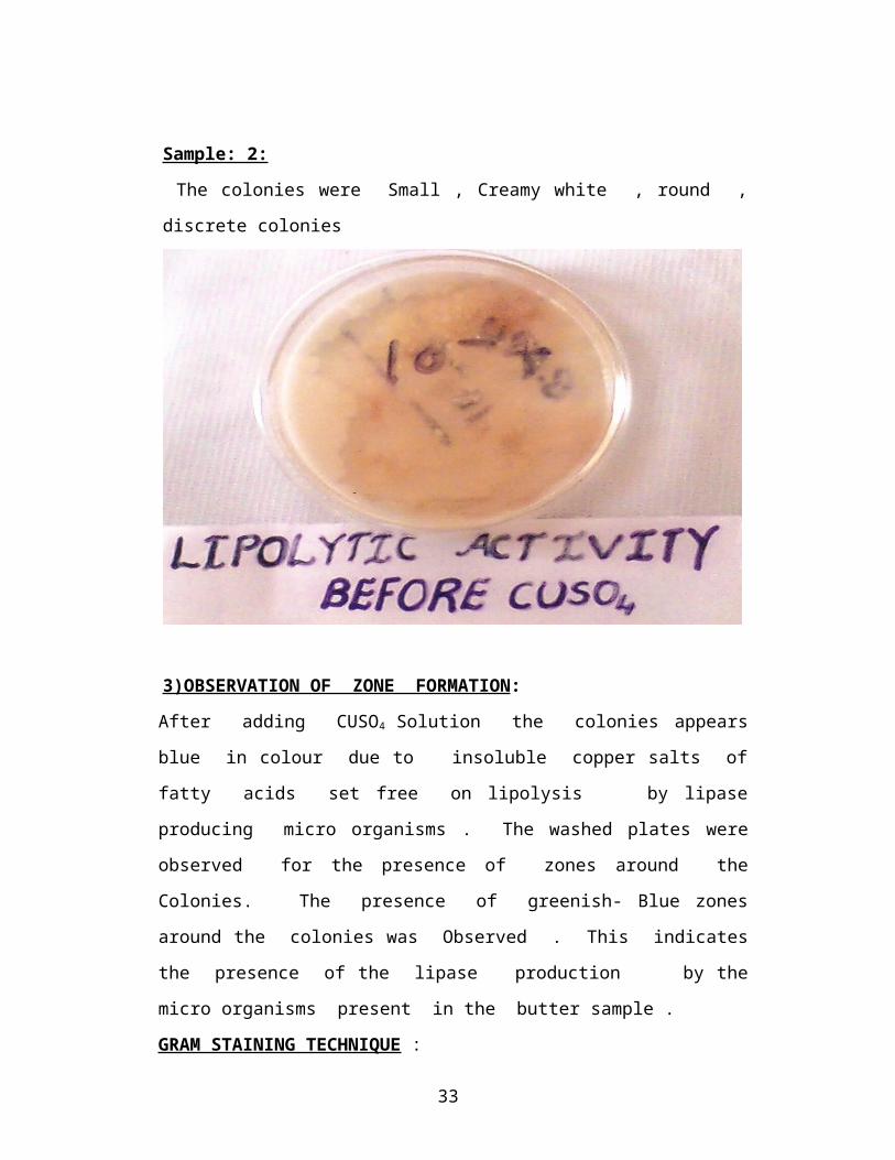
Sample: 2:
The colonies were Small , Creamy white , round , discrete colonies
21

3)OBSERVATION OF ZONE FORMATION:
After adding CUSO4 Solution the colonies appears blue in colour due to
insoluble copper salts of fatty acids set free on lipolysis by lipase producing
micro organisms . The washed plates were observed for the presence of zones
around the Colonies. The presence of greenish- Blue zones around the colonies
was Observed . This indicates the presence of the lipase production by the
micro organisms present in the butter sample .
GRAM STAINING TECHNIQUE :
The test was originally developed by Christian Gram in 1884, but was
modified by Hucker in 1921. The modified procedure provided greater reagent
stability and better differentiation of organisms.
The Gram stain is used to classify bacteria on the basis of their forms,
sizes, cellular morphologies, and Gram reactions; in a clinical microbiology
laboratory, it is additionally a critical test for the rapid presumptive diagnosis of
infectious agents and serves to assess the quality of clinical specimens.
Gram stain permits the separation of all bacteria into two large groups,
those which retain the primary dye (gram-positive) and those that take the color of
the counterstain (gram-negative).
The primary dye is crystal violet and the secondary dye is usually either
safranin O or basic fuchsin. Some of the moreCommon
formulations include: saturated crystal violet (approximately 1%), Hucker’s
crystal violet, and 2% alcoholic crystal violet.
PROTOCOL :
1. Deparaffinize sample and hydrate to distilled water
2. Place slides of staining rack
3. Add 1ml of crystal violet solution to the sample. Alternativley gentian
violet can be used
4. Add 250ul of 5% sodium biocarbonate
5. Gently mix solutions for one minute
6. Rinse with water
7. Cover sample with Gram's iodine solution and incubate for 1 minute
8. Rinse with water
22
9. Blot with filter paper until dry
10. Incubate breifly in acetone. This step should be short as longer incubation
will cause some gram positives to become gram negative. The time of
incubation is usually determined by the decolorization of the background
tissue. Repeat if necessary.
11. Cover sample with basic fuchsin solution for 3 minutes
12. Rinse with water
13. Blot excess water with filter paper
14. Dip slide in acetone
15. Dip slide in 0.1% picric acid in acetone
16. Rinse in acetone
17. Rinse in 50% acetone - 50% histolene
18. Rinse in 100% histolene. Repeat if necessary
19. Mount slide
Gram positive bacteria stain blue and gram negative bacteria stain red.
PROCEDURE :
1)The gram-positive cell envelope consists of a thick layer of peptidoglycan
embedded with techoic acids and a plasma membrane comprised of phospholipids
with integral membrane proteins traversing the bilayer.
2)The cells are flooded with crystal violet dye. Crystal violet is a water-soluble,
basic dye. In solution, basic dyes produce dye particles with positive charges
(cations). Sometimes the crystal violet dye particle is abbreviated CV+.
3)The individual crystal violet ions penetrate the thick peptidoglycan layer of the
cell as well as the plasma membrane, making their way through the matrix created
by the crosslinking of polysaccharides and proteins within the peptidoglycan layer.
4)Gram's iodine solution is added. This solution consists of a mixture of iodine
and potassium iodide. The active constituent in this solution is the iodide ion or an
iodine-iodine complex.
5) Like the crystal violet dye particles, the iodide ions are also able to penetrate
the thick peptidoglycan layer of the cell. Here, the iodide ions mix with the
crystal violet dye particles that were added in the previous step.
23

6) The crystal violet and iodide ions react, forming a crystal violet-iodine complex.
This complex is insoluble in water and produces particles much larger than either
the iodide ions or the crystal violet ions individually.
7) The alcohol/acetone mixture displaces water in the peptidoglycan layer,
resulting in dehydration. This loss of water causes the thick peptidoglycan layer to
shrink, tightening the matrix created by the crosslinking of polysaccharides and
proteins.
8) It is important to know that this decolorizing step is a critical step in the Gram
stain protocol. Exposure to the alcohol for too long can cause cells that are gram-
positive to lose too much of the dye complex due to damage to the peptidoglycan
layer. These cells will not appear gram-positive when the staining procedure is
complete.
9) The counterstain, normally safranin, is added. Like crystal violet, safranin is a
weakly water-soluble, basic dye that produces cationic stain particles in solution
that bind negatively charged moieties such as the techoic acids, peptides and
phospholipid heads within the envelope and in the cytoplasm.
10) Safranin, because of its small size, is able to penetrate the dehydrated
peptidoglycan layer and bind to negatively charged moieties. Because the safranin
is much lighter in color than the crystal violet-iodine complex.
11) When viewed under a microscope, gram-positive cells appear purple due to the
crystal violet-iodine complex retained inside.
12) The gram-negative cell envelope consists of a thin layer of peptidoglycan
surrounded by two phospholipid membranes, one interior and one exterior.
Polysaccharide chains are bound to the phosphate heads of the outer membrane to
form lipopolysaccharides. Both the membranes contain integral membrane
proteins. Place cursor over each membrane for ID.
13) The cells are flooded with crystal violet dye. Crystal violet is a water-soluble,
basic dye. In solution, basic dyes produce dye particles with positive charges
(cations). Sometimes the crystal violet dye particle is abbreviated CV+.
14) The individual crystal violet ions penetrate the thin peptidoglycan layer of the
cell as well as the plasma membrane, making their way through the matrix created
by the crosslinking of polysaccharides and proteins within the peptidoglycan layer.
24

15) Gram's iodine solution is added. This solution consists of a mixture of iodine
and potassium iodide. The active constituent in this solution is the iodide ion or an
iodine-iodine complex.
16) Gram's iodine solution is added. This solution consists of a mixture of iodine
and potassium iodide. The active constituent in this solution is the iodide ion or an
iodine-iodine complex.
17) Gram's iodine solution is added. This solution consists of a mixture of iodine
and potassium iodide. The active constituent in this solution is the iodide ion or an
iodine-iodine complex.
18) A decolorizing solution, normally consisting of a mixture of ethyl alcohol and
acetone, is added. Numerous variations of the decolorizing solution formula are
used in labs.
19) The alcohol/acetone mixture displaces water in the peptidoglycan layer,
resulting in dehydration. This loss of water causes the thin peptidoglycan layer to
shrink slightly, tightening the matrix created by the crosslinking of
polysaccharides and proteins. The alcohol/acetone mixture also disrupts and
dissolves the outer membrane, exposing the peptidoglycan layer to the
environment.
20) Safranin, because of its small size, is able to penetrate the dehydrated
peptidoglycan layer and bind to negatively charged moieties. Because
safranin is the only stain present, the cells will have a pink or red Colour.
GRAM’S STAINING :
Sample-1 :
Rod shaped cells was observed with purple colour . Therefore it was identified as
Gram positive bacteria .
Sample-2 :
Rod shaped cells was Observed with purple colour . Therefore it was also
identified As Gram positive bacteria .
25
BIOCHEMICAL TESTS – PROCEDURES 1.CITRATE UTILIZATION:
This test is one of several techniques used to assist in the identification of
enterobacteria. The test is based on the ability of an organism to use citrate as its
only source of carbon and ammonia as its only source of nitrogen.
PRINCIPLE :
The test organism is cultured in a medium which contains sodium citrate,
an ammonium salt, and the indicator bromo-thymol blue.Growth in the medium is
shown by turbidity and a change in color of the indicator from light green to blue,
due to the alkaline reaction, following citrate utilization.
REQUIREMENTS: Koser’s citrate medium:
Formula and preparation:Oxoid dehydrated medium
Grams per litreSodium ammonium phosphate 1.5Potassium dihydrogen phosphate 1.0Magnesium sulphate 0.2Sodium citrate 2.5Bromothymol blue 0.016
The medium is used at a concentration of 0.52 grams in every 100ml of
distilled water. Prepare the medium as per the instructions in the kit. Distribute
in 3ml amounts in small screw cap bottles or tubes. Sterilize by autoclaving (with
caps loosened) at 121oC for 15 minutes .When cool, tighten the container tops.
Date the medium and give it a batch number.
QUALITY CONTROL :
pH of medium: This should be within the range pH 6.6-7.0 at room temperature.
Performance: Test the medium by inoculating it with bacterial species of known
positive and negative citrate activity
Storage: Store in a cool dark place.
26

Shelf life: Up to 2 years providing there is no change in the volume or appearance
of the medium to suggest contamination or an alteration of pH.
INOCULATION :
Use a straight wire to inoculate the medium to prevent any carry over of
nutrients from the test culture .A light inoculum must be used, and the bottle caps
should be loosened during incubation.
METHOD:
1. Using a sterile straight wire, inoculate 3-4ml of sterile Koser’s citrate
medium with a broth culture of the test organism. Care must be taken not
to contaminate the medium with carbon particles, such as from a
frequently flamed wire.
2. Incubate the inoculated broth at 35-37oC for up to 4 days, checking daily
for growth.
RESULTS :
Turbidity and blue color………….Positive test (Citrate utilized)
No Growth……………………….Negative test (Citrate not utilized)
CONTROLS :
Positive citrate control: Klebsiella pneumoniae.
Negative citrate control: Escherichia coli
3)HYDROGEN SULPHIDE PRODUCTION :
The detection of hydrogen sulphide gas is used mainly to assist in the
identification of enterobacteria and occasionally to differentiate other bacteria
such as Bacteroides and Brucella species. Hydrogen sulphide is produced when
sulphur containing amino acids are decomposed.The technique used to detect the
release of hydrogen sulphide gas must not be too sensitive other wise it will detect
the small places of hydrogen sulphide produced by most enterobacteria.
27
USE OF KLIGLER IRON AGAR(KIA) TO DETECT HYDROGEN
SULPHIDE
This medium is suitable for detecting H2S production by enterobacteria.H2S is
detected by the ferric citrate contained in the medium.
LEAD ACETATE PAPER TO DETECT HYDROGEN SULPHIDE
When a sensitive technique for detecting H2S production is required,the lead
acetate paper test is recommended.
1. Inoculate a tube or bottle of sterile peptone water or nutrient broth with the
test organism.
2. Insert a lead acetate paper strip in the neck of the bottle tube above the
medium and stopper well.
3. Incubate the inoculated medium at 35-37oC and examine daily for a
blackening of the lower part of the strip.
RESULTS :
Blackening……………………………Positive test (H2S produced)
No blackening………………………. Negative test ( No H2S is produced)
4)INDOLE TEST :
Testing for indole production is important in the identification of
enterobacteria. Most strains of E.coli, P.vulgaris, P.rettgeri, M.morganii, and
Providencia species break down the amino acid tryptophan with the release of
indole.
PRINCIPLE :
The test organism is cultured in the medium which contains tryptophan.
Indole production is detected by Kovac’s or Ehrlich’s reagent which contains 4
(p)-dimethylaminobenzaldehyde. This reacts with the indole to produce a red
colored compound.
28

In the following method the use of the combined motility Indole Urea
(MIU) is described. A Kovac’s reagent paper strip is inserted in the neck of the
tube and indole production is indicated by a reddening of the strip. Indole is a
volatile substance (easily vaporized). The tube must be well stoppered during
incubation.
The indole test can also be carried out by culturing the organism in
tryptone water or peptone water containing tryptophan, and detecting indole
production by adding Kovac’s or Ehrlich’s reagent to an18-24 h culture.
REQUIREMENTS:
1. MOTILITY INDOLE UREA MEDIUM (MIU ):
MIU semi solid medium is used to differentiate enterobacteria species by
their motility, urease and indole reactions.
Formula and Preparation…………… To make 1 litre of MIU base medium
Tryptone………………………………………………………….30g
(or pancreatic digest of casein)
Potassium dihydrogen Phosphate…………………………… 1g
Sodium Chloride………………………………………………….5g
Agar…………………………………………………………… 4g
Phenol red 2.5g/l (0.25%)……………………………………… 2ml
Distilled water………………………………………………… 1 litre
Mix the dry ingredients in the water and heat to 100oC to dissolve the
chemicals (place the flask in a container of boiling water).
Allow to cool to 50-55oC and then add the phenol red solution. Mix well.
Dispense in 95ml amounts in screw-cap bottles. Sterilize by autoclaving
(with caps loosened) at 121oC for 15 minutes. When the medium has
cooled, tighten the bottle caps.
2. KOVAC’S REAGENT STRIPS
METHOD :
29

Using a sterile straight wire, inoculate 5 ml of sterile MIU medium with a
smooth colony of the test organism.
Place an indole paper strip in the neck of the MIU tube above the medium,
and stopper the tube. Incubate at 35-37 oC overnight.
Examine for indole production by looking for a reddening of the lower
part of the strip.
RESULTS:
Reddening of strip…………………………Positive test (Indole produced)No red color……………………………… .Negative test ( No Indole produced)
5) VOGES - PROSKAUER TEST :
This test is occasionally used to assist in the differentiation of
enterobacteria. K.pneumoniea, Vibrio cholerae and some strains of Enterobacter,
ferment glucose with the production of acetylmethylcarbinol ( acetoin ) which can
be detected by an oxidation reaction.
PRINCIPLE:
The test organism is cultured in a glucose phosphate peptone water for 48
hrs. sodium hydroxide and a small amount of creatine are then added. Under
alkaline conditions and exposure to the air, the acetoin produced from the
fermentation of the glucose is oxidized to diacetyl which forms a pink compound
with the creatine.
REQUIREMENTS :
1. GLUCOSE PHOSPHATE PEPTONE WATER :
Glucose phosphate peptone water is a fluid medium used in the Vogues-
Proskauer test and Methyl Red test.
Formula and Preparation --------- To make about 50 bottles
Peptone………………………………………….0.5g
Glucose (dextrose)…………………………… 0.5g
Di-Potassium hydrogen phosphate……… 0.5g
Distilled Water………………………………….100ml.
30

Dissolve the peptone and phosphate salt in the water by steaming. When
cool, filter, and adjust the pH to 7.5.
Add the glucose and mix well.
Dispense the medium in 2 ml amounts in small screw-cap tubes or bottles.
Sterilize by autoclaving (with caps loosened) at 115oC for 10 minutes.
When cool, tighten the container tops.
3. Sodium hydroxide, 400g/l.
4. Creatine powder.
METHOD ;
1. Inoculate 2 ml of sterile glucose phosphate peptone water with the test
organism. Incubate at 35-37 oC for 48 hrs.
2. Add a very small amount (knife point) of creatine and mix.
3. Add about 3 ml of the sodium hydroxide reagent and shake well. The
sodium hydroxide reagent is corrosive, therefore handle with care and do
not mouth- pipette.
4. Remove the bottle cap, and leave for 1 hour at room temperature. Look for
the slow development of a pink-red color.
RESULTS:
Pink-red color……………………… Positive test (Acetoin produced)
No pink-red color……………………Negative test (No acetoin produced)
6) NITRATE REDUCTION TEST :
This test is used to differentiate members of the Enterobacteriaceae that
produce the enzyme nitrate reductase, from gram negative bacteria that do not
produce the enzyme. The test is also helpful in differentiating Mycobacterium
species.
PRINCIPLE :
A heavy inoculum of the test organism is incubated in a broth containing
nitrate. After 4 hours, the broth is tested for the reduction of nitrate to nitrite by
adding sulphanilic acid reagent. If nitrite is present, the acid reagent is diazotized
and forms a pink-red compound with alpha-naphthylamine.
31

When nitrite is not detected it is necessary to test whether the organism
has reduced the nitrate beyond nitrite. This is done indirectly by checking whether
the broth still contains nitrate. Zinc dust is added which will convert any nitrate to
nitrite. If no nitrite is detected when the zinc dust is added, it can be assumed that
the entire nitrate has been reduced beyond nitrite to nitrogen gas or ammonia by a
nitrate reducing organism.
REQUIREMENTS:
1. NITRATE BROTH :
Nitrate broth can be prepared by adding 0.1 gram of potassium nitrate to
100 ml of peptone water.
The medium is used at a concentration of 0.9 grams in every 100 ml of
distilled water.
Sterilize by autoclaving (with loose caps) at 121oC for 15 minutes. When
the medium has cooled, stopper tightly.
1. Sulphanilic acid reagent
2. Alpha- naphthalamine reagent.
3. Zinc dust.
METHOD:
1. Inoculate 0.5 ml of sterile nitrate broth with a heavy growth of the test
organism.
2. Incubate at 35-37oC for 4 hours.
3. Add 1 drop of sulphanilic acid reagent and 1 drop of alpha- naphthylamine
reagent.
4. Shake to mix and look for a red color.
RESULTS:
Red color………………………………Positive test(Nitrate reduced)
32
If no red color is produced, add a very small amount (knife point) of zinc
dust powder. Look again for a red color and interpret as follows:
Red color…………………………Negative test(No reduction of nitrate)
No red color………………………… Positive test (Nitrate reduced)
7) UREASE TEST
Testing for urease enzyme activity is important in differentiating
enterobacteria. Proteus strains are strong urease producers. Y. enterocolitica also
shows urease activity (weekly at 35-37C). Salmonellae and shigellae do not
produce urease.
PRINCIPLE
The test organism is cultured in a medium which contains urea and the
indicator phenol red. If the strain is urease-producing, the enzyme will break
down the urea (by hydrolysis) to give ammonia and carbon dioxide. With the
release of ammonia, the medium becomes alkaline as shown by a change in color
of the indicator to red-pink. The method described is that which uses the
combined motility indole urea (MIU) medium.
REQUIREMENTS :
Motility indole urea (MIU) medium:
MIU semi solid medium is used to differentiate enterobacteria species by
their motility, urease and indole reactions.
Formula and PreparationTo make 1 litre of MIU base mediumTryptone………………………………………………………… 30g(Or pancreatic digest of casein)Potassium dihydrogen Phosphate…………………………… 1gSodium Chloride………………………………………………… 5gAgar……………………………………………………………….4gPhenol red 2.5g/l (0.25%)……………………………………… 2mlDistilled water……………………………………………………1 litre
33
Mix the dry ingredients in the water and heat to 100oC to dissolve the
chemicals (place the flask in a container of boiling water).
Allow to cool to 50-55oC and then add the phenol red solution. Mix well.
Dispense in 95ml amounts in screw-cap bottles. Sterilize by autoclaving
(with caps loosened) at 121oC for 15 minutes. When the medium has
cooled, tighten the bottle caps.
METHOD:
1. Using a sterile straight wire, inoculate a tube of sterile MIU medium with
a smooth colony of the test organism. When inoculating KIA medium at
the same time, inoculate the KIA tube first and then stab the MIU
medium.
2. Place an indole paper strip in the neck of the MIU tube above the medium.
Stopper the tube and incubate at 35-37o C overnight.
3. Examine for urease production by looking for a red-pink color in the
medium.
RESULTS:
Red-pink color…………………Positive test(Urease produced)
No red-pink color………………Negative test (No urease produced)
8) OXIDASE TEST :
The oxidase test is used to assist in the identification of Pseudomonas, Neisseria,
Vibrio, and Pastuerella species, all of which produce oxidase.
PRINCIPLE:
A piece of filter paper is soaked with a few drops of oxidase reagent. A
colony of the test organism is then smeared on the filter paper. If the organism is
oxidase producing, the phenyldiamine in the reagent will be oxidized to a deep
purple color.Occasionally the test is performed by flooding the culture plate with
oxidase reagent but this technique is not recommended for routine use because the
reagent rapidly kills bacteria. It can be useful, however, when attempting to
isolate N.gonorrhoeae colonies from mixed cultures in the absence of selected
34

medium.The oxidase positive colonies must be removed and subcultured within
30 seconds of flooding the plate.
REQUIREMENTS :
1. Oxidase reagent (freshly prepared)
This is a 10 g/l solution of tetra methyl-p-phenylenediamineihydrochloride.
METHOD :
1. Place a piece of filter paper in a clean Petri dish and add 2 or 3 drops of freshly prepared oxidase reagent.
2. Using a piece of stick or glass rod (not an oxidized wire loop), remove a colony of the test organism, and smear it on the filter paper.
3. Look for the development of a blue-purple color within a few seconds.
RESULT:
Blue-purple color………………… Positive test (Within 10 seconds) Oxidase
produced.
No blue-purple color……………… Negative test (Within 10 seconds) No oxidase
produced
9) CATALASE TEST:
This test is used to differentiate those bacteria that produce the enzyme
catalase, such as staphylococci, from non-catalase producing bacteria such as
streptococci.
PRINCIPLE :
Catalase acts as a catalyst in the breakdown of hydrogen peroxide to oxygen and
water.An organism is tested for catalase production by bringing it into contact
with hydrogen peroxide. Bubbles of oxygen are released if the organism is a
catalase producer. The culture should not be more than 24 hours old.
35

Care must be taken if testing an organism cultured on a medium containing blood
because catalase is present in red cells. If any of the blood agar is removed with
the colony, a false positive reaction will occur. It is usually recommended,
therefore that catalase testing be performed from a blood-free culture medium
such as nutrient agar.
REQUIREMENTS:
Hydrogen peroxide, 3% H2O2 -------- (10 volume solution)
METHOD:
Pour 2-3ml of the hydrogen peroxide solution into a test tube.
Using a sterile wooden stick or a glass rod, remove a good growth of the
test organism and immerse it in the hydrogen peroxide solution.
Look for immediate bubbling.
RESULTS:
Active bubbling……………………… Positive test (Catalase produced)
No release of bubbles………………… Negative test (No catalase produced)
36

results AND DISCUSSIONS
37
RESULTS:
BIOCHEMICAL CHARACTERISTICS :
S.NO Characteristics Sample -1 Sample-2 Positive Negative
1 Motility + _ NA NA
2 Gram stain Gram-Positive
rods
Gram
Positive rods
NA NA
3 Growth onButter
Fat Agar
Small,Creamy
white round ,
discrete colonies
Small,Creamy
white round ,
discrete
colonies
NA NA
4 Catalase Bacillus Lactobacillus Sample -1 Sample -2
5 Oxidase Bacillus Lactobacillus Sample-1 Sample-2
6 Indole Bacillus Lactobacillus ------ Sample-1
Sample-2
7 Methyl Red Bacillus Lactobacillus Sample-1
Sample-2
---------------
8 Voges Proskauer Bacillus Lactobacillus Sample- 1
Sample-2
---------------
9 Citrate Bacillus Lactobacillus Sample-1 Sample-2
10 Urease Bacillus Lactobacillus Sample-1 Sample-2
1 Glucose Bacillus Lactobacillus Sample-1
Sample-2
2 Galactose Bacillus Lactobacillus Sample-1
Sample-2
3 Xylose Bacillus Lactobacillus Sample-1
Sample-2
4 Sucrose Bacillus Lactobacillus Sample-1
Sample-2
38

DISCUSSION: Pathogenic organisms of both bovine and human origin have been isolated
from milk. Milk, therefore, can serve as a carrier of diseases. Many serious
epidemics were caused by the consumption of such products before this fact was
clearly recognized. However, this became less common as milk sanitation has
improved and pasteurization is being more widely practised.
The disease organisms present in milk may be derived from (1) diseased
animals or (2) persons collecting and handling milk: Thus the danger is due to the
inoculum and not to the growth of organisms in the milk.
The health of animal is an important factor. Several diseases of cattle
including staphylococcal and streptococcal infections, tuberculosis, brucellosis,
Salmonellosis, Q fever and Foot and mouth disease may be transmitted to man.
The organisms causing these diseases may get into the milk either directly from the
udder, or indirectly from infected body discharges, which may drop, splash, or be
blown into the milk. Some of the important diseases of human origin that have
been transmitted by milk are (1) typhoid fever (2) diphtheria, (3) scarlet fever,
(4) dysentery (5) septic sore throat and (6) poliomyelitis.
It is also possible for humans to infect animals. For example, mastitis may
be caused by a variety of organisms, including Staphylococcus aureus. The
infecting organism, in some cases, has been traced to humans.The organisms exist
in milk as dormant spores which, unless the milk is subjected to the action of
certain physical and chemical stimuli such as heat, cold, or the action of alkalis,
remain un germinated.
MILK FERMENTATION:
Raw milk produced under normal conditions develops acidity. It has long
been recognised that highly acid milk does not putrefy. Therefore, allowing milk
to develop acidity naturally preserves the other milk constituents. Bacteria in
milk are responsible for acid development. They produce acid by the anaerobic
breakdown of milk carbohydrate—lactose—to lactic acid and other organic acids.
Anaerobic breakdown of carbohydrate to organic acids or alcohols is called
fermentation.A number of sugar fermentations are recognized in milk. They can
39
be either homofermentative, with one end product, or heterofermentative, with
more than one end product.
1. Streptococci and Lactobacilli., 2. Propionibacteria. , 3. Yeasts – Candida and
Torula. 4. Coliform bacteria. are the organisms responsible for fermentation .
40

The lactic acid fermentation is the most important one in milk and is
central to many processes. Propionic fermentation is a mixed-acid fermentation
and is used in the manufacture of Swiss cheese varieties. Alcohol fermentation
can be used to prepare certain fermented milks and also to make ethyl alcohol
from whey.
The coliform gassy fermentation is an example of a spoilage fermentation.
Large numbers of coliform bacteria in milk indicates poor hygiene. The coliform
gassy fermentation disrupts lactic acid fermentation, and also causes spoilage in
cheese.
The factors that affect microbial growth also affect milk fermentation.
Fermentation rates will generally parallel the microbial growth curve up to the
stationary phase. The type of fermentation obtained will depend on the numbers
and types of bacteria in the milk, storage temperature and the presence or absence
of inhibitory substances.
The desired fermentations can be obtained by temperature manipulation or
by adding a selected culture of micro-organisms—starter—to pasteurised or
sterilised milk. In smallholder milk processing, traces of milk from previous
batches are often used to provide `starter' for subsequent batches. Other sources
include the container and additives such as cereal grains.
The fermentation will be established once the organisms dominate the medium
and will continue until either the substrate is depleted or the end product
accumulates. In milk, accumulation of end product usually arrests the
fermentation. For example, accumulation of lactic acid reduces milk pH to below
4.5, which inhibits the growth of most micro-organisms, including lactic-acid
producers. The fermentation then slows and finally stops.
41

CONCLUSIONS
42

CONCLUSION :
Bacteria are contaminants of all fresh foods. In order to avoid excessive
spoilage, various measures can be employed to kill bacteria or to retard bacterial
growth. These include keeping foods cold (or frozen), boiling (as is done for
canned foods), salting (pickling), dehydrating (as in beef jerky), and adding anti-
bacterial preservatives. In the particular case of milk, pasteurization combined
with refrigeration is the most common technique used. Pasteurization does not kill
all the bacteria (or spores) in milk, but does eliminate most of the pathogenic
bacteria that have been historically associated with milk, such as tuberculosis,
brucellosis, and typhoid. Pasteurization was first developed in order to kill these
pathogens, but it was soon discovered that this process also improved the keeping
quality of the milk without sacrificing the taste. Pasteurization can be
accomplished by heating milk to 63-650 C for 30 minutes or to 71° c for 15
seconds (flash pasteurization) followed by rapid cooling. Flash pasteurization is
the most common technique used. Pasteurization does not prevent spoilage, but it
reduces the bacterial population so that spoilage occurs more slowly. Milk can be
essentially sterilized by Ultra High Temperature (UHT) pasteurization in which it
is heated to a higher temperature than is used for normal pasteurization, but just
for a few seconds (149° C for 6 to 9 seconds). This milk can be stored for several
months at room temperature without spoiling. There are a variety of bacteria that
can be present in raw milk due to improper Collection preservation and
maintenance .
From the results data it was observed that the good hygienic conditions
maintained during collection of milk will minimize the microbial colony count
and the Lipolytic activity of the bacteria.
43
REFERENCES
44

References :
Alatosavva .M. and Alato ssava ,T. (2006) Phenotypic characterization of raw milk associated psychrotrophic bacteria . Microbiol.
APHA (1978) . American public health Association in standard methods for examination of dairy products (Ed.Elmer ,H.Marth .Washington ,D.C)
APHA, 1980. Compendium of methods for examination of foods. 12th Edition of American Public Health Association Inc., Washington, D.C.
Austain, B. (1988). Methods in Aquatic Bacteriology. A Wiley -Interscience Publication . 222-231.
Braun, P. and K. Fehlhaber, 2002. Combined w effect of temperature, a and pH on enzymatic a c tiv ity of spoi lage causing bacte ria .Milchwissenschaft, 57: 134-136. 23. Santos, E. de S., E.de Carvalho and L.R. de Abrew, 1999 .
Psych ro troph ic bacte ria : consequences of their presence in milk and cheeses. Boletim de Sociedade Braisileria de Ciencia e Technologia de Alimentos, 33: 129-138.
Brock, T., (1986). introduction: an overview of the thermophiles. in: Brock, T. thermophiles: general, molecular, and applied microbiology, John Wiley and Sons, New York, 2-15.
Buchanan , R.E. and N.E.Gibbons , 1974 , Bergey’s manual of determination bacteriology .5th Edn. The Williams and Wilkins company , Baltimore, USA.
Campos, L and Felix, C. R. (1995). Purification and Characterization of a Glucoamylase from Humicola grisea. Applied Environmental Microbiology.61. 2436-2438.
Canigova, M. and E. Benczova, 2001. The microflora changes of raw milk during its refrigerated storage. Acta Fytotechnica et Zootechnica, 4: 104-106.
Celestino, E.L., M. Iyer and H. Roginski, 1996. The effects of refrigerated storage on the quality of raw milk. Australian J. Dairy Tech., 51: 59-63.
45
Cempirkova, R., 2002. Psychrotrophic vs. total bacterial counts in bulk milk samples. Vet. Med. Czech., 47: 227-233.
Cappuccino, J.G. and N. Sherman, 1999. Microbiology: A laboratory manual . Benjamin/Cummings Publ. Company, New York., pp: 477.
Dicks LM, Silvester M, Lawson PA, Collins MD (2000). Lactobacillus fornicalis sp. nov., isolated from the posterior fornix of the human vagina.
Dogan,B.and Brook ,K.J. (2003) Genetic diversity and spoilage potentials among pseudomonaas sp. Isolated from fluid milk products and dairy processing plants. Appl.Environ.Microbiol., 69: 130-138.
Doolittle, W. F. (1999). Phylogenetic classification and the universal tree. Science, Vol. 284. 2124-2128.
Fairbairn, D.J. and B.A. Law, 1986. Proteinases of psychrotrophic bacteria: their production, properties, effects and control. J. Dairy Res.,
53: 139-177.
Fujio, Y. and Kume, S. (1991). Characteristics of highly thermostable neutral protease produced from Bacillus stearotherrtmophilus. World Journal of Microbiology and Biotechnology.7. 12-16
Geetha, R. and V. Prasad, 2001. Studies on the performance of cultures on lactic acid bacteria in lactose hydrolysed buffalo skim milk. Cheiron,
30: 81-84
Gopi,H., Parthiban M. and Dhana lakshmi ,B.(2001). Bacteriological quality of private brands of milk in chennai city .Cheiron ,30: 106-107.
Harrigan.W.F. and Mc.Cance , M.E(1976). Laboratory method in food and beverage Microbiology, Academic press, London.
Hisotsuyanagi, K. (1979). Stepwise introduction of regulatory genes stimulating production of α -amylase into Bacillus subtilis: Construction of α-amylase extrahyper producing strain. Agric boil chem. 43:2343-2349.
Harris ,P.L.Cuppett,L. and Buller man, L.B(1990) A technique for comparison of isolation of lipolytic bacteria .J. Food prot. 53: 176-177.
Holm, C., Jespen , L.Larsen, M. and Jespersen.L,(2004). Predominant microflora of down graded Danish bulk tank milk .J. Dairy Sci. 87 : 1151-1157.
46
Khali. A, M. salim and A.K. Sallal, (1998). Enumeration of thermotolerant bacteria from recreational thermal ponds in Jordan", Cytobios. 57-63.
Kohlmann, K.L., S.S. Nielsen, L.R. Steenson and M.R. Ladisch, 1991. Production or proteases by psychrotrophic microorganisms. J. Dairy Sci.,
74: 3275-3283
Kuzin , A.A.Gudkov , A.V. and Shergin , N.A(1992). Contamination of raw milk by psychrotrophs. Molochnaya promyshlennost, 5: 31-34 .
Lutz Fisher et al. (1995). Purification and characterization of thermotolerant β-Galactosidases from thermomyces lanuginose. Applied and Environmental Microbiology.61. 1497-1501.
Mac, Faddin., J. F, (1980). biochemical tests for Identification of medical bacteria "2nd ed., Waverly Press Inc. U.S.A. 50-53.
Marshall ,R.T(1993) Standard methods for the examination of dairy products , 16th Ed. American Public Health Association , Washington,D.C.
Matta, H. and V. Punj, 1996. Isolation and identification of proteolytic psychrotrophic spore forming bacteria from milk. Indian J. Dairy Sci.,49: 695-699
Prakash, M., V. Karthikeyan and N. Karmegam, 2006. Determination of total counts of psychrotrophic bacteria and coliforms in milk samples. J. Ecotox. Environ. Monit., 16: 291-296.
Priest, f., Good, M. and Todd, C. (1988). Anumerical classification of the genus bacillus. Journal of general microbiology. Vol.134.184.
Putz, M. and H. Gierl, 1991. Investigation of total bacterial count and of selected bacteria in different milk samples. Bayerisches Kandwirtschaftliches Jahrbuch, 68: 515-520.
Rahman, A.H. and H.A. Aait, 1988. Incidence and level of occurrences of proteolytic microorganisms in some selected foods. Ass. Vet. Medical J., 19: 72-78.
Saleha A.A.(1992) Psychrotrophic bacteria in pasteurized milk in Malaysia . Proceedings of the 3rd World Congress On Food borne infections and Intoxications , Berlin, Germany , 16-19 June 1992:1.pp190-192.
47

Sorhaug, T. and L. Stepaniak, 1997. Psychrotrophs and their enzymes in milk and dairy products: quality aspects. Trends Food Sci.Technol.,
8: 35-41.
Snedecor G.W .and Cochran E.G.(1989).Statistical Methods .8th Edn. The Iowa state university press, Ames ,Iowa, USA.
Tachibena, Y., et al. (1999). Purification and Characterization of an Extremely Thermostable Cyclomaltodextrin Glucanotransferase from a Newly Isolated Hyperthermophilic Archaeon, a Thermococcus sp. Applied Environmental Microbiology.65. 1991-1997.
Tapan, K et al (1998). Oxidation of Methyl-Substituted Naphthalenes: Pathway in a Versatile Sphingomonas Paucimobilis Strain. Applied and Environmental Microbiology. 64. 1884-1889.
The Ohio State University Food Science and Technology Department. Lactic Acid Bacteria - Carbohydrate and Protein Metabolism.
48
Related Documents