www.afm-journal.de FULL PAPER © 2012 WILEY-VCH Verlag GmbH & Co. KGaA, Weinheim 1632 www.MaterialsViews.com wileyonlinelibrary.com Adv. Funct. Mater. 2012, 22, 1632–1638 Jiseok Lee, Sungbaek Seo, and Jinsang Kim* 1. Introduction Nerve agents, often organophosphates (OPs), are a class of lethal weapons of mass destruction (WMD) that kill by disrupting the nerve transfer mechanism. When nerve agents are inhaled into the human body, they make strong covalent bonds with acetylcholinesterase (neurotransmitter enzyme) and inhibit the enzyme from degrading acetylcholine. [1] As a result, acetylcholine continues to transmit nerve impulses and muscle contractions do not stop. Consequently, the entire nervous system begins to operate abnormally causing vomiting, muscle twitching, con- vulsions and, ultimately, death. Common nerve agents include the G-agents (Tarbun, Soman, Cyclosarin, Sarin) and VX. For example, Sarin was released by terror- ists in a Tokyo subway station in 1995, resulting in more than 5000 human casu- alties. VX (O-ethyl S-[2-(diisopropylamino) ethyl] methylphosphonothioate) is ten-fold more toxic than G-agents and is consid- ered to be the most toxic chemical ever synthesized. Unfortunately, these highly adhesive and volatile nerve agents are colorless, odorless and tasteless, making detection very difficult. Therefore, a reli- able nerve agent detection system is criti- cally desirable in the current climate of terrorism awareness. Conventional analytical methods such as gas chromatography [2] and mass spec- troscopy [3] are still considered to be the most reliable techniques despite the recent development of sensors based on fluorescence, [4] carbon nano-tubes, [5] microcantilever, [6] and liquid crystals. [7] The conventional analytical detection systems, however, require expensive equipment that is often not port- able due to weight and size and is only operable by well-trained personnel. Therefore, a simple and rapid yet reliable detection system is highly desirable. Particularly, equipment-free sensor systems such as colorimetric detection by the naked eye would be among the best and most practically useful methods. In this contribution, we present our recent development of a rapid, highly selective and sensitive, and convenient colorimetric nerve agent detection system based on rationally designed polydiacetylene (PDA) liposomes both in solutions and in solid films. PDA-based sensory systems are unique in that they have a colorimetric/fluorescence dual detection capa- bility and a self-signaling property. [8] Furthermore, the prepa- ration protocol is very simple through a convenient molecular self-assembly followed by fast photo-polymerization. The photo-induced topochemical polymerization converts diacety- lene monomers into conjugated PDA having a blue color (absorption λ max at 640 nm) via 1,4 addition polymerization. Colorimetric Detection of Warfare Gases by Polydiacetylenes Toward Equipment-Free Detection Rationally designed polydiacetylene (PDA) molecules have been developed for rapid, selective, sensitive, and convenient colorimetric detection of orga- nophosphate (OP) nerve agents, a mass destruction weapon. Oxime (OX) functionality was incorporated into diacetylene molecules to utilize its strong affinity toward organophosphates. The diacetylene molecules having an OX functional group (OX-PDA) were self-assembled to form PDA liposomes in an aqueous solution. Upon exposure to organophosphate nerve agent simu- lants, OX at the OX-PDA liposome surface interacts with nerve agent simu- lants, which results in intraliposomal repulsive stress due to steric repulsion between OP-occupied OX units at the liposome surface as well as interli- posomal aggregation induced by increased hydrophobicity of the liposome surface via OP-OX complex formation. The resulting intra- and interliposomal stress causes disturbance of the conjugated backbone of OX-PDA, producing color change as a label-free and sensitive sensory signal. The effects of molec- ular structure on selectivity and sensitivity of OX-PDA liposome solution, OX-PDA liposome-embedded agarose gels, and OX-PDA liposome-coated cellulose acetate membranes were systematically investigated. The optimized OX-PDA liposome in the solid state showed selective and rapid optical transi- tion upon exposure down to 160 ppb of diisopropylfluorophosphate (DFP), a nerve agent simulant. The results provide an insightful molecular design principle of PDA-based colorimetric sensor and suggest portable sensory patches for rapid, selective, sensitive, and convenient colorimetric detection of organophosphate nerve agents. DOI: 10.1002/adfm.201102486 Dr. J. Lee, S. Seo Macromolecular Science and Engineering University of Michigan 2300 Hayward St., Ann Arbor, MI 48109-2136, USA Prof. J. Kim Macromolecular Science and Engineering Materials Science and Engineering Chemical Engineering Biomedical Engineering University of Michigan 2300 Hayward St., Ann Arbor, MI 48109-2136, USA E-mail: [email protected]

Welcome message from author
This document is posted to help you gain knowledge. Please leave a comment to let me know what you think about it! Share it to your friends and learn new things together.
Transcript

www.afm-journal.de
FULL
PAPER
1632
www.MaterialsViews.com
Jiseok Lee , Sungbaek Seo , and Jinsang Kim *
Colorimetric Detection of Warfare Gases by Polydiacetylenes Toward Equipment-Free Detection
Rationally designed polydiacetylene (PDA) molecules have been developed for rapid, selective, sensitive, and convenient colorimetric detection of orga-nophosphate (OP) nerve agents, a mass destruction weapon. Oxime (OX) functionality was incorporated into diacetylene molecules to utilize its strong affi nity toward organophosphates. The diacetylene molecules having an OX functional group (OX-PDA) were self-assembled to form PDA liposomes in an aqueous solution. Upon exposure to organophosphate nerve agent simu-lants, OX at the OX-PDA liposome surface interacts with nerve agent simu-lants, which results in intraliposomal repulsive stress due to steric repulsion between OP-occupied OX units at the liposome surface as well as interli-posomal aggregation induced by increased hydrophobicity of the liposome surface via OP-OX complex formation. The resulting intra- and interliposomal stress causes disturbance of the conjugated backbone of OX-PDA, producing color change as a label-free and sensitive sensory signal. The effects of molec-ular structure on selectivity and sensitivity of OX-PDA liposome solution, OX-PDA liposome-embedded agarose gels, and OX-PDA liposome-coated cellulose acetate membranes were systematically investigated. The optimized OX-PDA liposome in the solid state showed selective and rapid optical transi-tion upon exposure down to 160 ppb of diisopropylfl uorophosphate (DFP), a nerve agent simulant. The results provide an insightful molecular design principle of PDA-based colorimetric sensor and suggest portable sensory patches for rapid, selective, sensitive, and convenient colorimetric detection of organophosphate nerve agents.
1. Introduction
Nerve agents, often organophosphates (OPs), are a class of lethal weapons of mass destruction (WMD) that kill by disrupting the nerve transfer mechanism. When nerve agents are inhaled into the human body, they make strong covalent bonds with
© 2012 WILEY-VCH Verlag GmbH & Co. KGaA, Weinhewileyonlinelibrary.com
DOI: 10.1002/adfm.201102486
Dr. J. Lee , S. Seo Macromolecular Science and EngineeringUniversity of Michigan2300 Hayward St., Ann Arbor, MI 48109-2136, USAProf. J. Kim Macromolecular Science and EngineeringMaterials Science and EngineeringChemical EngineeringBiomedical EngineeringUniversity of Michigan2300 Hayward St., Ann Arbor, MI 48109-2136, USA E-mail: [email protected]
acetylcholinesterase (neurotransmitter enzyme) and inhibit the enzyme from degrading acetylcholine. [ 1 ] As a result, acetylcholine continues to transmit nerve impulses and muscle contractions do not stop. Consequently, the entire nervous system begins to operate abnormally causing vomiting, muscle twitching, con-vulsions and, ultimately, death. Common nerve agents include the G-agents (Tarbun, Soman, Cyclosarin, Sarin) and VX. For example, Sarin was released by terror-ists in a Tokyo subway station in 1995, resulting in more than 5000 human casu-alties. VX (O-ethyl S-[2-(diisopropylamino)ethyl] methylphosphonothioate) is ten-fold more toxic than G-agents and is consid-ered to be the most toxic chemical ever synthesized. Unfortunately, these highly adhesive and volatile nerve agents are colorless, odorless and tasteless, making detection very diffi cult. Therefore, a reli-able nerve agent detection system is criti-cally desirable in the current climate of terrorism awareness.
Conventional analytical methods such as gas chromatography [ 2 ] and mass spec-troscopy [ 3 ] are still considered to be the most reliable techniques despite the recent development of sensors based on
fl uorescence, [ 4 ] carbon nano-tubes, [ 5 ] microcantilever, [ 6 ] and liquid crystals. [ 7 ] The conventional analytical detection systems, however, require expensive equipment that is often not port-able due to weight and size and is only operable by well-trained personnel. Therefore, a simple and rapid yet reliable detection system is highly desirable. Particularly, equipment-free sensor systems such as colorimetric detection by the naked eye would be among the best and most practically useful methods.
In this contribution, we present our recent development of a rapid, highly selective and sensitive, and convenient colorimetric nerve agent detection system based on rationally designed polydiacetylene (PDA) liposomes both in solutions and in solid fi lms. PDA-based sensory systems are unique in that they have a colorimetric/fl uorescence dual detection capa-bility and a self-signaling property. [ 8 ] Furthermore, the prepa-ration protocol is very simple through a convenient molecular self-assembly followed by fast photo-polymerization. The photo-induced topochemical polymerization converts diacety-lene monomers into conjugated PDA having a blue color (absorption λ max at 640 nm) via 1,4 addition polymerization.
im Adv. Funct. Mater. 2012, 22, 1632–1638

FULL P
APER
www.afm-journal.dewww.MaterialsViews.com
Interestingly, the absorption λ max shifts from 640 nm (blue phase) to 540 nm (red phase) when an external stimulus is applied to PDA, and this red phase also fl uorescences, pro-viding the dual signaling capability. [ 9 ] This optical transition is believed to be caused by the conformational change of the con-jugated backbone of PDA induced by various external stimuli. Sensors have been designed to make use of this property to detect bacteria, [ 10 ] pH, [ 11 ] temperature, [ 12 ] ions, [ 13 ] and mechan-ical stress. [ 14 ]
2. Results and Discussion
We designed diacetylene molecules having an oxime (OX) functional group to utilize the attractive dual signaling capa-bility of PDA liposomes for the development of a nerve agent detection system. OX has been used to develop antidotes such as 2-pralidoxime (2-PAM) to detoxify organophosphorous nerve agents. Typically 2-PAM is a chloride salt having an OX func-tional group that reversibly binds to OP-inactivated acetylcho-linesterase then attaches to the free OP, effi ciently reversing and preventing the otherwise irreversible binding of orga-nophosphates to acetylcholinesterase. Because the reversible binding of 2-PAM does not inactivate the enzyme function, 2-PAM is an effective antidote. [ 15 ] We applied the good reactivity between 2-PAM and OP to our PDA-based sensor system. We designed the oxime-modifi ed polydiacetylene (OX-PDA) deriva-tives shown in Scheme 1 A. Our earlier work has demonstrated that steric repulsion of probe/target complexes at the liposome surface induces rearrangement of the conjugated backbone of PDA and produces a color transition from blue to red and also fl uorescence development. [ 13 , 16 ] We also found that inter-liposomal interactions play a critical role in sensitive optical signal generation. [ 16b ] Similar steric perturbation and inter-lipo-somal interactions occur in our PDA-based nerve agent detec-tion system as schematically described in Scheme 1 C. The OX at the surface of PDA liposomes reacts rapidly with OP mol-ecules, and the resulting OX/OP complexes produce repulsive strain to the conjugated backbone of the self-assembled PDA liposomes. In addition, the liposome surface becomes more hydrophobic by OX/OP complex formation, causing aggrega-tion of liposomes in an aqueous environment. These two simul-taneous factors of intra-liposomal repulsion and inter-liposomal hydrophobic aggregation impose large stress on the conjugated backbone of PDA, inducing the conformational change of PDA backbone as illustrated schematically in Scheme 1 C. The con-formational change appears as a rapid and sensitive colori-metric transition from blue to red.
Scheme 1 A and 1 B show the chemical structure of the inves-tigated PDA monomers having oxime (OX-PDAs) and aldehyde functionality. The reactivity of oxygen in oxime nucleophile toward OP electrophiles is signifi cantly enhanced by the α -effect of the adjacent heteroatom (nitrogen). [ 4b ] For instance, OX is known to be 100-fold more reactive toward organophosphorous electrophiles than phenols having a similar pKa. [ 17 ] Therefore, we synthesized OX-modifi ed PDA monomers, PCDA- p BO and PCDA-HBO, having an ester linkage. It is well established that the sensitivity of the PDA sensory system is closely related to the stability of PDA liposomes and can be tuned by adjusting
© 2012 WILEY-VCH Verlag GmAdv. Funct. Mater. 2012, 22, 1632–1638
the intermolecular self-assembling force between the diacety-lene molecules, for example by adding or removing hydrogen bonding capability. [ 12b ] As can be seen in Figure 1 , PDA lipo-some solutions having an ester linkage (PCDA-CPE) showed less stability and represented a saturated red color at a lower temperature (70 ° C) than PDA liposomes ( > > 90 ° C) having an amide linkage (PCDA- p BzA) due to the lack of hydrogen bonding. However, despite this heightened instability, neither PCDA- p BO nor PCDA-HBO liposome solution having an ester linkage showed noticeable color change to red after incubation with 100 mM of DCP or DFP. We hypothesized that the strong intermolecular hydrogen bonding between OXs of PCDA- p BO generated strong aromatic stacking resulting in the low sensitivity ( Figure 2 A). In the case of PCDA-HBO, the strong intramolecular hydrogen bonding between OX and β -hydroxy will produce a planar structure of PCDA-HBOs and form strong aromatic stacking between the planarized PCDA-HBOs. This strong intermolecular packing would give good stability to the resulting PDA liposomes and reduce signaling sensitivity [ 18 ] (Figure 2 C). Therefore, we designed analogous diacetylene mol-ecules (PCDA- p BA and PCDA-HBA) having an aldehyde instead of OX to remove hydrogen bonding between PCDA- p BO mol-ecules (Figure 2 B) and to also inhibit the potentially strong aromatic stacking of PCDA-HBO molecules (Figure 2 D and E). Figure 1 C indeed shows sensitive color change of PCDA- p BA. We could observe a saturated red color at the lowest temper-ature (40 ° C) compared to the acid-modifi ed PCDA-CPE (70 ° C) and PCDA-pBzA ( > > 90 ° C), which has strong head group hydrogen bonding. From this, we hypothesized that when the aldehyde-modifi ed PDAs are co-assembled with their oxime-modifi ed equivalent into liposomes, intermolecular hydrogen bonding and aromatic stacking between the oximes would be disrupted, providing better mobility between OX-PCDA molecules and ensuing good sensitivity.
2.1. Detection of Nerve Agent Simulants
We studied the color/fl uorescence transition of OX-PDA lipo-somes upon exposure to diethylchlorophosphate (DCP) and diisopropylfl uorophosphate (DFP) nerve gas simulants in three different conditions: aqueous phase, semi-wet phase (agarose gel), and solid phase.
2.1.1. Aqueous Phase Detection
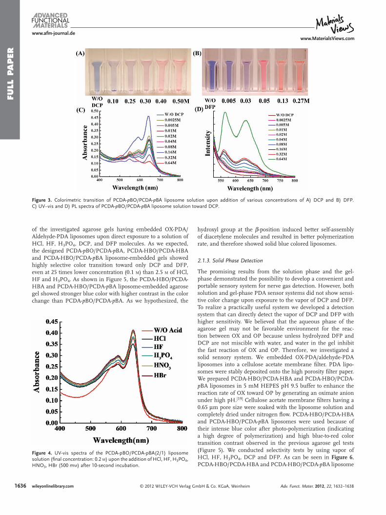
We initially conducted selectivity tests with PCDA- p BO/PCDA- p BA (1/1 mole ratio) liposomes in an aqueous solution. We confi rmed that PCDA- p BO/PCDA- p BA liposome solution selec-tively and rapidly detected DCP and DFP as shown in Figure 2 B and Figure 3 . The absorption peak at 650 nm (blue phase) decreased and the 550 nm (red phase) absorption peak appeared upon addition of various concentrations of DCP (Figure 3 C). The fl uorescence intensity of the PCDA- p BO/PCDA- p BA lipo-some also increased upon the addition of DCP (Figure 3 D). Moreover, we did not observe any noticeable color change upon addition of HCl, HF, HNO 3 , and H 3 PO 4 implying good selec-tivity ( Figure 4 ). The colorimetric transition was almost sponta-neous and saturated within 10 seconds. Although PCDA- p BO/
1633wileyonlinelibrary.combH & Co. KGaA, Weinheim

FULL
PAPER
1634
www.afm-journal.dewww.MaterialsViews.com
wileyonlinelibrary.com © 2012 WILEY-VCH Verlag GmbH & Co. KGaA, Weinheim Adv. Funct. Mater. 2012, 22, 1632–1638
Scheme 1 . Chemical structure of the investigated A) oxime-functionalized and B) aldehyde-functionalized PDA monomers. C) Schematic illustration of the PDA liposome-based organophosphate detection strategy by means of a) intraliposomal repulsion and b) interliposomal aggregation due to the surface property change from hydrophilic to hydrophobic.
Figure 1 . Chemical structure and UV–vis spectra of the A) PCDA-CPE, B) PCDA-pBzA and C) PCDA-pBA liposome solution (fi nal concentration: 0.5 m M ) upon heating.

FULL P
APER
www.afm-journal.dewww.MaterialsViews.com
Figure 2 . Molecular packing and color transition of the following PDA-liposome A) PCDA-pBO, B) PCDA-pBO/PCDA-pBA, C) PCDA-HBO, D) PCDA-HBO/PCDA-HBA (1/1) and E) PCDA-HBO/PCDA-pBA. (DFP concentration: 100 m M )
PCDA- p BA liposome solution showed rapid and selective detec-tion, a portable solid-state detection device will be much more useful for real application. In this regard, we also investigated solid phase detection systems.
© 2012 WILEY-VCH Verlag GmAdv. Funct. Mater. 2012, 22, 1632–1638
2.1.2. Gel Phase Detection
We prepared a gel phase sensory system with agarose. The OX-PDA liposomes were simply embedded into agarose gel and exposed to DCP and DFP. Figure 5 shows the color transition
1635wileyonlinelibrary.combH & Co. KGaA, Weinheim
FULL
PAPER
163
www.afm-journal.dewww.MaterialsViews.com
Figure 3 . Colorimetric transition of PCDA- p BO/PCDA- p BA liposome solution upon addition of various concentrations of A) DCP and B) DFP. C) UV–vis and D) PL spectra of PCDA- p BO/PCDA- p BA liposome solution toward DCP.
of the investigated agarose gels having embedded OX-PDA/Aldehyde-PDA liposomes upon direct exposure to a solution of HCl, HF, H 3 PO 4 , DCP, and DFP molecules. As we expected, the designed PCDA- p BO/PCDA- p BA, PCDA-HBO/PCDA-HBA and PCDA-HBO/PCDA- p BA liposome-embedded gels showed highly selective color transition toward only DCP and DFP, even at 25 times lower concentration (0.1 M) than 2.5 M of HCl, HF and H 3 PO 4 . As shown in Figure 5 , the PCDA-HBO/PCDA-HBA and PCDA-HBO/PCDA- p BA liposome-embedded agarose gel showed stronger blue color with higher contrast in the color change than PCDA- p BO/PCDA- p BA. As we hypothesized, the
6 wileyonlinelibrary.com © 2012 WILEY-VCH Verlag G
Figure 4 . UV-vis spectra of the PCDA-pBO/PCDA-pBA(2/1) liposome solution (fi nal concentration: 0.2 M ) upon the addition of HCl, HF, H 3 PO 4 , HNO 3 , HBr (500 m M ) after 10-second incubation.
hydroxyl group at the β -position induced better self-assembly of diacetylene molecules and resulted in better polymerization rate, and therefore showed solid blue colored liposomes.
2.1.3. Solid Phase Detection
The promising results from the solution phase and the gel-phase demonstrated the possibility to develop a convenient and portable sensory system for nerve gas detection. However, both solution and gel-phase PDA sensor systems did not show sensi-tive color change upon exposure to the vapor of DCP and DFP. To realize a practically useful system we developed a detection system that can directly detect the vapor of DCP and DFP with higher sensitivity. We believed that the aqueous phase of the agarose gel may not be favorable environment for the reac-tion between OX and OP because unless hydrolyzed DFP and DCP are not miscible with water, and water in the gel inhibit the fast reaction of OX and OP. Therefore, we investigated a solid sensory system. We embedded OX-PDA/aldehyde-PDA liposomes into a cellulose acetate membrane fi lter. PDA lipo-somes were stably deposited onto the high porosity fi lter paper. We prepared PCDA-HBO/PCDA-HBA and PCDA-HBO/PCDA- p BA liposomes in 5 mM HEPES pH 9.5 buffer to enhance the reaction rate of OX toward OP by generating an oximate anion under high pH. [ 19 ] Cellulose acetate membrane fi lters having a 0.65 μ m pore size were soaked with the liposome solution and completely dried under nitrogen fl ow. PCDA-HBO/PCDA-HBA and PCDA-HBO/PCDA- p BA liposomes were used because of their intense blue color after photo-polymerization (indicating a high degree of polymerization) and high blue-to-red color transition contrast observed in the previous agarose gel tests (Figure 5 ). We conducted selectivity tests by using vapor of HCl, HF, H 3 PO 4 , DCP and DFP. As can be seen in Figure 6 , PCDA-HBO/PCDA-HBA and PCDA-HBO/PCDA- p BA liposome
mbH & Co. KGaA, Weinheim Adv. Funct. Mater. 2012, 22, 1632–1638

FULL P
APER
www.afm-journal.dewww.MaterialsViews.com
Figure 5 . Color transition image of A) PCDA-HBO/PCDA-HBA (1/1), B) PCDA-HBO/PCDA- p BA(1/1) and C) PCDA- p BO/PCDA- p BA(1/1) lipo-some-embedded agarose gels upon addition of 2.5 M HCl, HF, H 3 PO 4 and 100 m M DCP and DFP.
Figure 7 . Colorimetric transition of A) PCDA-HBO/PCDA-HBA and B) PCDA-HBO/PCDA-pBA(1/1) liposomes embedded fi lter upon exposure to various concentrations of DFP vapor for 30 s (color transition was observed almost instantaneously upon exposure to the vapor) (ppb; mg/m 3 )
showed rapid color transition upon exposure to only DFP vapor in 30 seconds.
2.2. Detection Limit Study of Nerve Agent Simulants
We further carried out a detection limit study. PCDA-HBO/PCDA-HBA and PCDA-HBO/PCDA- p BA liposome-embedded cellulose acetate membrane fi lters showed a noticeable color change by naked eye upon exposure to 160 ppb DFP vapor for 30 seconds ( Figure 7 ). PCDA-HBO/PCDA- p BA liposome-embedded cellulose acetate fi lters showed a more noticeable color transition than PCDA-HBO/PCDA-HBA. As hypothesized in Figure 2 E, the benzaldehyde without the β - hydroxy likely produces better mobility because the freely rotating aldehyde inhibits the dense packing of hydroxyl-benzoxime (HBO). This, as a result, induces stronger conformational change to the con-jugated PDA backbone. The concentration of DFP vapor in the detection chamber was precisely quantifi ed by gas chromato-graphy (GC). We used the concentration of DFP vapor and measured the color transition after 30 sec incubation. There was no further color transition of the PDA sensory fi lm after several minutes. Because our sensor can detect 160 mg/m 3 within a minute, one can say that the sensitivity of the investigated PDA sensor system is more than 5 times as sensitive as the lethal
© 2012 WILEY-VCH Verlag GmAdv. Funct. Mater. 2012, 22, 1632–1638
Figure 6 . Colorimetric transition of A) PCDA-HBO/PCDA-HBA and B) PCDA-HBO/PCDA-pBA(1/1) liposomes-embedded fi lter upon expo-sure to the vapor of HCl, HF, H3PO4, DCP and DFP for 30 s (color transi-tion was observed almost instantaneously upon exposure to the vapor)
DFP inhalation dose for monkeys (800 mg min − 1 m − 3 ). [ 20 ] VX has the highest toxicity (LC 50 : 0.3 ppm) of all nerve agents. Most G agents and VX are considered to have much higher reactivity toward an OX than DCP and DFP by their vaporization mecha-nism through explosive devices and aerosolized dispersion as a weapon usage. [ 21 ] Therefore, we anticipate that our OX-PDA based sensory system will have much better sensitivity toward G and VX in real applications.
Another important feature in the investigated OX-PDA lipo-some sensory system is the possibility of neutralizing nerve agents. OX-PDA monomers were designed by mimicking the OP antidote 2-PAM, and as a result nerve agent simulants (DCP and DFP) were detected by the reaction between OP and OX of the OX-PDA liposomes. Thus OX-PDA liposomes may be used as a potential decomposition material as well as sensi-tive indicator.
3. Conclusions
In summary, we developed a rapid, convenient, sensitive and selective nerve agent simulants detection system using oxime-modifi ed PDA (OX-PDA) liposomes. The rapid reaction between OX and OP induced purturbation of the conjugated PDA backbone by simultaneous intra-repulsion and inter-hydrophobic aggregation to produce a rapid and sensitive color-imetric change. The developed OX-PDA liposome-based sensor assay can be coveniently prepared by simple self-assembly of the diacetylene molecules. We further developed a portable solid-state PDA sensory system by using cellulose acetate fi lter papers. PCDA-HBO/PCDA-HBA (1/1) liposome-embedded fi lter papers can detect as small as 160 ppb (mg/m 3 ) of DFP vapor. The presented design principle and sensory device fabrication protocol render the possibility of readily applicable PDA-based optical sensory systems to detect nerve gases and also to detoxify OP-based WMD.
4. Experimental Section PDA Liposome Solution : A mixture of the OX-PCDA and aldehyde
PCDA monomer mixture was dissolved in 100 μ L of tetrahydrofuran. The mixture solution was injected rapidly into 20 mL of 5 mM HEPES buffer at pH 8.0 (solution and gel phase) and 9.5 (solid phase), and
1637wileyonlinelibrary.combH & Co. KGaA, Weinheim

FULL
PAPER
1638
www.afm-journal.dewww.MaterialsViews.com
[ 1 ] J. Bajgar , in Advances in Clinical Chemistry, Vol. 38 (Ed: S. M. Gregory ), Elsevier , 2004 , pp. 151 .
[ 2 ] R. Subramaniam , C. Åstot , L. Juhlin , C. Nilsson , A. Östin , Anal. Chem. 2010 , 82 , 7452 .
was sonicated for 20 min. The suspension was fi ltered with 0.8 μ o syringe fi lter and stored for 4 hr at 5 ° C. The PDA liposome solution was polymerized for 30 s under 254 nm UV lamp before use. PDA liposomes in solution were analyzed to estimate their mean particle diameter using dynamic light scattering (Zetasizer Nano ZS series, Malvern Instruments) with Non-Invasive Back Scatter technology (NIBS) at 25 ° C. The mean diameter of the investigated PDA liposomes was 135–145 nm (PCDA-pBO/PCDA-pBA, PCDA-HBO/PCDA-HBA, PCDA-HBO/PCDA-pBA, PCDA-pBzA: 135 nm (PDI 0.22), 145 nm (PDI 0.27), 135 nm (PDI 0.27), 140 nm (PDI 0.25)).
PDA Liposome Embedded Agarose gel : A 1 wt% solution of Agarose was mixed with 0.2 mM PDA liposomes in 5 mM HEPES buffer pH 8.0 and the mixture solution was heated gently. The residue solution was stored at 5 ° C for 30 min. PDA liposome embedded gel was photo-polymerized under exposure to 254 nm UV light for 1 min.
PDA Liposome Embedded Cellulose Membrane Filter : A 0.2 m M PDA liposome solution was penetrated through 0.65 μ m sized cellulose acetate membrane fi lter using Millipore Stainless-steel Filter Holder. PDA liposome embedded membrane fi lter was dried under nitrogen and photo-polymerized under 254 nm UV light for 30 s.
Vapor concentration of DFP : The vaporized DFP was collected from the sealed detection chamber using a micro-syringe (250 μ L) after 30 incubation. The collected DFP vapor was injected into a gas chromatograph (GC) and the absolute amount of the DFP was calculated.
Supporting Information Supporting Information is available from the Wiley Online Library or from the author.
Acknowledgements We gratefully acknowledge Karthik Reddy and Prof. Xudong (Sherman) Fan for GC measurement. We acknowledge fi nancial supports from the National Science Foundation (DMR Career 0644864). This work was also partly supported by a WCU program through National Research Foundation of Korea funded by the Ministry of Education, Science and Technology (R31-2008-000-10075-0).
Received: October 15, 2011 Revised: December 1, 2011
Published online: February 6, 2012
wileyonlinelibrary.com © 2012 WILEY-VCH Verlag G
[ 3 ] W. A. Harris , P. T. A. Reilly , W. B. Whitten , Anal. Chem. 2007 , 79 , 2354 .
[ 4 ] a) T. J. Dale , J. Rebek , J. Am. Chem. Soc. 2006 , 128 , 4500 ; b) T. Dale , J. Rebek , Angew. Chem. Int. Ed. 2009 , 48 , 7850 ; c) S.-W. Zhang , T. M. Swager , J. Am. Chem. Soc. 2003 , 125 , 3420 .
[ 5 ] K. A. Joshi , M. Prouza , M. Kum , J. Wang , J. Tang , R. Haddon , W. Chen , A. Mulchandani , Anal. Chem. 2005 , 78 , 331 .
[ 6 ] Y. Yang , H.-F. Ji , T. Thundat , J. Am. Chem. Soc. 2003 , 125 , 1124 .
[ 7 ] K. D. Cadwell , N. A. Lockwood , B. A. Nellis , M. E. Alf , C. R. Willis , N. L. Abbott , Sens. Actuators, B 2007 , 128 , 91 .
[ 8 ] a) B. Yoon , S. Lee , J.-M. Kim , Chem. Soc. Rev. 2009 , 38 , 1958 ; b) M. A. Reppy , B. A. Pindzola , Chem. Commun. 2007 , 4317 ; c) K. Lee , L. K. Povlich , J. Kim , Analyst 2010 , 135 , 2179 .
[ 9 ] a) R. R. Chance , Macromolecules 1980 , 13 , 396 ; b) J. Olmsted , M. Strand , J. Phys. Chem. 1983 , 87 , 4790 .
[ 10 ] a) D. Meir , L. Silbert , R. Volinsky , S. Kolusheva , I. Weiser , R. Jelinek , J. Appl. Micro. 2008 , 104 , 787 ; b) S. Kolusheva , T. Shahal , R. Jelinek , J. Am. Chem. Soc. 2000 , 122 , 776 .
[ 11 ] D. J. Ahn , E.-H. Chae , G. S. Lee , H.-Y. Shim , T.-E. Chang , K.-D. Ahn , J.-M. Kim , J. Am. Chem. Soc. 2003 , 125 , 8976 .
[ 12 ] a) S. Lee , J.-M. Kim , Macromolecules 2007 , 40 , 9201 ; b) J.-M. Kim , J.-S. Lee , H. Choi , D. Sohn , D. J. Ahn , Macromolecules 2005 , 38 , 9366 ; c) J. Pang , L. Yang , B. F. McCaughey , H. Peng , H. S. Ashbaugh , C. J. Brinker , Y. Lu , J. Phys. Chem. B 2006 , 110 , 7221 ; d) H. Peng , X. Sun , F. Cai , X. Chen , Y. Zhu , G. Liao , D. Chen , Q. Li , Y. Lu , Y. Zhu , Q. Jia , Nat. Nanotechnol 2009 , 4 , 738 .
[ 13 ] a) J. Lee , H. Jun , J. Kim , Adv. Mater. 2009 , 21 , 3674 ; b) J. Lee , H.-J. Kim , J. Kim , J. Am. Chem. Soc. 2008 , 130 , 5010 .
[ 14 ] a) K. Tashiro , H. Nishimura , M. Kobayashi , Macromolecules 1996 , 29 , 8188 ; b) R. W. Carpick , D. Y. Sasaki , A. R. Burns , Lang-muir 2000 , 16 , 1270 ; c) R. W. Carpick , D. Y. Sasaki , M. S. Marcus , M. A. Eriksson , A. R. Burns , J. Phys.: Condens. Matter 2004 , 16 , R679 .
[ 15 ] D. Milatovic , M. Jokanovic , in Handbook of Toxicology of Chemical Warfare Agents , (Ed: C. G. Ramesh ), Academic Press , San Diego 2009 , pp. 985 .
[ 16 ] a) D. Seo , J. Kim , Adv. Funct. Mater. 2010 , 20 , 1397 ; b) J. Lee , E. Jeong Jeong , J. Kim , Chem. Commun. 2011 .
[ 17 ] F. Terrier , P. Rodriguez-Dafonte , E. Le Guevel , G. Moutiers , Org. Biomol. Chem. 2006 , 4 , 4352 .
[ 18 ] A. G. Smith , P. A. Tasker , D. J. White , Coord. Chem. Rev. 2003 , 241 , 61 .
[ 19 ] I. Oh , R. I. Masel , Electrochem. Solid-State Lett. 2007 , 10 , J19 .
[ 20 ] J. Brodeur , K. P. DuBois , J. Occup. Environ. Med. 1964 , 6 , 164 .
[ 21 ] J. B. Leikin , R. G. Thomas , F. G. Walter , R. Klein , H. W. Meislin , Crit. Care Med. 2002 , 30 , 2346 .
mbH & Co. KGaA, Weinheim Adv. Funct. Mater. 2012, 22, 1632–1638
Related Documents