Natural Peptides with Potential Applications in Drug Development, Diagnosis, and/or Biotechnology Guest Editors: Mirian A. F. Hayashi, Frédéric Ducancel, and Katsuhiro Konno International Journal of Peptides

Welcome message from author
This document is posted to help you gain knowledge. Please leave a comment to let me know what you think about it! Share it to your friends and learn new things together.
Transcript

Natural Peptides with Potential Applications in Drug Development, Diagnosis, and/or BiotechnologyGuest Editors: Mirian A. F. Hayashi, Frédéric Ducancel, and Katsuhiro Konno
International Journal of Peptides
Natural Peptides with Potential Applications inDrug Development, Diagnosis,and/or Biotechnology
International Journal of Peptides
Natural Peptides with Potential Applications inDrug Development, Diagnosis,and/or Biotechnology
Guest Editors: Mirian A. F. Hayashi, Frederic Ducancel,and Katsuhiro Konno

Copyright © 2012 Hindawi Publishing Corporation. All rights reserved.
This is a special issue published in “International Journal of Peptides.” All articles are open access articles distributed under the CreativeCommons Attribution License, which permits unrestricted use, distribution, and reproduction in any medium, provided the originalwork is properly cited.
Editorial Board
Andrew Abell, AustraliaEttore Benedetti, ItalyEva Ekblad, SwedenAyman El-Faham, EgyptA. Ferguson, CanadaPeter R. Flatt, UKLloyd D. Fricker, USAI. Gozes, IsraelRemo Guerrini, ItalyC. Haskell-Luevano, USAPer Hellstrom, SwedenKarl-Heinz Herzig, FinlanSuhn Hee Kim, KoreaMichal Lebl, USAYuan-Jian Li, China
M. Massi, ItalyKevin Mayo, USATzi Bun Ng, Hong KongToshio Nishikimi, JapanWeihong Pan, USAKailash N. Pandey, USAYong F. Qi, ChinaDomenico C. Regoli, ItalyJuan M. Saavedra, USASevero Salvadori, ItalyWolfgang Schmidt, GermanySeiji Shioda, JapanTeruna J. Siahaan, USAJirina Slaninova, Czech RepublicRobert C. Speth, USA
Yvette Tache, USAKazuhiro Takahashi, JapanGyula Telegdy, HungaryP. Andrea Temussi, ItalyElvar Theodorsson, SwedenGeza Toth, HungaryHubert Vaudry, FranceJohn D. Wade, AustraliaBrian Walker, UKJohn W. Wright, USADavid A. York, USAM. Yoshikawa, JapanJean-Marie Zajac, France
Contents
Natural Peptides with Potential Applications in Drug Development, Diagnosis, and/or Biotechnology,Mirian A. F. Hayashi, Frederic Ducancel, and Katsuhiro KonnoVolume 2012, Article ID 757838, 2 pages
A Meta-Analysis of the Therapeutic Effects of Glucagon-Like Peptide-1 Agonist in Heart Failure,Mohammed Munaf, Pierpaolo Pellicori, Victoria Allgar, and Kenneth WongVolume 2012, Article ID 249827, 7 pages
Antimicrobial Peptides as Infection Imaging Agents: Better Than Radiolabeled Antibiotics,Muammad Saeed Akhtar, Muhammad Babar Imran, Muhammad Afzal Nadeem, and Abubaker ShahidVolume 2012, Article ID 965238, 19 pages
Pyrazinamide Effects on Cartilage Type II Collagen Amino Acid Composition,Larysa B. Bondarenko and Valentina M. KovalenkoVolume 2012, Article ID 781785, 3 pages
Molecular Cloning and Sequence Analysis of the cDNAs Encoding Toxin-Like Peptides from the VenomGlands of Tarantula Grammostola rosea, Tadashi Kimura, Seigo Ono, and Tai KuboVolume 2012, Article ID 731293, 10 pages
Platelet-Rich Plasma Peptides: Key for Regeneration, Dolores Javier Sanchez-Gonzalez,Enrique Mendez-Bolaina, and Nayeli Isabel Trejo-BahenaVolume 2012, Article ID 532519, 10 pages
Diverse Effects of Glutathione and UPF Peptides on Antioxidant Defense System in HumanErythroleukemia Cells K562, Ceslava Kairane, Riina Mahlapuu, Kersti Ehrlich, Kalle Kilk, Mihkel Zilmer,and Ursel SoometsVolume 2012, Article ID 124163, 5 pages
Hindawi Publishing CorporationInternational Journal of PeptidesVolume 2012, Article ID 757838, 2 pagesdoi:10.1155/2012/757838
Editorial
Natural Peptides with Potential Applications in DrugDevelopment, Diagnosis, and/or Biotechnology
Mirian A. F. Hayashi,1 Frederic Ducancel,2 and Katsuhiro Konno3
1 Laboratory of Molecular Pharmacology, Departamento de Farmacologıa, Universidade Federal de Sao Paulo(Medical School of Sao Paulo), Sao Paulo, SP, Brazil
2 Responsable du Laboratoire d’Ingenierie des Anticorps pour la Sante Head of the Laboratory of Antibody Engineering for Health(CEA/iBiTecS/SPI/LIAS), CEA de Saclay, Bt 152, 91191 Gif-sur-Yvette Cedex, France
3 Institute of Natural Medicine, University of Toyama, 2630 Sugitani, Toyama-shi, Toyama 930-0194, Japan
Correspondence should be addressed to Mirian A. F. Hayashi, [email protected]
Received 19 July 2012; Accepted 19 July 2012
Copyright © 2012 Mirian A. F. Hayashi et al. This is an open access article distributed under the Creative Commons AttributionLicense, which permits unrestricted use, distribution, and reproduction in any medium, provided the original work is properlycited.
Natural peptides are central and crucial in many physiolog-ical processes playing either direct or indirect roles. Peptidesare short linear chains of up to fifty amino acid residues,stabilized or not by disulphide bonds. They occur naturally inall living beings and exert highly specific biological activities,whose specificity is mainly based on and dependent on theirprimary sequence and, ultimately, to their conformationalstructure. The primary function of most peptides is the cellsignalling role aiming to translate and deliver the biochemi-cal “message” that triggers structural, molecular, cellular, andeventually biological effects. Thus, peptides can play rolesas agonists, antagonists, modulators, mediators, hormones,effectors, cofactors, activators, stimulators, and so on.
Also, many peptides can act directly as enzyme inhibitorsor as antimicrobial compounds with possible activity on bio-logical membranes, although with no necessary membranelipid bilayer permeabilisation ability, acting by interferingwith metabolism and targeting cytoplasmic components.They are also potentially antigenic compounds and severalother peptides are used as pathological biomarkers, sincethey can be easily and specifically detected and quantified invarious biological fluids.
Based on the huge variety of mode of actions and physi-ological/pathological roles played by the peptides, in general,their structural and functional relationship has been widelystudied by scientific researchers. Their functional roles, theirreduced size, their low immunogenicity, their stability, inaddition to the recent development of powerful strategies
for chemical synthesis and/or recombinant expression, havegiven to the peptides the status of the most promisingfamily of compounds with potential application for humandiagnosis and therapy. Furthermore, their scaffold can beenengineered to design compounds with modified biochem-ical, functional, or biophysical properties, allowing theirlabelling for in vivo imaging and vectorization applications,or also to functionalize nanoparticles.
This special issue aims to gather a recent set of six originalarticles that mainly further emphasizes the molecular diver-sity and the variety of mode of action of natural peptides.
Thus, C. Kairane and colleagues, from Estonia (Facultyof Medicine of University of Tartu), have examined theinfluence of the replacement of γ-Glu moiety to α-Glu in twogluthatione- (GSH-) related tetrapeptides UPF1 (Tyr (Me)-γ-Glu-Cys-Gly) and UPF17 (Tyr (Me)-α-Glu-Cys-Gly) inthe antioxidative defense system in a human erythroleukemiaK562 cell line. By monitoring the effects in these K562cells via measurements of the cytosolic superoxide dis-mutase CuZnSOD activity and variations of intracellularGSH levels, followed by addressing the question of thestability of these two peptides against the action of the γ-glutamyltranspeptidase (GGT), allowed to the authors toopen promising perspectives for the usage of GSH analoguesas regulators of the oxidative status of cells. In fact, UPF1 wasshown to be resistant to the degradation by GGT. Nonethe-less, attention was brought to the fact that UPF1/GSH andUPF17/α-GSH have paradoxal effects, suggesting that the
2 International Journal of Peptides
effective antioxidative character of peptides is not dependsolely on the reactivity of the thiol group, but it might alsobe dependent of other functional groups and on the spatialstructure of peptides.
The short communication by L. B. Bondarenko and V.M. Kovalenko aimed at investigating the potential effectof pyrazinamide on the type II collagen amino acid com-position. Indeed, pyrazinamide is a drug classically usedfor tuberculosis treatment, and the establishment of itseffect on a so important cell structural protein is clearly ofworth. A dose-dependent quantitative and qualitative effectof pyrazinamide on the male rat extracellular matrix cartilagetype II collagen amino acid composition was demonstrated,but additional studies are now necessary to precise andcomplete this preliminary study.
D. J. Sanchez-Gonzalez and colleagues, from Mexico,provided to the readers of this special issue a review articleon platelet-rich plasma peptides, revealing the central andimportant roles of these nonnuclear cellular fragmentsin mammals. Indeed, platelets are characterized by animportant role on proteins and peptides synthesis, whosepattern and release in the plasma seems to be modulated inresponse to different cellular activations. Numerous peptidicgrowth factors present in the platelet-rich plasma are listedand their activities are also described. Also, the content inbioactive molecules, among which several peptides, presentin the alpha granules of platelets are described, and theirclassification accordingly to their general known activity isshown. Finally, the therapeutic potential of several plasma-derived plasma peptides and their actual clinical status arepresented, shedding some light on their potential use in bothtissue repair and regenerative medicine.
The K. Wong group’s article, from United Kingdom,consisted in a meta-analysis of the existing literature aboutthe therapeutic effects of glucagon-like peptide-1 (GLP-1)agonist in the treatment of heart failure due to ischaemia.The leading cause of systolic heart failure is myocardialischemia, resulting in the lack of chemical energy transferfrom the metabolism of carbon fuels to the contractilework. Thus, metabolic modulators are able to improve thecardiac energetics by altering the substrate from free fattyacids to glucose. This shift results in an optimization ofthe metabolic efficiency of the heart. The GLP-1 agonistis among these metabolic modulator agents. This compre-hensive review of medical literature (including informationon preclinical or clinical trials) gives an overall estimate ofthe therapeutic effectiveness of using GLP-1 agonist in heartfailure.
And, in a different topic, the review article presentedby M. S. Akhtar and colleagues, from Pakistan, illustratesthe particular interest that represents antimicrobial peptidesas infection imaging agents. Indeed, differentiation betweeninfection and inflammation by nuclear techniques usingradiolabeled compounds is usually difficult. In this review,the authors describe and discuss the merits and demeritsthat can be attributed to specific radiotracers such as anti-microbial peptides compared to radiolabeled antibiotics forinfection localization. Thus, antimicrobial peptides seemsto be more specific agents for localizing infections, as they
bind specifically to bacterial cell membranes. In fact, grampositive and negative bacteria, Candida albicans and alsoAspergillus fumigatus infections are detected by such tracers.Furthermore, the use of these radiolabeled peptides formonitoring the efficacy and duration of antibiotic treatmentsis also proposed.
Venom fluids from venomous animals are complex mix-tures of several hundreds of components, among whichincluding a number of peptides reticulated or not by disulfidebridges, or sometimes posttranslationaly modified by for ins-tance amidation or phosphorylation. Classically, they bindwith high affinity and specificity to different target proteinssuch as enzymes, ion channels, and receptors. Consequently,they constitute useful and powerful tools for physiologi-cal, biochemical and pharmacological studies, supportingfurther progress in the understanding of the sophisticatedrelationships between the main biological molecular actors.Interestingly, several of these natural peptides are either thetherapeutic molecules by themselves or they have inspiredthe design of synthetic chemical small molecule drugs. Thusthe importance of performing an inventory of the existingnatural molecular biodiversity is unquestionable. Classically,mass spectrometry analysis and/or precursors cloning fol-lowed by sequencing are the most frequently employedtechniques. Also, several groups have successfully applied thenext generation sequencing (NGS) strategies, initially usedin genome elucidation, to perform exhaustive transcriptomicstudies. Actually, T. Kubo’s group, from Japan, described theidentification of a large variety of venom bioactive peptidesby sequencing of cDNA library clones isolated from theChilean common tarantula Grammostola rosea venom gland.The cDNA sequences analysis of about 1,500 clones out of4,000 clones allowed the identification of 48 novel toxin-like peptides (GTx1 to GTx7, and GTx-TCTP and GTx-CRISP), and among them 24 toxins are ICK motif peptides,11 peptides are MIT1-like peptides, and 7 are ESTX-likepeptides. Peptides similar to JZTX-64, aptotoxin, CRISPor TCTP were also described. Moreover, GTx-CRISP isthe first CRISP-like protein identified from the arthropodvenom, demonstrating once more the power of applyingESTs techniques to cDNA library to the discovery of novelpeptide sequences with potential application in biomedicine.
Together, these articles composing this special issuepapers provide to the readers a new and recent set of infor-mation on bioactive peptide studies, either in form oforiginal papers or as concise review articles. The commonmotivation of these different publications is to illustratethe high therapeutic or diagnostic potential associated tothe use of natural peptides, or to the design of new drugsinspired in the natural biodiversity of sequences and theirwide biological roles.
Mirian A. F. HayashiFrederic DucancelKatsuhiro Konno
Hindawi Publishing CorporationInternational Journal of PeptidesVolume 2012, Article ID 249827, 7 pagesdoi:10.1155/2012/249827
Review Article
A Meta-Analysis of the Therapeutic Effects of Glucagon-LikePeptide-1 Agonist in Heart Failure
Mohammed Munaf, Pierpaolo Pellicori, Victoria Allgar, and Kenneth Wong
Department of Cardiovascular and Respiratory Studies, Hull and East Yorkshire Medical Research and Teaching Centre, Daisy Building,Castle Hill Hospital, Castle Road, Kingston upon Hull HU16 5JQ, UK
Correspondence should be addressed to Kenneth Wong, [email protected]
Received 15 September 2011; Revised 8 March 2012; Accepted 9 March 2012
Academic Editor: Frederic Ducancel
Copyright © 2012 Mohammed Munaf et al. This is an open access article distributed under the Creative Commons AttributionLicense, which permits unrestricted use, distribution, and reproduction in any medium, provided the original work is properlycited.
We conducted a meta-analysis of the existing literature of the therapeutic effects of using GLP-1 agonists to improve the metabolismof the failing heart. Animal studies showed significant improvement in markers of cardiac function, such as left ventricular ejectionfraction (LVEF), with regular GLP-1 agonist infusions. In clinical trials, the potential effects of GLP-1 agonists in improving cardiacfunction were modest: LVEF improved by 4.4% compared to placebo (95% C.I 1.36–7.44, P = 0.005). However, BNP levels werenot significantly altered by GLP-1 agonists in heart failure. In two trials, a modest increase in heart rate by up to 7 beats per minutewas noted, but meta-analysis demonstrated this was not significant statistically. The small number of studies plus variation in theconcentration and length of the regime between the trials would limit our conclusions, even though statistically, heterogeneitychi-squared tests did not reveal any significant heterogeneity in the endpoints tested. Moreover, studies in non-diabetics withheart failure yielded conflicting results. In conclusion, the use of GLP-1 agonists has at best a modest effect on ejection fractionimprovement in heart failure, but there was no significant improvement in BNP levels in the meta-analysis.
1. Introduction
Heart failure (HF) is defined as “a complex clinical syndromethat can result from any structural or functional cardiacdisorder that impairs the ability of the ventricle to fill withor eject blood” [1]. HF is a major public health issue,with a prevalence of over 5.8 million in the USA, andover 23 million (and rising) worldwide. The lifetime riskof developing HF is one in five [2]. Despite advances intreatment, the number of deaths from heart failure hasincreased steadily and only one quarter to one-third ofpeople with heart failure survive 5 years after admission[3]. The cause of heart failure has shifted in the last twodecades: in the late 1970s, rheumatic valvular disease wasthe primary cause, nowadays the leading cause is ischemicheart disease [4]. A deficit in the “pump” function as cause ofsigns or symptoms attributed to HF, or systolic dysfunction,is frequently well diagnosed due to widespread availabilityof echocardiography but, an increased left ventricular (LV)
“stiffness,” or diastolic dysfunction, is often missed. Tofurther complicate matters, the two components—systolicand diastolic dysfunction—often coexist. Some studies [5,6] reported that isolated diastolic dysfunction could beresponsible for up to 50% of heart failure admissions (oftenlabelled as “heart failure with normal ejection fraction,”HFnEF), with a major impact on patient outcome. Moreover,in patients with impaired glucose tolerance, the extent ofdiastolic dysfunction seems to be more severe [7] and HFnEFseems to be more common in patients with a history ofhypertension and/or diabetes [8, 9].
The standard treatment of systolic heart failure iscurrently angiotensin-converting enzyme (ACE) inhibitors,angiotensin II receptor blockers (ARBs), beta blockers, andaldosterone antagonists. These all improve prognosis of heartfailure. However, there is no specific treatment for HFnEF:diuretics are often used for symptom control; digoxin isparticularly beneficial for ventricular rate control when atrialfibrillation (AF) is the predominant rhythm.

2 International Journal of Peptides
In recent years, progress in basic research has led to theidentification of multiple new possible therapeutic targetsfor the treatment of systolic heart failure, and manypromising drugs have subsequently been developed. Theseinclude novel vasodilators, such as natriuretic peptides,metabolic substrates, urocortins, guanylyl cyclase activators,and adrenomedullin. They also include drugs such as directrenin inhibitors, and aldosterone synthase inhibitors [10].There have been numerous large randomised controlledtrials (RCT) of these new drugs. They have not yet beenlicensed as results regarding the efficacy of these new drugshave not been entirely positive. Further evidence is neededas many of the positive results that have been observed inpreclinical studies and Phase II trials have not always beenconfirmed in Phase III studies [10].
As mentioned above, the leading cause of systolic HF ismyocardial ischaemia, whereby the myocardium is oxygenstarved and thus has a decreased ability to generate ATP byoxidative metabolism. As a result, it is unable to effectivelytransfer the chemical energy from the metabolism of carbonfuels to contractile work. This leads the myocardium toutilise other compounds, such as free fatty acids (FFAs),for energy production. However, if the heart uses FFAs asa substrate for energy generation, there is much greateroxygen consumption per unit ATP produced than there iswith glucose. This increased demand for oxygen can leadto worsening heart failure. Thus, improvement of cardiacenergetics is an important therapeutic target in patients withheart failure [10].
Metabolic modulators do exactly this by altering thesubstrate that is oxidized by the myocardium to deriveenergy. They shift this substrate from FFA to glucose andthus optimize metabolic efficiency of the heart. Thesecompounds exert their effects through several mechanisms:inhibiting carnitine O-palmitoyltransferase 1, long-chain3-ketoacyl-CoA thiolase or malonyl-CoA decarboxylase,reducing plasma levels of FFA and myocardial uptake ofFFA, and/or activating the 5′-AMP-activated protein kinase(AMPK). Thus it follows that, using metabolic manipulatingagents to either promote glucose utilisation or reduce fattyacid utilisation, will improve the metabolic efficiency ofthe heart by decreasing oxygen demand and thus be usedtherapeutically in heart failure. Amongst these metabolicagents are glucagon like peptide-1 (GLP-1) agonists [10].
GLP-1 is an incretin that is released from intestinal Lcells in response to glucose ingestion and is known to bea potent glucose-dependent insulinotropic hormone. It hasimportant actions on gastric motility, on the suppressionof plasma glucagon levels, and possibly on the promotionof satiety and stimulation of glucose disposal in peripheraltissues independent of the actions of insulin. It does thisby increasing insulin secretion from the pancreas andmyocardial glucose uptake via the translocation of glucose-transporting vesicles (glucose transporter type 1 (GLUT1)and GLUT4) to the sarcolemma. GLP-1 exerts its direct car-dioprotective effects through the stimulation of G-protein-coupled receptors (i.e., GLP1Rs) that are coupled to adenylylcyclase, and via its rapid metabolism to the GLP1 (9–36)amide [11].
Therefore, GLP-1 agonists can be used to bring about thesame effects. These agents have been investigated widely asan adjunct to therapy in diabetes as they offer an obviousalternative to insulin, but their metabolic effect could also beextended to the heart as they can enable the heart to switch tothe more energy-efficient glucose-dependent pathway [10].Moreover, there are GLP-1 specific receptors in cardiactissue so the potential for using these peptide agonists holdspromise for treating heart failure [12].
However, whilst GLP-1-related compounds have provenefficacy in the treatment of hyperglycaemia associated withtype 2 diabetes [13, 14], little was known about theeffectiveness of GLP-1 agonist or other peptides substratesin improving cardiac function in heart failure. Because thehalf-life of GLP-1 in only a few minutes, several Phase III-Phase IV trials are analysing the effects of its analogues, suchas exenatide, which are not degraded so quickly [15].
2. Aims and Objectives
We aimed to carry out a comprehensive review of medicalliterature on the therapeutic advantage of using peptideagonists to improve cardiac metabolism in heart failure. Weincluded all papers regardless of size, whether they were pre-clinical or clinical trials, either randomized, blinded, or not.The results of these papers have been combined to give anoverall estimate of the effectiveness of using GLP-1 agonistsin heart failure. Furthermore, we conducted a meta-analysisof each primary outcome if contained in more than twopapers.
3. Methods
3.1. Search Strategy of the Meta-Analysis. Highly sensitivesearch strategies were developed using appropriate subjectheadings and text word terms. Full details of the searchstrategies used are appended. The following electronicdatabases were searched: the Cochrane Library (Issue 7,2011); MEDLINE (via OVID, from 1948 to August week1 2011); Pubmed (via NCBI); EMBASE (via OVID, from1996 to week 30, 2011); BMJ’s Clinical Evidence; DARE(Issue 7, 2011). British and American medical journals werealso hand-searched, such as The Lancet, NEJM, and BMJ.In addition, conference proceedings and reference lists ofall included studies were scanned to identify additionallypotentially relevant studies. There were no start year orlanguage restrictions.
3.2. Data Extraction. One reviewer screened the titles (andabstracts if available) of all reports identified by the searchstrategy. Full copies of potentially relevant reports wereobtained, studied, and assessed for inclusion. Data wasdiscussed with the senior author, and disagreements wereresolved by consensus.
3.3. Selection Criteria. Papers that had details of trials con-ducted of peptide agonists versus placebo or usual treatmentalone for heart failure were included. All papers, whether
International Journal of Peptides 3
they included human or animal trials were included. Forhumans, randomized controlled trials, regardless of whetherthey were blinded, were included along with pilot andobservational studies.
3.4. Meta-Analysis Methodology
3.4.1. Data Synthesis. The eligible trials were entered intoRevMan 5 software package, and the statistical methods werethose programmed into RevMan 5.1 analysis software.
For continuous data, the mean difference and 95%confidence intervals were calculated. Where applicable, fordichotomous data, the relative risk and 95% confidenceintervals would be calculated. The results from the trials werepooled using the fixed effects models. We tested for hetero-geneity with the chi squared statistic, which was consideredto be significant at P < 0.10. If significant, a random effectmodel would be used to allow generalisation of the resultsand sources of heterogeneity would be investigated. Z testswere used to test for the overall effect.
4. Results
A total of 16 papers were found in Medline and 32 in Embase.Handsearching in Pubmed yielded a further 22 papers. Therewere no Cochrane or DARE reviews of the use of GLP-1agonist due to the scarcity of clinical trials on these agentsand there were no additional papers found in Americanor British journals. The full references of the papers whichcontained studies are listed below in the references section.
The general finding from Medline, Embase, and Pubmedwas that the papers that were found to mention GLP-1agonists in HF, generally only detailed their pharmacologyand suggested their potential for therapeutic benefit withvery few containing any experimental evidence for theapplication of these agents [10–23]. When these paperscontaining studies were examined, they pertained to theuse of GLP-1 agonists in diabetics with HF due to theirinsulinotropic effects instead of looking at their use asmetabolic substrates for the ischaemic heart as has beensuggested by some other papers. In the present paper, we onlyfocused on papers that had experimental evidence for the useof GLP-1 agonists as therapeutic agents. These are discussedbelow.
4.1. Preclinical Experiments. Work on rats [24, 25], rabbits[26], mice [27], and dogs [28, 29] showed favourablefunctional effects of GLP-1 in failing hearts with significantimprovements in LV systolic and diastolic function.
Nikolaidis et al. [28] found that short-term infusionof recombinant GLP-1 over 48 hours increased myocardialinsulin sensitivity and glucose uptake in a canine model ofrapid pacing-induced dilated cardiomyopathy. Interestingly,GLP-1 (9–36) was found to exert similar beneficial effectsto native GLP-1 in this model, supporting the growingsuggestion that the metabolically inactive form of GLP-1[GLP-1 (9–36)] may play an active role in the cardiovascularsystem.
Furthermore, spontaneously hypertensive heart-failure-prone rats (characterized by obesity, insulin resistance,hypertension, and dilated cardiomyopathy), treated chron-ically with GLP-1 from 9 months of age (when they beginto progress to advanced heart failure and death) exhibitedpreserved cardiac contractile function, increased myocar-dial glucose uptake, improved survival, and a significantreduction in cardiac myocyte apoptosis [22]. Althoughthis study also reported GLP-1 to stimulate myocardialglucose uptake in the failing myocardium, it was unclearwhether its beneficial effects on contractile function occurreddue to a direct cardiac action or was secondary to itsestablished insulinotropic effects. These promising findingsled the way for clinical trials and these are discussedbelow.
4.2. Clinical Trials. The beneficial effects on contractilefunction seen in animals treated with GLP-1 were supportedby preliminary clinical studies in humans, indicating thatGLP-1 may also improve LV contractile function in patientswith chronic heart failure.
Thrainsdottir et al. [30], in an early nonrandomised pilotinvestigation conducted on 6 hospitalised type 2 diabetichospitalised with ischaemic but stable heart failure New YorkHeart Association (NYHA) class II-III, with LVEF < 40%,found that short-term GLP-1 infusion for 3 days tended toimprove both systolic and diastolic function, although thesechanges did not reach statistical significance.
However, we also found another three-day study that wasconducted on 10 patients with acute myocardial infarction(AMI) or left ventricular ejection fraction (LVEF) of <40%compared with 11 controls [20]. Baseline demographicsand background therapy were similar, and both groups hadsevere LV dysfunction at baseline (LVEF = 29 ± 2%). Thestudy demonstrated that GLP-1 significantly improved LVEF(from 29 ± 2% to 39 ± 2%, P ≤ 0.01), global wall motionscore indexes (1.94 ± 0.11 → 1.63 ± 0.09, P ≤ 0.01),and regional wall motion score indexes (2.53 ± 0.08 →2.02 ± 0.11, P ≤ 0.01) compared with control subjects.The benefits of GLP-1 were independent of AMI location orhistory of diabetes. Moreover, GLP-1 was well tolerated, withonly transient gastrointestinal effects.
Moreover, longer-term treatment with GLP-1 has shownpositive results in both diabetics and nondiabetics. Sokos andcolleagues [31] compared a 5-week infusion of GLP-1 addedto standard therapy in 12 patients with NYHA class III/IVheart failure and the results were compared with those of 9patients with heart failure on standard therapy. They foundthat patients treated with GLP-1 infusion had significantlybetter LV systolic function (LVEF changed from 21 ± 3% to27 ± 3% P < 0.01), exercise tolerance (VO2 max changedfrom 10.8 ± .9 mL/O2/min/kg to 13.9 ± .6 mL/O2/min/kg;P < 0.001, as well as the 6-minute walk distance, from232 ± 15 m to 286 ± 12 m; P < 0.001), and quality of life(Minnesota Living with Heart Failure quality of life score(MNQOL) score: from 64± 4 to 44± 5; P < 0.01). However,no significant changes in any of the parameters wereobserved in the control group on standard therapy. GLP-1
4 International Journal of Peptides
was well tolerated with minimal episodes of hypoglycaemiaand gastrointestinal side effects. Like the aforementionedstudy [20], this study suggests a role for GLP-1 agonistsbeyond glycaemic control as significant improvements wereseen in both diabetic and nondiabetic patients.
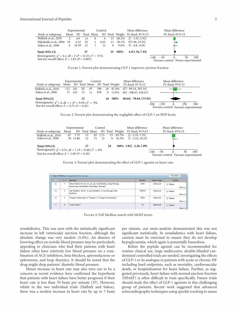
However, we found no further evidence for the extensionof GLP-1 to nondiabetics. In a randomized, double-blindcrossover trial of 20 normoglycaemic patients withoutdiabetes and with HF with ischemic heart disease, severe leftventricular impairment, NYHA II, and III, Halbirk et al. [32]found that GLP-1 infusion over 48 h increased circulatinginsulin levels and reduced plasma glucose concentrationbut had no major cardiovascular effects in patients withchronic heart failure when compared with a placebo. Theonly significant cardiovascular impacts of the infusion wereincreases in heart rate (67 ± 2 beats/min versus 65 ± 2beats/min; P = 0.016) and diastolic blood pressure (71 ±2 mmHg versus 68 ± 2 mmHg; P = 0.008). GLP-1 hadno effect on systolic blood pressure (113 ± 5 mmHg versus113 ± 4 mmHg; P = 0.95) or on LVEF (GLP-1 treatmentfrom 28 ± 2% to 30 ± 2% versus placebo 30 ± 2% to30 ± 2%; P = 0.93). Importantly, also, GLP-1 infusion didnot affect exercise capacity, VO2 max, cardiac index, strokevolume, and systemic vascular resistance during exercise.Unlike other studies, hypoglycemia was frequent with eightpatients experiencing nine episodes of hypoglycaemia (capil-lary glucose < 3.5 mmol/L) versus none with placebo. Thiscalls for caution in patients without diabetes but with HFand also reiterates the need for further studies with regard tothe use of GLP-1 agonists in nondiabetics. Intriguingly, bothGLP-1 and placebo significantly dropped BNP, although theeffects of the two infusions did not differ (−112 ± 54 pg/mLversus −65 ± 54 pg/mL, P = 0.17). Future trials looking atchanges in BNP in heart failure should bear in mind thatsmall changes need to be interpreted with caution, as it wasintriguing that placebo might have produced a significantreduction in BNP. The authors of that paper attributed thisdrop in natriuretic peptide to be due to patients’ reducedexercise during their hospital stay, more than a direct effectof the infusion. However, a recent study conducted in healthysubjects found exenatide had significant haemodynamiceffects, including natriuretic properties [33].
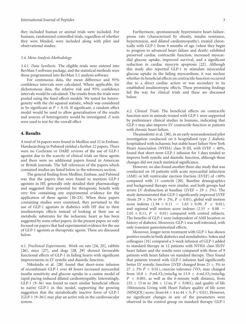
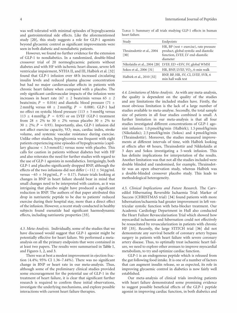
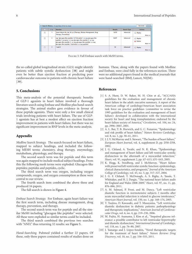
4.3. Meta-Analysis. Individually, some of the studies that wehave discussed would suggest that GLP-1 agonist might bepotentially effective for heart failure. We performed a meta-analysis on all the primary endpoints that were contained inat least two papers. The results were summarised in Table 1,and Figures 1, 2, and 3.
There was at best a modest improvement in ejection frac-tion (4.4%; 95% CI 1.36–7.44%). There was no significantchange in BNP or heart rate in our meta-analysis. Thus,although some of the preliminary clinical studies providedsome encouragement for the potential use of GLP-1 in thetreatment of heart failure, it is clear that significant furtherresearch is required to confirm these initial observations,investigate the underlying mechanisms, and explore possibleinteractions with current heart failure therapies.
Table 1: Summary of all trials studying GLP-1 effects in humanheart failure.
Study Endpoints
Thrainsdottir et al., 2004[30]
HR, BP (rest + exercise), rate pressureproduct, global systolic and diastolicfunction, LVEF, LV end-diastolicdiameter
Nikolaidis et al., 2004 [20] LVEF, ED +ESV, SV, global WMSI
Sokos et al., 2006 [31] HR, BNP, LVEF, VO2, 6-min walk
Halbirk et al., 2010 [32]BNP, BP, HR, SV, CI, LVEF, SVR, 6min hall walk test
4.4. Limitations of Meta-Analysis. As with any meta-analysis,the quality is dependent on the quality of the studiesand any limitations the included studies have. Firstly, themost obvious limitation is the lack of a large number ofstudies available to meta-analyse. Secondly, the total samplesize of patients in all four studies combined is small. Afurther limitation in our meta-analysis is that all fourstudies investigated different concentrations of GLP-1 ago-nist infusion: 1.0 pmol/kg/min (Halbirk); 1.5 pmol/kg/min(Nikolaidis); 2.5 pmol/kg/min (Sokos) and 4 pmol/kg/min(Thrainsdottir). Moreover, the studies measured improve-ments at different intervals of time, with Halbirk lookingat effects after 48 hours, Thrainsdottir and Nikkolaidis at3 days and Sokos investigating a 5-week infusion. Thishas definite implications for interpretation of the results.Another limitation was that not all the studies included weredouble blinded and randomised, for example, Thrainsdot-tir was an open observation study, whereas Halbirk wasa double-blinded crossover placebo study. This leads tomethodological heterogeneity.
4.5. Clinical Implications and Future Research. The Carv-edilol Hibernating Reversible Ischaemia Trial: Marker ofSuccess (CHRISTMAS trial) [34] found patients with morehibernation/ischaemia had greater improvement in left ven-tricular systolic function with beta-blocker treatment. OurAcademic Cardiology Department in Hull also conductedthe Heart Failure Revascularisation Trial which showed howmyocardial ischaemia and hibernation could not effectivelybe resuscitated by revascularization in patients with chronicHF [35]. Recently, the large STITCH trial [36] did notdemonstrate any survival benefit of coronary artery bypasssurgery in patients with heart failure with severe coronaryartery disease. Thus, to optimally treat ischaemic heart fail-ure, we need to explore other avenues to improve myocardialmetabolism, to try and optimize cardiac function.
GLP-1 is an endogenous peptide which is released fromthe gut following food intake. It is one of a number of factorsthat can augment insulin release, so as expected, its role inimproving glycaemic control in diabetics is now fairly wellestablished.
Our meta-analysis of clinical trials involving patientswith heart failure demonstrated some promising evidenceto suggest possible beneficial effects of the GLP-1 peptideagonist in improving cardiac function, in both diabetics and
International Journal of Peptides 5
Experimental Control Mean differenceIV, fixed, 95 % Cl
Mean differenceIV, fixed, 95% ClStudy or subgroup Mean SD Total Mean SD Total Weight
Total (95% Cl)
210
6
4.96.32
10.39
151012
011
66.63
12
15119
60.2%30.1%9.6%
2[−1.92, 5.92]9[3.46, 14.54]5[−4.8, 14.8]
100%3537
−100 −50 0 50 100Favours control
4.4[1.36, 7.44]
Halbirk et al., 2010Nikolaidis et al., 2004Sokos et al., 2006
Test for overall effect: Z = = 0.005)2.83 (P Favors experimental
Heterogeneity: χ2 = 4.1, df = 2 (P = 0.13); 12 = 51%
Figure 1: Forrest plot demonstrating GLP-1 improves ejection fraction.
Experimental Control Mean differenceIV, fixed, 95 % Cl
Mean differenceIV, fixed, 95% ClStudy or subgroup Mean SD Total Mean SD Total Weight
Favours control Favours experimental
Halbirk et al., 2010Sokos et al., 2006
112 241 20 65 196 2071 353 12 11 456 9
87.4%
Total (95%Cl) 32 29 100%
−500 −250 0 250 500
47[−89.14, 183.14]60[−298.67, 418.67]
48.64[−78.64, 175.92]
= 0.75 (P = 0.45)Heterogeneity: χ2= 0, df = 1 (P = 0.95); l2 = 0%Test for overall effect: Z
12.6%
Figure 2: Forrest plot demonstrating the negligible effect of GLP-1 on BNP levels.
Experimental Control Mean differenceIV, fixed, 95 %Cl
Mean differenceIV, fixed, 95% ClStudy or subgroup Mean SD Total Mean SD Total Weight
Halbirk et al., 2010Sokos et al., 2006
67 7.7513.86
1512
6571
7.7515
159
83.7%16.3%
2[−3.55, 7.55]7[−5.55, 19.55]
27 24 100% 2.82[−2.26, 7.89]Total (95%Cl)
Test for overall effect: Z = 1.09 (P = 0.28)Heterogeneity: χ2= −100 −50 0 50 100
Favours control Favours experimental
0.51, df = 1 (P = 0.48); l2 = 0%
78
Figure 3: Forrest plot demonstrating the effect of GLP-1 agonist on heart rate.
Figure 4: Full Medline search with MeSH terms.
nondiabetics. This was seen with the statistically significantincrease in left ventricular ejection fraction, although theabsolute change was very modest (4.4%). An absence oflowering effect on systolic blood pressure may be particularlyappealing to clinicians who find their patients with heartfailure often have relatively low blood pressure on a com-bination of ACE-inhibitors, beta blockers, spironolactone oreplerenone, and loop diuretics. It should be noted that thedrug might drop patients’ diastolic blood pressure.
Minor increase in heart rate may also turn out to be aconcern as recent evidence have confirmed the hypothesisthat patients with heart failure have better prognosis if theirheart rate is less than 70 beats per minute [37]. However,whilst in the two individual trials (Halbirk and Sokos),there was a modest increase in heart rate by up to 7 beats
per minute, our meta-analysis demonstrated this was notsignificant statistically. In nondiabetics with heart failure,caution must be exercised to ensure they do not develophypoglycaemia, which again is potentially hazardous.
Before the peptide agonist can be recommended forroutine clinical use, large multicentre, double-blinded ran-domised controlled trials are needed, investigating the effectsof GLP-1 or its analogue in patients with acute or chronic HFincluding hard endpoints, such as mortality, cardiovasculardeath, or hospitalization for heart failure. Further, as sug-gested previously, heart failure with normal ejection fraction(HFnEF) is often difficult to treat specifically. Future trialsshould study the effect of GLP-1 agonists in this challenginggroup of patients. Recent work suggested that advancedechocardiography techniques using speckle tracking to assess
6 International Journal of Peptides
Figure 5: Full Embase search with MeSH terms.
the so-called global longitudinal strain (GLS) might identifypatients with subtle systolic dysfunction [38] and mighteven be better than ejection fraction at predicting poorcardiovascular outcome in patients with chronic heart failure[39].
5. Conclusions
This meta-analysis of the potential therapeutic benefitsof GLP-1 agonists in heart failure involved a thoroughliterature search using Embase and Medline plus hand-searchstrategies. The animal studies gave evidence in favour ofthese peptide agonists. There were only a few small clinicaltrials involving patients with heart failure. The use of GLP-1 agonists has at best a modest effect on ejection fractionimprovement in patients with heart failure, but there was nosignificant improvement in BNP levels in the meta-analysis.
Appendix
Medline Search Strategy. The search focused on heart failure,mapped to subject headings, and included the follow-ing MESH terms: chemistry, drug therapy, enzymology,metabolism, physiology, and therapy.
The second search term was for peptide and this termwas again mapped to include medical subject headings. Fromthis the following mesh terms were exploded: Glucagon-likepeptides; peptides and peptides, cyclic.
The third search term was oxygen, including oxygencompounds, oxygen, and oxygen consumption as these werecentral to our review.
The fourth search item combined the above three andproduced 16 papers.
The full search is shown in Figure 4.
Embase Search Strategy. For Embase, again heart failure wasthe first search term, including disease management, drugtherapy, prevention, and therapy.
The second search term was for peptide and all the sim-ilar MeSH including “glucagon like peptides” were selected.All these were exploded so similar terms could be included.
The third search combined the previous two searcheswith “AND,” thus returning 32 results: see Figure 5.
Hand-Searching. Pubmed yielded a further 22 papers. Ofthese, only three papers contained results of studies done on
humans. These, along with the papers found with Medlineand Embase, were cited fully in the references section. Therewere no additional papers found in the medical journals thatwere hand-searched (BMJ, Lancet, NEJM).
References
[1] S. A. Hunt, D. W. Baker, M. H. Chin et al., “ACC/AHAguidelines for the evaluation and management of chronicheart failure in the adult: executive summary. A report of theAmerican college of cardiology/American heart associationtask force on practice guidelines (committee to revise the1995 guidelines for the evaluation and management of heartfailure): developed in collaboration with the internationalsociety for heart and lung transplantation; endorsed by theheart failure society of America,” Circulation, vol. 104, no. 24,pp. 2996–3007, 2001.
[2] A. L. Bui, T. B. Horwich, and G. C. Fonarow, “Epidemiologyand risk profile of heart failure,” Nature Reviews Cardiology,vol. 8, no. 1, pp. 30–41, 2011.
[3] J. J. V. McMurray and S. Stewart, “The burden of heart failure,”European Heart Journal, vol. 4, supplement D, pp. D50–D58,2002.
[4] J. G. Cleland, A. Torabi, and N. K. Khan, “Epidemiologyand management of heart failure and left ventricular systolicdysfunction in the aftermath of a myocardial infarction,”Heart, vol. 91, supplement 2, pp. ii7–ii13, ii31–ii43, 2005.
[5] K. Hogg, K. Swedberg, and J. McMurray, “Heart failurewith preserved left ventricular systolic function: epidemiology,clinical characteristics, and prognosis,” Journal of the AmericanCollege of Cardiology, vol. 43, no. 3, pp. 317–327, 2004.
[6] J. G. F. Cleland, T. McDonagh, A. S. Rigby, A. Yassin, T.Whittaker, and H. J. Dargie, “The national heart failure auditfor England and Wales 2008-2009,” Heart, vol. 97, no. 11, pp.876–886, 2011.
[7] A. M. Salmasi, P. Frost, and M. Dancy, “Left ventriculardiastolic function in normotensive subjects 2 months afteracute myocardial infarction is related to glucose intolerance,”American Heart Journal, vol. 150, no. 1, pp. 168–174, 2005.
[8] T. Tsujino, D. Kawasaki, and T. Masuyama, “Left ventriculardiastolic dysfunction in diabetic patients: pathophysiologyand therapeutic implications,” American Journal of Cardiovas-cular Drugs, vol. 6, no. 4, pp. 219–230, 2006.
[9] M. Fujita, H. Asanuma, J. Kim et al., “Impaired glucose tol-erance: a possible contributor to left ventricular hypertrophyand diastolic dysfunction,” International Journal of Cardiology,vol. 118, no. 1, pp. 76–80, 2007.
[10] J. Tamargo and J. Lopez-Sendon, “Novel therapeutic targetsfor the treatment of heart failure,” Nature Reviews DrugDiscovery, vol. 10, no. 7, pp. 536–555, 2011.
International Journal of Peptides 7
[11] T. J. Kieffer and J. F. Habener, “The glucagon-like peptides,”Endocrine Reviews, vol. 20, no. 6, pp. 876–913, 1999.
[12] C. Saraceni and T. L. Broderick, “Effects of glucagon-likepeptide-1 and long-acting analogues on cardiovascular andmetabolic function,” Drugs in R and D, vol. 8, no. 3, pp. 145–153, 2007.
[13] M. B. Toft-Nielsen, S. Madsbad, and J. J. Holst, “Determinantsof the effectiveness of glucagon-like peptide-1 in type 2diabetes,” Journal of Clinical Endocrinology and Metabolism,vol. 86, no. 8, pp. 3853–3860, 2001.
[14] J. J. Meier, D. Weyhe, M. Michaely et al., “Intravenousglucagon-like peptide 1 normalizes blood glucose after majorsurgery in patients with type 2 diabetes,” Critical CareMedicine, vol. 32, no. 3, pp. 848–851, 2004.
[15] M. Monami, F. Cremasco, C. Lamanna et al., “Glucagon-likepeptide-1 receptor agonists and cardiovascular events: a meta-analysis of randomized clinical trials,” Experimental DiabetesResearch, vol. 2011, Article ID 215764, 2011.
[16] W. C. Stanley, F. A. Recchia, and G. D. Lopaschuk, “Myocardialsubstrate metabolism in the normal and failing heart,” Physio-logical Reviews, vol. 85, no. 3, pp. 1093–1129, 2005.
[17] M. F. Essop and L. H. Opie, “Metabolic therapy for heartfailure,” European Heart Journal, vol. 25, no. 20, pp. 1765–1768, 2004.
[18] H. Taegtmeyer, “Cardiac metabolism as a target for thetreatment of heart failure,” Circulation, vol. 110, no. 8, pp.894–896, 2004.
[19] D. J. Grieve, R. S. Cassidy, and B. D. Green, “Emergingcardiovascular actions of the incretin hormone glucagon-likepeptide-1: potential therapeutic benefits beyond glycaemiccontrol?” British Journal of Pharmacology, vol. 157, no. 8, pp.1340–1351, 2009.
[20] L. A. Nikolaidis, S. Mankad, G. G. Sokos et al., “Effects ofglucagon-like peptide-1 in patients with acute myocardialinfarction and left ventricular dysfunction after successfulreperfusion,” Circulation, vol. 109, no. 8, pp. 962–965, 2004.
[21] K. Ban, M. H. Noyan-Ashraf, J. Hoefer, S. S. Bolz, D. J.Drucker, and M. Husain, “Cardioprotective and vasodilatoryactions of glucagon-like peptide 1 receptor are mediatedthrough both glucagon-like peptide 1 receptor-dependent and-independent pathways,” Circulation, vol. 117, no. 18, pp.2340–2350, 2008.
[22] E. Mannucci and C. M. Rotella, “Future perspectives onglucagon-like peptide-1, diabetes and cardiovascular risk,”Nutrition, Metabolism and Cardiovascular Diseases, vol. 18, no.9, pp. 639–645, 2008.
[23] A. K. Bose, M. M. Mocanu, R. D. Carr, C. L. Brand, and D. M.Yellon, “Glucagon-like peptide 1 can directly protect the heartagainst ischemia/reperfusion injury,” Diabetes, vol. 54, no. 1,pp. 146–151, 2005.
[24] I. Poornima, S. B. Brown, S. Bhashyam, P. Parikh, H.Bolukoglu, and R. P. Shannon, “Chronic glucagon-likepeptide-1 infusion sustains left ventricular systolic functionand prolongs survival in the spontaneously hypertensive, heartfailure-prone rat,” Circulation, vol. 1, no. 3, pp. 153–160, 2008.
[25] T. Zhao, P. Parikh, S. Bhashyam et al., “Direct effects ofglucagon-like peptide-1 on myocardial contractility and glu-cose uptake in normal and postischemic isolated rat hearts,”Journal of Pharmacology and Experimental Therapeutics, vol.317, no. 3, pp. 1106–1113, 2006.
[26] M. Matsubara, S. Kanemoto, B. G. Leshnower et al., “Singledose GLP-1-tf ameliorates myocardial ischemia/reperfusioninjury,” Journal of Surgical Research, vol. 165, no. 1, pp. 38–45,2011.
[27] M. H. Noyan-Ashraf, M. Abdul Momen, K. Ban et al., “GLP-1R agonist liraglutide activates cytoprotective pathways andimproves outcomes after experimental myocardial infarctionin mice,” Diabetes, vol. 58, no. 4, pp. 975–983, 2009.
[28] L. A. Nikolaidis, D. Elahi, T. Hentosz et al., “Recombinantglucagon-like peptide-1 increases myocardial glucose uptakeand improves left ventricular performance in conscious dogswith pacing-induced dilated cardiomyopathy,” Circulation,vol. 110, no. 8, pp. 955–961, 2004.
[29] L. A. Nikolaidis, D. Elahi, Y. T. Shen, and R. P. Shannon, “Activemetabolite of GLP-1 mediates myocardial glucose uptakeand improves left ventricular performance in conscious dogswith dilated cardiomyopathy,” American Journal of Physiology-Heart and Circulatory Physiology, vol. 289, no. 6, pp. H2401–H2408, 2005.
[30] I. Thrainsdottir, K. Malmberg, A. Olsson, M. Gutniak, and L.Ryden, “Initial experience with GLP-1 treatment on metaboliccontrol and myocardial function in patients with type 2diabetes mellitus and heart failure,” Diabetes & VascularDisease Research, vol. 1, no. 1, pp. 40–43, 2004.
[31] G. G. Sokos, L. A. Nikolaidis, S. Mankad, D. Elahi, and R. P.Shannon, “Glucagon-Like Peptide-1 Infusion Improves LeftVentricular Ejection Fraction and Functional Status in PatientsWith Chronic Heart Failure,” Journal of Cardiac Failure, vol.12, no. 9, pp. 694–699, 2006.
[32] M. Halbirk, H. Nørrelund, N. Møller et al., “Cardiovascularand metabolic effects of 48-h glucagon-like peptide-1 infusionin compensated chronic patients with heart failure,” AmericanJournal of Physiology-Heart and Circulatory Physiology, vol.298, no. 3, pp. H1096–H1102, 2010.
[33] B. Mendis, E. Simpson, I. Macdonald, and P. Mansell, “Inves-tigation of the haemodynamic effects of exenatide in healthymale subjects,” British Journal of Clinical Pharmacology. Inpress.
[34] J. G. Cleland, D. J. Pennell, S. G. Ray et al., “Carvedilolhibernating reversible ischaemia trial: marker of successinvestigators. Myocardial viability as a determinant of theejection fraction response to carvedilol in patients with heartfailure (CHRISTMAS trial): randomised controlled trial,” TheLancet, vol. 362, no. 9377, pp. 14–21, 2003.
[35] A. P. Coletta, J. G. F. Cleland, D. Cullington, and A. L. Clark,“Clinical trials update from heart rhythm 2008 and heartfailure 2008: ATHENA, URGENT, INH study, HEART andCK-1827452,” European Journal of Heart Failure, vol. 10, no.9, pp. 917–920, 2008.
[36] E. J. Velazquez, K. L. Lee, M. A. Deja et al., “Coronary-arterybypass surgery in patients with left ventricular dysfunction,”The New England Journal of Medicine, vol. 364, no. 17, pp.1607–1616, 2011.
[37] K. Fox, I. Ford, P.G. Steg, M. Tendera, M. Robertson, andR. Ferrari, “On behalf of the BEAUTIFUL investigators/Heartrate as a prognostic risk factor in patients with coronaryartery disease and left-ventricular systolic dysfunction (BEAU-TIFUL): a subgroup analysis of a randomised controlled trial,”The Lancet, vol. 372, no. 9641, pp. 817–821, 2008.
[38] M. Galderisi, V. S. Lomoriello, A. Santoro et al., “Differencesof myocardial systolic deformation and correlates of diastolicfunction in competitive rowers and young hypertensives:a speckle-tracking echocardiography study,” Journal of theAmerican Society of Echocardiography, vol. 23, no. 11, pp.1190–1198, 2010.
[39] J. Nahum, A. Bensaid, C. Dussault et al., “Impact of longi-tudinal myocardial deformation on the prognosis of chronicheart failure patients,” Circulation Cardiovascular Imaging, vol.3, no. 3, pp. 249–256, 2010.
Hindawi Publishing CorporationInternational Journal of PeptidesVolume 2012, Article ID 965238, 19 pagesdoi:10.1155/2012/965238
Review Article
Antimicrobial Peptides as Infection Imaging Agents:Better Than Radiolabeled Antibiotics
Muammad Saeed Akhtar, Muhammad Babar Imran,Muhammad Afzal Nadeem, and Abubaker Shahid
Nuclear Medicine Division, Punjab Institute of Nuclear Medicine (PINUM), Faisalabad 38000, Pakistan
Correspondence should be addressed to Muammad Saeed Akhtar, saeed [email protected]
Received 8 December 2011; Revised 9 February 2012; Accepted 11 March 2012
Academic Editor: Mirian A. F. Hayashi
Copyright © 2012 Muammad Saeed Akhtar et al. This is an open access article distributed under the Creative CommonsAttribution License, which permits unrestricted use, distribution, and reproduction in any medium, provided the original work isproperly cited.
Nuclear medicine imaging techniques offer whole body imaging for localization of number and site of infective foci inspite oflimitation of spatial resolution. The innate human immune system contains a large member of important elements includingantimicrobial peptides to combat any form of infection. However, development of antibiotics against bacteria progressed rapidlyand gained popularity over antimicrobial peptides but even powerful antimicrobials failed to reduce morbidity and mortalitydue to emergence of mutant strains of bacteria resulting in antimicrobial resistance. Differentiation between infection andinflammation using radiolabeled compounds with nuclear medicine techniques has always been a dilemma which is still to beresolved. Starting from nonspecific tracers to specific radiolabeled tracers, the question is still unanswered. Specific radiolabeledtracers included antibiotics and antimicrobial peptides which bind directly to the bacteria for efficient localization with advancednuclear medicine equipments. However, there are merits and demerits attributed to each. In the current paper, radiolabeledantibiotics and radiolabeled peptides for infection localization have been discussed starting with the background of primitivenonspecific tracers. Radiolabeled antimicrobial peptides have certain merits compared with labeled antibiotics which make themsuperior agents for localization of infective focus.
1. General Introduction
Blood-derived antimicrobial proteins and peptides beingpart of innate immunity target the microbial membranesleading to growth arrest and, in some instants, neutralizationof proinflammatory surface components like lipopolysaccha-rides. Different inflammatory response blood cells like neu-trophils, eosinophils, macrophages, and platelets contain an-timicrobial proteins and peptides which have affinity for sur-face lipids of microbial as opposed to eukaryotic cells. Neu-trophils contain primary and secondary granules in their cy-toplasm which contain antimicrobial proteins and peptides.Lactoferrin is localized in the secondary granules, which hasdirect microbicidal effect, presumably via membrane disrup-tion. Activated neutrophils release bactericidal/permeabilityincreasing protein (BPI) into inflammatory fluids where it ispotentially bactericidal. Serprocidins are proteases with cyto-toxic activity localized in neutrophil primary granules.
Cathelicidins are also antimicrobial peptides within second-ary granules of neutrophils. The defensins are a family of 4-Kd peptides with broad cytotoxic activity against bacteria, fu-ngi, parasites, viruses, and host cells. Humans express α-de-fensins in neutrophils and β-defensins in intestinal Panethcell, as well as pulmonary and reproductive epithelia. The de-fensins peptides, calprotectin protein, and ubiquicidin ca-tionic peptides are found in macrophages [1]. Platelet α-grounds contain certain cationic antimicrobial peptides hav-ing broad spectrum antimicrobial activity [2]. Multiple pro-teins and peptides have been radiolabelled by multiple inves-tigators for specific localization of infection foci but each hadcertain demerits. More attention diverted to development ofnew antibiotics followed by radiolabeling but these face thegrowing problem of microbial resistance.
Differentiation between infection and inflammation isusually difficult. Clinicians use a variety of clues, for example,clinical, laboratory, and radiological tests, to aid in diagnosis
2 International Journal of Peptides
and influence decision making. Commonly employed anduseful modalities for demonstration of any focal lesion in-clude conventional radiological techniques such as X-ray, ul-trasound, computerized tomography (CT), magnetic reso-nance imaging (MRI), which demonstrate structural abnor-malities which take some time to become visible, may notalways be present, and their resolution lag behind cure. Inaddition they are neither inflammation nor infection speci-fic. Nuclear medicine has enhanced infection imaging be-cause it depends on the demonstration of pathophysiologicaland pathological changes, which occur earlier in infectionprocess and also resolve quicker after cure of the infectioncompared with gross changes in structure [3]. Scintigraphicimaging of inflammation can be achieved in two ways. Thefirst is to utilize the locally enhanced vascular permeability byinjecting radiolabelled molecules that show increased extra-vasation at the site of infection/inflammation. The alterna-tive is to exploit the diapedesis and chemotaxis of leucocytes,either by radiolabelling white blood cells of the patientsex vivo or by directly targeting leukocyte antigens or receptorsin vivo via administration of radiolabelled antigranulocytemonoclonal antibodies on receptor-binding ligands [4].However, nuclear medicine utilizes radiation and must beused as a diagnostic modality in cases where other noniso-topic and noninvasive techniques fail to achieve the target.
Scintigraphy has the advantage of early elucidation ofpathophysiological changes in the infective process; however,it is limited by poor resolution. Recent advances in nuclearmedicine technology resulted in commercially availableinstrumentation such as single-photon emission computedtomography (SPECT) and positron emission tomography(PET) that have markedly improved anatomical details.Autologous in vitro 111In-oxinate- or 99mTc-HMPAO-labeledleukocytes are still the gold standard for imaging infections[5–7]. Planar images with gamma cameras are handicappedwith limited resolution that is not sufficient for assessingthe extent of disease. SPECT increases the sensitivity of thenuclear medicine procedures [8, 9], but precise anatomicallocalization of organs is still not possible. Hybrid SPECT/CTimproves the diagnostic accuracy when subjected to 99mTc-HMPAO-labeled leukocytes in patients with suspected osteo-myelitis [10]. Marked improvement in sensitivity and def-inition of the extent of infection has been documented withSPECT/CT using 67Ga- and 111In-labeled leukocytes [11].PET/CT with 18F-FDG-labeled autologous leukocytes hasfurther improved the diagnosis and localization of infectionlesions [12]; however, the technique is time consuming, de-mands a sterile environment, and carries the risk of transmis-sion of blood-borne diseases. Antimicrobial compounds thatbind to the bacteria would be specific for infection loca-lization if labeled with a suitable isotope because of theirselective adhesion to the causative agents [3]. An early anti-biotic radiopharmaceutical was 99mTc-ciprofloxacin, whichis an analog of a broad-spectrum quinolone antibiotic havingthe property of binding to DNA gyrase of bacteria and in-hibiting DNA synthesis [13]. This 99mTc agent showed en-couraging results in various infections [14–16]; however,specificity was lower than expected, and its accumulation innoninfectious/inflammatory sites has also been reported
[17]. Due to nonspecific accumulation in inflammatory sites,this agent has been proposed for identifying the presence anddistribution of inflammation within joints [18]. Bacterialresistance to ciprofloxacin is another disadvantage, which re-sults in false-negative results [19].
Antimicrobial peptides, produced by phagocytes, epithe-lial cells, endothelial cells, and many other cell types, are animportant component of innate immunity against infectionby a variety of pathogens [20]. These peptides show anti-bacterial, antiviral, and antifungal activities in vitro. Bacterialinfections with Staphylococcus aureus and Klebsiella pneumo-niae have been visualized in mice by 99mTc-labeled humanneutrophil peptide-1 [21]. The basis of the antimicrobialactivity of these peptides is the interaction of the cation-ic domains with the negatively charged surface of the micro-organisms. The antimicrobial peptide ubiquicidin UBI (29–41) (TGRAKRRMQYNRR; 1,693 Da) was originally isolatedfrom mouse macrophage cells. This peptide is identical orhighly homologous to S30, a protein that was purified fromthe small ribosomal subunit fraction of rat liver and shownto be present in various human and murine tissues [22]. La-ter, an identical UBI was isolated from human airway epi-thelial cells. This peptide was labeled with 99mTc, whichtargeted bacterial cells but not sterile inflammatory processesin experimental animals [23]. In later experiments, it alsoshowed accumulation with high accuracy in fungal infec-tions. This tracer was also used for detection of Staphylo-coccus aureus infections in mice and Klebsiella pneumoniaein rabbits. As controls, inflammation was produced by lipo-polysaccharides (LPSs) and heat-killed microorganisms [24].Interactions of cationic peptides with bacterial envelopes in-volve insertion of the peptide into microbial membranes [25]and possibly a sequence-dependent interaction of the antimi-crobial peptides with microorganisms [26]. Multiple animalstudies using 99mTc-labeled ubiquicidin (29–41) showedencouraging results for differentiation between infectionand inflammation model. 99mTc-UBI (29–41) scintigraphyshowed more accumulation of tracer in Staphylococcus-aureus-induced infection compared with that of Escherichiacoli infection model. Optimum time for imaging was 60 minafter tracer injection [27]. In another study with this radio-labelled peptide, it was concluded that its accumulation isdirectly related to viable number of bacteria as tracer accu-mulation in infective foci declined after administration ofciprofloxacin which reduced the number of bacteria sensitiveto this antibiotic. However, serial imaging with 99mTc-UBIcan be utilized for monitoring efficacy and direction of an-tibiotic treatment [28]. Use of radiolabeled antimicrobial pe-ptides is only recommended in cases where physician or sur-geon is in dilemma to differentiate infection from inflamma-tion. This would avoid blind use of prophylactic antibioticsor as broad spectrum coverage of infection, which results inheavy expenditure and side effects of unnecessary medicines.
Phase-I clinical trial with this novel radiolabelled peptideshowed overall sensitivity, specificity, and accuracy of 100%,80%, and 94.4%, respectively, in patients with soft tissue in-fections and osteomyelitis. However, optimum time forimaging was 30 min after intravenous administration of ra-diotracer [29].
International Journal of Peptides 3
2. Detection of Infection byNonspecific Tracers
2.1. Gallium-67-Citrate. The oldest radiopharmaceuticalproposed for imaging inflammation is Gallium-67 citratewhich has been used for infection and inflammation eversince its discovery in 1971 [30]. 67Ga is a cyclotron-producedradionuclide, with a half-life of 78 hours, emits a broad spec-trum of gamma rays between 93 keV and 880 keV. The en-ergy peaks that are most suitable for gamma camera imag-ing are 93 keV, 184 keV, 296 keV, and 388 keV [31]. Afterintravenous injection, 67Ga binds to transferrin. This com-plex extravasates at the site of inflammation due to the lo-cally enhanced vascular permeability, and in the inflamma-tory lesion it may transchelate to lactoferrin as present inleukocytes. The B-lymphocytes have lactoferrin-binding siteson their surface, which have high affinity for gallium. Ad-ditionally, macrophages engulf protein iron complexes andcellular debris, thereby accumulating gallium. Bacteria andfungi contain siderophores which are released for the pur-pose of scavenging iron and consequently bind gallium as agallium-siderophore complex [32]. The agent is excreted pa-rtly via the kidneys (especially during the first 24 hours afterinjection) and via the gastrointestinal tract; therefore colon isthe target organ. Oral laxatives to reduce bowel activity andto reduce dose to large bowel are not required [33, 34]. Phy-siological uptake of the radiolabel also occurs in liver, bone,bone marrow, salivary glands, nasopharynx, and lacrimalglands. For infection or inflammation, imaging can often beaccomplished at 48 hours, or even 24 hours, after injection.Planar imaging is performed in the anterior and posteriorprojection, to include the head, neck, chest, abdomen, pelvis,and proximal extremities. SPECT imaging is performed at 72hours, which improves the sensitivity and specificity. Mostpatients exhibit bowel activity at this time; therefore planarand SPECT imaging of abdomen can be performed at 5–7 days after injection. Inspite of SPECT imaging, there arelow spatial resolution and the lack of anatomic landmarks ofscintigraphy [35].
Although 67Ga-citrate scintigraphy has high sensitivityfor both acute and chronic infection and noninfectious in-flammation, there are several shortcomings that limit its clin-ical application. The specificity of the technique is low, dueto physiological bowel excretion and accumulation in ma-lignant tissues and areas of bone remodeling. In addition, theradiopharmaceutical has unfavorable imaging characteristics(long physical half-life and high energy gamma radiations),causing high radiation-absorbed doses. Furthermore, opti-mal imaging often requires delayed recordings up to 72hours. These unfavorable characteristics, in combinationwith the development of newer radiopharmaceuticals, havenarrowed the clinical indication for gallium scintigraphy tocertain conditions such as lung infections and chronic os-teomyelitis. The sensitivity and specificity for chronic osteo-myelitis are lower than for acute osteomyelitis [36, 37]. Use ofSPECT/CT with 67Ga improves diagnostic efficiency as com-pared with planar or SPECT scanning [11]. Gallium scanis most often used in patients with fever of knownorigin (FUO), suspected vertebral osteomyelitis, chronic
osteomyelitis, pulmonary/mediastinal infections, tuberculo-sis, sarcoidosis, and retroperitoneal fibrosis. This agent isalso valuable for evaluation and followup of drug-inducedpulmonary toxic agents like bleomycin and amiodarone. Im-munocompromised and neutropenic patients are also candi-dates for evaluation with gallium scanning [38].
2.2. Nonspecific Immunoglobulins. Initially it was hypothe-sized that human polyclonal immunoglobulin (HIG) wasretained in infectious foci owing to the interaction with Fc-Υreceptors as expressed on infiltrating leucocytes [39]. Laterstudies showed that radiolabelled HIG accumulates ininfectious foci by nonspecific extravasation due the locallyenhanced vascular permeability [40]. For clinical use, HIGhas been labeled with 111In-oxinate as well as with 99mTc.Both agents have slow blood clearance and physiological up-take in the liver, the spleen, and the kidneys. The 99mTc-label-ed preparation has the known ideal radiation characteristics,while the 111In-labeled preparation allows imaging at timepoints beyond 24 hours after injection. 111In-oxinate or99mTc-labeled HIG has been extensively tested in a largenumber of clinical studies. It has shown excellent perfor-mance in the localization of musculoskeletal infection andinflammation [41]. In addition, good results have been re-ported in pulmonary infection particularly in immunocom-promised patients [42] and abdominal inflammation. A ge-neral limitation is the long time span between injection andfinal diagnosis (24–48 hours) [43, 44].
2.3. Liposomes. Liposomes are spheres consisting of one ormore lipid bilayers surrounding an aqueous space. They wereproposed as vehicles to image infection some 20 years ago,but the preparations used in those early years were clearedfrom the circulation very rapidly by the mononuclear phago-cyte system (MPS). However, if the surface of the liposomesis coated with a hydrophilic polymer such as polyethyleneglycol (PEG), they circumvent recognition by the MPS,leading to a prolonged residence time in the circulationand enhanced uptake at pathological sites by extravasationdue to locally enhanced vascular permeability [45]. Suchstabilized PEG-liposomes can be labeled with 111In-oxinateand with 99mTc, either using hexamethylpropylene amineoxime (HMPAO) as an internal label or via HYNIC as anexternal chelator. Labeling is easy and takes only minutes[46]. The first clinical evaluation showed good imaging offocal infection. In patients suspected of harboring infectiousor inflammatory disease, 99mTc-PEG-liposomes were directlycompared with 111In-IgG scintigraphy. 99mTc-PEG-liposomescintigraphy has shown high sensitivity (94%) and specificity(89%) [47].
2.4. The Avidin-Biotin System. Avidins are a family of pro-teins present in the eggs of amphibians, reptiles, and birds;streptavidin is a member of the same family. Avidin andstreptavidin (mol. wt. 66,000 and 60,000, resp.) bind to bio-tin with extremely high affinity. Biotin is a compoundof low molecular weight that can be radiolabelled. Theavidin-biotin approach is based on the fact that avidin (orstreptavidin) will nonspecifically localize at sites of infection
4 International Journal of Peptides
owing to increased vascular permeability. Avidin (or strep-tavidin) is injected as a pretargeting agent, followed hourslater by a second injection with radiolabelled biotin. Gooddiagnostic accuracy was demonstrated in studies of vascularinfection and chronic osteomyelitis [48–51].
3. Detection of Infection by Specific Tracers
3.1. Radiolabeled White Blood Cells. Ex vivo labeled autol-ogous leucocytes were developed in the 1970s and 1980sand their use is still considered the “gold standard” nuclearmedicine technique for infection and inflammation imaging[5–7]. Although a variety of in vitro leukocyte-labeling tech-niques have been used, the most commonly used proceduremakes use of the lipophilic compounds 111In-Oxyquinolineand 99mTc-HMPAO. The radiolabeling procedure takesabout 2-3 hours. Because all the cellular components of theblood can be labeled, it is necessary to separate the leukocytesfrom the erythrocytes and platelets. After withdrawal, there-fore, the syringe containing the blood is kept in the uprightposition for about 1-2 hour to promote erythrocyte sedimen-tation. After the erythrocytes have been separated, the leuko-cytes must be separated from platelets. The leukocyte-richplasma is centrifuged, and the leukocyte pellet that formsat the bottom of the tube is removed, incubated with the ra-diolabel, washed, and reinjected into the patient. The us-ual dose of 111In-labeled leukocytes is 10–18.5 MBq (300–500 μCi); the usual dose of 99mTc-HMPAO-labeled leuko-cytes is 185–370 MBq (5–10 mCi). Uptake of labeled leuko-cytes is dependent on intact chemotoxis, the number andtypes of cells labeled, and the cellular component of a par-ticular inflammatory response. A total white cell count of atleast 2000/mm3 is needed to obtain satisfactory images. Neu-trophils can be radiolabeled and hence the procedure is mostuseful for identifying neutrophil-mediated inflammatoryprocesses, such as bacterial infections. The procedure is lessuseful for those illnesses in which the predominant cellularresponse is other than neutrophilic, such as tuberculosis [52].
3.1.1. 111In-Oxine-Labeled Leukocytes. For over two decades,111In-oxine-labeled leukocytes have been used to imageinfection and inflammation. The scintigraphic images reflectthe distribution of white blood cells in the body. Since anabscess or other localized infection consists primarily ofleukocytes, the radiopharmaceutical localizes at the site ofinfection [53–61]. After intravenous administration, there isinitial sequestration of the labeled leucocytes in the lungs,with subsequent rapid clearance of the activity from thelungs. The radiolabel rapidly clears from the blood and inmost cases there is high uptake in granulocytic infiltrates,while a substantial portion of the leucocytes (presumably thedamaged cells) accumulate in the spleen. Thus, as a radio-pharmaceutical, radiolabelled leucocytes are a specific indi-cator for leukocytic infiltration, but not for infection [62, 63].At 24 hour, after injection, the usual imaging time for 111In-labeled leukocytes, the normal distribution of activity islimited to the liver, spleen, and bone marrow. Large field ofview gamma camera equipped with medium energy parallel
Table 1: Causes of false-negative and false-positive 111In leukocytestudies.
False Negative
Encapsulated nonpyogenic abscess
Vertebral osteomyelitis
Chronic low-grade infection
Parasitic, mycobacterial or fungal infections
Intrahepatic, perihepatic, or splenic infection
Hyperglycemia
Steroids
False Positive
Gastrointestinal bleeding
Pseudoaneurysm
Healing fracture
Soft tissue tumor
Surgical wounds, stomas, or catheter sites
Tumors
Accessory spleens
hole collimator is used with 15% window centered on 174-Kev photopeak and 20% window centered on the 247-Kevphotopeak. Advantages of the 111In label are a very stablelabel and constant normal distribution of activity limited toliver, spleen, and bone marrow. The 67-hour half-life ofisotope allows delayed imaging, which is particularly valuablein musculoskeletal infection. Another advantage is conduc-tion of bone or bone marrow scan immediately after com-pletion of 111In-labeled study which is a limitation with99mTc-labeled tracers [9]. Disadvantages of the 111In label in-clude a low photon flux, less than ideal photon energies, andthe fact that a 24-hour interval between injection and ima-ging is generally required. Causes of false-negative and false-positive 111In-leukocyte study are summarized in Table 1[64]. Drawbacks of 111In-labeled white blood cells are labori-ous and time-consuming preparation, requiring specializedequipment and can be hazardous. Almost 3 hours are re-quired for isolating and labeling a patient’s white blood cellsby a trained technician. In addition, the need to handle po-tentially contaminated blood can lead to transmission ofblood-borne pathogens such as HIV and HBV. As anatomicalland marks are not properly outlined with scintigraphy, thesame is the limitation with 111In-WBC planar images. How-ever, SPECT/CT with 111In-WBC scintigraphy markedlyimproves accurate identification of infection sites [11].
3.1.2. 99mTc-HMPAO-Labeled Leukocytes. The normal bio-distribution of 99mTc-HMPAO-labeled leukocytes is morevariable. In addition to the reticuloendothelial system, ac-tivity is also normally present in the genitourinary tract, largebowel, blood pool, and occasionally the gall bladder [65].The interval between injection and imaging varies with indi-cation; in general, imaging is usually performed within a fewhours after injection. 99mTc-HMPAO has theoretical advan-tages over 111In-labeled leukocytes. 99mTc, being generatorproduced on site, could be immediately available for radi-olabeling. The radiation dose to the patient would be
International Journal of Peptides 5
significantly lower, permitting a higher administered activity.The higher photon yield of 99mTc would result in superiorimage resolution and improved infection detectability andaccuracy.
HMPAO preferentially labels granulocytes, a potentialadvantage for imaging acute purulent processes. Unlike111In-oxine-labeled leukocytes, 99mTc-HMPAO-labeled leu-kocytes are cleared by the hepatobiliary and renal systems[65–70]. Disadvantages include genitourinary tract activity,which appears shortly after injection and colonic activitywhich appears 4 hours after injection. The instability of thelabel and the short half-life of 99mTc are disadvantages when24-hour imaging is needed. This occurs in those infectionsthat tend to be indolent and for which several hours may benecessary for accumulation of a sufficient quantity of labeledleukocytes to be successfully imaged. Bone or bone marrowscan if indicated after 99mTc-HMPAO-WBC scan has to bedelayed at least for 48 hours and preferably 72 hours. For bet-ter anatomical localization of infection site, SPECT/CT with99mTc-HMPAO-WBC scintigraphy is far better than pla-nar/SPECT imaging with other infecting imaging agents[10].
3.2. Antigranulocyte Antibodies and Antibody Fragment. Sev-eral monoclonal antibodies reactive with antigens expressedon granulocytes (NCA, CD15, CD66, and CD67) havebeen developed. At least three antigranulocyte antibodieshave been tested for infection imaging: anti-NCA-95 IgG(BW250/183) [71, 72], anti-NCA-90 Fab’ (Immu-MN3, Leu-koScan: anti-CD66) [73], and anti-SSEA-1 IgM (LeuTech:anti-CD15) [74–76]. Each of these antigranulocyte antibod-ies labeled with 99mTc or 123I allowed accurate delineationof infection [59]. The antigranulocyte antibody-based radio-pharmaceuticals visualized infectious foci in patients withsensitivity between 80% and 90% [77].
The use of antibody fragments instead of the whole anti-body seems to be more advantageous, since such fragmentsappear to be less immunogenic. In addition, antibody frag-ments show faster blood clearance and may thus provide ear-lier diagnosis. 99mTc-labeled antigranulocyte Fab’ fragment(LeukoScan) has been registered in Europe as an infectionimaging agent.
3.3. Chemotactic Peptides. A wide variety of peptides thatbind to receptors expressed on white blood cells have beentested for the detection of infection. One of the first receptorbinding peptide tested for its ability to image infectious fociwas chemotactic peptide formyl-Met-Leu-Phe. This tripep-tide, which is N-terminally formylated, is a chemotactic fac-tor produced by bacteria which binds to receptors on gran-ulocytes and monocytes with high affinity [78–80]. A draw-back of using these biologically potent peptides is a transientbut severe reduction in peripheral leukocyte counts. Severalantagonists were developed to circumvent this undesirablebiological activity of the radiolabelled chemotactic peptide.However, these antagonists had lower uptake in the infec-tious focus, most likely owing to reduced affinity for the re-ceptor [81].
3.4. Cytokines. The attraction of leukocytes to tissues is es-sential for inflammation and host response to infection. Theprocess is controlled by chemokines, which are chemotacticcytokines. Over 40 chemokines have been identified to date,most of them in the past few years. Chemokines induce cellmigration and activation by binding to specific G-protein-coupled cell surface receptors on target cells. Their receptorsare expressed on different types of leukocytes. Some are re-stricted to certain cells, whereas others are more widelyexpressed [82]. Labeled cytokines are an interesting classof protein radiopharmaceuticals of low molecular weight(<20,000).
3.5. Interleukin-1. Interleukin-1 (IL-1) binds receptors ex-pressed mainly on granulocytes, monocytes, and lympho-cytes, with high affinity. Studies in mice with focal Staphylo-coccus aureus infections showed specific uptake of radioiodi-nated IL-1 at the site of infection [83]. Fever, haemodynamicand hematological side effects, occurring even at very lowconcentrations, are the major drawbacks of the use of sucha biologically active protein [84].
3.6. Interleukin-2. The IL-2 is considered to bind specificallyto IL-2 receptors expressed on activated T lymphocytes. In astudy in an animal model of human autoimmune diabetesmellitus, Hashimoto’s thyroiditis, Grave’s disease, Crohn’sdisease, Coeliac disease and other autoimmune diseases, de-monstrated localization of 123I or 99mTc-labeled IL-2 at thesite of lymphocytic infiltration [85, 86].
3.7. Interleukin-8. IL-8 binds to receptors on neutrophilswith high affinity. In rabbits with focal Escherichia coli infec-tion, accumulation of 123I-labeled IL-8 in the abscess wasrapid and high. The specific activity of this IL-8 prepara-tion was relatively low, resulting in a transient reductionin peripheral leukocyte counts to 45% after a dose of25 μg/kg 123I-IL-8, followed by leukocytosis for several hours.Recently, a 99mTc-labelled IL-8 preparation was developedusing HYNIC as a chelator. In rabbits with Escherichia coliinfection, high abscess uptake of 99mTc-HYNIC-IL-8 andhigh abscess-to-background ratios were obtained comparedwith those obtained using the radioiodinated preparation[87–91].
3.8. Platelet Factor-4. Platelet factor 4 (PF-4), like IL-8, is amember of the CXC chemokines. PF-4 binds the CXC type II(= IL-8 type B) receptors expressed on neutrophils and mo-nocytes. In a rabbit model of infection, 99mTc-P483H clearlydelineated the infectious foci as early as 4 hours after in-jection. No systemic side effects were observed. 99mTc-P483Hhas been studied in patients to test its applicability as animaging agent for scintigraphic detection of infection andinflammation, with fair results (82% sensitivity, 77% speci-ficity) [92, 93].
3.9. Detection of Infection by 18F-Deoxyglucose (FDG). FDGis transported into cells by glucose transporters and is phos-phorylated by hexokinase enzyme to 18F-2-FDG-6 phosphate
6 International Journal of Peptides
but is not metabolized. The degree of cellular FDG uptake isrelated to the cellular metabolic rate and the number of glu-cose transporters [94–96]. Activated inflammatory cells alsodemonstrate increased expression of glucose transporters. Inaddition, in inflammatory conditions, the affinity of gluc-ose transporters for deoxyglucose is apparently increased byvarious cytokines and growth factors, a phenomenon thathas not been observed in tumors [97]. Although FDG up-take can produce false-positive results in patients with knownor suspected malignancy, FDG represents another potentiallyuseful radiotracer in the setting of infection and inflamma-tion. The areas of normal distribution of FDG include thebrain, myocardium, and genitourinary tract. Activity in thebone marrow, stomach, and bowel is variable [98]. Thymicuptake, especially in children, can also be observed [99].Hepatic and splenic uptake are generally of low grade anddiffuse; however intense uptake in spleen may be visualizedin the setting of infection [100].
Positron emission tomography (PET) with 18FDG is nowwidely used. FDG is taken up by inflammatory cells with in-creased metabolic requirements. In contrast to glucose, de-oxyglucose cannot leave the cell once it is taken up, so it canbe used to image scintigraphically cells with high glucose up-take such as tumor cells and inflammatory cells. FDG-PEThas been studied in a wide variety of infections, includinglesions of bacterial, tuberculous, fungal, soft tissue, and boneinfections [101, 102]. Sensitivity and specificity have gener-ally exceeded 90%. This radiotracer has high positive pre-dictive value to localize in infective sites of patients withAIDS and fever of unknown origin (FUO). This techniqueaccurately helps identify sites of infective endocarditis andis promising supplement to conventional echocardiography[103]. FDG-PET has been especially successful in cases ofosteomyelitis [104–107]. For vertebral osteomyelitis, highsensitivity, specificity, and accuracy comparable to those ofgallium imaging have been reported [108]. High spatial re-solution and rapid accumulation in infectious foci are sig-nificant advantages over conventional imaging techniquessuch as the use of labeled leucocytes. However, the fact thatuptake occurs in any cell type with high glycolytic activityis a serious limitation of the use of FDG-PET for infectionimaging, restricting its specificity [109]. PET is highly sen-sitive but may be unable to define the anatomic locationof a focus of increased 18F-FDG accumulation. The hybridPET/CT technology improved diagnostic accuracy withprecise registration of metabolic and structural imaging data.18F-FDG PET/CT utilized for workup of osteomyelitis indiabetic foot correctly identified 93% of all infected sites[110]. However, 18F-FDG-WBC is a nonspecific tracer of in-creased glucose metabolism and does not accumulate onlyin sites of infection and inflammation but also shows false-positive results in tumor and postoperative changes [111].18F-FDG-labeled autologous leukocytes PET/CT for infec-tion detection has demonstrated sensitivity, specificity, andaccuracy of 91%, 85%, and 90%, respectively. Negativepredictive value of 100% with this technique is a hallmark.18F-FDG-WBC imaging is superior to 18F-FDG alone for in-fection detection as well as for assessment of response of
infection to antibiotic treatment; however limited availabilityand cost are the limitations [12].
3.10. Detection of Infection by Radiolabeled Antibiotics
3.10.1. Ciprofloxacin. The first antibiotic developed as a ra-diopharmaceutical was 99mTc-ciprofloxacin having many ofthe properties of an ideal infection-specific agent [3]. Cipro-floxacin is a broad spectrum quinolone which binds specif-ically to bacterial DNA gyrase, inhibits DNA synthesis, andhas been proposed to distinguish infection from inflamma-tion [112, 113]. It is retained at sites of infection and as-sociates freely with metal ions, allowing it to be labeled withtechnetium. Ciprofloxacin also binds to the equivalent mam-malian enzyme, topoisomerase II, but with 100 to 1000 timeslesser affinity and the binding is readily reversible. Similarly,it penetrates neutrophils, macrophages, and other cells andtissues, but is not retained for prolonged periods. Thus afterthe initial distribution phase, as the serum concentrationfalls, the antibiotic readily leaches out of the cells and tissuesinto tissue fluid and then the blood and excreted freely,predominantly in the urine. However, it is retained at sites ofinfection, giving high target to background ratio permittinginfection-specific imaging when sequential images are takenat 1, 4 and if required 24 hours after injection [3].
In vitro, ciprofloxacin is taken up by a wide variety ofGram-positive, Gram-negative, and anaerobic bacteria (in-cluding ciprofloxacin-resistant bacteria as long as the resis-tance is not mediated by cell membrane impermeability,which prevents entry of the antibiotic into the bacterial cell)but not by dead bacteria or white cells [114, 115]. Biodis-tribution and dosimetry of 99mTc-ciprofloxacin show rapid,predominantly urinary excretion of the tracer, with low-to-absent brain, lung, and bone marrow uptake and low liveruptake and excretion. Highest radiation dose was receivedby urinary bladder. Imaging conditions were excellent forboth thoracic and abdominal regions, even at early time (60minutes) after injection [116]. In a comparative study in51 patients, it demonstrated greater specificity (96%) forimaging infection compared with white cell imaging (84%)[113]. High specificity for bacterial infection was confirmedin a subsequent study involving 90 patients [117]. It alsoshowed that some infections due to ciprofloxacin-resistantbacteria could be imaged and that prior antibiotic treatmentdid not significantly affect the imaging result. The sensitivityand specificity of ciprofloxacin imaging have been validatedfurther in a large multicentre study involving 879 patients,with a wide variety of infective and noninfective conditions(including noninfective inflammatory disorders), in eightcountries, under the auspices of the International AtomicEnergy Agency (IAEA) [118]. No adverse reactions occurredand antibiotic-resistant organisms did not emerge as a re-sult of the administration of antibiotic into patients. This wasexpected because only a tracer dose of ciprofloxacin ispresent in the kit, 2 mg, which is 1/200th of a single thera-peutic intravenous dose of ciprofloxacin (400 mg). Sensi-tivity (85.4%) and specificity (81.7%) for imaging sites ofinfection were good, but varied according to the type of

International Journal of Peptides 7
infections imaged. The highest sensitivity (in excess of 90%)was seen in osteomyelitis, septic arthritis, infection of ortho-pedic prostheses (which is often difficult to diagnose by stan-dard techniques and differentiate from aseptic loosening),and culture-proven soft tissue and abdominal infections andtuberculosis. Ciprofloxacin has several advantages over esta-blished, for example, radiolabeled leucocytes, and othermethods for imaging infection, which include the following:(1) specificity for infection, (2) lack of bone marrow uptake,which is a significant advantage in imaging bone and jointand orthopedic prostheses infections, (3) ease and cost ofpreparation of the agent, (4) ex vivo labeling, which avoidscontact with blood and hence the risk of acquiring blood-borne infections such as H1V and hepatitis B and C, (5) in-dependence of the host inflammatory response and neu-trophil count and hence it can be used to image infections inimmunocompromised patients, including those who areneutropaenic, where culture is often negative and white cellimaging unreliable, and (6) availability in a kit format withlong shelf-life, making it user friendly and more widely avail-able [3]. Certain studies have revealed false positive uptake of99mTc-ciprofloxacin in sterile inflammation questioning itsspecificity [14–19].
3.10.2. 99mTc Sparfloxacin. Sparfloxacin is fluoroquinolon,antibiotic effective against a wide range of Gram-positive ba-cteria. It was successfully labeled and evaluated in invitro ani-mal model in which 99mTc-labeled antibiotic accumulated ininfective sites of live Staphylococcus aureus bacteria and re-vealed no uptake in control models with heat-killed bacteria[119].
3.10.3. 99mTc-Enroflaxacin. This is also quinolone antibioticused mainly in veterinary practice. Labeled with 99mTc,in vitro studies did not show encouraging results in differ-entiating infection from inflammatory foci [120].
3.10.4. 99mTc-Ceftizoxime. Ceftizoxime is a 3rd-generationcephalosporin effective against a wide range of Gram-posi-tive bacteria especially Staphylococcus aureus, Streptococci,and Enterobacteriaceae. This drug is widely used at surgicaland medical floors as prophylactic and treatment antibiotic.As per hypothesis, if such drugs are effectively labeled withradiotracer without altering the efficacy and effective bindingwith bacteria, localization of infective foci and monitoringthe efficacy and duration of antibiotic treatment would bea big hallmark. This drug was effectively labeled with 99mTcand used to localize bone infections as the penetration of thisantibiotic in bone is far better as compared with other an-tibiotics [121].
3.10.5. 99mTc-Ethambutol. Ethambutol is a narrow-spectrumantibiotic, which is active against mycobacteria (inhibits cellwall mycolic acid synthesis) and is used as a first-line drugfor the treatment of tuberculosis (TB). Hence radiolabeledethambutol is an attractive candidate for specifically imagingmycobacterial infections, including early TB. Good resultswere obtained in a thigh model of Mycobacterium tuberculosis
infection in mice and rabbits. 99mTc-ethambutol accumu-lated at the site of infection as early as 2 hours after injection,which increased at 4 hours and persisted till 24 hours. Fur-ther studies with this agent are eagerly awaited [122].
3.10.6. 99mTc-INH. Isoniazid (INH) is a specific antituber-culous drug showing selective uptake in live mycobacteriabased on its specific interaction with mycolic acid which is animportant constituent of bacterial cell wall. This drug wassuccessfully labeled with 99mTc followed by studies on miceand rabbits for labeling efficiency, in vitro and in vivostability, blood kinetics, and organ distribution. Thigh modelof localized tubercular lesion was prepared in rabbits afterinjecting 500 μL of 3× 10⁸ cells/mL of Mycobacterium tuber-culosis live bacteria in growing phase (clinical human isolate).The localization of radiolabeled complex was studied with70–75 MBq of 99mTc-INH intravenously with successfulimaging at 2, 4, and 24 hours after tracer injection withincrease in target to nontarget (T/NT) ratio gradually up to24 hours. Labeling efficiency of the kit was >95% and only 2–3.5% of tracer leaked out from the complex or 24 hours whenincubated in serum as 37◦C, confirming its stability. Organdistribution studies showed renal source of excretion withno gastric or thyroid uptake suggesting good in vitro labelingefficiency and stability. As controls, infection was induced inrabbits with live Staphylococcus aureus after injection of 107
live bacteria followed by injection of same dose of 99mTc-INH and imaging under same parameters. Tracer accumu-lation was visualized in the infective foci from 2 hoursafter injection; however, delayed imaging revealed gradualclearance at 4 and 24 hours, which was in contradiction tofindings with tuberculous lesions. Overall more than 95%sensitivity and high specificity were observed in the animalstudy. Therefore it was concluded that this radiolabeled agentcan be used for detection and followup of tuberculous lesionsin patients especially to determine the treatment endpointof antituberculous drugs [123]. Similar labeling of INH with99mTc and study in humans was tried in our set up but wewere unable to localize lesions of tuberculous lymphadenitisin humans with this radiotracer.
3.10.7. 99mTc-Fluconazole. Anticancer therapy, transplanta-tion and AIDS give rise to infections with fungi such as Can-dida albicans and Aspergillus Fumigatus. Fluconazole is anti-fungal drug which was successfully labeled with 99mTc.This labeled compound successfully detected infections withCandida albicans but not bacterial infections or sterile in-flammatory sites in animals. There was good correlation be-tween 99mTc-Fluconazole accumulation and the number ofviable Candida albicans which could be used as parameter formonitoring antifungal therapy. This agent is mainly excretedvia the kidneys with little accumulation in the liver. However,it is not suited to detect Aspergillus fumigatus infections.However, this radiotracer is able to distinguish between Can-dida albicans infections from bacterial infections and sterileinflammations [124].
8 International Journal of Peptides
3.11. Detection of Infection by Antimicrobial Peptides. Pep-tides are composed of relatively small components, the ami-no acids. A difference between peptides and proteins is theirsize. Peptides are compounds with up to about 50 aminoacids and a molecular mass below about 10,000 Dalton. Incontrast, they generally do not possess a well-defined three-dimensional (tertiary) structure. Because of the lack oftertiary structure, small peptides are less susceptible to aloss of integrity through labeling conditions and are less im-munogenic than proteins [125, 126]. Antimicrobial peptides,produced by phagocytes, epithelial cells, endothelial cells,and many other cell types, are an important component ofinnate immunity against infection by a variety of pathogens.They can be expressed constitutively or induced during in-flammation or microbial challenge. These peptides, whichnow number more than 100, with proven microbicidal ac-tivity against a variety of microorganisms, share certain pro-perties such as their small size and cationic charge. Thelater allows them to bind preferentially to a broad spectrumof microorganisms. Interestingly, the antibacterial effect ofantimicrobial peptides in experimentally infected animalsmight also be attributed to synergistic effects with endoge-nous antimicrobial peptides and proteins such as lysozymeand secretary leukoprotease inhibitor (SLPI), reactive oxygenintermediates, or other local factors (such as pH, Ca+2 andZn+2 concentrations) or to interactions with host cells, lead-ing to enhanced antibacterial activities of the cells [127–129].
4. Key Features of Antimicrobial Peptides
(i) Antimicrobial peptides usually contain <50 aminoacids with a net positive charge created by an excess ofbasic residues, such as lysine and arginine, and∼50%hydrophobic amino acids.
(ii) Antimicrobial peptides are essential components ofthe innate host defense because of their ability to killa wide range of pathogens.
(iii) They have a wide distribution throughout the animaland plant kingdoms.
(iv) They are effectors of local and systemic immune re-sponses. The latter is essentially found in insects.
(v) Although they share basic features such as small size,hydrophobicity, and cationic character, antimicrobialpeptides have a great structural diversity.
(vi) The majority of antimicrobial peptides are derivedfrom larger precursors that harbor a signal sequence,whereas other peptides are generated by proteolysisfrom larger proteins (such as lactoferrin).
(vii) In addition, some antimicrobial peptides such asmammalian defensins have other activities contrib-uting to host defenses by mediating an acute inflam-matory reaction and linking the innate with the ac-quired immune response [130].
4.1. Preparation of Radiolabeled Peptides. Difficulties arisingin purifying natural antimicrobial peptides from varioussources have prompted the recombinant production of
Table 2: Natural and synthetic human antimicrobial peptides.
Peptide Aminoacids
Amino acid sequence Code
Ubiquicidin
1–59 [20] UBI 1–59
1–18 KVHGSLARAGKVRGQTPK UBI 1–18
29–41 TGRAKRRMQYNRR UBI 29–41
18–29 KVAKQEKKKKKT UBI 18–29
18–35 KVAKQEKKKKKTGRAKRR UBI 18–35
31–38 RAKRRMQY UBI 31–38
22–35 QEKKKKKTGRAKRR UBI 22–35
Lactoferrin
1–692 [27] hLF
1–11 GRRRRSVQWCA hLF 1–11
2–11 RRRRSVQWCA hLF 2–11
3–11 RRRSVQWCA hLF 3–11
4–11 RRSVQWCA hLF 4–11
5–11 RSVQWCA hLF 5–11
6–11 SVQWCA hLF 6–11
21–31 FQWQRNMRKVR hLF 21–30
Defensin 1–3 1–30 [28] —
antimicrobial peptides by genetically engineered bacteriaor by peptide synthesis. Such methods result in sufficientamounts of antimicrobial peptides produced under goodlaboratory practice conditions, which is essential for use inanimal and human studies. Synthetic peptides are usuallysmall, rapidly removed from the circulation and other bodycompartments, and flexible, because they do not hold a pa-rticular structure in a hydrophilic environment, and displaya favorable adverse effect profile.
4.2. Radiolabeling of Peptides. The aim of radiolabeling tech-niques is to firmly attach or incorporate the radionuclide intothe peptide without altering its biological functions, thusallowing a reliable evaluation of its pharmacokinetics afterintravenous administration. The various methods of labelingpeptides with 99mTc including indirect labeling using thepreformed chelate approach or bifunctional chelating agentsand the direct labeling method have been discussed exten-sively [131]. The direct labeling method is a simple proce-dure in which the peptide is labeled in absence of an exo-genous chelator. The labeling of antimicrobial peptides is ra-pid (within 10 min), effective (impurities, 5% of the totalradioactivity), stable (minimal release of radioactivity fromthe 99mTc-peptide in diluted human serum), and safe (noadverse effects in mice and rabbits). Unfortunately, the re-action mechanism underlying this 99mTc-labeling of peptideshas not been elucidated. It may, however, involve the reduc-tion of 99mTc, the production of a 99mTc intermediate, andthe substitution reaction transferring the reduced 99mTcfrom this intermediate to the peptide [132].
4.3. Selection of 99mTc-Labeled Antimicrobial Peptides forScintigraphic Studies. In vitro binding studies were used toselect peptides displaying a preferential binding to microor-ganisms over human cells from a range of 99mTc-labeled
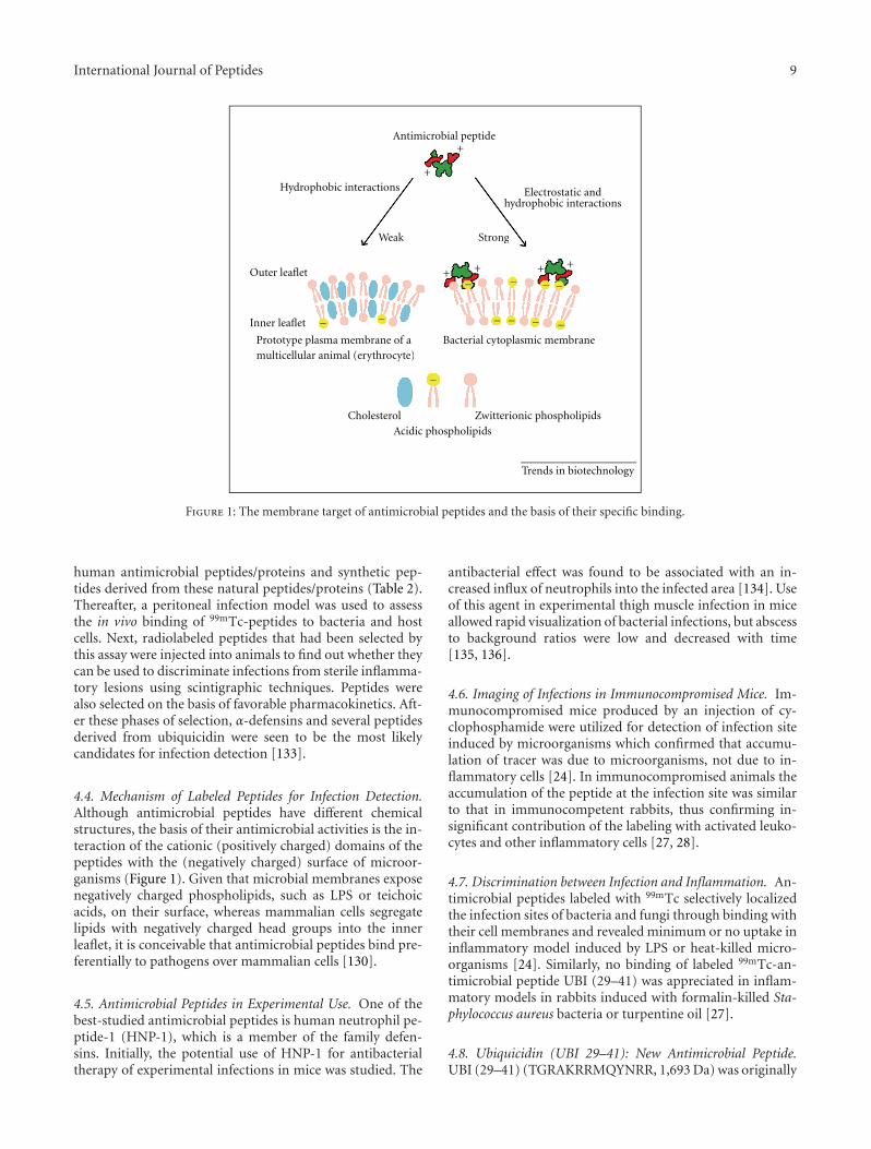
International Journal of Peptides 9
Antimicrobial peptide
Inner leaflet
Outer leaflet
Prototype plasma membrane of amulticellular animal (erythrocyte)
Bacterial cytoplasmic membrane
Cholesterol Zwitterionic phospholipids
Acidic phospholipids
Trends in biotechnology
Weak Strong
+
t
plasma membrane of aar animal (erythrocyte)
Bacterial cytoplasmic membra
Weak Strong
+Hydrophobic interactions Electrostatic and
hydrophobic interactions
+
−
− − − − − −
−−−−+ + + +
Figure 1: The membrane target of antimicrobial peptides and the basis of their specific binding.
human antimicrobial peptides/proteins and synthetic pep-tides derived from these natural peptides/proteins (Table 2).Thereafter, a peritoneal infection model was used to assessthe in vivo binding of 99mTc-peptides to bacteria and hostcells. Next, radiolabeled peptides that had been selected bythis assay were injected into animals to find out whether theycan be used to discriminate infections from sterile inflamma-tory lesions using scintigraphic techniques. Peptides werealso selected on the basis of favorable pharmacokinetics. Aft-er these phases of selection, α-defensins and several peptidesderived from ubiquicidin were seen to be the most likelycandidates for infection detection [133].
4.4. Mechanism of Labeled Peptides for Infection Detection.Although antimicrobial peptides have different chemicalstructures, the basis of their antimicrobial activities is the in-teraction of the cationic (positively charged) domains of thepeptides with the (negatively charged) surface of microor-ganisms (Figure 1). Given that microbial membranes exposenegatively charged phospholipids, such as LPS or teichoicacids, on their surface, whereas mammalian cells segregatelipids with negatively charged head groups into the innerleaflet, it is conceivable that antimicrobial peptides bind pre-ferentially to pathogens over mammalian cells [130].
4.5. Antimicrobial Peptides in Experimental Use. One of thebest-studied antimicrobial peptides is human neutrophil pe-ptide-1 (HNP-1), which is a member of the family defen-sins. Initially, the potential use of HNP-1 for antibacterialtherapy of experimental infections in mice was studied. The
antibacterial effect was found to be associated with an in-creased influx of neutrophils into the infected area [134]. Useof this agent in experimental thigh muscle infection in miceallowed rapid visualization of bacterial infections, but abscessto background ratios were low and decreased with time[135, 136].
4.6. Imaging of Infections in Immunocompromised Mice. Im-munocompromised mice produced by an injection of cy-clophosphamide were utilized for detection of infection siteinduced by microorganisms which confirmed that accumu-lation of tracer was due to microorganisms, not due to in-flammatory cells [24]. In immunocompromised animals theaccumulation of the peptide at the infection site was similarto that in immunocompetent rabbits, thus confirming in-significant contribution of the labeling with activated leuko-cytes and other inflammatory cells [27, 28].
4.7. Discrimination between Infection and Inflammation. An-timicrobial peptides labeled with 99mTc selectively localizedthe infection sites of bacteria and fungi through binding withtheir cell membranes and revealed minimum or no uptake ininflammatory model induced by LPS or heat-killed micro-organisms [24]. Similarly, no binding of labeled 99mTc-an-timicrobial peptide UBI (29–41) was appreciated in inflam-matory models in rabbits induced with formalin-killed Sta-phylococcus aureus bacteria or turpentine oil [27].
4.8. Ubiquicidin (UBI 29–41): New Antimicrobial Peptide.UBI (29–41) (TGRAKRRMQYNRR, 1,693 Da) was originally
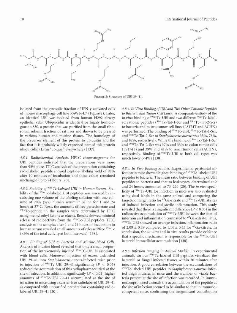
10 International Journal of Peptides
O
HN
OH
HN
NH
NH2
H2N
O
HN
HN
O
HN
O
HN
O
HN
NH2
S
O
HN
HN
NH2HN
O
HN
HN
O
O
O
HN
OH
O
HN
O
HN NH2
HN
NH2HN
NHOH
O
NH2
HN
OH
O
O
HN NH2
HN
Figure 2: Structure of UBI 29–41.
isolated from the cytosolic fraction of IFN-γ-activated cellsof mouse macrophage cell line RAW264.7 (Figure 2). Later,an identical UBI was isolated from human H292 airwayepithelial cells. Ubiquicidin is identical or highly homolo-gous to S30, a protein that was purified from the small ribo-somal subunit fraction of rat liver and shown to be presentin various human and murine tissues. The homology ofthe precursor element of this protein to ubiquitin and thefact that it is probably widely expressed named this proteinubiquicidin (Latin “ubique,” everywhere) [137].
4.8.1. Radiochemical Analysis. HPLC chromatograms forUBI peptides indicated that the preparations were morethan 95% pure. ITLC analysis of the preparation containingradiolabeled peptide showed peptide-labeling yield of 98%after 10 minutes of incubation and these values remainedunchanged up to 24 hours [20].
4.8.2. Stability of 99mTc-Labeled UBI in Human Serum. Sta-bility of the 99mTc-labeled UBI peptides was assessed by in-cubating one volume of the labeling solution with one vol-ume of 20% (v/v) human serum in saline for 1 and 24hours at 37◦C. Next, the amounts of free pertechnetate and99mTc-peptide in the samples were determined by ITLCusing methyl ethyl ketone as eluent. Results showed minimalrelease of radioactivity from the 99mTc-UBI peptides. ITLCanalysis of the samples after 1 and 24 hours of incubation inhuman serum revealed small amounts of released/free 99mTc(<3% of the total activity at both intervals) [138].
4.8.3. Binding of UBI to Bacteria and Murine Blood Cells.Analysis of murine blood revealed that only a small propor-tion of the intravenously injected 99mTC-UBI is associatedwith blood cells. Moreover, injection of excess unlabeledUBI 29–41 into Staphylococcus-aureus-infected mice priorto injection of 99mTc UBI 29–41 significantly (P < 0.05)reduced the accumulation of this radiopharmaceutical at thesite of infection. In addition, significantly (P < 0.01) higheramounts of 99mTc-UBI 29–41 accumulated at the site ofinfection in mice using a carrier-free radiolabeled UBI 29–41as compared with unpurified preparation containing radio-labeled UBI 29–41 [20].
4.8.4. In Vitro Binding of UBI and Two Other Cationic Peptidesto Bacteria and Tumor Cell Lines. A comparative study of thein vitro binding of 99mTc-UBI and two different 99mTc-label-ed cationic peptides (99mTc-Tat-1-Scr and 99mTc-Tat-2-Scr)to bacteria and to two tumor cell lines (LS174T and ACHN)was performed. The binding of 99mTc-UBI, 99mTc-Tat-1-Scr,and 99mTc-Tat-2-Scr to Staphylococcus aureus was 35%, 78%,and 87%, respectively. While the binding of 99mTc-Tat-1-Scrand 99mTc-Tat-2-Scr was 37% and 33% to colon tumor cells(LS174T) and 39% and 41% to renal tumor cells (ACHN),respectively. Binding of 99mTc-UBI to both cell types wasmuch lower (<4%) [138].
4.8.5. In Vivo Binding Studies. Experimental peritoneal in-fection in mice showed highest binding of 99mTc-labeled UBIpeptides to bacteria. The mean ratio between binding of UBIpeptides to bacteria and that to leukocytes, determined at 2and 24 hours, amounted to 73–220 [20]. The in vivo speci-ficity of 99mTc-UBI for infection in mice was also evaluatedusing dual labels in the same animal and comparing thetarget/nontarget ratio for 67Ga-citrate and 99mTc-UBI at sitesof induced infection and sterile inflammation. This studyrevealed that there is a significant difference (P < 0.05) in theradioactive accumulation of 99mTc-UBI between the sites ofinfection and inflammation compared to 67Ga-citrate. Thus,99mTc-UBI showed an average infection/inflammation ratioof 2.08 ± 0.49 compared to 1.14 ± 0.45 for 67Ga-citrate. Inconclusion, the in vitro and in vivo results provide evidencethat a specific mechanism is responsible for the 99mTc-UBIbacterial intracellular accumulation [138].
4.8.6. Infection Imaging in Animal Models. In experimentalanimals, various 99mTc-labeled UBI peptides visualized thebacterial or fungal infected tissues within 30 minutes afterinjection. A good correlation between the accumulations of99mTc-labeled UBI peptides in Staphylococcus-aureus-infec-ted thigh muscles in mice and the number of viable bac-teria present at the site of infection was recorded. In immu-nocompromised animals the accumulation of the peptide atthe site of infection seemed to be similar to that in immuno-competent mice, confirming the insignificant contribution
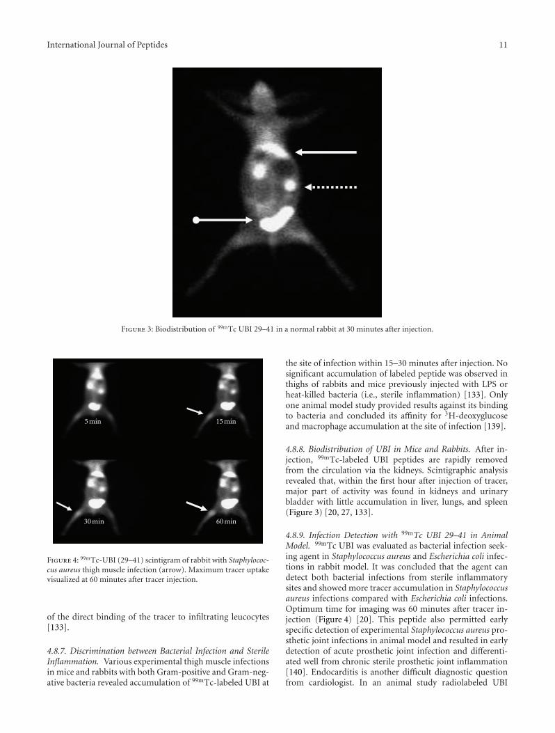
International Journal of Peptides 11
Figure 3: Biodistribution of 99mTc UBI 29–41 in a normal rabbit at 30 minutes after injection.
5 min 15 min
30 min 60 min
Figure 4: 99mTc-UBI (29–41) scintigram of rabbit with Staphylococ-cus aureus thigh muscle infection (arrow). Maximum tracer uptakevisualized at 60 minutes after tracer injection.
of the direct binding of the tracer to infiltrating leucocytes[133].
4.8.7. Discrimination between Bacterial Infection and SterileInflammation. Various experimental thigh muscle infectionsin mice and rabbits with both Gram-positive and Gram-neg-ative bacteria revealed accumulation of 99mTc-labeled UBI at
the site of infection within 15–30 minutes after injection. Nosignificant accumulation of labeled peptide was observed inthighs of rabbits and mice previously injected with LPS orheat-killed bacteria (i.e., sterile inflammation) [133]. Onlyone animal model study provided results against its bindingto bacteria and concluded its affinity for 3H-deoxyglucoseand macrophage accumulation at the site of infection [139].
4.8.8. Biodistribution of UBI in Mice and Rabbits. After in-jection, 99mTc-labeled UBI peptides are rapidly removedfrom the circulation via the kidneys. Scintigraphic analysisrevealed that, within the first hour after injection of tracer,major part of activity was found in kidneys and urinarybladder with little accumulation in liver, lungs, and spleen(Figure 3) [20, 27, 133].
4.8.9. Infection Detection with 99mTc UBI 29–41 in AnimalModel. 99mTc UBI was evaluated as bacterial infection seek-ing agent in Staphylococcus aureus and Escherichia coli infec-tions in rabbit model. It was concluded that the agent candetect both bacterial infections from sterile inflammatorysites and showed more tracer accumulation in Staphylococcusaureus infections compared with Escherichia coli infections.Optimum time for imaging was 60 minutes after tracer in-jection (Figure 4) [20]. This peptide also permitted earlyspecific detection of experimental Staphylococcus aureus pro-sthetic joint infections in animal model and resulted in earlydetection of acute prosthetic joint infection and differenti-ated well from chronic sterile prosthetic joint inflammation[140]. Endocarditis is another difficult diagnostic questionfrom cardiologist. In an animal study radiolabeled UBI

12 International Journal of Peptides
Figure 5: Anterior whole body image at 30 minutes after 99mTc-UBI29–41 injection in normal human subject showing kidneys (dottedarrows), liver (solid arrows), and urinary bladder (ball arrow).
29–41 scintigraphy revealed early and specific detectionof multidrug-resistant Staphylococcus-aureus-induced endo-carditis. Furthermore it was concluded that accumulationof tracer depends on the number of viable bacteria in thevegetation and declared it as dedicated noninvasive imagingtool for early detection of infective endocarditis [141].
4.8.10. 99mTc UBI Scan for Detection of Fungal Infections inAnimals. 99mTc UBI imaging was also studied in animalmodels for localization of infection induced with Candidaalbicans. It was observed that this antimicrobial peptideshowed significant accumulation at the site of infection com-pared with sterile inflammation [142]. This peptide showedbinding with Candida albicans and Aspergillus fumigatus inaddition to viable bacteria. Therefore this agent is usefulfor detection of fungal infections; however differentiationwould be difficult from bacterial infections. However 99mTc-fluconazole can be used later on, which binds only with Ca-ndida albicans, shows no binding to bacteria and no accumu-lation at site of sterile inflammation [143].
4.8.11. 99mTc UBI for Monitoring Efficacy of AntibacterialAgents. 99mTc-UBI when injected into Staphylococcus-au-reus-infected mice after treatment with various doses ofcloxacillin and erythromycin showed inverse correlation
between accumulation of the peptide at the site of infectionand the dose of antibacterial agents. Good correlation wasobserved between the accumulation of 99mTc-UBI and thenumber of viable bacteria. These results indicated the poten-tial of the peptide for evaluating the efficacy of antibiotictherapy. However, minimum number of bacteria that can bedetected was 103–104, which is the limitation for monitoringthe effects of the antibacterial agents [144]. Same conclusionwas drawn in another study in which commonly used broad-spectrum antibiotic ciprofloxacin was used in a rabbit mod-el. It also revealed that the radiotracer can be used for moni-toring efficacy and duration of antibiotic treatment [28].
4.8.12. Biodistribution in Humans. 99mTc-UBI was investiga-ted in a biokinetic model to evaluate its feasibility as aninfection imaging agent in humans. Whole body images from6 children with suspected bone infection were acquired at 1,30, 120, 240 minutes and 24 hours after tracer administra-tion. Regions of interest (ROIs) were drawn around sourceorgans (heart, liver, kidneys, and bladder) on each timeframe. The same set of ROIs was used for all 6 scans and thecounts per minutes (cpm) of each ROI were converted toactivity using the conjugate view counting method. Countswere corrected by physical decay and by the background cor-rection factor derived from preclinical phantom studies. Theimage sequence was used to extrapolate 99mTc-UBI time-ac-tivity curves in each organ and calculate the cumulatedactivity. Urine samples were used to obtain the cumulativepercent of injected activity versus time renal elimination. Theabsorbed dose in organs was evaluated according to the gen-eral equation described in the MIRD formula. In addition,67Ga-citrate images were obtained from all the patients andused as a control. Biokinetic data showed a fast blood clear-ance with a mean residence time of 0.52 hour. Approximately85% of the injected activity was eliminated by renal clearance24 hours after 99mTc-UBI administration. Images showedminimal accumulation in nontarget tissues with an averagetarget/nontarget ratio of 2.18 ± 0.74 in positive lesions at 2hours. All infection positive images were in agreement withthose obtained with 67Ga-citrate. The mean radiation-ab-sorbed dose calculated was 0.13 mGy/MBq for kidneys andthe effective dose was 4.34 × 10⁻³ mSv/MBq [145]. Biodis-tribution of the peptide was also studied in 3 normal subjectsin another study by taking anterior and posterior whole bodyimages and using geometric mean method. It was observedthat the tracer mainly excreted through the kidneys into urin-ary bladder followed by liver with no other site of accumula-tion in the body (Figure 5) [27].
4.8.13. Bacterial Infection Detection in Humans with 99mTcUBI 29–41. 99mTc UBI 29–41 showed good correlation forinfection detection in humans when compared with 67Gaimaging of the same subjects [145]. In another study theagent was tested in patients suffering from bone, soft tis-sue, or prosthesis infections and encouraging results were ob-served with overall sensitivity, specificity, and accuracy of100%, 80%, and 94.4%, respectively. Maximum traceraccumulation was noted at 30 minutes after tracer injection(Figure 6) [29].
International Journal of Peptides 13
30 min60 min
120 min
Figure 6: Positive 99mTc-UBI 29–41 scan in a patient with infection in medial aspect of right hand (arrow). Maximum focal increased traceruptake was seen at 30 minutes after tracer injection.
This peptide yielded fast and promising results in pa-tients with suspected mediastinitis after cardiac surgery.Qualitative analysis correctly identified infection in 5/6 pa-tients with proven mediastinitis on bacterial culture [146].Fever of unknown origin (FUO) is also a diagnostic dilemmafor physicians and surgeons. Antibiotics are blindly usedwithout localizing the infective focus. This antimicrobial pe-ptide revealed specificity of 95.35% for localizing infectionand discarding sterile inflammation. Sensitivity was 97.52%with high accuracy [147]. Vertebral osteomyelitis is difficultto diagnose by noninvasive diagnostic modalities includingCT, MRI, radionuclide bone scan, and X-Ray. Even 67Ga scanis nonspecific test which is considered better than other diag-nostic techniques. As UBI 29–41 has special affinity for bind-ing to viable bacteria, this has been used for confirmation ofvertebral infection which showed 100% sensitivity and 88%specificity [148]. Noninvasive diagnostic techniques includ-ing three-phase bone scan, MRI, and 99mTc-UBI 29–41 scanfor detection of osteomyelitis were compared. Antimicrobialpeptide showed 100% accuracy with maximum mean targetto nontarget ratio at 15 minutes after tracer injection, whilethree-phase bone scan and MRI revealed 90% and 75% accu-racy, respectively. This study revealed superiority of antimi-crobial peptide imaging over bone scan and MRI [149].
4.8.14. 99mTc-UBI Scan for Monitoring Efficacy of AntibioticTreatment in Human Infections. As mentioned in animalstudies, binding of 99mTc UBI 29–41 to viable bacteria is pro-portional to their number in the infective focus. Intensity oftracer uptake decreased when number of viable bacteria wasreduced. Therefore it was concluded that this tracer can beused for monitoring efficacy and duration of treatment
[28, 144]. Similar study for monitoring antibiotic therapyin patients with orthopaedic infections showed better resultson quantitative analysis of scan at 30, 60, and 120 minutesafter tracer injection after 10–14, day interval with antibiotictreatment when compared with erythrocyte sedimentationrate, C-reactive protein, and radioisotope bone scan [150].
5. Limitations of Antimicrobial Peptides
5.1. Lack of Discrimination between Different InfectiousAgents. 99mTc-labeled antimicrobial peptides bind to fungiin addition to bacterial cell membranes; however, infectioneither from bacteria or fungi can be differentiated from ster-ile inflammatory sites [24]. Similarly, there is nonuniformaccumulation of radiolabeled peptides in different types ofbacteria. Staphylococcus aureus showed more uptake thanEscherichia coli which may be due to different mode of toxi-city [27].
5.2. Lack of Detection of Intracellular Pathogens. Radiolabeledpeptides bind to the cell membranes of the bacteria. How-ever, if the bacteria are engulfed by the anti-inflammatorycells or become intracellular after invasion of the host im-mune cells, their detection would become difficult with scin-tigraphy [123].
5.3. Resistance against Antimicrobial Peptides. Major concernwith use of the labeled antibiotics as specific infection locali-zation tracers is the development of resistance. Similarsituation may also be encountered with antimicrobial pep-tides. Some Gram-negative bacteria can modify the lipid-Amoiety of the endotoxin [151]. Similarly Gram-positivebacteria can reduce negative charge of the bacterial surface
14 International Journal of Peptides
by esterification of phospholipids of Staphylococcus aureus[152]. Inactivation of antimicrobial peptides by bacterialserine proteases can prevent intracellular accumulation ofthe peptides [153]. However, in multiple studies conductedwith different radiolabeled antimicrobial peptides includingUBI (29–41) have not revealed such evidence up till now.
6. Conclusion
The medical community often faces the dilemma of discrim-ination between infection and inflammation on medical aswell as on surgical floors. Nonspecific radiotracers for loca-lization of infection/inflammation do not solve the problem.Among the specific radiotracers for localization of infection,antibiotics gained more popularity due to easy availability,labeling, low cost, and high sensitivity. Among the labeledantibiotics ciprofloxacin was the most successful specific ba-cterial localization agent which showed sensitivity of 85.4%and specificity of 81.7% [117]. However, emerging antibi-otic resistance against antibiotics is also associated withciprofloxacin [154]. False uptake of 99mTc-ciprofloxacin insterile inflammation is also a big disadvantage [14]. Due tononspecific accumulation in inflammatory sites, this agenthas been proposed for identifying the presence and distribu-tion of inflammation in joints [18].
On the other hand, antimicrobial peptides labeled withisotopes are better specific infection localizing agents as theybind specifically to bacterial cell membranes. These tracersdetect Gram-positive, Gram-negative bacteria, Candida albi-cans and Aspergillus Fumigatus infections. The amount ofradiolabeled peptides at the site of infection depends on thenumber of viable organisms present at the focus. Recentlyinvestigated antimicrobial peptide, ubiquicidin UBI (29–41)has shown encouraging results in human clinical trials. Thispeptide can also be used for monitoring efficacy and dura-tion of antibiotic treatment in patients which is very im-portant issue from prophylactic, therapeutic, and socioeco-nomic point of view. No doubt, there are limitations at-tributed to synthesis/isolation of such peptides, labeling withisotopes, minimum detection limit of 103 Colony-FormingUnit (CFU) of bacteria, and inability to distinguish betweenbacterial and fungal infections. In addition, different bac-terial types reveal different tracer accumulation (Staphylo-coccus aureus versus Escherichia coli). Currently no evidenceregarding resistance against antimicrobial peptides has beenreported. Considering the merits and demerits of radiola-beled peptides and radiolabeled antibiotics, it can currentlybe concluded that radiolabeled peptides are better specificinfection localizing agents.
References
[1] O. Levy, “Antimicrobial proteins and peptides of blood:templates for novel antimicrobial agents,” Blood, vol. 96, no.8, pp. 2664–2672, 2000.
[2] M. R. Yeaman, A. S. Ibrahim, J. E. J. Edwards, A. S. Bayer,and M. A. Ghannoum, “Thrombin-induced rabbit plateletmicrocidal protein is fungicidal in vitro,” Antimicrob AgentsChemother, vol. 37, pp. 546–553, 1993.
[3] S. S. Das, A. V. Hall, D. W. Wareham, and K. E. Britten, “In-fection imaging with radiopharmaceuticals in the 21st cen-tury,” Brazilian Archives of Biology and Technology, vol. 45,pp. 25–37, 2002.
[4] F. H. M. Corstens and J. W. M. Van Der Meer, “Nuclear me-dicine’s role in infection and inflammation,” Lancet, vol. 354,no. 9180, pp. 765–770, 1999.
[5] A. M. Peters, H. J. Danpure, S. Osman et al., “Preliminaryclinical experience with 99mTc-hexamethylpropylene-amin-eoxime for labelling leucocytes and imaging infection,” TheLancet, vol. 2, pp. 945–949, 1986.
[6] A. M. Peters, “The utility of [99mTc]HMPAO-leukocytes forimaging infection,” Seminars in Nuclear Medicine, vol. 24, no.2, pp. 110–127, 1994.
[7] S. Gratz, H. J. J. M. Rennen, O. C. Boerman et al., “99mTc-HMPAO-labeled autologous versus heterologous leukocytesfor imaging infection,” Journal of Nuclear Medicine, vol. 43,no. 7, pp. 918–924, 2002.
[8] W. Becker and J. Meller, “The role of nuclear medicine in in-fection and inflammation,” Lancet Infectious Diseases, vol. 1,no. 5, pp. 326–333, 2001.
[9] C. Love and C. J. Palestro, “Radionuclide imaging of in-fection,” Journal of Nuclear Medicine Technology, vol. 32, no.2, pp. 47–57, 2004.
[10] L. Filippi and O. Schillaci, “Usefulness of hybrid SPECT/CTin 99mTC-HMPAO-labeled leukocyte scintigraphy for boneand joint infections,” Journal of Nuclear Medicine, vol. 47, no.12, pp. 1908–1913, 2006.
[11] R. Bar-Shalom, N. Yefremov, L. Guralnik et al., “SPECT/CTusing 67Ga and 111In-labeled leukocyte scintigraphy fordiagnosis of infection,” Journal of Nuclear Medicine, vol. 47,no. 4, pp. 587–594, 2006.
[12] N. Dumarey, D. Egrise, D. Blocklet et al., “Imaging infectionwith 18F-FDG-labeled leukocyte PET/CT: initial experiencein 21 patients,” Journal of Nuclear Medicine, vol. 47, no. 4, pp.625–632, 2006.
[13] B. Fournier, X. Zhao, T. Lu, K. Drlica, and D. C. Hooper,“Selective targeting of topoisomerase IV and DNA gyrase inStphylococcus aureus: different patterns of quinolone-in-duced inhibition of DNA Synthesis,” Antimicrob AgentsChemother, vol. 44, pp. 2160–2165, 2000.
[14] Z. Yapar, M. Kibar, A. F. Yapar, E. Togrul, U. Kayaselcuk,and Y. Sarpel, “The efficacy of technetium-99m ciprofloxacin(Infecton) imaging in suspected orthopaedic infection: acomparison with sequential bone/gallium imaging,” Euro-pean Journal of Nuclear Medicine, vol. 28, no. 7, pp. 822–830,2001.
[15] K. Sonmezoglu, M. Sonmezoglu, M. Halac et al., “Use-fulness of 99mTc-ciprofloxacin (infecton) scan in diagnosisof chronic orthopedic infections: comparative study with99mTc-HMPAO leukocyte scintigraphy,” Journal of NuclearMedicine, vol. 42, no. 4, pp. 567–574, 2001.
[16] M. J. Larikka, A. K. Ahonen, O. Niemela et al., “99mTc-cipro-flaxacin (infection) imaging in the diagnosis of knee pros-thesis infections,” Nuclear Medicine Communications, vol. 23,pp. 167–170, 2002.
[17] L. Sarda, A. C. Cremieux, Y. Lebellec et al., “Inability of99mTc-ciprofloxacin scintigraphy to discriminate between se-ptic and sterile osteoarticular diseases,” Journal of NuclearMedicine, vol. 44, no. 6, pp. 920–926, 2003.
[18] T. Appelboom, P. Emery, L. Tant, N. Dumarey, and A. Schou-tens, “Evaluation of technetium-99m-ciprofloxacin (infec-tion) for detecting sites of inflammation in arthritis,” Rheu-matology, vol. 42, no. 10, pp. 1179–1182, 2003.

International Journal of Peptides 15
[19] M. E. Jones, N. M. Boenink, J. Verhoef, K. Kohrer, and F. J.Schmitz, “Multiple mutations conferring ciprofloxacin resis-tance in staphylococcus aureus demonstrate long-term sta-bility in an antibiotic free environment,” Journal of Antimi-crobial Chemotherapy, vol. 45, pp. 353–356, 2000.
[20] M. Zasloff, “Antimicrobial peptides of multicellular organ-isms,” Nature, vol. 415, no. 6870, pp. 389–395, 2002.
[21] M. M. Welling, P. H. Nibbering, A. Paulusma-Annema, P. S.Hiemstra, E. K. J. Pauwels, and W. Calame, “Imaging of ba-cterial infections with 99mTc-labeled human neutrophil pe-ptide-1,” Journal of Nuclear Medicine, vol. 40, no. 12, pp.2073–2080, 1999.
[22] P. S. Hiemstra, M. T. Van Den Barselaar, M. Roest, P. H.Nibbering, and R. Van Furth, “Ubiquicidin, a novel murinemicrobicidal protein present in the cytosolic fraction of ma-crophages,” Journal of Leukocyte Biology, vol. 66, no. 3, pp.423–428, 1999.
[23] M. M. Welling, S. Mongera, A. Lupetti et al., “Radiochemicaland biological characteristics of 99mTc-UBI 29-41 for imagingof bacterial infections,” Nuclear Medicine and Biology, vol. 29,no. 4, pp. 413–422, 2002.
[24] M. M. Welling, A. Lupetti, H. S. Balter et al., “99mTc-labeledantimicrobial peptides for detection of bacterial and Candidaalbicans infections,” Journal of Nuclear Medicine, vol. 42, no.5, pp. 788–794, 2001.
[25] R. M. Epand and H. J. Vogel, “Diversity of antimicrobial pep-tides and their mechanisms of action,” Biochimica et Biophys-ica Acta, vol. 1462, no. 1-2, pp. 11–28, 1999.
[26] M. Edgerton, S. E. Koshlukova, T. E. Lo, B. G. Chrzan, R. M.Straubinger, and P. A. Raj, “Candidacidal activity of salivaryhistatins: identification of a histatin 5-binding protein onCandida albicans,” Journal of Biological Chemistry, vol. 273,no. 32, pp. 20438–20447, 1998.
[27] M. S. Akhtar, J. Iqbal, M. A. Khan et al., “99mTc-labeled an-timicrobial peptide ubiquicidin (29-41) accumulates less inEscherichia coli infection than in Staphlococcus aureus in-fection,” Journal of Nuclear Medicine, vol. 45, no. 5, pp. 849–856, 2004.
[28] M. S. Akhtar, M. E. Khan, B. Khan et al., “An imaging analysisof 99mTc-UBI (29-41) uptake in S. aureus infected thighsof rabbits on ciprofloxacin treatment,” European Journal ofNuclear Medicine and Molecular Imaging, vol. 35, no. 6, pp.1056–1064, 2008.
[29] M. S. Akhtar, A. Qaisar, J. Irfanullah et al., “Antimicrobialpeptide 99mTc-Ubiquicidin 29-41 as human infection-imag-ing agent: clinical trial,” Journal of Nuclear Medicine, vol. 46,no. 4, pp. 567–573, 2005.
[30] Y. Ito, S. Okuyama, T. Awano, K. Takahashi, and T. Sato,“Diagnostic evaluation of 67 Ga scanning of lung cancer andother diseases.,” Radiology, vol. 101, no. 2, pp. 355–362, 1971.
[31] R. D. Neuman and J. G. McAfee, “Gallium-67 imaging in in-fection,” in Diagnostic Nuclear Medicine, M. P. Sandler, J. A.Patton, R. E. Coleman, A. Gottschalk, F. J. Wackers, and P.B. Hoffer, Eds., pp. 1493–1507, Williams and Wilkins, Bal-timore, Md, USA, 3rd edition, 1996.
[32] M. F. Tsan, “Mechanism of gallium-67 accumulation in in-flammatory lesions,” Journal of Nuclear Medicine, vol. 26, no.1, pp. 88–92, 1985.
[33] R. K. Zeman and T. W. Ryerson, “The value of bowel prepa-ration in Ga-67 citrate scanning: concise communication,”Journal of Nuclear Medicine, vol. 18, no. 9, pp. 886–889, 1977.
[34] O. James, E. J. Wood, and S. Sherlock, “67Gallium scanningin the diagnosis of liver disease,” Gut, vol. 15, no. 5, pp. 404–410, 1974.
[35] D. Front, O. Israel, R. Epelbaum et al., “Ga-67 SPECT beforeand after treatment of lymphoma,” Radiology, vol. 175, no. 2,pp. 515–519, 1990.
[36] C. J. Palestro, “The current role of gallium imaging in infec-tion,” Seminars in Nuclear Medicine, vol. 24, no. 2, pp. 128–141, 1994.
[37] E. V. Staab and W. H. McCartney, “Role of Gallium 67 ininflammatory disease,” Seminars in Nuclear Medicine, vol. 8,no. 3, pp. 219–234, 1978.
[38] M. A. Auler, S. Bagg, and L. Gordon, “The role of nuclear me-dicine in imaging infection,” Seminars in Roentgenology, vol.42, no. 2, pp. 117–121, 2007.
[39] A. J. Fischman, R. H. Rubin, J. A. White et al., “Localization ofFc and Fab fragments of nonspecific polyclonal IgG at focalsites of inflammation,” Journal of Nuclear Medicine, vol. 31,no. 7, pp. 1199–1205, 1990.
[40] A. J. Fischman, A. J. Fucello, J. L. Pellegrino-Gensey et al.,“Effect of carbohydrate modification on the localization ofhuman polyclonal IgG at focal sites of bacterial infection,”Journal of Nuclear Medicine, vol. 33, no. 7, pp. 1378–1382,1992.
[41] M. W. Nijhof, W. J. G. Oyen, A. Van Kampen, R. A. M. J.Ciaessens, J. W. M. Van Der Meer, and F. H. M. Corstens,“Evaluation of infections of the locomotor system with in-dium-111 -labeled human IgG scintigraphy,” Journal of Nu-clear Medicine, vol. 38, no. 8, pp. 1300–1305, 1997.
[42] J. R. Buscombe, W. J. G. Oyen, A. Grant et al., “Indium-111-labeled polyclonal human immunoglobulin: identifying focalinfection in patients positive for human immunodeficiencyvirus,” Journal of Nuclear Medicine, vol. 34, no. 10, pp. 1621–1625, 1993.
[43] L. Mairal, P. A. De Uma, J. Martin-Comin et al., “Simulta-neous administration of 111In-human immunoglobulin and99mTc-HMPAO labelled leucocytes in inflammatory boweldisease,” European Journal of Nuclear Medicine, vol. 22, no.7, pp. 664–670, 1995.
[44] W. Becker, J. Bair, T. Behr et al., “Detection of soft-tissue infections and osteomyelitis using a technetium- 99m-labeled anti-granulocyte monoclonal antibody fragment,”Journal of Nuclear Medicine, vol. 35, no. 9, pp. 1436–1443,1994.
[45] O. C. Boerman, G. Storm, W. J. G. Oyen et al., “Stericallystabilized liposomes labeled with Indium-111 to image focalinfection,” Journal of Nuclear Medicine, vol. 36, no. 9, pp.1639–1644, 1995.
[46] P. Laverman, E. T. M. Dams, W. J. G. Oyen et al., “A novelmethod to label liposomes with 99mTc by the hydrazinonicotinyl derivative,” Journal of Nuclear Medicine, vol. 40, no.1, pp. 192–197, 1999.
[47] E. T. M. Dams, W. J. G. Oyen, O. C. Boerman et al., “99mTc-PEG liposomes for the scintigraphic detection of infectionand inflammation: clinical evaluation,” Journal of NuclearMedicine, vol. 41, no. 4, pp. 622–630, 2000.
[48] D. J. Hnatowich, F. Virzi, and M. Rusckowski, “Investigationsof avidin and biotin for imaging applications,” Journal ofNuclear Medicine, vol. 28, no. 8, pp. 1294–1302, 1987.
[49] M. Rusckowski, B. Fritz, and D. J. Hnatowich, “Localizationof infection using streptavidin and biotin: an alternative tononspecific polyclonal immunoglobulin,” Journal of NuclearMedicine, vol. 33, no. 10, pp. 1810–1815, 1992.
[50] A. Samuel, G. Paganelli, R. Chiesa et al., “Detection of pro-sthetic vascular graft infection using avidin/indium-111-bio-tin scintigraphy,” Journal of Nuclear Medicine, vol. 37, no. 1,pp. 55–61, 1996.

16 International Journal of Peptides
[51] M. Rusckowski, G. Paganelli, D. J. Hnatowich et al., “Imag-ing osteomyelitis with streptavidin and Indium-111-labeledbiotin,” Journal of Nuclear Medicine, vol. 37, no. 10, pp. 1655–1662, 1996.
[52] C. J. Palestro and M. A. Torres, “Radionuclide imaging ofnonosseous infection,” Quarterly Journal of Nuclear Medicine,vol. 43, no. 1, pp. 46–60, 1999.
[53] E. Outwater, E. Oates, and R. C. Sarno, “Indium-111-labeledleukocyte scintigraphy: diagnosis of subperiosteal abscessescomplicating osteomyelitis in a child,” Journal of NuclearMedicine, vol. 29, no. 11, pp. 1871–1874, 1988.
[54] F. L. Datz and D. A. Thorne, “Cause and significance of coldbone defects on Indium-111-labeled leukocyte imaging,”Journal of Nuclear Medicine, vol. 28, no. 5, pp. 820–823, 1987.
[55] K. Uno, N. Matsui, and K. Nohira, “Indium-111 leukocyteimaging in patients with rheumatoid arthritis,” Journal ofNuclear Medicine, vol. 27, no. 3, pp. 339–344, 1986.
[56] D. S. Schauwecker, H. M. Park, R. W. Burt, B. H. Mock, andH. N. Wellman, “Combined bone scintigraphy and indium-111 leukocyte scans in neuropathic foot disease,” Journal ofNuclear Medicine, vol. 29, no. 10, pp. 1651–1655, 1988.
[57] L. M. Lamki, L. P. Kasi, and T. P. Haynie, “Localization ofIndium-111 leukocytes in noninfected neoplasms,” Journal ofNuclear Medicine, vol. 29, no. 12, pp. 1921–1926, 1988.
[58] C. J. Palestro, H. H. Mehta, M. Patel et al., “Marrow ver-sus infection in the Charcot joint: indium-111 leukocyte andtechnetium-99m sulfur colloid scintigraphy,” Journal of Nu-clear Medicine, vol. 39, no. 2, pp. 346–350, 1998.
[59] W. J. G. Oyen, R. A. M. J. Claessens, J. W. M. Van der Meer,and F. H. M. Corstens, “Detection of subacute infectious fociwith indium-111-labeled autologous leukocytes and indium-111-labeled human nonspecific immunoglobulin G: a pro-spective comparative study,” Journal of Nuclear Medicine, vol.32, no. 10, pp. 1854–1860, 1991.
[60] J. G. McAfee, G. Gagne, G. Subramanian, and R. F. Schneider,“The localization of indium-111-leukocytes, gallium-67-polyclonal IgG and other radioactive agents in acute focalinflammatory lesions,” Journal of Nuclear Medicine, vol. 32,no. 11, pp. 2126–2131, 1991.
[61] C. J. Palestro, C. K. Kim, A. J. Swyer, S. Vallabhajosula, and S.J. Goldsmith, “Radionuclide diagnosis of vertebral osteo-myelitis: indium-111-leukocyte and technetium-99m-me-thylene diphosphonate bone scintigraphy,” Journal of NuclearMedicine, vol. 32, no. 10, pp. 1861–1865, 1991.
[62] F. L. Datz, “Indium-111-labeled leukocytes for the detectionof infection: current status,” Seminars in Nuclear Medicine,vol. 24, no. 2, pp. 92–109, 1994.
[63] S. L. Kipper, “Radiolabelled leukocyte imaging of the ab-domen,” in Nuclear Medicine Annual, L. M. Freeman, Ed., pp.81–126, Raven Press, New York, NY, USA, 1995.
[64] J. H. Thrall and H. A. Ziessman, “Infection and inflamma-tion,” in Nuclear Medicine: The Requisites, J. H. Thrall, Ed.,pp. 167–192, Mosby, 2nd edition, 2001.
[65] M. E. Roddie, A. M. Peters, H. J. Danpure et al., “Inflamma-tion: imaging with Tc-99m HMPAO-labeled leukocytes,”Radiology, vol. 166, no. 3, pp. 767–772, 1988.
[66] M. Vorne, I. Soini, T. Lantto, and S. Paakkinen, “Technetium-99m HM-PAO-labeled leukocytes in detection of inflamma-tory lesions: comparison with Gallium-67 citrate,” Journal ofNuclear Medicine, vol. 30, no. 8, pp. 1332–1336, 1989.
[67] I. Hovi, M. Taavitsainen, T. Lantto, M. Vorne, R. Paul, and K.Remes, “Technetium-99m-HMPAO-labeled leukocytes and
technetium-99m-labeled human polyclonal immunoglobu-lin G in diagnosis of focal purulent disease,” Journal of Nu-clear Medicine, vol. 34, no. 9, pp. 1428–1434, 1993.
[68] E. H. Lantto, T. J. Lantto, and M. Vorne, “Fast diagnosisof abdominal infections and inflammations with techne-tium-99m-HMPAO labeled leukocytes,” Journal of NuclearMedicine, vol. 32, no. 11, pp. 2029–2034, 1991.
[69] E. H. Lantto, T. J. Lantto, and M. Vorne, “Fast diagnosisof abdominal infections and inflammations with techne-tium-99m-HMPAO labeled leukocytes,” Journal of NuclearMedicine, vol. 32, no. 11, pp. 2029–2034, 1991.
[70] F. Palermo, F. Boccaletto, A. Paolin et al., “Comparisonof technetium-99m-MDP, technetium-99-m-WBC and tech-netium- 99m-HIG in musculoskeletal inflammation,” Jour-nal of Nuclear Medicine, vol. 39, no. 3, pp. 516–521, 1998.
[71] W. Becker, U. Borst, W. Fischbach, B. Pasurka, R. Schafer, andW. Borner, “Kinetic data of in-vivo labeled granulocytes inhumans with a murine Tc-99m-labelled monoclonal anti-body,” European Journal of Nuclear Medicine, vol. 15, no. 7,pp. 361–366, 1989.
[72] W. S. Becker, A. Saptogino, and F. G. Wolf, “The single late99Tcm granulocyte antibody scan in inflammatory diseases,”Nuclear Medicine Communications, vol. 13, no. 3, pp. 186–192, 1992.
[73] W. Becker, J. Bair, T. Behr et al., “Detection of soft-tissueinfections and osteomyelitis using a technetium- 99m-label-ed anti-granulocyte monoclonal antibody fragment,” Journalof Nuclear Medicine, vol. 35, no. 9, pp. 1436–1443, 1994.
[74] M. L. Thakur, C. S. Marcus, P. Henneman et al., “Imaging in-flammatory diseases with neutrophil-specific technetium-99m-labeled monoclonal antibody anti-SSEA-1,” Journal ofNuclear Medicine, vol. 37, no. 11, pp. 1789–1795, 1996.
[75] S. L. Kipper, E. B. Rypins, D. G. Evans, M. L. Thakur, T. D.Smith, and B. Rhodes, “Neutrophil-specific 99mTc-labeledanti-CD 15 monoclonal antibody imaging for diagnosis ofequivocal appendicitis,” Journal of Nuclear Medicine, vol. 41,no. 3, pp. 449–455, 2000.
[76] S. Gratz, T. Behr, A. Herrmann et al., “Intraindividual com-parison of 99mTc-labelled anti-SSEA-1 antigranulocyte anti-body and 99mTc-HMPAO labelled white blood cells for theimaging of infection,” European Journal of Nuclear Medicine,vol. 25, no. 4, pp. 386–393, 1998.
[77] W. Becker, D. M. Goldenberg, and F. Wolf, “The use ofmonoclonal antibodies and antibody fragments in the imag-ing of infectious lesions,” Seminars in Nuclear Medicine, vol.24, no. 2, pp. 142–153, 1994.
[78] A. J. Fischman, M. C. Pike, D. Kroon et al., “Imaging focalsites of bacterial infection in rats with indium-111- labeledchemotactic peptide analogs,” Journal of Nuclear Medicine,vol. 32, no. 3, pp. 483–491, 1991.
[79] J. W. Babich, W. Graham, S. A. Barrow et al., “Technetium-99m-labeled chemotactic peptides: comparison with Indi-um-111- labeled white blood cells for localizing acute bac-terial infection in the rabbit,” Journal of Nuclear Medicine, vol.34, no. 12, pp. 2176–2181, 1993.
[80] A. J. Fischman, D. Rauh, H. Solomon et al., “In vivo bioac-tivity and biodistribution of chemotactic peptide analogs innonhuman primates,” Journal of Nuclear Medicine, vol. 34,no. 12, pp. 2130–2134, 1993.
[81] J. W. Babich, Q. Dong, W. Graham et al., “A novel high affin-ity chemotactic peptide antagonist for infection imaging,”Journal of Nuclear Medicine, vol. 38: 268, 1997.
International Journal of Peptides 17
[82] A. D. Luster, “Mechanisms of disease: chemokines-chemo-tactic cytokines that mediate inflammation,” New EnglandJournal of Medicine, vol. 338, no. 7, pp. 436–445, 1998.
[83] C. J. Van der Laken, O. C. Boerman, W. J. G. Oyen et al.,“Specific targeting of infectious foci with radioiodinated hu-man recombinant interleukin-1 in an experimental model,”European Journal of Nuclear Medicine, vol. 22, no. 11, pp.1249–1255, 1995.
[84] C. J. vanderLaken, O. C. Boerman, W. J. G. Oyen et al., “Com-parison of radiolabeled human recombinant interleukin-1with its receptor antagonist in a model of infection,” Euro-pean Journal of Nuclear Medicine, vol. 22: 916, 1995.
[85] A. Signore, M. Chianelli, A. Annovazzi et al., “123I-Inter-leukin-2 scintigraphy for in vivo assessment of intestinalmononuclear cell infiltration in Crohn’s disease,” Journal ofNuclear Medicine, vol. 41, no. 2, pp. 242–249, 2000.
[86] A. Signore, M. Chianelli, A. Annovazzi et al., “Imaging activelymphocytic infiltration in coeliac disease with iodine- 123-interleukin-2 and the response to diet,” European Journal ofNuclear Medicine, vol. 27, no. 1, pp. 18–24, 2000.
[87] M. D. Gross, B. Shapiro, R. S. Skinner, P. Shreve, L. M. Fig,and R. V. Hay, “Scintigraphy of osteomyelitis in man withhuman recombinant interleukin-8,” Journal of Nuclear Me-dicine, vol. 37: 25, 1996.
[88] R. V. Hay, R. S. Skinner, O. C. Newman et al., “Scintigraphyof acute inflammatory lesions in rats with radliolabelled re-combinant human interleukin-8,” Nuclear Medicine Commu-nications, vol. 18, no. 4, pp. 367–378, 1997.
[89] C. J. Van Der Laken, O. C. Boerman, W. J. G. Oyen, M. T. P.Van De Ven, J. W. M. Van Der Meer, and F. H. M. Corstens,“Radiolabeled interleukin-8: specific scintigraphic detectionof infection within a few hours,” Journal of Nuclear Medicine,vol. 41, no. 3, pp. 463–469, 2000.
[90] H. J. J. M. Rennen, O. C. Boernan, W. J. G. Oyen, J. W. M.Van der Meer, and F. H. M. Corstens, “Specific and rapidscintigraphic detection of infection with 99mTc-labeled in-terleukin-8,” Journal of Nuclear Medicine, vol. 42, no. 1, pp.117–123, 2001.
[91] S. Gratz, H. J. J. M. Rennen, O. C. Boerman, W. J. G. Oyen,and F. H. M. Corstens, “Rapid imaging of experimentalcolitis with 99mTc-interleukin-8 in rabbits,” Journal of NuclearMedicine, vol. 42, no. 6, pp. 917–923, 2001.
[92] B. R. Moyer, S. Vallabhajosula, J. Lister-James et al., “Tech-netium-99m-White Blood Cell-Specific Imaging Agent De-veloped from Platelet Factor 4 to Detect Infection,” Journal ofNuclear Medicine, vol. 37, no. 4–6, pp. 673–679, 1996.
[93] C. J. Palestro, M. B. Tomas, K. K. Bhargava et al., “Tc-99mP483H for imaging infection: phase 2 multicenter trialresults,” Journal of Nuclear Medicine, vol. 40:15, 1999.
[94] G. I. Bell, C. F. Burant, J. Takeda, and G. W. Gould, “Structureand function of mammalian facilitative sugar transporters,”Journal of Biological Chemistry, vol. 268, no. 26, pp. 19161–19164, 1993.
[95] E. K. J. Pauwels, M. J. Ribeiro, J. H. M. B. Stoot, V. R.McCready, M. Bourguignon, and B. Maziere, “FDG accumu-lation and tumor biology,” Nuclear Medicine and Biology, vol.25, no. 4, pp. 317–322, 1998.
[96] H. Zhuang and A. Alavi, “18-Fluorodeoxyglucose positronemission tomographic imaging in the detection and moni-toring of infection and inflammation,” Seminars in NuclearMedicine, vol. 32, no. 1, pp. 47–59, 2002.
[97] J. Y. Paik, K. H. Lee, S. C. Yearn, Y. Choi, and B. T. Kim,“Augmented 18F-FDG uptake in activated monocytes occursduring the priming process and involves tyrosine kinases and
protein kinase C,” Journal of Nuclear Medicine, vol. 45, no. 1,pp. 124–128, 2004.
[98] P. D. Shreve, Y. Anzai, and R. L. Wahl, “Pitfalls in oncologicdiagnosis with FDG PET imaging: physiologic and benignvariants,” Radiographics, vol. 19, no. 1, pp. 61–77, 1999.
[99] T. Nakahara, H. Fujii, M. Ide et al., “FDG uptake inthe morphologically normal thymus: comparison of FDGposition emission tomography and CT,” British Journal ofRadiology, vol. 74, pp. 821–824, 2001.
[100] C. Love, M. B. Tomas, G. G. Tronco, and C. J. Palestro, “FDGPET of infection and inflammation,” Radiographics, vol. 25,no. 5, pp. 1357–1368, 2005.
[101] A. Guhlmann, D. Brecht-Krauss, G. Suger et al., “Fluorine-18-FDG PET and technetium-99m antigranulocyte antibodyscintigraphy in chronic osteomyelitis,” Journal of NuclearMedicine, vol. 39, no. 12, pp. 2145–2152, 1998.
[102] Y. Sugawara, D. K. Braun, P. V. Kison, J. E. Russo, K. R.Zasadny, and R. L. Wahl, “Rapid detection of human in-fections with fluorine-18 fluorodeoxyglucose and positronemission tomography: preliminary results,” European Journalof Nuclear Medicine, vol. 25, no. 9, pp. 1238–1243, 1998.
[103] R. F. Yen, Y. C. Chen, Y. W. Wu, M. H. Pan, and S. C. Chang,“Using 18-fluoro-2-deoxyglucose position emission tomog-raphy in detecting infectious endocarditis/endoarteritis: apreliminary report,” Academic Radiology, vol. 11, pp. 316–321, 2004.
[104] A. Guhlmann, D. Brecht-Krauss, G. Suger et al., “Chronicosteomyelitis: detection with FDG PET and correlation withhistopathologic findings,” Radiology, vol. 206, no. 3, pp. 749–754, 1998.
[105] T. Kalicke, A. Schmitz, J. H. Risse et al., “Fluorine-18 flu-orodeoxyglucose PET in infectious bone diseases: results ofhistologically confirmed cases,” European Journal of NuclearMedicine, vol. 27, no. 5, pp. 524–528, 2000.
[106] K. D. M. Stumpe, H. Dazzi, A. Schaffner, and G. K. VonSchulthess, “Infection imaging using whole-body FDG-PET,”European Journal of Nuclear Medicine, vol. 27, no. 7, pp. 822–832, 2000.
[107] M. Schiesser, K. D. M. Stumpe, O. Trentz, T. Kossmann, andG. K. Von Schulthess, “Detection of metallic implant-as-sociated infections with FDG PET in patients with trauma:correlation with microbiologic results,” Radiology, vol. 226,no. 2, pp. 391–398, 2003.
[108] C. Love and C. J. Palestro, “18F-FDG and 67Ga-SPECT imag-ing in suspected vertebral osteomyelitis: an intraindividualcomparison,” Journal of Nuclear Medicine, vol. 45, supple-ment: 148, 2003.
[109] S. Yamada, K. Kubota, R. Kubota, T. Ido, and N. Tamahashi,“High accumulation of fluorine-18-fluorodeoxyglucose inturpentine-induced inflammatory tissue,” Journal of NuclearMedicine, vol. 36, no. 7, pp. 1301–1306, 1995.
[110] Z. Keidar, D. Militianu, E. Melamed, R. Bar-Shalom, and O.Israel, “The diabetic foot: initial experience with 18F-FDGPET/CT,” Journal of Nuclear Medicine, vol. 46, no. 3, pp. 444–449, 2005.
[111] A. Kjaer, A. M. Lebech, A. Eigtved, and L. Højgaard, “Feverof unknown origin: prospective comparison of diagnostic va-lue of 18F-FDG PET and 111In-granulocyte scintigraphy,”European Journal of Nuclear Medicine and Molecular Imaging,vol. 31, no. 5, pp. 622–626, 2004.
[112] D. C. Hooper, J. S. Wolfson, E. Y. Ng, and M. N. Swartz,“Mechanisms of action of and resistance to ciprofloxacin,”American Journal of Medicine, vol. 82, no. 4, pp. 12–20, 1987.
18 International Journal of Peptides
[113] S. Vinjamuri, A. V. Hall, K. K. Solanki et al., “Comparison of99mTc infecton imaging with radiolabelled white-cell imagingin the evaluation of bacterial infection,” Lancet, vol. 347, no.8996, pp. 233–235, 1996.
[114] A. V. Hall, K. K. Solanki, S. Vinjamuri et al., “Evaluation ofthe efficacy of 99mTc-Infecton: a novel agent for detecting sitesof infection,” Journal of Clinical Pathology, vol. 51, pp. 215–219, 1996.
[115] J. L. Martinez, A. Alonso, J. M. Gonez-Gomez, and F. Ba-quero, “Quinolone resistance by mutations in chromosomalgyrase genes,” Journal of Antimicrobial Chemotherapy, vol. 42,pp. 683–671, 1998.
[116] F. De Winter, C. Van de Wiele, F. Dumont et al., “Biodistribu-tion and dosimetry of 99mTc-ciprofloxacin, a promising agentfor the diagnosis of bacterial infection,” European Journal ofNuclear Medicine, vol. 28, no. 5, pp. 570–574, 2001.
[117] K. E. Britton, S. Vinjamuri, A. V. Hall et al., “Clinical eva-luation of technetium-99m infecton for the localisation ofbacterial infection,” European Journal of Nuclear Medicine,vol. 24, no. 5, pp. 553–556, 1997.
[118] K. E. Britton, D. W. Wareham, S. S. Das et al., “Imaging bac-terial infection with 99mTc-Ciprofloxacin (infecton),” Journalof Clinical Pathology, vol. 55, pp. 817–823, 2002.
[119] A. K. Singh, J. Verma, A. Bhatnagar, and A. Ali, “99mTc-labelled sparfloxacin: a specific infection imaging agent,”World Journal of Nuclear Medicine, vol. 1, pp. 103–109, 2003.
[120] R. H. Siaens, H. J. Rennen, O. C. Boerman, R. Dierckx, andG. Slegers, “Synthesis and comparison of 99mTc-enrofloxacinand 99mTc-ciprofloxacin,” Journal of Nuclear Medicine, vol.45, no. 12, pp. 2088–2094, 2004.
[121] V. Gomes Barreto, F. Iglesias, M. Roca, F. Tabau, and J.Martin-Comin, “Labelling of cefizoxime with 99mTc,” RevistaEspanola de Medicina Nuclear, vol. 19, pp. 479–483, 2000.
[122] J. Verma, A. k. Singh, A. Bhatnagar et al., “Radio-labelingof Ethambutol with technetium-99m and its evaluation fordetection of tuberculosis,” World Journal of Nuclear Medicine,vol. 4, pp. 35–46, 2005.
[123] A. K. Singh, J. Verma, A. Bhatnagar, S. Sen, and M. Bose, “Tc-99m isoniazid: a specific agent for diagnosis of tuberculosis,”World Journal of Nuclear Medicine, vol. 2, pp. 292–305, 2003.
[124] A. Lupetti, M. M. Welling, U. Mazzi, P. H. Nibbering,and E. K. Pauwels, “Technetium-99m labelled fluconazoleand antimicrobial peptides for imaging of Candida albicansand Aspergillus fumigatus infections,” European Journal ofNuclear Medicine, vol. 29, no. 5, pp. 674–679, 2002.
[125] A. Lupetti, M. M. Welling, E. K. J. Pauwels, and P. H. Nib-bering, “Radiolabelled antimicrobial peptides for infectiondetection,” Lancet Infectious Diseases, vol. 3, no. 4, pp. 223–229, 2003.
[126] A. J. Fischman, J. W. Babich, and H. W. Strauss, “A ticketto ride: peptide radiopharmaceuticals,” Journal of NuclearMedicine, vol. 34, no. 12, pp. 2253–2263, 1993.
[127] D. Blok, R. I. J. Feitsma, P. Vermeij, and E. J. K. Pauwels,“Peptide radiopharmaceuticals in nuclear medicine,” Euro-pean Journal of Nuclear Medicine, vol. 26, no. 11, pp. 1511–1519, 1999.
[128] P. H. Nibbering, M. M. Welling, P. J. Van Den Broek, K. E. VanWyngaarden, E. K. J. Pauwels, and W. Calame, “Radiolabelledantimicrobial peptides for imaging of infections: a review,”Nuclear Medicine Communications, vol. 19, no. 12, pp. 1117–1121, 1998.
[129] R. M. Epand and H. J. Vogel, “Diversity of antimicrobial pe-ptides and their mechanisms of action,” Biochimica et Bio-physica Acta, vol. 1462, no. 1-2, pp. 11–28, 1999.
[130] A. Lupetti, P. H. Nibbering, M. M. Welling, and E. K. J.Pauwels, “Radiopharmaceuticals: new antimicrobial agents,”Trends in Biotechnology, vol. 21, no. 2, pp. 70–73, 2003.
[131] S. M. Okarvi, “Recent developments in 99Tcm-labelled pep-tide-based radiopharmaceuticals: an overview,” NuclearMedicine Communications, vol. 20, no. 12, pp. 1093–1112,1999.
[132] E. K. J. Pauwels, M. M. Welling, R. I. J. Feitsma, D. E. Atsma,and W. Nieuwenhuizen, “The labeling of proteins and LDLwith 99mTc: a new direct method employing KBH4 and stan-nous chloride,” Nuclear Medicine and Biology, vol. 20, no. 7,pp. 825–833, 1993.
[133] M. M. Welling, A. Paulusma-Annema, H. S. Balter, E. K.J. Pauwels, and P. H. Nibbering, “Technetium-99m labelledantimicrobial peptides discriminate between bacterial infec-tions and sterile inflammations,” European Journal of NuclearMedicine, vol. 27, no. 3, pp. 292–301, 2000.
[134] M. M. Welling, P. S. Hiemstra, M. T. Van Den Barselaar et al.,“Antibacterial activity of human neutrophil defensins in ex-perimental infections in mice is accompanied by increasedleukocyte accumulation,” Journal of Clinical Investigation,vol. 102, no. 8, pp. 1583–1590, 1998.
[135] M. M. Welling, P. H. Nibbering, A. Paulusma-Annema, P. S.Hiemstra, E. K. J. Pauwels, and W. Calame, “Imaging ofbacterial infections with 99mTc-labeled human neutrophilpeptide-1,” Journal of Nuclear Medicine, vol. 40, no. 12, pp.2073–2080, 1999.
[136] P. H. Nibbering, M. M. Welling, A. Paulusma-Annema, M.T. vandenBarselaar, and E. K. J. Pauwels, “Monitoring theefficacy of antibacterial treatments of infections with Tc-99mlabeled antimicrobial peptides,” Nuclear Medicine Communi-cations, vol. 21, pp. 575–576, 2000.
[137] P. S. Hiemstra, M. T. Van Den Barselaar, M. Roest, P. H.Nibbering, and R. Van Furth, “Ubiquicidin, a novel murinemicrobicidal protein present in the cytosolic fraction ofmacrophages,” Journal of Leukocyte Biology, vol. 66, no. 3, pp.423–428, 1999.
[138] G. Ferro-Flores, C. Arteaga De Murphy, M. Pedraza-Lopezet al., “In vitro and in vivo assessment of 99mTc-UBI speci-ficity for bacteria,” Nuclear Medicine and Biology, vol. 30, no.6, pp. 597–603, 2003.
[139] D. Salber, J. Gunawan, K.-J. Langen et al., “Comparison of99mTc-and 18F-ubiquicidin autoradiography to anti-staphy-lococcus aureus immunoflorescence in rat muscle abscesses,”Journal of Nuclear Medicine, vol. 49, pp. 995–999, 2008.
[140] L. Sarda-Mantel, A. Saleh-Mghir, M. M. Welling et al., “Eva-luation of 99mTc-UBI 29-41 scintigraphy for specific detec-tion of experimental Staphylococcus aureus prosthetic jointinfections,” European Journal of Nuclear Medicine and Molec-ular Imaging, vol. 34, no. 8, pp. 1302–1309, 2007.
[141] C. P. J. M. Brouwer, F. F. A. Y. Gemmel, and M. M. Welling,“Evaluation of99mTc-UBI 29-41 scintigraphy for specificdetection of experimental multidrug-resistant Staphylococ-cus aureus bacterial endocarditis,” Quarterly Journal of Nu-clear Medicine and Molecular Imaging, vol. 54, no. 4, pp. 442–450, 2010.
[142] M. M. Welling, A. Lupetti, H. S. Balter et al., “99mTc-labeledantimicrobial peptides for detection of bacterial and Candidaalbicans infections,” Journal of Nuclear Medicine, vol. 42, no.5, pp. 788–794, 2001.
[143] A. Lupetti, M. M. Welling, U. Mazzi, P. H. Nibbering, and E.K. Pauwels, “Technetium-99m labelled fluconazole and anti-microbial peptides for imaging of Candida albicans and

International Journal of Peptides 19
Aspergillus fumigatus infections,” European Journal of Nu-clear Medicine, vol. 29, no. 5, pp. 674–679, 2002.
[144] P. H. Nibbering, M. M. Welling, A. Paulusma-Annema, C. P.J. M. Brouwer, A. Lupetti, and E. K. J. Pauwels, “99mTc-labeled UBI 29–41 peptide for monitoring the efficacy ofantimicrobial agents in mice infected with staphylococcusaureus,” Journal of Nuclear Medicine, vol. 45, pp. 321–326,2004.
[145] L. Melendez-Alafort, J. Rodrıguez-Cortes, G. Ferro-Floreset al., “Biokinetics of 99mTc-UBI 29-41 in humans,” NuclearMedicine and Biology, vol. 31, no. 3, pp. 373–379, 2004.
[146] E. Vallejo, I. Martinz, A. Tejero, S. Hermandez et al., “Clinicalutility of 99mTc-labeled ubiquicidin 29-41 antimicrobial pep-tide for sctigraphic detection of mediastinitis after cardiacsurgery,” Archives of Medical Research, vol. 39, no. 8, pp. 768–774, 2008.
[147] J. Supulveda-Mendez, C. A. de Murphy, J. C. Rojas-Bautista,and M. Pedraza-Lopez, “Specificity of 99m-Tc-UBI fordetecting foci in patients with fever in study,” NuclearMedicine Communications, vol. 31, no. 10, pp. 889–895, 2010.
[148] C. Dillmann-Arroyo, R. Cantu-Leal, H. Camp-Nunez, C. Lo-pez-Carazosc et al., “Application of the ubiquicidin 29-41scan in the diagnosis of pyogenic vertebral osteomyelitis,”Acta Ortopedica Mexicana, vol. 25, no. 1, pp. 27–31, 2011.
[149] M. Assadi, K. Vahdat, I. Nabipour, M. R. Sehhat, F. Hadan-vand et al., “Diagnostic value of 99mTc-ubiquicidin scintigra-phy for osteomyelitis and comparison with 99mTc-methylenediphosphonate scintigraphy and magnetic resonance imag-ing,” Nuclear Medicine Communications, vol. 32, no. 8, pp.716–723, 2011.
[150] B. Nazari, Z. Azizmohammadi, M. Rajaei et al., “Role of99mTc-ubiquicidin 29-41 scintigraphy to monitor antibiotictherapy in patients with orthopaedic infection: a preliminarystudy,” Nuclear Medicine Communications, vol. 32, no. 8, pp.745–751, 2011.
[151] R. E. Bishop, H. S. Gibbons, T. Guina, M. S. Trent, S. I.Miller, and C. R. H. Raetz, “Transfer of palmitate from pho-spholipids to lipid A in outer membranes of Gram-negativebacteria,” EMBO Journal, vol. 19, no. 19, pp. 5071–5080,2000.
[152] A. Peschel, R. W. Jack, M. Otto et al., “Staphylococcus aureusresistance to human defensins and evasion of neutrophilkilling via the novel virulence factor MprF is based on modi-fication of membrane lipids with L-lysine,” Journal of Experi-mental Medicine, vol. 193, no. 9, pp. 1067–1076, 2001.
[153] A. Peschel, “How do bacteria resist human antimicrobialpeptides?” Trends in Microbiology, vol. 10, no. 4, pp. 179–186,2002.
[154] M. H. Limoncu, S. Ermertcan, C. B. Cetin, G. Cosar, and G.Dinc, “Emergence of phenotypic resistance to ciprofloxacinand levofloxacin in methicillin resistant and methicillin-sensitive staphylococcus aureus strains,” International Journalof Antimicrobial Agents, vol. 5, pp. 420–424, 2003.
Hindawi Publishing CorporationInternational Journal of PeptidesVolume 2012, Article ID 781785, 3 pagesdoi:10.1155/2012/781785
Research Article
Pyrazinamide Effects on Cartilage Type IICollagen Amino Acid Composition
Larysa B. Bondarenko and Valentina M. Kovalenko
SI “Institute of Pharmacology and Toxicology” National Academy of Medical Sciences of Ukraine,Eugene Potier 14, 03680 Kyiv, Ukraine
Correspondence should be addressed to Larysa B. Bondarenko, [email protected]
Received 5 December 2011; Revised 13 February 2012; Accepted 16 February 2012
Academic Editor: Katsuhiro Konno
Copyright © 2012 L. B. Bondarenko and V. M. Kovalenko. This is an open access article distributed under the Creative CommonsAttribution License, which permits unrestricted use, distribution, and reproduction in any medium, provided the original work isproperly cited.
Introduction. Current therapeutic regimens with first-line antitubercular agents are associated to a high rate of adverse effectswhich could cause pronounced changes in collagen’s contents and structure. Investigation of these changes is very important foroptimization of antitubercular therapy and minimization of treatment-caused harm. The aim of present paper was to investigatepotential effect of pyrazinamide on male rats’ cartilage type II collagen amino acid composition. Materials and Methods. Wistaralbino male rats (160–200 g b.w.) were divided into three groups: I—received pyrazinamide per os at a dose of 1000 mg/kg b.w./day;II—at a dose of 2000 mg/kg b.w./day, in both groups it was given for 60 days; III—control. After 60 days of the experiment, ratsof the experimental (groups I and II) and control groups were sacrificed and the amino acids contents of male rat cartilage typeII collagens were determined using amino acid analyzer. Results and Discussion. The study of pyrazinamide effects (administeredin different doses) on rat cartilage type II collagen amino acid contents demonstrated presence of dose-dependent pyrazinamide-mediated quantitative and qualitative changes in these rat extracellular matrix proteins in comparison with control.
1. Introduction
There has been resurgence in tuberculosis worldwide. Ap-proximately 2 billion people have latent infection, 8 millionwould develop active tuberculosis annually, and 2-3 mil-lion would die due to tuberculosis. With this resurgence,cases with extrapulmonary tuberculosis have also shown anincrease. Approximately 10-11% of extrapulmonary tuber-culosis involves joints and bones, which is approximately 1–3% of all tuberculosis cases. The global prevalence of latentjoint and bone tuberculosis is approximately 19–38 millioncases [1].
Collagens are major structural proteins of the extracel-lular matrix, joints, and bones and their correct structure iscrucial for the proper functioning of locomotor apparatus.Both tuberculosis per se and its chemotherapy with antitu-bercular drugs could cause pronounced changes in collagen’scontents and structure [1, 2]. Investigation of these changesis very important for improving first-line antituberculartherapy and minimization of its adverse effects.
Previously we have demonstrated putative changes in ratbone and skin type I collagens amino acid contents withusing different doses of pyrazinamide [2, 3]. Type II collagenhas been classically recognized as the major collagenouscomponent of cartilage.
The aim of present study was to investigate potentialeffect of pyrazinamide on male rats cartilage type II collagenamino acid composition.
2. Materials and Methods
Cartilage type II collagens were extracted and purifiedaccording to Trelstad et al. [4]. All procedures were carriedout at 4◦C. Cartilages (20 g) were grinded. Extraction ofproteoglycans was carried out by 100 mL 2 M MgCl2, 0.05 MTris (pH 7.6) during 3 days. Extract was decanted. Cartilageswere washed by distilled water (3 times). Collagen wasextracted by 100 mL 0.1 M acetic acid (pH 2.5) with pepsin.Pepsin (20 mg/g of tissue) was added into this solutionand mixtures were left for 3 days in refrigerator at 4◦C.
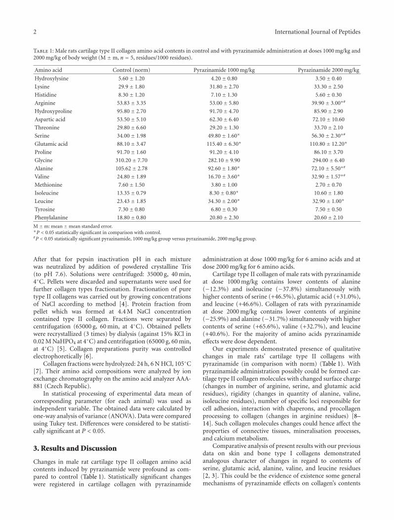
2 International Journal of Peptides
Table 1: Male rats cartilage type II collagen amino acid contents in control and with pyrazinamide administration at doses 1000 mg/kg and2000 mg/kg of body weight (M ±m, n = 5, residues/1000 residues).
Amino acid Control (norm) Pyrazinamide 1000 mg/kg Pyrazinamide 2000 mg/kg
Hydroxylysine 5.60± 1.20 4.20± 0.80 3.50± 0.40
Lysine 29.9± 1.80 31.80± 2.70 33.30± 2.50
Histidine 8.30± 1.20 7.10± 1.30 5.60± 0.30
Arginine 53.83± 3.35 53.00± 5.80 39.90± 3.00∗#
Hydroxyproline 95.80± 2.70 91.70± 4.70 85.90± 2.90
Aspartic acid 53.50± 5.10 62.30± 6.40 72.10± 10.60
Threonine 29.80± 6.60 29.20± 1.30 33.70± 2.10
Serine 34.00± 1.98 49.80± 1.60∗ 56.30± 2.30∗#
Glutamic acid 88.10± 3.47 115.40± 6.30∗ 110.80± 12.20∗
Proline 91.70± 1.60 91.20± 4.10 86.10± 3.70
Glycine 310.20± 7.70 282.10± 9.90 294.00± 6.40
Alanine 105.62± 2.78 92.60± 1.80∗ 72.10± 5.50∗#
Valine 24.80± 1.89 16.70± 3.60∗ 32.90± 1.57∗#
Methionine 7.60± 1.50 3.80± 1.00 2.70± 0.70
Isoleucine 13.35± 0.79 8.30± 0.80∗ 10.60± 1.80
Leucine 23.43± 1.85 34.30± 2.00∗ 32.90± 1.00∗
Tyrosine 7.30± 0.80 6.80± 0.30 7.50± 0.50
Phenylalanine 18.80± 0.80 20.80± 2.30 20.60± 2.10
M ±m: mean ±mean standard error.∗P < 0.05 statistically significant in comparison with control.#P < 0.05 statistically significant pyrazinamide, 1000 mg/kg group versus pyrazinamide, 2000 mg/kg group.
After that for pepsin inactivation pH in each mixturewas neutralized by addition of powdered crystalline Tris(to pH 7.6). Solutions were centrifuged: 35000 g, 40 min,4◦C. Pellets were discarded and supernatants were used forfurther collagen types fractionation. Fractionation of puretype II collagens was carried out by growing concentrationsof NaCl according to method [4]. Protein fraction frompellet which was formed at 4.4 M NaCl concentrationcontained type II collagen. Fractions were separated bycentrifugation (65000 g, 60 min, at 4◦C). Obtained pelletswere recrystallized (3 times) by dialysis (against 15% KCl in0.02 M NaHPO4 at 4◦C) and centrifugation (65000 g, 60 min,at 4◦C) [5]. Collagen preparations purity was controlledelectrophoretically [6].
Collagen fractions were hydrolyzed: 24 h, 6 N HCl, 105◦C[7]. Their amino acid compositions were analyzed by ionexchange chromatography on the amino acid analyzer AAA-881 (Czech Republic).
In statistical processing of experimental data mean ofcorresponding parameter (for each animal) was used asindependent variable. The obtained data were calculated byone-way analysis of variance (ANOVA). Data were comparedusing Tukey test. Differences were considered to be statisti-cally significant at P < 0.05.
3. Results and Discussion
Changes in male rat cartilage type II collagen amino acidcontents induced by pyrazinamide were profound as com-pared to control (Table 1). Statistically significant changeswere registered in cartilage collagen with pyrazinamide
administration at dose 1000 mg/kg for 6 amino acids and atdose 2000 mg/kg for 6 amino acids.
Cartilage type II collagen of male rats with pyrazinamideat dose 1000 mg/kg contains lower contents of alanine(−12.3%) and isoleucine (−37.8%) simultaneously withhigher contents of serine (+46.5%), glutamic acid (+31.0%),and leucine (+46.6%). Collagen of rats with pyrazinamideat dose 2000 mg/kg contains lower contents of arginine(−25.9%) and alanine (−31.7%) simultaneously with highercontents of serine (+65.6%), valine (+32.7%), and leucine(+40.6%). For the majority of amino acids pyrazinamideeffects were dose dependent.
Our experiments demonstrated presence of qualitativechanges in male rats’ cartilage type II collagens withpyrazinamide (in comparison with norm) (Table 1). Withpyrazinamide administration possibly could be formed car-tilage type II collagen molecules with changed surface charge(changes in number of arginine, serine, and glutamic acidresidues), rigidity (changes in quantity of alanine, valine,isoleucine residues), number of specific loci responsible forcell adhesion, interaction with chaperons, and procollagenprocessing to collagen (changes in arginine residues) [8–14]. Such collagen molecules changes could hence affect theproperties of connective tissues, mineralisation processes,and calcium metabolism.
Comparative analysis of present results with our previousdata on skin and bone type I collagens demonstratedanalogous character of changes in regard to contents ofserine, glutamic acid, alanine, valine, and leucine residues[2, 3]. This could be the evidence of existence some generalmechanisms of pyrazinamide effects on collagen’s contents
International Journal of Peptides 3
and structure. Moreover, having compared these data withour previous experiments, we found out analogous characterof these changes in regard to changes of free serine, glutamicacid, alanine, valine, and leucine contents in liver, kidney,lung and spleen pools with different doses of pyrazinamide[8]. Thus adverse effects of pyrazinamide (this widelyused antitubercular drug) are much more serious andmore profound than it was considered earlier. Among thispyrazinamide treatment could cause qualitative changes innucleic acid molecules, change their length, and structure[9].
We can suppose that such changes could be causedby pyrazinamide via its influence on nucleic acids (codinginformation for this proteins synthesis) as it was mentionedpreviously [2, 9, 10]. In our previous experiments wedemonstrated epigenetic changes induced by pyrazinamidetreatment, pyrazinamide-mediated alterations in male ratsDNA fragmentation processes, bone type I collagen aminoacid composition, spermatogenesis indices, reproductivecapability, and posterity antenatal and postnatal develop-ment. Besides these, on changes in collagen metabolismand structure pathologic changes in amino acid metabolismcould also be affected [8]. And at last, due to collagengenes polymorphism [11–14], collagen structures containin norm 4 different α-chains of the same type in differentconcentrations. Pathology [14] changed concentrations inwhich these 4 different α-chains of the same type ofcollagen are present in connective tissue structures. Possiblypyrazinamide-caused disturbances in amino acid composi-tions in our experiments could be a result of such changes intranscription rates of genes coding different α-chains fromthe same type collagen superfamily as it was previouslydemonstrated for other pathology [14].
References
[1] A. N. Malaviya and P. P. Kotwal, “Arthritis associated withtuberculosis,” Best Practice and Research, vol. 17, no. 2, pp.319–343, 2003.
[2] L. B. Bondarenko, G. M. Shayakhmetova, T. F. Byshovets,and V. M. Kovalenko, “Pyrazinamide-mediated changes in rattype I collagen and spermatogenesis indices,” Acta PoloniaePharmaceutica—Drug Research, vol. 68, no. 2, pp. 285–290,2011.
[3] L. B. Bondarenko, G. M. Shayakhmetova, T. F. Byshovets, andV. M. Kovalenko, Acta Poloniae Pharmaceutica. In press.
[4] R. L. Trelstad, V. M. Catanese, and D. F. Rubin, “Collagenfractionation: separation of native types I,II and III bydifferential precipitation,” Analytical Biochemistry, vol. 71, no.1, pp. 114–118, 1976.
[5] A. L. Rubin, M. P. Drake, P. F. Davison, D. Pfahl, P. T.Speakman, and F. O. Schmitt, “Effects of pepsin treatment onthe interaction properties of tropocollagen macromolecules,”Biochemistry, vol. 4, no. 2, pp. 181–190, 1965.
[6] G. Maurer, in The Disk-Electrophoresis, p. 247, Mir, Moscow,Russia, 1971.
[7] T. Deveni and J. Gherghey, in The Aminoacids, Peptides andProteins, p. 364, Mir, Moscow, Russia, 1976.
[8] L. B. Bondarenko, N. A. Saprykina, and V. M. Kovalenko,“Lung and spleen contents of free amino acids after pyrazi-namide treatment,” Acta Toxicologica, vol. 14, no. 1-2, pp. 79–86, 2006.
[9] V. M. Kovalenko, T. V. Bagnyukova, O. V. Sergienko et al.,“Epigenetic changes in the rat livers induced by pyrazinamidetreatment,” Toxicology and Applied Pharmacology, vol. 225, no.3, pp. 293–299, 2007.
[10] L. Bondarenko, G. Shayakhmetova, T. Byshovets, and V.Kovalenko, International Journal of Infectious Diseases, vol. 15,supplement S99, 2011.
[11] S. R. Kimball, M. Yancisin, R. L. Horetsky, and L. S. Jefferson,“Translational and pretranslational regulation of proteinsynthesis by amino acid availability in primary cultures ofrat hepatocytes,” International Journal of Biochemistry and CellBiology, vol. 28, no. 3, pp. 285–294, 1996.
[12] J. Inamasu, B. H. Guiot, and D. C. Sachs, “Ossification ofthe posterior longitudinal ligament: an update on its biology,epidemiology, and natural history,” Neurosurgery, vol. 58, no.6, pp. 1027–1038, 2006.
[13] J. D. Kurt, S. Makoto, K. Tomoatsu, and Y. Yoshihiko,“Complete coding sequence and deduced primary structure ofthe human cartilage large aggregating proteoglycan, aggrecan,”The Journal of Biological Chemistry, vol. 266, no. 2, pp. 894–902, 1991.
[14] B. Lee, M. D’Alessio, H. Vissing, F. Ramirez, B. Steinmann,and A. Superti-Furga, “Characterization of a large deletionassociated with a polymorphic block of repeated dinucleotidesin the type III procollagen gene (COL3AI) of a patient withEhlers-Danlos syndrome type IV,” American Journal of HumanGenetics, vol. 48, no. 3, pp. 511–517, 1991.
Hindawi Publishing CorporationInternational Journal of PeptidesVolume 2012, Article ID 731293, 10 pagesdoi:10.1155/2012/731293
Research Article
Molecular Cloning and Sequence Analysis ofthe cDNAs Encoding Toxin-Like Peptides from the Venom Glandsof Tarantula Grammostola rosea
Tadashi Kimura,1, 2 Seigo Ono,1 and Tai Kubo1, 2
1 Molecular Neurophysiology Group, Neuroscience Research Institute, National Institute of Advanced Industrial Science andTechnology (AIST), Central 6, 1-1-1 Higashi, Tsukuba, Ibaraki 305-8566, Japan
2 United Graduate School of Drug Discovery and Medical Information Science, Gifu University, 1-1 Yanagido, Gifu 501-1193, Japan
Correspondence should be addressed to Tadashi Kimura, [email protected] and Tai Kubo, [email protected]
Received 16 September 2011; Accepted 26 November 2011
Academic Editor: Mirian A. F. Hayashi
Copyright © 2012 Tadashi Kimura et al. This is an open access article distributed under the Creative Commons AttributionLicense, which permits unrestricted use, distribution, and reproduction in any medium, provided the original work is properlycited.
Tarantula venom glands produce a large variety of bioactive peptides. Here we present the identification of venom componentsobtained by sequencing clones isolated from a cDNA library prepared from the venom glands of the Chilean common tarantula,Grammostola rosea. The cDNA sequences of about 1500 clones out of 4000 clones were analyzed after selection using severalcriteria. Forty-eight novel toxin-like peptides (GTx1 to GTx7, and GTx-TCTP and GTx-CRISP) were predicted from the nucleotidesequences. Among these peptides, twenty-four toxins are ICK motif peptides, eleven peptides are MIT1-like peptides, and sevenare ESTX-like peptides. Peptides similar to JZTX-64, aptotoxin, CRISP, or TCTP are also obtained. GTx3 series possess a cysteineframework that is conserved among vertebrate MIT1, Bv8, prokineticins, and invertebrate astakines. GTx-CRISP is the first CRISP-like protein identified from the arthropod venom. Real-time PCR revealed that the transcripts for TCTP-like peptide are expressedin both the pereopodal muscle and the venom gland. Furthermore, a unique peptide GTx7-1, whose signal and prepro sequencesare essentially identical to those of HaTx1, was obtained.
1. Introduction
Venoms are complex mixtures of many different componentsproven to be useful tools for biochemical, physiological, andpharmacological studies of ion channels and receptors. Tox-ins that recognize ion channel subgroups are versatile toolsfor channel studies and thus contribute to drug discovery [1,2]. For example, a 25-amino-acid peptide isolated from themarine fish-hunting cone snail Conus magus, ω-conotoxin-MVIIA, blocks N-type voltage-dependent calcium channels.In 2004, ziconotide, the synthetic version of ω-conotoxin-MVIIA, was approved in the United States for the treatmentof chronic severe pain refractory to other current painmedications.
About 40000 different kinds of spiders are known atpresent. Spider venoms contain peptide neurotoxins and areexpected to be a rich source of ion channel blockers [3–5].Tarantulas, comprising more than 860 species, like all other
spiders are predators that feed on a variety of vertebrateand invertebrate prey [6]. Tarantulas do not use webs forcapture but are well-equipped predators, possessing a varietyof venoms that target receptors in the nervous system,probably with adaptation to a certain type of prey [7, 8].Tarantula venom has been suggested to contain 1000 ormore peptide toxins [8]. Despite their diverse activities, thesetoxins display only a few widely conserved structural motifsthat share remarkable similarities in their primary sequencesand tertiary structures [9–11]. In a similar fashion to theevolution of snake toxins, several molecular scaffolds havebeen used during the evolution of toxin “cocktails” in spidervenoms. The selected genes are duplicated several times, and,while the core of each protein scaffold is conserved, the loopsand surfaces are altered through mutations [12]. We recentlyfound T-type voltage-dependent calcium channel blockerfrom venom of Chilean common tarantula, Grammostolarosea [13].
2 International Journal of Peptides
Expressed sequence tags (ESTs) are short single-passsequence reads generated from either 5′ or 3′ end of cDNAs.They provide a quick and inexpensive route for discoveringnew genes and obtaining data on gene expression. The ESTsapproach has been used in several reports, because it isa rapid and reliable method for gene discovery in gene-ral, mainly in this case, related to secretory glands from ven-omous animals [14–16].
In this paper, we focused on the tarantula toxins, andby applying improved molecular biological techniques, werevealed novel peptide sequences after ESTs techniquesapplied to a cDNA library prepared from the Chilean com-mon tarantula Grammostola rosea venom glands.
2. Materials and Methods
2.1. Animals and Venom Glands. Grammostola rosea taran-tulas were obtained from a local pet supplier. The venomglands were dissected from the chelicera and the pereopodalmuscles were from the prosoma using sharp forceps, frozenimmediately with liquid nitrogen, and then stored at −80◦Cuntil use.
2.2. cDNA Library Construction. Preparation of the venomgland cDNA library was reported previously [13]. Briefly, thevenom glands were dissected from 30 spiders, and total RNAwas extracted using TRIZOL reagent (Invitrogen, Carlsbad,CA). Poly(A)+ RNA was prepared using Oligotex-dT30Super (Takara Bio, Otsu, Japan). The first-strand cDNAswere synthesized from 2.5 μg of poly(A)+ RNA using theprimer, VNXho(dT)30, which installs oligo dT and XhoIsequences, by ReverTra Ace (Toyobo, Osaka, Japan) andSuperscript II (Invitrogen). The second strands were synthe-sized with DNA polymerase I (Takara Bio), RNase H (TakaraBio), and Escherichia coli DNA ligase (Takara Bio). Eco RIadaptors (Clontech, Palo Alto, CA) were ligated to the cDNAsafter both ends of the double-stranded cDNAs were filledin with a DNA blunting kit (Takara Bio). The cDNAs werethen digested with XhoI and fractionated by 1.2% agarosegel electrophoresis. DNA fragments with lengths of 0.8–2.0 kbp were eluted from dissected gel. The resulting DNAfragments were ligated into EcoRI and XhoI restriction sitesof pSD64TRER [17]. E. coli XL1-Blue MRA (Agilent Tech-nologies, Santa Clara, CA) was transformed with the plas-mid. An aliquot of the cDNA library in E. coli was spreadonto LB agar plates containing 50 μg/mL ampicillin, and theplasmid DNA was prepared for the PCR template.
2.3. Fingerprinting of Clones. An aliquot of cDNA library wasspread onto LB agar plates with ampicillin (50 μ g/mL) andincubated at 37◦C overnight. Formed colonies were pickedup by automated colony picker (Microtec Nichion, Japan)and inoculated into1 mL of 2 × LB medium supplementedwith ampicillin (50 μg/mL) in 96 deep-well plates andincubated at 37◦C overnight with vigorous shaking. Plasmidswere purified with an automated machine, BIOMEK2000,(Beckman Coulter, USA) using MultiScreen-FB and -NA(Millipore, USA) and eluted by 50 μL TE solution, thenstored −20◦C until use. PCR was performed with an SP6
primer and a pSD64-specific reverse primer, SDA (5′-TTATGTAGCTTAGAGACT-3′), to amplify the inserts ofthe cDNA library. Each PCR reaction mixture consists of10 pmol of the forward and reverse primers, 0.25 U EXTaq polymerase (Takara Bio), 200 mM each of dATP, dCTP,dGTP and dTTP, 2 mM MgCl2, PCR buffer, and 1 μL DNAtemplate. The reaction was performed in a thermal cyclerPTC-200 (MJ research, USA) for 30 cycles, each consistingof denaturation at 94◦C for 30 s, annealing at 42◦C for 45 s,and polymerization at 72◦C for 1 min, after the initial cycleof 94◦C for 5 min. At the end of all the cycles, samples weremaintained at 72◦C for 9 min and then kept at 4◦C.
PCR reaction products were digested by DdeI restrictionenzyme at 37◦C and analyzed by electrophoresis in 3%agarose/TBE gel, then visualized with ethidium bromide,and digitized using gel documentation system, Gel Doc 1000(Bio-Rad, USA). The band patterns of the digested PCRproducts were clustered by the similarity using MolecularAnalyst Fingerprinting plus software (Bio-Rad).
2.4. Sequencing and Data Analysis. We manually selectedclones to be sequenced based on the fingerprinting catego-rization described previously. Single run DNA sequencingswere performed using an SP6 primer by a sequencer ModelABI Prism 377 (Applied Biosystems, CA, USA) or performedby Shimadzu. The obtained DNA sequences were translatedinto amino acid sequences with all three frames using theVector NTI program (Invitrogen, USA). After translationinto three amino acid sequences, both protein and cDNAsequences were stored into in-house database software,KIROKU (World Fusion, Tokyo, Japan). Homology searchof translated protein sequences was performed at in-housesequence database using the BLAST program. The pre-diction of signal sequence was performed by SignalP 3.0 pro-gram (http://www.cbs.dtu.dk/services/SignalP/). Amino acidalignment and phylogenetic tree construction were perform-ed using the MegAlign program by Clustal W and neighbor-joining method (DNASTAR, Madison, USA).
2.5. PCR Cloning Based on the Signal Sequence of Toxins. Wesynthesized oligonucleotide primers based on the conservedinitiation codon (ATG) and its juxtaposed sequences includ-ing 5′-noncoding region and the signal sequences of theGTxs (Table 1). Using these primers and an SDA primer, PCRamplifications were carried out with the venom glands cDNAlibrary as a template. The reaction conditions were essentiallythe same as described in Section 2.3. Amplified fragmentswere cloned into pCR 2.1-TOPO (Invitrogen). The full-length nucleotide sequences of the clones were determinedby Hitachi Soft Co Ltd.
2.6. Quantification of Tissue Expression of
GTx-TCTP and GTx-CRISP cDNAs
2.6.1. Cloning and Sequence Analysis of GTx-TCTP andGTx-CRISP cDNAs. Single run sequencings of the venomgland cDNA library revealed the first half of GTx-TCTPand GTx-CRISP cDNA including 5′-UTR region with start
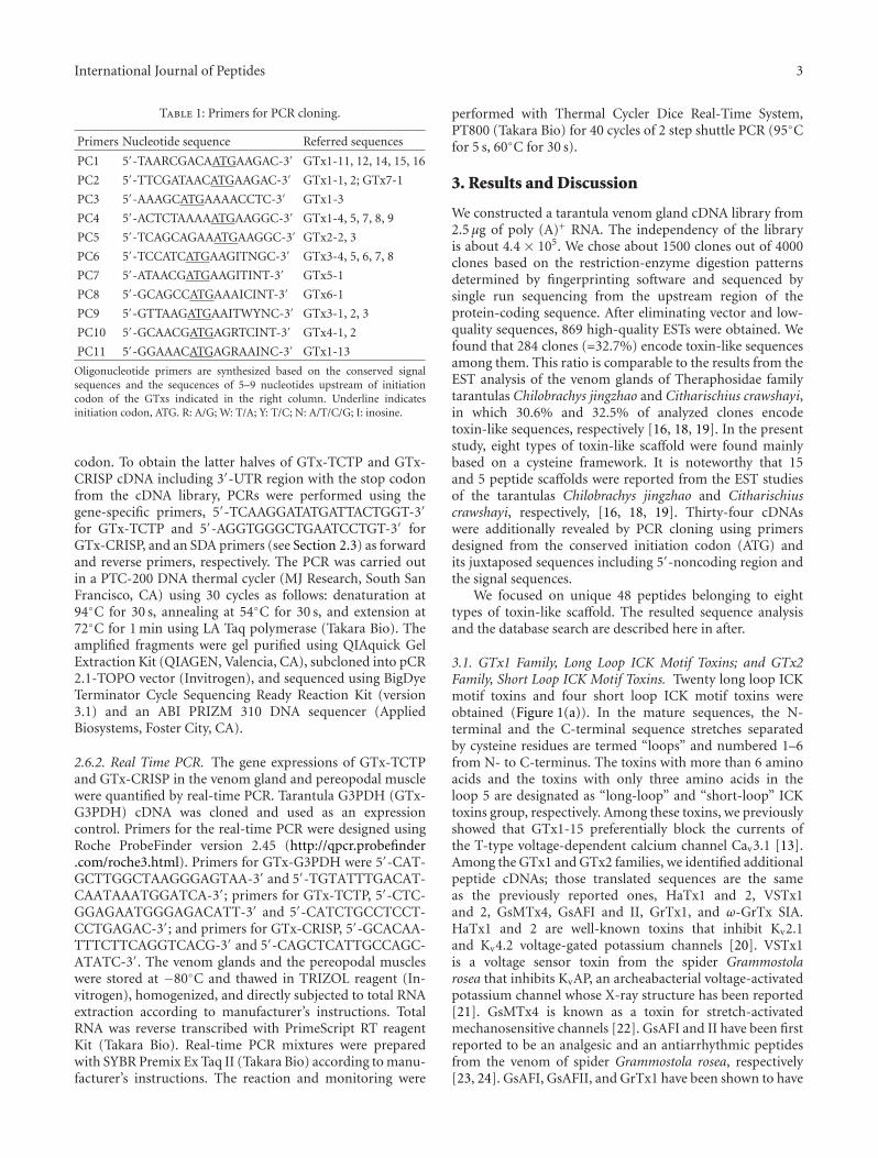
International Journal of Peptides 3
Table 1: Primers for PCR cloning.
Primers Nucleotide sequence Referred sequences
PC1 5′-TAARCGACAATGAAGAC-3′ GTx1-11, 12, 14, 15, 16
PC2 5′-TTCGATAACATGAAGAC-3′ GTx1-1, 2; GTx7-1
PC3 5′-AAAGCATGAAAACCTC-3′ GTx1-3
PC4 5′-ACTCTAAAAATGAAGGC-3′ GTx1-4, 5, 7, 8, 9
PC5 5′-TCAGCAGAAATGAAGGC-3′ GTx2-2, 3
PC6 5′-TCCATCATGAAGITNGC-3′ GTx3-4, 5, 6, 7, 8
PC7 5′-ATAACGATGAAGITINT-3′ GTx5-1
PC8 5′-GCAGCCATGAAAICINT-3′ GTx6-1
PC9 5′-GTTAAGATGAAITWYNC-3′ GTx3-1, 2, 3
PC10 5′-GCAACGATGAGRTCINT-3′ GTx4-1, 2
PC11 5′-GGAAACATGAGRAAINC-3′ GTx1-13
Oligonucleotide primers are synthesized based on the conserved signalsequences and the sequcences of 5–9 nucleotides upstream of initiationcodon of the GTxs indicated in the right column. Underline indicatesinitiation codon, ATG. R: A/G; W: T/A; Y: T/C; N: A/T/C/G; I: inosine.
codon. To obtain the latter halves of GTx-TCTP and GTx-CRISP cDNA including 3′-UTR region with the stop codonfrom the cDNA library, PCRs were performed using thegene-specific primers, 5′-TCAAGGATATGATTACTGGT-3′
for GTx-TCTP and 5′-AGGTGGGCTGAATCCTGT-3′ forGTx-CRISP, and an SDA primers (see Section 2.3) as forwardand reverse primers, respectively. The PCR was carried outin a PTC-200 DNA thermal cycler (MJ Research, South SanFrancisco, CA) using 30 cycles as follows: denaturation at94◦C for 30 s, annealing at 54◦C for 30 s, and extension at72◦C for 1 min using LA Taq polymerase (Takara Bio). Theamplified fragments were gel purified using QIAquick GelExtraction Kit (QIAGEN, Valencia, CA), subcloned into pCR2.1-TOPO vector (Invitrogen), and sequenced using BigDyeTerminator Cycle Sequencing Ready Reaction Kit (version3.1) and an ABI PRIZM 310 DNA sequencer (AppliedBiosystems, Foster City, CA).
2.6.2. Real Time PCR. The gene expressions of GTx-TCTPand GTx-CRISP in the venom gland and pereopodal musclewere quantified by real-time PCR. Tarantula G3PDH (GTx-G3PDH) cDNA was cloned and used as an expressioncontrol. Primers for the real-time PCR were designed usingRoche ProbeFinder version 2.45 (http://qpcr.probefinder.com/roche3.html). Primers for GTx-G3PDH were 5′-CAT-GCTTGGCTAAGGGAGTAA-3′ and 5′-TGTATTTGACAT-CAATAAATGGATCA-3′; primers for GTx-TCTP, 5′-CTC-GGAGAATGGGAGACATT-3′ and 5′-CATCTGCCTCCT-CCTGAGAC-3′; and primers for GTx-CRISP, 5′-GCACAA-TTTCTTCAGGTCACG-3′ and 5′-CAGCTCATTGCCAGC-ATATC-3′. The venom glands and the pereopodal muscleswere stored at −80◦C and thawed in TRIZOL reagent (In-vitrogen), homogenized, and directly subjected to total RNAextraction according to manufacturer’s instructions. TotalRNA was reverse transcribed with PrimeScript RT reagentKit (Takara Bio). Real-time PCR mixtures were preparedwith SYBR Premix Ex Taq II (Takara Bio) according to manu-facturer’s instructions. The reaction and monitoring were
performed with Thermal Cycler Dice Real-Time System,PT800 (Takara Bio) for 40 cycles of 2 step shuttle PCR (95◦Cfor 5 s, 60◦C for 30 s).
3. Results and Discussion
We constructed a tarantula venom gland cDNA library from2.5 μg of poly (A)+ RNA. The independency of the libraryis about 4.4 × 10⁵. We chose about 1500 clones out of 4000clones based on the restriction-enzyme digestion patternsdetermined by fingerprinting software and sequenced bysingle run sequencing from the upstream region of theprotein-coding sequence. After eliminating vector and low-quality sequences, 869 high-quality ESTs were obtained. Wefound that 284 clones (=32.7%) encode toxin-like sequencesamong them. This ratio is comparable to the results from theEST analysis of the venom glands of Theraphosidae familytarantulas Chilobrachys jingzhao and Citharischius crawshayi,in which 30.6% and 32.5% of analyzed clones encodetoxin-like sequences, respectively [16, 18, 19]. In the presentstudy, eight types of toxin-like scaffold were found mainlybased on a cysteine framework. It is noteworthy that 15and 5 peptide scaffolds were reported from the EST studiesof the tarantulas Chilobrachys jingzhao and Citharischiuscrawshayi, respectively, [16, 18, 19]. Thirty-four cDNAswere additionally revealed by PCR cloning using primersdesigned from the conserved initiation codon (ATG) andits juxtaposed sequences including 5′-noncoding region andthe signal sequences.
We focused on unique 48 peptides belonging to eighttypes of toxin-like scaffold. The resulted sequence analysisand the database search are described here in after.
3.1. GTx1 Family, Long Loop ICK Motif Toxins; and GTx2Family, Short Loop ICK Motif Toxins. Twenty long loop ICKmotif toxins and four short loop ICK motif toxins wereobtained (Figure 1(a)). In the mature sequences, the N-terminal and the C-terminal sequence stretches separatedby cysteine residues are termed “loops” and numbered 1–6from N- to C-terminus. The toxins with more than 6 aminoacids and the toxins with only three amino acids in theloop 5 are designated as “long-loop” and “short-loop” ICKtoxins group, respectively. Among these toxins, we previouslyshowed that GTx1-15 preferentially block the currents ofthe T-type voltage-dependent calcium channel Cav3.1 [13].Among the GTx1 and GTx2 families, we identified additionalpeptide cDNAs; those translated sequences are the sameas the previously reported ones, HaTx1 and 2, VSTx1and 2, GsMTx4, GsAFI and II, GrTx1, and ω-GrTx SIA.HaTx1 and 2 are well-known toxins that inhibit Kv2.1and Kv4.2 voltage-gated potassium channels [20]. VSTx1is a voltage sensor toxin from the spider Grammostolarosea that inhibits KvAP, an archeabacterial voltage-activatedpotassium channel whose X-ray structure has been reported[21]. GsMTx4 is known as a toxin for stretch-activatedmechanosensitive channels [22]. GsAFI and II have been firstreported to be an analgesic and an antiarrhythmic peptidesfrom the venom of spider Grammostola rosea, respectively[23, 24]. GsAFI, GsAFII, and GrTx1 have been shown to have
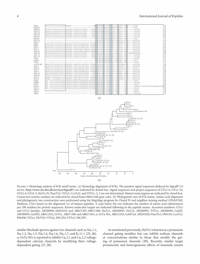
4 International Journal of Peptides
VSTx1 63HnTx-IV 35GsMTx4 82GTx1-16 82GTx1-20 82GTx1-19 82GTx1-15 82PaurTx3 34GTx1-24 36GTx1-6 37HaTx1 85HaTx2 85GTx1-1 85GTx1-3 86GTx1-21 86GTx1-14 88GTx1-11 81GTx1-12 81TLTx1 35CcoTx2 34VSTx3 35
89GTx1-7 86GTx1-8 85GTx1-9 86GTx1-2 85GTx1-13 83GTx1-22 83GTx1-4 84GTx1-5 84GsAFI 80
GrTx1 80GTx2-2 105GTx2-3 80GTx2-5 30GTx2-4 30VSTx2 32
GSAFII 81
ω-GrTx-SIA
(a)
0
185.8
20406080100120140160180
GTx1-4GTx1-5
GTx1-6GTx1-8
GTx1-9
GTx1-7GTx1-3GTx1-21
GTx1-2GTx1-24
GTx1-1GTx1-13
GTx1-22
HnTx-IV(Na Ch)
GTx1-20GTx1-16
VSTx3(GTx1-11
GTx1-12TLTx1(
CcoTx2(PaurTx3
GTx1-19GTx1-15(
GTx2-3GTx2-5
GTx2-4
GTx2-2
VSTx2(GTx1-14
Amino acid substitution per 100 residues
ω-GrTx SIA(Ca2+Ch)
HaTx2(K+, Na+ Ch)
HaTx1(K+, Na+ Ch)
VSTx1(Na+, K+
+
(Na Ch)+
(Na Ch)+
(Ca Ch)2+
Ch)
GrTx1(Na+, K+ Ch)GsAFI(Na +, K+ Ch)
GsMTx4(MSC, Na+, K+ Ch)
K+ Ch)
K+ Ch)
K+ Ch)GsAFII(SAC, Na+, K+, Ca2+ Ch)
(b)
Figure 1: Homology analysis of ICK motif toxins. (a) Homology alignment of ICKs. The putative signal sequences deduced by SignalP 3.0server (http://www.cbs.dtu.dk/services/SignalP/) are indicated by dotted box. Signal sequences and prepro-sequences of GTx1-6, GTx1-24,GTx2-4, GTx2-5, HnTx-IV, PaurTx3, TLTx1, CcoTx2, and VSTx1, 2, 3 are not determined. Mature toxin regions are indicated by closed box.Conserved cysteine residues are indicated by closed boxes filled with gray color. (b) Phylogenetic tree of ICK toxins. Amino acid alignmentand phylogenetic tree construction were performed using the MegAlign program by Clustal W and neighbor-joining method (DNASTAR,Madison, USA) based on the alignment (a) of mature peptides. A scale below the tree indicates the number of amino acid substitutionsper 100 residues for protein sequences. Known molecular targets are indicated following to the peptide names. Accession numbers: GTx1and GTx2 families, AB200996-AB201024 and AB671302-AB671308; HaTx1, AB200991; HaTx2, AB200992; VSTx1, AB200994; GsAFI,AB200995; GsAFII, AB612242; GrTx1, AB671300 and AB671301; ω-GrTx SIA, AB612243; GsMTx4, AB201020; PaurTx3, P84510; CcoTx2,P84508; TLTx1, P83745; VSTx2, P0C2P4; VSTx3, P0C2P5.
similar blockade spectra against ion channels such as Nav1.1,Nav1.2, Nav1.3, Nav1.4, Nav1.6, Nav1.7, and Kv11.1 [25, 26].ω-GrTx SIA is reported to inhibit Cav2.1 and Cav2.2 voltage-dependent calcium channels by modifying their voltage-dependent gating [27, 28].
As mentioned previously, HaTx1 is known as a potassiumchannel gating modifier but can inhibit sodium channelsat concentrations similar to those that modify the gat-ing of potassium channels [29]. Recently, similar targetpromiscuity and heterogeneous effects of tarantula venom

International Journal of Peptides 5
Prokineticin1 61
Prokineticin2 69
MIT1 42
Bv8 61
De astakine2 66
Ag astakine2 64
Pl astakine2a 66
Ra astakine2 67
Is astakine2 66
Av astakine2 68
Pl astakine1 65
Ag astakine1 64
GTx3-4 57
GTx3-5 57
GTx3-6 57
GTx3-7 57
GTx3-8 57
GTx3-9 57
HWTX-XIVa1 55
PRTx16C0 35
ACTX-Hvf17 33
GTx3-2 59
GTx3-1 57
GTx3-11 59
GTx3-3 59
GTx3-10 59
Prokineticin1 105
Prokineticin2 108
MIT1 81
Bv8 96
De astakine2 106
Ag astakine2 127
Pl astakine2a 122
Ra astakine2 127
Is astakine2 118
Av astakine2 127
Pl astakine1 104
Ag astakine1 89
GTx3-4 86
GTx3-5 86
GTx3-6 86
GTx3-7 86
GTx3-8 86
GTx3-9 86
HWTX-XIVa1 84
ACTX-Hvf17 68
GTx3-2 109
GTx3-1 107
GTx3-11 109
GTx3-3 111
GTx3-10 111
PRTx16C0 64
(a)
0
159.4
20406080100120140
GTx3-6GTx3-7
GTx3-9GTx3-8GTx3-4
GTx3-5HWTX-XIVa1
PRTx16C0Pl astakine2a
Pl astakine1De astakine2
ACTX-Hvf17Ra astakine2
Av astakine2Is astakine2
Prokineticin1MIT1
Prokineticin2Bv8
Ag astakine2Ag astakine1
GTx3-3GTx3-10
GTx3-11GTx3-2
GTx3-1
Amino acid substitution per 100 residues
(b)
Figure 2: Homology analysis of GTx3 series toxins. (a) Sequence alignment of GTx3 series and their related peptides. The putative signalsequence deduced by SignalP 3.0 server (http://www.cbs.dtu.dk/services/SignalP/) is indicated by dotted box. Signal sequence of MIT1is not determined. Conserved cysteine residues are indicated by closed boxes filled with gray color. AVIT N-terminals of Prokineticin1,Prokineticin2, MIT1, and Bv8 are indicated closed boxes. (b) Phylogenetic tree of GTx3 series and related peptides. Amino acid alignmentand phylogenetic tree construction were performed using the MegAlign program by Clustal W and neighbor-joining method (DNASTAR)based on the alignment (a) of mature peptides. A scale below the tree indicates the number of amino acid substitutions per 100 residuesfor protein sequences. Accession numbers: GTx3 series, AB201025-AB201034 and AB671309-AB671311; Bv8, AF168790; Prokineticin1, AF333024; Prokineticin 2, AAH96695; MIT1, P25687; HWTX-XIVa1, ABY77690; ACTX-Hvf17, P81803; Pacifastacus leniusculus (Pl)astakine1, AY787656; Pl astakine2a, EF568370; Acanthoscurria gomesiana (Ag) astakine1, DR447331; Ag astakine2, DR445103; Rhipicephalusappendiculatus (Ra) astakine2, CD794853; Ixodes scapularis (Is) astakine2, EW845057; Amblyomma variegatum (Av) astakine2, BM292046;Dysdera erythrina (De) astakine2, CV178181.
voltage-sensor toxins are discussed including GsAFI andII, GrTx1 described previously [25, 30]. We also suggestsimilar target promiscuity; that is, a toxin family includingGsAFII, GsMTx2, PaTx2, and ProTx-II could affect severaltypes of ion channels such as stretch-activated channelsand the voltage-dependent sodium, potassium, and calciumchannels [13]. Figure 1(b) shows phylogenetic tree of GTx1,GTx2, and several ICK toxins. The correlations betweenthe peptide groups and their target molecules are not clear.Vega discussed the pharmacological diversification of ICKmotif toxin by phylogenetic analysis of 171 homologousICK toxins using Bayesian inference [31]. Although therelationship between clusters is not satisfactorily solved, sev-eral trustable monophyletic groups appear from the analy-sis. The main conclusion from the tree is a plausible lin-age-specific process of paralogous diversification fromseveral independent recruiting events. Further investigation
is needed to elucidate the relationships between evolutionalprocesses and the pharmacological diversification and targetpromiscuity of ICK toxins.
3.2. GTx3 Series: Similar to Mamba Intestinal Toxin 1 (MIT1),Bv8/Prokineticins, and Invertebrate Astakines. Eleven tox-ins similar to MIT1, Bv8/prokineticins, and invertebrateastakines were identified (Figure 2). Bv8, prokineticins, andMIT1 consist in a group known as AVIT family due to theirN-terminal residues A-V-I-T [32]. MIT1 shows contractileeffects on longitudinal ileal muscle and distal colon [33]. Thesolution structure of MIT1 was determined at a resolutionof 0.5 A and revealed a new type of folding for venom tox-ins similar to that of colipase, a protein involved in fattyacid digestion [34]. Bv8 is bioactive peptide found fromfrog skin to induce hyperalgesic effects [35] and belongsto a family of secretory proteins (Bv8-prokineticin family)
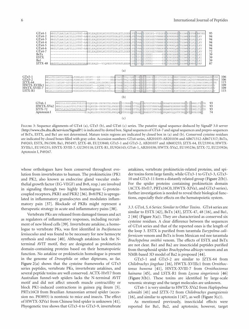
6 International Journal of Peptides
GTx4-1 95GTx4-2 94GTx4-3 94GTx4-4 94GTx4-5 94GTx4-6 94GTx4-7 72BsTx 39ESTX 39Ba1 39JZTX-48 87
(a)
GTx5-1 106GTx5-2 106JZTX-64 115HWTX-XVIIIc1 109HNTX-XVIII-7 109LSTX-R1 115
(b)
GTx6-1 93HWTX-XVa2 93JZTX-72 93Aptotoxin I 74
(c)
Figure 3: Sequence alignments of GTx4 (a), GTx5 (b), and GTx6 (c) series. The putative signal sequence deduced by SignalP 3.0 server(http://www.cbs.dtu.dk/services/SignalP/) is indicated by dotted box. Signal sequences of GTx4-7 and signal sequences and prepro-sequencesof BsTx, ESTX, and Ba1 are not determined. Mature toxin regions are indicated by closed box in (a) and (b). Conserved cysteine residuesare indicated by closed boxes filled with gray color. Accession numbers: GTx4 series, AB201035-AB201036 and AB671312-AB671317; BsTx,P49265; ESTX, P61509; Ba1, P85497; JZTX-48, EU233840; GTx5-1 and GTx5-2, AB201037 and AB683255; JZTX-64, EU233914; HWTX-XVIIIc1, EU195231; HNTX-XVIII-7, GU293118; LSTX-R1, EU926143; GTx6-1, AB201038; HWTX-XVa2, EU195236; JZTX-72, EU233926;Aptotoxin I, P49267.
whose orthologues have been conserved throughout evo-lution from invertebrates to human. The prokineticins (PK1and PK2, also known as endocrine gland vascular endo-thelial growth factor (EG-VEGF) and Bv8, resp.) are involvedin signaling through two highly homologous G-protein-coupled receptors, PKR1 and PKR2 [36]. Bv8/PK2 is upregu-lated in inflammatory granulocytes and modulates inflam-matory pain [37]. Blockade of PKRs might represent atherapeutic strategy in acute and inflammatory pains [38].
Vertebrate PKs are released from damaged tissues and actas regulators of inflammatory responses, including recruit-ment of new blood cells [39]. Invertebrate astakine, a homo-logue to vertebrate PKs, was first identified in Pacifastacusleniusculus and was found to be necessary for new hemocytesynthesis and release [40]. Although astakines lack the N-terminal AVIT motif, they are designated as prokineticindomain-containing proteins based on their hematopoieticfunction. No astakine or prokineticin homologue is presentin the genome of Drosophila or other dipterans, so far.Figure 2(a) shows that the cysteine frameworks of GTx3series peptides, vertebrate PKs, invertebrate astakines, andseveral peptide toxins are well conserved. ACTX-Hvf17 fromAustralian funnel-web spiders lacks the N-terminal AVITmotif and did not affect smooth muscle contractility orblock PK1-induced contractions in guinea pig ileum [5].PRTx16C0 from Brazilian Amazonian armed spider (acces-sion no. P83893) is nontoxic to mice and insects. The effectof HWTX-XIVa1 from Chinese bird spider is unknown [41].Phyogenetic tree shows that GTx3-4 to GTx3-9, invertebrate
astakines, vertebrate prokineticin-related proteins, and spi-der toxins form large family, while GTx3-1 to GTx3-3, GTx3-10 and GTx3-11 form a distantly related group (Figure 2(b)).For the spider proteins containing prokineticin domain(ACTX-Hvf17, PRTx16C0, HWTX-XIVa1, and GTx3 series),further investigation is needed to reveal their biological func-tions, especially their effects on the hematopoietic system.
3.3. GTx4, 5, 6 Series: Similar to Other Toxins. GTx4 series aresimilar to ESTX [42], BsTx [43], JZTX-47, 48 [16], and Ba1,2 [44] (Figure 3(a)). They are characterized as conserved sixcysteine residues. A clear difference between the sequencesof GTx4 series and that of the reported ones is the length ofthe loop 3. ESTX is purified from tarantula Eurypelma cali-fornicum venom and BsTx is from Mexican red nee tarantulaBrachypelma smithii venom. The effects of ESTX and BsTxare not clear. Ba1 and Ba2 are insecticidal peptides purifiedfrom theraphosid spider Brachypelma albiceps venom and anNMR-based 3D model of Ba2 is proposed [44].
GTx5-1 and GTx5-2 are similar to JZTX-64 fromChilobrachys jingzhao [16], HWTX-XVIIIc1 from Ornithoc-tonus huwena [41], HNTX-XVIII-7 from Ornithoctonushainana [45], and LSTX-R1 from Lycosa singoriensis [46](Figure 3(b)). These toxins are identified by large-scalevenomic strategy and the target molecules are unknown.
GTx6-1 is very similar to HWTX-XVa2 from Haplopelmaschmidti [41] and JZTX-72 from Chilobrachys guangxiensis[16], and similar to aptotoxin I [47], as well (Figure 3(c)).
As mentioned previously, insecticidal effects werereported for Ba1, Ba2, and aptotoxin; however, target

International Journal of Peptides 7
76GTx-TCTP80HumanTCTP76BmTCTP76DaHRF
154GTx-TCTP1155HumanTCTP153BmTCTP154DaHRF
173GTx-TCTP172HumanTCTP172BmTCTP173DaHRF
(a)
GTx-CRISPPseudecinHuman-CRISP1Helothermine
7580726765Ablomin
153GTx-CRISP159Pseudecin150Human-CRISP1144Helothermine142Ablomin
231GTx-CRISP239Pseudecin225Human-CRISP1219Helothermine216Ablomin
260GTx-CRISP1275Pseudecin249Human-CRISP1242Helothermine240Ablomin
(b)
G3PDH GTx-CRISP GTx-TCTP
PM VG PM VG PM VG
0.01
0.1
1
Rel
ativ
e qu
anti
ty
(c)
Figure 4: Sequence alignment of GTx-TCTP (a) and GTx-CRISP (b) families. The putative signal sequence deduced by SignalP 3.0 server(http://www.cbs.dtu.dk/services/SignalP/) is indicated by dotted box. Conserved cysteine residues are indicated by closed boxes filled withgray color. (c) The results of real-time PCR. G3PDH is equally expressed in the pereopodal muscle (PM) and the venom gland (VG). GTx-CRISP transcript is predominantly expressed in the venom gland. The GTx-CRISP transcript in the PM is one-400th of that in the VG.GTx-TCTP transcript is expressed in both the pereopodal muscle and the venom gland. The GTx-TCTP transcript in the PM is one-13thof that in the VG. Results were confirmed in triplicate experiments. Accession numbers: GTx-TCTP, AB201040; HumanTCTP, NM 003295;Bombyx mori (Bm) TCTP, NM 001044107; Dermacentor andersoni (Da) HRF, DQ009480; GTx-CRISP, AB201041; Pseudecin (Harpegnathossaltator), EFN80524; Human-CRISP1, NM 001205220; Helothermine, U13619; Ablomin, AF384218.
molecules of GTx4, 5, 6, and their homologues are not yetknown.
3.4. GTx-TCTP and GTx-CRISP. We also obtained onetranslationally controlled tumor protein- (TCTP-) like pep-
tide (Figure 4(a)) and one cysteine-rich secretory protein-(CRISP-) like peptide (Figure 4(b)).
TCTP was first identified as a growth-related tumorprotein whose synthesis is controlled mainly at the transla-tional level [48]. This protein has been recognized as a cell
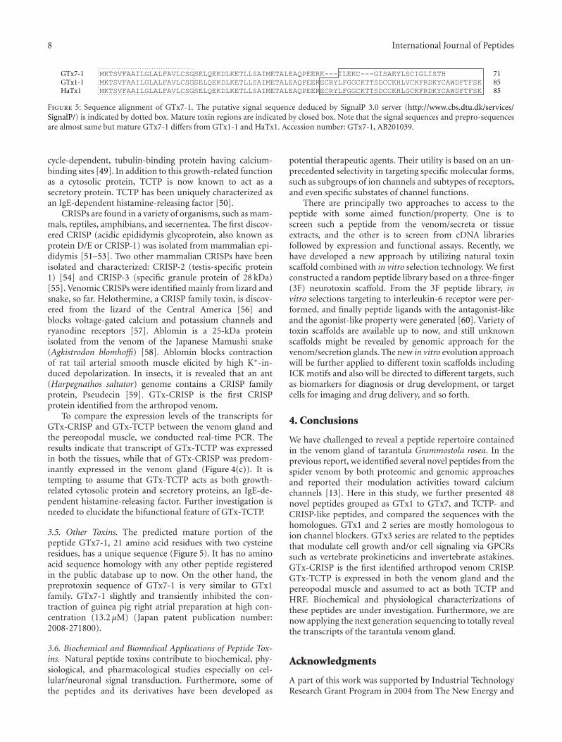
8 International Journal of Peptides
GTx7-1 71GTx1-1 85HaTx1 85
Figure 5: Sequence alignment of GTx7-1. The putative signal sequence deduced by SignalP 3.0 server (http://www.cbs.dtu.dk/services/SignalP/) is indicated by dotted box. Mature toxin regions are indicated by closed box. Note that the signal sequences and prepro-sequencesare almost same but mature GTx7-1 differs from GTx1-1 and HaTx1. Accession number: GTx7-1, AB201039.
cycle-dependent, tubulin-binding protein having calcium-binding sites [49]. In addition to this growth-related functionas a cytosolic protein, TCTP is now known to act as asecretory protein. TCTP has been uniquely characterized asan IgE-dependent histamine-releasing factor [50].
CRISPs are found in a variety of organisms, such as mam-mals, reptiles, amphibians, and secernentea. The first discov-ered CRISP (acidic epididymis glycoprotein, also known asprotein D/E or CRISP-1) was isolated from mammalian epi-didymis [51–53]. Two other mammalian CRISPs have beenisolated and characterized: CRISP-2 (testis-specific protein1) [54] and CRISP-3 (specific granule protein of 28 kDa)[55]. Venomic CRISPs were identified mainly from lizard andsnake, so far. Helothermine, a CRISP family toxin, is discov-ered from the lizard of the Central America [56] andblocks voltage-gated calcium and potassium channels andryanodine receptors [57]. Ablomin is a 25-kDa proteinisolated from the venom of the Japanese Mamushi snake(Agkistrodon blomhoffi) [58]. Ablomin blocks contractionof rat tail arterial smooth muscle elicited by high K+-in-duced depolarization. In insects, it is revealed that an ant(Harpegnathos saltator) genome contains a CRISP familyprotein, Pseudecin [59]. GTx-CRISP is the first CRISPprotein identified from the arthropod venom.
To compare the expression levels of the transcripts forGTx-CRISP and GTx-TCTP between the venom gland andthe pereopodal muscle, we conducted real-time PCR. Theresults indicate that transcript of GTx-TCTP was expressedin both the tissues, while that of GTx-CRISP was predom-inantly expressed in the venom gland (Figure 4(c)). It istempting to assume that GTx-TCTP acts as both growth-related cytosolic protein and secretory proteins, an IgE-de-pendent histamine-releasing factor. Further investigation isneeded to elucidate the bifunctional feature of GTx-TCTP.
3.5. Other Toxins. The predicted mature portion of thepeptide GTx7-1, 21 amino acid residues with two cysteineresidues, has a unique sequence (Figure 5). It has no aminoacid sequence homology with any other peptide registeredin the public database up to now. On the other hand, thepreprotoxin sequence of GTx7-1 is very similar to GTx1family. GTx7-1 slightly and transiently inhibited the con-traction of guinea pig right atrial preparation at high con-centration (13.2 μM) (Japan patent publication number:2008-271800).
3.6. Biochemical and Biomedical Applications of Peptide Tox-ins. Natural peptide toxins contribute to biochemical, phy-siological, and pharmacological studies especially on cel-lular/neuronal signal transduction. Furthermore, some ofthe peptides and its derivatives have been developed as
potential therapeutic agents. Their utility is based on an un-precedented selectivity in targeting specific molecular forms,such as subgroups of ion channels and subtypes of receptors,and even specific substates of channel functions.
There are principally two approaches to access to thepeptide with some aimed function/property. One is toscreen such a peptide from the venom/secreta or tissueextracts, and the other is to screen from cDNA librariesfollowed by expression and functional assays. Recently, wehave developed a new approach by utilizing natural toxinscaffold combined with in vitro selection technology. We firstconstructed a random peptide library based on a three-finger(3F) neurotoxin scaffold. From the 3F peptide library, invitro selections targeting to interleukin-6 receptor were per-formed, and finally peptide ligands with the antagonist-likeand the agonist-like property were generated [60]. Variety oftoxin scaffolds are available up to now, and still unknownscaffolds might be revealed by genomic approach for thevenom/secretion glands. The new in vitro evolution approachwill be further applied to different toxin scaffolds includingICK motifs and also will be directed to different targets, suchas biomarkers for diagnosis or drug development, or targetcells for imaging and drug delivery, and so forth.
4. Conclusions
We have challenged to reveal a peptide repertoire containedin the venom gland of tarantula Grammostola rosea. In theprevious report, we identified several novel peptides from thespider venom by both proteomic and genomic approachesand reported their modulation activities toward calciumchannels [13]. Here in this study, we further presented 48novel peptides grouped as GTx1 to GTx7, and TCTP- andCRISP-like peptides, and compared the sequences with thehomologues. GTx1 and 2 series are mostly homologous toion channel blockers. GTx3 series are related to the peptidesthat modulate cell growth and/or cell signaling via GPCRssuch as vertebrate prokineticins and invertebrate astakines.GTx-CRISP is the first identified arthropod venom CRISP.GTx-TCTP is expressed in both the venom gland and thepereopodal muscle and assumed to act as both TCTP andHRF. Biochemical and physiological characterizations ofthese peptides are under investigation. Furthermore, we arenow applying the next generation sequencing to totally revealthe transcripts of the tarantula venom gland.
Acknowledgments
A part of this work was supported by Industrial TechnologyResearch Grant Program in 2004 from The New Energy and
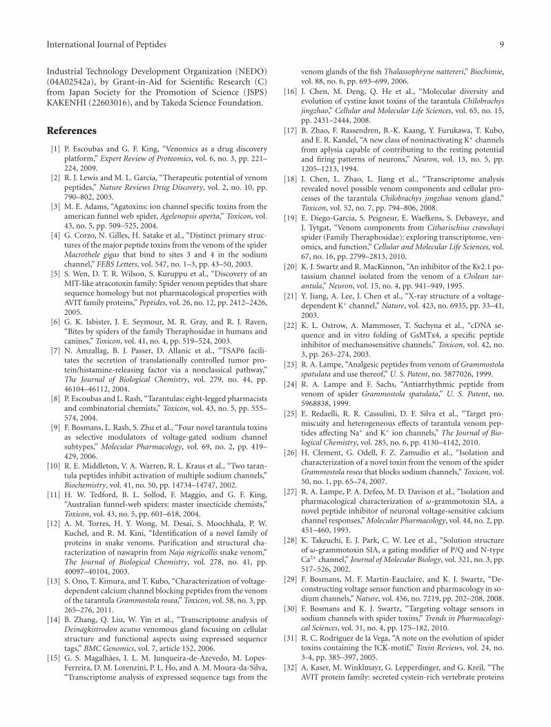
International Journal of Peptides 9
Industrial Technology Development Organization (NEDO)(04A02542a), by Grant-in-Aid for Scientific Research (C)from Japan Society for the Promotion of Science (JSPS)KAKENHI (22603016), and by Takeda Science Foundation.
References
[1] P. Escoubas and G. F. King, “Venomics as a drug discoveryplatform,” Expert Review of Proteomics, vol. 6, no. 3, pp. 221–224, 2009.
[2] R. J. Lewis and M. L. Garcia, “Therapeutic potential of venompeptides,” Nature Reviews Drug Discovery, vol. 2, no. 10, pp.790–802, 2003.
[3] M. E. Adams, “Agatoxins: ion channel specific toxins from theamerican funnel web spider, Agelenopsis aperta,” Toxicon, vol.43, no. 5, pp. 509–525, 2004.
[4] G. Corzo, N. Gilles, H. Satake et al., “Distinct primary struc-tures of the major peptide toxins from the venom of the spiderMacrothele gigas that bind to sites 3 and 4 in the sodiumchannel,” FEBS Letters, vol. 547, no. 1–3, pp. 43–50, 2003.
[5] S. Wen, D. T. R. Wilson, S. Kuruppu et al., “Discovery of anMIT-like atracotoxin family: Spider venom peptides that sharesequence homology but not pharmacological properties withAVIT family proteins,” Peptides, vol. 26, no. 12, pp. 2412–2426,2005.
[6] G. K. Isbister, J. E. Seymour, M. R. Gray, and R. J. Raven,“Bites by spiders of the family Theraphosidae in humans andcanines,” Toxicon, vol. 41, no. 4, pp. 519–524, 2003.
[7] N. Amzallag, B. J. Passer, D. Allanic et al., “TSAP6 facili-tates the secretion of translationally controlled tumor pro-tein/histamine-releasing factor via a nonclassical pathway,”The Journal of Biological Chemistry, vol. 279, no. 44, pp.46104–46112, 2004.
[8] P. Escoubas and L. Rash, “Tarantulas: eight-legged pharmacistsand combinatorial chemists,” Toxicon, vol. 43, no. 5, pp. 555–574, 2004.
[9] F. Bosmans, L. Rash, S. Zhu et al., “Four novel tarantula toxinsas selective modulators of voltage-gated sodium channelsubtypes,” Molecular Pharmacology, vol. 69, no. 2, pp. 419–429, 2006.
[10] R. E. Middleton, V. A. Warren, R. L. Kraus et al., “Two taran-tula peptides inhibit activation of multiple sodium channels,”Biochemistry, vol. 41, no. 50, pp. 14734–14747, 2002.
[11] H. W. Tedford, B. L. Sollod, F. Maggio, and G. F. King,“Australian funnel-web spiders: master insecticide chemists,”Toxicon, vol. 43, no. 5, pp. 601–618, 2004.
[12] A. M. Torres, H. Y. Wong, M. Desai, S. Moochhala, P. W.Kuchel, and R. M. Kini, “Identification of a novel family ofproteins in snake venoms. Purification and structural cha-racterization of nawaprin from Naja nigricollis snake venom,”The Journal of Biological Chemistry, vol. 278, no. 41, pp.40097–40104, 2003.
[13] S. Ono, T. Kimura, and T. Kubo, “Characterization of voltage-dependent calcium channel blocking peptides from the venomof the tarantula Grammostola rosea,” Toxicon, vol. 58, no. 3, pp.265–276, 2011.
[14] B. Zhang, Q. Liu, W. Yin et al., “Transcriptome analysis ofDeinagkistrodon acutus venomous gland focusing on cellularstructure and functional aspects using expressed sequencetags,” BMC Genomics, vol. 7, article 152, 2006.
[15] G. S. Magalhaes, I. L. M. Junqueira-de-Azevedo, M. Lopes-Ferreira, D. M. Lorenzini, P. L. Ho, and A. M. Moura-da-Silva,“Transcriptome analysis of expressed sequence tags from the
venom glands of the fish Thalassophryne nattereri,” Biochimie,vol. 88, no. 6, pp. 693–699, 2006.
[16] J. Chen, M. Deng, Q. He et al., “Molecular diversity andevolution of cystine knot toxins of the tarantula Chilobrachysjingzhao,” Cellular and Molecular Life Sciences, vol. 65, no. 15,pp. 2431–2444, 2008.
[17] B. Zhao, F. Rassendren, B.-K. Kaang, Y. Furukawa, T. Kubo,and E. R. Kandel, “A new class of noninactivating K+ channelsfrom aplysia capable of contributing to the resting potentialand firing patterns of neurons,” Neuron, vol. 13, no. 5, pp.1205–1213, 1994.
[18] J. Chen, L. Zhao, L. Jiang et al., “Transcriptome analysisrevealed novel possible venom components and cellular pro-cesses of the tarantula Chilobrachys jingzhao venom gland,”Toxicon, vol. 52, no. 7, pp. 794–806, 2008.
[19] E. Diego-Garcıa, S. Peigneur, E. Waelkens, S. Debaveye, andJ. Tytgat, “Venom components from Citharischius crawshayispider (Family Theraphosidae): exploring transcriptome, ven-omics, and function,” Cellular and Molecular Life Sciences, vol.67, no. 16, pp. 2799–2813, 2010.
[20] K. J. Swartz and R. MacKinnon, “An inhibitor of the Kv2.1 po-tassium channel isolated from the venom of a Chilean tar-antula,” Neuron, vol. 15, no. 4, pp. 941–949, 1995.
[21] Y. Jiang, A. Lee, J. Chen et al., “X-ray structure of a voltage-dependent K+ channel,” Nature, vol. 423, no. 6935, pp. 33–41,2003.
[22] K. L. Ostrow, A. Mammoser, T. Suchyna et al., “cDNA se-quence and in vitro folding of GsMTx4, a specific peptideinhibitor of mechanosensitive channels,” Toxicon, vol. 42, no.3, pp. 263–274, 2003.
[23] R. A. Lampe, “Analgesic peptides from venom of Grammostolaspatulata and use thereof,” U. S. Patent, no. 5877026, 1999.
[24] R. A. Lampe and F. Sachs, “Antiarrhythmic peptide fromvenom of spider Grammostola spatulata,” U. S. Patent, no.5968838, 1999.
[25] E. Redaelli, R. R. Cassulini, D. F. Silva et al., “Target pro-miscuity and heterogeneous effects of tarantula venom pep-tides affecting Na+ and K+ ion channels,” The Journal of Bio-logical Chemistry, vol. 285, no. 6, pp. 4130–4142, 2010.
[26] H. Clement, G. Odell, F. Z. Zamudio et al., “Isolation andcharacterization of a novel toxin from the venom of the spiderGrammostola rosea that blocks sodium channels,” Toxicon, vol.50, no. 1, pp. 65–74, 2007.
[27] R. A. Lampe, P. A. Defeo, M. D. Davison et al., “Isolation andpharmacological characterization of ω-grammotoxin SIA, anovel peptide inhibitor of neuronal voltage-sensitive calciumchannel responses,” Molecular Pharmacology, vol. 44, no. 2, pp.451–460, 1993.
[28] K. Takeuchi, E. J. Park, C. W. Lee et al., “Solution structureof ω-grammotoxin SIA, a gating modifier of P/Q and N-typeCa2+ channel,” Journal of Molecular Biology, vol. 321, no. 3, pp.517–526, 2002.
[29] F. Bosmans, M. F. Martin-Eauclaire, and K. J. Swartz, “De-constructing voltage sensor function and pharmacology in so-dium channels,” Nature, vol. 456, no. 7219, pp. 202–208, 2008.
[30] F. Bosmans and K. J. Swartz, “Targeting voltage sensors insodium channels with spider toxins,” Trends in Pharmacologi-cal Sciences, vol. 31, no. 4, pp. 175–182, 2010.
[31] R. C. Rodrıguez de la Vega, “A note on the evolution of spidertoxins containing the ICK-motif,” Toxin Reviews, vol. 24, no.3-4, pp. 385–397, 2005.
[32] A. Kaser, M. Winklmayr, G. Lepperdinger, and G. Kreil, “TheAVIT protein family: secreted cystein-rich vertebrate proteins

10 International Journal of Peptides
with diverse functions,” EMBO Reports, vol. 4, no. 5, pp. 469–473, 2003.
[33] H. Schweitz, P. Pacaud, S. Diochot, D. Moinier, and M.Lazdunski, “MIT1, a black mamba toxin with a new and highlypotent activity on intestinal contraction,” FEBS Letters, vol.461, no. 3, pp. 183–188, 1999.
[34] J. Boisbouvier, J. P. Albrand, M. Blackledge et al., “A structuralhomologue of colipase in black mamba venom revealed byNMR floating disulphide bridge analysis,” Journal of MolecularBiology, vol. 283, no. 1, pp. 205–219, 1998.
[35] C. Mollay, C. Wechselberger, G. Mignogna et al., “Bv8, a smallprotein from frog skin and its homologue from snake ve-nom induce hyperalgesia in rats,” European Journal of Phar-macology, vol. 374, no. 2, pp. 189–196, 1999.
[36] L. Negri, R. Lattanzi, E. Giannini, and P. Melchiorri, “Bv8/Pro-kineticin proteins and their receptors,” Life Sciences, vol. 81,no. 14, pp. 1103–1116, 2007.
[37] E. Giannini, R. Lattanzi, A. Nicotra et al., “The chemokineBv8/prokineticin 2 is up-regulated in inflammatory granulo-cytes and modulates inflammatory pain,” Proceedings of theNational Academy of Sciences of the United States of America,vol. 106, no. 34, pp. 14646–14651, 2009.
[38] L. Negri, R. Lattanzi, E. Giannini, M. Canestrelli, A. Nicotra,and P. Melchiorri, “Bv8/prokineticins and their receptors: anew pronociceptive system,” International Review of Neurobi-ology, vol. 85, pp. 145–157, 2009.
[39] M. Dorsch, Y. Qiu, D. Soler et al., “PK1/EG-VEGF inducesmonocyte differentiation and activation,” Journal of LeukocyteBiology, vol. 78, no. 2, pp. 426–434, 2005.
[40] I. Soderhall, Y. A. Kim, P. Jiravanichpaisal, S. Y. Lee, andK. Soderhall, “An ancient role for a prokineticin domain ininvertebrate hematopoiesis,” The Journal of Immunology, vol.174, no. 10, pp. 6153–6160, 2005.
[41] L. Jiang, L. Peng, J. Chen, Y. Zhang, X. Xiong, and S. Liang,“Molecular diversification based on analysis of expressedsequence tags from the venom glands of the Chinese birdspider Ornithoctonus huwena,” Toxicon, vol. 51, no. 8, pp.1479–1489, 2008.
[42] A. Savel-Niemann, “Tarantula (Eurypelma californicum) ven-om, a multicomponent system,” Biological Chemistry Hoppe-Seyler, vol. 370, no. 5, pp. 485–498, 1989.
[43] I. I. Kaiser, P. R. Griffin, S. D. Aird et al., “Primary structuresof two proteins from the venom of the Mexican red kneetarantula (Brachypelma smithii),” Toxicon, vol. 32, no. 9, pp.1083–1093, 1994.
[44] G. Corzo, C. Bernard, H. Clement et al., “Insecticidal peptidesfrom the theraposid spider Brachypelma albiceps: an NMR-based model of Ba2,” Biochimica et Biophysica Acta, vol. 1794,no. 8, pp. 1190–1196, 2009.
[45] X. Tang, Y. Zhang, and S. Liang, “Large-scale identificationand analysis of peptide toxins from the tarantula Ornithoc-tonus hainana venom using a venomic strategy,” NCBI, DirectSubmission, 2010, Accession no. GU293118.
[46] Y. Zhang, “Transcriptome analysis of Lycosa singoriensis spidervenomous gland,” NCBI, Direct Submission, 2008, Accessionno. FM864147.
[47] W. S. Skinner, P. A. Dennis, J. P. Li, and G. B. Quistad,“Identification of insecticidal peptides from venom of thetrap-door spider, Aptostichus schlingeri (Ctenizidae),” Toxicon,vol. 30, no. 9, pp. 1043–1050, 1992.
[48] U. A. Bommer, A. Lazaris-Karatzas, A. De Benedetti et al.,“Translational regulation of the mammalian growth-relatedprotein p23: involvement of eIF-4E,” Cellular and MolecularBiology Research, vol. 40, no. 7-8, pp. 633–641, 1994.
[49] Y. Gachet, S. Tournier, M. Lee, A. Lazaris-Karatzas, T. Poulton,and U. A. Bommer, “The growth-related, translationally con-trolled protein P23 has properties of a tubulin binding proteinand associates transiently with microtubules during the cellcycle,” Journal of Cell Science, vol. 112, part 8, pp. 1257–1271,1999.
[50] S. M. MacDonald, T. Rafnar, J. Langdon, and L. M.Lichtenstein, “Molecular identification of an IgE-Dependenthistamine-releasing factor,” Science, vol. 269, no. 5224, pp.688–690, 1995.
[51] D. A. Ellerman, V. G. Da Ros, D. J. Cohen, D. Busso, M. M.Morgenfeld, and P. S. Cuasnicu, “Expression and structure-function analysis of DE, a sperm cysteine-rich secretory pro-tein that mediates gamete fusion,” Biology of Reproduction, vol.67, no. 4, pp. 1225–1231, 2002.
[52] N. J. Charest, D. R. Joseph, E. M. Wilson, and F. S. French,“Molecular cloning of complementary deoxyribonucleic acidfor an androgen-regulated epididymal protein: sequence ho-mology with metalloproteins,” Molecular Endocrinology, vol.2, no. 10, pp. 999–1004, 1988.
[53] A. L. Kierszenbaum, O. Lea, P. Petrusz, F. S. French, andL. L. Tres, “Isolation, culture, and immunocytochemicalcharacterization of epididymal epithelial cells from pubertaland adult rats,” Proceedings of the National Academy of Sciencesof the United States of America, vol. 78, no. 3, pp. 1675–1679,1981.
[54] M. Kasahara, H. C. Passmore, and J. Klein, “A testis-specificgene Tpx-1 maps between Pgk-2 and Mep-1 on mouse chro-mosome 17,” Immunogenetics, vol. 29, no. 1, pp. 61–63, 1989.
[55] L. Kjeldsen, J. B. Cowland, A. H. Johnsen, and N. Borregaard,“SGP28, a novel matrix glycoprotein in specific granules ofhuman neutrophils with similarity to a human testis-specificgene product and to a rodent sperm-coating glycoprotein,”FEBS Letters, vol. 380, no. 3, pp. 246–250, 1996.
[56] J. Mochca-Morales, B. M. Martin, and L. D. Possani, “Iso-lation and characterization of helothermine, a novel toxinfrom Heloderma horridum horridum (Mexican beaded lizard)venom,” Toxicon, vol. 28, no. 3, pp. 299–309, 1990.
[57] M. Nobile, F. Noceti, G. Prestipino, and L. D. Possani,“Helothermine, a lizard venom toxin, inhibits calcium currentin cerebellar granules,” Experimental Brain Research, vol. 110,no. 1, pp. 15–20, 1996.
[58] Y. Yamazaki, H. Koike, Y. Sugiyama et al., “Cloning and char-acterization of novel snake venom proteins that block smoothmuscle contraction,” European Journal of Biochemistry, vol.269, no. 11, pp. 2708–2715, 2002.
[59] R. Bonasio, G. Zhang, C. Ye et al., “Genomic comparison ofthe ants Camponotus floridanus and Harpegnathos saltator,”Science, vol. 329, no. 5995, pp. 1068–1071, 2010.
[60] M. Naimuddin, S. Kobayashi, C. Tsutsui et al., “Directedevolution of a three-finger neurotoxin by using cDNA displayyields antagonists as well as agonists of interleukin-6 receptorsignaling,” Molecular Brain, vol. 4, no. 1, article 2, 2011.

Hindawi Publishing CorporationInternational Journal of PeptidesVolume 2012, Article ID 532519, 10 pagesdoi:10.1155/2012/532519
Review Article
Platelet-Rich Plasma Peptides: Key for Regeneration
Dolores Javier Sanchez-Gonzalez,1, 2 Enrique Mendez-Bolaina,3, 4
and Nayeli Isabel Trejo-Bahena2, 5
1 Subseccion de Biologıa Celular y Tisular, Escuela Medico Militar, Universidad del Ejercito y Fuerza Aerea,11200 Mexico City, MEX, Mexico
2 Sociedad Internacional para la Terapia Celular con Celulas Madre, Medicina Regenerativa y Antienvejecimiento S.C. (SITECEM),53840 Naucalpan, MEX, Mexico
3 Facultad de Ciencias Quımicas, Universidad Veracruzana, 94340 Orizaba, VER, Mexico4 Centro de Investigaciones Biomedicas-Doctorado en Ciencias Biomedicas, Universidad Veracruzana, 91000 Xalapa, VER, Mexico5 Area de Medicina Fısica y Rehabilitacion, Hospital Central Militar, 11200 Mexico City, MEX, Mexico
Correspondence should be addressed to Dolores Javier Sanchez-Gonzalez, [email protected]
Received 15 September 2011; Revised 13 December 2011; Accepted 14 December 2011
Academic Editor: Frederic Ducancel
Copyright © 2012 Dolores Javier Sanchez-Gonzalez et al. This is an open access article distributed under the Creative CommonsAttribution License, which permits unrestricted use, distribution, and reproduction in any medium, provided the original work isproperly cited.
Platelet-derived Growth Factors (GFs) are biologically active peptides that enhance tissue repair mechanisms such as angiogenesis,extracellular matrix remodeling, and cellular effects as stem cells recruitment, chemotaxis, cell proliferation, and differentiation.Platelet-rich plasma (PRP) is used in a variety of clinical applications, based on the premise that higher GF content should promotebetter healing. Platelet derivatives represent a promising therapeutic modality, offering opportunities for treatment of wounds,ulcers, soft-tissue injuries, and various other applications in cell therapy. PRP can be combined with cell-based therapies such asadipose-derived stem cells, regenerative cell therapy, and transfer factors therapy. This paper describes the biological backgroundof the platelet-derived substances and their potential use in regenerative medicine.
1. Introduction
Platelets are nonnuclear cellular fragments derived frommegakaryocytes in the bone marrow; they are specializedsecretory elements that release the contents of their intracel-lular granules in response to activation. They were discoveredby Bizzozero in the 19th century [1] and after Wright ob-served that megakaryocytes are platelet precursors [2]. Actu-ally we know that platelets synthesize proteins and that pat-tern of peptides synthesis changes in response to cellularactivation [3].
Platelets contain a great variety of proteins molecules,among which are the high presence of signaling, membraneproteins, protein processing, cytoskeleton regulatory pro-teins, cytokines, and other bioactive peptides that initiateand regulate basic aspects of wound healing [3]. It is known,through efforts such as the platelet proteome project, thatmore than 300 proteins are released by human plateletsin response to thrombin activation [4]. Proteome plateletincludes 190 membrane-associated and 262 phosphorylated
proteins, which were identified via independent proteomicand phospho proteomic profiling [5].
When platelets fall precipitously below critical levels(usually under 10,000 to 20,000 per cubic millimeter), mo-lecular disassembly opens the zippers formed by adjacentintercellular endothelial junctions, causing extravasationof erythrocytes into the surrounding tissues. In additionto their well-known function in hemostasis, platelets alsorelease substances that promote tissue repair, angiogene-sis, and inflammation [6]. Furthermore, they induce themigration and adherence of bone-marrow-derived cells tosites of angiogenesis; platelets also induce differentiation ofendothelial-cell progenitors into mature endothelial cells [7].
At the site of the injury, platelets release an arsenalof potent regenerative and mitogenic substances that areinvolved in all aspects of the wound-healing process includ-ing a potential point-of-care biologic treatment followingmyocardial injury [8]. Based on this, platelet called Platelet-rich Plasma (PRP) has been extensively used for orthopaedic

2 International Journal of Peptides
Table 1: Peptidic growth factors present in platelet-rich plasma (PRP).
Name Cytogenetic location Biologic activities
Transforming growth factor, beta-I; TGFB1 19q13.2Controls proliferation, differentiation, and otherfunctions in many cell types
Platelet-derived growth factor, alpha polypeptide;PDGFA
7p22.3Potent mitogen for connective tissue cells and exertsits function by interacting with related receptortyrosine kinases
Platelet-derived growth factor, beta polypeptide;PDGFB
22q13.1Promotes cellular proliferation and inhibitsapoptosis
Platelet-derived growth factor C; PDGFC 4q32.1Increases motility in mesenchymal cells, fibroblasts,smooth muscle cells, capillary endothelial cells, andneurons
Platelet-derived growth factor D; PDGFD 11q22.3Involved in developmental and physiologicprocesses, as well as in cancer, fibrotic diseases, andarteriosclerosis
Insulin-like growth factor I; IGF1 12q23.2Mediates many of the growth-promoting effects ofgrowth hormone
Fibroblast growth factor I; FGF1 5q31.3Induces liver gene expression, angiogenesis andfibroblast proliferation
Epidermal growth factor; EGF 4q25Induces differentiation of specific cells, is a potentmitogenic factor for a variety of cultured cells ofboth ectodermal and mesodermal origin
Vascular endothelial growth factor A; VEGFA 6p21.1Is a mitogen primarily for vascular endothelial cells,induces angiogenesis
Vascular endothelial growth factor B; VEGFB 11q13.1Is a regulator of blood vessel physiology, with a rolein endothelial targeting of lipids to peripheral tissues
Vascular endothelial growth factor C; VEGFC 4q34.3Angiogenesis and endothelial cell growth, and canalso affect the permeability of blood vessels
Includes, name, cytogenetic location, and biologic activities of platelet growth factors. Furthermore, PRP content other proteins like interleukin-8, macrophageinflammatory protein-1 alpha, and platelet factor-4.
applications; for topical therapy of various clinical condi-tions, including wounds and soft tissue injuries; and suitablealternative to fetal calf serum for the expansion of mesenchy-mal stem cells from adipose tissue (see Table 1) [9–12].
In this paper we are going to talk about the platelets,platelet-derived particles, and their biological effects in re-generative medicine.
2. Platelets
Platelets are the first element to arrive at the site of tissueinjury and are particularly active in the early inflammatoryphases of the healing process [6]. They play a role in aggre-gation, clot formation, homeostasis through cell membraneadherence, and release of substances that promote tissuerepair and that influence the reactivity of blood vesselsand blood cell types involved in angiogenesis, regeneration,and inflammation [13]. Platelet secretory granules containgrowth factors (GFs), signaling molecules, cytokines, inte-grins, coagulation proteins, adhesion molecules, and someother molecules, which are synthesized in megakaryocytesand packaged into the granules through vesicle traffick-ing processes [14]. Three major storage compartments inplatelets are alpha granules, dense granules, and lysosomes[14].
Platelets mediate these effects through degranulation, inwhich platelet-derived GF (PDGF), insulin-like GF (IGF1),
transforming GF-beta 1 (TGF-β1), vascular endothelial GF(VEGF), basic fibroblastic GF (bFGF), and epidermal GF(EGF) are released from alpha granules [15]. In fact, themajority of the platelet substances are contained in alphagranules (see Table 2) [15]. When platelets are activated, theyexocytose the granules; this process is mediated by molecularmechanisms homologous to other secretory cells, uniquelycoupled to cell activation by intracellular signaling events[16].
Among bioactive molecules stored and released fromplatelets dense granules are catecholamines, histamine, sero-tonin, ADP, ATP, calcium ions, and dopamine, which areactive in vasoconstriction, increased capillary permeability,attract and activate macrophages, tissue modulation andregeneration. These non-GF molecules have fundamentaleffects on the biologic aspects of wound healing [5].
For their numerous functions, platelets have developeda set of platelet receptors that are the contact betweenplatelets and their surroundings; they determine the reac-tivity of platelets with a wide range of agonists and adhe-sive proteins. Some of these receptors are expressed onlyon activated platelets [6]. Certain biological mechanismspresent in the platelets are shared with other cells, andtherefore they contain some common cytoplasmic enzymes,signal transduction molecules, and cytoskeletal components[14].

International Journal of Peptides 3
Table 2: Some bioactive peptides present in the alpha granules of platelets.
General activity categories Specific moleculesCytogenetic
locationBiologic activities
Tissue factor pathwayinhibitor; TFPI
2q32.1Regulates the tissue factor-(TF-) dependentpathway of blood coagulation
Kininogen; KNG 3q27.3Plays an important role in assembly of theplasma kallikrein
Growth arrest-specific 6;GAS6
13q34 Stimulates cell proliferation
Multimerin; MMRN 4q22 Carrier protein for platelet factor V
Clotting factors and related proteins Antithrombin; AT 1q25.1 Is the most important inhibitor of thrombin
Protein S; PROS1 3q11.1 Inhibits blood clotting
Coagulation factor V; F5 1q24.2Acts as a cofactor for the conversion of pro-thrombin to thrombin by factor Xa
Coagulation factor XI; F11 4q35.2It participates in blood coagulation as a cat-alyst in the conversion of factor IX to factorIXa in the presence of calcium ions
Plasminogen; PLG 6q26Induces plasmin production (leads to fibrin-olysis)
Plasminogen activatorinhibitor 1; PAI1
7q22.1 Regulation of plasmin production
Alpha-2-plasmin inhibitor 17p13.3 Inactivation of plasmin
Fibrinolytic factors and related proteins Osteonectin; ON 5q33.1Inhibits cell-cycle progression and influencesthe synthesis of extracellular matrix (ECM)
Histidine-rich glycoprotein;HRG
3q27.3 Interacts with heparin and thrombospondin
Thrombin-activatablefibrinolysis inhibitor; TAFI
13q14.13 Attenuates fibrinolysis
Alpha-2-Macroglobulin; A2M 12p13.31Carrier of specific growth factors and indu-ces cell signaling
Tissue inhibitor ofmetalloproteinase 4; TIMP4
3p25.2Inhibits matrix metalloproteinases (MMPs),a group of peptidases involved in degrada-tion of the extracellular matrix
Complement component 1inhibitor; C1NH
11q12.1Inhibits serine proteinases including plas-min, kallikrein, and coagulation factors XIaand XIIa
Proteases and antiproteasesAlpha-1-antitrypsin (serpin
peptidase inhibitor)14q32.13
Acute phase protein, inhibits a wide varietyof proteases and enzymes
Nexin 2; SNX2 5q23.2Modulates intracellular trafficking of pro-teins to various organelles
Platelet factor 4; PF4 4q13.3 Inhibition of angiogenesis
Basic proteinsβ-thromboglobulin
(Pro-platelet basic protein;PPBP)
4q13.3 Platelet activation, inhibition of angiogenesis
Endostatin (Collagen, typeXVIII, Alpha-1; COL18A1)
21q22.3Inhibitors of endothelial cell migration andangiogenesis
Fibrinogen; FG 4q31.3Blood clotting cascade (fibrin clot forma-tion)
Fibronectin; FN 2q35Binds to cell-surface integrins, affecting celladhesion, cell growth, migration, and differ-entiation
Adhesive proteins Vitronectin; VTN 17q11.2 Induces cell adhesion, chemotaxis
Thrombospondin I; THBS1 15q14 Inhibition of angiogenesis
Laminin-818p11.31-
p11.23Modulates cell contact interactions
It is described general activity categories, specific molecules, cytogenetic location and biologic activities. Furthermore, alpha granules include growth factorsof Table 1, membrane glycoproteins, and others proteins like albumin and immunoglobulins.
4 International Journal of Peptides
The platelet lifespan is approximately 7 to 9 days, whichthey spend circulating in the blood in their resting form.When adhered to exposed endothelium or activated byagonists, they change their shape and secrete the contentsof the granules (including ADP, fibrinogen, and serotonin),which is followed by platelet aggregation [7]. Initiation of thesignaling event within the platelet leads to the reorganizationof the platelet cytoskeleton, which is visible as an extremelyrapid shape change [17].
3. Platelet-Rich Plasma (PRP)
Platelets are activated either by adhesion to the moleculesthat are exposed on an injured endothelium, such as vonWillebrand Factor (vWF), collagen, fibronectin, and laminin,or by physiologic agonists such as thrombin, ADP, collagen,thromboxane A2, epinephrine, and platelet-activating fac-tors [18].
PRP has been used clinically in humans since the 1970sfor its healing properties attributed of autologous GF andsecretory proteins that may enhance the healing process ona cellular level [19]. Furthermore, PRP enhances the recruit-ment, proliferation, and differentiation of cells involved intissue regeneration [20]. PRP-related products, also knownas platelet-rich concentrate, platelet gel, preparation rich ingrowth factors (PRGF), and platelet releasate, have beenstudied with in vitro and in vivo experiments in the fieldsof surgical sciences mainly [21].
Depending on the device and technique used, PRP cancontain variable amounts of plasma, erythrocytes, whiteblood cells, and platelets. The platelet concentration shouldbe increased above baseline or whole blood concentration. Itis generally agreed upon that PRP should have a minimum of5 times the number of platelets compared to baseline valuesfor whole blood to be considered “platelet rich” [22].
This conclusion is supported by in vitro work showing apositive dose-response relationship between platelet concen-tration and proliferation of human mesenchymal stem cells,proliferation of fibroblasts, and production of type I collagen[23]. This suggests that the application of autologous PRPcan enhance wound healing, as has been demonstrated incontrolled animal studies for both soft and hard tissues[24, 25].
Autologous PRP represents an efficacious treatment forits use in wound healing like chronic diabetic foot ulcerationdue to multiple growth factors, is safe for its autologousnature, and is produced as needed from patient blood. Likewe said, key for self-regeneration [26].
Upon activation, platelets release their granular contentsinto the surrounding environment. The platelet alpha gran-ules are abundant and contain many of the GFs responsiblefor the initiation and maintenance of the healing response[14]. These GFs have been shown to play an important role inall phases of healing. The active secretion of these proteins byplatelets begins within 10 minutes after clotting, with morethan 95% of the presynthesized GFs secreted within 1 hour.After this initial burst, the platelets synthesize and secreteadditional proteins for the balance of their life (5–10 days)[10].
The fibrin matrix formed following platelet activationalso has a stimulatory effect on wound healing. The fibrinmatrix forms by polymerization of plasma fibrinogen follow-ing either external activation with calcium or thrombin orinternal activation with endogenous tissue thromboplastin[23]. This matrix traps platelets allowing a slow release ofa natural combination of GF while providing a provisionalmatrix that provides a physical framework for wound stemcells and fibroblast migration and presentation of otherbiological mediators such as adhesive glycoproteins [27, 28].
PRP with a platelet concentration of at least1 000 000 platelets/μL in 5 mL of plasma is associated with theenhancement of healing [29]. PRP can potentially enhancehealing by the delivery of various GF and cytokines fromthe alpha granules contained in platelets and has an 8-foldincrease in GF concentrations compared with that of wholeblood [30].
The use of PRP to enhance bone regeneration and softtissue maturation has increased dramatically in the fieldsof orthopedics, periodontics, maxillofacial surgery, urology,and plastic surgery over the last years. However, controversiesexist in the literature regarding the added benefit of thisprocedure. While some authors have reported significantincreases in bone formation and maturation rates [21],others did not observe any improvement [31].
The wound-healing process is a complex mechanismcharacterized by four distinct, but overlapping, phases: he-mostasis, inflammation, proliferation, and remodeling [8].The proliferative phase includes blood vessel formation byendothelial cells and bone synthesis by osteoblasts. All theseevents are coordinated by cell-cell interactions and by solubleGF released by various cell types. Recent reviews have em-phasized the need for additional research aiming to charac-terize PRP in terms of GF content and their physiologicalroles in wound healing [32–34].
Thrombin represents a strong inducer of platelet activa-tion leading to GF release [35]. It is also known that par-ticulate grafts, when combined with calcium and thrombintreated PRP, possess better handling characteristics andhigher GF content [31]. Typically, thrombin concentrationsused in clinical applications vary between 100 and 200 unitsper mL [21], while platelet aggregation is maximum in therange of 0.5 to 4 units per mL [36].
The basic cytokines identified in platelets play importantroles in cell proliferation, chemotaxis, cell differentiation,regeneration, and angiogenesis [28]. A particular value ofPRP is that these native cytokines are all present in “normal”biologic ratios. The platelets in PRP are delivered in a clot,which contains several cell adhesion molecules includingfibronectin, fibrin, and vitronectin. These cell adhesionmolecules play a role in cell migration and thus also addto the potential biologic activity of PRP. The clot itself canalso play a role in wound healing by acting as conductivematrix or “scaffold” upon which cells can adhere and beginthe wound-healing process [28].
PRP can only be made from anticoagulated blood. Prepa-ration of PRP begins by addition of citrate to whole bloodto bind the ionized calcium and inhibit the clotting cascade[15]. This is followed by one or two centrifugation steps. The
International Journal of Peptides 5
first centrifugation step separates the red and white bloodcells from plasma and platelets. The second centrifugationstep further concentrates the platelets, producing the PRPseparate from platelet-poor plasma [19].
An important point is that clotting leads to platelet acti-vation, resulting in release of the GF from the alpha granules,otherwise known as degranulation. Approximately 70% ofthe stored GFs are released within 10 minutes, and nearly100% of the GFs are released within 1 hour. Small amountsof GF may continue to be produced by the platelet during therest of its lifespan (1 week) [21].
A method to delay the release of GF is possible by addi-tion of calcium chloride (CaCl2) to initiate the formationof autogenous thrombin from prothrombin. The CaCl2 isadded during the second centrifugation step and resultsin formation of a dense fibrin matrix. Intact platelets aresubsequently trapped in the fibrin matrix and release GFslowly over a 7-day period. The fibrin matrix itself may alsocontribute to healing by providing a conductive scaffold forcell migration and new matrix formation [27].
4. Growth Factors (GFs)
Platelets are known to contain high concentrations of differ-ent GF and are extremely important in regenerative process;activation of the platelet by endothelial injury initiates thewound-healing process [30]. When platelets are activated,their alpha granules are released, resulting in an increasedconcentration of GF in the wound milieu [14].
There is increasing evidence that the platelet cell mem-branes themselves also play a crucial role in wound healingthrough their GF receptor sites [28]. GFs are found in a widearray of cells and in platelet alpha granules [37]. Table 1 givesan overview of some of the more extensively studied GFsand their involvement in wound healing. There are manymore, both discovered and undiscovered, GFs. The plateletis an extremely important cell in wound healing because itinitiates and plays a major role in the wound regenerativeprocess [38].
The first discovered GF was EGF in 1962 by Cohen[39]. It was not until 1989 before clinical trials with EGFwere attempted to demonstrate enhanced wound healing.Studies did demonstrate that EGF can accelerate epidermalregeneration and enhance healing of chronic wounds [40].
PDGF was discovered in 1974 and is ubiquitous in thebody. It is known to be released by platelet alpha granulesduring wound healing and stimulate the proliferation ofmany cells, including connective tissue cells. In fact, thus far,high-affinity cell-surface receptors specific for PDGF haveonly been demonstrated on connective tissue cells. Whenreleased, PDGF is chemotactic for monocytes, neutrophils,and fibroblasts. These cells release their own PDGF, thuscreating a positive autocrine feedback loop [41]. Otherfunctions of PDGF include effects on cell growth, cellularmigration, metabolic effects, and modulation of cell mem-brane receptors [42].
PDGFs were first identified as products of platelets whichstimulated the proliferation in vitro of connective tissue celltypes such as fibroblasts [43].
The PDGF system, comprising four isoforms (PDGF-A, -B, -C, and -D) and two receptor chains (PDGFR-alphaand -beta), plays important roles in wound healing, ath-erosclerosis, fibrosis, and malignancy. Components of thesystem are expressed constitutively or inducibly in most renalcells [42]. They regulate a multitude of pathophysiologicevents, ranging from cell proliferation and migration toextracellular matrix accumulation, production of pro- andanti-inflammatory mediators, tissue permeability, and regu-lation of hemodynamics [43].
Inactivation of PDGF-B and PDGF beta receptor(PDGFRb) genes by homologous recombination in embry-onic stem cells shows cardiovascular, hematological, and re-nal defects. The latter is particularly interesting since it con-sists of a specific cellular defect: the complete loss of kidneyglomerular mesangial cells and the absence of urine collec-tion in the urinary bladder [43].
PDGF-C and PDGFR-alpha contribute to the formationof the renal cortical interstitium. Almost all experimental andhuman renal diseases are characterized by altered expressionof components of the PDGF system. Infusion or systemicoverexpression of PDGF-B or -D induces prominent mesan-gioproliferative changes and renal fibrosis. Intervention stud-ies identified PDGF-C as a mediator of renal interstitial fibro-sis and PDGF-B and -D as key factors involved in mesangio-proliferative disease and renal interstitial fibrosis [43–45].
Frechette et al., demonstrated that the release of PDGF-B, TGF-beta1, bFGF, and VEGF is significantly regulated bythe amount of calcium and thrombin added to the PRP andthat PRP supernatants are more mitogenic for endothelialcells than whole-blood supernatants [11]. Other GFs such asepidermal growth factor (EGF), transforming growth factor-alpha (TGF-alpha), insulin-like growth factor-1 (IGF-1),angiopoietin-2 (Ang-2), and interleukin-1beta (IL-1beta) arealso known to play important roles in the wound-healingprocess [28].
In 2008, Wahlstrom et al., demonstrated that growthfactors released from platelets had potent effects on fractureand wound healing. The acidic tide of wound healing, that is,the pH within wounds and fractures, changes from acidic pHto neutral and alkaline pH as the healing process progresses[44]. They investigated the influence of pH on lysed plateletconcentrates regarding the release of growth factors. Theplatelet concentrates free of leukocyte components were lysedand incubated in buffers with pH between 4.3 and 8.6. Bonemorphogenetic protein-2 (BMP-2), platelet-derived growthfactor (PDGF), transforming growth factor-beta (TGF-beta),and vascular endothelial growth factor (VEGF) were mea-sured by quantitative enzyme-linked immunosorbent assays.BMP-2 was only detected in the most acidic preparation (pH4.3), which is interesting since BMP-2 has been reportedto be an endogenous mediator of fracture repair and tobe responsible for the initiation of fracture healing. Thesefindings indicate that platelets release substantial amountsof BMP-2 only under conditions of low pH, the milieuassociated with the critical initial stage of fracture healing[44].
6 International Journal of Peptides
Recently, Bir et al., demonstrated stromal cell-derivedfactor 1-α (SDF-1α) PRP from diabetic mice. The con-centration (pg/mL) of different growth factors was signif-icantly higher in the PRP group than in the platelet-poorplasma (PPP) group. The concentrations (pg/mL) of SDF-1α(10,790 ± 196 versus 810 ± 39), PDGF-BB (45,352 ± 2,698versus 958 ± 251), VEGF (53± 6 versus 30 ± 2), bFGF (29 ±5 versus 9± 5), and IGF-1 (20,628 ± 1,180 versus 1,214 ±36) were significantly higher in the PRP group than in thePPP group, respectively [46].
5. Platelet GF as Treatment
5.1. In Vitro Studies. Knowledge of GF and their functionis far from complete. Many of the known functions werelearned through in vitro study. Although many GFs are asso-ciated with wound healing, PDGF and TGF-β1 appear to betwo of the more integral modulators [46]. PDGF has activityin early wound healing (during the acid tide). In vitro studieshave shown that at lower pH (5.0), platelet concentrate lysatehas increased concentrations of PDGF, with an increasedcapacity to stimulate fibroblast proliferation [23]. TGF-βincreases the production of collagen from fibroblasts [47]. Itsrelease in vitro is enhanced by neutral or alkaline pH, whichcorrespond to the later phases of healing [10]. Throughmodulation of interleukin-1 production by macrophages,PRP may inhibit excessive early inflammation that couldlead to dense scar tissue formation [48]. Insulin-like GF-I(IGF-1) has also been extensively studied for its ability toinduce proliferation, differentiation, and hypertrophy ofmultiple cell lines. Separate analyses of GF in PRP haveshown significant increases in PDGF, VEGF, TGF-β1, andEGF, compared with their concentrations in whole blood[15, 49].
IGF-1 has two important functions: chemotaxis forvascular endothelial cells into the wound which results inangiogenesis and promoting differentiation of several celllines including chondroblasts, myoblasts, osteoblasts, andhematopoietic cells [50].
TGF-β is a member of the newest family of proteinsdiscovered. Two major sources of this protein are the plateletand macrophage. TGF-β causes chemotactic attraction andactivation of monocytes, macrophages, and fibroblasts. Theactivated fibroblasts enhance the formation of extracellularmatrix and collagen and also stimulate the cells ability to con-tract the provisional wound matrix [51]. Macrophages infil-tration promotes TGF-β that induces extracellular matrixsuch as collagen and fibronectin; however alpha-mangostinprevents the increase in this molecule in rats with Cisplatin-induced nephrotoxicity [52].
5.2. In Vivo Studies. In vivo study is much more complexdue to the inability to control the environment. A furthercomplexing matter is the fact that the same GF, depending onthe presence or absence of other peptides, may display eitherstimulatory or inhibitory activity within the same cell. Also,a particular GF can alter the binding affinity of another GFreceptor [53].
Release of PDGF can have a chemotactic effect on mono-cytes, neutrophils, fibroblasts, stem cells, and osteoblasts.This peptide is a potent mitogen for mesenchymal cells in-cluding fibroblasts, smooth muscle cells and glial cells [54]and is involved in all three phases of wound healing, includ-ing angiogenesis, formation of fibrous tissue, and reepithe-lialization [41].
TGF beta released from platelet alpha granules is a mito-gen for fibroblasts, smooth muscle cells, and osteoblasts. Inaddition, it promotes angiogenesis and extracellular matrixproduction [41]. VEGF promotes angiogenesis and canpromote healing of chronic wounds and aid in endochondralossification. EGF, another platelet-contained GF, is a mitogenfor fibroblasts, endothelial cells, and keratinocytes and also isuseful in healing chronic wounds [55].
IGF, another platelet-contained GF regulates bone main-tenance and is also an important modulator of cell apoptosis,and, in combination with PDGF, can promote bone regener-ation [56].
However, there are conflicting results with regard to IGF-1, where the majority of studies reported no increase inIGF-1 in PRP, compared with whole blood. There are alsoconflicting results regarding the correlation between the GFcontent and platelet counts in PRP [57]. The basis of thesecontradictions is not fully understood and may be relatedto variability in patient age, health status, or platelet count.Alternatively, differences in GF content and platelet countmay be due to the various methods of processing, handling,and storing of samples, in addition to the type of assayperformed. The diversity of PRP products should be takeninto account when interpreting and comparing results andmethods for generating PRP [10].
VEGF, discovered 25 years ago, was initially referred toas vascular permeability factor [58]. In mammals, there areat least four members of the VEGF family: VEGF-A, VEGF-B, and the VEGF-C/VEGF-D pair, which has a commonreceptor, VEGF receptor 3 (VEGF-R3) [59]. VEGF-A isa proangiogenic cytokine during embryogenesis and con-tributes to vascular integrity: selective knockout of VEGF-Ain endothelial cells increases apoptosis, which compromisesthe integrity of the junctions between endothelial cells [60,61]. VEGF-B, which can form heterodimers with VEGF-A, occurs predominantly in brown fat, myocardium, andskeletal muscle [62]. VEGF-C and VEGF-D seem to regulatelymphangiogenesis. The expression of VEGF-R3 in adultsis restricted to the lymphatics and fenestrated endothe-lium [63]. Neuropilin 1 and neuropilin 2 are receptorsthat bind specific VEGF family members and are impor-tant in neuronal development and embryonic vasculogenesis[64].
Megakaryocytes and platelets contain the three majorisoforms of VEGF-A; after exposure to thrombin in vitro,they release VEGF-A [65–67]. VEGF-A alters the endothelial-cell phenotype by markedly increasing vascular permeability,upregulating expression of urokinase, tissue plasminogenactivator, connexin, osteopontin, and the vascular-cell adhe-sion molecule [68].
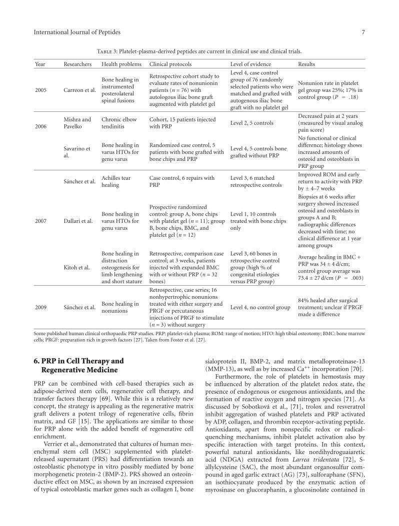
International Journal of Peptides 7
Table 3: Platelet-plasma-derived peptides are current in clinical use and clinical trials.
Year Researchers Health problems Clinical protocols Level of evidence Results
2005 Carreon et al.
Bone healing ininstrumentedposterolateralspinal fusions
Retrospective cohort study toevaluate rates of nonunioninpatients (n = 76) withautologous iliac bone graftaugmented with platelet gel
Level 4, case controlgroup of 76 randomlyselected patients who werematched and grafted withautogenous iliac bonegraft with no platelet gel
Nonunion rate in plateletgel group was 25%; 17% incontrol group (P = .18)
2006Mishra andPavelko
Chronic elbowtendinitis
Cohort, 15 patients injectedwith PRP
Level 2, 5 controlsDecreased pain at 2 years(measured by visual analogpain score)
Savarino etal.
Bone healing invarus HTOs forgenu varus
Randomized case control, 5patients with bone grafted withbone chips and PRP
Level 4, 5 controls bonegrafted without PRP
No functional or clinicaldifference; histology showsincreased amounts ofosteoid and osteoblasts inPRP group
Sanchez et al.Achilles tearhealing
Case control, 6 repairs withPRP
Level 3, 6 matchedretrospective controls
Improved ROM and earlyreturn to activity with PRPby ± 4–7 weeks
2007 Dallari et al.Bone healing invarus HTOs forgenu varus
Prospective randomizedcontrol: group A, bone chipswith platelet gel (n = 11); groupB, bone chips, BMC, andplatelet gel (n = 12)
Level 1, 10 controlstreated with bone chipsonly
Biopsies at 6 weeks aftersurgery showed increasedosteoid and osteoblasts ingroups A and B;radiographic differencesdecreased with time; noclinical difference at 1 yearamong groups
Kitoh et al.
Bone healing indistractionosteogenesis forlimb lengtheningand short stature
Retrospective, comparison casecontrol; at 3 weeks, patientsinjected with expanded BMCwith or without PRP (n = 32bones)
Level 3, 60 bones inretrospective controlgroup (high % ofcongenital etiologiesversus PRP group)
Average healing in BMC +PRP was 34± 4 d/cm;control group average was73.4± 27 d/cm (P = .003)
2009 Sanchez et al.Bone healing innonunions
Retrospective, case series; 16nonhypertrophic nonunionstreated with either surgery andPRGF or percutaneousinjections of PRGF to stimulate(n = 3) without surgery
Level 4, no control group84% healed after surgicaltreatment; unclear if PRGFmade a difference
Some published human clinical orthopaedic PRP studies. PRP: platelet-rich plasma; ROM: range of motion; HTO: high tibial osteotomy; BMC: bone marrowcells; PRGF: preparation rich in growth factors [27]. Taken from Foster et al. [27].
6. PRP in Cell Therapy andRegenerative Medicine
PRP can be combined with cell-based therapies such asadipose-derived stem cells, regenerative cell therapy, andtransfer factors therapy [69]. While this is a relatively newconcept, the strategy is appealing as the regenerative matrixgraft delivers a potent trilogy of regenerative cells, fibrinmatrix, and GF [15]. The applications are similar to thosefor PRP alone with the added benefit of regenerative cellenrichment.
Verrier et al., demonstrated that cultures of human mes-enchymal stem cell (MSC) supplemented with platelet-released supernatant (PRS) had differentiation towards anosteoblastic phenotype in vitro possibly mediated by bonemorphogenetic protein-2 (BMP-2). PRS showed an osteoin-ductive effect on MSC, as shown by an increased expressionof typical osteoblastic marker genes such as collagen I, bone
sialoprotein II, BMP-2, and matrix metalloproteinase-13(MMP-13), as well as by increased Ca++ incorporation [70].
Furthermore, the role of platelets in hemostasis maybe influenced by alteration of the platelet redox state, thepresence of endogenous or exogenous antioxidants, and theformation of reactive oxygen and nitrogen species [71]. Asdiscussed by Sobotkova et al., [71], trolox and resveratrolinhibit aggregation of washed platelets and PRP activatedby ADP, collagen, and thrombin receptor-activating peptide.Antioxidants, apart from nonspecific redox or radical-quenching mechanisms, inhibit platelet activation also byspecific interaction with target proteins. In this context,powerful natural antioxidants, like nordihydroguaiareticacid (NDGA) extracted from Larrea tridentata [72], S-allylcysteine (SAC), the most abundant organosulfur com-pound in aged garlic extract (AG) [73], sulforaphane (SFN),an isothiocyanate produced by the enzymatic action ofmyrosinase on glucoraphanin, a glucosinolate contained in
8 International Journal of Peptides
cruciferous vegetables [74], and acetonic and methanolicextracts of Heterotheca inuloides, can be administered withample safety margin in patients treated with PRP [75].
7. Advantages, Limitations, and Precautions
PRP as autologous procedure eliminates secondary effectsand unnecessary risks for chemically processed strangemolecules, is a natural reserver of various growth factors thatcan be collected autologously, and is costeffective [76–81](see Table 3). Thus for clinical use, no special considera-tions concerning antibody formation and infection risk areneeded. The key of our health and our own regenerationresides in our own body. Nevertheless this treatment is notthe panacea, it is only the beginning in this new age of theregenerative medicine. Some clinical devices to automaticallyprepare PRPs are available at present. PRP are consistentlybeing used clinically in the department of orthopedics andplastic surgery (oral, maxillary facial) for a long time. On thebasis of research evidence, some publications have reportedpositive results in either bone or soft tissue healing. Itis recommended to avoid the abuse and use generalizedof this procedure before any disease. Until now, theirclinical applications still are limited. However, some researchconcludes that there is no or little benefit from PRP. Thisis likely due to faster degradation of growth factors in PRPsince some authors suggest using sustained release form ofPRP to deliver optimal effect of PRP. Gelatin hydrogel is alsobeing used clinically as a slow, sustained release of carrier forgrowth factors in our center recently [47].
8. Conclusions
PRP as therapeutic option is a powerful tool nowadays for thelocalized delivery of great variety of biologically active GF tothe site of injury and is supported by its simplicity, potentialcost effectiveness, safety, and permanent availability [11].
Platelet concentrates are potentially useful in wound-healing applications because they function as both a tissuesealant and a drug delivery system that contains a host ofpowerful mitogenic and chemotactic GFs. However, themethod of PRP preparation has a potentially significantimpact on the different levels of platelet recovery and acti-vation [10]. Platelet activation during preparation of theplatelet concentrate can result in early alpha granule releaseand loss of the GF during the collection process. It is there-fore critical to recognize that each PRP preparation methodmay differ in regard to platelet number, platelet activationrates, and GF profiles [15].
In this regard, therefore, it is critical to define the extentof platelet activation that occurs during graft preparation.If platelets become activated and release the contents of thealpha granules during the centrifugation process, the GF willbe diluted and lost into the plasma. To ensure that plateletsare intact until the PRP fraction has been collected, plateletsurface marker for platelet activation P selectin can be mea-sured [30]. However, in all methods, the application of PRPis really providing a sufficient dose of these useful bioactivepeptides for wound healing and regenerative process [82].
Acknowledgments
This work was supported by award SNI-33834, to D. J.Sanchez-Gonzalez and SNI-38264 to E. Mendez-Bolainafrom CONACYT Investigators National System from Mexicoand International Society for Cellular Therapy with StemCells, Regenerative Medicine and Anti-aging (SITECEM,Mexico).
References
[1] G. Bizzozero, “Su di un nuovo elemento morfologico delsangue dei mammiferi e della sua importanza nella trombosi enella coagulazione,” L’Osservatore, vol. 17, pp. 785–787, 1881.
[2] J. H. Wright, “The origin and nature of blood plates,” BostonMedical Surgical Journal, vol. 154, pp. 643–645, 1906.
[3] A. S. Weyrich, H. Schwertz, L. W. Kraiss, and G. A. Zim-merman, “Protein synthesis by platelets: historical and newperspectives,” Journal of Thrombosis and Haemostasis, vol. 7,no. 2, pp. 241–246, 2009.
[4] J. A. Coppinger, G. Cagney, S. Toomey et al., “Characterizationof the proteins released from activated platelets leads tolocalization of novel platelet proteins in human atheroscleroticlesions,” Blood, vol. 103, no. 6, pp. 2096–2104, 2004.
[5] A. H. Qureshi, V. Chaoji, D. Maiguel et al., “Proteomic andphospho-proteomic profile of human platelets in basal, restingstate: insights into integrin signaling,” PLoS One, vol. 4, no. 10,Article ID e7627, 2009.
[6] R. L. Nachman and S. Rafii, “Platelets, petechiae, and preser-vation of the vascular wall,” The New England Journal of Med-icine, vol. 359, no. 12, pp. 1261–1270, 2008.
[7] K. Jurk and B. E. Kehrel, “Platelets: physiology and biochem-istry,” Semin Thromb Hemost, vol. 31, pp. 381–392, 2005.
[8] A. Mishra, J. Velotta, T. J. Brinton et al., “RevaTen platelet-richplasma improves cardiac function after myocardial injury,”Cardiovascular Revascularization Medicine, vol. 12, no. 3, pp.158–163, 2011.
[9] P. Borzini and L. Mazzucco, “Platelet-rich plasma (PRP) andplatelet derivatives for topical therapy. What is true from thebiological view point?” ISBT Science Series, vol. 2, pp. 272–281,2007.
[10] B. Cole and S. Seroyer, “Platelet-rich plasma: where are we nowand where are we going?” Sports Health, vol. 2, no. 3, pp. 203–210, 2010.
[11] J. P. Frechette, I. Martineau, and G. Gagnon, “Platelet-richplasmas: growth factor content and roles in wound healing,”Journal of Dental Research, vol. 84, no. 5, pp. 434–439, 2005.
[12] A. Kocaoemer, S. Kern, H. Kluter, and K. Bieback, “HumanAB serum and thrombin-activated platelet-rich plasma aresuitable alternatives to fetal calf serum for the expansion ofmesenchymal stem cells from adipose tissue,” Stem Cells, vol.25, no. 5, pp. 1270–1278, 2007.
[13] N. Borregaard and J. B. Cowland, “Granules of the humanneutrophilic polymorphonuclear leukocyte,” Blood, vol. 89,no. 10, pp. 3503–3521, 1997.
[14] F. Rendu and B. Brohard-Bohn, “The platelet release reaction:granules’ constituents, secretion and functions,” Platelets, vol.12, no. 5, pp. 261–273, 2001.
[15] E. Anitua, I. Andia, B. Ardanza, P. Nurden, and A. T. Nurden,“Autologous platelets as a source of proteins for healing andtissue regeneration,” Thrombosis and Haemostasis, vol. 91, no.1, pp. 4–15, 2004.
International Journal of Peptides 9
[16] A. Garcia, N. Zitzmann, and S. P. Watson, “Analyzing theplatelet proteome,” Seminars in Thrombosis and Hemostasis,vol. 30, no. 4, pp. 485–489, 2004.
[17] A. I. Mininkova, “Platelet structure and functions (a review ofliterature). Part 1,” Klinichescheskaya Laboratornaya Diagnos-tika, no. 11, pp. 21–26, 2010.
[18] A. I. Mininkova, “Investigation of platelets by the flow cytoflu-orometric technique (a review of literature). Part 2,” Klinich-escheskaya Laboratornaya Diagnostika, no. 4, pp. 25–30, 2011.
[19] O. Mei-Dan, L. Laver, M. Nyska, and G. Mann, “Plateletrich plasma—a new biotechnology for treatment of sportsinjuries,” Harefuah, vol. 150, no. 5, pp. 453–457, 2011.
[20] A. T. Nurden, “Platelets, inflammation and tissue regenera-tion,” Thrombosis and Haemostasis, vol. 105 suppl 1, pp. S13–S33, 2011.
[21] R. E. Marx, E. R. Carlson, R. M. Eichstaedt, S. R. Schimmele, J.E. Strauss, and K. R. Georgeff, “Platelet-rich plasma: growthfactor enhancement for bone grafts,” Oral Surgery, OralMedicine, Oral Pathology, Oral Radiology, and Endodontics, vol.85, no. 6, pp. 638–646, 1998.
[22] L. Brass, “Understanding and evaluating platelet function,”Hematology / the Education Program of the American Society ofHematology Education Program, vol. 2010, pp. 387–396, 2010.
[23] Y. Liu, A. Kalen, O. Risto, and O. Wahlstrom, “Fibroblast pro-liferation due to exposure to a platelet concentrate in vitro ispH dependent,” Wound Repair and Regeneration, vol. 10, no.5, pp. 336–340, 2002.
[24] C. A. Carter, D. G. Jolly, C. E. Worden, D. G. Hendren, and C.J. M. Kane, “Platelet-rich plasma gel promotes differentiationand regeneration during equine wound healing,” Experimentaland Molecular Pathology, vol. 74, no. 3, pp. 244–255, 2003.
[25] B. L. Eppley, W. S. Pietrzak, and M. Blanton, “Platelet-rich plasma: a review of biology and applications in plasticsurgery,” Plastic and Reconstructive Surgery, vol. 118, no. 6, pp.147e–159e, 2006.
[26] K. M. Lacci and A. Dardik, “Platelet-rich plasma: support forits use in wound healing,” Yale Journal of Biology and Medicine,vol. 83, no. 1, pp. 1–9, 2010.
[27] T. E. Foster, B. L. Puskas, B. R. Mandelbaum, M. B. Gerhardt,and S. A. Rodeo, “Platelet-rich plasma: from basic science toclinical applications,” American Journal of Sports Medicine, vol.37, no. 11, pp. 2259–2272, 2009.
[28] S. Werner and R. Grose, “Regulation of wound healing bygrowth factors and cytokines,” Physiological Reviews, vol. 83,no. 3, pp. 835–870, 2003.
[29] R. E. Marx, “Platelet-rich plasma (PRP): what is PRP and whatis not PRP?” Implant Dentistry, vol. 10, no. 4, pp. 225–228,2001.
[30] B. L. Eppley, J. E. Woodell, and J. Higgins, “Platelet quantifi-cation and growth factor analysis from platelet-rich plasma:implications for wound healing,” Plastic and ReconstructiveSurgery, vol. 114, no. 6, pp. 1502–1508, 2004.
[31] S. J. Froum, S. S. Wallace, D. P. Tarnow, and S. C. Cho, “Effectof platelet-rich plasma on bone growth and osseointegrationin human maxillary sinus grafts: three bilateral case reports,”International Journal of Periodontics and Restorative Dentistry,vol. 22, no. 1, pp. 45–53, 2002.
[32] A. R. Sanchez, P. J. Sheridan, and L. I. Kupp, “Is platelet-richplasma the perfect enhancement factor? A current review,”International Journal of Oral and Maxillofacial Implants, vol.18, no. 1, pp. 93–103, 2003.
[33] T. F. Tozum and B. Demiralp, “Platelet-rich plasma: a promis-ing innovation in dentistry,” Journal Canadian Dental Associa-tion, vol. 69, no. 10, p. 664, 2003.
[34] E. G. Freymiller and T. L. Aghaloo, “Platelet-rich plasma: readyor not?” Journal of Oral and Maxillofacial Surgery, vol. 62, no.4, pp. 484–488, 2004.
[35] M. I. Furman, L. Liu, S. E. Benoit, R. C. Becker, M. R. Barnard,and A. D. Michelson, “The cleaved peptide of the thrombinreceptor is a strong platelet agonist,” Proceedings of the Nation-al Academy of Sciences of the United States of America, vol. 95,no. 6, pp. 3082–3087, 1998.
[36] J. P. Maloney, C. C. Silliman, D. R. Ambruso, J. Wang, R. M.Tuder, and N. F. Voelkel, “In vitro release of vascular endo-thelial growth factor during platelet aggregation,” AmericanJournal of Physiology, vol. 275, no. 3, pp. H1054–H1061, 1998.
[37] N. T. Bennett and G. S. Schultz, “Growth factors and woundhealing: biochemical properties of growth factors and theirreceptors,” American Journal of Surgery, vol. 165, no. 6, pp.728–737, 1993.
[38] D. R. Knighton, T. K. Hunt, K. K. Thakral, and W. H.Goodson, “Role of platelets and fibrin in the healing sequence:an in vivo study of angiogenesis and collagen synthesis,”Annals of Surgery, vol. 196, no. 4, pp. 379–388, 1982.
[39] S. Cohen, “Isolation of a mouse submaxillary gland proteinaccelerating incisor eruption and eyelid opening in the newborn animal,” Journal of Biological Chemistry, vol. 237, pp.1555–1562, 1962.
[40] G. L. Brown, L. B. Nancy, J. Griffen et al., “Enhancement ofwound healing by topical treatment with epidermal growthfactor,” The New England Journal of Medicine, vol. 321, no.2, pp. 76–79, 1989.
[41] G. Hosgood, “Wound healing: the role of platelet-derivedgrowth factor and transforming growth factor beta,” Veteri-nary Surgery, vol. 22, no. 6, pp. 490–495, 1993.
[42] H. N. Antoniades and L. T. Williams, “Human platelet-derivedgrowth factor: structure and function,” Federation Proceedings,vol. 42, no. 9, pp. 2630–2634, 1983.
[43] J. Floege, F. Eitner, and C. E. Alpers, “A new look at platelet-derived growth factor in renal disease,” Journal of the AmericanSociety of Nephrology, vol. 19, no. 1, pp. 12–23, 2008.
[44] O. Wahlstrom, C. Linder, A. Kalen, and P. Magnusson, “Acidicpreparations of platelet concentrates release bone morpho-genetic protein-2,” Acta Orthopaedica, vol. 79, no. 3, pp. 433–437, 2008.
[45] C. Betsholtz, “Role of platelet-derived growth factors in mousedevelopment,” International Journal of Development Biology,vol. 39, no. 5, pp. 817–825, 1995.
[46] S. C. Bir, J. Esaki, A. Marui et al., “Therapeutic treatmentwith sustained-release platelet-rich plasma restores bloodperfusion by augmenting ischemia-induced angiogenesis andarteriogenesis in diabetic mice,” Journal of Vascular Research,vol. 48, no. 3, pp. 195–205, 2011.
[47] S. C. Bir, J. Esaki, A. Marui et al., “Angiogenic propertiesof sustained release platelet-rich plasma: characterization in-vitro and in the ischemic hind limb of the mouse,” Journal ofVascular Surgery, vol. 50, no. 4, pp. 870–879, 2009.
[48] A. Mishra and T. Pavelko, “Treatment of chronic elbow tendi-nosis with buffered platelet-rich plasma,” American Journal ofSports Medicine, vol. 34, no. 11, pp. 1774–1778, 2006.
[49] G. Weibrich, W. K. Kleis, G. Hafner, and W. E. Hitzler,“Growth factor levels in platelet-rich plasma and correlationswith donor age, sex, and platelet count,” Journal of Craniomax-illofacial Surgery, vol. 30, no. 2, pp. 97–102, 2002.
[50] M. M. Rechler and S. P. Nissley, “Insulin-like growth factors,”in Handbook of Experimental Pharm: Peptide Growth Factorsand Their Receptors, M. B. Sporn and A. B. Roberts, Eds., vol.96, pp. 263–367, Springer, Berlin, Germany, 1990.
10 International Journal of Peptides
[51] G. F. Pierce, T. A. Mustoe, J. Lingelbach, V. R. Masakowski,P. Gramates, and T. F. Deuel, “Transforming growth factor βreverses the glucocorticoid-induced wound healing deficit inrats. Possible regulation in macrophages by platelet-derivedgrowth factor,” Proceedings of the National Academy of Sciencesof the United States of America, vol. 86, no. 7, pp. 2229–2233,1989.
[52] J. M. Perez-Rojas, C. Cruz, P. Garcıa-Lopez et al., “Renopro-tection byα-mangostin is related to the attenuation in renaloxidative/nitrosative stress induced by cisplatin nephrotoxic-ity,” Free Radical Research, vol. 43, no. 11, pp. 1122–1132, 2009.
[53] M. G. Goldner, “The fate of the second leg in the diabeticamputee,” Diabetes, vol. 9, pp. 100–103, 1960.
[54] J. Yu, C. Ustach, and H. R. Kim, “Platelet-derived growthfactor signaling and human cancer,” Journal of Biochemistryand Molecular Biology, vol. 36, no. 1, pp. 49–59, 2003.
[55] S. P. Bennett, G. D. Griffiths, A. M. Schor, G. P. Leese, andS. L. Schor, “Growth factors in the treatment of diabetic footulcers,” British Journal of Surgery, vol. 90, no. 2, pp. 133–146,2003.
[56] E. M. Spencer, A. Tokunaga, and T. K. Hunt, “Insulin-likegrowth factor binding protein-3 is present in the α-granules ofplatelets,” Endocrinology, vol. 132, no. 3, pp. 996–1001, 1993.
[57] T. McCarrel and L. Fortier, “Temporal growth factor releasefrom platelet-rich plasma, trehalose lyophilized platelets, andbone marrow aspirate and their effect on tendon and ligamentgene expression,” Journal of Orthopaedic Research, vol. 27, no.8, pp. 1033–1042, 2009.
[58] H. F. Dvorak, “Discovery of vascular permeability factor(VPF),” Experimental Cell Research, vol. 312, no. 5, pp. 522–526, 2006.
[59] T. Tammela, B. Enholm, K. Alitalo, and K. Paavonen, “Thebiology of vascular endothelial growth factors,” CardiovascularResearch, vol. 65, no. 3, pp. 550–563, 2005.
[60] S. Lee, T. T. Chen, C. L. Barber et al., “Autocrine VEGFsignaling is required for vascular homeostasis,” Cell, vol. 130,no. 4, pp. 691–703, 2007.
[61] P. Carmeliet, V. Ferreira, G. Breier et al., “Abnormal bloodvessel development and lethality in embryos lacking a singleVEGF allele,” Nature, vol. 380, no. 6573, pp. 435–439, 1996.
[62] B. Olofsson, M. Jeltsch, U. Eriksson, and K. Alitalo, “Currentbiology of VEGF-B and VEGF-C,” Current Opinion in Biotech-nology, vol. 10, no. 6, pp. 528–535, 1999.
[63] T. A. Partanen, J. Arola, A. Saaristo et al., “VEGF-C and VEGF-D expression in neuroendocrine cells and their receptor,VEGFR-3, in fenestrated blood vessels in human tissues,”FASEB Journal, vol. 14, no. 13, pp. 2087–2096, 2000.
[64] M. Klagsbrun, S. Takashima, and R. Mamluk, “The roleof neuropilin in vascular and tumor biology,” Advances inExperimental Medicine and Biology, vol. 515, pp. 33–48, 2002.
[65] R. J. Levine, S. E. Maynard, C. Qian et al., “Circulatingangiogenic factors and the risk of preeclampsia,” The NewEngland Journal of Medicine, vol. 350, no. 7, pp. 672–683, 2004.
[66] J. Folkman, “Angiogenesis: an organizing principle for drugdiscovery?” Nature Reviews Drug Discovery, vol. 6, no. 4, pp.273–286, 2007.
[67] R. Mohle, D. Green, M. A. Moore, R. L. Nachman, and S. Rafii,“Constitutive production and thrombin-induced release ofvascular endothelial growth factor by human megakaryocytesand platelets,” Proceedings of the National Academy of Sciencesof the United States of America, vol. 94, no. 2, pp. 663–668,1997.
[68] M. Lucerna, A. Zernecke, R. de Nooijer et al., “Vascular en-dothelial growth factor-A induces plaque expansion in ApoE
knock-out mice by promoting de novo leukocyte recruit-ment,” Blood, vol. 109, no. 1, pp. 122–129, 2007.
[69] D. J. Sanchez-Gonzalez, C. A. Sosa-Luna, and I. Vasquez-Moctezuma, “Transfer factors in medical therapy,” MedicinaClinica, vol. 137, no. 6, pp. 273–277, 2011.
[70] S. Verrier, T. R. Meury, L. Kupcsik, P. Heini, T. Stoll, and M.Alini, “Platelet-released supernatant induces osteoblastic dif-ferentiation of human mesenchymal stem cells: potential roleof BMP-2,” European Cells & Materials, vol. 20, pp. 403–414,2010.
[71] A. Sobotkova, L. Masova-Chrastinova, J. Suttnar et al., “An-tioxidants change platelet responses to various stimulatingevents,” Free Radical Biology and Medicine, vol. 47, no. 12, pp.1707–1714, 2009.
[72] E. Floriano-Sanchez, C. Villanueva, O. N. Medina-Campos etal., “Nordihydroguaiaretic acid is a potent in vitro scavengerof peroxynitrite, singlet oxygen, hydroxyl radical, superoxideanion and hypochlorous acid and prevents in vivo ozone-induced tyrosine nitration in lungs,” Free Radical Research, vol.40, no. 5, pp. 523–533, 2006.
[73] C. Cruz, R. Correa-Rotter, D. J. Sanchez-Gonzalez et al.,“Renoprotective and antihypertensive effects of S-allylcysteinein 5/6 nephrectomized rats,” American Journal of Physiology,vol. 293, no. 5, pp. F1691–F1698, 2007.
[74] C. E. Guerrero-Beltran, M. Calderon-Oliver, E. Tapia et al.,“Sulforaphane protects against cisplatin-induced nephrotox-icity,” Toxicology Letters, vol. 192, no. 3, pp. 278–285, 2010.
[75] E. Coballase-Urrutia, J. Pedraza-Chaverri, N. Cardenas-Rodrıguez et al., “Hepatoprotective effect of acetonic andmethanolic extracts of Heterotheca inuloides against CCl(4)-induced toxicity in rats,” Experimental and Toxicologic Pathol-ogy, vol. 63, no. 4, pp. 363–370, 2011.
[76] M. Sanchez, E. Anitua, J. Azofra, I. Andıa, S. Padilla, and I.Mujika, “Comparison of surgically repaired achilles tendontears using platelet-rich fibrin matrices,” American Journal ofSports Medicine, vol. 35, no. 2, pp. 245–251, 2007.
[77] L. Savarino, E. Cenni, C. Tarabusi et al., “Evaluation of bonehealing enhancement by lyophilized bone grafts supplementedwith platelet gel: a standardized methodology in patientswith tibial osteotomy for genu varus,” Journal of BiomedicalMaterials Research, vol. 76, no. 2, pp. 364–372, 2006.
[78] D. Dallari, L. Savarino, C. Stagni et al., “Enhanced tibialosteotomy healing with use of bone grafts supplemented withplatelet gel or platelet gel and bone marrow stromal cells,”Journal of Bone and Joint Surgery, vol. 89, no. 11, pp. 2413–2420, 2007.
[79] H. Kitoh, T. Kitakoji, H. Tsuchiya, M. Katoh, and N. Ishiguro,“Transplantation of culture expanded bone marrow cells andplatelet rich plasma in distraction osteogenesis of the longbones,” Bone, vol. 40, no. 2, pp. 522–528, 2007.
[80] L. Y. Carreon, S. D. Glassman, Y. Anekstein, and R. M. Puno,“Platelet gel (AGF) fails to increase fusion rates in instru-mented posterolateral fusions,” Spine, vol. 30, no. 9, pp. E243–E247, 2005.
[81] M. Sanchez, E. Anitua, R. Cugat et al., “Nonunions treatedwith autologous preparation rich in growth factors,” Journalof Orthopaedic Trauma, vol. 23, no. 1, pp. 52–59, 2009.
[82] P. R. Siljander, “Platelet-derived microparticles - an updatedperspective,” Thrombosis Research, vol. 127, no. 2, suppl 2, pp.S30–S33, 2011.
Hindawi Publishing CorporationInternational Journal of PeptidesVolume 2012, Article ID 124163, 5 pagesdoi:10.1155/2012/124163
Research Article
Diverse Effects of Glutathione and UPF Peptides on AntioxidantDefense System in Human Erythroleukemia Cells K562
Ceslava Kairane, Riina Mahlapuu, Kersti Ehrlich, Kalle Kilk,Mihkel Zilmer, and Ursel Soomets
The Centre of Excellence of Translational Medicine, Department of Biochemistry, Faculty of Medicine, University of Tartu,Ravila Street 19, 50411 Tartu, Estonia
Correspondence should be addressed to Kersti Ehrlich, [email protected]
Received 15 September 2011; Accepted 2 December 2011
Academic Editor: Katsuhiro Konno
Copyright © 2012 Ceslava Kairane et al. This is an open access article distributed under the Creative Commons AttributionLicense, which permits unrestricted use, distribution, and reproduction in any medium, provided the original work is properlycited.
The main goal of the present paper was to examine the influence of the replacement of γ-Glu moiety to α-Glu in glutathione andin its antioxidative tetrapeptidic analogue UPF1 (Tyr(Me)-γ-Glu-Cys-Gly), resulting in α-GSH and UPF17 (Tyr(Me)-Glu-Cys-Gly), on the antioxidative defense system in K562 cells. UPF1 and GSH increased while UPF17 and α-GSH decreased the activityof CuZnSOD in K562 cells, at peptide concentration of 10 μM by 42% and 38% or 35% and 24%, respectively. After three-hourincubation, UPF1 increased and UPF17 decreased the intracellular level of total GSH. Additionally, it was shown that UPF1 is notdegraded by γ-glutamyltranspeptidase, which performs glutathione breakdown. These results indicate that effective antioxidativecharacter of peptides does not depend only on the reactivity of the thiol group, but also of the other functional groups, and on thespatial structure of peptides.
1. Introduction
Glutathione (GSH) system is an attractive target for drugdiscovery because of its importance and versatility [1]. GSH(γ-L-Glu-L-Cys-Gly) is a prevalent low molecular weightthiol in eukaryotic cells and has antioxidative, detoxificative,and regulatory roles [2, 3]. Decrease of GSH level and shiftedGSH redox status are related to several pathological states,including neurodegenerative, cardiovascular, pulmonary,and immune system diseases [4]. Exogenous administrationof GSH to compensate the decrease of GSH levels is notreasonable because of its degradation in the plasma andpoor cellular uptake [5–7]. GSH and its oxidized disulfideform (GSSG) are degraded by γ-glutamyltranspeptidase(GGT) via cleavage of the amino acid γ-glutamate fromthe N-terminal end of the peptide. GGT is located in theouter side of the cell membrane, and one of its functions,in cooperation with dipeptidases, is to provide cells withprecursor amino acids needed for GSH de novo synthesis.To overcome the problems with GSH administration, severalGSH analogues have been created to increase the GSH
level and support the functionality of the GSH system [8].We have previously designed and synthesized a library ofpeptidic GSH analogues [9]. For this study, two of them,UPF1 (Tyr(Me)-γ-Glu-Cys-Gly) and UPF17 (Tyr(Me)-Glu-Cys-Gly), were selected. Both molecules have an O-methyl-L-tyrosine residue added to the N-terminus of GSH-likeGlu-Cys-Gly sequence to increase the antioxidativity andhydrophobicity. Previously, different groups have shownthat various low molecular weight antioxidants, includingmelatonin, carvedilol, and its metabolite SB 211475, carrya methoxy moiety in their aromatic structures [10, 11].The only structural difference between the peptides usedis that UPF17 contains α-glutamyl moiety while UPF1 hasγ-glutamyl moiety similarly to GSH. This switch from γ-to α-glutamyl moiety improved hydroxyl radical scavengingability of UPF17 by approximately 500-fold compared toUPF1 whereas UPF1 itself is about 60-fold better hydroxylradical scavenger than GSH [9]. In addition to being anexcellent in vitro free radical scavenger, UPF1 has shown pro-tective properties against oxidative damage in a global brainischemia/reperfusion model and in an ischemia/reperfusion
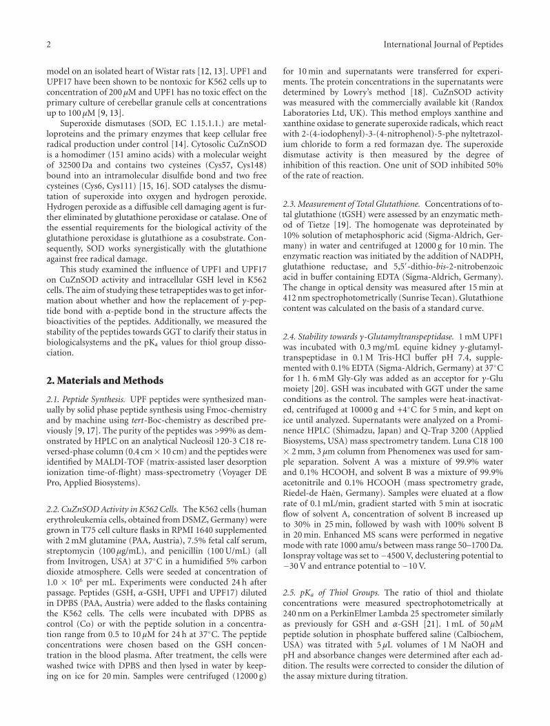
2 International Journal of Peptides
model on an isolated heart of Wistar rats [12, 13]. UPF1 andUPF17 have been shown to be nontoxic for K562 cells up toconcentration of 200 μM and UPF1 has no toxic effect on theprimary culture of cerebellar granule cells at concentrationsup to 100 μM [9, 13].
Superoxide dismutases (SOD, EC 1.15.1.1.) are metal-loproteins and the primary enzymes that keep cellular freeradical production under control [14]. Cytosolic CuZnSODis a homodimer (151 amino acids) with a molecular weightof 32500 Da and contains two cysteines (Cys57, Cys148)bound into an intramolecular disulfide bond and two freecysteines (Cys6, Cys111) [15, 16]. SOD catalyses the dismu-tation of superoxide into oxygen and hydrogen peroxide.Hydrogen peroxide as a diffusible cell damaging agent is fur-ther eliminated by glutathione peroxidase or catalase. One ofthe essential requirements for the biological activity of theglutathione peroxidase is glutathione as a cosubstrate. Con-sequently, SOD works synergistically with the glutathioneagainst free radical damage.
This study examined the influence of UPF1 and UPF17on CuZnSOD activity and intracellular GSH level in K562cells. The aim of studying these tetrapeptides was to get infor-mation about whether and how the replacement of γ-pep-tide bond with α-peptide bond in the structure affects thebioactivities of the peptides. Additionally, we measured thestability of the peptides towards GGT to clarify their status inbiologicalsystems and the pKa values for thiol group disso-ciation.
2. Materials and Methods
2.1. Peptide Synthesis. UPF peptides were synthesized man-ually by solid phase peptide synthesis using Fmoc-chemistryand by machine using tert-Boc-chemistry as described pre-viously [9, 17]. The purity of the peptides was >99% as dem-onstrated by HPLC on an analytical Nucleosil 120-3 C18 re-versed-phase column (0.4 cm× 10 cm) and the peptides wereidentified by MALDI-TOF (matrix-assisted laser desorptionionization time-of-flight) mass-spectrometry (Voyager DEPro, Applied Biosystems).
2.2. CuZnSOD Activity in K562 Cells. The K562 cells (humanerythroleukemia cells, obtained from DSMZ, Germany) weregrown in T75 cell culture flasks in RPMI 1640 supplementedwith 2 mM glutamine (PAA, Austria), 7.5% fetal calf serum,streptomycin (100 μg/mL), and penicillin (100 U/mL) (allfrom Invitrogen, USA) at 37◦C in a humidified 5% carbondioxide atmosphere. Cells were seeded at concentration of1.0 × 106 per mL. Experiments were conducted 24 h afterpassage. Peptides (GSH, α-GSH, UPF1 and UPF17) dilutedin DPBS (PAA, Austria) were added to the flasks containingthe K562 cells. The cells were incubated with DPBS ascontrol (Co) or with the peptide solution in a concentra-tion range from 0.5 to 10 μM for 24 h at 37◦C. The peptideconcentrations were chosen based on the GSH concen-tration in the blood plasma. After treatment, the cells werewashed twice with DPBS and then lysed in water by keep-ing on ice for 20 min. Samples were centrifuged (12000 g)
for 10 min and supernatants were transferred for experi-ments. The protein concentrations in the supernatants weredetermined by Lowry’s method [18]. CuZnSOD activitywas measured with the commercially available kit (RandoxLaboratories Ltd, UK). This method employs xanthine andxanthine oxidase to generate superoxide radicals, which reactwith 2-(4-iodophenyl)-3-(4-nitrophenol)-5-phe nyltetrazol-ium chloride to form a red formazan dye. The superoxidedismutase activity is then measured by the degree ofinhibition of this reaction. One unit of SOD inhibited 50%of the rate of reaction.
2.3. Measurement of Total Glutathione. Concentrations of to-tal glutathione (tGSH) were assessed by an enzymatic meth-od of Tietze [19]. The homogenate was deproteinated by10% solution of metaphosphoric acid (Sigma-Aldrich, Ger-many) in water and centrifuged at 12000 g for 10 min. Theenzymatic reaction was initiated by the addition of NADPH,glutathione reductase, and 5,5′-dithio-bis-2-nitrobenzoicacid in buffer containing EDTA (Sigma-Aldrich, Germany).The change in optical density was measured after 15 min at412 nm spectrophotometrically (Sunrise Tecan). Glutathionecontent was calculated on the basis of a standard curve.
2.4. Stability towards γ-Glutamyltranspeptidase. 1 mM UPF1was incubated with 0.3 mg/mL equine kidney γ-glutamyl-transpeptidase in 0.1 M Tris-HCl buffer pH 7.4, supple-mented with 0.1% EDTA (Sigma-Aldrich, Germany) at 37◦Cfor 1 h. 6 mM Gly-Gly was added as an acceptor for γ-Glumoiety [20]. GSH was incubated with GGT under the sameconditions as the control. The samples were heat-inactivat-ed, centrifuged at 10000 g and +4◦C for 5 min, and kept onice until analyzed. Supernatants were analyzed on a Promi-nence HPLC (Shimadzu, Japan) and Q-Trap 3200 (AppliedBiosystems, USA) mass spectrometry tandem. Luna C18 100× 2 mm, 3 μm column from Phenomenex was used for sam-ple separation. Solvent A was a mixture of 99.9% waterand 0.1% HCOOH, and solvent B was a mixture of 99.9%acetonitrile and 0.1% HCOOH (mass spectrometry grade,Riedel-de Haen, Germany). Samples were eluated at a flowrate of 0.1 mL/min, gradient started with 5 min at isocraticflow of solvent A, concentration of solvent B increased upto 30% in 25 min, followed by wash with 100% solvent Bin 20 min. Enhanced MS scans were performed in negativemode with rate 1000 amu/s between mass range 50–1700 Da.Ionspray voltage was set to−4500 V, declustering potential to−30 V and entrance potential to −10 V.
2.5. pKa of Thiol Groups. The ratio of thiol and thiolateconcentrations were measured spectrophotometrically at240 nm on a PerkinElmer Lambda 25 spectrometer similarlyas previously for GSH and α-GSH [21]. 1 mL of 50 μMpeptide solution in phosphate buffered saline (Calbiochem,USA) was titrated with 5 μL volumes of 1 M NaOH andpH and absorbance changes were determined after each ad-dition. The results were corrected to consider the dilution ofthe assay mixture during titration.
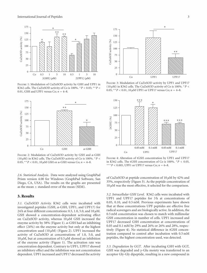
International Journal of Peptides 3
Co 0.5 1 5 10 0.5 1 5 100
25
50
75
100
125
150
175
Cu
Zn
SOD
act
ivit
y (%
)
[GSH] (μM) [UPF1] (μM)
∗∗ ∗∗∗ ∗
∗
Figure 1: Modulation of CuZnSOD activity by GSH and UPF1 inK562 cells. The CuZnSOD activity of Co is 100%. ∗P < 0.05; ∗∗P <0.01, GSH and UPF1 versus Co; n = 4–8.
Co GSH0
25
50
75
100
125
150
175
Cu
Zn
SOD
act
ivit
y (%
)
∗∗∗
∗
α-GSH
Figure 2: Modulation of CuZnSOD activity by GSH and α-GSH(10 μM) in K562 cells. The CuZnSOD activity of Co is 100%. ∗P <0.05; ∗∗P < 0.01, 10 μM GSH or α-GSH versus Co; n = 4–8.
2.6. Statistical Analysis. Data were analyzed using GraphPadPrism version 4.00 for Windows (GraphPad Software, SanDiego, CA, USA). The results on the graphs are presentedas the mean ± standard error of the mean (SEM).
3. Results
3.1. CuZnSOD Activity. K562 cells were incubated withinvestigated peptides (GSH, α-GSH, UPF1, and UPF17) for24 h at four different concentrations: 0.5, 1.0, 5.0, and 10 μM.GSH showed a concentration-dependent activating effecton CuZnSOD activity, whereas 10 μM GSH increased theenzyme activity by 38% (Figure 1). α-GSH had an inhibitingeffect (24%) on the enzyme activity but only at the highestconcentration used (10 μM) (Figure 2). UPF1 increased theactivity of CuZnSOD at concentrations of 1.0, 5.0, and10 μM, but at concentration of 0.5 μM showed an inhibitionof the enzyme activity (Figure 1). The activation rate wasconcentration dependent. Contrary to UPF1, UPF17 showedan inhibitory effect and the inhibition was not concentrationdependent. UPF1 increased and UPF17 decreased the activity
Co UPF1 UPF170
25
50
75
100
125
150
175
Cu
Zn
SOD
act
ivit
y (%
)
∗∗
∗
Figure 3: Modulation of CuZnSOD activity by UPF1 and UPF17(10 μM) in K562 cells. The CuZnSOD activity of Co is 100%. ∗P <0.05; ∗∗P < 0.01, 10 μM UPF1 or UPF17 versus Co; n = 4–8.
0
25
50
75
100
125
150
UPF1 UPF17Co
[tG
SH]
(%)
0.05 mM 0.1 mM 0.1 mM0.05 mM
∗∗∗
∗∗∗ ∗∗∗
∗
Figure 4: Alteration of tGSH concentration by UPF1 and UPF17in K562 cells. The tGSH concentration of Co is 100%. ∗P < 0.05,∗∗∗P < 0.005, UPF1 or UPF17 versus Co; n = 6–8.
of CuZnSOD at peptide concentration of 10 μM by 42% and35%, respectively (Figure 3). As the peptide concentration of10 μM was the most effective, it selected for the comparison.
3.2. Intracellular GSH Level. K562 cells were incubated withUPF1 and UPF17 peptides for 3 h at concentrations of0.05, 0.10, and 0.5 mM. Previous experiments have shownthat at these concentrations UPF peptides are effective freeradical scavengers and are biologically active. In addition, the0.5 mM concentration was chosen to match with millimolarGSH concentration in number of cells. UPF1 increased andUPF17 decreased GSH concentration at concentrations of0.05 and 0.1 mM by 29% and 26% or 26% and 28%, respec-tively (Figure 4). No statistical difference in tGSH concen-tration compared to control after incubation with 0.5 mMpeptides, the highest concentration used, was detected.
3.3. Degradation by GGT. After incubating GSH with GGT,GSH was degraded and γ-Glu moiety was transferred to anacceptor Gly-Gly dipeptide, resulting in a new compound in

4 International Journal of Peptides
mass spectra with MW 261.2 Da [γ-Glu-Gly-Gly]. The ques-tion arose: can the bond between γ-glutamate and cysteine bedegraded by GGT in UPF1, where the access to the bond isobstructed by an additional amino acid methylated tyrosine?Results obtained from the mass spectrometry measurementsdemonstrated that UPF1 is not degraded by GGT as theexpected peaks with or without acceptor dipeptide MW438.4 Da [Tyr(Me)-γ-Glu-Gly-Gly] or 324.3 Da [Tyr(Me)-γ-Glu], respectively, did not appear. During the incubation,UPF1 was dimerised over disulphide bridge. GGT is alsoable to breakdown dimeric form of GSH, but degradation ofdimerised UPF1 was not detected.
3.4. pKa. The pKa values of thiol groups of the peptides weremeasured. For GSH and α-GSH, the values were 9.0±0.3 and9.1 ± 0.1, respectively, whereas pKa values for UPF peptideswere slightly higher: 9.3 ± 0.1 for UPF1 and 9.4 ± 0.2 forUPF17.
4. Discussion
The present study focused on the effects of UPF1 and UPF17on CuZnSOD activity and intracellular GSH level in K562cells. For the first time we described and compared counter-point biological activities of structural antioxidative peptideanalogs differing from each other by spacial arrangementof Glu residue (γ-peptide bond in UPF1 changed to the α-peptide bond in UPF17). Previously we have shown thatUPF1 and UPF17 have a tendency for MnSOD activation.However, the γ-glutamyl moiety containing UPF1 neededmore time for MnSOD activation compared to UPF17,which had the effect already after 5 min incubation. UPF1and UPF17 have also different influence on glutathioneperoxidase activity (GPx): at higher concentrations than usedin in vivo experiments, both UPF1 and UPF17 inhibitedactivity concentration dependently whereas the α-peptidebond containing UPF17 had stronger inhibitory effect [22].In the present work we investigated how the replacementof γ-peptide bond with α-peptide bond on GSH and itsanalogue UPF1 affects CuZnSOD activity and level of GSH inK562 cells. The results showed that γ-Glu moiety containingGSH and UPF1 stimulated CuZnSOD activity and increasedintracellular tGSH level, whereas α-GSH and UPF17, whichhave α-Glu moiety in the structure, inhibited enzymaticactivity and decreased GSH level. The stability of UPF1towards GGT activity indicated that UPF1 affects GSH leveland CuZnSOD activity as intact molecule instead of beinga GSH precursor. Previously, it has been shown that GSHand UPF1 are able to act as signaling molecules through G-protein activation in frontocortical membrane preparations[23]. It has been reported that plasma membranes havespecific binding sites of GSH which have an interactionwith the glutamate binding sites [24]. By this way GSH andUPF peptides may affect the metabolism of cells as signalmolecules. The effects on the level of GSH and CuZnSODactivity may be different depending on the replacement of γ-peptide bond with α-peptide bond. GSH has been shown tobind to ionotropic glutamate receptors via gamma-glutamyl
residue in the nervous tissue [25]. Additionally, glutamatereceptors have been found also in the plasma membrane ofmegakaryocytes and rat erythrocytes [26, 27]. By interactingwith the latter receptors, GSH and UPF peptides may affectthe metabolism of cells as signal molecules through the PKCpathway and affect CuZnSOD activity. The various effects ofthe studied molecules may be caused by structural differencesbetween the GSH and UPF peptides (replacement of γ-peptide bond with α-peptide bond).
UPF1 and UPF17 have also shown different effectsin free radical scavenging experiments. According to theclassification of kinetic behavior by Sanches-Moreno et al.,UPF17 is classified as fast and UPF1 as intermediate DPPHradical scavenger [9, 28]. In silica modeling of noncovalentcomplex formation by docking calculations revealed a moreaffine complex between DPPH radical and α-GSH comparedto the complex with GSH [21]. This raised a question aboutpKa values for the thiol groups of UPF peptides. Previously,the change of γ-peptide bond to α-peptide bond has alsobeen investigated for GSH and its α-analogue: pKa of thiolgroups were similar for GSH and α-GSH (9.0 ± 0.1 and9.1± 0.1) [21]. The comparison of these results with currentmeasurements for UPF1 and UPF17 demonstrated that pKa
value is rather influenced by the addition of a methylatedtyrosine moiety to the GSH backbone than by the changeof the peptide bond type. Smaller pKa values for GSHand its α-analogue showed that these molecules donate thesulfhydryl proton more easily than UPF peptides; however,UPF peptides are better radical scavengers. This indicatesthat reactive species elimination does not depend only of thereactivity of the thiol group.
The results of the current paper show that γ-peptidebond and α-peptide bond containing UPF peptides mayinfluence enzyme activities in different direction, whichoffers a wider perspective for the usage of glutathione ana-logues as protective diverse regulators of the oxidative state.
Acknowledgments
This paper was financially supported by the EstonianScience Foundation Grants no. 7856 and 7494, by targetedfinancing from Ministry of Education and Science of Estonia(SF0180105s08) and by European Union through the Euro-pean Regional Development Fund.
References
[1] M. Zilmer, U. Soomets, A. Rehema, and U. Langel, “The glu-tathione system as an attractive therapeutic target,” Drug Des-ign Reviews Online, vol. 2, no. 2, pp. 121–127, 2005.
[2] N. H. P. Cnubben, I. M. C. M. Rietjens, H. Wortelboer, J. VanZanden, and P. J. Van Bladeren, “The interplay of glutathione-related processes in antioxidant defense,” Environmental Toxi-cology and Pharmacology, vol. 10, no. 4, pp. 141–152, 2001.
[3] D. A. Dickinson, A. L. Levonen, D. R. Moellering et al., “Hu-man glutamate cysteine ligase gene regulation through the ele-ctrophile response element,” Free Radical Biology and Medi-cine, vol. 37, no. 8, pp. 1152–1159, 2004.
[4] N. Ballatori, S. M. Krance, S. Notenboom, S. Shi, K. Tieu, andC. L. Hammond, “Glutathione dysregulation and the etiology
International Journal of Peptides 5
and progression of human diseases,” Biological Chemistry, vol.390, no. 3, pp. 191–214, 2009.
[5] R. Franco, O. J. Schoneveld, A. Pappa, and M. I. Panayiotidis,“The central role of glutathione in the pathophysiology ofhuman diseases,” Archives of Physiology and Biochemistry, vol.113, no. 4-5, pp. 234–258, 2007.
[6] O. Ortolani, A. Conti, A. R. De Gaudio, E. Moraldi, Q. Cantini,and G. Novelli, “The effect of glutathione and N-acetylcysteineon lipoperoxidative damage in patients with early septicshock,” American Journal of Respiratory and Critical CareMedicine, vol. 161, no. 6, pp. 1907–1911, 2000.
[7] A. Wendel and P. Cikryt, “The level and half-life of glutathionein human plasma,” FEBS Letters, vol. 120, no. 2, pp. 209–211,1980.
[8] I. Cacciatore, C. Cornacchia, F. Pinnen, A. Mollica, and A. DiStefano, “Prodrug approach for increasing cellular glutathionelevels,” Molecules, vol. 15, no. 3, pp. 1242–1264, 2010.
[9] K. Ehrlich, S. Viirlaid, R. Mahlapuu et al., “Design, synthesisand properties of novel powerful antioxidants, glutathioneanalogues,” Free Radical Research, vol. 41, no. 7, pp. 779–787,2007.
[10] A. Gozzo, D. Lesieur, P. Duriez, J. C. Fruchart, and E. Teissier,“Structure-activity relationships in a series of melatoninanalogues with the low-density lipoprotein oxidation model,”Free Radical Biology and Medicine, vol. 26, no. 11-12, pp. 1538–1543, 1999.
[11] T. L. Yue, P. J. Mckenna, P. G. Lysko et al., “SB 211475, a meta-bolite of carvedilol, a novel antihypertensive agent, is a potentantioxidant,” European Journal of Pharmacology, vol. 251, no.2-3, pp. 237–243, 1994.
[12] J. Kals, J. Starkopf, M. Zilmer et al., “Antioxidant UPF1 atten-uates myocardial stunning in isolated rat hearts,” InternationalJournal of Cardiology, vol. 125, no. 1, pp. 133–135, 2008.
[13] P. Poder, M. Zilmer, J. Starkopf et al., “An antioxidanttetrapeptide UPF1 in rats has a neuroprotective effect intransient global brain ischemia,” Neuroscience Letters, vol. 370,no. 1, pp. 45–50, 2004.
[14] I. Fridovich, “Superoxide anion radical (O •2 ), superoxide dis-
mutases, and related matters,” Journal of Biological Chemistry,vol. 272, no. 30, pp. 18515–18517, 1997.
[15] M. D. De Beus, J. Chung, and W. Colon, “Modification ofcysteine 111 in Cu/Zn superoxide dismutase results in alteredspectroscopic and biophysical properties,” Protein Science, vol.13, no. 5, pp. 1347–1355, 2004.
[16] M. A. Hough and S. S. Hasnain, “Structure of fully reducedbovine copper zinc superoxide dismutase at 1.15 A,” Structure,vol. 11, no. 8, pp. 937–946, 2003.
[17] U. Soomets, M. Zilmer, and U. Langel, “Manual solid-phasesynthesis of glutathione analogues: a laboratory-based shortcourse,” in Peptide Synthesis and Applications, J. Howl, Ed., pp.241–257, Humana Press, Totowa, NJ, USA, 2006.
[18] O. H. Lowry, N. J. Rosebrough, A. L. Farr, and R. J. Randall,“Protein measurement with the Folin phenol reagent,” TheJournal of biological chemistry, vol. 193, no. 1, pp. 265–275,1951.
[19] F. Tietze, “Enzymic method for quantitative determination ofnanogram amounts of total and oxidized glutathione: appli-cations to mammalian blood and other tissues,” AnalyticalBiochemistry, vol. 27, no. 3, pp. 502–522, 1969.
[20] D. Burg, D. V. Filippov, R. Hermanns, G. A. Van der Marel, J.H. Van Boom, and G. J. Mulder, “Peptidomimetic glutathioneanalogues as novel γGT stable GST inhibitors,” Bioorganic andMedicinal Chemistry, vol. 10, no. 1, pp. 195–205, 2002.
[21] S. Viirlaid, R. Mahlapuu, K. Kilk, A. Kuznetsov, U. Soomets,and J. Jarv, “Mechanism and stoichiometry of 2,2-diphenyl-1-picrylhydrazyl radical scavenging by glutathione and its novelα-glutamyl derivative,” Bioorganic Chemistry, vol. 37, no. 4, pp.126–132, 2009.
[22] K. Ehrlich, K. Ida, R. Mahlapuu et al., “Characterization ofUPF peptides, members of the glutathione analogues library,on the basis of their effects on oxidative stress-related en-zymes,” Free Radical Research, vol. 43, no. 6, pp. 572–580, 2009.
[23] E. Karelson, R. Mahlapuu, M. Zilmer, U. Soomets, N. Bog-danovic, and U. Langel, “Possible signaling by glutathione andits novel analogue through potent stimulation of frontocor-tical G proteins in normal aging and in Alzheimer’s disease,”Annals of the New York Academy of Sciences, vol. 973, pp. 537–540, 2002.
[24] R. Janaky, K. Ogita, B. A. Pasqualotto et al., “Glutathione andsignal transduction in the mammalian CNS,” Journal of Neuro-chemistry, vol. 73, no. 3, pp. 889–902, 1999.
[25] Z. Jenei, R. Janaky, V. Varga, P. Saransaari, and S. S. Oja, “Inter-ference of S-alkyl derivatives of glutathione with brain iono-tropic glutamate receptors,” Neurochemical Research, vol. 23,no. 8, pp. 1085–1091, 1998.
[26] P. G. Genever, D. J. P. Wilkinson, A. J. Patton et al., “Expressionof a functional N-methyl-D-aspartate-type glutamate receptorby bone marrow megakaryocytes,” Blood, vol. 93, no. 9, pp.2876–2883, 1999.
[27] A. Makhro, J. Wang, J. Vogel et al., “Functional NMDA rece-ptors in rat erythrocytes,” American Journal of Physiology, vol.298, no. 6, pp. C1315–C1325, 2010.
[28] C. Sanchez-Moreno, J. A. Larrauri, and F. Saura-Calixto, “Aprocedure to measure the antiradical efficiency of polyphe-nols,” Journal of the Science of Food and Agriculture, vol. 76,no. 2, pp. 270–276, 1998.
Related Documents