http://ionsource.com/tutorial/DeNovo/introduction.htm - De Novo Peptide Sequencing Tutorial 1

Welcome message from author
This document is posted to help you gain knowledge. Please leave a comment to let me know what you think about it! Share it to your friends and learn new things together.
Transcript
http://ionsource.com/tutorial/DeNovo/introduction.htm
- De Novo Peptide Sequencing Tutorial
1

http://ionsource.com/tutorial/DeNovo/introduction.htm
Table of Contents
Introduction - Peptide Fragmentation Nomenclature - b and y Ions - The Rules (16 simple rules) - The Protocol - Example MS/MS Spectrum - • low mass • mid mass • high mass - First De Novo Exercise (Ion Trap Data)- • Answer Page - Second De Novo Exercise (q-TOF Data)- • Answer page
• Tutorial References and Additional Suggested Reading -
• De Novo Tables - AA Residue Mass, Mass Conflict, Di-peptide Mass Residue Combinations
2

http://ionsource.com/tutorial/DeNovo/introduction.htm
3

http://ionsource.com/tutorial/DeNovo/introduction.htm
Introduction
De novo is Latin for, "over again", or "anew". A popular definition for "de novo peptide sequen-cing" is, peptide sequencing performed without prior knowledge of the amino acid sequence. Usually this rule is imposed by Edman degradation practitioners who perform de novo sequencing day in and day out, and perhaps feel a little bit threatened by that half million dollar mass spectrometer sitting down the hall, that can supposedly sequence peptides in a matter of seconds, and not days. Actually, any research project should be started with as much information as pos-sible, there should never be a need to restrict your starting knowledge, unless of course you are performing a clinical trial or some other highly controlled experiment. Mass spectrometers do have the advantage when it comes to generating sequence data for peptides in low femtomole quantities. However, Edman degradation will always enjoy the advantage when pmol quantities of a peptide are available. At higher pmol quantities (2-10 pmol), Edman will often provide the exact amino acid sequence without ambiguity for a limited run of amino acids, 6-30 amino acids, usually taking 30-50 min per cycle of the sequencer. However, at lower quantities, gaps and uncertainties are often encountered, even with Edman sequencing. MS/MS enjoys sensitivity, and speed, and does not require an external standard for each amino acid or amino acid variant. MS/MS sequencing does have difficulty with isobaric or near isobaric masses, for example telling K from Q on low resolution, low mass accuracy mass spectrometers. Another advantage is that MS/MS sequencing is never stopped by a blocked amino terminus, as is the case for Edman degradation.
Edman practitioners will often blast MS/MS sequencing on it deficiencies. We do need to approach de novo sequencing with our eyes wide open to all of its challenges and also to all of it's advantages. As scientists we need to have faith in the derived de novo sequence without knowing the sequence ahead of time, I guess this is at the heart of de novo. Especially when software is in-volved, we need to be confident enough to point to the top ranked output sequence and say, "Yes, this is the most correct sequence!" It is appropriate to test your skills or the skills of a software package with blinded but know sequences. Throughout the tutorial we will look at some known and some blinded sequences to demonstrate some of the de novo sequencing principles and also to test your newly learned de novo sequencing skills.
This tutorial leans heavily on a de novo sequencing course that was presented in 1992 at the University of Virginia, taught by Professor Donald F. Hunt. Dr. Hunt and his colleagues have generously taught this course for many years, educating generations of mass spectroscopists. It is impossible to calculate the enormity of the contribution that Dr. Hunt and his teaching efforts have made to countless research projects, both influencing basic, and medical, and drug research. One of the most notable early applications was the sequencing of peptides bound to MHC molecules. This was truly ground breaking work by the Hunt lab at the University of Virginia.
Let's Start:
A common question when one begins to talk about peptide fragmentation is, "What are b and y ions?" First we will look at the classical nomenclature, and then we will look at our first example peptide
4
http://ionsource.com/tutorial/DeNovo/introduction.htm
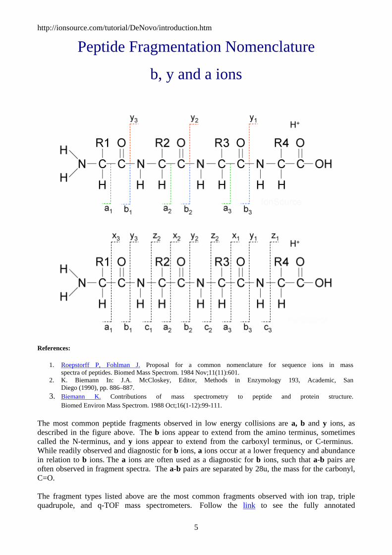
Peptide Fragmentation Nomenclature
b, y and a ions
References:
1. Roepstorff P, Fohlman J. Proposal for a common nomenclature for sequence ions in mass spectra of peptides. Biomed Mass Spectrom. 1984 Nov;11(11):601.
2. K. Biemann In: J.A. McCloskey, Editor, Methods in Enzymology 193, Academic, San Diego (1990), pp. 886–887.
3. Biemann K. Contributions of mass spectrometry to peptide and protein structure. Biomed Environ Mass Spectrom. 1988 Oct;16(1-12):99-111.
The most common peptide fragments observed in low energy collisions are a, b and y ions, as described in the figure above. The b ions appear to extend from the amino terminus, sometimes called the N-terminus, and y ions appear to extend from the carboxyl terminus, or C-terminus. While readily observed and diagnostic for b ions, a ions occur at a lower frequency and abundance in relation to b ions. The a ions are often used as a diagnostic for b ions, such that a-b pairs are often observed in fragment spectra. The a-b pairs are separated by 28u, the mass for the carbonyl, C=O.
The fragment types listed above are the most common fragments observed with ion trap, triple quadrupole, and q-TOF mass spectrometers. Follow the link to see the fully annotated
5

http://ionsource.com/tutorial/DeNovo/introduction.htm
fragmentation nomenclature as proposed by Biemann. An important note: an earlier nomenclature was proposed by Roepstorff and Fohlman and later modified by Biemann. The Biemann adaptation has been widely accepted.
More on b and y ions
Peptides do not fragment sequentially, that is to say, the first fragmentation event does not start at the amino terminus and proceed sequentially one residue at a time down the amino acid chain. The fragmentation events are somewhat random and definitely not sequential. In addition, some fragmentations are preferred over others as noted by the variation in the abundance of observed peaks in the spectrum below. Most of us can recognize a peptide fragment spectrum just by glancing at it. The peaks will appear to differ by the approximate mass of an amino acid residue as shown below.
Figure 1. This is an MS/MS spectrum of the tryptic peptide GLSDGEWQQVLNVWGK. This data was collected on an ion trap mass spectrometer. This spectrum will be the subject of our first unblinded de novo sequencing example.
The mess of peaks normally observed in a fragment spectrum are a reflection of the population of fragment ions produced in the collision cell of a mass spectrometer. The sequence of the peptide is determined by the mass difference between these peaks. To complicate matters there will be y and b ions intermixed that may allow you to to establish a sequence, both forward and backward.
Those fragment peaks that appear to extend from the amino terminus are termed "b ions". Figure 2. below demonstrates the ladder or family of "b ions" that may be observed in the fragment mass
6

http://ionsource.com/tutorial/DeNovo/introduction.htm
spectrum for this tryptic peptide. The b fragment peaks are labeled from the amino to the carboxyl terminus. The fragment containing only the amino terminal amino acid is termed b1. The fragment containing the first two amino terminal amino acids is termed the b2 ion, and so forth. The nomenclature is very simple to follow.
Figure 2.
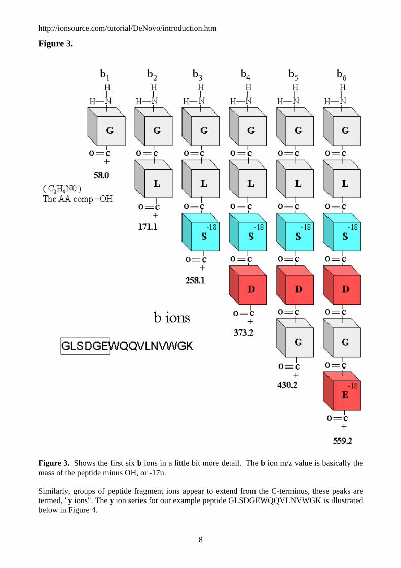
Below is a closer look at the generic structure of the first six amino terminal b ions. You can calculate the mass of any b ion, basically it is the mass of the shortened peptide (M)-17 (OH) = b ion m/z or or more simply M-17 = b ion m/z. To keep it simple this is the calculation for a singly charged b ion.
7
http://ionsource.com/tutorial/DeNovo/introduction.htm
Figure 3.
Figure 3. Shows the first six b ions in a little bit more detail. The b ion m/z value is basically the mass of the peptide minus OH, or -17u. Similarly, groups of peptide fragment ions appear to extend from the C-terminus, these peaks are termed, "y ions". The y ion series for our example peptide GLSDGEWQQVLNVWGK is illustrated below in Figure 4.
8

http://ionsource.com/tutorial/DeNovo/introduction.htm
Figure 4.
9
http://ionsource.com/tutorial/DeNovo/introduction.htm
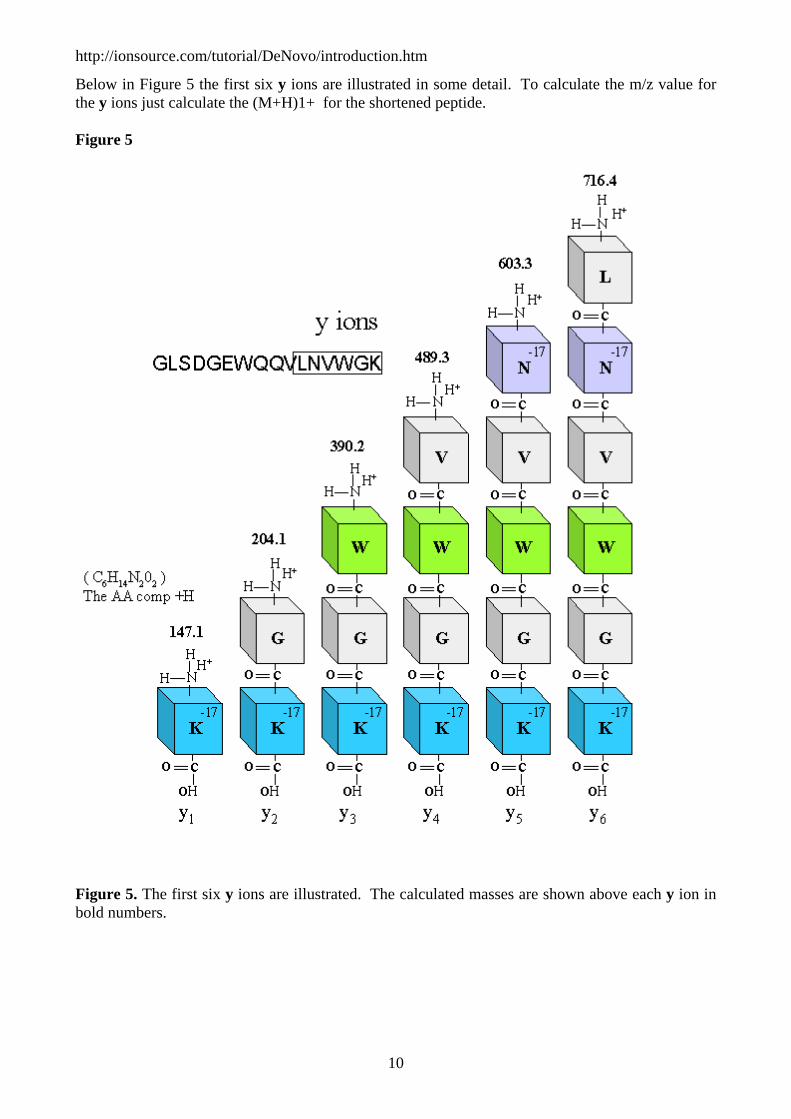
Below in Figure 5 the first six y ions are illustrated in some detail. To calculate the m/z value for the y ions just calculate the (M+H)1+ for the shortened peptide.
Figure 5
Figure 5. The first six y ions are illustrated. The calculated masses are shown above each y ion in bold numbers.
-
10

http://ionsource.com/tutorial/DeNovo/introduction.htm
y and b ion mnemonic: To remember which are y ions and which are b ions you can remember that b ions are the series that extend from the amino terminus, or the front of the peptide. To us, it would make more sense if the b ions extended from the back or C-terminus, but just the opposite is true, b ions extend from the front of the peptide, the amino terminus.
The screen shot below is the output of a free on-line calculator provided by the Institute of Systems Biology. All you need to do is paste in the sequence of your peptide and it will output the expected y and b ions. The URL for this resource is http://db.systemsbiology.net:8080/proteomicsToolkit/index.html
You can use these masses to casually match up the masses to the peaks in Figure 1, at the top of the page.
Figure 6.
De Novo sequencing seems petty simple right? Well, before we proceed onto our first example we should learn some of the rules and observations that scientists have previously made that will help us when we start looking at real data.
11

http://ionsource.com/tutorial/DeNovo/introduction.htm
The Rules
(the observations)
Before we go though our first MS/MS example we should take a look at some of the rules that are generally applied to de novo sequencing. These rules or observations were adapted from a 1991 de novo sequencing course taught at the University of Virginia by Professor Donald F. Hunt and Dr. Jeffrey Shabanowitz. Here are a few of the rules and observations that were introduced in that course.
The Rules
Loss of Ammonia and Water
1. y and b ion fragments containing the amino acid residues R, K, Q, and N may appear to lose ammonia, -17. -
2. y and b ion fragments containing the amino acid residues S, T, and E may appear to lose water, -18. In the case of glutamic acid, E must be at the N-terminus of the fragment for this observation to be made.
Spectral Intensity Rules
1. b ion intensity will drop when the next residue is P, G or also H, K, and R. -
2. Internal cleavages can occur at P and H residues. An internal cleavage fragment is a fragment that appears to be a shortened peptide with P and or H at its amino terminus, for example the peptide EFGLPGLQNK may display the b ions PGLQNK, PGLQN, PGLQ, etc. These are the result of a double cleavage event. The y ion intensity will often be the most prominent peak in the spectrum. -
3. It is common for b and y ions or y and b ions to swap intensity when a P is encountered in a sequence. This can also be true when the basic residues H, K, or R are encountered in the sequence. -
4. When a cleavage appears before or after R, the -17 (loss of ammonia) peak can be more prominent than the corresponding y or b ion. -
5. When encountering aspartic acid in a sequence, the ion series can die out.
Amino Acid Composition
1. It is possible to observe immonium ions at the low end of the spectrum that can give a clue to the amino acid composition of a peptide. One caveat is that if you do not see an immonium ion for a particular amino acid, this does not mean that that amino acid is absent from the sequence. You can follow this link to learn more about immonium ions.
Isobaric Mass
12
http://ionsource.com/tutorial/DeNovo/introduction.htm
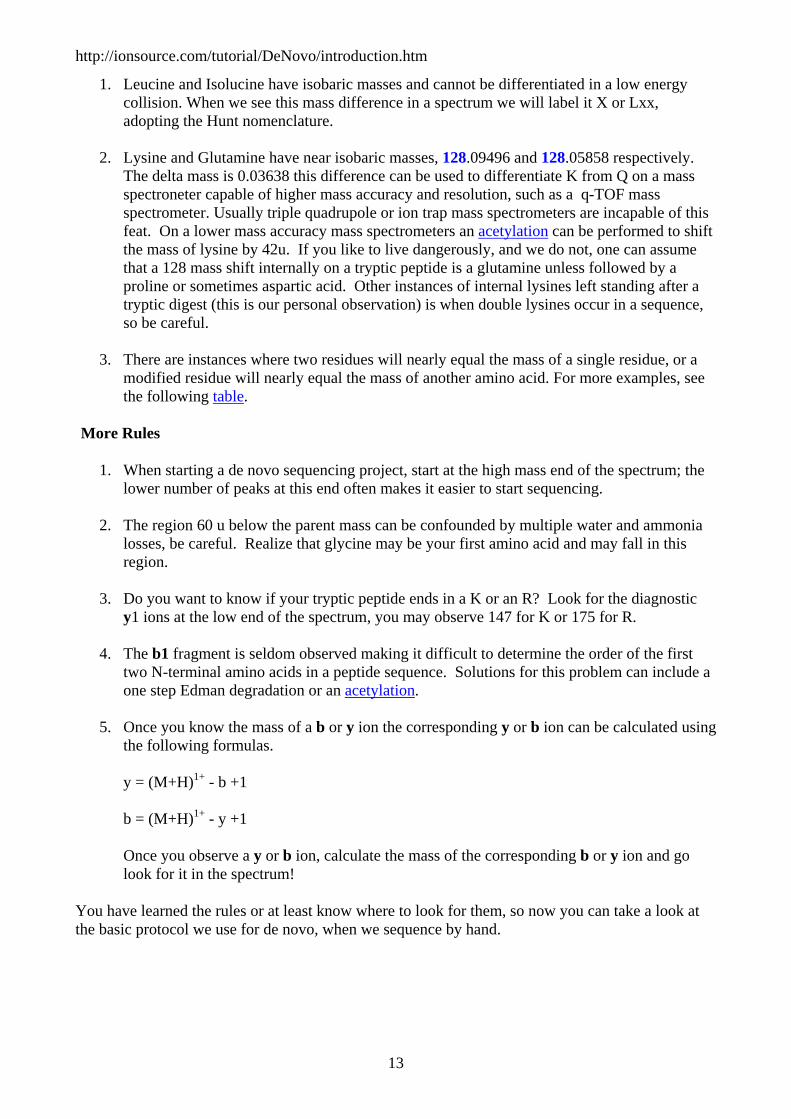
1. Leucine and Isolucine have isobaric masses and cannot be differentiated in a low energy collision. When we see this mass difference in a spectrum we will label it X or Lxx, adopting the Hunt nomenclature. -
2. Lysine and Glutamine have near isobaric masses, 128.09496 and 128.05858 respectively. The delta mass is 0.03638 this difference can be used to differentiate K from Q on a mass spectroneter capable of higher mass accuracy and resolution, such as a q-TOF mass spectrometer. Usually triple quadrupole or ion trap mass spectrometers are incapable of this feat. On a lower mass accuracy mass spectrometers an acetylation can be performed to shift the mass of lysine by 42u. If you like to live dangerously, and we do not, one can assume that a 128 mass shift internally on a tryptic peptide is a glutamine unless followed by a proline or sometimes aspartic acid. Other instances of internal lysines left standing after a tryptic digest (this is our personal observation) is when double lysines occur in a sequence, so be careful. -
3. There are instances where two residues will nearly equal the mass of a single residue, or a modified residue will nearly equal the mass of another amino acid. For more examples, see the following table.
-More Rules
1. When starting a de novo sequencing project, start at the high mass end of the spectrum; the lower number of peaks at this end often makes it easier to start sequencing. -
2. The region 60 u below the parent mass can be confounded by multiple water and ammonia losses, be careful. Realize that glycine may be your first amino acid and may fall in this region. -
3. Do you want to know if your tryptic peptide ends in a K or an R? Look for the diagnostic y1 ions at the low end of the spectrum, you may observe 147 for K or 175 for R. -
4. The b1 fragment is seldom observed making it difficult to determine the order of the first two N-terminal amino acids in a peptide sequence. Solutions for this problem can include a one step Edman degradation or an acetylation. -
5. Once you know the mass of a b or y ion the corresponding y or b ion can be calculated using the following formulas. - y = (M+H)1+ - b +1 - b = (M+H)1+ - y +1 - Once you observe a y or b ion, calculate the mass of the corresponding b or y ion and go look for it in the spectrum!
You have learned the rules or at least know where to look for them, so now you can take a look at the basic protocol we use for de novo, when we sequence by hand.
13

http://ionsource.com/tutorial/DeNovo/introduction.htm
The Protocol
Following the b Ion Series
1. If you are working with a tryptic peptide, look for the arginine or lysine y1 ion at the low end of the spectrum: 147 indicates a lysine, and a 175 indicates an arginine. This may give you a clue to the C-terminal residue of your tryptic peptide. Use this information to calculate the b ion that is the result of losing the C-terminal amino acid. Use this formula for Arginine (M+H)1+ - 18 -156 = penultimate b ion Use this formula for Lysine (M+H)1+ - 18 -128 = penultimate b ion (protonated peptide - 18 - AA residue mass = penultimate b ion) or you could use the standard formula for calculating the corresponding b ion once the y ion is known. (M+H)1+ - y1+1 = penultimate b ion If you are using an ion trap you may not be able to observe the low end of the spectrum, and in this case, you will need to do both of these calculations Go back to the high end of the spectrum and look for this b ion that you just calculated.
2. Whenever you identify a b ion look for an a ion at -28u. This gives some assurance that your assignment is correct. Also look for ammonia and water losses, -17 and -18u respectively. Whenever you identify a b ion do the math to find the corresponding y ion. y = (M+H)1+ - b +1 Go and look for this calculated y ion. All of this data should fit together to help firm up your assignments.
3. Look for the next b ion residue in the series. Use the amino acid residue masses to look for the next peak, see table. Soon you will have the residue masses memorized. Take the smallest amino acid jump possible to search for the next b ion. It is important to make the smallest jump because some residue combinations equal the mass of a single residue, for example GG = N, see our conflicting masses table. Label the b ion that you find and look for the related a ion, and calculate the corresponding y ion. You will not always be able to find an a ion, however, sometimes you can, and it is an assurance that you are on the right track.
4. Continue to follow the b ions down to the low end of the spectrum. Once you reach the low end and cannot go any further construct the b ion series. Since you will not see a b1 ion you will often need to calculate the mass residue combinations that compose the gap at the end, in which case you will not be able to determine the order of these two amino terminal amino acids.
14
http://ionsource.com/tutorial/DeNovo/introduction.htm
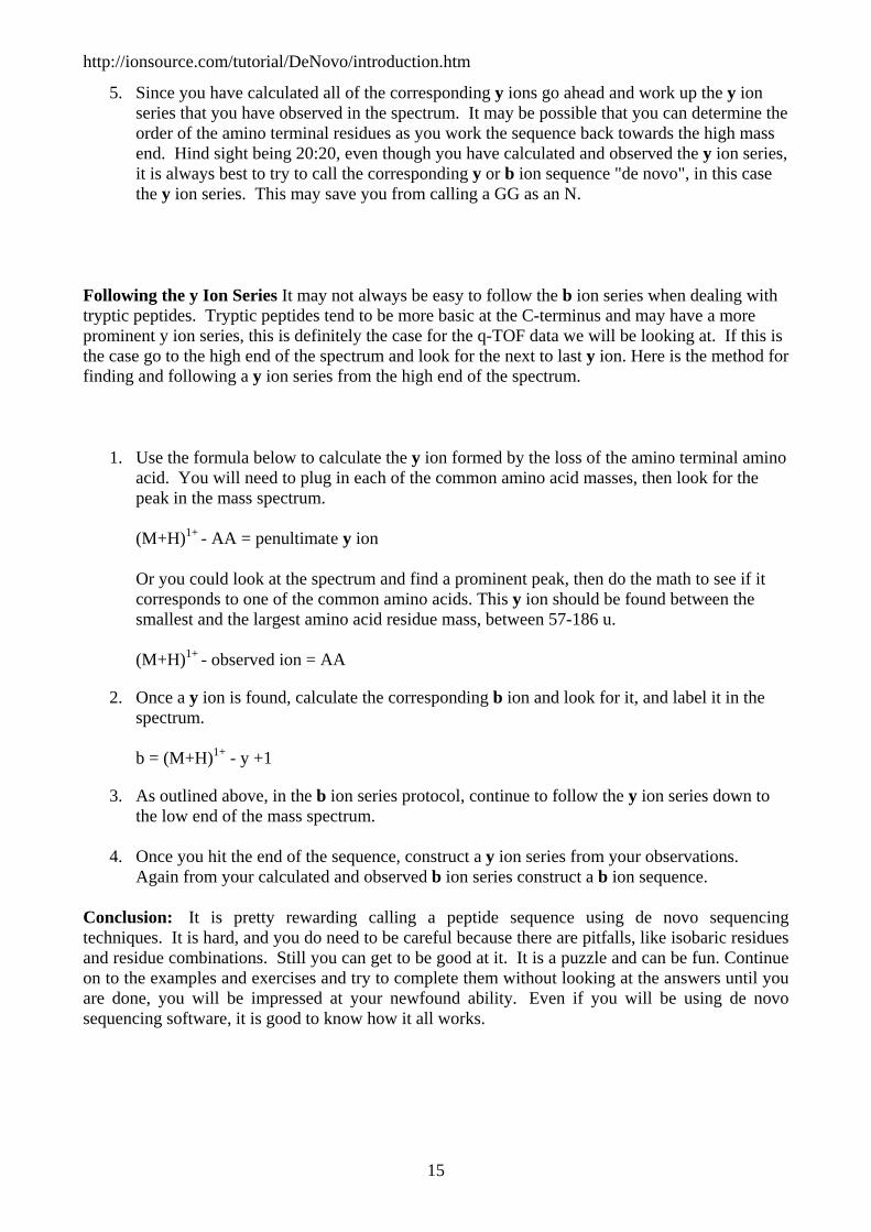
5. Since you have calculated all of the corresponding y ions go ahead and work up the y ion series that you have observed in the spectrum. It may be possible that you can determine the order of the amino terminal residues as you work the sequence back towards the high mass end. Hind sight being 20:20, even though you have calculated and observed the y ion series, it is always best to try to call the corresponding y or b ion sequence "de novo", in this case the y ion series. This may save you from calling a GG as an N.
-
Following the y Ion Series It may not always be easy to follow the b ion series when dealing with tryptic peptides. Tryptic peptides tend to be more basic at the C-terminus and may have a more prominent y ion series, this is definitely the case for the q-TOF data we will be looking at. If this is the case go to the high end of the spectrum and look for the next to last y ion. Here is the method for finding and following a y ion series from the high end of the spectrum.
1. Use the formula below to calculate the y ion formed by the loss of the amino terminal amino acid. You will need to plug in each of the common amino acid masses, then look for the peak in the mass spectrum. (M+H)1+ - AA = penultimate y ion Or you could look at the spectrum and find a prominent peak, then do the math to see if it corresponds to one of the common amino acids. This y ion should be found between the smallest and the largest amino acid residue mass, between 57-186 u. (M+H)1+ - observed ion = AA
2. Once a y ion is found, calculate the corresponding b ion and look for it, and label it in the spectrum. b = (M+H)1+ - y +1
3. As outlined above, in the b ion series protocol, continue to follow the y ion series down to the low end of the mass spectrum. -
4. Once you hit the end of the sequence, construct a y ion series from your observations. Again from your calculated and observed b ion series construct a b ion sequence.
Conclusion: It is pretty rewarding calling a peptide sequence using de novo sequencing techniques. It is hard, and you do need to be careful because there are pitfalls, like isobaric residues and residue combinations. Still you can get to be good at it. It is a puzzle and can be fun. Continue on to the examples and exercises and try to complete them without looking at the answers until you are done, you will be impressed at your newfound ability. Even if you will be using de novo sequencing software, it is good to know how it all works.
15

http://ionsource.com/tutorial/DeNovo/introduction.htm
Example Spectrum part one, low mass
For this example we will start at the low end of the spectrum and work our way up to the high end. This is just an example spectrum used for illustrative purposes. We will basically know the ions we are looking for and go look for them, and also we will learn what we can learn. Later on with the exercises we will follow the rules of our protocol and start at the high end of the spectrum. Below, in Figure 1, are six of the b ions described previously for the tryptic peptide GLSDGEWQQVLNVWGK. You may notice that we have annotated the serine and glutamic acid blocks with the notation -18, this is to remind us that these residues may be associated in the spectrum with water losses. In Figure 2 we have made a screen shot of the low end of the MS/MS spectrum to look for some of the predicted b ions and their associated fragment ions.
b Ion Details
Figure 1.
16

http://ionsource.com/tutorial/DeNovo/introduction.htm
Figure 1. We have taken the first 6 amino acids from the peptide GLSDGEWQQVLNVWGK to show in detail, b ions 1-6. The calculated b ion masses are noted in bold numbers below the block figures.
In Figure 2 below, it is easy to see the that the b ion series is not the predominant series. Data was not acquired below m/z 240 because this is ion trap data, and traps typically have a low mass cutoff, sometimes called the 1/3 rule. Due to this fact we have lost our diagnostic low mass immonium ions and several other important diagnostic ions. We were easily able to observe b ions 4, 6 and 7. Ion b5 was too low to call, most likely due to to the overall low abundance of the b ion series at the lower end of this spectrum. Calling a true de novo sequence with this b ion data would have been a challenge with these low ion intensities. There is an interesting feature in this spectrum that confirms that we are looking at a b ion series, which is that we observe a ions associated with b6 and b7. The loss of C=O or 28 u is the characteristic mass difference for the a / b pair. One can also observe water losses for b ions 4, 6 and 7 as predicted by the presence of serine and glutamic acid in this sequence.
Figure 2.
17

http://ionsource.com/tutorial/DeNovo/introduction.htm
Why did the b5 ion drop out in Figure 2? It looks like there was a significant water loss with a peak at m/z 412 which could have diminished the already small peak at m/z 430? Or, it could be the de novo sequencing rule, "b ion intensity may drop when the next residue is P, G or also H, K, and R", pretty cool, huh!
Figure 3.
Figure 3 above illustrates the y ion series. Again note that we have annotated the lysine and asparagine blocks with -17. This is to remind us that these residues appear to lose ammonia, and for the majority of y ions observed below in Figure 4, we also observe a peak that is 17 u lower. The apparent loss of ammonia is diagnostic for these residues.
Figure 4.
18

http://ionsource.com/tutorial/DeNovo/introduction.htm
So far we have discussed b and y ions, and for simplicity we have relegated ourselves to the terminal 6 or 7 amino acids. We will detail the entire spectrum in the following slides. It is apparent that the amino terminal sequence may be difficult to determine from the b ion series, one because of the relative abundance, and two because of the limitations of the ion trap. At the end of this tutorial we will provide spectra obtained on a q-TOF mass spectrometer for you to practice with, which does not share the same ion trap low mass limitation.
If you are interested in following this sequence proceed onto the next page. It is getting kind of interesting, isn't it? I wonder what will happen next?
19
http://ionsource.com/tutorial/DeNovo/introduction.htm
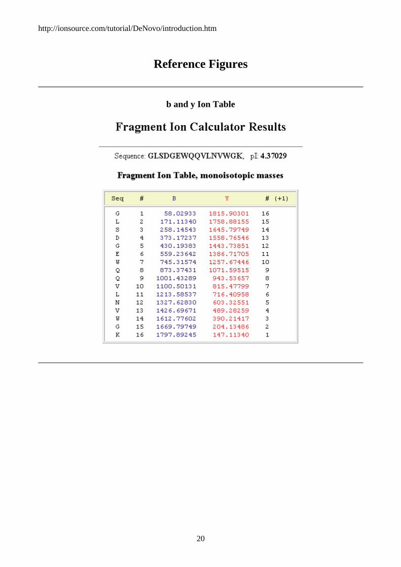
Reference Figures
b and y Ion Table
20

http://ionsource.com/tutorial/DeNovo/introduction.htm
Full Fragment Spectrum
21

http://ionsource.com/tutorial/DeNovo/introduction.htm
Example Spectrum part two, mid mass
As we move into the mid spectrum range the intensity of the b ions increase, see Figure 1 below. The b ions in Figure 1 are losing water and ammonia which is consistent with S (-18) and Q (-17) residues. Peaks consistent with a ions are also observed at -28u from two of the b ions.
Figure 1.
The predominant ion in the mid range is the y ion. Notice that only ammonia loss (-17) is noted for the y ions in Figure 2. The -17u loss is consistent for fragments containing the residues K, Q and N. Also note that the greatest ammonia loss is observed for fragment y9 which contains the greatest number of these residues.
Figure 2.
22

http://ionsource.com/tutorial/DeNovo/introduction.htm
Shall we proceed onto the high mass range of the spectrum? Let's.
23

http://ionsource.com/tutorial/DeNovo/introduction.htm
Reference Figures
b and y Ion Table
24

http://ionsource.com/tutorial/DeNovo/introduction.htm
Full Fragment Spectrum
25

http://ionsource.com/tutorial/DeNovo/introduction.htm
Example Spectrum part three, high mass
At the high end of the spectrum, the b ions increase dramatically in intensity as compared to the b ion intensity at the low end of the spectrum. In addition, y ion intensity has greatly decreased. It is as if the y and b ion intensity have swapped.
Figure 1.
26

http://ionsource.com/tutorial/DeNovo/introduction.htm
Figure 2.
Figure 2 shows the greatly diminished y ion intensity at the high end of the spectrum. This occurs as we encounter an E. The reduction in peak intensity is pretty dramatic. Out of curiosity we plotted the pIs of the y and b ions, take a look. Amazing, between the y and b ion coverage we have accounted for almost all of the major peaks.
Feeling pretty bold? Proceed on to the next page for a true de novo example.
27

http://ionsource.com/tutorial/DeNovo/introduction.htm
Reference Figures
b and y Ion Table
28

http://ionsource.com/tutorial/DeNovo/introduction.htm
Full Fragment Spectrum
29

http://ionsource.com/tutorial/DeNovo/introduction.htm
1st De Novo Exercise
Here is an MS/MS spectrum for you to test out your newly acquired de novo skills. Below we have an MS/MS spectrum acquired on an ion trap mass spectrometer. The first figure is the full fragment spectrum; the spectrum is then divided into three sections to make the details easier for you to see. Try not to be intimidated, just follow the simple protocol that we have described previously. Start with the high end of the spectrum, see Figure 2. We have made a pdf file for you so that you can download the spectrum and print them out so that you can get a pencil and a calculator and start sequencing. To download the spectra, right click on the link and save the pdf document to your computer.
Here are the hints:
1. This is a tryptic peptide ending in K or R. 2. The Parent [M+H]1+ mass is 1379.4 3. Since this is ion trap data, mass accuracy should be within 0.8u
Figure 1.
Here is a zoom in on the high end of the mass spectrum.
30

http://ionsource.com/tutorial/DeNovo/introduction.htm
Figure 2.
Figure 3.
31

http://ionsource.com/tutorial/DeNovo/introduction.htm
Figure 4.
Once you have done your best you can proceed onto the answer page to check out the known sequence and see some of the issues we ran into with this spectrum.
32

http://ionsource.com/tutorial/DeNovo/introduction.htm
1st De Novo Exercise the answer page
The correct sequence for this peptide is,
HGTVVLTALGGILK
or more accurately the best that low energy CID can do
HGTVVXTAXGGXXK
The first thing that we did was to do the math to determine the penultimate b ion.
If we could determine the C-terminal residue from the observed immonium ions we could calculate the penultimate b ion using the formulas below.
1379.4 - 18 -156 = 1205.4 for Arginine
1379.4 - 18 -128 = 1235.4 for Lysine
Since we could not observe the low end of the ion trap spectrum we had to do the math for both likely tryptic peptide C-terminal residues. We found that the C-terminal residue matched for Lysine. The b ion series extended all of the way into the low end of the spectrum. The a ion type fragments did accompany some of the b ions down the spectrum giving us some confidence that we were following the correct sequence. For the b ion series, once we got down to the low end of the spectrum we had to use mass and amino acid residue combinations to determine the last two amino terminal residues. From the observed b ion series we could not determine the order of the remaining two amino terminal amino acids. Once the b ions were determined we calculated the expected y ion series using the formula [M+H]+ - observed b ion + 1 = corresponding y ion. We then went through and redid the delta masses on all of the calculated and observed y ions, obtaining the y ion sequence. The y ions were more abundant and let us determine the order of the two amino terminal amino acids as well as removing the N-X ambiguity that we observed in the b ion series. We had this N-X ambiguity because the b ion series was much less abundant that the y ion series, and a more accurate mass was easier to determine for the y ion series With the b ion series, we still missed the G-G sequence and substituted an asparagine, a rookie mistake.
Here is the sequence that we determined (called) from both the b and y ion series.
33

http://ionsource.com/tutorial/DeNovo/introduction.htm
All considered, we feel that we did pretty good. We did mistake the double glycine for an asparagine. The peak that would have indicated the glycine in the b ion series was very small and we fell into the N = GG trap. Once this mistake was made we missed the glycine peak in the y ion series even though the peak was fairly observable. Even though one can calculate the corresponding y or b ion once a b or y ion is observed we would suggest calling both the y and b series independently to remove any bias. This would have saved us from calling GG as an N. Perhaps we could do better with q-TOF data, let's see as we proceed onto the next exercise. Still, we did pretty good; hope you did too. Don't let anyone tell you that trap data is bad for de novo, you just need to try a little harder and be a little wary.
If you would like to see the scribbling of a mad man, as I marked my spectrum up in this exercise:
34

http://ionsource.com/tutorial/DeNovo/introduction.htm
35

http://ionsource.com/tutorial/DeNovo/introduction.htm
2nd De Novo Exercise q-TOF Data
Here is an MS/MS q-TOF spectrum for you to test your de novo skills on. The first figure is the full fragment spectrum, the spectrum is then divided into four sections to make the details easier for you to see. Start with the high end of the spectrum, Figure 2. We have made a pdf file for you so that you can download the spectra and print them out so that you can get a pencil and a calculator and start sequencing. To download the spectra, right click on the link and save the pdf document to your computer.
Once you have done your best you can proceed onto the answer page to check out the known sequence and see some of the issues we ran into with this spectrum.
Here are several hints
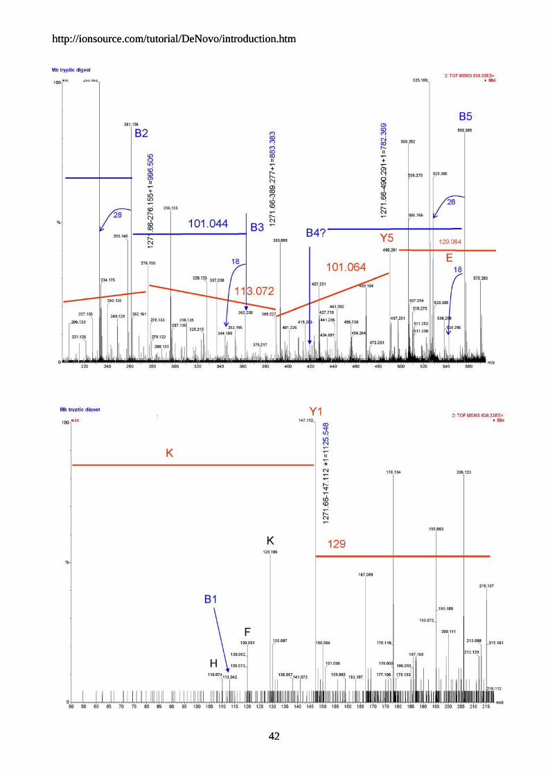
1. The parent ion is m/z 636.33 which is a doubly charged ion, [M+2H]2+ . 2. This is a tryptic peptide ending in K or R 3. Since this is q-TOF data start by following the y ion series, try to start at the high mass end. 4. Another note is, y ions dominate this set of q-TOF data. 5. Mass accuracy will be on your side, mass accuracy should be better than 30 mmu, that's
0.030u.
Figure 1.
-
36

http://ionsource.com/tutorial/DeNovo/introduction.htm
Figure 2.
Figure 3. .
-
37

http://ionsource.com/tutorial/DeNovo/introduction.htm
Figure 4.
-
Figure 5.
38

http://ionsource.com/tutorial/DeNovo/introduction.htm
2nd De Novo Exercise the q-TOF answer page
The correct sequence for this peptide is, LFTGHPETLEK
The first thing that we did was to look at the low end of the spectrum for low mass clues. We saw the immonium ions for H, F, and K, which let us know that we may encounter these amino acids in the sequence.
We did the math to determine the next to last y ion.
1271.66 - AA residue mass = penultimate y ion
We then proceeded to look for the next amino acid residue and labeled the peaks. We then did the calculation to determine the corresponding b ion and looked for a ions to confirm. With this data set, as long as you start with the y ion series the going is pretty straightforward. The mass accuracy of the q-TOF helps because you know that any amino acid call should be within 30 mmu.
If you tried to follow the b ion series your going would be tougher, because there are gaps that would force you to make several double amino acid jumps, and that's pretty hard, but still easier than with ion trap or triple quad data.. We don't have a lot of experience with q-TOF data, but it appears that the y ion series is dominant. Luckily we were able to determine the b1 ion which was a good surprise.
Here is the correct sequence
LFTGHPETLEK Here is the sequence that we determined from the y ion series
XFTGHPETXEK
We determined the entire sequence, yippee! Hopefully you got it right too!. If you would like to look at our scribblings,
39

http://ionsource.com/tutorial/DeNovo/introduction.htm
40

http://ionsource.com/tutorial/DeNovo/introduction.htm
41
http://ionsource.com/tutorial/DeNovo/introduction.htm
42
http://ionsource.com/tutorial/DeNovo/introduction.htm
42
http://ionsource.com/tutorial/DeNovo/introduction.htm
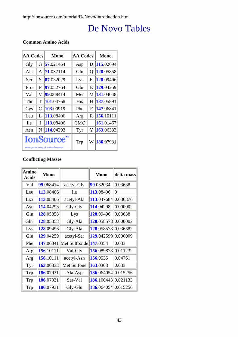
De Novo Tables Common Amino Acids
AA Codes Mono. AA Codes Mono.
Gly G 57.021464 Asp D 115.02694Ala A 71.037114 Gln Q 128.05858
Ser S 87.032029 Lys K 128.09496Pro P 97.052764 Glu E 129.04259Val V 99.068414 Met M 131.04048Thr T 101.04768 His H 137.05891Cys C 103.00919 Phe F 147.06841Leu L 113.08406 Arg R 156.10111Ile I 113.08406 CMC 161.01467
Asn N 114.04293 Tyr Y 163.06333
Trp W 186.07931
Conflicting Masses
Amino Acids Mono Mono delta mass
Val 99.068414 acetyl-Gly 99.032034 0.03638 Leu 113.08406 Ile 113.08406 0 Lxx 113.08406 acetyl-Ala 113.047684 0.036376 Asn 114.04293 Gly-Gly 114.04298 0.000002 Gln 128.05858 Lys 128.09496 0.03638 Gln 128.05858 Gly-Ala 128.058578 0.000002 Lys 128.09496 Gly-Ala 128.058578 0.036382 Glu 129.04259 acetyl-Ser 129.042599 0.000009 Phe 147.06841 Met Sulfoxide 147.0354 0.033 Arg 156.10111 Val-Gly 156.089878 0.011232 Arg 156.10111 acetyl-Asn 156.0535 0.04761 Tyr 163.06333 Met Sulfone 163.0303 0.033 Trp 186.07931 Ala-Asp 186.064054 0.015256 Trp 186.07931 Ser-Val 186.100443 0.021133 Trp 186.07931 Gly-Glu 186.064054 0.015256
43

http://ionsource.com/tutorial/DeNovo/introduction.htm
44
Di-peptide Table Note: Where di-peptides conflict with single amino acid the masses are noted in blue. Masses in red are notations where di-peptides conflict with other di-peptides.
G A S P V T C L I N D Q K E M H F R CMC Y W
57 71 87 97 99 101 103 113 113 114 115 128 128 129 131 137 147 156 161 163 186
G 57 114 128 144 154 156 158 160 170 170 171 172 185 185 186 188 194 204 213 218 220 243
A 71 128 142 158 168 170 172 174 184 184 185 186 199 199 200 202 208 218 227 232 234 257
S 87 144 158 174 184 186 188 190 200 200 201 202 215 215 216 218 224 234 243 248 250 273
P 97 154 168 184 194 196 198 200 210 210 211 212 225 225 226 228 234 244 253 258 260 283
V 99 156 170 186 196 198 200 202 212 212 213 214 227 227 228 230 236 246 255 260 262 285
T 101 158 172 188 198 200 202 204 214 214 215 216 229 229 230 232 238 248 257 262 264 287
C 103 160 174 190 200 202 204 206 216 216 217 218 231 231 232 234 240 250 259 264 266 289
L 113 170 184 200 210 212 214 216 226 226 227 228 241 241 242 244 250 260 269 274 276 299
I 113 170 184 200 210 212 214 216 226 226 227 228 241 241 242 244 250 260 269 274 276 299
N 114 171 185 201 211 213 215 217 227 227 228 229 242 242 243 245 251 261 270 275 277 300
D 115 172 186 202 212 214 216 218 228 228 229 230 243 243 244 246 252 262 271 276 278 301
Q 128 185 199 215 225 227 229 231 241 241 242 243 256 256 257 259 265 275 284 289 291 314
K 128 185 199 215 225 227 229 231 241 241 242 243 256 256 257 259 265 275 284 289 291 314
E 129 186 200 216 226 228 230 232 242 242 243 244 257 257 258 260 266 276 285 290 292 315
M 131 188 202 218 228 230 232 234 244 244 245 246 259 259 260 262 268 278 287 292 294 317
H 137 194 208 224 234 236 238 240 250 250 251 252 265 265 266 268 274 284 293 298 300 323
F 147 204 218 234 244 246 248 250 260 260 261 262 275 275 276 278 284 294 303 308 310 333
R 156 213 227 243 253 255 257 259 269 269 270 271 284 284 285 287 293 303 312 317 319 342
CMC 161 218 232 248 258 260 262 264 274 274 275 276 289 289 290 292 298 308 317 322 324 347
Y 163 220 234 250 260 262 264 266 276 276 277 278 291 291 292 294 300 310 319 324 326 349
W 186 243 257 273 283 285 287 289 299 299 300 301 314 314 315 317 323 333 342 347 349 372
http://ionsource.com/tutorial/DeNovo/introduction.htm
45
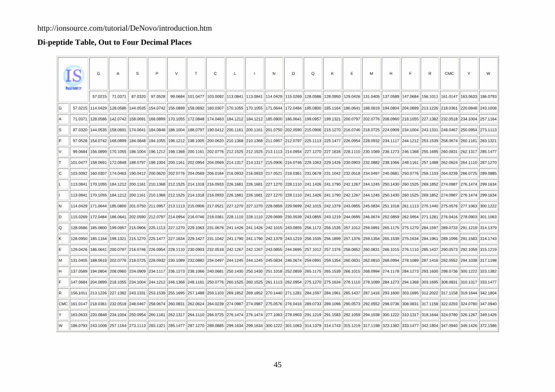
Di-peptide Table, Out to Four Decimal Places
G A S P V T C L I N D Q K E M H F R CMC Y W
-
57.0215 71.0371 87.0320 97.0528 99.0684 101.0477 103.0092 113.0841 113.0841 114.0429 115.0269 128.0586 128.0950 129.0426 131.0405 137.0589 147.0684 156.1011 161.0147 163.0633 186.0793
G 57.0215 114.0429 128.0586 144.0535 154.0742 156.0899 158.0692 160.0307 170.1055 170.1055 171.0644 172.0484 185.0800 185.1164 186.0641 188.0619 194.0804 204.0899 213.1226 218.0361 220.0848 243.1008
A 71.0371 128.0586 142.0742 158.0691 168.0899 170.1055 172.0848 174.0463 184.1212 184.1212 185.0800 186.0641 199.0957 199.1321 200.0797 202.0776 208.0960 218.1055 227.1382 232.0518 234.1004 257.1164
S 87.0320 144.0535 158.0691 174.0641 184.0848 186.1004 188.0797 190.0412 200.1161 200.1161 201.0750 202.0590 215.0906 215.1270 216.0746 218.0725 224.0909 234.1004 243.1331 248.0467 250.0954 273.1113
P 97.0528 154.0742 168.0899 184.0848 194.1055 196.1212 198.1005 200.0620 210.1368 210.1368 211.0957 212.0797 225.1113 225.1477 226.0954 228.0932 234.1117 244.1212 253.1539 258.0674 260.1161 283.1321
V 99.0684 156.0899 170.1055 186.1004 196.1212 198.1368 200.1161 202.0776 212.1525 212.1525 213.1113 214.0954 227.1270 227.1634 228.1110 230.1089 236.1273 246.1368 255.1695 260.0831 262.1317 285.1477
T 101.0477 158.0691 172.0848 188.0797 198.1004 200.1161 202.0954 204.0569 214.1317 214.1317 215.0906 216.0746 229.1063 229.1426 230.0903 232.0882 238.1066 248.1161 257.1488 262.0624 264.1110 287.1270
C 103.0092 160.0307 174.0463 190.0412 200.0620 202.0776 204.0569 206.0184 216.0933 216.0933 217.0521 218.0361 231.0678 231.1042 232.0518 234.0497 240.0681 250.0776 259.1103 264.0239 266.0725 289.0885
L 113.0841 170.1055 184.1212 200.1161 210.1368 212.1525 214.1318 216.0933 226.1681 226.1681 227.1270 228.1110 241.1426 241.1790 242.1267 244.1245 250.1430 260.1525 269.1852 274.0987 276.1474 299.1634
I 113.0841 170.1055 184.1212 200.1161 210.1368 212.1525 214.1318 216.0933 226.1681 226.1681 227.1270 228.1110 241.1426 241.1790 242.1267 244.1245 250.1430 260.1525 269.1852 274.0987 276.1474 299.1634
N 114.0429 171.0644 185.0800 201.0750 211.0957 213.1113 215.0906 217.0521 227.1270 227.1270 228.0859 229.0699 242.1015 242.1379 243.0855 245.0834 251.1018 261.1113 270.1440 275.0576 277.1063 300.1222
D 115.0269 172.0484 186.0641 202.0590 212.0797 214.0954 216.0746 218.0361 228.1110 228.1110 229.0699 230.0539 243.0855 243.1219 244.0695 246.0674 252.0859 262.0954 271.1281 276.0416 278.0903 301.1063
Q 128.0586 185.0800 199.0957 215.0906 225.1113 227.1270 229.1063 231.0678 241.1426 241.1426 242.1015 243.0855 256.1172 256.1535 257.1012 259.0991 265.1175 275.1270 284.1597 289.0733 291.1219 314.1379
K 128.0950 185.1164 199.1321 215.1270 225.1477 227.1634 229.1427 231.1042 241.1790 241.1790 242.1379 243.1219 256.1535 256.1899 257.1376 259.1354 265.1539 275.1634 284.1961 289.1096 291.1583 314.1743
E 129.0426 186.0641 200.0797 216.0746 226.0954 228.1110 230.0903 232.0518 242.1267 242.1267 243.0855 244.0695 257.1012 257.1376 258.0852 260.0831 266.1015 276.1110 285.1437 290.0573 292.1059 315.1219
M 131.0405 188.0619 202.0776 218.0725 228.0932 230.1089 232.0882 234.0497 244.1245 244.1245 245.0834 246.0674 259.0991 259.1354 260.0831 262.0810 268.0994 278.1089 287.1416 292.0552 294.1038 317.1198
H 137.0589 194.0804 208.0960 224.0909 234.1117 236.1273 238.1066 240.0681 250.1430 250.1430 251.1018 252.0859 265.1175 265.1539 266.1015 268.0994 274.1178 284.1273 293.1600 298.0736 300.1222 323.1382
F 147.0684 204.0899 218.1055 234.1004 244.1212 246.1368 248.1161 250.0776 260.1525 260.1525 261.1113 262.0954 275.1270 275.1634 276.1110 278.1089 284.1273 294.1368 303.1695 308.0831 310.1317 333.1477
R 156.1011 213.1226 227.1382 243.1331 253.1539 255.1695 257.1488 259.1103 269.1852 269.1852 270.1440 271.1281 284.1597 284.1961 285.1437 287.1416 293.1600 303.1695 312.2022 317.1158 319.1644 342.1804
CMC 161.0147 218.0361 232.0518 248.0467 258.0674 260.0831 262.0624 264.0239 274.0987 274.0987 275.0576 276.0416 289.0733 289.1096 290.0573 292.0552 298.0736 308.0831 317.1158 322.0293 324.0780 347.0940
Y 163.0633 220.0848 234.1004 250.0954 260.1161 262.1317 264.1110 266.0725 276.1474 276.1474 277.1063 278.0903 291.1219 291.1583 292.1059 294.1038 300.1222 310.1317 319.1644 324.0780 326.1267 349.1426
W 186.0793 243.1008 257.1164 273.1113 283.1321 285.1477 287.1270 289.0885 299.1634 299.1634 300.1222 301.1063 314.1379 314.1743 315.1219 317.1198 323.1382 333.1477 342.1804 347.0940 349.1426 372.1586

http://ionsource.com/tutorial/DeNovo/introduction.htm
References and Additional Reading
Denovo Analysis:
1. Hunt DF, Yates JR 3rd, Shabanowitz J, Winston S, Hauer CR. Protein sequencing by tandem mass spectrometry. Proc Natl Acad Sci. 1986 Sep;83(17):6233-7.
2. Papayannopoulos, IA, The interpretation of collision-induced dissociation tandem mass spectra of peptides. Mass Spectrom. Rev., 14(1) 49-73 (1995). Martin SE, Shabanowitz J, Hunt DF, Marto JA.3. Subfemtomole MS and MS/MS peptide sequence analysis using nano-HPLC micro-ESI fourier transform ion cyclotron resonance mass spectrometry. Anal Chem. 2000 Sep 15;72(18):4266-74.
4. Schroeder MJ, Shabanowitz J, Schwartz JC, Hunt DF, Coon JJ. A neutral loss activation method for improved phosphopeptide sequence analysis by quadrupole ion trap mass spectrometry. Anal Chem. 2004 Jul 1;76(13):3590-8.
5. Syka JE, Coon JJ, Schroeder MJ, Shabanowitz J, Hunt DF.Peptide and protein sequence analysis by electron transfer dissociation mass spectrometry. Proc Natl Acad Sci U S A. 2004 Jun 29;101(26):9528-33. Epub 2004 Jun 21.
6. Deutzmann R. Structural characterization of proteins and peptides. Methods Mol Med. 2004;94:269-97.
7. Medzihradszky KF Peptide sequence analysis. Methods Enzymol. 2005;402:209-44. 8. Coon JJ, Syka JE, Shabanowitz J, Hunt DF. Tandem mass spectrometry for peptide and
protein sequence analysis. Biotechniques. 2005 Apr;38(4):519, 521, 523.
Sequence Tag:
1. Mann M, Wilm M., Error-tolerant identification of peptides in sequence databases by peptide sequence tags. Anal Chem. 1994 Dec 15;66(24):4390-9.
2. Mortz E, O'Connor PB, Roepstorff P, Kelleher NL, Wood TD, McLafferty FW, Mann M. Sequence tag identification of intact proteins by matching tanden mass spectral data against sequence data bases. Proc Natl Acad Sci U S A. 1996 Aug 6;93(16):8264-7
3. Sunyaev S, Liska AJ, Golod A, Shevchenko A, Shevchenko A. MultiTag: multiple error-tolerant sequence tag search for the sequence-similarity identification of proteins by mass spectrometry. Anal Chem. 2003 Mar 15;75(6):1307-15.
Software:
1. Horn DM, Zubarev RA, McLafferty FW.Automated de novo sequencing of proteins by tandem high-resolution mass spectrometry. Proc Natl Acad Sci 2000 Sep 12;97(19):10313-7.
2. Bruni R, Gianfranceschi G, Koch G. On peptide de novo sequencing: a new approach. J Pept Sci. 2005 Apr;11(4):225-34.
3. Han Y, Ma B, Zhang K. SPIDER: software for protein identification from sequence tags with de novo sequencing error. J Bioinform Comput Biol. 2005 Jun;3(3):697-716. Savitski MM, Nielsen ML, Kjeldsen F, Zubarev RA.4. Proteomics-grade de novo sequencing approach. J Proteome Res. 2005 Nov-Dec;4(6):2348-54.
5. Zhong H, Li L. An algorithm for interpretation of low-energy collision-induced dissociation product ion spectra for de novo sequencing of peptides. Rapid Commun Mass Spectrom. 2005;19(8):1084-96.
46
http://www.ncbi.nlm.nih.gov/entrez/query.fcgi?cmd=Retrieve&db=pubmed&dopt=Abstract&list_uids=7847635

http://ionsource.com/tutorial/DeNovo/introduction.htm
6. Zhong H, Li L. An algorithm for interpretation of low-energy collision-induced dissociation product ion spectra for de novo sequencing of peptides. Rapid Commun Mass Spectrom. 2005;19(8):1084-96.
7. Searle BC, Dasari S, Wilmarth PA, Turner M, Reddy AP, David LL, Nagalla SR. Identification of protein modifications using MS/MS de novo sequencing and the OpenSea alignment algorithm. J Proteome Res. 2005 Mar-Apr;4(2):546-54.
Peptide Fragmentation Details:
1. Roepstorff P, Fohlman J. Proposal for a common nomenclature for sequence ions in mass spectra of peptides. Biomed Mass Spectrom. 1984 Nov;11(11):601.
2. Johnson RS, Martin SA, Biemann K, Stults JT, Watson JT. Novel fragmentation process of peptides by collision-induced decomposition in a tandem mass spectrometer: differentiation of leucine and isoleucine. Anal Chem. 1987 Nov 1;59(21):2621-5.
3. Biemann K. Contributions of mass spectrometry to peptide and protein structure. Biomed Environ Mass Spectrom. 1988 Oct;16(1-12):99-111.
4. K. Biemann In: J.A. McCloskey, Editor, Methods in Enzymology 193, 1990, pp. 886–887. 5. Falick, AM, Hines, WM, Medzihradszky, KF, Baldwin, MA and Gibson, BW, Low-mass
ions produced from peptides by high-energy collision-induced dissociation in tandem mass spectrometry. J. Am. Soc. Mass Spectrom., 4(11) 882-93 (1993).
6. Tang XJ, Thibault P, Boyd RK. Fragmentation reactions of multiply-protonated peptides and implications for sequencing by tandem mass spectrometry with low-energy collision-induced dissociation. Anal Chem. 1993 Oct 15;65(20):2824-34.
7. Patterson SD, Katta V. Prompt fragmentation of disulfide-linked peptides during matrix-assisted laser desorption ionization mass spectrometry. Anal Chem. 1994 Nov 1;66(21):3727-32.
8. Cramer R, Corless S. The nature of collision-induced dissociation processes of doubly protonated peptides: comparative study for the future use of matrix-assisted laser desorption/ionization on a hybrid quadrupole time-of-flight mass spectrometer in proteomics. Rapid Commun Mass Spectrom. 2001;15(22):2058-66.
9. Sadagopan N, Watson JT. Mass spectrometric evidence for mechanisms of fragmentation of charge-derivatized peptides. J Am Soc Mass Spectrom. 2001 Apr;12(4):399-409.
10. Samyn B, Debyser G, Sergeant K, Devreese B, Van Beeumen J. A case study of de novo sequence analysis of N-sulfonated peptides by MALDI TOF/TOF mass spectrometry. J Am Soc Mass Spectrom. 2004 Dec;15(12):1838-52.
De novo Approaches:
1. Schnolzer M, Jedrzejewski P, Lehmann WD. Protease-catalyzed incorporation of 18O into peptide fragments and its application for protein sequencing by electrospray and matrix-assisted laser desorption/ionization mass spectrometry. Electrophoresis 1996 May;17(5):945-53.
2. Keough T, Lacey MP, Youngquist RS. Derivatization procedures to facilitate de novo sequencing of lysine-terminated tryptic peptides using postsource decay matrix-assisted laser desorption/ionization mass spectrometry. Rapid Commun Mass Spectrom. 2000;14(24):2348-56.
3. Yergey AL, Coorssen JR, Backlund PS Jr, Blank PS, Humphrey GA, Zimmerberg J, Campbell JM, Vestal ML.De novo sequencing of peptides using MALDI/TOF-TOF. J Am Soc Mass Spectrom. 2002 Jul;13(7):784-91.
4. Heller M, Mattou H, Menzel C, Yao X. Trypsin catalyzed 16O-to-18O exchange for comparative proteomics: tandem mass spectrometry comparison using MALDI-TOF,
47
http://ionsource.com/tutorial/DeNovo/introduction.htm
ESI-QTOF, and ESI-ion trap mass spectrometers. J Am Soc Mass Spectrom. 2003 Jul;14(7):704-18.
5. Dancik V, Addona TA, Clauser KR, Vath JE, Pevzner PA. De novo peptide sequencing via tandem mass spectrometry. J Comput Biol. 1999 Fall-Winter;6(3-4):327-42.
6. Heredia-Langner A, Cannon WR, Jarman KD, Jarman KH. Sequence optimization as an alternative to de novo analysis of tandem mass spectrometry data. Bioinformatics 2004 Sep 22;20(14):2296-304. Epub 2004 Apr 15.
7. Olsen JV, Mann M. Improved peptide identification in proteomics by two consecutive stages of mass spectrometric fragmentation. Proc Natl Acad Sci 2004 Sep 14;101(37):13417-22. Epub 2004 Sep 3.
8. Zhang Z. De novo peptide sequencing based on a divide-and-conquer algorithm and peptide tandem spectrum simulation. Anal Chem. 2004 Nov 1;76(21):6374-83.
9. Spengler B. De novo sequencing, peptide composition analysis, and composition-based sequencing: a new strategy employing accurate mass determination by fourier transform ion cyclotron resonance mass spectrometry. J Am Soc Mass Spectrom. 2004 May;15(5):703-14.
De Novo Combined with Database Searching
1. Sheng QH, Xie T, Ding DF. De Novo Interpretation of MS/MS Spectra and Protein Identification via Database Searching. Sheng Wu Hua Xue Yu Sheng Wu Wu Li Xue Bao (Shanghai). 2000;32(6):595-600.
2. Johnson RS, Taylor JA. Searching sequence databases via de novo peptide sequencing by tandem mass spectrometry. Mol Biotechnol. 2002 Nov;22(3):301-15.
3. Savitski MM, Nielsen ML, Zubarev RA. New data base-independent, sequence tag-based scoring of peptide MS/MS data validates Mowse scores, recovers below threshold data, singles out modified peptides, and assesses the quality of MS/MS techniques. Mol Cell Proteomics 2005 Aug;4(8):1180-8. Epub 2005 May 22.
4. Halligan BD, Ruotti V, Twigger SN, Greene AS. DeNovoID: a web-based tool for identifying peptides from sequence and mass tags deduced from de novo peptide sequencing by mass spectroscopy. Nucleic Acids Res. 2005 Jul 1;33(Web Server issue):W376-81.
48
Related Documents