+ CASE CONFERENCE Sept. 14, 2011 Vincent Patrick Tiu Uy, MD

+ CASE CONFERENCE Sept. 14, 2011 Vincent Patrick Tiu Uy, MD.
Dec 28, 2015
Welcome message from author
This document is posted to help you gain knowledge. Please leave a comment to let me know what you think about it! Share it to your friends and learn new things together.
Transcript

+
CASE CONFERENCE
Sept. 14, 2011Vincent Patrick Tiu Uy, MD

5 year old male presents to the emergency department with ABDOMINAL PAIN

HISTORY OF PRESENT ILLNESS
1 week PTC
Intermittent episodes of vague abdominal pain localized at the periumbilical area. The pain was mild and the child can tolerate. Episodes of NBNB emesis x 3Denies fever, weaknessPoor appetite, otherwise hydrated
Few hours PTC
Pain became continuous, diffused, and severeGraded 10/10 on FACES. Several episodes of vomiting this time biliousNo appetite; child refuses to drink anythingChild became sicker
Abdominal PainVomiting x 3-5

+History
Review of Systems
Unremarkable. Most mentioned in the HPI
Past Medical History
Denies any medical problems, no previous hospitalizations, no previous surgeries, NKDA
Family History Denies any medical/surgical problems among immediate family members
Social History Child lives in an apartment with parents and siblings. No pets at home. No recent travel. Denies any introduction of new foods. No smokers at home. Child feels safe at home.

+Physical Examination
General Appearance Alert and awake, appears to be in moderate pain and prefers to lie down
Vital Signs T 97.4 HR 108 RR 22 BP 94/58 SO2 100% RA
Head, Eyes, Ears, Nose Throat, Neck
NCAT, pinkish conjunctivae, anicteric sclerae, nasal septum midline, TM’s intact, dry oral mucosa, non-hyperemic OP, supple neck, no CLAD
Chest and Cardiovascular CTAB, +S1/S2, no murmurs
Abdominal Exam Flat abdomen, hypoactive bowel sounds, (+) direct tenderness with guarding on 4 quadrants, no palpable masses, (-) rebound, (-) Rovsing’s sign, (-) Psoas sign, (-) Obturator sign, (-) Murphy’s sign*Liver span not mentioned
GU/Rectal *Was not performed at the ED
Extremities No edema, no cyanosis, brisk capillary refill
Neurologic Exam No focal neurologic findings

+Differentials?

+Management at the ED
IV line placement + Fluid bolus and then maintenance
Laboratories obtained: CBC, CMP, Amylase and Lipase, Coagulation panel, Blood typing, Urinalysis, Blood culture*, Urine Culture*
Serial abdominal exams still with diffuse tenderness and muscle guarding.
Diagnostic Imaging: Ultrasound of the abdomen and pelvis

+Laboratories
Hgb 11.7 g/dL
Hct 33.5%
WBC 13.2 10^3/uL
N 50
L 40
M 7
E 2
Na+ 135 mEq/L
K+ 3.5 mEq/L
Cl- 103 mEq/L
CO2 23 mEq/L
AST 111 IU/L
ALT 44 IU/L
Alk Phos 159 IU/L
Ca2+ 9.7 mg/dL
Glucose 216 mg/dL
Amylase 366 IU/L
Lipase 904 IU/L
Complete Blood Count Chemistries

+Ultrasound of the Abdomen and Pelvis

+Ultrasound of the Abdomen and Pelvis

+Eventually…
Diagnostic imaging: Contrast CT scan of the Abdomen and Pelvis

+CT scan of the abdomen

+CT SCAN OF THE ABDOMEN


+
CYSTIC DISEASE OF THE BILIARY TREE

+Biliary Duct Cysts
Encompasses both intrahepatic and extrahepatic cysts
Incidence: 1:1,000 – 1:1,500
Higher incidence in the Asian population
Cholangiocarcinoma (9-28%) is the most feared complication.
Approximately 67% of patients will be symptomatic by 10 years of age.

+Pathogenesis
Multifactorial
Abnormal Pancreatic-Biliary Junction (APBJ) Pancreatic duct empties into the CBD by >1 cm proximal to
the ampulla of Vater. Reflux of pancreatic secretions weakness of CBD wall
Other congenital causes Poor epithelialization and recanalization of the biliary tree Congenital weakness of the ductal walls

+Abnormal Pancreaticobiliary Junction

+Clinical Presentation
Pathologic jaundice and Acholic stools (representing biliary obstruction)
Palpable RUQ mass
Hepatomegaly
Infants

+Clinical Presentation
Older Children Intermittent bouts of pancreatitis (subclinical) Intermittent biliary obstruction Palpable mass Jaundice

+Differential Diagnosis
Acute Pancreatitis
Biliary Atresia
Cholecystitis, Choledolithiasis, Ascending Cholangitis
Biliary duct Obstruction
Bile duct tumors

+Laboratory Work-up
No laboratory work-up is specific
May suggest a co-diagnosis Elevated LFTs: Cholangitis, Obstruction Bilirubins: Pathologic Jaundice Amylase and Lipase: Pancreatitis WBC Counts: Cholangitis Electrolytes: Hypochloremic, Hypokalemic Alkalosis in
vomiting

+Imaging Studies
Ultrasound Best initial test Can identify the cyst prenatally (2nd trimester)
CT Scan Delineates the anatomy Presence and extent of intrahepatic involvement Usually enough to confirm the diagnosis and plan further
management/surgery
MRCP Best non-invasive test Able to detect APBJ Useful in planning further management/surgery

+Invasive Testing
ERCP & PTC If non-invasive tests fail to delineate the anatomy. Helpful in determining if there is an APBJ Delineates any intrahepatic or extrahepatic strictures or
stones

+

+Type I Cyst
Most common (50-85%)
Type IA saccular dilatation of majority or almost all of the CBD
Type IB saccular dilatation of a limited segment.
Type IC fusiform dilatation of majority or almost all of the CBD

+Type II Cyst
Isolated outpouching or a diverticulum protruding from the wall of the CBD
May be penduculated (with an adjoining stalk)

+Type III Cyst
AKA Choledochocoele
Arises from the intraduodenal portion of the CBD
Choledochoceles can be lined by duodenal or biliary epithelium.
Arise embryologically as duodenal duplications involving the ampulla.

+Type IV Cyst
Second most common (15-35%)
Type IVA multiple dilatations of both intrahepatic and extrahepatic sites.
Type IVB multiple dilatations of extrahepatic sites only.

+Type V Cyst
Multiple dilatations limited to intrahepatic bile ducts only.
Also known as Caroli’s Disease
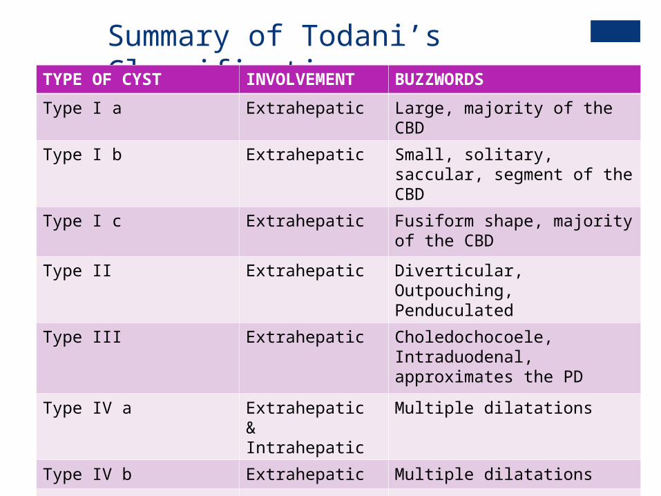
Summary of Todani’s ClassificationTYPE OF CYST INVOLVEMENT BUZZWORDS
Type I a Extrahepatic Large, majority of the CBD
Type I b Extrahepatic Small, solitary, saccular, segment of the CBD
Type I c Extrahepatic Fusiform shape, majority of the CBD
Type II Extrahepatic Diverticular, Outpouching, Penduculated
Type III Extrahepatic Choledochocoele, Intraduodenal, approximates the PD
Type IV a Extrahepatic & Intrahepatic
Multiple dilatations
Type IV b Extrahepatic Multiple dilatations
Type V – Caroli’s Disease
Intrahepatic Multiple dilatations

+Treatment
Surgical Cystoduodenostomy and Cystojejunostomy was a thing of
the past as it left the cyst wall behind. Total excision is preferred with hepaticojejunostomy (Roux-
en-Y)

+Current treatment strategiesType Typical procedure Extra Procedures
Type I Complete excision + Roux-en-Y
Type II Diverticular excision with ductoplasty
T-tube placement
Type III < 3mm: Endoscopic Sphincterotomy> 3mm: Excision (Transduodenal approach)
Reimplantation of pancreatic duct
Type IV Complete excision of extrahepatic component + Roux-en-YIntrahepatic components left untouched
Lobar excision for intrahepatic components if with stone, strictures, or hepatic abscess or coalescing in one lobe
Type V Medical management Liver transplant if two lobes are affected.

+“Extraordinary” Strategies
Lilly Technique For cysts that are adherent to the portal vein Can also be done in older patients with repeated cholangitis
and marked pericystic inflammation.
Liver Transplantation

+Adults Versus Children
Acquired
History of prior biliary surgery, pancreatitis, cholangitis, early/late post-op complications.
Vague symptoms
Malignant transformation and fibrosis is more common
Long term complications 30%
Congenital
No prior history
Classic triad is more common Abdominal pain Jaundice Abdominal mass
Fibrosis is rare
Long term complications 9.2%
ADULTS CHILDREN

+Patient Update
Transferred and admitted to CHAM
Diagnostic MRCP confirms Type IV A Choledochal cysts + Anomalous Pancreaticobiliary Junction (APBJ)
Edematous pancreas
Underwent surgery to remove the cyst, relieve the pressure on the pancreas
Patient tolerated procedure

+Easy Pop Quiz!
A 9 day old baby girl presents to you with jaundice. The mom noticed yellowing of the skin and eyeballs since she was 4 days old. A work-up was done which showed total bilirubin levels to be elevated at 15 mg/dL. The direct bilirubin was 20% of the TSB. There were no abnormalities in the physical examination.
Mom asks you: “What do you think it is?”
A. Alagille Syndrome
B. Viral Hepatitis
C. Biliary Atresia
D. Choledochal Cyst
E. Sepsis

+An even Easier Pop Quiz!
A 10 year old boy was brought to the ED with abdominal pain. There was diffuse tenderness and a palpable mass over the right upper quadrant. USG revealed a fusiform choledochal cyst. MRCP was done. The radiologist reading the study mentions an abnormal pancreatico-biliary junction and quizzes the residents: “With the pancreatic duct joining the CBD X cm. before reaching the duodenum.”
What is a possible value for X to justify the reading?
A. 1
B. 0.5
C. 0.3
D. 1.2
E. 0.8

+Sources
Ching Shui Huang, et al. Choledochal Cysts: Differences Between Pediatric and Adult Patients. J. Gastrointestinal Surgery (2010) 14:1105-1110
Irie, H., et. al. Value of MRCP in evaluating Choledochal Cysts American Journal of Roentgenology
Medscape References
Up-to-date

+Thank you!
LIVER
CYSTIC DUCT
GALL BLADDER
Related Documents