© 2009 The McGraw-Hill Companies, Inc. All rights reserved The Digestive System The Digestive System PowerPoint® presentation to accompany: Medical Assisting Third Edition Booth, Whicker, Wyman, Pugh, Thompson

© 2009 The McGraw-Hill Companies, Inc. All rights reserved The Digestive System PowerPoint® presentation to accompany: Medical Assisting Third Edition.
Dec 22, 2015
Welcome message from author
This document is posted to help you gain knowledge. Please leave a comment to let me know what you think about it! Share it to your friends and learn new things together.
Transcript

© 2009 The McGraw-Hill Companies, Inc. All rights reserved
The Digestive SystemThe Digestive SystemPowerPoint® presentation to accompany:
Medical AssistingThird Edition
Booth, Whicker, Wyman, Pugh, Thompson
33-2
© 2009 The McGraw-Hill Companies, Inc. All rights reserved
Learning Outcomes33.1 List the functions of the digestive system.
33.2 Trace the pathway of food through the alimentary canal.
33.3 Describe the structure and functions of the mouth, teeth, tongue, and salivary glands.
33.4 Describe the structure and function of the pharynx.
33-3
© 2009 The McGraw-Hill Companies, Inc. All rights reserved
Learning Outcomes (cont.)
33.5 Describe the swallowing process.
33.6 Describe the structure of the esophagus and tell how it propels food into the stomach.
33.7 Describe the structure and functions of the stomach.
33.8 List the substances secreted by the stomach and give their functions.

33-4
© 2009 The McGraw-Hill Companies, Inc. All rights reserved
Learning Outcomes (cont.)
33.9 Describe the structure and functions of the small intestine.
33.10 List the substances secreted by the small intestine and describe the importance of each.
33.11 Describe the structure and functions of the large intestine, including the anal canal and rectum.
33.12 Explain the structures and functions of the liver, gallbladder, and pancreas.
33-5
© 2009 The McGraw-Hill Companies, Inc. All rights reserved
Learning Outcomes (cont.)
33.13 List the substances released by the liver, gallbladder, and pancreas into the small intestine and give the function of each secretion.
33.14 Tell what types of nutrients are absorbed by the digestive system and where they are absorbed.
33.15 Describe the causes, signs and symptoms, and treatments of various diseases and disorders of the digestive system.

33-6
© 2009 The McGraw-Hill Companies, Inc. All rights reserved
Introduction Digestion
Mechanical and chemical breakdown of foods into forms that body cells can absorb
The organs of the digestive system carry out digestion
Two categories Alimentary canal organs Accessory organs

33-7
© 2009 The McGraw-Hill Companies, Inc. All rights reserved
Characteristics of the Alimentary Canal Wall of alimentary canal
Mucosa Inner most layer; epithelial tissue Secretes enzymes and mucus into lumen Absorbs nutrients
Submucosa Inferior to mucosa; loose connective tissue, blood vessels,
glands, and nerves Blood vessels carries away absorbed nutrients
Muscular layer Just outside submucosa; layers of smooth muscle Contracts to move materials through the canal
33-8
© 2009 The McGraw-Hill Companies, Inc. All rights reserved
Characteristics of the Alimentary Canal (cont.)
Serosa Double-walled outermost layer: peritoneum
Visceral peritoneum Innermost wall of serosa Secretes serous fluid to keep outside of canal moist
Parietal peritoneum Abdominal lining
Movements Churning – mixes substances in the canal Peristalsis – propels substances through the tract
33-9
© 2009 The McGraw-Hill Companies, Inc. All rights reserved
Apply Your KnowledgeWhat are the layers of the wall of the alimentary canal and what do they do?
ANSWER: The layers are:
Mucosa: innermost layer; secretes enzymes and mucus into the canal and absorbs nutrients
Submucosa: inferior to the mucosa; carries away absorbed nutrients
Muscular layer: just outside the submucosa; contracts to move materials through the canal
Serosa: double-walled outer layer; secretes serous fluid to keep outside of canal moist
Bravo!

33-10
© 2009 The McGraw-Hill Companies, Inc. All rights reserved
The Mouth Buccal cavity Mechanical digestion
Takes in food and reduces its size by chewing
Starts chemical digestion Saliva contains enzyme
amylase, which breaks down carbohydrates

33-11
© 2009 The McGraw-Hill Companies, Inc. All rights reserved
The Mouth (cont.)
Cheeks hold food in mouth Lips – sensory nerve fibers that judge temperature of
food Tongue
Skeletal muscles covered by mucous membrane Lingual frenulum – holds tongue to floor of mouth Mixes food, holds food between teeth, contains taste buds Lingual tonsils – lymphatic tissue destroys bacteria and
viruses on back of tongue

33-12
© 2009 The McGraw-Hill Companies, Inc. All rights reserved
The Mouth (cont.)
Palate Roof of mouth Separates oral cavity from nasal cavity Uvula – portion of soft palate that hangs down in
throat Lymph tissue
Palatine tonsils (oropharynx) Pharyngeal tonsils – adenoids (nasopharynx)
33-13
© 2009 The McGraw-Hill Companies, Inc. All rights reserved
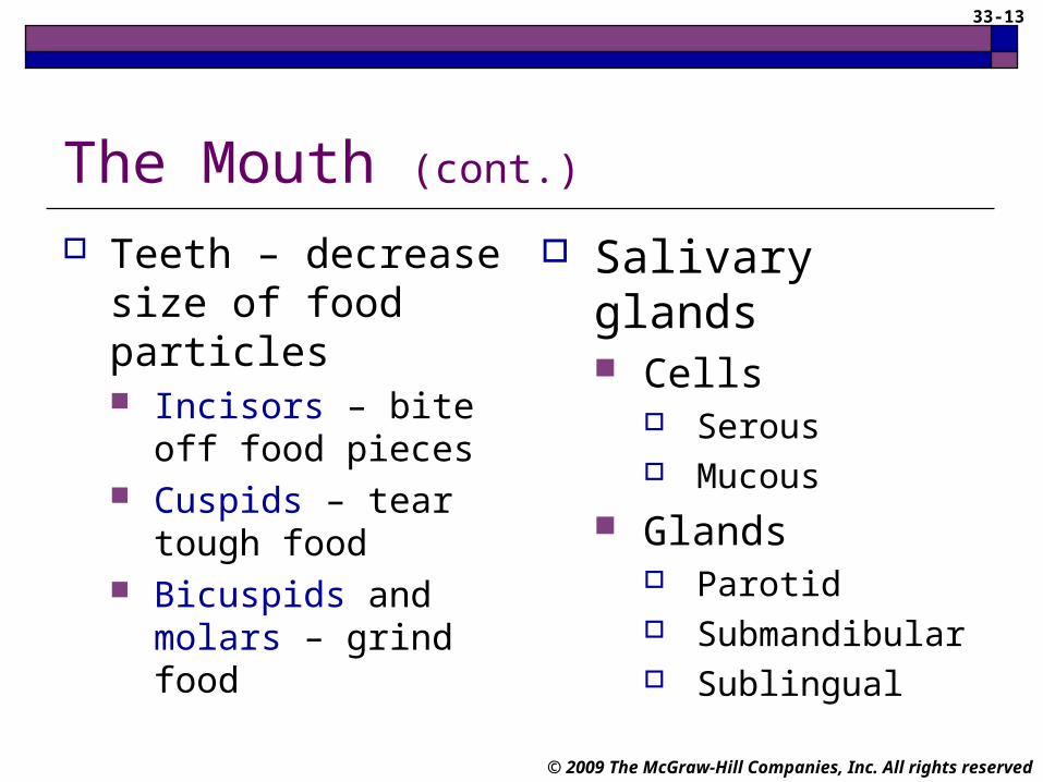
The Mouth (cont.)
Teeth – decrease size of food particles Incisors – bite off
food pieces Cuspids – tear tough
food Bicuspids and
molars – grind food
Salivary glands Cells
Serous Mucous
Glands Parotid Submandibular Sublingual

33-14
© 2009 The McGraw-Hill Companies, Inc. All rights reserved
Apply Your KnowledgeMatching:
___ Buccal cavity A. Saliva
___ Roof of mouth B. Mouth
___ Grind food C. Bolus
___ Adenoids D. Palate
___ Water, enzymes, and mucus E. Bicuspids
___ Mass of food mixed with saliva and mucus F. Pharyngeal gland
D
E
F
A
B
BANSWER:
33-15
© 2009 The McGraw-Hill Companies, Inc. All rights reserved
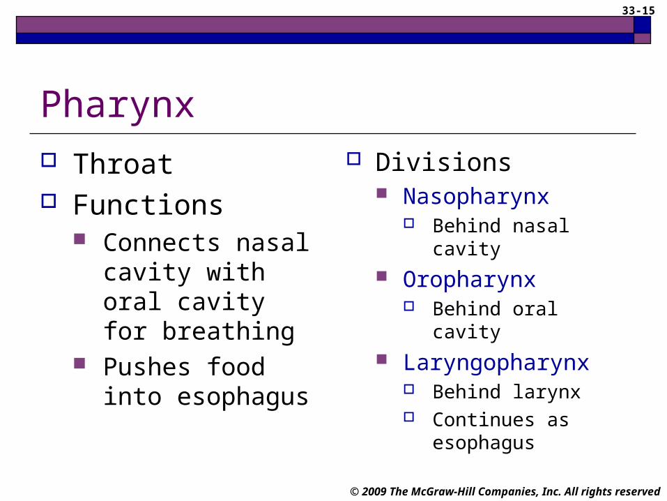
Pharynx Throat Functions
Connects nasal cavity with oral cavity for breathing
Pushes food into esophagus
Divisions Nasopharynx
Behind nasal cavity
Oropharynx Behind oral cavity
Laryngopharynx Behind larynx Continues as
esophagus

33-16
© 2009 The McGraw-Hill Companies, Inc. All rights reserved
Pharynx (cont.)
Swallowing – automatic process
1. Soft palate raises, uvula covers opening between nasal and oral cavity
2. Epiglottis covers larynx, keeping food out of it
3. Tongue presses against roof of mouth, forcing food into oropharynx

33-17
© 2009 The McGraw-Hill Companies, Inc. All rights reserved
Pharynx (cont.)
4. Muscles in pharynx contract, moving food toward esophagus
5. Esophagus opens
6. Food is pushed into esophagus by muscles of pharynx

33-18
© 2009 The McGraw-Hill Companies, Inc. All rights reserved
The Esophagus Muscular tube connecting pharynx to stomach
Esophageal hiatus – hole in diaphragm through which esophagus passes
Cardiac sphincter Circular band of muscle at the opening of the
stomach controls movement of food into stomach

33-19
© 2009 The McGraw-Hill Companies, Inc. All rights reserved
Apply Your Knowledge
Matching:
___ Connects nasal cavity with oral cavity A. Cardiac sphincter
___ Covers the opening of larynx B. Esophageal hiatus
___ Hole in diaphragm C. Sphincter
___ Controls movement of food into stomach D. Epiglottis
___ Circular bands of muscle E. PharynxC
A
B
D
E
ANSWER:

33-20
© 2009 The McGraw-Hill Companies, Inc. All rights reserved
The Stomach Below the diaphragm in
the upper left quadrant of the abdominal cavity
Functions Receive food from
esophagus Mix bolus with gastric juice Start protein digestion Move food into small
intestine
Sections Cardiac region Fundus Body Pylorus
Pyloric sphincter Controls movement of
substances into small intestine Stomach

33-21
© 2009 The McGraw-Hill Companies, Inc. All rights reserved
The Stomach (cont.)
Lining of stomach Rugae – folds of the inner lining Gastric glands
Mucous cells – secrete mucus to protect the lining Chief cells – secrete pepsinogen pepsin, which
digests protein Parietal cells
Hydrochloric acid needed to convert pepsinogen to pepsin Intrinsic factor needed for vitamin B12 absorption Stomach

33-23
© 2009 The McGraw-Hill Companies, Inc. All rights reserved
The Stomach (cont.)
Gastric glands stimulated by Parasympathetic nervous system Gastrin (hormone)
Cholesystokinin (hormone) secreted by the small intestine inhibits gastric glands
Stomach absorbs alcohol, water, and some fat-soluble drugs
Chyme – mixture of food and gastric juices
33-24
© 2009 The McGraw-Hill Companies, Inc. All rights reserved
Apply Your Knowledge
What are the functions of the stomach?
ANSWER: The stomach’s functions are to receive the bolus of food, mix it with gastric juice, start protein digestion, and move food into the small intestine. It also absorbs alcohol, water, and some drugs.
Right Answer!
33-25
© 2009 The McGraw-Hill Companies, Inc. All rights reserved
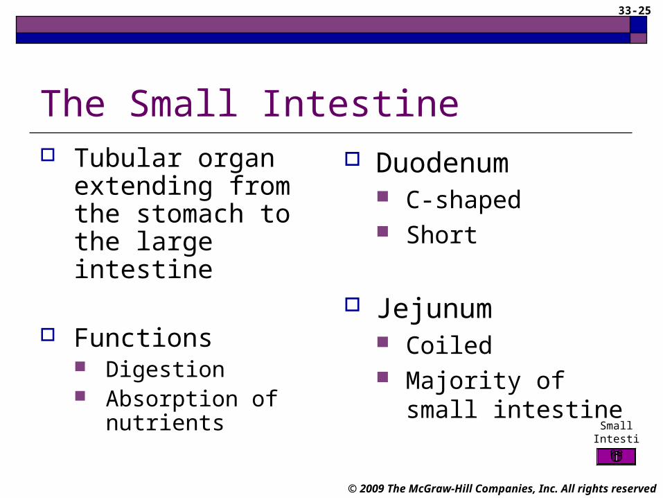
The Small Intestine Tubular organ
extending from the stomach to the large intestine
Functions Digestion Absorption of nutrients
Small Intestine
Duodenum C-shaped Short
Jejunum Coiled Majority of small
intestine

33-26
© 2009 The McGraw-Hill Companies, Inc. All rights reserved
The Small Intestine Ileum
Attached to large intestine Mesentery
Fan-like tissue that holds jejunum and ileum in the abdominal cavity
Attaches to the posterior wall of the abdomen Ileocecal sphincter
Controls movement of chyme from the ileum to the cecum of the large intestine Small
Intestine

33-27
© 2009 The McGraw-Hill Companies, Inc. All rights reserved
The Small Intestine (cont.)
Lining of small intestine Microvilli – increase surface area Intestinal glands
Mucus and water Enzymes
Peptidases – digest proteins Sucrase, maltase, and lactase – digest sugars Intestinal lipase – digests fats
Primary controls Parasympathetic nervous system Stretching of intestinal wall

33-29
© 2009 The McGraw-Hill Companies, Inc. All rights reserved
Apply Your Knowledge
ANSWER: She cannot produce lactase and cannot digest lactose, which is the sugar in dairy products.
Your patient states that she is lactose intolerant. What does that mean?
33-30
© 2009 The McGraw-Hill Companies, Inc. All rights reserved
The Large Intestine Extends from the ileum to the anus
Cecum Beginning of large intestine Veriform appendix
Ascending colon Portion that goes up the right side of the
abdominal cavity Large Intestine
33-31
© 2009 The McGraw-Hill Companies, Inc. All rights reserved
The Large Intestine (cont.)
Transverse colon Crosses abdominal cavity from right to left
Descending colon Down left side of abdominal cavity
Sigmoid colon S-shaped portion in pelvic cavity
Absorbs water and electrolytes Large Intestine

33-32
© 2009 The McGraw-Hill Companies, Inc. All rights reserved
The Rectum and Anal Canal Rectum – off sigmoid colon
Anal canal Last few centimeters of rectum Opening to outside of body is the anus
Large Intestine

33-33
© 2009 The McGraw-Hill Companies, Inc. All rights reserved
The Rectum and Anal Canal Feces
Leftover chyme Consists of undigested solid materials, little
water, ions, mucus, cells of intestinal lining, and bacteria
Defecation reflex Triggered by periodic contractions of large
intestine Allows anal sphincters to relax

33-35
© 2009 The McGraw-Hill Companies, Inc. All rights reserved
Apply Your KnowledgeMatching:
___ Vermiform appendix A. Feces
___ Crosses the abdomen B. Ascending colon
___ Up right side of abdomen C. Anal canal
___ S-shaped tube D. Defecation reflex
___ Down left side of abdomen E. Sigmoid
___ Leftover chyme F. Transverse colon
___ Last section of rectum G. Cecum
___ Allows anal sphincters to relax H. Descending colon
H
G
F
E
D
C
B
A
ANSWER: Super!

33-36
© 2009 The McGraw-Hill Companies, Inc. All rights reserved
The Liver Lobular organ in right upper quadrant
Large right lobe and smaller left lobe Hepatic lobules – contain macrophages Hepatocytes – process nutrients in blood and
make bile
Part of liver’s function Store vitamins and iron

33-37
© 2009 The McGraw-Hill Companies, Inc. All rights reserved
The Liver (cont.)
Hepatic portal vein – carries blood from digestive organs to hepatic lobules
Hepatic duct – bile from liver Hepatic duct merges with cystic
duct from gallbladder and forms common bile duct
Common bile duct – delivers bile to duodenum
33-38
© 2009 The McGraw-Hill Companies, Inc. All rights reserved
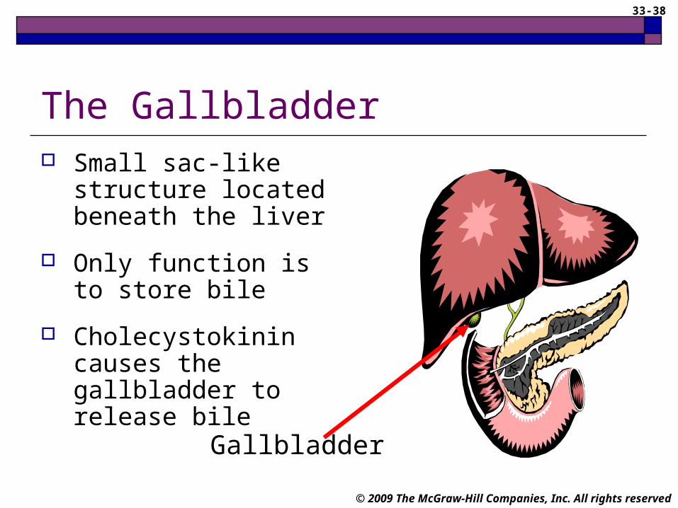
The Gallbladder Small sac-like structure
located beneath the liver
Only function is to store bile
Cholecystokinin causes the gallbladder to release bile
Gallbladder

33-39
© 2009 The McGraw-Hill Companies, Inc. All rights reserved
Apply Your KnowledgeWhat is the route of bile through the liver and gall bladder?
ANSWER: Bile is made in the hepatocytes and leaves the liver through the hepatic duct. The hepatic duct merges with the cystic duct from the gall bladder to form the common bile duct, which delivers bile to the duodenum.

33-40
© 2009 The McGraw-Hill Companies, Inc. All rights reserved
The Pancreas
Pancreatic amylase – digests carbohydrates
Pancreatic lipase – digests lipids
Nucleases – digestsnucleic acids
Trypsin, chymotrypsin, and carboxypeptidase – digest proteins
Located behind the stomach Acinar cells produce pancreatic juice, which contains these enzymes:

33-41
© 2009 The McGraw-Hill Companies, Inc. All rights reserved
The Pancreas (cont.)
Also secretes bicarbonate ions into duodenum Neutralize acidic chyme
Enzyme release stimulated by Parasympathetic nervous system Hormones secretin and cholecystokinin (from
small intestine)

33-42
© 2009 The McGraw-Hill Companies, Inc. All rights reserved
Apply Your Knowledge
What are the pancreatic enzymes and what do they do?
ANSWER: They are:
Pancreatic amylase – digests carbohydrates
Pancreatic lipase – digests lipids
Nucleases – digest nucleic acids
Trypsin, chymotrypsin, and carboxypeptidase – digest proteins
Good Job!

33-43
© 2009 The McGraw-Hill Companies, Inc. All rights reserved
The Absorption of Nutrients Nutrients are necessary food
substances Carbohydrates Proteins Lipids Vitamins Minerals Water
33-44
© 2009 The McGraw-Hill Companies, Inc. All rights reserved
The Absorption of Nutrients (cont.)
Carbohydrates – provide energy Polysaccharides – starches Monosaccharides and disaccharides – simple sugars Cellulose – provides fiber or bulk
Lipids – used for energy when glucose levels are low Triglycerides Cholesterol – essential for cell growth and function

33-45
© 2009 The McGraw-Hill Companies, Inc. All rights reserved
The Absorption of Nutrients (cont.)
Protein – used for growth and repair of tissue Essential amino acids body can not make
Vitamins Fat-soluble Water-soluble
Minerals – used to make enzymes, cell membranes, and proteins
33-46
© 2009 The McGraw-Hill Companies, Inc. All rights reserved
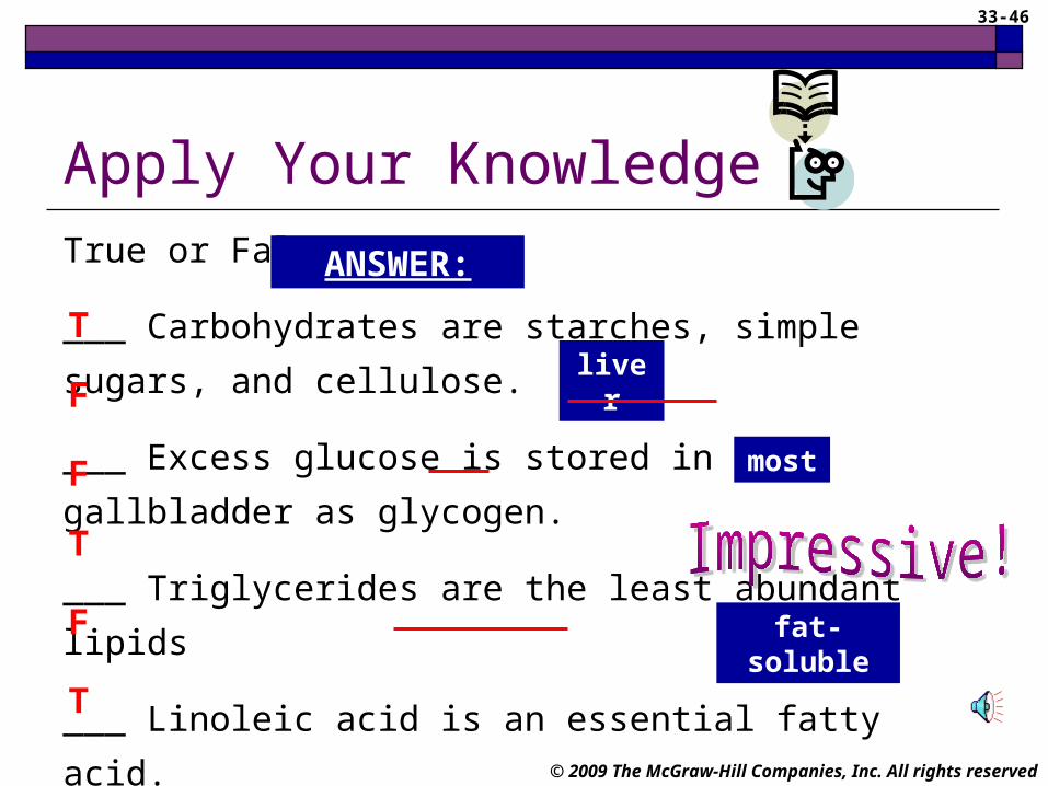
Apply Your KnowledgeTrue or False:
___ Carbohydrates are starches, simple sugars, and cellulose.
___ Excess glucose is stored in the gallbladder as glycogen.
___ Triglycerides are the least abundant lipids
___ Linoleic acid is an essential fatty acid.
___ A, D, E and K are water-soluble vitamins.
___ Minerals are used by cells to make enzymes.
F
F
F
T
T
ANSWER:
Tliver
most
fat-soluble

33-47
© 2009 The McGraw-Hill Companies, Inc. All rights reserved
Aging and the Digestive System Decreased motility –
GERD
Decreased absorption
More likely to develop ulcers and cancers
Decreased ability to detoxify blood
Sense of taste altered
Dietary changes due to Isolation Depression

33-48
© 2009 The McGraw-Hill Companies, Inc. All rights reserved
Common Diseases and Disorders
Disease / Disorder Description
Appendicitis Inflammation of the appendix; can be life-threatening if not treated promptly
Cirrhosis Chronic liver disease; normal tissue is replaced with nonfunctional scar tissue
Colitis Inflammation of the large intestine; can be acute or chronic
Colorectal cancer Arises from lining of rectum or colon; curable if treated early

33-49
© 2009 The McGraw-Hill Companies, Inc. All rights reserved
Common Diseases and Disorders (cont.)
Disease / Disorder Description
Constipation Difficult defecation
Crohn’s disease Inflammatory bowel disease; typically effects small intestine
Diarrhea Watery and frequent feces; usually self-limiting
Diverticulosis Abnormal pouches in the intestinal wall; no inflammation present
33-50
© 2009 The McGraw-Hill Companies, Inc. All rights reserved
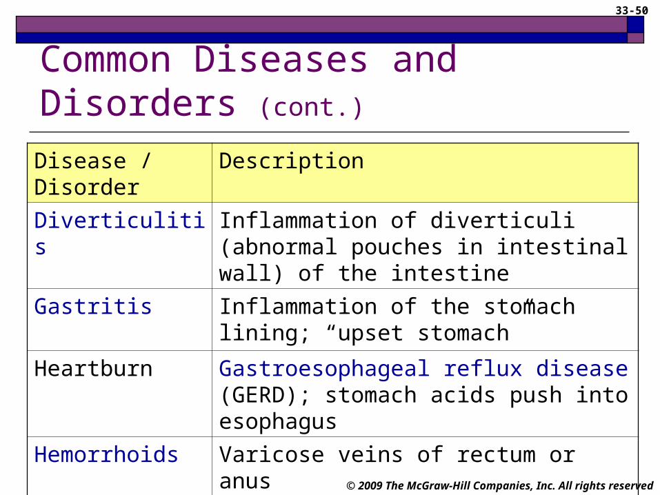
Common Diseases and Disorders (cont.)
Disease / Disorder Description
Diverticulitis Inflammation of diverticuli (abnormal pouches in intestinal wall) of the intestine
Gastritis Inflammation of the stomach lining; “upset stomach”
Heartburn Gastroesophageal reflux disease (GERD); stomach acids push into esophagus
Hemorrhoids Varicose veins of rectum or anus
Hepatitis Inflammation of the liver; various types
33-51
© 2009 The McGraw-Hill Companies, Inc. All rights reserved
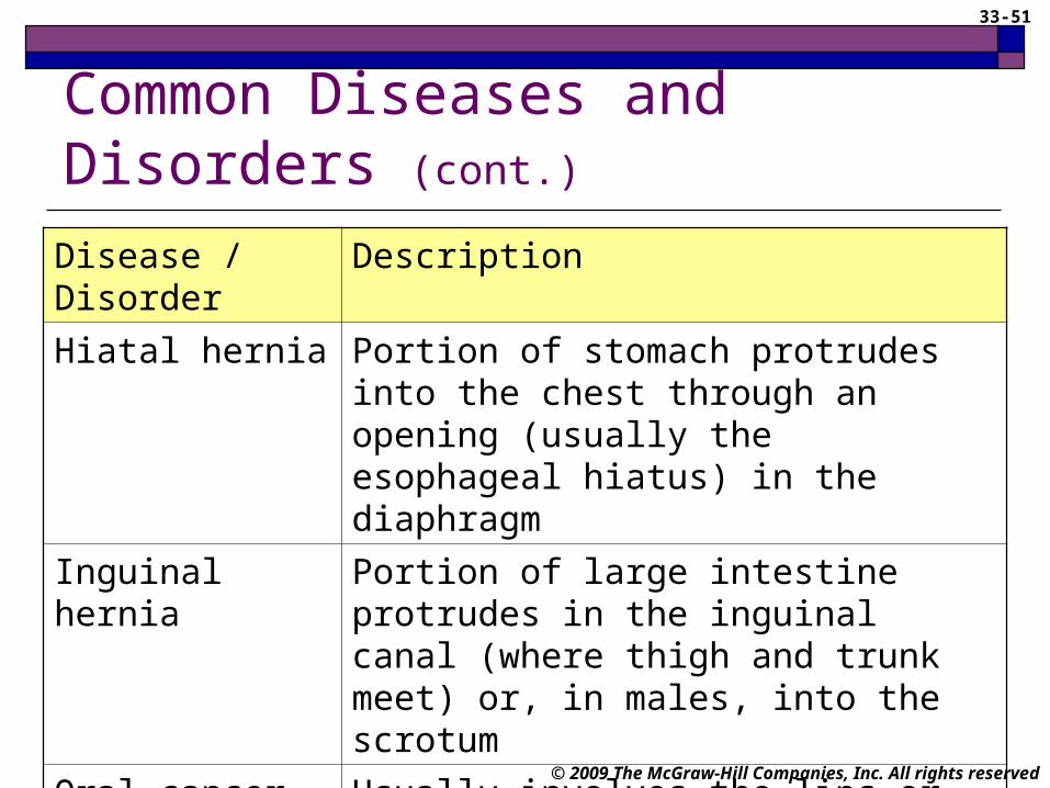
Common Diseases and Disorders (cont.)
Disease / Disorder Description
Hiatal hernia Portion of stomach protrudes into the chest through an opening (usually the esophageal hiatus) in the diaphragm
Inguinal hernia Portion of large intestine protrudes in the inguinal canal (where thigh and trunk meet) or, in males, into the scrotum
Oral cancer Usually involves the lips or tongue but can occur anywhere in the mouth; tends to spread rapidly
33-52
© 2009 The McGraw-Hill Companies, Inc. All rights reserved
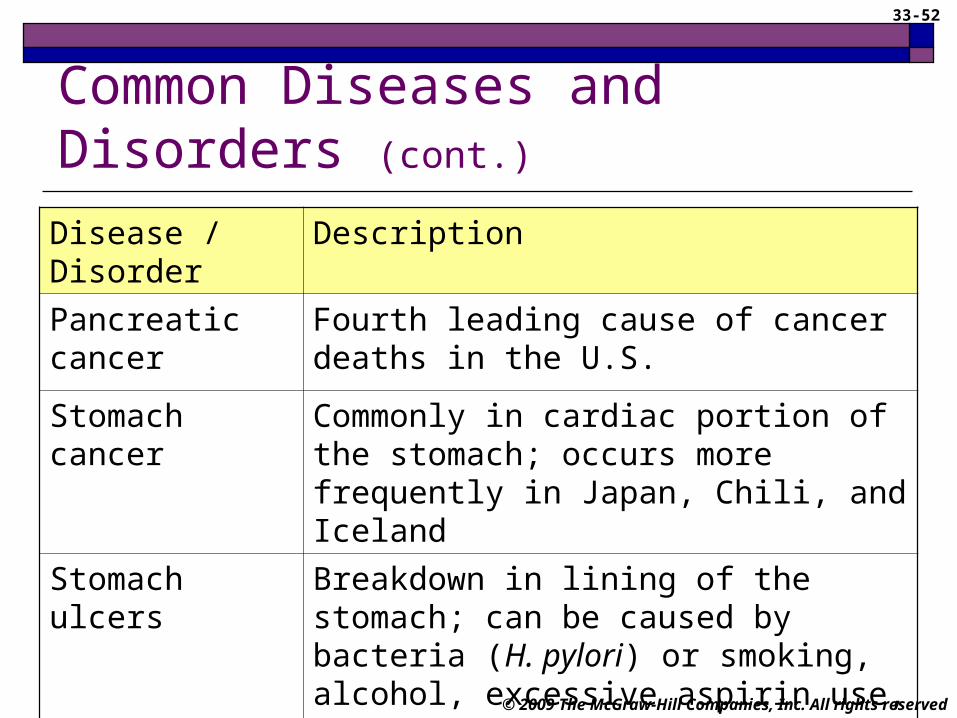
Common Diseases and Disorders (cont.)
Disease / Disorder Description
Pancreatic cancer Fourth leading cause of cancer deaths in the U.S.
Stomach cancer Commonly in cardiac portion of the stomach; occurs more frequently in Japan, Chili, and Iceland
Stomach ulcers Breakdown in lining of the stomach; can be caused by bacteria (H. pylori) or smoking, alcohol, excessive aspirin use, and hypersecretion of stomach acid
33-53
© 2009 The McGraw-Hill Companies, Inc. All rights reserved
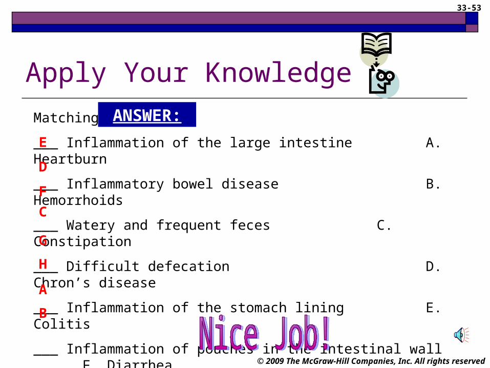
Apply Your Knowledge
Matching:
___ Inflammation of the large intestine A. Heartburn
___ Inflammatory bowel disease B. Hemorrhoids
___ Watery and frequent feces C. Constipation
___ Difficult defecation D. Chron’s disease
___ Inflammation of the stomach lining E. Colitis
___ Inflammation of pouches in the intestinal wall F. Diarrhea
___ GERD G. Gastritis
___ Varicose veins of rectum H. Diverticulitis
H
G
F
D
C
B
A
E
ANSWER:

33-54
© 2009 The McGraw-Hill Companies, Inc. All rights reserved
In Summary Purpose of the digestive system is to provide
nutrients to the body Organs of alimentary canal are responsible for
mechanical and chemical breakdown of food Accessory organs
Assist in breakdown of food Eliminate waste
Medical assistant must have knowledge of this system Assist with procedures Patient education

33-55
© 2009 The McGraw-Hill Companies, Inc. All rights reserved
End of Chapter
Take all that is given whether wealth, love or language; nothing
comes by mistake and with good digestion all
can be turned to health.
~ George Herbert
Related Documents