2007 McGraw-Hill Higher Education. All rights reserved. Chapter 8: Skeletal Muscle EXERCISE PHYSIOLOGY Theory and Application to Fitness and Performance, 6 th edition Scott K. Powers & Edward T. Howley

© 2007 McGraw-Hill Higher Education. All rights reserved. Chapter 8: Skeletal Muscle EXERCISE PHYSIOLOGY Theory and Application to Fitness and Performance,
Dec 22, 2015
Welcome message from author
This document is posted to help you gain knowledge. Please leave a comment to let me know what you think about it! Share it to your friends and learn new things together.
Transcript

© 2007 McGraw-Hill Higher Education. All rights reserved.
Chapter 8:Skeletal Muscle
EXERCISE PHYSIOLOGY
Theory and Application to Fitness and Performance, 6th edition
Scott K. Powers & Edward T. Howley

© 2007 McGraw-Hill Higher Education. All rights reserved.
Objectives
• Draw & label the microstructure of skeletal muscle
• Outline the steps leading to muscle shortening
• Define the concentric and isometric • Discuss: twitch, summation & tetanus• Discus the major biochemical and mechanical
properties of skeletal muscle fiber types

© 2007 McGraw-Hill Higher Education. All rights reserved.
Objectives
• Discuss the relationship between skeletal muscle fibers types and performance
• List & discuss those factors that regulate the amount of force exerted during muscular contraction
• Graph the relationship between movement velocity and the amount of force exerted during muscular contraction
• Discuss structure & function of muscle spindle• Describe the function of a Golgi tendon organ

© 2007 McGraw-Hill Higher Education. All rights reserved.
Skeletal Muscle• Human body contains over 400 skeletal muscles
– 40-50% of total body weight• Functions of skeletal muscle
– Force production for locomotion and breathing– Force production for postural support– Heat production during cold stress

© 2007 McGraw-Hill Higher Education. All rights reserved.
Connective Tissue Covering Skeletal Muscle
• Epimysium– Surrounds entire muscle
• Perimysium– Surrounds bundles of muscle fibers
• Fascicles• Endomysium
– Surrounds individual muscle fibers

© 2007 McGraw-Hill Higher Education. All rights reserved.
Connective Tissue
Covering Skeletal Muscle
Fig 8.1
© 2007 McGraw-Hill Higher Education. All rights reserved.
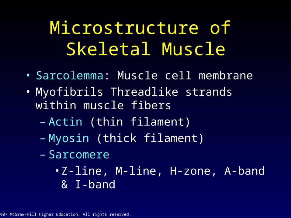
Microstructure of Skeletal Muscle
• Sarcolemma: Muscle cell membrane• Myofibrils Threadlike strands within muscle
fibers– Actin (thin filament)– Myosin (thick filament)– Sarcomere
• Z-line, M-line, H-zone, A-band & I-band

© 2007 McGraw-Hill Higher Education. All rights reserved.
Microstructure of Skeletal
Muscle
Fig 8.2

© 2007 McGraw-Hill Higher Education. All rights reserved.
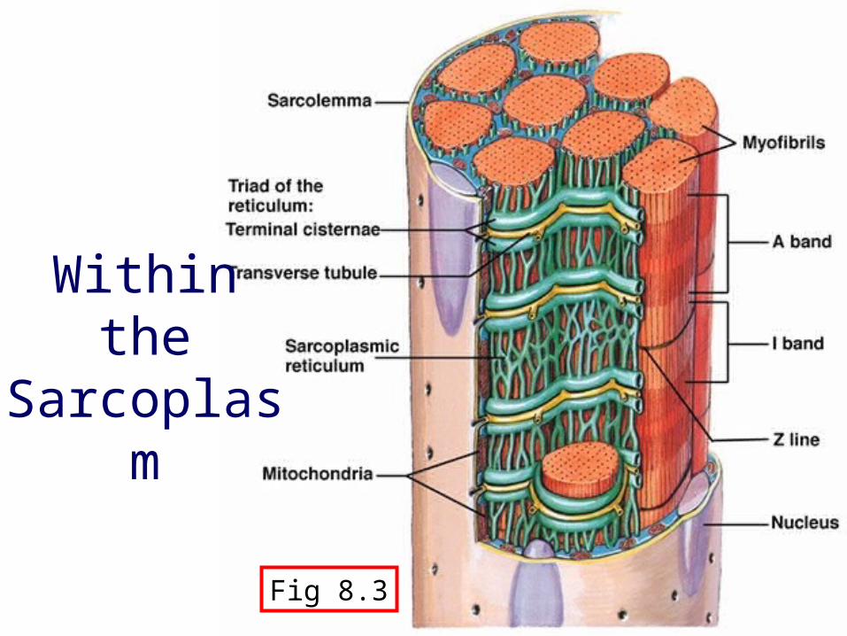
Microstructure of Skeletal Muscle
• Within the sarcoplasm– Sarcoplasmic reticulum
• Storage sites for calcium– Transverse tubules– Terminal cisternae– Mitochondria
© 2007 McGraw-Hill Higher Education. All rights reserved.
Within the Sarcoplasm
Fig 8.3

© 2007 McGraw-Hill Higher Education. All rights reserved.
The Neuromuscular Junction
• Where motor neuron meets the muscle fiber
• Motor end plate: pocket formed around motor neuron by sarcolemma
• Neuromuscular cleft: short gap • Ach is released from the motor neuron
– Causes an end-plate potential (EPP)• Depolarization of muscle fiber

© 2007 McGraw-Hill Higher Education. All rights reserved.
Neuromuscular Junction
Fig 8.4

© 2007 McGraw-Hill Higher Education. All rights reserved.
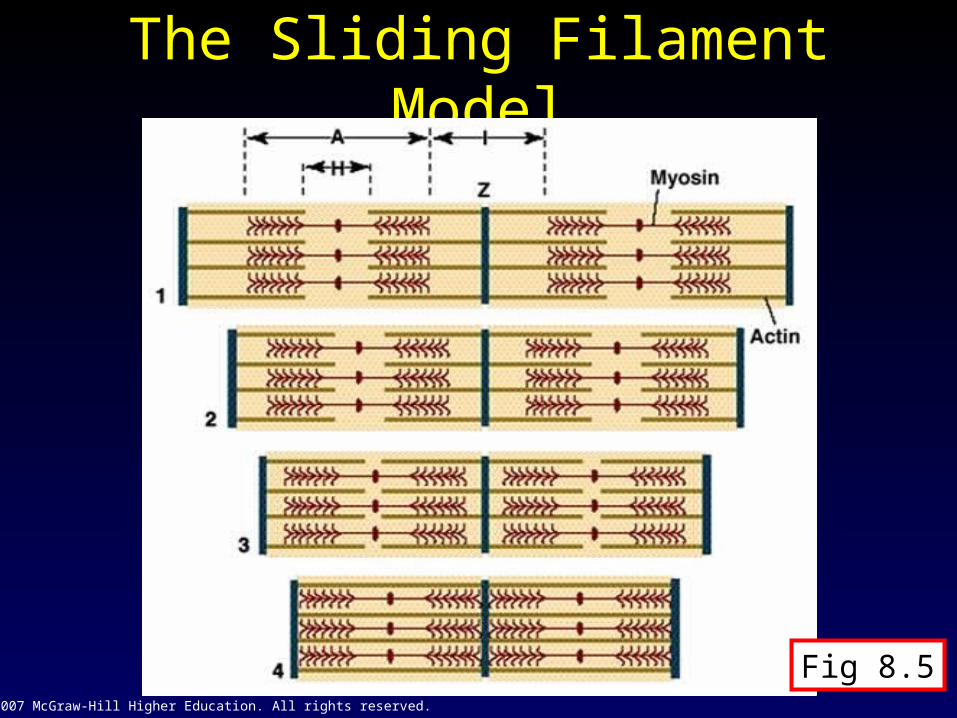
Muscular Contraction
• The sliding filament model– Muscle shortening occurs due to the
movement of the actin filament over the myosin filament
– Formation of cross-bridges between actin and myosin filaments “Power stroke”• 1 power stroke only shorten muscle 1%
– Reduction in the distance between Z-lines of the sarcomere
© 2007 McGraw-Hill Higher Education. All rights reserved.
The Sliding Filament Model
Fig 8.5

© 2007 McGraw-Hill Higher Education. All rights reserved.
Actin & Myosin Relationship
• Actin– Actin-binding site– Troponin with calcium binding site– Tropomyosin
• Myosin– Myosin head– Myosin tais

© 2007 McGraw-Hill Higher Education. All rights reserved.
Actin & Myosin Relationship
Fig 8.6

© 2007 McGraw-Hill Higher Education. All rights reserved.
Energy for Muscle Contraction
• ATP is required for muscle contraction– Myosin ATPase breaks down ATP as fiber
contracts• Sources of ATP
– Phosphocreatine (PC)– Glycolysis– Oxidative phosphorylation

© 2007 McGraw-Hill Higher Education. All rights reserved.
Sources of ATP for Muscle Contraction
Fig 8.7

© 2007 McGraw-Hill Higher Education. All rights reserved.
Excitation-Contraction Coupling
• Depolarization of motor end plate (excitation) is coupled to muscular contraction– Nerve impulse travels down T-tubules and
causes release of Ca++ from SR– Ca++ binds to troponin and causes position
change in tropomyosin, exposing active sites on actin
– Permits strong binding state between actin and myosin and contraction occurs

© 2007 McGraw-Hill Higher Education. All rights reserved.
Excitation-Contraction
Coupling
Fig 8.9

© 2007 McGraw-Hill Higher Education. All rights reserved.
Steps Leading to Muscular Contraction
Fig 8.10

© 2007 McGraw-Hill Higher Education. All rights reserved.
Properties of Muscle Fiber Types
• Biochemical properties– Oxidative capacity– Type of ATPase
• Contractile properties– Maximal force production– Speed of contraction– Muscle fiber efficiency
© 2007 McGraw-Hill Higher Education. All rights reserved.
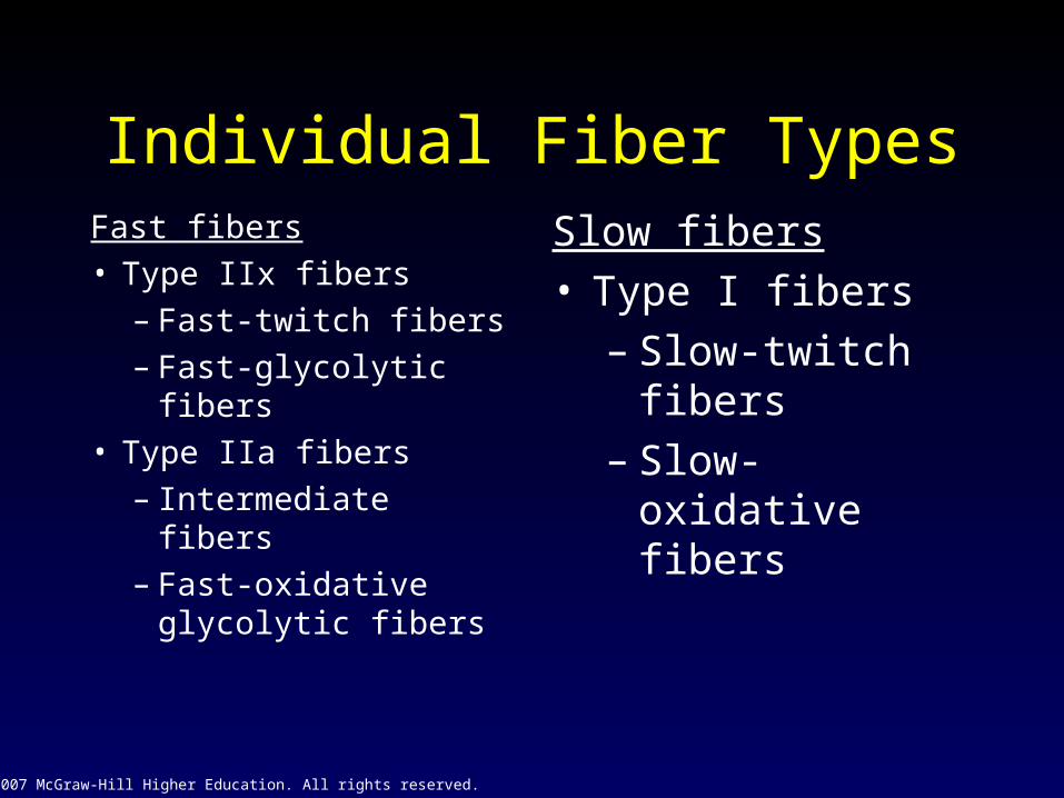
Individual Fiber TypesFast fibers• Type IIx fibers
– Fast-twitch fibers– Fast-glycolytic
fibers• Type IIa fibers
– Intermediate fibers– Fast-oxidative
glycolytic fibers
Slow fibers• Type I fibers
– Slow-twitch fibers– Slow-oxidative
fibers
© 2007 McGraw-Hill Higher Education. All rights reserved.
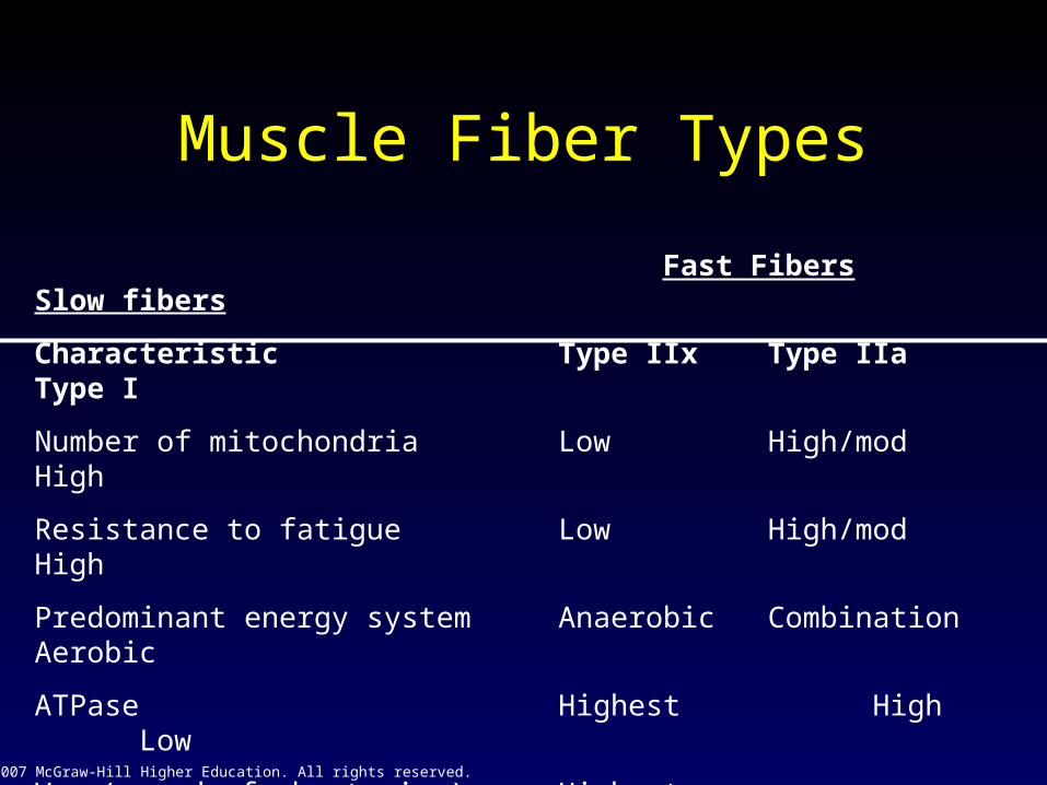
Muscle Fiber Types
Fast Fibers Slow fibers
Characteristic Type IIx Type IIa Type I
Number of mitochondria Low High/mod High
Resistance to fatigue Low High/mod High
Predominant energy system Anaerobic Combination Aerobic
ATPase Highest High Low
Vmax (speed of shortening) Highest Intermediate Low
Efficiency Low Moderate High
Specific tension High High Moderate
© 2007 McGraw-Hill Higher Education. All rights reserved.
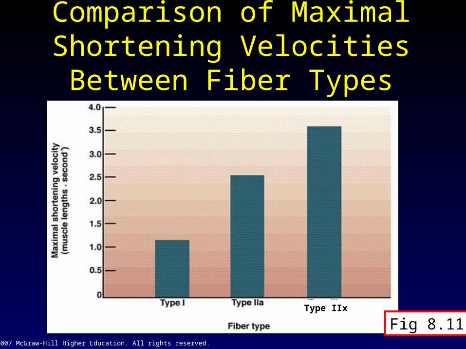
Comparison of Maximal Shortening Velocities Between
Fiber Types
Type IIx
Fig 8.11

© 2007 McGraw-Hill Higher Education. All rights reserved.
Histochemical Staining of Fiber Type
Type IIa
Type IIx
Type I
Fig 8.12

© 2007 McGraw-Hill Higher Education. All rights reserved.
Fiber Typing• Gel electrophoresis: myosin isoforms
– different weight move different distances
Table 10.2
+
_
Type IType IIAType IIx
1
1 – Marker
2
2 – Soleus
3
3 – Gastroc
4
4 – Quads
5
5 - Biceps
© 2007 McGraw-Hill Higher Education. All rights reserved.
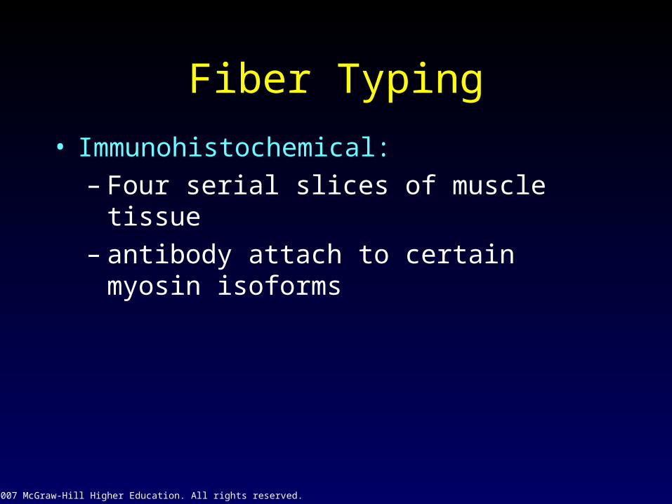
Fiber Typing
• Immunohistochemical: – Four serial slices of muscle tissue– antibody attach to certain myosin isoforms

© 2007 McGraw-Hill Higher Education. All rights reserved.
Muscle Tissue: Rat Diaphragm
Type 2x fibers - light/white antibody: BF-35
Type 2a fibers - dark antibody: SC-71
Type 2b fiber - dark Antibody: BF-F3
Type 1 fibers - darkantibody: BA-D5
© 2007 McGraw-Hill Higher Education. All rights reserved.
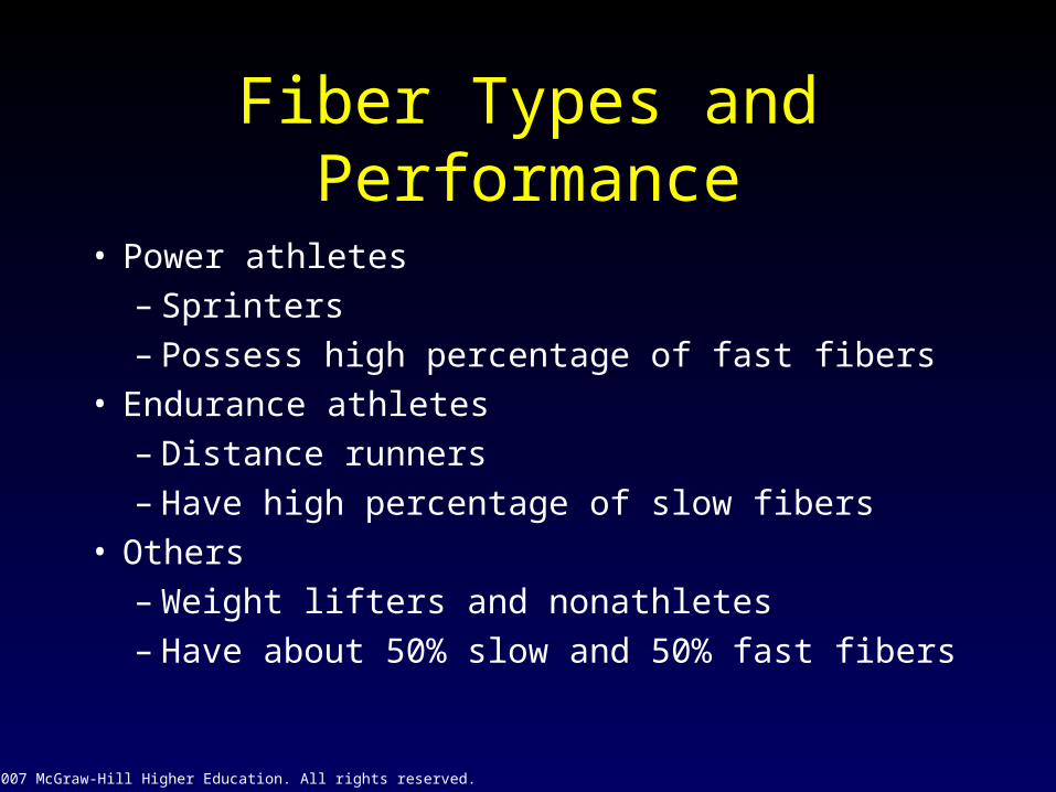
Fiber Types and Performance
• Power athletes – Sprinters– Possess high percentage of fast fibers
• Endurance athletes – Distance runners– Have high percentage of slow fibers
• Others– Weight lifters and nonathletes– Have about 50% slow and 50% fast fibers
© 2007 McGraw-Hill Higher Education. All rights reserved.
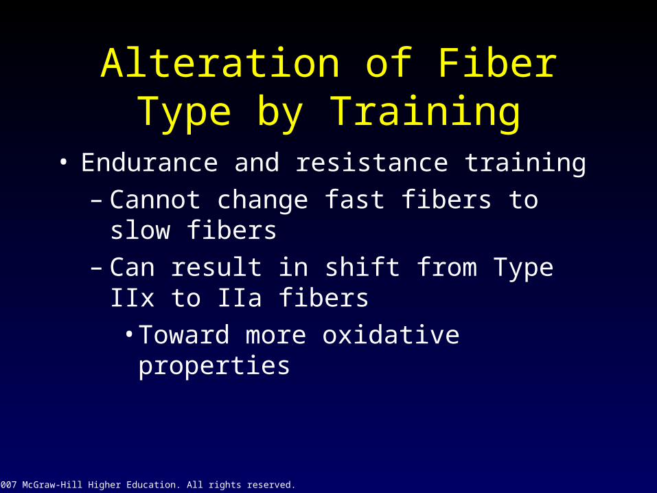
Alteration of Fiber Type by Training
• Endurance and resistance training– Cannot change fast fibers to slow fibers– Can result in shift from Type IIx to IIa fibers
• Toward more oxidative properties

© 2007 McGraw-Hill Higher Education. All rights reserved.
Training-Induced Changes in Muscle Fiber Type
Fig 8.13

© 2007 McGraw-Hill Higher Education. All rights reserved.
Age-Related Changes in Skeletal Muscle
• Aging is associated with a loss of muscle mass– Rate increases after 50 years of age
• Regular exercise training can improve strength and endurance– Cannot completely eliminate the age-
related loss in muscle mass

© 2007 McGraw-Hill Higher Education. All rights reserved.
Types of Muscle Contraction • Isometric
– Muscle exerts force without changing length– Pulling against immovable object– Postural muscles
• Isotonic (dynamic)– Concentric
• Muscle shortens during force production– Eccentric
• Muscle produces force but length increases

© 2007 McGraw-Hill Higher Education. All rights reserved.
Isotonic and Isometric Contractions
Fig 8.14

© 2007 McGraw-Hill Higher Education. All rights reserved.
Speed of Muscle Contraction and Relaxation
• Muscle twitch– Contraction as the result of a single stimulus– Latent period
• Lasting only ~5 ms– Contraction
• Tension is developed• 40 ms
– Relaxation• 50 ms

© 2007 McGraw-Hill Higher Education. All rights reserved.
Muscle Twitch
Fig 8.15
© 2007 McGraw-Hill Higher Education. All rights reserved.
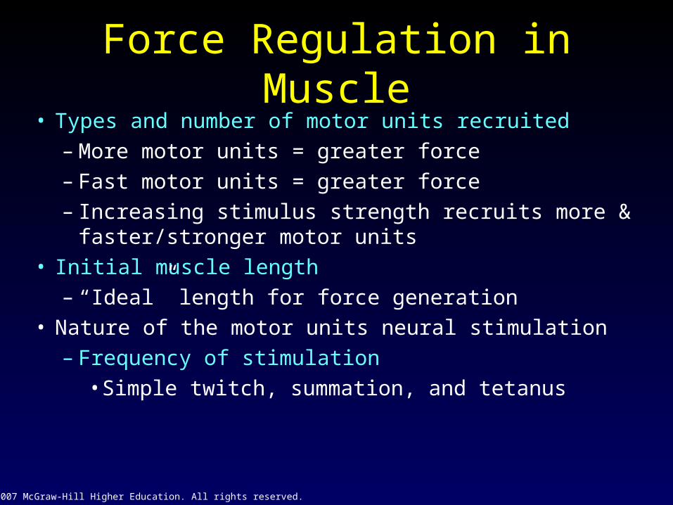
Force Regulation in Muscle• Types and number of motor units recruited
– More motor units = greater force– Fast motor units = greater force– Increasing stimulus strength recruits more &
faster/stronger motor units• Initial muscle length
– “Ideal” length for force generation• Nature of the motor units neural stimulation
– Frequency of stimulation• Simple twitch, summation, and tetanus

© 2007 McGraw-Hill Higher Education. All rights reserved.
Relationship Between Stimulus Frequency and
Force Generation
Fig 8.16

© 2007 McGraw-Hill Higher Education. All rights reserved.
Length-Tension
Relationship
Fig 8.17

© 2007 McGraw-Hill Higher Education. All rights reserved.
Simple Twitch, Summation, and Tetanus
Fig 8.18

© 2007 McGraw-Hill Higher Education. All rights reserved.
Force-Velocity Relationship
• At any absolute force the speed of movement is greater in muscle with higher percent of fast-twitch fibers
• The maximum velocity of shortening is greatest at the lowest force– True for both slow and fast-twitch fibers

© 2007 McGraw-Hill Higher Education. All rights reserved.
Force-Velocity Relationship
Fig 8.19

© 2007 McGraw-Hill Higher Education. All rights reserved.
Force-Power Relationship
• At any given velocity of movement the power generated is greater in a muscle with a higher percent of fast-twitch fibers
• The peak power increases with velocity up to movement speed of 200-300 degrees•second-1
– Force decreases with increasing movement speed beyond this velocity

© 2007 McGraw-Hill Higher Education. All rights reserved.
Force-Power Relationship
• At any given velocity of movement the power generated is greater in a muscle with a higher percent of fast-twitch fibers
• The peak power increases with velocity up to movement speed of 200-300 degrees/sec– Force decreases with increasing
movement speed beyond this velocity

© 2007 McGraw-Hill Higher Education. All rights reserved.
Force-Power Relationship
Fig 8.20

© 2007 McGraw-Hill Higher Education. All rights reserved.
Receptors in Muscle• Muscle spindle
– Changes in muscle length– Rate of change in muscle length – Intrafusal fiber contains actin & myosin, and
therefore has the ability to shorten– Gamma motor neuron stimulate muscle spindle to
shorten• Stretch reflex
– Stretch on muscle causes reflex contraction

© 2007 McGraw-Hill Higher Education. All rights reserved.
Muscle Spindle
Fig 8.21
© 2007 McGraw-Hill Higher Education. All rights reserved.
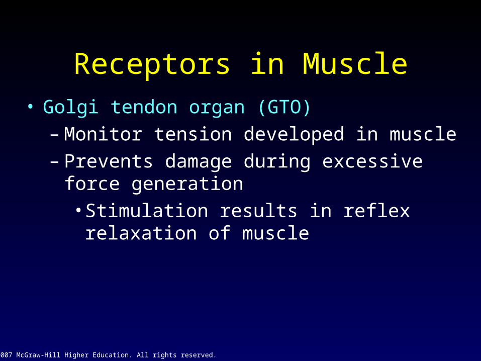
Receptors in Muscle• Golgi tendon organ (GTO)
– Monitor tension developed in muscle– Prevents damage during excessive force
generation• Stimulation results in reflex relaxation of
muscle

© 2007 McGraw-Hill Higher Education. All rights reserved.
Golgi Tendon Organ
Fig 8.22

© 2007 McGraw-Hill Higher Education. All rights reserved.
Chapter 8:Skeletal Muscle
Related Documents