1 ن المصريم قوات ا

Welcome message from author
This document is posted to help you gain knowledge. Please leave a comment to let me know what you think about it! Share it to your friends and learn new things together.
Transcript

1
قوات االمن المصري

2
Incubation periods Questions may either ask directly about incubation periods or they may be used to provide a clue in a differential diagnosis Less than 1 week
meningococcus diphtheria influenza scarlet fever
1 - 2 weeks
malaria dengue fever typhoid measles
2 - 3 weeks
mumps rubella chickenpox
Longer than 3 weeks
infectious mononucleosis cytomegalovirus viral hepatitis HIV
Congenital infections The major congenital infections encountered in examinations are rubella, toxoplasmosis
and cytomegalovirus Cytomegalovirus is the most common congenital infection in the UK. Maternal infection is usually asymptomatic
Rubella Toxoplasmosis Cytomegalovirus
Characteristic
features
1) Sensorineural deafness
2) Congenital cataracts
3) Glaucoma
4) Congenital heart disease
(e.g. patent ductus
arteriosus)
1) Cerebral calcification
2) Chorioretinitis
3) Hydrocephalus
1) Growth retardation
2) Purpuric skin lesions
Other features 1) Growth retardation
2) Hepatosplenomegaly
3) Purpuric skin lesions
'Salt and pepper'
4) Chorioretinitis
5) Microphthalmia
6) Cerebral palsy
1) Anaemia
2) Hepatosplenomegaly
3) Cerebral palsy
1) Sensorineural deafness
2) Encephalitis/seizures
3) Cerebral palsy
Pneumonitis
4) Hepatosplenomegaly
5) Anaemia
6) Jaundice

3
Bacterial Infections
Classification of bacteria Remember:
Gram positive cocci = staphylococci + streptococci (including enterococci)
Gram negative cocci = Neisseria meningitidis + Neisseria gonorrhea, also Moraxella
Therefore, only a small list of Gram positive rods (bacilli) need to be memorized to categorize
all bacteria - mnemonic = ABCD L
Actinomyces
Bacillus anthracis (anthrax)
Clostridium
Diphtheria: Corynebacterium diphtheriae
Listeria monocytogenes
Remaining organisms are Gram negative rods

4
Staphylococci Staphylococci are a common type of bacteria which are often found normal commensal
organisms but may also cause invasive disease.
Some basic facts include:
1) Gram-positive cocci
2) facultative anaerobes
3) produce catalase
The two main types of Staphylococci you need to know about are Staphylococcus
aureusand Staphylococcus epidermidis.
Staphylococcus aureus Staphylococcus epidermidis
• Coagulase-positive
• Causes skin infections (e.g. cellulitis),
abscesses, osteomyelitis, toxic shock
syndrome
• Coagulase-negative
• Cause of central line infections and
infective endocarditis
Osteomyelitis Osteomyelitis describes an infection of the bone.
Staph. aureus is the most common cause except in patients with sickle-cell anaemia
where Salmonella species predominate.
Predisposing conditions:
1) diabetes mellitus
2) sickle cell anaemia
3) intravenous drug user
4) immunosuppression due to either medication or HIV
5) alcohol excess
Investigations:
MRI is the imaging modality of choice, with a sensitivity of 90-100%
Management:
flucloxacillin for 6 weeks
clindamycin if penicillin-allergic
5
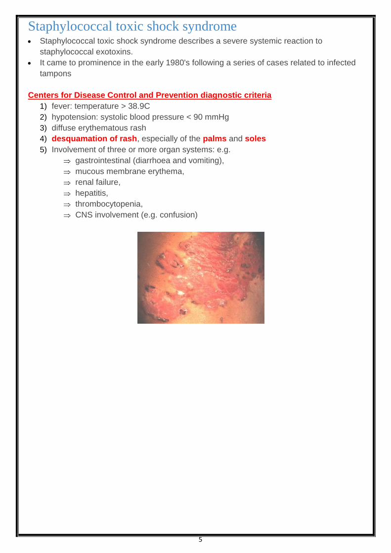
Staphylococcal toxic shock syndrome Staphylococcal toxic shock syndrome describes a severe systemic reaction to
staphylococcal exotoxins.
It came to prominence in the early 1980's following a series of cases related to infected
tampons
Centers for Disease Control and Prevention diagnostic criteria
1) fever: temperature > 38.9C
2) hypotension: systolic blood pressure < 90 mmHg
3) diffuse erythematous rash
4) desquamation of rash, especially of the palms and soles
5) Involvement of three or more organ systems: e.g.
gastrointestinal (diarrhoea and vomiting),
mucous membrane erythema,
renal failure,
hepatitis,
thrombocytopenia,
CNS involvement (e.g. confusion)

6
MRSA Methicillin-resistant Staphylococcus aureus (MRSA) was one of the first organisms which
highlighted the dangers of hospital-acquired infections.
Who should be screened for MRSA?
1) All patients awaiting elective admissions
Exceptions include:
day patients having terminations of pregnancy and ophthalmic surgery
Patients admitted to mental health trusts are also excluded
2) from 2011 all emergency admissions will be screened
How should a patient be screened for MRSA?
1) nasal swab and skin lesions or wounds
2) the swab should be wiped around the inside rim of a patient's nose for 5 seconds
3) the microbiology form must be labelled 'MRSA screen'
Suppression of MRSA from a carrier once identified
1) nose: mupirocin 2% in white soft paraffin, tds for 5 days
2) Skin:
chlorhexidine gluconate, od for 5 days
Apply all over but particularly to the axilla, groin and perineum
The following antibiotics are commonly used in the treatment of MRSA infections:
1) vancomycin
2) teicoplanin
3) linezolid
Some strains may be sensitive to the antibiotics listed below but they should not generally
be used alone because resistance may develop:
1) rifampicin
2) macrolides
3) tetracyclines
4) aminoglycosides
5) clindamycin
Relatively new antibiotics have activity against MRSA but should be reserved for resistant
cases such as:
1) linezolid,
2) quinupristin/dalfopristin combinations
3) tigecycline

7
Streptococci Streptococci are gram-positive cocci.
They may be divided into alpha and beta haemolytic types
Alpha haemolytic streptococci (partial haemolysis)
The most important alpha haemolytic Streptococcus is Streptococcus pneumonia
(pneumococcus).
Pneumococcus is a common cause of:
1) pneumonia,
2) meningitis and
3) otitis media
Another clinical example is Streptococcus viridans
Beta haemolytic streptococci (complete haemolysis)
These can be subdivided into groups A-H.
Only groups A, B & D are important in humans.
Group A
most important organism is Streptococcus pyogenes
responsible for:
1) erysipelas,
2) impetigo,
3) cellulitis,
4) type 2 necrotizing fasciitis and
5) pharyngitis/tonsillitis
immunological reactions can cause:
1) rheumatic fever or
2) post-streptococcal glomerulonephritis
erythrogenic toxins cause scarlet fever
Group B
Streptococcus agalactiae may lead to:
neonatal meningitis and septicaemia
Group D
Enterococcus

8
Gram Negative Cocci Gram negative cocci = Neisseria meningitidis + Neisseria gonorrhea, also Moraxella
Meningococcal septicaemia:
Investigations: Meningococcal septicaemia is a frightening condition for patients, parents and doctors.
It is associated with a high morbidity and mortality unless treated early –
Meningococcal disease is the leading infectious cause of death in early childhood.
A high index of suspicion is therefore needed.
Much of the following is based on the 2010 NICE guidelines (please see link).
Presentation of meningococcal disease:
15% - meningitis
25% - septicaemia
60% - a combination of meningitis and septicaemia
Investigations:
blood cultures
blood PCR
lumbar puncture is usually contraindicated
full blood count and clotting to assess for disseminated intravascular coagulation

9
Meningitis:
Causes 0 - 3 months
Group B Streptococcus (Streptococcus agalactiae) (most common cause in neonates)
E. coli
Listeria monocytogenes
3 months - 6 years
Neisseria meningitidis
Streptococcus pneumoniae (Alpha haemolytic streptococci)
Haemophilus influenzae
6 years - 60 years
Neisseria meningitidis
Streptococcus pneumoniae (Alpha haemolytic streptococci)
> 60 years
Streptococcus pneumoniae (Alpha haemolytic streptococci)
Neisseria meningitidis
Listeria monocytogenes
Immunosuppressed
Listeria monocytogenes
Investigations suggested by NICE
1) full blood count
2) CRP
3) coagulation screen
4) blood culture
5) whole-blood PCR
6) blood glucose
7) blood gas
Lumbar puncture if no signs of raised intracranial pressure

10
Meningitis: management Management
All patients should be transferred to hospital urgently.
If patients are in a pre-hospital setting (for example a GP surgery) and meningococcal
disease is suspected then intramuscular benzylpenicillin may be given, as long as this
doesn't delay transit to hospital.
BNF recommendations on antibiotics
Scenario BNF recommendation
Initial empirical therapy aged < 3 months Intravenous cefotaxime + amoxicillin
Initial empirical therapy aged > 50 years Intravenous cefotaxime + amoxicillin
Initial empirical therapy aged 3 months – 50yr Intravenous cefotaxime
Meningococcal meningitis Intravenous benzylpenicillin or cefotaxime
Pneuomococcal meningitis
Meningitis caused by Haemophilus influenza
Intravenous cefotaxime
Meningitis caused by Listeria Intravenous amoxicillin + gentamicin
If the patient has a history of immediate hypersensitivity reaction to penicillin or to
cephalosporins the BNF recommends using chloramphenicol.
Management of contacts:
1) prophylaxis needs to be offered to household and close contacts of patients affected with
meningococcal meningitis
2) Oral ciprofloxacin or rifampicin or may be used.
3) The Health Protection Agency (HPA) guidelines now state that whilst either may be used
ciprofloxacin is the drug of choice as it is widely available and only requires one dose
4) the risk is highest in the first 7 days but persists for at least 4 weeks
5) meningococcal vaccination should be offered to close contacts when serotype results
are available, including booster doses to those who had the vaccine in infancy
6) For pneumococcal meninigitis no prophylaxis is generally needed.
7) There are however exceptions to this. If a cluster of cases of pneumococcal meninigitis
occur the HPA have a protocol for offering close contacts antibiotic prophylaxis. Please see
the link for more details

11
Gram Negative Cocci Gram negative cocci = Neisseria meningitidis + Neisseria gonorrhea, also Moraxella
Gonorrhea Gonorrhea is caused by the Gram negative diplococcus Neisseria gonorrhea.
Acute infection can occur on any mucous membrane surface, typically genitourinary but
also rectum and pharynx.
The incubation period of gonorrhea is 2-5 days
Features:
1) males: urethral discharge, dysuria
2) females: cervicitis e.g. leading to vaginal discharge
3) rectal and pharyngeal infection is usually asymptomatic
4) Local complications that may develop include urethral strictures, epididymitis and
salpingitis (hence may lead to infertility).
5) Disseminated infection may occur - see below
Management:
1) Ciprofloxacin:
Used to be the treatment of choice.
However, there is increased resistance to ciprofloxacin and therefore
cephalosporins are now used
2) The 2011 British Society for Sexual Health and HIV (BASHH) guidelines recommend:
Ceftriaxone 500 mg intramuscularly as a single dose with azithromycin 1 g oral
as a single dose.
The azithromycin is thought to act synergistically with ceftriaxone and is also useful
for eradicating any co-existent Chlamydia infections
3) if ceftriaxone is refused or contraindicated other options include cefixime 400mg PO
(single dose)
Disseminated gonococcal infection (DGI) and gonococcal arthritis
Gonococcal infection being the most common cause of septic arthritis in young adults.
The pathophysiology of DGI is not fully understood
It is thought to be due to haematogenous spread from mucosal infection (e.g.
Asymptomatic genital infection).
Initially there may be a classic triad of symptoms: tenosynovitis, migratory polyarthritis and
dermatitis.
Later complications include septic arthritis, endocarditis and perihepatitis (Fitz-Hugh-Curtis
syndrome)
Key features of disseminated gonococcal infection
1) tenosynovitis
2) migratory polyarthritis
3) dermatitis (lesions can be maculopapular or vesicular)

12
Gram positive bacillus Anthrax Anthrax is caused by Bacillus anthracis, a Gram positive rod.
It is spread by infected carcasses. جثث
It is also known as Woolsorters' disease. الصوف فارزي
Bacillus anthracis produces a tripartite protein toxin
1) protective antigen
2) oedema factor: a bacterial adenylate cyclase which increases cAMP
3) lethal factor: toxic to macrophages
Features:
1) causes painless black eschar (cutaneous 'malignant pustule', but no pus)
2) typically painless and non-tender
3) may cause marked oedema
4) anthrax can cause gastrointestinal bleeding
Management:
1) the current Health Protection Agency advice for the initial management of cutaneous
anthrax is ciprofloxacin
2) further treatment is based on microbiological investigations and expert advice
Listeria low temperatures Listeria monocytogenes is a Gram positive bacillus
has the unusual ability to multiply at low temperatures
It is typically spread via contaminated food, typically unpasteurized dairy products.
Infection is particularly dangerous to the unborn child where it can lead to miscarriage
Features - can present in a variety of ways
1) diarrhoea, flu-like illness
2) pneumonia , meningoencephalitis
3) ataxia and seizures
Investigation:
1) Suspected Listeria infection should be investigated by taking blood cultures.
2) CSF may reveal a pleocytosis, with 'tumbling motility' on wet mounts
Management:
1) Listeria is sensitive to amoxicillin/ ampicillin (cephalosporins usually inadequate)
2) Listeria meningitis should be treated with IV amoxicillin/ampicillin + gentamicin

13
Diphtheria Diphtheria is caused by the Gram positive (rods) bacterium Corynebacterium diphtheriae
Pathophysiology:
releases an exotoxin encoded by a β-prophage
exotoxin inhibits protein synthesis by catalyzing ADP-ribosylation of elongation factor EF-2
Diphtheria toxin commonly causes a 'diphtheric membrane' on tonsils caused by necrotic
mucosal cells.
Systemic distribution may produce necrosis of myocardial, neural and renal tissue
Possible presentations:
1) recent visitors to Eastern Europe/Russia/Asia
2) sore throat with a 'diphtheric membrane' - see above
3) bulky cervical lymphadenopathy
4) neuritis e.g. cranial nerves
5) heart block

14
Exotoxins and endotoxins Exotoxins are secreted by bacteria where as
Endotoxins are only released following lysis of the cell.
Exotoxins: Exotoxins are generally released by Gram positive bacteria with the notable exceptions of
Vibrio cholerae and some strains of E. coli.
There may be classified into a number of different groups:
1) Superantigens (bridges the MHC class II protein on antigen-presenting cells with the T cell
receptor on the surface of T cells resulting in massive cytokine release)
Staphylococcus aureus exotoxins: lead to
acute gastroenteritis (enterotoxins),
toxic shock syndrome (TSST-1 superantigen) and
staphylococcal scalded skin syndrome (exfoliatin)
Streptococcus pyogenes: scarlet fever
2) AB toxins - ADP ribosylating
Diphtheria toxin:
inhibits elogation factor (EF-2) causing a 'diphtheric membrane' on tonsils caused
by necrotic mucosal cells
Systemic distribution may produce necrosis of myocardial, neural and renal tissue
Pseudomonas aeruginosa:
produces exotoxin A which also inhibits EF-2
cholera toxin
causes activation of adenylate cyclase (via Gs) leading to increases in cAMP levels,
which in turn leads to increased chloride secretion and reduced sodium absorption
pertussis exotoxin inhibits Gi leading to increases in cAMP levels
Escherichia coli
heat labile: activates adenylate cyclase (via Gs), increasing cAMP → watery diarrhoea
heat stabile: activates guanylate cyclase, increasing cGMP → watery diarrhoea
Bacillus anthracis
produces oedema factor, a bacterial adenylate cyclase which increases cAMP
Clostridium tetani neurotoxin
Tetanospasmin which blocks the release of GABA and glycine.
Causes Lockjaw
Clostridium perfringens
produces α-toxin, a lecithinase, which causes gas gangrene (myonecrosis) and
haemolysis.
Clostridium botulinum
produces an exotoxin that blocks acetylcholine (ACh) release leading to flaccid
paralysis
Shigella dysenteriae
produces Shiga toxin which inactivates 60S ribosome

15
Endotoxins
Endotoxins are lipopolysaccharides that are released from Gram-negative bacteria such
as Neisseria meningitidis.
Tetanus Tetanus is caused by the tetanospasmin exotoxin released from Clostridium tetani (gram
positive rods).
Tetanus spores are present in soil and may be introduced into the body from a wound,
which is often unnoticed.
Tetanospasmin prevents release of GABA
Features
1) prodrome fever, lethargy, headache
2) trismus (lockjaw)
3) risus sardonicus
4) opisthotonus (arched back, hyperextended neck)
5) spasms (e.g. dysphagia)
Management:
1) supportive therapy including ventilatory support and muscle relaxants
2) intramuscular human tetanus immunoglobulin for high-risk wounds (e.g. compound
fractures, delayed surgical intervention, significant degree of devitalised tissue)
3) metronidazole is now preferred to benzylpenicillin as the antibiotic of choice
4) If vaccination history is incomplete or unknown then a dose of tetanus vaccine should be
given combined with intramuscular human tetanus immunoglobulin for high-risk wounds
Tetanus vaccination: The tetanus vaccine is a cell-free purified toxin that is normally given as part of a combined
vaccine.
Tetanus vaccine is currently given in the UK as part of the routine immunisation schedule
at:
2 months
3 months
4 months
3-5 years
13-18 years
This therefore provides 5 doses of tetanus-containing vaccine.
Five doses is now considered to provide adequate long-term protection against tetanus.
Intramuscular human tetanus immunoglobulin should be given to patients with high-risk
wounds (e.g. Compound fractures, delayed surgical intervention, significant degree of
devitalised tissue) irrespective of whether 5 doses of tetanus vaccine have previously been
given
If vaccination history is incomplete or unknown then a dose of tetanus vaccine should be
given combined with intramuscular human tetanus immunoglobulin for high-risk wounds

16
Gram Negative Rods
Escherichia coli Escherichia coli is a facultative anaerobic, lactose-fermenting, Gram negative rod which is a
normal gut commensal.
E. coli infections lead to a variety of diseases in humans including:
1) diarrhoeal illnesses
2) UTIs
3) neonatal meningitis
Serotypes
E. coli may be classified according to the antigens which may trigger an immune response:
Antigen Origin Notes
O Lipopolysaccharide layer
K Capsule Neonatal meningitis secondary to E. coli is usually
caused by a serotype that contains the capsular
antigen K-1
H Flagellin
E. coli O157:H7 is a particular strain associated with severe, haemorrhagic, watery
diarrhoea.
It has a high mortality rate and can be complicated by haemolytic uraemic syndrome.
It is often spread by contaminated ground beef.

17
Salmonella The Salmonella group contains many members, most of which cause diarrhoeal diseases.
They are aerobic, Gram negative rods which are not normally present as commensals in
the gut.
Typhoid and paratyphoid are caused by Salmonella typhi and Salmonella paratyphi (types
A, B & C) respectively.
They are often termed enteric fevers, producing systemic symptoms such as headache,
fever, arthralgia
Features:
1) initially systemic upset as above
2) relative bradycardia
3) abdominal pain, distension
4) constipation: although Salmonella is a recognised cause of diarrhoea, constipation is
more common in typhoid
5) rose spots: present on the trunk in 40% of patients, and are more common in
paratyphoid
Possible complications include
1) osteomyelitis (especially in sickle cell disease where Salmonella is one of the most
common pathogens)
2) GI bleed/perforation
3) meningitis
4) cholecystitis
5) chronic carriage (1%, more likely if adult females)
Shigella Overview
causes bloody diarrhoea, abdo pain
severity depends on type: S sonnei (e.g. from UK) may be mild, S flexneri or S
dysenteriae from abroad may cause severe disease
treat with ciprofloxacin
Cat scratch disease Cat scratch disease is generally caused by the Gram negative rod Bartonella henselae
Features:
1) fever
2) history of a cat scratch
3) regional lymphadenopathy
4) headache, malaise

18
Cholera Overview
caused by Vibro cholerae - Gram negative bacteria
Features:
1) profuse 'rice water' diarrhoea
2) dehydration
3) hypoglycaemia
Management
1) oral rehydration therapy
2) antibiotics: doxycycline, ciprofloxacin

19
Brucellosis Brucellosis is a zoonosis more common in the Middle East and in farmers.
Four major species cause infection in humans: B melitensis (sheep), B abortus (cattle), B
canis and B suis (pigs).
Brucellosis has an incubation period 2 - 6 weeks
Features
1) non-specific: fever, malaise
2) hepatosplenomegaly
3) sacroilitis: spinal tenderness may be seen
4) complications: osteomyelitis, infective endocarditis, meningoencephalitis, orchitis
5) leukopenia often seen
Diagnosis
1) the Rose Bengal plate test can be used for screening but other tests are required to
confirm the diagnosis
2) Brucella serology is the best test for diagnosis
3) blood and bone marrow cultures may be suitable in certain patients, but these tests are
often negative
Management:
doxycycline and streptomycin

20
Malaria Falciparum Feature of severe malaria:
1) schizonts on a blood film
2) parasitaemia > 2%
3) hypoglycaemia
4) temperature > 39 C
5) severe anaemia
6) complications as below
Complications:
1) hypoglycaemia
2) cerebral malaria: seizures, coma
3) acute renal failure: blackwater fever, secondary to intravascular haemolysis, mechanism
unknown
4) acute respiratory distress syndrome (ARDS)
5) disseminated intravascular coagulation (DIC)
Treatment:
A) Uncomplicated falciparum malaria:
1) strains resistant to chloroquine are prevalent in certain areas of Asia and Africa
2) the 2010 WHO guidelines recommend artemisinin-based combination therapies
(ACTs) as first-line therapy
Examples include:
artemether plus lumefantrine,
artesunate plus amodiaquine,
artesunate plus mefloquine,
artesunate plus sulfadoxine-pyrimethamine,
dihydroartemisinin plus piperaquine
B) Severe falciparum malaria:
1) If parasite counts of more than 2%:
will usually need parenteral treatment irrespective of clinical state
intravenous artesunate is now recommended by WHO in preference to
intravenous quinine
2) If parasite count > 10%:
exchange transfusion should be considered
3) shock may indicate coexistent bacterial septicaemia - malaria rarely causes
haemodynamic collapse

21
Non-falciparum Malaria: The most common cause of non-falciparum malaria is Plasmodium vivax, with Plasmodium
ovale and Plasmodium malariae accounting for the other cases.
Plasmodium vivax is often found in Central America and the Indian Subcontinent
whilst Plasmodium ovale typically comes from Africa
Features:
general features of malaria: fever, headache, splenomegaly
Plasmodium vivax/ovale: cyclical fever every 48 hours.
Plasmodium malariae: cyclical fever every 72 hours
Plasmodium malariae: is associated with nephrotic syndrome
Ovale and vivax malaria have a hypnozoite stage and may therefore relapse following
treatment.
Treatment:
1) non-falciparum malarias are almost always chloroquine sensitive
2) patients with ovale or vivax malaria should be given primaquine following acute treatment
with chloroquine to destroy liver hypnozoites and prevent relapse
Malaria prophylaxis: There are around 1,500-2,000 cases each year of malaria in patients returning from
endemic countries.
The majority of these cases (around 75%) are caused by the potentially
fatal Plasmodium falciparum protozoa.
The majority of patients who develop malaria did not take prophylaxis.
It should also be remembered that UK citizens who originate from malaria endemic areas
quickly lose their innate immunity. Up-to-date charts with recommended regimes for malarial zones should be consulted prior to prescribing
Drug Side-effects + notes
Time to begin
before travel
Time to end
after travel
Atovaquone + proguanil
(Malarone)
GI upset 1 - 2 days 7 days
Chloroquine 1) Headache
2) Contraindicated in epilepsy
3) Taken weekly
1 week 4 weeks
Doxycycline 1) Photosensitivity
2) Oesophagitis
1 - 2 days 4 weeks
Mefloquine (Lariam) 1) Dizziness
2) Neuropsychiatric disturbance
3) Contraindicated in epilepsy
4) Taken weekly
2 - 3 weeks 4 weeks
Proguanil (Paludrine) 1 week 4 weeks
Proguanil + chloroquine See above 1 week 4 weeks

22
Pregnant women:
should be advised to avoid travelling to regions where malaria is endemic
Diagnosis can also be difficult as parasites may not be detectable in the blood film due to
placental sequestration.
However, if travel cannot be avoided:
1) chloroquine can be taken
2) proguanil: folate supplementation (5mg od) should be given
3) Malarone (atovaquone + proguanil): the BNF advises to avoid these drugs unless
essential.
4) If taken then folate supplementation should be given
5) mefloquine: caution advised
6) doxycycline is contraindicated
It is again advisable to avoid travel to malaria endemic regions with children if avoidable.
However, if travel is essential then children should take malarial prophylaxis as they are
more at risk of serious complications.
1) diethyltoluamide (DEET) 20-50% can be used in children over 2 months of age
2) doxycycline is only licensed in the UK for children over the age of 12 years
23
Leptospirosis Also known as Weil's disease*,
Leptospirosis is commonly seen in questions referring to sewage workers الصحي الصرفعمال ,
farmers, vets البيطريون االطباء or people who work in abattoir مجزر مسلخ .
It is caused by the spirochaete Leptospira interrogans (serogroup L icterohaemorrhagiae),
Classically being spread by contact with infected rat urine.
Weil's disease should always be considered in high-risk patients with hepatorenal failure
Features:
1) fever
2) flu-like symptoms
3) renal failure (seen in 50% of patients)
4) jaundice
5) subconjunctival haemorrhage
6) headache, may herald ينذر the onset of meningitis
Management
high-dose benzylpenicillin or doxycycline
*the term Weil's disease is sometimes reserved for the most severe 10% of cases that are
associated with jaundice
Q fever Q fever is caused by Coxiella burnetii, a rickettsia.
The source of infection is typically an abattoir, cattle/sheep or it may be inhaled from
infected dust
Features:
1) typically prodrome: fever, malaise
2) causes pyrexia of unknown origin,
3) atypical pneumonia,
4) endocarditis (culture negative)
Management:
Doxycycline

24
Lyme disease Lyme disease is caused by the spirochaete Borrelia burgdorferi and is spread by ticks
Features: Early features:
1) Erythema chronicum migrans
Small papule often at site of the tick bite
which develops into a larger annular lesion with central clearing, 'bulls-eye'
Occurs in 70% of patients
2) systemic symptoms: malaise, fever, arthralgia
Later features:
1) CVS: heart block, myocarditis
2) neurological: cranial nerve palsies, meningitis
3) polyarthritis
Investigation
serology: antibodies to Borrelia burgdorferi
Management
1) Doxycycline if early disease.
2) Amoxicillin is an alternative if doxycycline is contraindicated (e.g. pregnancy)
3) ceftriaxone if disseminated disease
4) Jarisch-Herxheimer reaction: (also seen in syphilis)
sometimes seen after initiating therapy
fever, rash, tachycardia after first dose of antibiotic
more commonly seen in syphilis, another spirochaetal disease

25
Tuberculosis Diagnosis: 1) In adults induction of sputum or bronchoscopy and lavage may be used in patients who
cannot produce sputum
2) In children who are unable to cough up sputum, the gold standard is gastric washings
for M tuberculosis culture
Tuberculosis drug therapy: 1) The standard therapy for treating active tuberculosis is:
A) Initial phase - first 2 months (RIPE)
1) Rifampicin
2) Isoniazid
3) Pyrazinamide
4) Ethambutol (the 2006 NICE guidelines now recommend giving a 'fourth drug' such as
ethambutol routinely - previously this was only added if drug-resistant tuberculosis
was suspected)
B) Continuation phase - next 4 months
1) Rifampicin
2) Isoniazid
2) The treatment for latent tuberculosis is isoniazid alone for 6 months
3) Patients with meningeal tuberculosis:
treated for a prolonged period (at least 12 months)
with the addition of steroids
4) Directly observed therapy:
a three times a week dosing regimen
May be indicated in certain groups, including:
1) homeless people with active tuberculosis
2) patients who are likely to have poor concordance
3) all prisoners with active or latent tuberculosis

26
Tuberculosis: drug side-effects and mechanism of action Rifampicin
1) mechanism of action: inhibits bacterial DNA dependent RNA polymerase
preventing transcription of DNA into mRNA
2) potent liver enzyme inducer
3) hepatitis, orange secretions
4) flu-like symptoms
Isoniazid
1) mechanism of action: inhibits mycolic acid synthesis
2) peripheral neuropathy: prevent with pyridoxine (Vitamin B6)
3) hepatitis, agranulocytosis
4) liver enzyme inhibitor
Pyrazinamide
1) mechanism of action: converted by pyrazinamidase into pyrazinoic acid which in
turn inhibits fatty acid synthase (FAS) I
2) hyperuricaemia causing gout
3) arthralgia, myalgia
4) hepatitis
Ethambutol
1) mechanism of action: inhibits the enzyme arabinosyl transferase which
polymerizes arabinose into arabinan
2) optic neuritis: check visual acuity before and during treatment
3) dose needs adjusting in patients with renal impairment

27
Tuberculosis: screening The Mantoux test is the main technique used to screen for latent tuberculosis.
In recent years the interferon-gamma blood test has also been introduced.
It is used in a number of specific situations such as:
the Mantoux test is positive or equivocal
people where a tuberculin test may be falsely negative (see below)
Mantoux test
0.1 ml of 1:1,000 purified protein derivative (PPD) injected intradermally
result read 2-3 days later
Diameter of
induration Positivity Interpretation
< 6mm Negative
no significant hypersensitivity to
tuberculin protein
Previously unvaccinated individuals
may be given the BCG
6 - 15mm Positive
hypersensitive to tuberculin protein
Should not be given BCG.
May be due to previous TB infection
or BCG
> 15mm Strongly positive
strongly hypersensitive to
tuberculin protein
Suggests tuberculosis infection.
False negative tests may be caused by:
1) miliary TB
2) sarcoidosis
3) HIV
4) lymphoma
5) very young age (e.g. < 6 months)
Heaf test
The Heaf test was previously used in the UK but has been since been discontinued. It involved
injection of PPD equivalent to 100,000 units per ml to the skin over the flexor surface of the left
forearm. It was then read 3-10 days later.

28
Intracellular
Chlamydia Chlamydia is the most prevalent sexually transmitted infection in the UK
Caused by Chlamydia trachomatis, an obligate intracellular pathogen.
Approximately 1 in 10 young women in the UK have Chlamydia.
The incubation period is around 7-21 days,
a large percentage of cases are asymptomatic
Features:
1) asymptomatic in around 70% of women and 50% of men
2) women: cervicitis (discharge, bleeding), dysuria
3) men: urethral discharge, dysuria
Potential complications:
1) epididymitis
2) pelvic inflammatory disease
3) endometritis
4) increased incidence of ectopic pregnancies
5) infertility
6) reactive arthritis
7) perihepatitis (Fitz-Hugh-Curtis syndrome)
Investigation:
1) traditional cell culture is no longer widely used
2) nuclear acid amplification tests (NAATs) are now rapidly emerging as the investigation of
choice
3) urine (first void urine sample), vulvovaginal swab or cervical swab may be tested using the
NAAT technique
Screening:
in England the National Chlamydia Screening Programme is open to all men and
women aged 15-24 years
the 2009 SIGN guidelines support this approach, suggesting screening all sexually
active patients aged 15-24 years
relies heavily on opportunistic testing
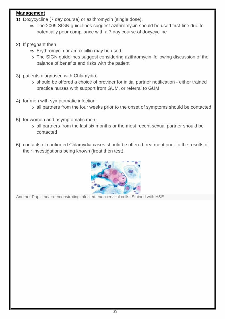
Pap smear demonstrating infected endocervical cells. Red inclusion bodies are typical
29
Management
1) Doxycycline (7 day course) or azithromycin (single dose).
The 2009 SIGN guidelines suggest azithromycin should be used first-line due to
potentially poor compliance with a 7 day course of doxycycline
2) If pregnant then
Erythromycin or amoxicillin may be used.
The SIGN guidelines suggest considering azithromycin 'following discussion of the
balance of benefits and risks with the patient'
3) patients diagnosed with Chlamydia:
should be offered a choice of provider for initial partner notification - either trained
practice nurses with support from GUM, or referral to GUM
4) for men with symptomatic infection:
all partners from the four weeks prior to the onset of symptoms should be contacted
5) for women and asymptomatic men:
all partners from the last six months or the most recent sexual partner should be
contacted
6) contacts of confirmed Chlamydia cases should be offered treatment prior to the results of
their investigations being known (treat then test)
Another Pap smear demonstrating infected endocervical cells. Stained with H&E

30
Legionella Legionnaire's disease is caused by the intracellular bacterium "Legionella pneumophilia".
It is typically colonizes water tanks and hence questions may hint at air-conditioning systems or
foreign holidays.
Person-to-person transmission is not seen
Features:
1) flu-like symptoms including fever (present in > 95% of patients)
2) dry cough
3) relative bradycardia
4) confusion
5) lymphopaenia
6) hyponatraemia
7) deranged liver function tests
8) pleural effusion: seen in around 30% of ptients
Diagnosis:
urinary antigen
Management:
treat with erythromycin

31
Viral Infections
Herpes simplex virus There are two strains of the herpes simplex virus (HSV) in humans: HSV-1 and HSV-2.
Whilst it was previously thought HSV-1 accounted for oral lesions (cold sores) and HSV-2
for genital herpes it is now known there is considerable overlap
Features:
1) primary infection: may present with a severe gingivostomatitis
2) cold sores
3) painful genital ulceration
Management:
1) gingivostomatitis:
oral aciclovir,
chlorhexidine mouthwash
2) Cold sores: topical aciclovir although the evidence base for this is modest
3) Genital herpes:
oral aciclovir
Some patients with frequent exacerbations may benefit from longer
term aciclovir
Pap smear. Multinucleated giant cells representing infection by the herpes simplex virus. Note the 3 M's; Multinucleation, Margination of the chromatin, Molding of the nuclei
Further Pap smear showing the cytopathic effect of HSV (multi-nucleation, ground glass & marginated chromatin)

32
Chickenpox Chickenpox is caused by primary infection with varicella zoster virus.
Shingles is reactivation of dormant virus in dorsal root ganglion
Chickenpox is highly infectious:
spread via the respiratory route
can be caught from someone with shingles
infectivity = 4 days before rash, until 5 days after the rash first appeared*
incubation period = 10-21 days
*it was traditionally taught that patients were infective until all lesions had scabbed over
Clinical features: (tend to be more severe in older children/adults)
1) fever initially
2) Itchy rash:
starting on head/trunk before spreading
Initially macular then papular then vesicular
3) systemic upset is usually mild
Management: is supportive
1) keep cool, trim nails
2) calamine lotion
3) School exclusion:
current HPA advice is 5 days from start of skin eruption
They also state 'Traditionally children have been excluded until all lesions are
crusted. However, transmission has never been reported beyond the fifth day of the
rash.'
4) Immunocompromised patients and newborns with peripartum exposure:
should receive varicella zoster immunoglobulin (VZIG)
If chickenpox develops then IV aciclovir should be considered
5) A common complication is secondary bacterial infection of the lesions. Rare complications
include:
pneumonia
encephalitis (cerebellar involvement may be seen)
disseminated haemorrhagic chickenpox
arthritis, nephritis and pancreatitis may very rarely be seen

33
Chickenpox exposure in pregnancy In pregnancy there is a risk to both the mother and also the fetus, a syndrome now termed
fetal varicella syndrome
Fetal varicella syndrome (FVS):
risk of FVS following maternal varicella exposure is around 1% if occurs before 20 weeks
gestation
studies have shown a very small number of cases occurring between 20-28 weeks
gestation and none following 28 weeks
Features of FVS include:
1) Skin scarring,
2) eye defects (microphthalmia),
3) limb hypoplasia,
4) microcephaly and
5) learning disabilities
Management of chickenpox exposure
1) if there is any doubt about the mother previously having chickenpox maternal blood should
be checked for varicella antibodies
2) If the pregnant woman is not immune to varicella:
She should be given varicella zoster immunoglobulin (VZIG) as soon as possible.
RCOG and Greenbook guidelines suggest VZIG is effective up to 10 days post
exposure
3) consensus guidelines suggest oral aciclovir should be given if pregnant women with
chickenpox present within 24 hours of onset of the rash
Ramsay Hunt syndrome
Ramsay Hunt syndrome (herpes zoster oticus) is caused by the reactivation of the varicella
zoster virus in the geniculate ganglion of the seventh cranial nerve.
Features
auricular pain is often the first feature
facial nerve palsy
vesicular rash around the ear
other features include vertigo and tinnitus
Management
oral aciclovir and corticosteroids are usually given

34
Reye's syndrome:
Reye's syndrome is a severe, progressive encephalopathy affecting children that is
accompanied by fatty infiltration of the liver, kidneys and pancreas.
The aetiology of Reye's syndrome is not fully understood although there is a known
association with aspirin use and a viral cause has been postulated
The peak incidence is 2 years of age, features include:
1) may be history of preceding viral illness
2) encephalopathy: confusion, seizures, cerebral oedema, coma
3) fatty infiltration of the liver, kidneys and pancreas
4) hypoglycaemia
Management is supportive
Although the prognosis has improved over recent years there is still a mortality rate of 15-25%.
35
H1N1 influenza pandemic الخنازير انفلونزا وباء The 2009 H1N1 influenza (swine flu) outbreak was first observed in Mexico in early 2009.
In June 2009, the WHO declared the outbreak to be a pandemic.
H1N1:
The H1N1 virus is a subtype of the influenza A virus and the most common cause of
flu in humans.
The 2009 pandemic was caused by a new strain of the H1N1 virus.
The following groups are particularly at risk:
1) patients with chronic illnesses and those on immunosuppressants
2) pregnant women
3) young children under 5 years old
Features:
The majority of symptoms are typical of those seen in a flu-like illness:
1) fever greater than 38C
2) myalgia
3) lethargy
4) headache
5) rhinitis
6) sore throat
7) cough
8) diarrhoea and vomiting
A minority of patients may go on to develop an acute respiratory distress syndrome which
may require ventilatory support.
Treatment:
There are two main treatments currently available:
A) Oseltamivir (Tamiflu):
1) oral medication
2) a neuraminidase inhibitor which prevents new viral particles from being released by
infected cells
3) common side-effects include:
1) nausea, vomiting, diarrhoea
2) headaches
B) Zanamivir (Relenza):
1) inhaled medication
2) intravenous preparations are available for patients who are acutely unwell
3) also a neuraminidase inhibitor
4) may induce bronchospasm in asthmatics

36
Cytomegalovirus Cytomegalovirus (CMV) is one of the herpes viruses.
It is thought that around 50% of people have been exposed to the CMV virus although it
only usually causes disease in the immunocompromised, for example people with HIV or
those on immunosuppressants following organ transplantation.
Pathophysiology
infected cells have a 'Owl's eye' appearance due to intranuclear inclusion bodies
Patterns of disease
A) Congenital CMV infection
features include:
1) growth retardation,
2) pinpoint petechial 'blueberry muffin' skin lesions,
3) microcephaly,
4) sensorineural deafness,
5) encephalitis (seizures)
6) hepatosplenomegaly
B) CMV mononucleosis
infectious mononucelosis-like illness
may develop in immunocompetent individuals
C) CMV retinitis
common in HIV patients with a low CD4 count (< 50)
Presents with visual impairment e.g. 'blurred vision'.
Fundoscopy shows retinal haemorrhages and necrosis, often called 'pizza' retina
IV ganciclovir is the treatment of choice
D) CMV encephalopathy
seen in patients with HIV who have low CD4 counts
E) CMV pneumonitis
F) CMV colitis

37
Measles Overview
RNA paramyxovirus
spread by droplets
infective from prodrome until 4 days after rash starts
incubation period = 10-14 days
Features:
1) prodrome: irritable, conjunctivitis, fever
2) Koplik spots (before rash): white spots ('grain of salt') on buccal mucosa
3) rash: starts behind ears then to whole body, discrete maculopapular rash becoming
blotchy & confluent
Koplik spots
Complications:
1) encephalitis: typically occurs 1-2 weeks following the onset of the illness
2) subacute sclerosing panencephalitis: very rare, may present 5-10 years following the
illness
3) febrile convulsions
4) giant cell pneumonia
5) keratoconjunctivitis, corneal ulceration
6) diarrhoea
7) increased incidence of appendicitis
8) myocarditis
The rash typically starts behind the ears and then spreads to the whole body
Management of contacts
A) if a child not immunized against measles comes into contact with measles:
MMR should be offered
(Vaccine-induced measles antibody develops more rapidly than that following natural
infection)
this should be given within 72 hours

38
Infectious mononucleosis Infectious mononucleosis (glandular fever)
Caused by the Epstein-Barr virus (also known as human herpes virus 4, HHV-4).
It is most common in adolescents and young adults.
Features:
1) sore throat
2) lymphadenopathy
3) pyrexia
4) malaise, anorexia, headache
5) palatal petechiae
6) splenomegaly:
occurs in around 50% of patients and
may rarely predispose to splenic rupture
7) hepatitis
8) presence of 50% lymphocytes with at least 10% atypical lymphocytes
9) haemolytic anaemia secondary to cold agglutins (IgM)
10) a maculopapular, pruritic rash:
develops in around 99% of patients who take ampicillin/amoxicillin whilst they
have infectious mononucleosis
Diagnosis:
heterophil antibody test (Monospot test)
Management is supportive and includes:
1) rest during the early stages, drink plenty of fluid, avoid alcohol
2) simple analgesia for any aches or pains
3) consensus guidance in the UK is to avoid playing contact sports for 8 weeks after
having glandular fever to reduce the risk of splenic rupture
Epstein-Barr virus: associated conditions Malignancies associated with EBV infection
Burkitt's lymphoma*
Hodgkin's lymphoma
nasopharyngeal carcinoma
HIV-associated central nervous system lymphomas
The non-malignant condition hairy leukoplakia is also associated with EBV infection.
*EBV is currently thought to be associated with both African and sporadic Burkitt's
39
Dengue fever Dengue fever is a viral infection which can progress to viral haemorrhagic fever
also yellow fever, Lassa fever, Ebola
Basics:
transmitted by the Aedes aegyti mosquito
incubation period of 7 days
a form of disseminated intravascular coagulation (DIC) known as dengue
haemorrhagic fever (DHF) may develop
Around 20-30% of these patients go on to develop dengue shock syndrome (DSS)
Features:
1) causes headache (often retro-orbital)
2) fever
3) myalgia
4) pleuritic pain
5) facial flushing (dengue)
6) maculopapular rash
Treatment is entirely symptomatic e.g. fluid resuscitation, blood transfusion etc

40
41
Genital warts (HPV) 6&11 also known as condylomata accuminata A common cause of attendance at genitourinary clinics. They are caused by the many varieties of the human papilloma virus HPV, especially
types 6 & 11. It is now well established that HPV (primarily types 16, 18 & 33) predisposes to cervical
cancer.
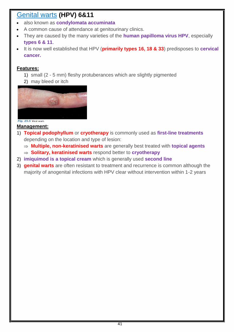
Features: 1) small (2 - 5 mm) fleshy protuberances which are slightly pigmented
2) may bleed or itch
Management:
1) Topical podophyllum or cryotherapy is commonly used as first-line treatments
depending on the location and type of lesion:
Multiple, non-keratinised warts are generally best treated with topical agents
Solitary, keratinised warts respond better to cryotherapy
2) imiquimod is a topical cream which is generally used second line
3) genital warts are often resistant to treatment and recurrence is common although the
majority of anogenital infections with HPV clear without intervention within 1-2 years

42
Rabies Rabies is a viral disease that causes acute encephalitis.
The rabies virus is classed as a RNA rhabdovirus and has a bullet shaped capsid.
It is commonly transmitted by bat, raccoon and skunk األمريكي الظربان bites.
Following a bite the virus travels up the nerve axons towards the central nervous system in
a retrograde fashion.
Features:
1) prodrome: headache, fever, agitation
2) hydrophobia: water-provoking muscle spasms
3) hypersalivation
4) Negri bodies: cytoplasmic inclusion bodies found in infected neurons
There is now considered to be 'no risk' of developing rabies following an animal bite in the
UK and the majority of developed countries.
Following an animal bite in at risk countries:
1) if an individual is already immunised:
2 further doses of vaccine should be given
2) if not previously immunised then
human rabies immunoglobulin (HRIG) should be given
along with a full course of vaccination

43
Hepatitis B Hepatitis B is a double-stranded DNA virus and is spread through exposure to infected
blood or body fluids, including vertical transmission from mother to child.
The incubation period is 6-20 weeks.
Hepatitis B serology Interpreting hepatitis B serology is a dying art form which still occurs at regular intervals in
medical exams.
It is important to remember a few key facts:
1) surface antigen (HBsAg) is the first marker to appear and causes the production of anti-
HBs
2) HBsAg normally implies acute disease (present for 1-6 months)
3) if HBsAg is present for > 6 months then this implies chronic disease (i.e. Infective)
4) Anti-HBs implies immunity (either exposure or immunisation). It is negative in chronic
disease
5) Anti-HBc:
implies previous (or current) infection.
IgM anti-HBc appears during acute or recent hepatitis B infection and is present
for about 6 months.
IgG anti-HBc persists
6) HbeAg results from breakdown of core antigen from infected liver cells as is therefore a
marker of infectivity
Example results:
previous immunisation: anti-HBs positive, all others negative
previous hepatitis B (> 6 months ago), not a carrier: anti-HBc positive, HBsAg negative
previous hepatitis B, now a carrier: anti-HBc positive, HBsAg positive
Complications of hepatitis B infection
1) Chronic hepatitis (5-10%).
2) Fulminant liver failure (1%)
3) hepatocellular carcinoma
4) glomerulonephritis
5) polyarteritis nodosa
6) cryoglobulinaemia

44
Management of hepatitis B:
Pegylated interferon-alpha
used to be the only treatment available
It reduces viral replication in up to 30% of chronic carriers.
A better response is predicted by:
1) being female,
2) < 50 years old,
3) low HBV DNA levels,
4) non-Asian,
5) HIV negative,
6) high degree of inflammation on liver biopsy
whilst NICE still advocate the use of pegylated interferon firstl-line other antiviral
medications are increasingly used with an aim to suppress viral replication (not in a
dissimilar way to treating HIV patients)
examples include tenofovir and entecavir
Immunisation against hepatitis B
contains HBsAg adsorbed onto aluminium hydroxide adjuvant and is prepared from yeast cells
using recombinant DNA technology
most schedules give 3 doses of the vaccine with a recommendation for a one-off booster 5
years following the initial primary vaccination
at risk groups who should be vaccinated include: healthcare workers, intravenous drug users,
sex workers, close family contacts of an individual with hepatitis B, individuals receiving blood
transfusions regularly, chronic kidney disease patients who may soon require renal
replacement therapy, prisoners, chronic liver disease patients
Around 10-15% of adults fail to respond or respond poorly to 3 doses of the vaccine.
Risk factors include age over 40 years, obesity, smoking, alcohol excess and
immunosuppression
Testing for anti-HBs is only recommended for those at risk of occupational exposure (i.e.
Healthcare workers) and patients with chronic kidney disease. In these patients anti-HBs levels
should be checked 1-4 months after primary immunisation
the table below shows how to interpret anti-HBs levels:
Anti-HBs level
(mIU/ml) Response
> 100 Indicates adequate response, no further testing required.
Should still receive booster at 5 years
10 - 100 Suboptimal response –
one additional vaccine dose should be given
If immunocompetent no further testing is required
< 10 Non-responder.
Test for current or past infection.
Give further vaccine course (i.e. 3 doses again) with testing following.
If still fails to respond then HBIG would be required for protection if exposed
to the virus
45
Hepatitis B and pregnancy Basics
all pregnant women are offered screening for hepatitis B
babies born to mothers who are chronically infected with hepatitis B or to mothers
who've had acute hepatitis B during pregnancy should receive a complete course of
vaccination + hepatitis B immunoglobulin
studies are currently evaluating the role of oral antiviral treatment (e.g. Lamivudine) in
the latter part of pregnancy
there is little evidence to suggest caesarean section reduces vertical transmission rates
hepatitis B cannot be transmitted via breastfeeding (in contrast to HIV)
Hepatitis E Overview
RNA hepevirus
spread by the faecal-oral route
incubation period: 3-8 weeks
common in Central and South-East Asia, North and West Africa, and in Mexico
causes a similar disease to hepatitis A, but carries a significant mortality (about 20%)
during pregnancy
does not cause chronic disease or an increased risk of hepatocellular cancer
a vaccine is currently in development*, but is not yet in widespread use
*New England Journal of Medicine 356:895, 2007
Pyogenic liver abscess Management
drainage (needle aspiration or catheter) should always be performed
amoxicillin + ciprofloxacin + metronidazole
if penicillin allergic: ciprofloxacin + clindamycin
46
Post-exposure prophylaxis Hepatitis A
Human Normal Immunoglobulin (HNIG)
or
hepatitis A vaccine may be used depending on the clinical situation
Hepatitis B
1) HBsAg positive source:
If the person exposed is a known responder to HBV vaccine then a booster dose
should be given.
If they are in the process of being vaccinated or are a non-responder they need to
have hepatitis B immune globulin (HBIG) and the vaccine
2) unknown source:
for known responders the green book advises considering a booster dose of HBV
vaccine
For known non-responders HBIG + vaccine should be given
whilst those in the process of being vaccinated should have an accelerated
course of HBV vaccine
Hepatitis C
monthly PCR if seroconversion then interferon +/- ribavirin
HIV:
1) a combination of oral antiretrovirals (e.g. Tenofovir, emtricitabine, lopinavir and ritonavir)
as soon as possible (i.e. Within 1-2 hours, but may be started up to 72 hours following
exposure) for 4 weeks
2) serological testing at 12 weeks following completion of post-exposure prophylaxis
3) reduces risk of transmission by 80%
Varicella zoster:
VZIG for IgG negative pregnant women/immunosuppressed
Estimates of transmission risk for single needle stick injury
Hepatitis B 20-30%
Hepatitis C 0.5-2%
HIV 0.3%

47
HIV and pregnancy With the increased incidence of HIV infection amongst the heterosexual population there
are an increasing number of HIV positive women giving birth in the UK.
In London the incidence may be as high as 0.4% of pregnant women.
The aim of treating HIV positive women during pregnancy is to minimize harm to both the
mother and fetus, and to reduce the chance of vertical transmission.
Guidelines regularly change on this subject and most recent guidelines can be found using
the links provided.
Factors which reduce vertical transmission: (from 25-30% to 2%)
1) maternal antiretroviral therapy
2) mode of delivery (caesarean section)
3) neonatal antiretroviral therapy
4) infant feeding (bottle feeding)
Screening:
NICE guidelines recommend offering HIV screening to all pregnant women
Antiretroviral therapy:
1) all pregnant women should be offered antiretroviral therapy regardless of whether they
were taking it previously
2) If women are not currently taking antiretroviral therapy
The RCOG recommend that it is commenced between 28 and 32 weeks of gestation
and should be continued intrapartum.
BHIVA recommend that antiretroviral therapy may be started at an earlier gestation
depending upon the individual situation
Mode of delivery
vaginal delivery is recommenced if viral load is less than 50 copies/ml at 36 weeks,
otherwise caesarian section is recommended
a zidovudine infusion should be started four hours before beginning the caesarean
section
Neonatal antiretroviral therapy
Zidovudine is usually administered orally to the neonate if maternal viral load is <50
copies/ml.
Otherwise triple ART should be used.
Therapy should be continued for 4-6 weeks.
Infant feeding
in the UK all women should be advised not to breast feed
48
HIV: biliary and pancreatic disease 1) The most common cause of biliary disease in patients with HIV is sclerosing cholangitis
due to infections such as CMV, Cryptosporidium and Microsporidia
2) Pancreatitis in the context of HIV infection may be secondary to:
1) anti-retroviral treatment (especially didanosine) or by
2) opportunistic infections e.g. CMV
HIV: Cytomegalovirus retinitis Cytomegalovirus (CMV) retinitis is common,
affecting 30-40% of patients who have a CD4 count < 50
Diagnosis is clinical as there are no diagnostic tests
Features:
visual impairment - 'blurred vision' etc
Fundoscopy:
characteristic appearance showing retinal haemorrhages and necrosis
often called 'pizza' retina
Management:
IV ganciclovir
treatment used to be life-long but new evidence suggests that it may be discontinued
once CD4 > 150 after HAART
alternative: IV foscarnet or cidofovir
HIV: diarrhoea Diarrhoea is common in patients with HIV.
This may be due to the effects of the virus itself (HIV enteritis) or opportunistic infections
Possible causes:
1) Cryptosporidium + other protozoa (most common)
2) Cytomegalovirus
3) Mycobacterium avium intracellulare
4) Giardia
Cryptosporidium:
The most common infective cause of diarrhoea in HIV patients.
It is an intracellular protozoa and has an incubation period of 7 days.
Presentation is very variable, ranging from mild to severe diarrhoea.
A modified Ziehl-Neelsen stain (acid-fast stain) of the stool may reveal the characteristic
red cysts of Cryptosporidium.
Treatment is difficult, with the mainstay of management being supportive therapy*

49
Mycobacterium avium intracellulare:
An atypical mycobacteria seen with the CD4 count is below 50.
Typical features include fever, sweats, abdominal pain and diarrhoea.
There may be hepatomegaly and deranged LFTs.
Diagnosis by:
1) blood cultures and
2) Bone marrow examination.
Management is with:
1) rifabutin,
2) ethambutol and
3) clarithromycin
*nitazoxanide is licensed in the US for immunocompetent patients
HIV: Kaposi's sarcoma caused by HHV-8 (human herpes virus 8)
presents as purple papules or plaques on the skin or mucosa (e.g. gastrointestinal and
respiratory tract)
skin lesions may later ulcerate
respiratory involvement may cause massive haemoptysis and pleural effusion
radiotherapy + resection
HIV: oesophageal candidiasis: Oesophageal candidiasis is the most common cause of oesophagitis in patients with HIV.
It is generally seen in patients with a CD4 count of less than 100.
Typical symptoms include dysphagia and odynophagia.
Fluconazole and itraconazole are first-line treatments
HIV: immunisation The Department of Health 'Greenbook' on immunisation defers to the British HIV Association
for guidelines relating to immunisation of HIV-infected adults
Vaccines that can be used in all
HIV-infected adults
Vaccines that can be used
if CD4 > 200
Contraindicated in HIV-infected
adults
Hepatitis A
Hepatitis B
Haemophilus influenzae B (Hib)
Influenza-parenteral
Japanese encephalitis
Meningococcus-MenC
Meningococcus-ACWY I
Pneumococcus-PPV23
Poliomyelitis-parenteral (IPV)
Rabies
Tetanus-Diphtheria (Td)
Measles, Mumps, Rubella
(MMR)
Varicella
Yellow Fever
Cholera CVD103-HgR
Influenza-intranasal
Poliomyelitis-oral (OPV)
Tuberculosis (BCG)

50
HIV: seroconversion
HIV seroconversion is symptomatic in 60-80% of patients and
typically presents as a glandular fever type illness
Increased symptomatic severity is associated with poorer long term prognosis.
It typically occurs 3-12 weeks after infection
Features:
1) sore throat
2) lymphadenopathy
3) malaise, myalgia, arthralgia
4) diarrhoea
5) maculopapular rash
6) mouth ulcers
7) rarely meningoencephalitis
Diagnosis
antibodies to HIV may not be present
HIV PCR and p24 antigen tests can confirm diagnosis

51
HIV Neurocomplications: Focal neurological lesions
Toxoplasmosis:
accounts for around 50% of cerebral lesions in patients with HIV
constitutional symptoms, headache, confusion, drowsiness
CT:
1) usually single or multiple ring enhancing lesions,
2) mass effect may be seen
Management:
1) sulfadiazine and
2) pyrimethamine
Cerebral toxoplasmosis: CT scan with contrast
showing multiple ring enhancing lesions
Cerebral
toxoplasmosis: MRI (T1 C+) demonstrates multiple
small peripherally enhancing nodules located
predominantly in the basal ganglia as well as the
central portions of the cerebellar hemispheres.
Only a small amount of surrounding oedema is
present.
Primary CNS lymphoma:
accounts for around 30% of cerebral lesions
associated with the Epstein-Barr virus
CT: single or multiple homogenous enhancing lesions
Treatment:
1) Generally involves steroids (may significantly reduce tumour size),
2) Chemotherapy (e.g. methotrexate) + with or without whole brain irradiation.
3) Surgical may be considered for lower grade tumours
A) Primary CNS lymphoma: MRI (T1 C+) demonstrates a large multilobulated mass in the right frontal lobe. It homogeneously enhances and extends to involve the caudate and the periventricular area. There is significant mass effect.
B) Primary CNS lymphoma: Non-contrast CT demonstrates a hyper-attenuating mass adjacent to the left lateral ventricle, with no calcification or haemorrhage.

52
Differentiating between toxoplasmosis and lymphoma is a common clinical scenario in HIV
patients.
It is clearly important given the vastly different treatment strategies.
Toxoplasmosis Lymphoma
Multiple lesions
Ring or nodular enhancement
Thallium SPECT negative
Single lesion
Solid (homogenous) enhancement
Thallium SPECT positive
Tuberculosis:
much less common than toxoplasmosis or primary CNS lymphoma
CT: single enhancing lesion
HIV neurocomplications:
Generalised neurological disease
Encephalitis:
may be due to CMV or HIV itself
HSV encephalitis but is relatively rare in the context of HIV
CT: oedematous brain
Cryptococcus:
most common fungal infection of CNS
meningitis is typical presentation but may occasionally cause a space occupying lesion
headache, fever, malaise, nausea/vomiting, seizures, focal neurological deficit
CSF:
high opening pressure,
India ink test positive
CT:
meningeal enhancement,
cerebral oedema
Progressive multifocal leukoencephalopathy (PML)
widespread demyelination
due to infection of oligodendrocytes by JC virus (a polyoma DNA virus)
symptoms, subacute onset :
behavioral changes,
speech, motor, visual impairment
CT:
Single or multiple lesions,
no mass effect,
Don’t usually enhance.
MRI:
Better
high-signal demyelinating white matter lesions are seen
AIDS dementia complex
caused by HIV virus itself
symptoms:
behavioral changes,
motor impairment
CT: cortical and subcortical atrophy

53
Orf Orf is generally a condition found in sheep and goats although it can be transmitted to
humans.
It is caused by the parapox virus.
In animals:
'scabby' lesions around the mouth and nose
In humans:
1) generally affects the hands and arms
2) initially small, raised, red-blue papules
3) later may increase in size to 2-3 cm and become flat-topped and haemorrhagic

54
Antiviral agents
Drug Mechanism of action Indications Adverse effects/toxicity
Aciclovir Guanosine analog,
phosphorylated by thymidine kinase
which in turn inhibits the viral DNA
polymerase
HSV, VZV Crystalline nephropathy
Ganciclovir Guanosine analog,
phosphorylated by thymidine kinase
which in turn inhibits the viral DNA
polymerase
CMV Myelosuppression/
agranulocytosis
Ribavirin Guanosine analog
inhibits inosine monophosphate (IMP)
dehydrogenase,
interferes with the capping of viral
Mrna
Chronic hepatitis
C, RSV
Haemolytic anaemia
Amantadine Inhibits uncoating (M2 protein) of
virus in cell.
Also releases dopamine from nerve
endings
Influenza,
Parkinson's
disease
Confusion,
ataxia,
slurred speech
Oseltamivir Inhibits neuraminidase Influenza
Foscarnet Pyrophosphate analog which
inhibits viral DNA polymerase
CMV,
HSV if not
responding to
aciclovir
Nephrotoxicity,
hypocalcaemia,
hypomagnesaemia,
seizures
Interferon-α Human glycoprotein
which inhibit synthesis of mRNA
Chronic hepatitis
B & C, hairy cell
leukaemia
Flu-like symptoms,
anorexia,
myelosuppression
Cidofovir Acyclic nucleoside phosphonate, and is
therefore independent of phosphorylation
by viral enzymes (compare and contrast
with aciclovir/ganciclovir)
CMV retinitis in
HIV
Nephrotoxicity
Anti-retroviral agent used in HIV should be started once CD4 <350
1) Nucleoside analogue reverse transcriptase inhibitors (NRTI)
examples: zidovudine (AZT), didanosine, lamivudine, stavudine, zalcitabine
2) Protease inhibitors (PI)
inhibits a protease needed to make the virus able to survive outside the cell
examples: indinavir, nelfinavir, ritonavir, saquinavir
3) Non-nucleoside reverse transcriptase inhibitors (NNRTI)
examples: nevirapine, efavirenz

55
Vaginal discharge Vaginal discharge is a common presenting symptom and is not always pathological
Common causes:
1) physiological
2) Candida
3) Trichomonas vaginalis
4) bacterial vaginosis
Less common causes:
1) whilst cervical infections such as Chlamydia and Gonorrhoea can cause a vaginal
discharge this is rarely the presenting symptoms
2) ectropion
3) foreign body
4) cervical cancer
Key features of the common causes are listed below
Condition Key features
Candida 'Cottage cheese' discharge
Vulvitis
Itch
Trichomonas vaginalis Offensive, yellow/green, frothy discharge
Vulvovaginitis
Strawberry cervix
Bacterial vaginosis Offensive, thin, white/grey, 'fishy' discharge

56
Bacterial vaginosis Bacterial vaginosis (BV) describes an overgrowth of predominately anaerobic organisms
such as Gardnerella vaginalis.
This leads to a consequent fall in lactic acid producing aerobic lactobacilli resulting in a
raised vaginal pH.
Whilst BV is not a sexually transmitted infection it is seen almost exclusively in sexually
active women.
Features
1) vaginal discharge: 'fishy', offensive
2) asymptomatic in 50%
Amsel's criteria for diagnosis of BV - 3 of the following 4 points should be present
1) thin, white homogenous discharge
2) clue cells on microscopy
3) vaginal pH > 4.5
4) positive whiff test (addition of potassium hydroxide results in fishy odour)
Management
1) oral metronidazole for 5-7 days
2) 70-80% initial cure rate
3) relapse rate > 50% within 3 months
4) the BNF suggests topical metronidazole or topical clindamycin as alternatives
© Image used on license from PathoPic
Clue cells - epithelial cells develop a stippled appearance due to being covered with bacteria
Bacterial vaginosis in pregnancy:
results in an increased risk of preterm labour, low birth weight and chorioamnionitis, late
miscarriage
It was previously taught that oral metronidazole should be avoided in the first trimester and
topical clindamycin used instead.
Recent guidelines however recommend that oral metronidazole is used throughout
pregnancy.
The BNF still advises against the use of high dose metronidazole regimes

57
Pelvic inflammatory disease Pelvic inflammatory disease (PID) is a term used to describe infection and inflammation of
the female pelvic organs including the uterus, fallopian tubes, ovaries and the surrounding
peritoneum.
It is usually the result of ascending infection from the endocervix
Causative organisms:
1) Chlamydia trachomatis - the most common cause
2) Neisseria gonorrhea
3) Mycoplasma genitalium
4) Mycoplasma hominis
Features:
1) lower abdominal pain
2) fever
3) deep dyspareunia
4) dysuria and menstrual irregularities may occur
5) vaginal or cervical discharge
6) cervical excitation
Investigation:
screen for Chlamydia and Gonorrhoea
Management:
1) due to the difficulty in making an accurate diagnosis, and the potential complications of
untreated PID, consensus guidelines recommend having a low threshold for treatment
2) oral ofloxacin + oral metronidazole or intramuscular ceftriaxone + oral doxycycline +
oral metronidazole
3) RCOG guidelines suggest that in mild cases of PID intrauterine contraceptive devices may
be left in.
4) The more recent BASHH guidelines suggest that the evidence is limited but that ' Removal
of the IUD should be considered and may be associated with better short term clinical
outcomes'
Complications:
1) infertility - the risk may be as high as 10-20% after a single episode
2) chronic pelvic pain
3) ectopic pregnancy

58
Urinary tract infection in adults: management A) Lower urinary tract infections in non-pregnant women:
local antibiotic guidelines should be followed if available
2012 SIGN guidelines recommend trimethoprim or nitrofurantoin for 3 days
B) Pregnant women with symptomatic bacteriuria:
should be treated with an antibiotic for 7 days
A urine culture should be sent.
C) For asymptomatic pregnant women:
a urine culture should be performed routinely at the first antenatal visit
if positive, a second urine culture should be sent to confirm the presence of bacteriuria
SIGN recommend to treat asymptomatic bacteriuria detected during pregnancy with an
antibiotic
a 7 day course of antibiotics should be given
a further urine culture should be sent following completion of treatment as a test of cure
D) For patients with sign of acute pyelonephritis:
hospital admission should be considered
local antibiotic guidelines should be followed if available
the BNF currently recommends a broad-spectrum cephalosporin or a quinolone for 10-
14 days

59
Antibiotics:
Anaerobic activity The following antibiotics have anti-anaerobic activity
1) penicillins
2) cephalosporins (except ceftazidime)
3) erythromycin
4) metronidazole
5) tetracycline
The following antibiotics do not have anti-anaerobic activity
1) gentamicin
2) ciprofloxacin
3) ceftazidime
Linezolid Linezolid is a type of oxazolidonone antibiotic which has been introduced in recent years.
It inhibits bacterial protein synthesis by stopping formation of the 70s initiation complex
I t is bacteriostatic nature.
Spectrum: highly active against Gram positive organisms including:
1) MRSA (Methicillin-resistant Staphylococcus aureus)
2) VRE (Vancomycin-resistant enterococcus)
3) GISA (Glycopeptide Intermediate Staphylococcus aureus)
Adverse effects:
thrombocytopenia (reversible on stopping)
monoamine oxidase inhibitor: avoid tyramine containing foods
Antibiotics: bactericidal vs. bacteriostatic Bactericidal antibiotics:
1) penicillins
2) cephalosporins
3) aminoglycosides
4) nitrofurantoin
5) metronidazole
6) quinolones
7) rifampicin
8) isoniazid
Bacteriostatic antibiotics
1) chloramphenicol
2) macrolides
3) sulphonamides
4) tetracyclines
5) trimethoprim

60
Antibiotics: mechanisms of action The lists below summarise the site of action of the commonly used antibiotics
Inhibit cell wall formation:
1) penicillins
2) cephalosporins
Inhibit protein synthesis:
1) aminoglycosides (cause misreading of mRNA)
2) chloramphenicol
3) macrolides (e.g. erythromycin)
4) tetracyclines
5) fusidic acid
Inhibit DNA synthesis:
1) quinolones (e.g. ciprofloxacin)
2) metronidazole
3) sulphonamides
4) trimethoprim
Inhibit RNA synthesis
rifampicin
Macrolides Erythromycin was the first macrolide used clinically.
Newer examples include clarithromycin and azithromycin.
Macrolides act by inhibiting bacterial protein synthesis.
If pushed to give an answer they are bacteriostatic in nature, but in reality this depends on
the dose and type of organism being treated.
Adverse effects:
1) Gastrointestinal side-effects are common. Nausea is less common with clarithromycin
than erythromycin
2) cholestatic jaundice: risk may be reduced if erythromycin stearate is used
3) P450 inhibitor (see below)
Common interactions:
Statins should be stopped whilst taking a course of macrolides:
Macrolides inhibit the cytochrome P450 isoenzyme CYP3A4 that metabolises statins.
Taking macrolides concurrently with statins significantly increases the risk of
myopathy and rhabdomyolysis.

61
Quinolones Quinolones are a group of antibiotics which work by inhibiting DNA synthesis.
They are bactericidal in nature. Examples include:
1) ciprofloxacin
2) levofloxacin
Mechanism of action:
inhibit topoisomeras II (DNA gyrase) and topoisomerase IV
Adverse effects:
1) lower seizure threshold in patients with epilepsy
2) tendon damage (including rupture) - the risk is increased in patients also taking steroids
3) cartilage damage has been demonstrated in animal models and for this reason quinolones
are generally avoided (but not necessarily contraindicated) in children
Quinupristin & dalfopristin antibiotics injectable streptogrammin antibiotic
combination of group A and group B streptogrammin
inhibits bacterial protein synthesis by blocking tRNA complexes binding to the ribosome
Spectrum:
most Gram positive bacteria
exception: Enterococcus faecalis*
Adverse effects
1) thrombophlebitis (give via a central line)
2) arthralgia
3) P450 inhibitor
*not to be confused with Enterococcus faecium, which is sensitive to Quinupristin & dalfopristin

62
Epididymo-orchitis Epididymo-orchitis describes an infection of the epididymis +/- testes resulting in pain and
swelling.
It is most commonly caused by local spread of infections from the genital tract (such
as Chlamydia trachomatis and Neisseria gonorrhea) or the bladder.
The most important differential diagnosis is testicular torsion.
This needs to be excluded urgently to prevent ischaemia of the testicle.
Features: 1) unilateral testicular pain and swelling
2) urethral discharge may be present, but urethritis is often asymptomatic
3) factors suggesting testicular torsion include:
patients < 20 years,
severe pain and an
acute onset
Management:
1) the British Association for Sexual Health and HIV (BASHH) produced guidelines in 2010
2) if the organism is unknown BASHH recommend:
ceftriaxone 500mg intramuscularly single dose,
plus
doxycycline 100mg by mouth twice daily for 10-14 days
3) further investigations following treatment are recommended to exclude any underlying
structural abnormalities

63
STI: ulcers A) Genital herpes:
1) most often caused by the herpes simplex virus (HSV) type 2 (cold sores are usually due
to HSV type 1) 2) Primary attacks are often severe and associated with fever whilst 3) subsequent attacks are generally less severe and localised to one site
B) Syphilis
1) a sexually transmitted infection caused by the spirochaete Treponema pallidum. 2) Infection is characterised by primary, secondary and tertiary stages. 3) A painless ulcer (chancre) is seen in the primary stage. 4) The incubation period= 9-90 days
C) Chancroid:
1) a tropical disease caused by Haemophilus ducreyi. 2) It causes painful genital ulcers associated with unilateral, painful inguinal lymph node
enlargement. 3) The ulcers typically have a sharply defined, ragged, undermined border.
D) Lymphogranuloma venereum (LGV)
1) caused by Chlamydia trachomatis 2) Typically infection comprises of three stages
stage 1: small painless pustule which later forms an ulcer
stage 2: painful inguinal lymphadenopathy
stage 3: proctocolitis
3) LGV is treated using doxycycline.
Other causes of genital ulcers
1) Behcet's disease
2) carcinoma
3) granuloma inguinale: Klebsiella granulomatis*
*previously called Calymmatobacterium granulomatis
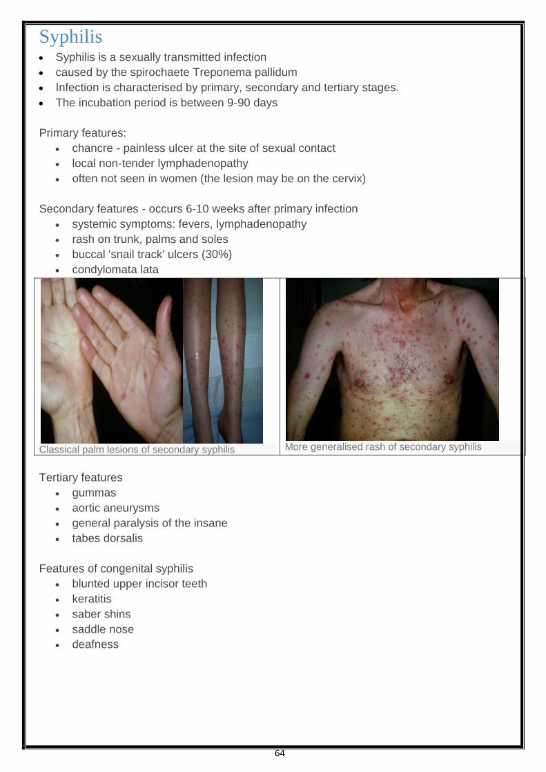
64
Syphilis Syphilis is a sexually transmitted infection
caused by the spirochaete Treponema pallidum
Infection is characterised by primary, secondary and tertiary stages.
The incubation period is between 9-90 days
Primary features:
chancre - painless ulcer at the site of sexual contact
local non-tender lymphadenopathy
often not seen in women (the lesion may be on the cervix)
Secondary features - occurs 6-10 weeks after primary infection
systemic symptoms: fevers, lymphadenopathy
rash on trunk, palms and soles
buccal 'snail track' ulcers (30%)
condylomata lata
Classical palm lesions of secondary syphilis
More generalised rash of secondary syphilis
Tertiary features
gummas
aortic aneurysms
general paralysis of the insane
tabes dorsalis
Features of congenital syphilis
blunted upper incisor teeth
keratitis
saber shins
saddle nose
deafness

65
Syphilis: investigation Treponema pallidum is a very sensitive organism and cannot be grown on artificial media.
The diagnosis is therefore usually based on clinical features, serology and microscopic
examination of infected tissue
Serological tests can be divided into
cardiolipin tests (not treponeme specific)
treponemal specific antibody tests
A) Cardiolipin tests:
syphilis infection leads to the production of non-specific antibodies that react to
cardiolipin
examples include VDRL (Venereal Disease Research Laboratory) & RPR (rapid plasma
reagin)
insensitive in late syphilis
becomes negative after treatment
B) Treponemal specific antibody tests
example: TPHA (Treponema pallidum HaemAgglutination test)
remains positive after treatment
Causes of false positive cardiolipin tests
1) pregnancy
2) SLE, anti-phospholipid syndrome
3) TB
4) leprosy
5) malaria
6) HIV
Syphilis: management Management:
1) benzylpenicillin
2) alternatives: doxycycline
3) The Jarisch-Herxheimer reaction ( also seen in lyme)
sometimes seen following treatment
Fever, rash, tachycardia after first dose of antibiotic.
It is thought to be due to the release of endotoxins following bacterial death and
typically occurs within a few hours of treatment.

66
Gastroenteritis Gastroenteritis may either occur whilst at home or whilst travelling abroad (travellers' diarrhoea) Travellers' diarrhoea: May be defined as at least 3 loose to watery stools in 24 hours with or without one or more of:
1) Abdominal cramps, 2) fever, 3) nausea, vomiting or 4) Blood in the stool.
The most common cause is Escherichia coli
Another pattern of illness is 'acute food poisoning'. This describes the sudden onset of nausea, vomiting and diarrhoea after the ingestion of a
toxin. Acute food poisoning is typically caused by Staphylococcus aureus, Bacillus cereus or
Clostridium perfringens. Stereotypical histories
Infection Typical presentation
Escherichia coli Common amongst travellers
Watery stools
Abdominal cramps and nausea
Amoebiasis Gradual onset bloody diarrhoea,
abdominal pain and tenderness which may last for several weeks
Giardiasis Prolonged, non-bloody diarrhoea
Cholera Profuse, watery diarrhoea
Severe dehydration resulting in weight loss
Not common amongst travellers
Shigella Bloody diarrhoea,
Vomiting and abdominal pain
Staphylococcusaureus Severe vomiting
Short incubation period
Campylobacter A flu-like prodrome is usually followed by crampy abdominal pains,
fever and diarrhoea which may be bloody
Complications include Guillain-Barre syndrome
Reiter s syndrome (can not pee , can not see , can not climb the
tree)
Bacillus cereus Two types of illness are seen:
vomiting within 6 hours, stereotypically due to rice
diarrhoeal illness occurring after 6 hours
associated with chineese food
67
Incubation period: 1-6 hrs: Staphylococcus aureus, Bacillus cereus*
12-48 hrs: Salmonella, Escherichia coli
48-72 hrs: Shigella, Campylobacter
> 7 days: Giardiasis, Amoebiasis
*vomiting subtype, the diarrhoeal illness has an incubation period of 6-14 hours
Giardiasis Giardiasis is caused by the flagellate protozoan Giardia lamblia.
It is spread by the faeco-oral route
Features:
1) often asymptomatic
2) lethargy, bloating, abdominal pain
3) non-bloody diarrhoea
4) chronic diarrhoea, malabsorption and lactose intolerance can occur
5) stool microscopy for trophozoite and cysts are classically negative, therefore duodenal
fluid aspirates or 'string tests' (fluid absorbed onto swallowed string) are sometimes
needed
Treatment is with metronidazole

68

69
Necrotising fasciitis Necrotising fasciitis is a medical emergency that is difficult to recognise in the early stages
It can be classified according to the causative organism:
type 1 is caused by mixed anaerobes and aerobes (often occurs post-surgery in
diabetics)
type 2 is caused by Streptococcus pyogenes
Features
acute onset
painful, erythematous lesion develops
extremely tender over infected tissue
Management
urgent surgical referral debridement
intravenous antibiotics
Acute epiglottitis Acute epiglottitis is rare but serious infection
caused by Haemophilus influenzae type B
Prompt recognition and treatment is essential as airway obstruction may develop.
Epiglottitis was generally considered a disease of childhood but in the UK it is now more
common in adults due to the immunisation programme.
The incidence of epiglottitis has decreased since the introduction of the Hib vaccine
Features:
1) rapid onset
2) high temperature, generally unwell
3) stridor
4) drooling of saliva
Animal bites The majority of bites seen in everyday practice involve dogs and cats.
These are generally polymicrobial but the most common isolated organism is Pasteurella
multocida.
Management:
1) cleanse wound
2) current BNF recommendation is co-amoxiclav
3) if penicillin-allergic then doxycycline + metronidazole is recommended

70
Protozoa
Leishmaniasis Leishmaniasis is caused by the intracellular protozoa Leishmania,
usually being spread by sand flies
Cutaneous, mucocutaneous leishmaniasis and visceral forms are seen
A) Cutaneous leishmaniasis:
1) caused by Leishmania tropica or Leishmania mexicana
2) crusted lesion at site of bite
3) may be underlying ulcer
B) Mucocutaneous leishmaniasis:
1) caused by Leishmania braziliensis
2) skin lesions may spread to involve mucosae of nose, pharynx etc
C) Visceral leishmaniasis (kala-azar)
1) mostly caused by Leishmania donovani
2) occurs in the Mediterranean, Asia, South America, Africa
3) fever, sweats, rigors
4) Massive splenomegaly& hepatomegaly
5) poor appetite*, weight loss
6) grey skin - 'kala-azar' means black sickness
7) pancytopaenia secondary to hypersplenism
*occasionally patients may report increased appetite with paradoxical weight loss
Toxoplasmosis Toxoplasma gondii is a protozoa which infects the body via the GI tract, lung or broken skin
It's oocysts release trophozoites which migrate widely around the body including to the eye,
brain and muscle.
The usual animal reservoir is the cat, although other animals such as rats carry the disease
Most infections are asymptomatic.
Symptomatic patients usually have a self-limiting infection, often having clinical features
resembling infectious mononucleosis (fever, malaise, lymphadenopathy)
Other less common manifestations include meningioencephalitis and myocarditis.
Investigation:
antibody test
Sabin-Feldman dye test
Treatment:
usually reserved for those with severe infections or patients who are immunosuppressed
pyrimethamine plus sulphadiazine for at least 6 weeks
Congenital toxoplasmosis:
is due to transplacental spread from the mother
It causes a variety of effects to the unborn child including microcephaly, hydrocephalus,
cerebral calcification and choroidoretinitis.
71
Trypanosomiasis Two main form of this protozoal disease are recognized:
1) African trypanosomiasis (sleeping sickness) and
2) American trypanosomiasis (Chagas' disease)
African trypanosomiasis Two forms of African trypanosomiasis, or sleeping sickness, are seen:
1) Trypanosoma gambiense in West Africa and
2) Trypanosoma rhodesiense in East Africa.
Both types are spread by the tsetse fly.
Trypanosoma rhodesiense tends to follow a more acute course.
Clinical features include:
1) Trypanosoma chancre - painless subcutaneous nodule at site of infection
2) intermittent fever
3) enlargement of posterior cervical lymph nodes
4) later: central nervous system involvement e.g. somnolence, headaches, mood changes,
meningoencephalitis
Management:
1) early disease:
IV pentamidine or
suramin
2) later disease or central nervous system involvement:
IV melarsoprol
American trypanosomiasis, or Chagas' disease:
Caused by the protozoanTrypanosoma cruzi.
Acute phase:
The vast majority of patients (95%) are asymptomatic in the acute phase
A chagoma (an erythematous nodule at site of infection) and periorbital oedema are
sometimes seen.
Chronic Chagas' disease mainly affects the heart and gastrointestinal tract
1) Myocarditis:
may lead to heart failure and arrhythmias
Cardiac involvement is the leading cause of death in Chagas diease
2) gastrointestinal features includes:
megaoesophagus causing dysphagia
megacolon causing constipation
Management:
1) treatment is most effective in the acute phase using azole or nitroderivatives such as
benznidazole or nifurtimox
2) chronic disease management involves treating the complications e.g., heart failure

72
Loiasis Loiasis is a filarial infection caused by Loa Loa.
It is transmitted by the Chrysops deerfly
tends to occur in rainforest regions of Western and Central Africa
Clinical features:
1) pruritus
2) urticaria
3) Calabar swellings: transient, non-erythematous, hot swelling of soft-tissue around joints
4) 'Eye worm' - the dramatic presentation of subconjuctival migration of the adult worm.
It has less pathological features than other the microfilarial infections Onchocerciasis and
Lymphatic Filariasis.
However high loa loa microfilaraemia is associated with encephalopathy following
treatment with either Ivermectin or DEC.
This occurs due to the death of vast numbers of blood microfilaria.
Both of these drugs are contraindicated if loa loa microfilaraemia exceeds 2500 mf/ml.
This has significant public health implications as Ivermectin is currently the drug of choice
for control of both Onchocerciasis and Lymphatic Filariasis in Africa.
Nematodes 1) Ancylostoma braziliense
most common cause of cutaneous larva migrans
common in Central and Southern America
2) Strongyloides stercoralis
acquired percutaneously (e.g. walking barefoot)
causes pruritus and larva currens - this has a similar appearance to cutaneous larva
migrans but moves through the skin at a far greater rate
abdo pain, diarrhoea, pneumonitis
may cause Gram negative septicaemia due carrying of bacteria into bloodstream
eosinophilia sometimes seen
Management:
1) thiabendazole, albendazole.
2) Ivermectin also used, particularly in chronic infections
Toxocara canis
commonly acquired by ingesting eggs from soil contaminated by dog faeces
commonest cause of visceral larva migrans
other features: eye granulomas, liver/lung involvement

73
Strongyloides stercoralis Strongyloides stercoralis is a human parasitic nematode worm.
The larvae are present in soil and gain access to the body by penetrating the skin.
Infection with Strongyloides stercoralis causes strongyloidiasis.
Features:
1) diarrhoea
2) abdominal pain
3) papulovesicular lesions where the skin has been penetrated by infective larvae e.g.
soles of feet and buttocks
4) larva currens: pruritic, linear, urticarial rash
5) if the larvae migrate to the lungs a pneumonitis similar to Loeffler's syndrome may be
triggered
Treatment
ivermectin and albendazole are used
Tape worms Tape worms are made up of repeated segments called proglottids.
These are often present in faeces and are useful diagnostically
Cysticercosis
caused by Taenia solium (from pork) and Taenia saginata (from beef)
management: niclosamide
Hydatid disease:
caused by the dog tapeworm Echinococcus granulosus
life-cycle involves dogs ingesting hydatid cysts from sheep liver
often seen in farmers
may cause liver cysts
management: albendazole

74
Schistosomiasis Schistosomiasis, or bilharzia, is a parasitic flatworm infection.
The following types of schistosomiasis are recognised:
Schistosoma mansoni and Schistosoma intercalatum: intestinal schistosomiasis
Schistosoma haematobium: urinary schistosomiasis
Schistosoma haematobium
This typically presents as a 'swimmer's itch' in patients who have recently returned from
Africa.
Schistosoma haematobium is a risk factor for squamous cell bladder cancer
Features:
1) frequency
2) haematuria
3) bladder calcification
Management
single oral dose of praziquantel
75
Vaccinations It is important to be aware of vaccines which are of the live-attenuated type as these may pose
a risk to immunocompromised patients. The main types of vaccine are as follows:
Live attenuated
BCG
measles, mumps, rubella (MMR)
influenza (intranasal)
oral rotavirus
oral polio
yellow fever
oral typhoid*
Inactivated preparations
rabies
influenza (intramuscular)
Detoxified exotoxins
tetanus
Extracts of the organism/virus (sometimes termed fragment)**
diphtheria
pertussis ('acellular' vaccine)
hepatitis B
meningococcus, pneumococcus, haemophilus
Notes
influenza: different types are available, including whole inactivated virus, split virion
(virus particles disrupted by detergent treatment) and sub-unit (mainly haemagglutinin
and neuraminidase)
cholera: contains inactivated Inaba and Ogawa strains of Vibrio cholerae together with
recombinant B-subunit of the cholera toxin
hepatitis B: contains HBsAg adsorbed onto aluminium hydroxide adjuvant and is
prepared from yeast cells using recombinant DNA technology
*whole cell typhoid vaccine is no longer used in the UK
**may also be produced using recombinant DNA technology

76
Pyrexia of unknown origin Defined as a prolonged fever of > 3 weeks which resists diagnosis after a week in hospital
Neoplasia:
1) lymphoma
2) hypernephroma
3) preleukaemia
4) atrial myxoma
Infections
abscess
TB
Connective tissue disorders
Splenectomy Following a splenectomy patients are particularly at risk from pneumococcus, Haemophilus,
meningococcus and Capnocytophaga canimorsus* infections
Vaccination
if elective, should be done 2 weeks prior to operation
Hib, meningitis A & C
annual influenza vaccination
pneumococcal vaccine every 5 years
Antibiotic prophylaxis
penicillin V:
unfortunately clear guidelines do not exist of how long antibiotic prophylaxis
should be continued
It is generally accepted though that penicillin should be continued for at least 2
years and at least until the patient is 16 years of age,
although the majority of patients are usually put on antibiotic prophylaxis for life
*usually from dog bites
77
Lymphadenopathy There are many causes of generalised lymphadenopathy
Infective
infectious mononucleosis
HIV, including seroconversion illness
eczema with secondary infection
rubella
toxoplasmosis
CMV
tuberculosis
roseola infantum
Neoplastic
leukaemia
lymphoma
Others
autoimmune conditions: SLE, rheumatoid arthritis
graft versus host disease
sarcoidosis
drugs: phenytoin and to a lesser extent allopurinol, isoniazid
© Image used on license from DermNet NZ
© Image used on license from PathoPic
© Image used on license from DermNet NZ
Image sourced from Wikipedia© Image used on license from PathoPic
© Image used on license from DermNet NZ
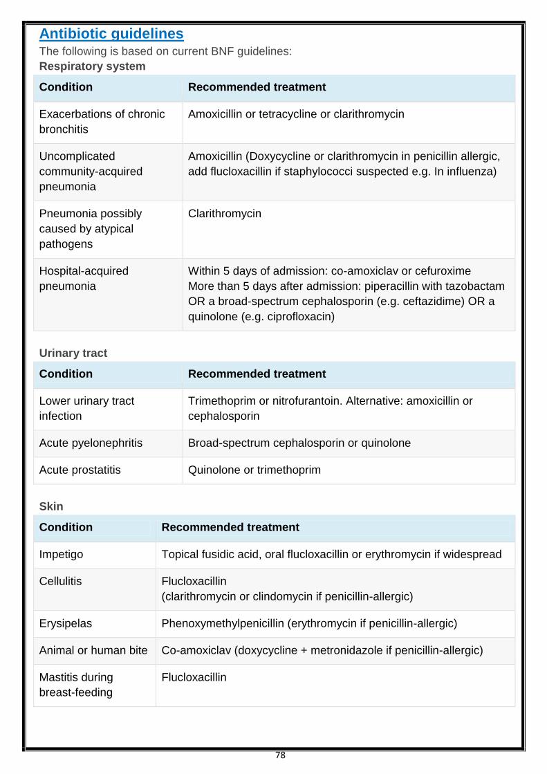
78
Antibiotic guidelines The following is based on current BNF guidelines:
Respiratory system
Condition Recommended treatment
Exacerbations of chronic
bronchitis
Amoxicillin or tetracycline or clarithromycin
Uncomplicated
community-acquired
pneumonia
Amoxicillin (Doxycycline or clarithromycin in penicillin allergic,
add flucloxacillin if staphylococci suspected e.g. In influenza)
Pneumonia possibly
caused by atypical
pathogens
Clarithromycin
Hospital-acquired
pneumonia
Within 5 days of admission: co-amoxiclav or cefuroxime
More than 5 days after admission: piperacillin with tazobactam
OR a broad-spectrum cephalosporin (e.g. ceftazidime) OR a
quinolone (e.g. ciprofloxacin)
Urinary tract
Condition Recommended treatment
Lower urinary tract
infection
Trimethoprim or nitrofurantoin. Alternative: amoxicillin or
cephalosporin
Acute pyelonephritis Broad-spectrum cephalosporin or quinolone
Acute prostatitis Quinolone or trimethoprim
Skin
Condition Recommended treatment
Impetigo Topical fusidic acid, oral flucloxacillin or erythromycin if widespread
Cellulitis Flucloxacillin
(clarithromycin or clindomycin if penicillin-allergic)
Erysipelas Phenoxymethylpenicillin (erythromycin if penicillin-allergic)
Animal or human bite Co-amoxiclav (doxycycline + metronidazole if penicillin-allergic)
Mastitis during
breast-feeding
Flucloxacillin

79
Ear, nose & throat
Condition Recommended treatment
Throat infections Phenoxymethylpenicillin (erythromycin alone if penicillin-allergic)
Sinusitis Amoxicillin or doxycycline or erythromycin
Otitis media Amoxicillin (erythromycin if penicillin-allergic)
Otitis externa* Flucloxacillin (erythromycin if penicillin-allergic)
Periapical or periodontal
abscess
Amoxicillin
Gingivitis: acute
necrotising ulcerative
Metronidazole
Genital system
Condition Recommended treatment
Gonorrhoea Intramuscular ceftriaxone + oral azithromycin
Chlamydia Doxycycline or azithromycin
Pelvic
inflammatory
disease
Oral ofloxacin + oral metronidazole or intramuscular
ceftriaxone + oral doxycycline + oral metronidazole
Syphilis Benzathine benzylpenicillin or doxycycline or
erythromycin
Bacterial vaginosis Oral or topical metronidazole or topical clindamycin
Gastrointestinal
Condition Recommended treatment
Clostridium difficile First episode: metronidazole
Second or subsequent episode of infection: vancomycin
Campylobacter enteritis Clarithromycin
Salmonella (non-typhoid) Ciprofloxacin
Shigellosis Ciprofloxacin
*a combined topical antibiotic and corticosteroid is generally used for mild/moderate cases of
otitis externa
Related Documents