J Appl Oral Sci. 409
ABSTRACT
www.scielo.br/jaoshttp://dx.doi.org/10.1590/1678-775720130472
Postretention stability after orthodontic closure of maxillary interincisor diastemas
Juliana Fernandes de MORAIS1, Marcos Roberto de FREITAS1, Karina Maria Salvatore de FREITAS2, Guilherme JANSON1, Nuria CASTELLO BRANCO2
1- Department of Pediatric Dentistry, Orthodontics and Community Health, Bauru School of Dentistry, University of São Paulo, Bauru, SP, Brazil.2- Private practice, Bauru, SP, Brazil.
Corresponding address: Juliana Fernandes de Morais - Departamento de Odontopediatria, Ortodontia e Saúde Coletiva - Faculdade de Odontologia de Bauru - Universidade de São Paulo - Alameda Octávio Pinheiro Brisolla 9-75 - Bauru - SP - 17012-901 - Brazil - Phone/Fax: 55 14 32342650 - e-mail: [email protected]
����������������� ������������������������� �� ����������������������� �!��"������
Anterior spaces may interfere with smile attractiveness and compromise dentofacial harmony. They are among the most frequent reasons why patients seek orthodontic
treatment. However, midline diastema is commonly cited as a malocclusion with high relapse incidence by orthodontists. Objectives: This study aimed to evaluate the stability of maxillary interincisor diastemas closure and the association of their relapse and interincisor width, overjet, overbite and root parallelism. Material and Methods: Sample comprised 30 patients with at least a pretreatment midline diastema of 0.5 mm or greater after eruption of the maxillary permanent canines. Dental casts and panoramic radiographs were taken at pretreatment, posttreatment and postretention. Results: Before treatment, midline diastema width was 1.52 mm [standard deviation(SD)=0.88] and right and left lateral diastema widths were 0.55 mm (SD=0.56) and 0.57 mm (SD=0.53), respectively. According to repeated measures analysis of variance, only midline diastema demonstrated ����������� �������������� ���� ������������ ��������� �����������������mm (SD=0.66), whilst the unstable patients showed a mean space reopening of 0.78 mm (SD=0.66). Diastema closure in the area between central and lateral incisors showed great ���� ����� ������������ ����������������� ������ ������������!"��#�$����� ����������%��!"��&�$����������������������� ��������� �����������'��� ������*��� ������������ �������������� �����������������������#�+��the sample, while lateral diastemas closure remained stable after treatment. Only initial diastema width and overjet relapse showed association with relapse of midline diastema. There was no association between relapse of interincisor diastema and root parallelism.
Keywords: Diastema. Relapse. Root tip. Corrective orthodontics.
INTRODUCTION
Anterior diastemas may interfere with smile attractiveness, compromise dentofacial harmony7,8,24, and provoke dyslalias5. Since they are easily visible, anterior spaces are one of the most important reasons why patients look for long-lasting stable treatment outcomes1.
In the primary and mixed dentitions, anterior spaces are common and considered as normal. In the permanent dentition, reported incidence ranges from 1.7% to 38%11,13,17,19,23 in different populations. This incidence is higher in black individuals than
among white or yellow racial groups11,13,17.Midline diastema is frequently cited as a
malocclusion with high relapse incidence by orthodontists2,4,15. Some have suggested that its recurrence is associated with initial diastema width18, inadequate root parallelism at the end of treatment4,15, sucking habits or imbalanced musculature4,15, abnormal labial frenum4, and intermaxillary osseous cleft18,21. An increase in overjet and overbite as the diastema reopens was also mentioned21.
However, information on stability following maxillary diastema closure is limited and the majority of reports consists of case reports and
2014;22(5):409-15
J Appl Oral Sci. 410
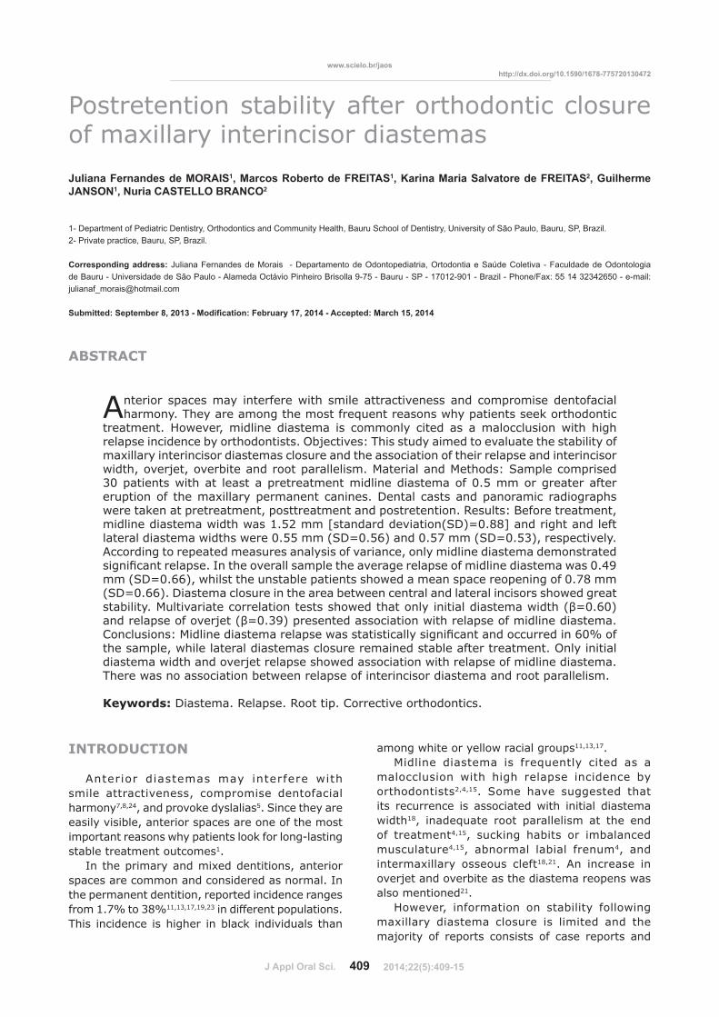
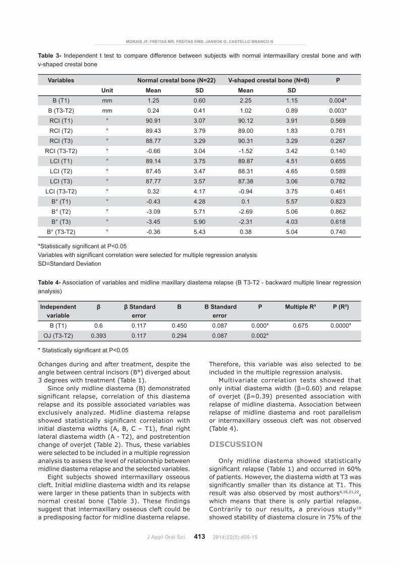
Figure 1- (1A) Sites where diastema widths were measured. A: Right lateral diastema; B: midline diastema; C: Left lateral diastema. (1B) Space width measurement using a round digital caliper
Postretention stability after orthodontic closure of maxillary interincisor diastemas
authors’ opinion. The rare follow-up evaluations on maxillary diastema treatment stability have divergent results. Edwards4 (1977) found diastema relapse on 84% of the sample (recurrent diastema width larger than 0.5 mm), and showed a strong correlation between labial frenum and diastema relapse. Contrarily, other studies18,21 demonstrated that relapse occurred at about one third of the subjects, but recurrent diastema widths were near 0.6 mm.
To date, no study has evaluated maxillary diastema relapse between central and lateral incisors, or the association between root parallelism and diastema closure stability.
The objectives of this study were: (1) to describe the frequency and severity of midline and lateral interincisor diastema relapses in patients with diastema before treatment; and (2) to identify treatment and posttreatment factors (interincisor widths, overjet, overbite and root parallelism) which could be associated to space reopening.
MATERIAL AND METHODS
A minimum sample size of 20 individuals was �����������<�+������������������ ��� ��0.05 to demonstrate a true difference of 0.4 mm in the diastema width, with 0.6 mm of estimated standard deviation, according to Sullivan, et al.21 (1996).
Ethical Committee approval was obtained from the Research Ethics Committee of Bauru School of Dentistry, University of São Paulo, to perform this retrospective study.
Thirty subjects were selected (17 female; 13 male) with Class I (18 subjects) and Class II (12 ���%���$ �� ��� ������ ������� ���� �� � ��of 4,331 patients treated in the Orthodontics Department at Bauru School of Dentistry, University of São Paulo. The subjects were treated nonextraction with standard edgewise appliances (0.022x0.028-inch) by graduate students. Inclusion criteria were patients presenting with at least a
maxillary midline diastema equal to or greater than 0.5 mm after maxillary canines full eruption. Subjects with missing anterior teeth, periodontal disease with bone loss, generalized microdontia, maxillary pathologies, mesiodens, diastema closure by a non-orthodontic method, post-orthodontic restoration of maxillary anterior teeth resulting in increase in mesiodistal width, and patients with deteriorated or missing dental casts or radiographs were excluded. After treatment, patients wore a Hawley plate in the maxillary arch. Patients with maxillary fixed retention were excluded. Pretreatment (T1), posttreatment (T2) and postretention (T3) mean ages were 12.94 years (SD 1.27), 15.32 years (SD 1.61), and 22.38 (SD 3.90), respectively. Dental casts and panoramic radiographs taken at T1, T2 and T3 were used. Periapical radiographs taken at T1 and T2 were also evaluated. Treatment changes were calculated as T2-T1, and postretention changes were calculated as T3-T2. Diastema relapse occurred when diastema width at T3-T2 was greater than zero.
Dental cast analysisAll dental cast measurements were made with
a 0.01 mm precision digital caliper (Mitutoyo, Kawasaki, Kanagawa, Japan) at T1, T2 and T3. The assessed variables were diastema width, overjet and overbite. To measure these variables, the following concepts were considered:
Diastema widths (A, B, C): The smallest distance between adjacent teeth at the level of gum papilla (Figure 1).
Overjet (OJ): Distance from the labial aspect of the medium point, on mesiodistal direction, of the central maxillary incisor incisal edge to the labial surface of the central mandibular incisor.
Overbite (OB): Distance between the incisal edges of maxillary and mandibular central incisors.
Values of overbite and overjet were obtained from right and left sides and averaged.
2014;22(5):409-15
J Appl Oral Sci. 411
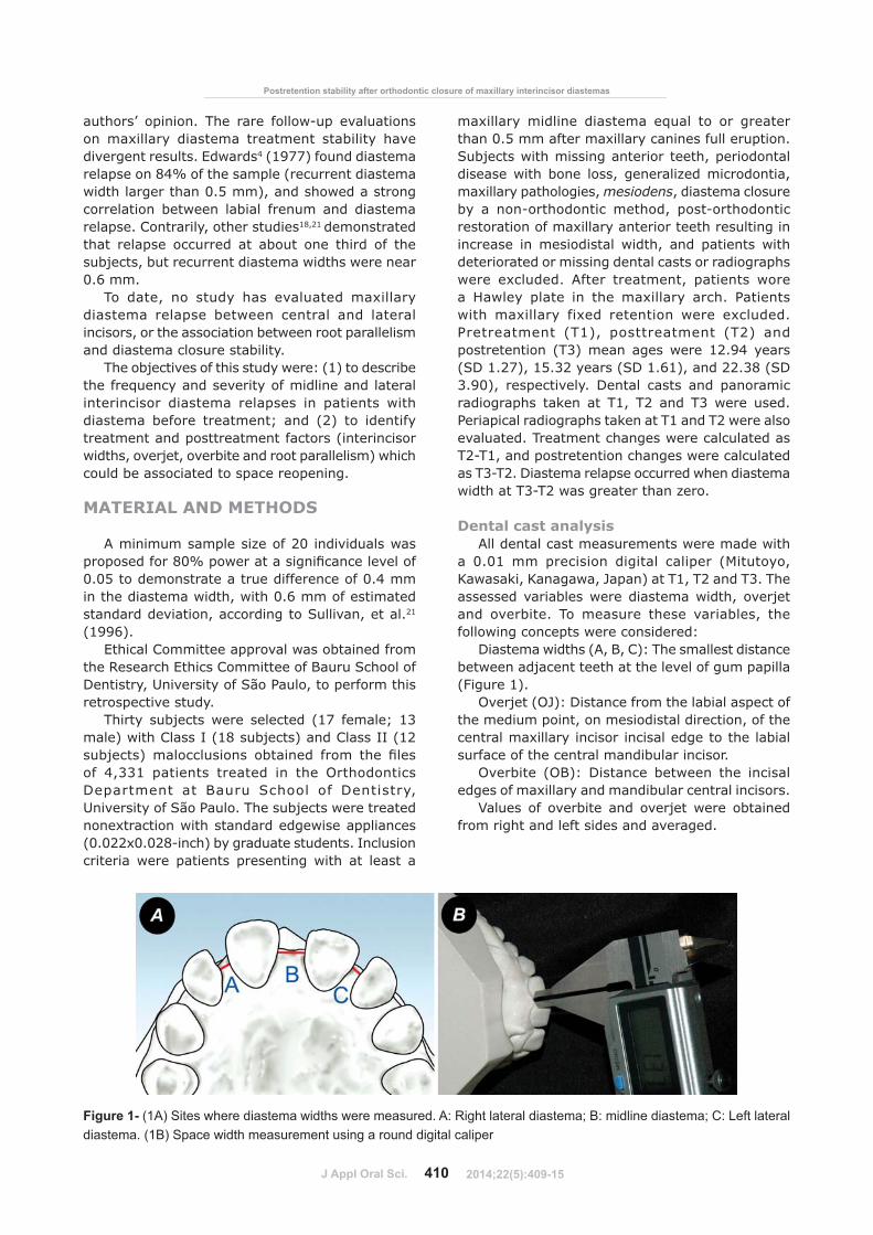
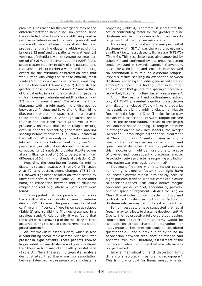
Figure 2- Panoramic radiographic tracing showing the angles built by root long axes of the maxillary incisors and the intermaxillary-zygomatic-processes (IZP) line, and the angles between adjacent incisors
MORAIS JF, FREITAS MR, FREITAS KMS, JANSON G, CASTELLO BRANCO N
Radiographic analysisPanoramic radiographs were taken at T1, T2, and
T3 under standard conditions, with the Frankfurt ����>��� � ��� ���� � � ?��� ��� �� ����� midline plane in a vertical position, by means of the Rotograph Plus (Villa Sistemi Medicali, Buccinasco, �� ��@�� �$������������������������������close to 25%.
Panoramic radiographs were traced using a 0.5 mm pencil on a sheet of acetate paper (14x21 ��$� ����������������������� ��G����������outlines of the zygomatic processes of the maxilla and the contours of the maxillary incisors were traced. A horizontal reference line (IZP line) was used, passing through the most inferior point of the right and left zygomatic processes of maxilla (Figure 2)10. Maxillary incisor angulations were measured, using the following variables: RLI (angle between the long axis of the right maxillary lateral incisor and the IZP line), RCI (angle between the long axis of the right maxillary central incisor and the IZP line), LCI (angle between the long axis of the left maxillary central incisor and the IZP line), LLI (angle between the long axis of the left maxillary lateral incisor and the IZP line). Interincisor angles were assessed by measuring the angles (A°, B°, C°) between adjacent maxillary incisors. Figure 2 describes the anatomical structures, lines and angles used in the panoramic radiographic analysis.
Intermaxillary osseous cleft was evaluated by comparing periapical radiographs taken at T1 and T2, and was considered present when a v-shaped radiolucency in crestal bone between the central ������������������������� ������ ������radiographs18,21.
Error studyAfter a month interval from the f irst
measurement, 30 randomly selected dental casts and 30 panoramic radiographs were reevaluated by the same examiner (JFM). The casual error
was calculated according to Dahlberg’s formula (S2"N�2/2n)3, where S2 is the error variance and d is the difference between two determinations of the same variable. The systematic errors were evaluated with dependent t tests at P<0.05.
Statistical analysesMeans and standard deviations of dental cast
and panoramic radiograph variables were calculated � �� ����� @��� @���������������������were compared by repeated measures ANOVA, followed by Tukey tests.
Y����������� ������������������������to evaluate associations between diastema relapse (A, B, C at T3-T2) and the independent variables: interincisor widths (A, B, C) at T1 and T2; overjet (OJ) and overbite (OB) at T1, T2, and T3-T2; and root parallelism (RLI, RCI, LCI, LLI, A°, B°, C°) at T1, T2, and T3.
Independent t tests were used to compare variables related with midline diastema relapse (B) in v-shaped crestal bone subjects and in subjects with normal intermaxillary crestal bone. Results ������������������������Y\���^�
Variables significantly correlated at P<0.05 �����Y����������� ��������������������������� �������������������� ���������������subjects with normal or v-shaped crestal bone were selected to be included in a backward multiple linear regression analysis.
RESULTS
From the 30 subjects, 8 presented with only a midline diastema. The retention time was 1.44 years (SD 0.48), and the mean period out of retention was 5.62 years (SD 3.89). The mean treatment time was 2.38 (SD 1.06).
There were no systematic errors. Casual errors ranged from 0.04 to 0.15 mm for linear (A, B, C, OJ, OB) and from 0.17° to 1.45° for angular variables
2014;22(5):409-15
J Appl Oral Sci. 412
Variables Initial (T1) Final (T2) Postretention (T3) ANOVAUnit Mean SD Mean SD Mean SD P
A mm 0.55A 0.56 0.03B 0.1 0.07B 0.15 0.000*
B mm 1.52A 0.88 0.04B 0.12 0.49C 0.68 0.000*
C mm 0.57A 0.53 0.04B 0.11 0.10B 0.21 0.000*
OJ mm 6.13A 2.65 3.05B 1.11 3.45B 1.49 0.000*
OB mm 3.91A 1.19 2.73B 0.81 2.99B 0.9 0.000*
RLI ° 98.37A 4.47 96.55AB 5.36 95.08B 4.28 0.031*
RCI ° 90.7 3.26 89.32 3.36 89.18 3.31 0.151
LCI ° 89.33 3.90 87.68 3.76 87.67 3.40 0.139
LLI ° 97.12A 5.05 94.25B 5.3 93.90B 3.16 0.015*
A° ° 7.7 3.90 7.30 4.00 6.00 3.30 0.188
B° ° -0.3 4.60 -3,00 5.50 -3.20 5.40 0.064
C° ° 8.00 5.00 6.60 5.60 6.30 4.20 0.396
������������ ��� ���� �� ��� � �������� ������� �� ������� � � ���� ����� � �� � ������� ���� ����� ��� ��� �� ����������� ��� ���� ��difference (Tukey test). SD=Standard Deviation
Table 1- Repeated measures ANOVA and Tukey tests to compare the occlusal and radiographic characteristics at the initial (T1), posttreatment (T2) and postretention (T3) stages
B T3-T2 T1 T2 T3-T2R P R P R P
A 0.498 0.005* 0.664 0.000* 0.282 0.132
B 0.734 0.000* 0.036 0.851 - -
C 0.401 0.028* 0.283 0.13 0.273 0.144
OJ 0.046 0.809 0.206 0.274 0.597 0.000*
OB -0.04 0.825 0.174 0.358 -0.19 0.315
T1 T2 T3
RLI 0.041 0.829 -0.112 0.557 -0.071 0.709
RCI -0.187 0.323 -0.076 0.689 0.173 0.361
LCI -0.246 0.19 -0.087 0.648 -0.011 0.953
LLI -0.128 0.499 0.036 0.849 0.003 0.986
B° -0.311 0.095 -0.107 0.573 0.099 0.604
���������������� ���� �������������� � � � � � � ������������������ ���� ����������� �������������������!�������������� �� ����
Table 2- Correlation of several variables with midline diastema relapse (B T3-T2)
Postretention stability after orthodontic closure of maxillary interincisor diastemas
(RLI, RCI, LCI, LLI, A°, B°, C°).Considering only recurrent diastemas, midline
diastema (B) relapse occurred in 18 patients (60%) with a mean increase of 0.78 mm (SD 0.66): six presented recurrent midline diastema of up to 0.5 mm, eight presented 0.5 to 1.0 mm widths, two had diastemas between 1.0 and 1.5 mm, and two diastemas larger than 1.5 mm. Only 5 patients (16.6%) showed right lateral diastema (A) relapse (mean increase of 0.37 mm, SD 0.12) and also only 5 patients (16.6%) presented left lateral diastema (C) reopening (mean increase of 0.47 mm, SD 0.10).
G����_�� ������ ������� ��� ���� ��closure of anterior spaces while eight patients �����������������������������`� ���interincisor distances greater than zero. Diastema width, overjet and overbite were significantly reduced during treatment. However, only midline ������� �{$ �������� � ������� � ���������postretention relapse. Central incisor angulations (RCI, LCI) showed no statistically significant changes during and after treatment, while lateral incisors (RLI, LLI) tended to have crown mesial tip during treatment and remain stable after treatment. Interincisor angles did not exhibit significant
2014;22(5):409-15
J Appl Oral Sci. 413
Variables Normal crestal bone (N=22) V-shaped crestal bone (N=8) PUnit Mean SD Mean SD
B (T1) mm 1.25 0.60 2.25 1.15 0.004*
B (T3-T2) mm 0.24 0.41 1.02 0.89 0.003*
RCI (T1) ° 90.91 3.07 90.12 3.91 0.569
RCI (T2) ° 89.43 3.79 89.00 1.83 0.761
RCI (T3) ° 88.77 3.29 90.31 3.29 0.267
RCI (T3-T2) ° -0.66 3.04 -1.52 3.42 0.140
LCI (T1) ° 89.14 3.75 89.87 4.51 0.655
LCI (T2) ° 87.45 3.47 88.31 4.65 0.589
LCI (T3) ° 87.77 3.57 87.38 3.06 0.782
LCI (T3-T2) ° 0.32 4.17 -0.94 3.75 0.461
B° (T1) ° -0.43 4.28 0.1 5.57 0.823
B° (T2) ° -3.09 5.71 -2.69 5.06 0.862
B° (T3) ° -3.45 5.90 -2.31 4.03 0.618
B° (T3-T2) ° -0.36 5.43 0.38 5.04 0.740
��������������� ���� ���������������������������� ���� ����������� �������������������!�������������� �� ����SD=Standard Deviation
Table 3- Independent t test to compare difference between subjects with normal intermaxillary crestal bone and with v-shaped crestal bone
Independent variable
# �#������ �error
B B Standard error
P Multiple R² P (R²)
B (T1) 0.6 0.117 0.450 0.087 0.000* 0.675 0.0000*
OJ (T3-T2) 0.393 0.117 0.294 0.087 0.002*
���������������� ���� �����������
Table 4- Association of variables and midline maxillary diastema relapse (B T3-T2 - backward multiple linear regression analysis)
MORAIS JF, FREITAS MR, FREITAS KMS, JANSON G, CASTELLO BRANCO N
0changes during and after treatment, despite the angle between central incisors (B°) diverged about 3 degrees with treatment (Table 1).
Since only midline diastema (B) demonstrated ��������� �� ����@ ����� ���� �� ��� �������relapse and its possible associated variables was exclusively analyzed. Midline diastema relapse ������ ������� � ��������� ����� ���� �������� �������������|@{@'}G~$@��� ����lateral diastema width (A - T2), and postretention change of overjet (Table 2). Thus, these variables were selected to be included in a multiple regression analysis to assess the level of relationship between midline diastema relapse and the selected variables.
Eight subjects showed intermaxillary osseous cleft. Initial midline diastema width and its relapse were larger in these patients than in subjects with normal crestal bone (Table 3). These findings suggest that intermaxillary osseous cleft could be a predisposing factor for midline diastema relapse.
Therefore, this variable was also selected to be included in the multiple regression analysis.
Multivariate correlation tests showed that �� � ����� ����������� �!"��#�$��� �� ������ ����%� �!"��&�$ �������� ���������� ���relapse of midline diastema. Association between relapse of midline diastema and root parallelism or intermaxillary osseous cleft was not observed (Table 4).
DISCUSSION
Only midline diastema showed statistically ����������� �����G�� �~$�������������#�+of patients. However, the diastema width at T3 was ��������� ���� ����� ����������G~�G���result was also observed by most authors4,18,21,22, which means that there is only partial relapse. Contrarily to our results, a previous study18 showed stability of diastema closure in 75% of the
2014;22(5):409-15
J Appl Oral Sci. 414
Postretention stability after orthodontic closure of maxillary interincisor diastemas
patients. One reason for this divergence may be the difference between sample inclusion criteria, since ������ ������������������� ������`����removable retention and the mean pretreatment space width was 1.22 mm. In our study, the mean pretreatment midline diastema width was slightly larger (1.52 mm) and the patients were at least 1.8 years out of retention, with an average postretention period of 5.6 years. Sullivan, et al.21 (1996) found space closure stability in 66% of the patients, and the sample selection criteria were similar to ours, except for the minimum postretention time that was 1 year. Analyzing the relapse amount, most studies18,21,22 also showed small space reopening. On the other hand, Edwards4 (1977) demonstrated greater relapse, between 2.4 and 2.7 mm in 84% of the patients, in a sample consisting of patients with an average pretreatment midline diastema of 3.2 mm (minimum 2 mm). Therefore, the initial diastema width might explain the discrepancy ���������������������������4. Unlike midline diastema area, lateral space closure appeared to be stable (Table 1). Although lateral space relapse had not been investigated yet, it was previously observed that, when relapse occurs, even in patients presenting generalized anterior spacing before treatment, it is usually located at the midline21. Whereas only 22 patients presented lateral diastemas before treatment, post-hoc power analysis calculation showed that a sample composed of 18 subjects provides 91.4% power ������������� ��� �����^�������������difference of 0.1 mm, with standard deviation 0.12.
Regarding the contributing factors for midline diastema relapse, spaces A, B, and C at T1, space A at T2, and posttreatment changes (T3-T2) in �������������������������������������univariate correlation test (Table 2). On the other hand, no association between midline diastema relapse and root angulations or parallelism were found.
�������������������� � �����?������the stability after orthodontic closure of anterior diastema4,15. However, the present results did not �����������?��������������������� �����G�� ��$�������� ����������������� ���previous study14. Additionally, it was found that the slight mesial crown tip of the maxillary incisors occurred during the space closure remained stable posttreatment14.
An intermaxillary osseous cleft, which is also a contributing factor for diastema relapse4,20 was present in eight patients. These patients showed larger initial midline diastema and greater relapse than those with normal intermaxillary crestal bone (Table 3). Nevertheless, multivariate analysis demonstrated that there was no association between intermaxillary osseous cleft and diastema
reopening (Table 4). Therefore, it seems that the actual contributing factor for the greater midline diastema relapse in the osseous cleft group was its larger width at the pretreatment stage.
According to the multivariate analysis, initial diastema width (B T1) was the only pretreatment ���������������������������� �����{G&_G�$(Table 4). This association was also supported by others18,22 ������������� ������ ���������tendency found in Edwards’ sample4. Conversely, spaces between lateral and central incisors showed no correlation with midline diastema relapse. Previous results showing no association between diastema reopening and initial generalized anterior spacing21 ������ ��� ������� '������ �@ ����������������������� �>�������������������more likely to suffer midline diastema recurrence18.
Among the treatment and posttreatment factors, �� � �� G&_G� �������� ��������� ����������with diastema relapse (Table 4). As the overjet increased, so did the midline diastema. Muscle function and relapse of Class II malocclusion may explain this association. Forward tongue posture induces incisor proclination, increase in arch length and anterior space opening. If tongue pressure is stronger on the maxillary incisors, the overjet increases. Camouflage orthodontic treatment of Class II division 1 malocclusion usually is reached by maxillary incisor retroclination and great overjet decrease. Therefore, patients with this malocclusion might be more prone to relapse of overjet and, consequently, diastema relapse. Association between diastema reopening and incisor proclination was previously determined21.
G������ �������� ��� ����������� ������remaining is another factor that might have ��?��������������� �������������@����������� ������ ������� ����� ���� �� � �����of anterior spaces. This could induce tongue abnormal pressure5 and, secondarily, provoke anterior space enlargement. Studies focusing on Class II malocclusion, on muscle function, and �������������������������������������diastema relapse may be of interest in the future.
Some investigators have suggested that labial frenum may contribute to diastema development2,4,8. Due to the retrospective follow-up study design, information about frenum anatomy would be available on clinical charts, photographies, or study models. These methods could be considered questionable21, and a previous study found no association between frequency of relapse and abnormal frenum18. Therefore, assessment of the ��?������� ���� ����������������� �������not performed.
Image magnification and distortion limit dimensional accuracy in panoramic radiography9. This is more critical for linear measurements.
2014;22(5):409-15
J Appl Oral Sci. 415
MORAIS JF, FREITAS MR, FREITAS KMS, JANSON G, CASTELLO BRANCO N
Contrarily, angular measurements in panoramic radiography showed less distortion6,12, especially in the anterior region16. Panoramic radiography was used because it is a low-dose radiographic technique which provides a comprehensive view of the entire maxillomandibular complex in a single � �� |������� �@ � �� ������ � �������� �� ��orthodontic practice and its use for this research avoided extra radiation exposure of the patients who comprised this sample.
This study showed that midline diastema closure is highly unstable. The amount of relapse is proportional to its pretreatment width and occurs associated with increase in the overjet. It is suggested lifetime wear of a well-adapted ��`� ����`�������������������������� ���diastema, especially in cases with initial large spaces and/or muscle unbalance.
CONCLUSIONS
Midline diastema relapse was statistically ����������������������#�+�������� �@��� �lateral diastemas showed great stability.
Only initial diastema severity width and overjet relapse showed association with relapse of midline diastema.
There was no association between relapse of interincisor diastemas and root parallelism.
REFERENCES
1- Bernabé E, Flores-Mir C. Influence of anterior occlusal characteristics on self-perceived dental appearance in young adults. Angle Orthod. 2007;77:831-6.2- Bishara SE. Management of diastemas in orthodontics. Am J Orthod. 1972;61:55-63.3- Dahlberg G. Statistical methods for medical and biological students. New York: Interscience; 1940.4- Edwards JG. The diastema, the frenum, the frenectomy: a clinical study. Am J Orthod. 1977;71:489-508.5- Farronato G, Giannini L, Riva R, Galbiati G, Maspero C. Correlations between malocclusions and dyslalias. Eur J Paediatr Dent. 2012;13:13-8.6- Frykholm A, Malmgren O, Sämfors KA, Welander U. Angular measurements in orthopantomography. Dentomaxillofac Radiol. 1977;6:77-81.
7- Furuse AY, Franco EJ, Mondelli J. Esthetic and functional restoration for an anterior open occlusal relationship with multiple diastemata: a multidisciplinary approach. J Prosthet Dent. 2008;99:91-4.8- Gardiner JH. Midline spaces. Dent Pract Dent Rec. 1967;17:287-97.9- Graber TM. Panoramic radiography in orthodontic diagnosis. Am J Orthod. 1967;53:799-821.10- Janson G, Putrick LM, Henriques JF, Freitas MR, Henriques RP. Maxillary third molar position in Class II malocclusions: the effect of treatment with and without maxillary premolar extractions. Eur J Orthod. 2006;28:573-9.11- Lavelle CL. Crowding and spacing within the human dental arch of different racial groups. Arch Oral Biol. 1970;15:1101-3.12- McKee IW, Williamson PC, Lam EW, Heo G, Glover KE, Major PW. The accuracy of 4 panoramic units in the projection of mesiodistal tooth angulations. Am J Orthod Dentofacial Orthop. 2002;121:166-75; quiz 92.13- McVay TJ, Latta GH Jr. Incidence of the maxillary midline diastema in adults. J Prosthet Dent. 1984;52:809-11.14- Morais JF, Freitas MR, Freitas KM, Janson G, Castello Branco N, Zanda M. Maxillary incisors mesiodistal angulation changes in patients with orthodontically treated anterior superior diastemas. Dental Press J Orthod. 2012;17:65-71.15- Mulligan TF. Diastema closure and long-term stability. J Clin Orthod. 2003;37:560-74.16- Peck JL, Sameshima GT, Miller A, Worth P, Hatcher DC. Mesiodistal root angulation using panoramic and cone beam CT. Angle Orthod. 2007;77:206-13.17- Richardson ER, Malhotra SK, Henry M, Little RG, Coleman HT. Biracial study of the maxillary midline diastema. Angle Orthod. 1973;43:438-43.18- Shashua D, Artun J. Relapse after orthodontic correction of maxillary median diastema: a follow-up evaluation of consecutive cases. Angle Orthod. 1999;69:257-63.19- Steigman S, Gershkovitz E, Harari D. Characteristics and stability of spaced dentition. Angle Orthod. 1985;55:321-8.��_��� ����G����?��������������� ���������������������and diastema formation. Am J Orthod. 1976;70:645-62.21- Sullivan TC, Turpin DL, Artun J. A postretention study of patients presenting with a maxillary median diastema. Angle Orthod. 1996;66:131-8.22- Surbeck BT, Artun J, Hawkins NR, Leroux B. Associations between initial, posttreatment, and postretention alignment of maxillary anterior teeth. Am J Orthod Dentofacial Orthop. 1998;113:186-95.23- Taylor JE. Clinical observations relating to the normal and abnormal frenum labii superioris. Am J Orthod Oral Surg 1939;25:646-50.24- Witt M, Flores-Mir C. Laypeople's preferences regarding frontal dentofacial esthetics: periodontal factors. J Am Dent Assoc. 2011;142:925-37.
2014;22(5):409-15