Slide
1 Foundations of Public Health
Immunology
Hypersensitivity
Type I hypersensitivity reactions can be caused by a variety of allergens
SLIDE 1 Hypersensitivity. This week’s lecture will elaborate on concepts from last week- immune responses directed against self antigens. Hypersensitivity diseases are disorders primarily caused by the adaptive immune response, where tissue damage is caused by the same effector mechanisms used by the immune system to protect against microbes. Specific hypersensitivity reactions can result in different autoimmune diseases.
Slide
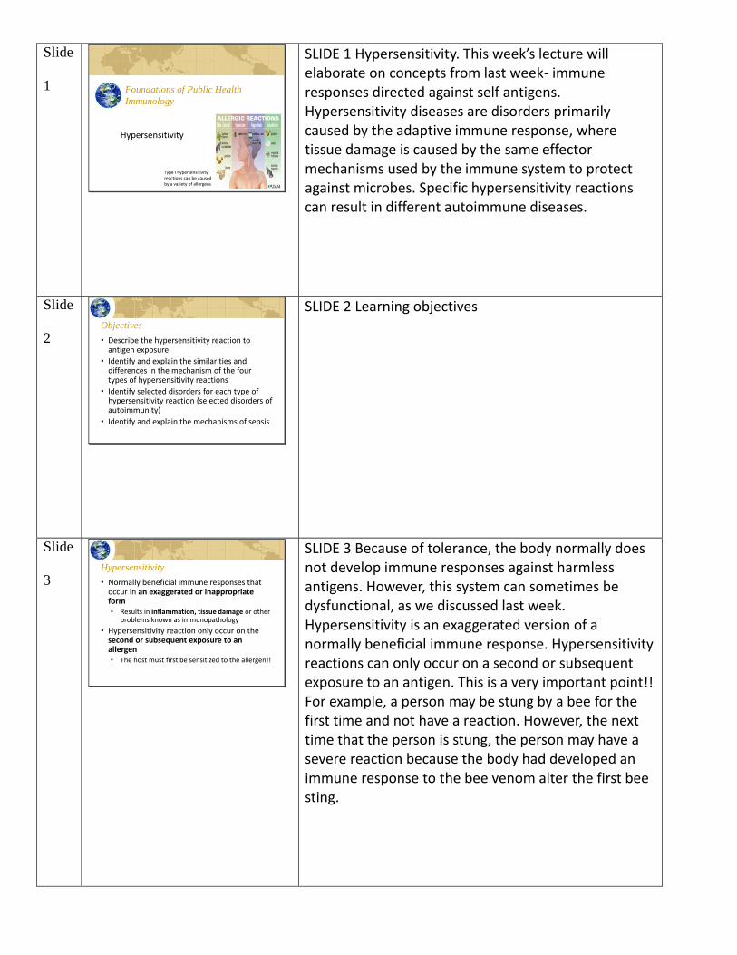
2 Objectives
• Describe the hypersensitivity reaction to antigen exposure
• Identify and explain the similarities and differences in the mechanism of the four types of hypersensitivity reactions
• Identify selected disorders for each type of hypersensitivity reaction (selected disorders of autoimmunity)
• Identify and explain the mechanisms of sepsis
SLIDE 2 Learning objectives
Slide
3 Hypersensitivity
• Normally beneficial immune responses that occur in an exaggerated or inappropriate form• Results in inflammation, tissue damage or other
problems known as immunopathology
• Hypersensitivity reaction only occur on the second or subsequent exposure to an allergen• The host must first be sensitized to the allergen!!
SLIDE 3 Because of tolerance, the body normally does not develop immune responses against harmless antigens. However, this system can sometimes be dysfunctional, as we discussed last week. Hypersensitivity is an exaggerated version of a normally beneficial immune response. Hypersensitivity reactions can only occur on a second or subsequent exposure to an antigen. This is a very important point!! For example, a person may be stung by a bee for the first time and not have a reaction. However, the next time that the person is stung, the person may have a severe reaction because the body had developed an immune response to the bee venom alter the first bee sting.
Slide
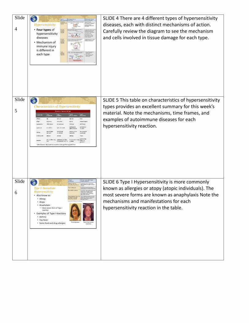
4 Hypersensitivity
• Four types of hypersensitivity diseases
• Mechanism of immune injury is different in each type
SLIDE 4 There are 4 different types of hypersensitivity diseases, each with distinct mechanisms of action. Carefully review the diagram to see the mechanism and cells involved in tissue damage for each type.
Slide
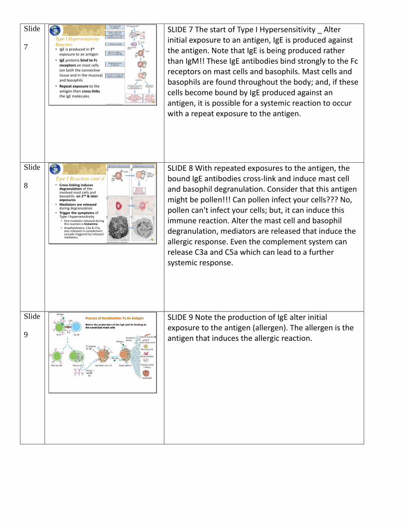
5 Characteristics of Hypersensitivity
SLIDE 5 This table on characteristics of hypersensitivity types provides an excellent summary for this week’s material. Note the mechanisms, time frames, and examples of autoimmune diseases for each hypersensitivity reaction.
Slide
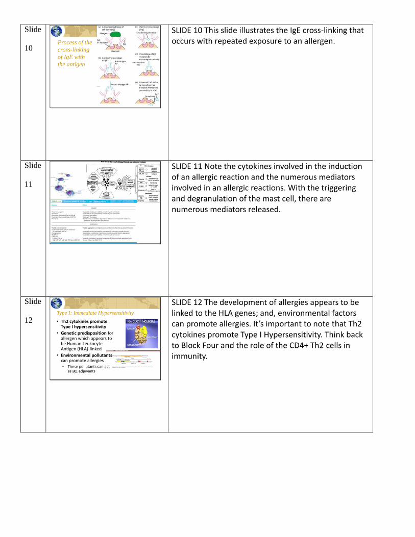
6 Type I: Immediate
Hypersensitivity
• Also know as:
• Allergy
• Atopy
• Anaphylaxis• Most severe form of Type I
reaction
• Examples of Type I reactions
• Asthma
• Hay fever
• Some food and drug allergies
SLIDE 6 Type I Hypersensitivity is more commonly known as allergies or atopy (atopic individuals). The most severe forms are known as anaphylaxis Note the mechanisms and manifestations for each hypersensitivity reaction in the table.
Slide
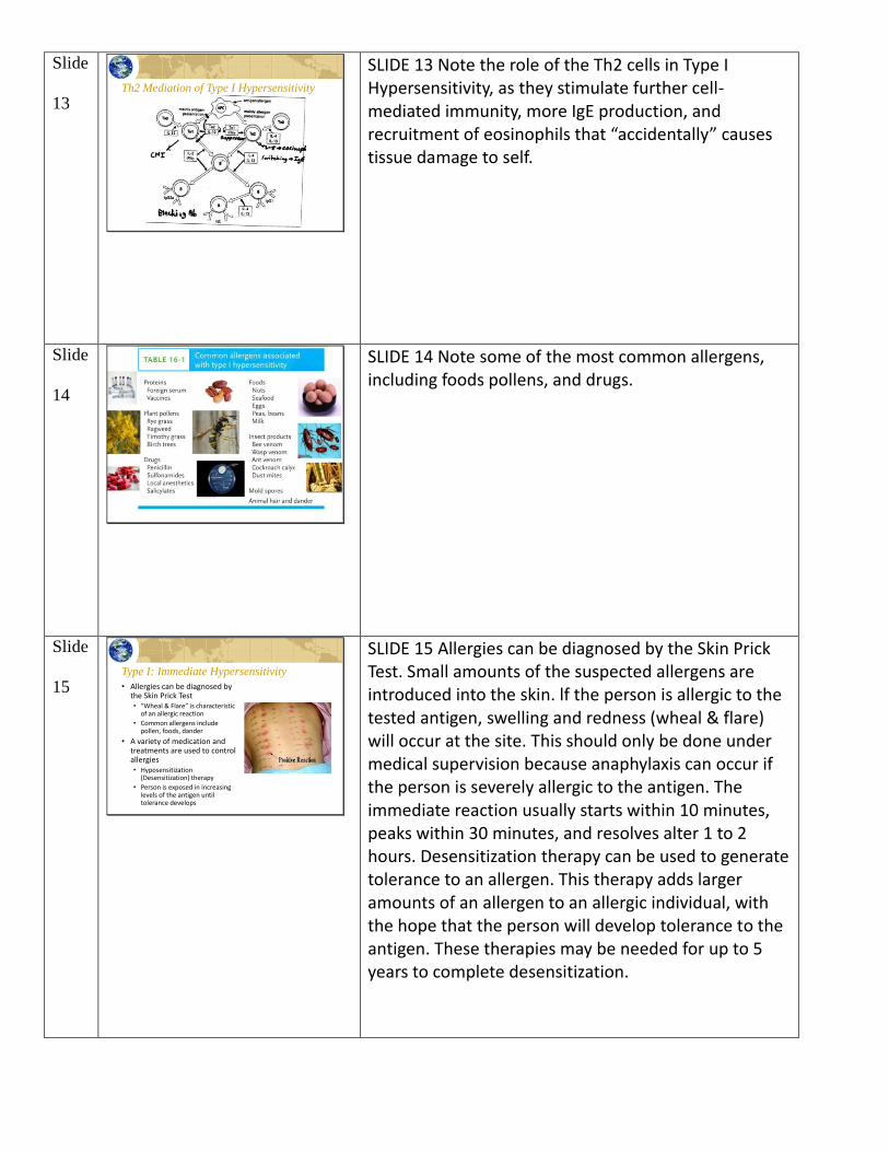
7
Type I Hypersensitivity
Reaction• IgE is produced in 1st
exposure to an antigen
• IgE proteins bind to Fc receptors on mast cells (on both the connective tissue and in the mucosa) and basophils
• Repeat exposure to the antigen then cross-linksthe IgE molecules
SLIDE 7 The start of Type I Hypersensitivity _ Alter initial exposure to an antigen, IgE is produced against the antigen. Note that IgE is being produced rather than IgM!! These IgE antibodies bind strongly to the Fc receptors on mast cells and basophils. Mast cells and basophils are found throughout the body; and, if these cells become bound by IgE produced against an antigen, it is possible for a systemic reaction to occur with a repeat exposure to the antigen.
Slide
8 Type I Reaction cont’d• Cross-linking induces
degranulation of the involved mast cells and basophils- on 2nd & later exposures
• Mediators are released during degranulation
• Trigger the symptoms of Type I hypersensitivity• One mediator released during
this reaction is histamine• Anaphylatoxins, C3a & C5a,
also released in complement cascade triggered by released mediators.
SLIDE 8 With repeated exposures to the antigen, the bound IgE antibodies cross-link and induce mast cell and basophil degranulation. Consider that this antigen might be pollen!!! Can pollen infect your cells??? No, pollen can't infect your cells; but, it can induce this immune reaction. Alter the mast cell and basophil degranulation, mediators are released that induce the allergic response. Even the complement system can release C3a and C5a which can lead to a further systemic response.
Slide
9
SLIDE 9 Note the production of IgE alter initial exposure to the antigen (allergen). The allergen is the antigen that induces the allergic reaction.
Slide
10 Process of the
cross-linking
of IgE with
the antigen
SLIDE 10 This slide illustrates the IgE cross-linking that occurs with repeated exposure to an allergen.
Slide
11
SLIDE 11 Note the cytokines involved in the induction of an allergic reaction and the numerous mediators involved in an allergic reactions. With the triggering and degranulation of the mast cell, there are numerous mediators released.
Slide
12 Type I: Immediate Hypersensitivity
• Th2 cytokines promote Type I hypersensitivity
• Genetic predisposition for allergen which appears to be Human Leukocyte Antigen (HLA)-linked
• Environmental pollutants can promote allergies• These pollutants can act
as IgE adjuvants
SLIDE 12 The development of allergies appears to be linked to the HLA genes; and, environmental factors can promote allergies. It’s important to note that Th2 cytokines promote Type I Hypersensitivity. Think back to Block Four and the role of the CD4+ Th2 cells in immunity.
Slide
13 Th2 Mediation of Type I Hypersensitivity
SLIDE 13 Note the role of the Th2 cells in Type I Hypersensitivity, as they stimulate further cell-mediated immunity, more IgE production, and recruitment of eosinophils that “accidentally” causes tissue damage to self.
Slide
14
SLIDE 14 Note some of the most common allergens, including foods pollens, and drugs.
Slide
15 Type I: Immediate Hypersensitivity
• Allergies can be diagnosed by the Skin Prick Test• “Wheal & Flare” is characteristic
of an allergic reaction
• Common allergens include pollen, foods, dander
• A variety of medication and treatments are used to control allergies• Hyposensitization
(Desensitization) therapy
• Person is exposed in increasing levels of the antigen until tolerance develops
SLIDE 15 Allergies can be diagnosed by the Skin Prick Test. Small amounts of the suspected allergens are introduced into the skin. lf the person is allergic to the tested antigen, swelling and redness (wheal & flare) will occur at the site. This should only be done under medical supervision because anaphylaxis can occur if the person is severely allergic to the antigen. The immediate reaction usually starts within 10 minutes, peaks within 30 minutes, and resolves alter 1 to 2 hours. Desensitization therapy can be used to generate tolerance to an allergen. This therapy adds larger amounts of an allergen to an allergic individual, with the hope that the person will develop tolerance to the antigen. These therapies may be needed for up to 5 years to complete desensitization.
Slide
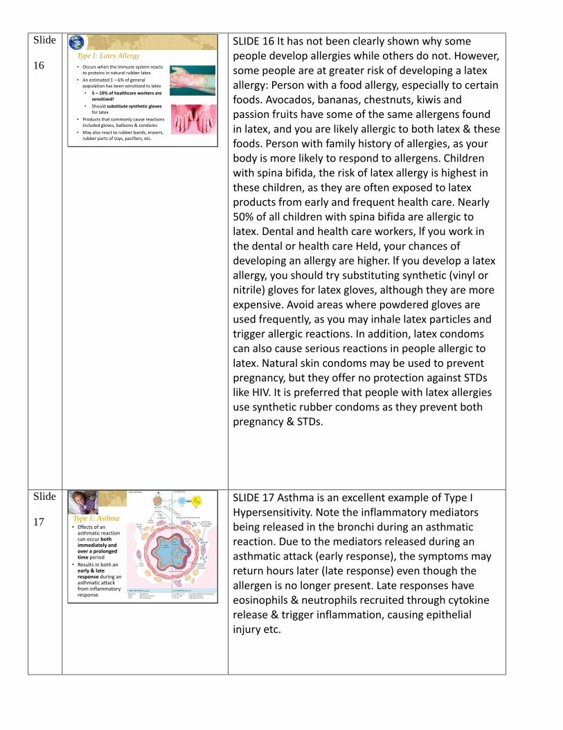
16 Type I: Latex Allergy
• Occurs when the immune system reacts to proteins in natural rubber latex
• An estimated 1 – 6% of general population has been sensitized to latex
• 5 – 10% of healthcare workers are sensitized!
• Should substitute synthetic gloves for latex
• Products that commonly cause reactions included gloves, balloons & condoms
• May also react to rubber bands, erasers, rubber parts of toys, pacifiers, etc.
SLIDE 16 It has not been clearly shown why some people develop allergies while others do not. However, some people are at greater risk of developing a latex allergy: Person with a food allergy, especially to certain foods. Avocados, bananas, chestnuts, kiwis and passion fruits have some of the same allergens found in latex, and you are likely allergic to both latex & these foods. Person with family history of allergies, as your body is more likely to respond to allergens. Children with spina bifida, the risk of latex allergy is highest in these children, as they are often exposed to latex products from early and frequent health care. Nearly 50% of all children with spina bifida are allergic to latex. Dental and health care workers, lf you work in the dental or health care Held, your chances of developing an allergy are higher. lf you develop a latex allergy, you should try substituting synthetic (vinyl or nitrile) gloves for latex gloves, although they are more expensive. Avoid areas where powdered gloves are used frequently, as you may inhale latex particles and trigger allergic reactions. In addition, latex condoms can also cause serious reactions in people allergic to latex. Natural skin condoms may be used to prevent pregnancy, but they offer no protection against STDs like HIV. It is preferred that people with latex allergies use synthetic rubber condoms as they prevent both pregnancy & STDs.
Slide
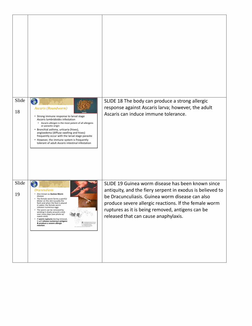
17 Type 1: Asthma• Effects of an
asthmatic reaction can occur both immediately and over a prolonged time period
• Results in both an early & late response during an asthmatic attack from inflammatory response
SLIDE 17 Asthma is an excellent example of Type I Hypersensitivity. Note the inflammatory mediators being released in the bronchi during an asthmatic reaction. Due to the mediators released during an asthmatic attack (early response), the symptoms may return hours later (late response) even though the allergen is no longer present. Late responses have eosinophils & neutrophils recruited through cytokine release & trigger inflammation, causing epithelial injury etc.
Slide
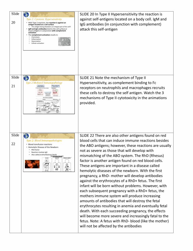
18 Ascaris (Roundworm)
• Strong immune response to larval stage Ascaris lumbridoides infestation• Ascaris allergen is the most potent of all allergens
or parasitic origin
• Bronchial asthma, urticaria (hives), angioedema (diffuse swelling and hives) frequently occur with the larval stage parasite
• However, the immune system is frequently tolerant of adult Ascaris intestinal infestation
SLIDE 18 The body can produce a strong allergic response against Ascaris larva; however, the adult Ascaris can induce immune tolerance.
Slide
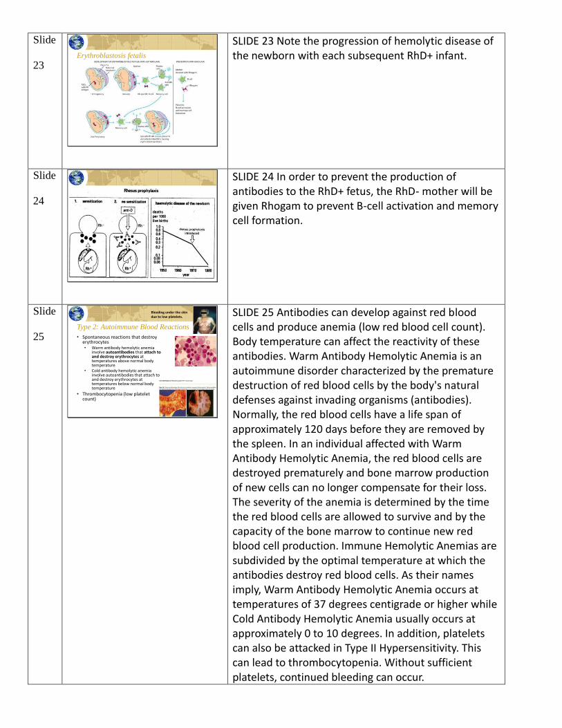
19 Dracunuliasis• Also known as Guinea Worm
disease• The female worm forms a painful
blister on the skin (usually the feet) and when the foot is placed in water, the female worm releases numerous eggs
• The worm can be removed by winding it slowly around a stick over many days (see photo w/ match stick)
• If worm ruptures during removal, it will release numerous antigens & produce a severe allergic reaction.
SLIDE 19 Guinea worm disease has been known since antiquity, and the fiery serpent in exodus is believed to be Dracunculiasis. Guinea worm disease can also produce severe allergic reactions. lf the female worm ruptures as it is being removed, antigens can be released that can cause anaphylaxis.
Slide
20 Type 2: Cytotoxic Hypersensitivity
• With Type 2 reactions, the reaction is against an antigen located on a cell surface• The antigen being attacked is an integral part of the cell!!
• IgM and IgG antibodies bind to the cell surface or tissue antigens in conjunction with complement activation
• The complement activation results in:• Chemotaxis• Inflammation• Opsonization• Cellular activation
SLIDE 20 In Type II Hypersensitivity the reaction is against self-antigens located on a body cell. IgM and IgG antibodies (in conjunction with complement) attack this self-antigen
Slide
21 Type 2 Mediated Immunopathology
SLIDE 21 Note the mechanism of Type II Hypersensitivity, as complement binding to Fc receptors on neutrophils and macrophages recruits these cells to destroy the self antigen. Watch the 3 mechanisms of Type II cytotoxicity in the animations provided.
Slide
22 Type 2: Blood Immunopathologies
• Blood transfusion reactions
• Hemolytic Disease of the Newborn• RhD factor
• Reaction involves IgG
• Also called erythorblastosis fetalis
SLIDE 22 There are also other antigens found on red blood cells that can induce immune reactions besides the ABO antigens; however, these reactions are usually not as severe as those that will develop with mismatching of the ABO system. The RhD (Rhesus) factor is another antigen found on red blood cells. These antigens are important in a disease called hemolytic diseases of the newborn. With the first pregnancy, a RhD- mother will develop antibodies against the erythrocytes of a RhD+ fetus. The first infant will be born without problems. However, with each subsequent pregnancy with a RhD+ fetus, the mothers immune system will produce increasing amounts of antibodies that will destroy the fetal erythrocytes resulting in anemia and eventually fetal death. With each succeeding pregnancy, the effects will become more severe and increasingly fatal to the fetus. Note: A fetus with RhD- blood (like the mother) will not be affected by the antibodies
Slide
23 Erythroblastosis fetalis
SLIDE 23 Note the progression of hemolytic disease of the newborn with each subsequent RhD+ infant.
Slide
24
SLIDE 24 In order to prevent the production of antibodies to the RhD+ fetus, the RhD- mother will be given Rhogam to prevent B-cell activation and memory cell formation.
Slide
25 Type 2: Autoimmune Blood Reactions
• Spontaneous reactions that destroy erythrocytes• Warm antibody hemolytic anemia
involve autoantibodies that attach to and destroy erythrocytes at temperatures above normal body temperature
• Cold antibody hemolytic anemia involve autoantibodies that attach to and destroy erythrocytes at temperatures below normal body temperature
• Thrombocytopenia (low platelet count)
Bleeding under the skin due to low platelets.
SLIDE 25 Antibodies can develop against red blood cells and produce anemia (low red blood cell count). Body temperature can affect the reactivity of these antibodies. Warm Antibody Hemolytic Anemia is an autoimmune disorder characterized by the premature destruction of red blood cells by the body's natural defenses against invading organisms (antibodies). Normally, the red blood cells have a life span of approximately 120 days before they are removed by the spleen. In an individual affected with Warm Antibody Hemolytic Anemia, the red blood cells are destroyed prematurely and bone marrow production of new cells can no longer compensate for their loss. The severity of the anemia is determined by the time the red blood cells are allowed to survive and by the capacity of the bone marrow to continue new red blood cell production. Immune Hemolytic Anemias are subdivided by the optimal temperature at which the antibodies destroy red blood cells. As their names imply, Warm Antibody Hemolytic Anemia occurs at temperatures of 37 degrees centigrade or higher while Cold Antibody Hemolytic Anemia usually occurs at approximately 0 to 10 degrees. In addition, platelets can also be attacked in Type II Hypersensitivity. This can lead to thrombocytopenia. Without sufficient platelets, continued bleeding can occur.
Slide
26
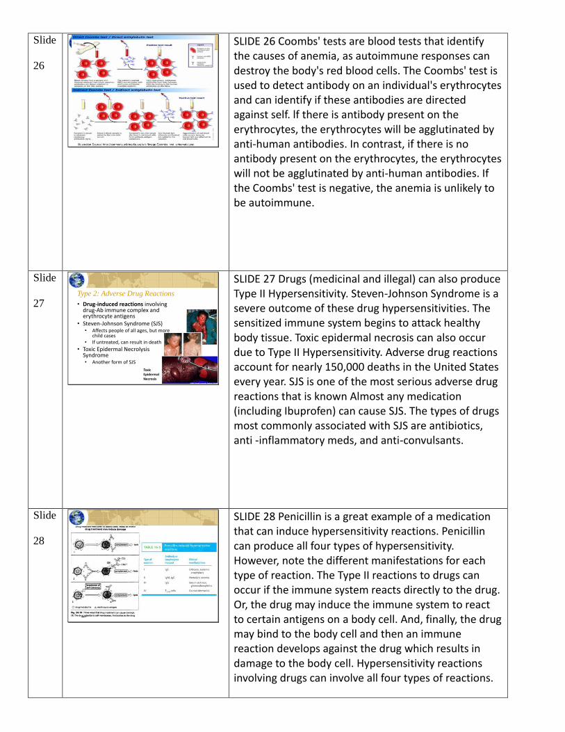
SLIDE 26 Coombs' tests are blood tests that identify the causes of anemia, as autoimmune responses can destroy the body's red blood cells. The Coombs' test is used to detect antibody on an individual's erythrocytes and can identify if these antibodies are directed against self. lf there is antibody present on the erythrocytes, the erythrocytes will be agglutinated by anti-human antibodies. In contrast, if there is no antibody present on the erythrocytes, the erythrocytes will not be agglutinated by anti-human antibodies. If the Coombs' test is negative, the anemia is unlikely to be autoimmune.
Slide
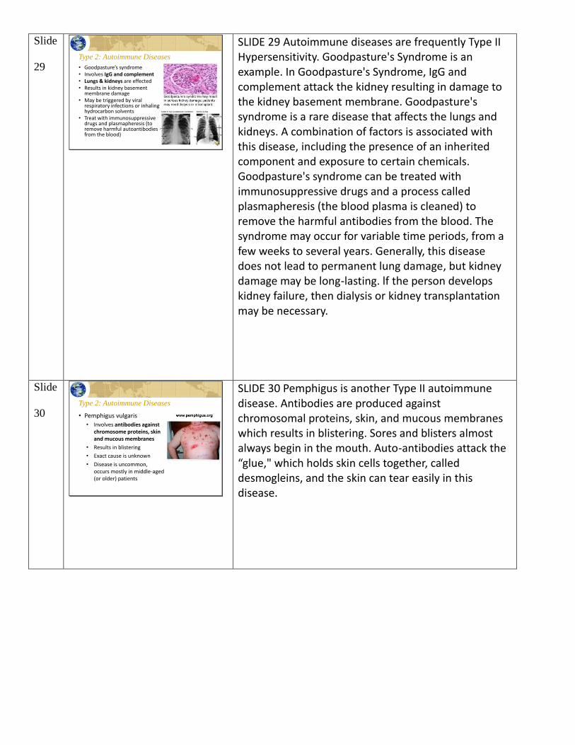
27 Type 2: Adverse Drug Reactions
• Drug-induced reactions involving drug-Ab immune complex and erythrocyte antigens
• Steven-Johnson Syndrome (SJS)• Affects people of all ages, but more
child cases• If untreated, can result in death
• Toxic Epidermal NecrolysisSyndrome• Another form of SJS
Toxic Epidermal Necrosis
SLIDE 27 Drugs (medicinal and illegal) can also produce Type II Hypersensitivity. Steven-Johnson Syndrome is a severe outcome of these drug hypersensitivities. The sensitized immune system begins to attack healthy body tissue. Toxic epidermal necrosis can also occur due to Type II Hypersensitivity. Adverse drug reactions account for nearly 150,000 deaths in the United States every year. SJS is one of the most serious adverse drug reactions that is known Almost any medication (including Ibuprofen) can cause SJS. The types of drugs most commonly associated with SJS are antibiotics, anti -inflammatory meds, and anti-convulsants.
Slide
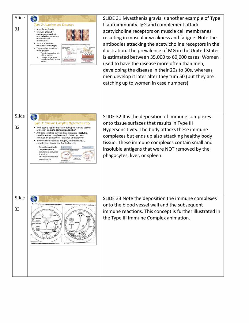
28
SLIDE 28 Penicillin is a great example of a medication that can induce hypersensitivity reactions. Penicillin can produce all four types of hypersensitivity. However, note the different manifestations for each type of reaction. The Type II reactions to drugs can occur if the immune system reacts directly to the drug. Or, the drug may induce the immune system to react to certain antigens on a body cell. And, finally, the drug may bind to the body cell and then an immune reaction develops against the drug which results in damage to the body cell. Hypersensitivity reactions involving drugs can involve all four types of reactions.
Slide
29 Type 2: Autoimmune Diseases
• Goodpasture’s syndrome• Involves IgG and complement• Lungs & kidneys are effected• Results in kidney basement
membrane damage• May be triggered by viral
respiratory infections or inhaling hydrocarbon solvents
• Treat with immunosuppressive drugs and plasmapheresis (to remove harmful autoantibodies from the blood)
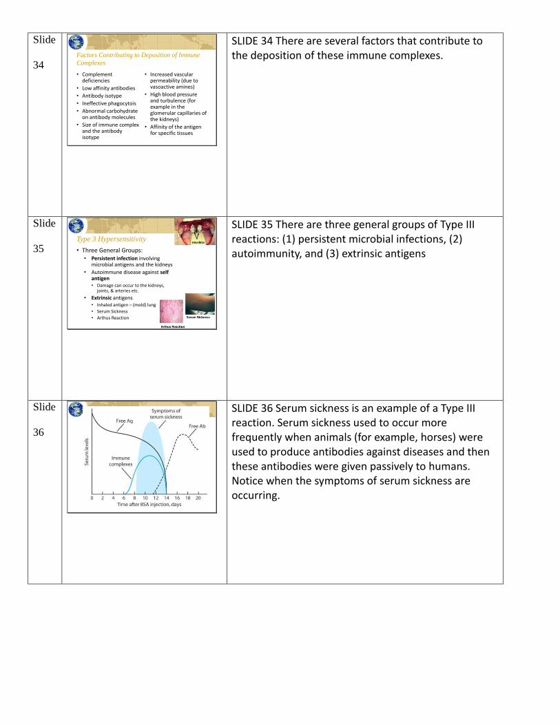
SLIDE 29 Autoimmune diseases are frequently Type II Hypersensitivity. Goodpasture's Syndrome is an example. In Goodpasture's Syndrome, IgG and complement attack the kidney resulting in damage to the kidney basement membrane. Goodpasture's syndrome is a rare disease that affects the lungs and kidneys. A combination of factors is associated with this disease, including the presence of an inherited component and exposure to certain chemicals. Goodpasture's syndrome can be treated with immunosuppressive drugs and a process called plasmapheresis (the blood plasma is cleaned) to remove the harmful antibodies from the blood. The syndrome may occur for variable time periods, from a few weeks to several years. Generally, this disease does not lead to permanent lung damage, but kidney damage may be long-lasting. lf the person develops kidney failure, then dialysis or kidney transplantation may be necessary.
Slide
30 Type 2: Autoimmune Diseases
• Pemphigus vulgaris
• Involves antibodies against chromosome proteins, skin and mucous membranes
• Results in blistering
• Exact cause is unknown
• Disease is uncommon, occurs mostly in middle-aged (or older) patients
SLIDE 30 Pemphigus is another Type II autoimmune disease. Antibodies are produced against chromosomal proteins, skin, and mucous membranes which results in blistering. Sores and blisters almost always begin in the mouth. Auto-antibodies attack the “glue," which holds skin cells together, called desmogleins, and the skin can tear easily in this disease.
Slide
31 Type 2: Autoimmune Diseases
• Myasthenia Gravis• Involves IgG and
complement against acetylcholine receptorson muscle cell membranes
• Results in muscle weakness and fatigue
• Thymus abnormalities often present• Thymic tumors found in
10% of patients• Changes in germinal
centers found in 70% of patients
SLIDE 31 Myasthenia gravis is another example of Type II autoimmunity. IgG and complement attack acetylcholine receptors on muscle cell membranes resulting in muscular weakness and fatigue. Note the antibodies attacking the acetylcholine receptors in the illustration. The prevalence of MG in the United States is estimated between 35,000 to 60,000 cases. Women used to have the disease more often than men, developing the disease in their 20s to 30s, whereas men develop it later alter they tum 50 (but they are catching up to women in case numbers).
Slide
32 Type 3: Immune Complex Hypersensitivity
• With type 3 hypersensitivity, damage occurs to tissues at sites of immune complex deposition
• Antigens involved in Type 3 reactions are insoluble, small immune complexes which have not been removed by phagocytes, the liver, or the spleen
• Involves the deposited antigen, antibodies (IgG), complement deposition & effector cells
• The antigen-antibody complexes induce complement activation & result in inflammation mediated by neutrophils
SLIDE 32 It is the deposition of immune complexes onto tissue surfaces that results in Type III Hypersensitivity. The body attacks these immune complexes but ends up also attacking healthy body tissue. These immune complexes contain small and insoluble antigens that were NOT removed by the phagocytes, liver, or spleen.
Slide
33
SLIDE 33 Note the deposition the immune complexes onto the blood vessel wall and the subsequent immune reactions. This concept is further illustrated in the Type III Immune Complex animation.
Slide
34 Factors Contributing to Deposition of Immune
Complexes
• Complement deficiencies
• Low affinity antibodies
• Antibody isotype
• Ineffective phagocytois
• Abnormal carbohydrate on antibody molecules
• Size of immune complex and the antibody isotype
• Increased vascular permeability (due to vasoactive amines)
• High blood pressure and turbulence (for example in the glomerular capillaries of the kidneys)
• Affinity of the antigen for specific tissues
SLIDE 34 There are several factors that contribute to the deposition of these immune complexes.
Slide
35 • Three General Groups:• Persistent infection involving
microbial antigens and the kidneys
• Autoimmune disease against self antigen• Damage can occur to the kidneys,
joints, & arteries etc.
• Extrinsic antigens• Inhaled antigen – (mold) lung
• Serum Sickness
• Arthus Reaction
Type 3 Hypersensitivity
SLIDE 35 There are three general groups of Type III reactions: (1) persistent microbial infections, (2) autoimmunity, and (3) extrinsic antigens
Slide
36
SLIDE 36 Serum sickness is an example of a Type III reaction. Serum sickness used to occur more frequently when animals (for example, horses) were used to produce antibodies against diseases and then these antibodies were given passively to humans. Notice when the symptoms of serum sickness are occurring.
Slide
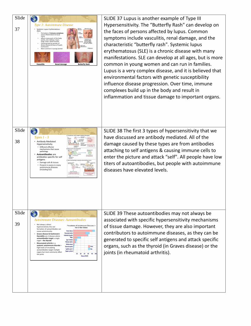
37 Type 3: Autoimmune Disease
• Systemic Lupus Erythematosus(SLE)• Formation of immune complexes
cause inflammation & tissue injury
• Affects many parts of the body (joints, skin, kidneys, heart, lungs, brain, blood vessels)
• Characterized by periods of illness (flares) & times of health (remission)
• More common in young women
SLIDE 37 Lupus is another example of Type III Hypersensitivity. The “Butterfly Rash" can develop on the faces of persons affected by lupus. Common symptoms include vasculitis, renal damage, and the characteristic “butterfly rash". Systemic lupus erythematosus (SLE) is a chronic disease with many manifestations. SLE can develop at all ages, but is more common in young women and can run in families. Lupus is a very complex disease, and it is believed that environmental factors with genetic susceptibility influence disease progression. Over time, immune complexes build up in the body and result in inflammation and tissue damage to important organs.
Slide
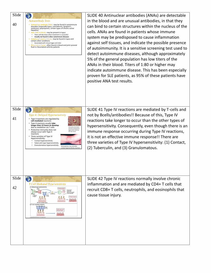
38 Types 1 - 3
• Antibody Mediated Hypersensitivity• Different effector
mechanisms that cause pathology
• Autoantibodies are antibodies specific for self antigens• Damage cells & tissues
• Present in excess in many autoimmune diseases (including SLE)
SLIDE 38 The first 3 types of hypersensitivity that we have discussed are antibody mediated. All of the damage caused by these types are from antibodies attaching to self antigens & causing immune cells to enter the picture and attack “self". All people have low titers of autoantibodies, but people with autoimmune diseases have elevated levels.
Slide
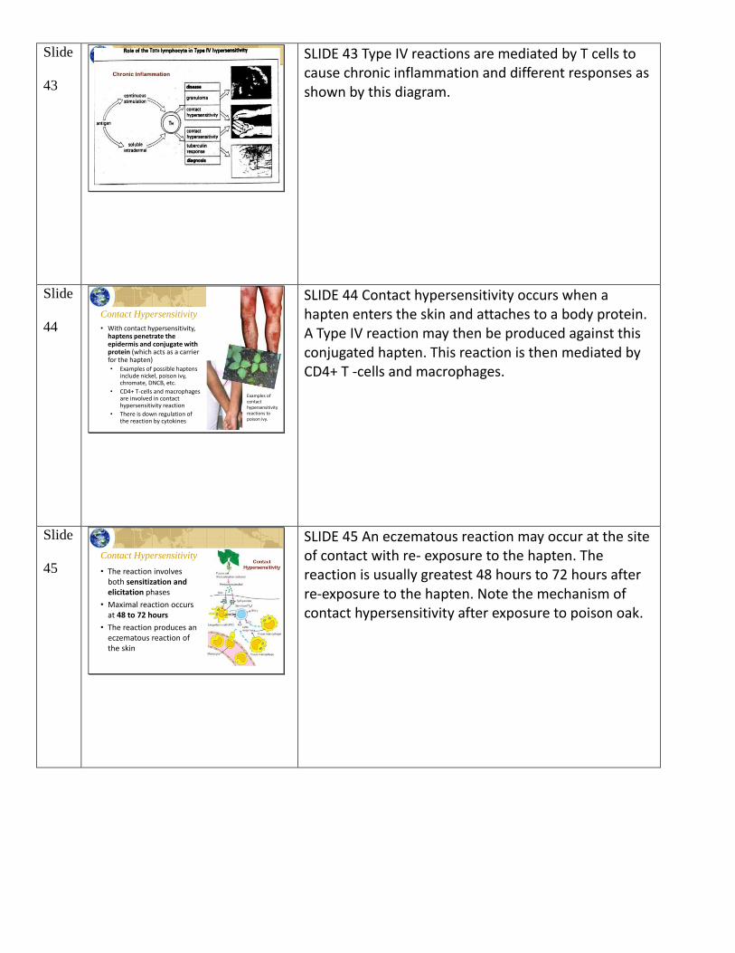
39 Autoimmune Diseases: Autoantibodies
• Not always a direct hypersensitivity link, but formation of autoantibodies can cause autoimmunity
• Graves disease & Hashimoto’s thyroiditis are 2 disease where autoantibodies target a single organ – the thyroid
• Rheumatoid arthritis is a systemic autoimmune disorder –high levels of circulating autoantibodies target multiple organs but most commonly affect the joints
SLIDE 39 These autoantibodies may not always be associated with specific hypersensitivity mechanisms of tissue damage. However, they are also important contributors to autoimmune diseases, as they can be generated to specific self antigens and attack specific organs, such as the thyroid (in Graves disease) or the joints (in rheumatoid arthritis).
Slide
40 Autoantibody Tests
• Antinuclear antibody (ANA): may be found in autoimmune disorders [especially lupus, scleroderma, Sjorgren’ssyndrome, polymyositis, certain types of chronic active hepatitis]
• Anti-DNA antibody: may be present in lupus• Titers will decrease when treatment is successful• Usually not found in other autoimmune diseases
• Antiphospholipid antibody: may be found in lupus and certain other conditions• Associated with miscarriages and clots
• Rheumatoid factor: often found in blood and joint synovial fluid in rheumatoid arthritis patients
SLIDE 40 Antinuclear antibodies (ANAs) are detectable in the blood and are unusual antibodies, in that they can bind to certain structures within the nucleus of the cells. ANAs are found in patients whose immune system may be predisposed to cause inflammation against self tissues, and indicate the possible presence of autoimmunity. It is a sensitive screening test used to detect autoimmune diseases, although approximately 5% of the general population has low titers of the ANAs in their blood. Titers of 1:80 or higher may indicate autoimmune disease. This has been especially proven for SLE patients, as 95% of these patients have positive ANA test results.
Slide
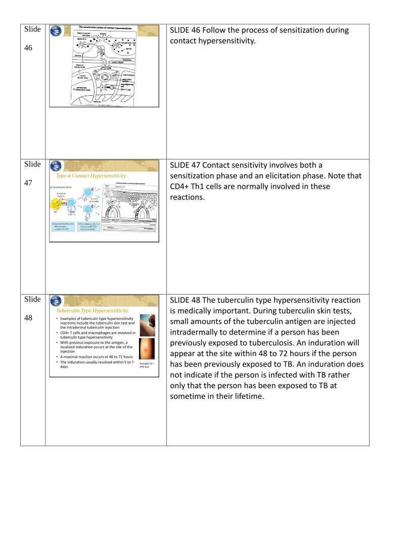
41 Type 4: Delayed Hypersensitivity
• Type 4 reactions are regulated by cell-mediated reactions
• Type 4 reactions usually take longer than 12 hours to develop due to mediation via T-cells
• Protective immunity does not always occur with Type 4 reactions
• Three varieties of Type IV Hypersensitivity:• Contact hypersensitivity• Tuberculin type hypersensitivity• Granulomatous hypersensitivity
Granuloma formation around a schistosomeegg (center) destroys the liver tissue.
Wristwatches may stimulate contact hypersensitivity reactions.
SLIDE 41 Type IV reactions are mediated by T-cells and not by Bcells/antibodies!! Because of this, Type IV reactions take longer to occur than the other types of hypersensitivity. Consequently, even though there is an immune response occurring during Type IV reactions, it is not an effective immune response!! There are three varieties of Type IV hypersensitivity: (1) Contact, (2) Tuberculin, and (3) Granulomatous.
Slide
42 T Cell Mediated Hypersensitivity
SLIDE 42 Type IV reactions normally involve chronic inflammation and are mediated by CD4+ T cells that recruit CD8+ T cells, neutrophils, and eosinophils that cause tissue injury.
Slide
43
SLIDE 43 Type IV reactions are mediated by T cells to cause chronic inflammation and different responses as shown by this diagram.
Slide
44 Contact Hypersensitivity
• With contact hypersensitivity, haptens penetrate the epidermis and conjugate with protein (which acts as a carrier for the hapten)• Examples of possible haptens
include nickel, poison ivy, chromate, DNCB, etc.
• CD4+ T-cells and macrophages are involved in contact hypersensitivity reaction
• There is down regulation of the reaction by cytokines
Examples of contact hypersensitivity reactions to poison ivy.
SLIDE 44 Contact hypersensitivity occurs when a hapten enters the skin and attaches to a body protein. A Type IV reaction may then be produced against this conjugated hapten. This reaction is then mediated by CD4+ T -cells and macrophages.
Slide
45 Contact Hypersensitivity
• The reaction involves both sensitization and elicitation phases
• Maximal reaction occurs at 48 to 72 hours
• The reaction produces an eczematous reaction of the skin
SLIDE 45 An eczematous reaction may occur at the site of contact with re- exposure to the hapten. The reaction is usually greatest 48 hours to 72 hours after re-exposure to the hapten. Note the mechanism of contact hypersensitivity after exposure to poison oak.
Slide
46
SLIDE 46 Follow the process of sensitization during contact hypersensitivity.
Slide
47 Type 4 Contact Hypersensitivity
SLIDE 47 Contact sensitivity involves both a sensitization phase and an elicitation phase. Note that CD4+ Th1 cells are normally involved in these reactions.
Slide
48 Tuberculin Type Hypersensitivity
• Examples of tuberculin type hypersensitivity reactions include the tuberculin skin test and the intradermal tuberculin injection
• CD4+ T cells and macrophages are involved in tuberculin type hypersensitivity
• With previous exposure to the antigen, a localized induration occurs at the site of the injection
• A maximal reaction occurs at 48 to 72 hours
• The induration usually resolved within 5 to 7 days
Example of + PPD test
SLIDE 48 The tuberculin type hypersensitivity reaction is medically important. During tuberculin skin tests, small amounts of the tuberculin antigen are injected intradermally to determine if a person has been previously exposed to tuberculosis. An induration will appear at the site within 48 to 72 hours if the person has been previously exposed to TB. An induration does not indicate if the person is infected with TB rather only that the person has been exposed to TB at sometime in their lifetime.
Slide
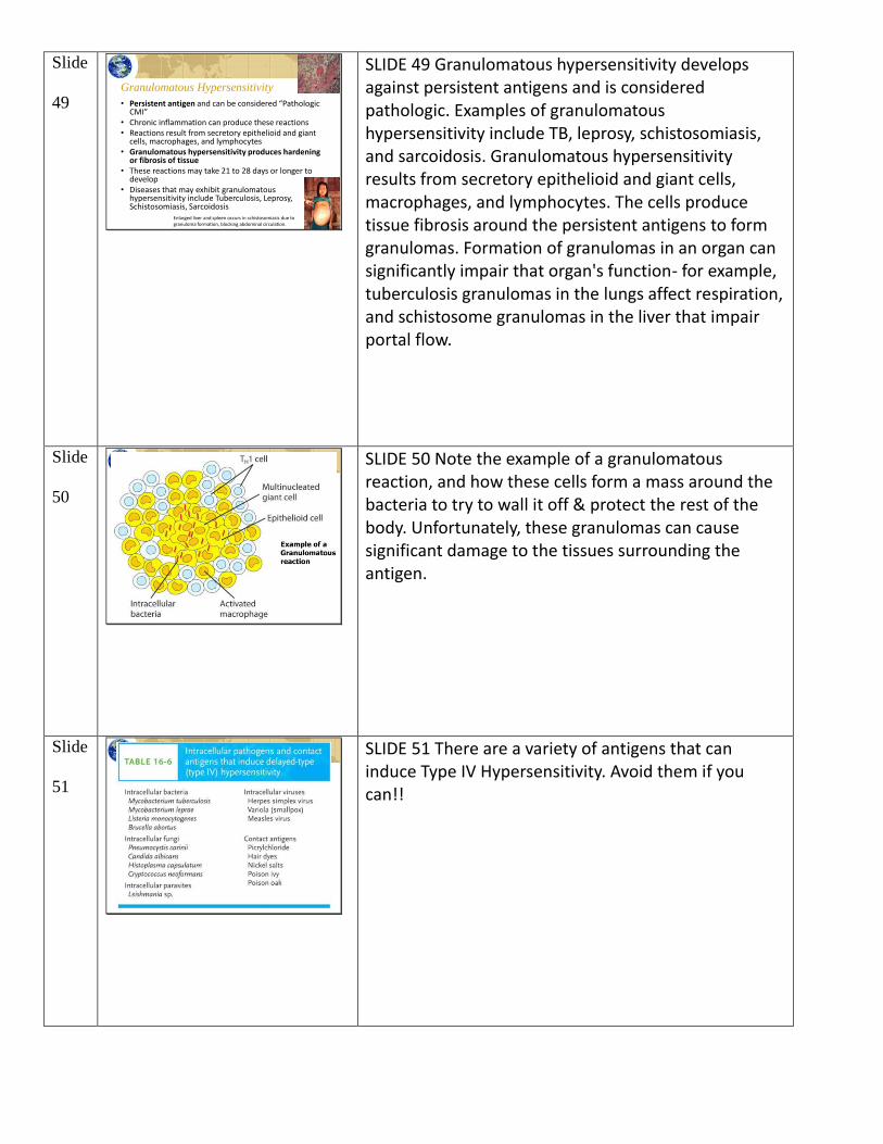
49 Granulomatous Hypersensitivity
• Persistent antigen and can be considered “Pathologic CMI”
• Chronic inflammation can produce these reactions• Reactions result from secretory epithelioid and giant
cells, macrophages, and lymphocytes• Granulomatous hypersensitivity produces hardening
or fibrosis of tissue• These reactions may take 21 to 28 days or longer to
develop• Diseases that may exhibit granulomatous
hypersensitivity include Tuberculosis, Leprosy, Schistosomiasis, Sarcoidosis
Enlarged liver and spleen occurs in schistosomiasis due to granuloma formation, blocking abdominal circulation.
SLIDE 49 Granulomatous hypersensitivity develops against persistent antigens and is considered pathologic. Examples of granulomatous hypersensitivity include TB, leprosy, schistosomiasis, and sarcoidosis. Granulomatous hypersensitivity results from secretory epithelioid and giant cells, macrophages, and lymphocytes. The cells produce tissue fibrosis around the persistent antigens to form granulomas. Formation of granulomas in an organ can significantly impair that organ's function- for example, tuberculosis granulomas in the lungs affect respiration, and schistosome granulomas in the liver that impair portal flow.
Slide
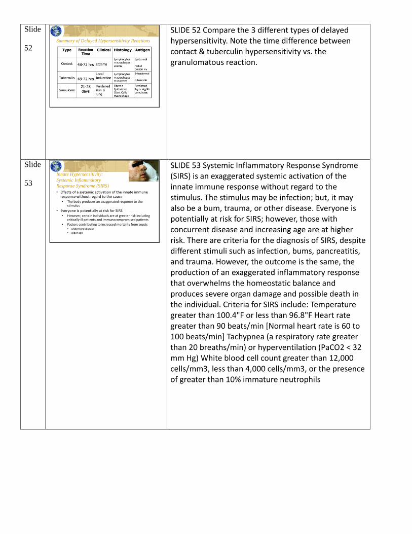
50
SLIDE 50 Note the example of a granulomatous reaction, and how these cells form a mass around the bacteria to try to wall it off & protect the rest of the body. Unfortunately, these granulomas can cause significant damage to the tissues surrounding the antigen.
Slide
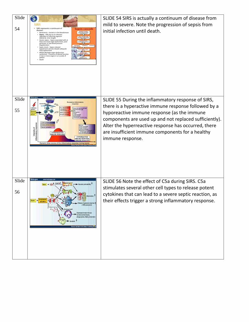
51
SLIDE 51 There are a variety of antigens that can induce Type IV Hypersensitivity. Avoid them if you can!!
Slide
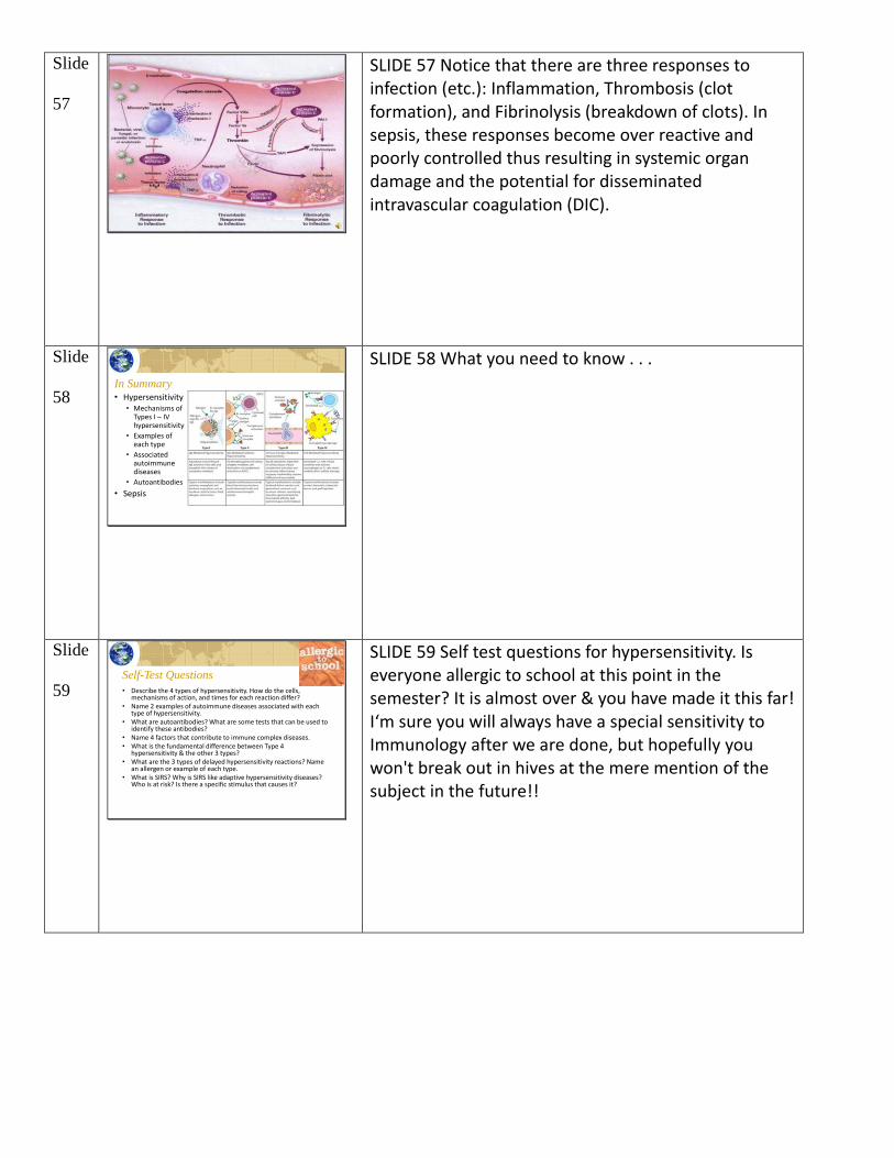
52 Summary of Delayed Hypersensitivity Reactions
SLIDE 52 Compare the 3 different types of delayed hypersensitivity. Note the time difference between contact & tuberculin hypersensitivity vs. the granulomatous reaction.
Slide
53
Innate Hypersensitivity:
Systemic Inflammatory
Response Syndrome (SIRS)
• Effects of a systemic activation of the innate immune response without regard to the cause• The body produces an exaggerated response to the
stimulus
• Everyone is potentially at risk for SIRS• However, certain individuals are at greater risk including
critically ill patients and immunocompromised patients
• Factors contributing to increased mortality from sepsis• underlying disease
• older age
SLIDE 53 Systemic Inflammatory Response Syndrome (SIRS) is an exaggerated systemic activation of the innate immune response without regard to the stimulus. The stimulus may be infection; but, it may also be a bum, trauma, or other disease. Everyone is potentially at risk for SIRS; however, those with concurrent disease and increasing age are at higher risk. There are criteria for the diagnosis of SIRS, despite different stimuli such as infection, bums, pancreatitis, and trauma. However, the outcome is the same, the production of an exaggerated inflammatory response that overwhelms the homeostatic balance and produces severe organ damage and possible death in the individual. Criteria for SIRS include: Temperature greater than 100.4"F or less than 96.8"F Heart rate greater than 90 beats/min [Normal heart rate is 60 to 100 beats/min] Tachypnea (a respiratory rate greater than 20 breaths/min) or hyperventilation (PaCO2 < 32 mm Hg) White blood cell count greater than 12,000 cells/mm3, less than 4,000 cells/mm3, or the presence of greater than 10% immature neutrophils
Slide
54 SIRS• SIRS represents a continuum of
disease:• Bacteremia – bacteria in the bloodstream• Sepsis – SIRS due to an infection
regardless of infecting organism (bacterial, viral, fungal)
• Severe sepsis – Sepsis associated with at least one acute organ dysfunction, poor perfusion, or low blood pressure (hypotension)
• Septic shock – Sepsis-induced hypotension persists despite adequate fluid replacement
• MODS (Multiple organ dysfunction syndrome) – Presence of altered function in two or more organs in an acutely ill patient
• Death
SLIDE 54 SIRS is actually a continuum of disease from mild to severe. Note the progression of sepsis from initial infection until death.
Slide
55
SLIDE 55 During the inflammatory response of SIRS, there is a hyperactive immune response followed by a hyporeactive immune response (as the immune components are used up and not replaced sufficiently). Alter the hyperreactive response has occurred, there are insufficient immune components for a healthy immune response.
Slide
56
SLIDE 56 Note the effect of C5a during SIRS. C5a stimulates several other cell types to release potent cytokines that can lead to a severe septic reaction, as their effects trigger a strong inflammatory response.
Slide
57
SLIDE 57 Notice that there are three responses to infection (etc.): Inflammation, Thrombosis (clot formation), and Fibrinolysis (breakdown of clots). In sepsis, these responses become over reactive and poorly controlled thus resulting in systemic organ damage and the potential for disseminated intravascular coagulation (DIC).
Slide
58 In Summary• Hypersensitivity
• Mechanisms of Types I – IV hypersensitivity
• Examples of each type
• Associated autoimmune diseases
• Autoantibodies
• Sepsis
SLIDE 58 What you need to know . . .
Slide
59 Self-Test Questions
• Describe the 4 types of hypersensitivity. How do the cells, mechanisms of action, and times for each reaction differ?
• Name 2 examples of autoimmune diseases associated with each type of hypersensitivity.
• What are autoantibodies? What are some tests that can be used to identify these antibodies?
• Name 4 factors that contribute to immune complex diseases. • What is the fundamental difference between Type 4
hypersensitivity & the other 3 types?• What are the 3 types of delayed hypersensitivity reactions? Name
an allergen or example of each type.• What is SIRS? Why is SIRS like adaptive hypersensitivity diseases?
Who is at risk? Is there a specific stimulus that causes it?
SLIDE 59 Self test questions for hypersensitivity. Is everyone allergic to school at this point in the semester? It is almost over & you have made it this far! I‘m sure you will always have a special sensitivity to Immunology after we are done, but hopefully you won't break out in hives at the mere mention of the subject in the future!!