Negative regulation of Caenorhabditis elegansepidermal damage responses by death-associatedprotein kinaseAmy Tonga,b, Grace Lynnb, Vy Ngoa, Daniel Wongc,d,e, Sarah L. Moseleya, Jonathan J. Ewbankc,d,e,Alexandr Goncharovb,f, Yi-Chun Wug, Nathalie Pujolc,d,e, and Andrew D. Chisholma,b,1
aDepartment of Molecular, Cell and Developmental Biology, Sinsheimer Laboratories, University of California, Santa Cruz, CA 95064; bDivision of BiologicalSciences and fHoward Hughes Medical Institute, University of California at San Diego, 9500 Gilman Drive, La Jolla, CA 92093; cCentre d’Immunologie deMarseille-Luminy, Universite de la Mediterranee, Case 906, 13288 Marseille Cedex 9, France; dInstitut National de la Sante et de la Recherche Medicale,Unite 631, 13288 Marseille, France; eCentre National de la Recherche Scientifique, Unite Mixte de Recherche 6102, 13288 Marseille, France; and gInstitute ofMolecular and Cellular Biology, National Taiwan University, No. 1, Sec. 4, Roosevelt Road, Taipei, Taiwan 10617, Republic of China
Edited by Iva S. Greenwald, Columbia University, New York, NY, and approved November 26, 2008 (received for review September 18, 2008)
Wounding of epidermal layers triggers multiple coordinated re-sponses to damage. We show here that the Caenorhabditis elegansortholog of the tumor suppressor death-associated protein kinase,dapk-1, acts as a previously undescribed negative regulator ofbarrier repair and innate immune responses to wounding. Loss ofDAPK-1 function results in constitutive formation of scar-like struc-tures in the cuticle, and up-regulation of innate immune responsesto damage. Overexpression of DAPK-1 represses innate immuneresponses to needle wounding. Up-regulation of innate immuneresponses in dapk-1 requires the TIR-1/p38 signal transductionpathway; loss of function in this pathway synergizes with dapk-1to drastically reduce adult lifespan. Our results reveal a previouslyundescribed function for the DAPK tumor suppressor family inregulation of epithelial damage responses.
antimicrobial peptide � epidermis � innate immunity �wound repair � cuticle
The epidermis forms an outer protective barrier againstenvironmental damage and pathogens for most animals.
Epidermal responses to physical wounding have been extensivelycharacterized in vertebrates and insects (1–3). Repair of a barrierepithelium such as the skin involves several coordinated pro-cesses: choreographed movement of cells at the wound edge,leading to reepithelialization (4); scab formation followed bysynthesis of new external extracellular matrix (5, 6); and activa-tion of local cutaneous innate immune defenses such as theexpression of antimicrobial peptides (AMPs) that can defendagainst opportunistic infection at the wound site (7–9).
Despite the differing structures of epidermal layers in differ-ent animals, recent molecular genetic studies suggest that epi-dermal wound healing pathways are evolutionarily conserved.Activation of JNK and AP-1 transcription factors appears cen-tral to promoting cell motility at the leading edge of an epidermalwound (10). In both insects and vertebrates, transcription factorsof the Grainyhead family are activated in response to woundsignals via an ERK kinase pathway and promote transcription ofextracellular matrix cross-linking enzymes (11, 12). Less iscurrently known about the pathways that induce innate immuneresponses to sterile wounding; in human skin, the EGFR path-way has been implicated in local activation of AMPs (13). As wellas acting as antibiotics at the wound site, some AMPs maydirectly promote reepithelialization (14), linking these 2 arms ofthe wound response.
Like other immune responses, cutaneous responses to damagemust be tightly regulated to prevent chronic responses to tran-sient stimuli (15). Negative regulatory pathways prevent innateimmune responses to infection or wounds from developing intopathological reactions (15); defects in such negative regulationcan underlie chronic skin inflammatory diseases such as atopic
dermatitis (16). Loss of barrier repair functions can also resultin chronic inflammation (e.g., loss of AP-1 function blocksreepithelialization; see ref. 17); and has been implicated inpsoriasis (18), suggesting an intimate link between wound repairand regulation of innate immunity.
The Caenorhabditis elegans epidermis allows wound repairprocesses to be studied in the context of a simple epithelium thatsecretes an external collagenous cuticle. As in other animals, thenematode skin is likely to have an active role in preventingorganismal damage from physical or biological challenges. Werecently showed that laser or puncture wounding of C. elegansactivates epidermal innate responses via the Toll-interleukin 1receptor (TIR) domain protein TIR-1 and a p38 MAPK cascade(19). Here, we identify a new negative regulator of epidermaldamage responses, the C. elegans ortholog of the tumor sup-pressor death-associated protein kinase, dapk-1. Loss ofDAPK-1 function results in constitutive formation of scar-likestructures in the cuticle, and up-regulation of antimicrobial geneexpression. Transient overexpression of DAPK-1 represses thetranscriptional response to puncture wounding. We show thatup-regulation of innate immune responses, but not barrierrepair, in dapk-1 mutants requires the TIR-1/p38 MAPK path-way, and that dapk-1 mutants depend on this innate immunepathway for adult survival. Our results reveal a previouslyundescribed role for the DAPK tumor suppressor in negativeregulation of epithelial damage responses.
ResultsIn genetic screens for mutants displaying progressive defects inepidermal morphogenesis, we identified multiple alleles ofdapk-1, which encodes the C. elegans member of the calcium-calmodulin activated DAPK family [supporting information (SI)Fig. S1]; dapk-1 mutations form an allelic series (Table S1), inwhich the strongest allele, ju4, results in a missense alteration(S179L) in the peptide-binding ledge of the DAPK-1 kinasedomain. RNA interference phenocopied these dapk-1 epidermalphenotypes (data not shown), indicating that these mutationsresult in loss of DAPK-1 function.
Author contributions: A.T. and A.D.C. designed research; A.T., G.L., V.N., D.W., S.L.M., A.G.,and N.P. performed research; A.T. and Y.-C.W. contributed new reagents/analytic tools;D.W., J.J.E., N.P., and A.D.C. analyzed data; A.T., J.J.E., N.P., and A.D.C. wrote the paper.
The authors declare no conflict of interest.
This article is a PNAS Direct Submission.
Freely available online through the PNAS open access option.
1To whom correspondence should be addressed. E-mail: [email protected].
This article contains supporting information online at www.pnas.org/cgi/content/full/0809339106/DCSupplemental.
© 2009 by The National Academy of Sciences of the USA
www.pnas.org�cgi�doi�10.1073�pnas.0809339106 PNAS � February 3, 2009 � vol. 106 � no. 5 � 1457–1461
DEV
ELO
PMEN
TAL
BIO
LOG
Y
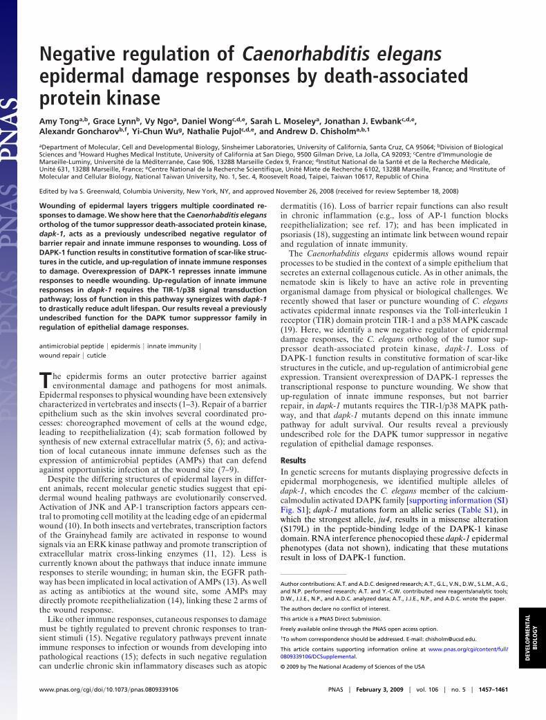
dapk-1 mutants appeared morphologically normal until midlarval development. Beginning in the L3 stage, dapk-1(ju4)mutants displayed striking and progressive defects in morphol-ogy of the epidermis and cuticle in specific body regions,especially in the nose, tail, vulva, and the dorsal midline in theregion of the posterior pharyngeal bulb (Fig. 1A). Cuticle inthese regions became up to 5–10 times thicker than the wild type,at the expense of underlying epidermis; this thickened cuticleappeared refractile under differential interference contrast(DIC) microscopy, and had aberrant ultrastructure with inclu-sions of electron-dense material (Fig. 1B). By using transgenicmarkers, we found that these regions of thickened cuticleaccumulated collagens and other cuticle components (Fig. 1C).The thickened cuticle of dapk-1 mutants also accumulatedproteins such as TSP-15, a component of the epithelial apicalmembrane, suggesting a breakdown of epithelial-cuticle integ-rity. Other epidermal compartments such as subapical adherensjunctions appeared normal (data not shown), suggesting dapk-1mutants have specific defects in synthesis or accumulation ofapically secreted proteins. The C. elegans cuticle is not normallyautofluorescent; in contrast, the regions of cuticle thickening indapk-1 contained autofluorescent aggregates (Fig. 1D).
The areas of thickened cuticle and autofluorescent aggregatesin dapk-1 mutants resemble the scars caused by needle or laserwounding of the C. elegans epidermis (Fig. 1D) (19). These
similarities, and the progressive nature of the dapk-1 epidermaldefects, suggested that dapk-1 mutants might have weakenedepidermal layers that undergo breakage and scarring in responseto mechanical stress caused by movement. However, inhibitionof movement using the unc-54 mutation (Table S2) or bylevamisole (data not shown) did not suppress the epidermaldefects of dapk-1 mutants. Also, other mutants known to havefragile epidermal layers (itermediate filaments/ifb-1, plectin/vab-10; see refs. 20 and 21) do not display scar-like structures (datanot shown). These findings suggest that the scar-like structuresof dapk-1 mutants are not a secondary result of a fragileepidermis. The scar-like areas appear to be structurally weak,because they occasionally rupture in dapk-1 adults and in assaysof cuticle fragility. Because DAPK has been implicated inregulation of endocytosis (22) and can phosphorylate syntaxin(23), we tested whether mutation or RNA interference of genesinvolved in cuticle secretion or endocytosis could enhance orsuppress a weak dapk-1 phenotype. Double mutants with thecuticle secretion gene che-14 (24) showed additive interactionswith dapk-1 (Fig. S2 A). Inhibition of genes involved in cuticlesecretion (e.g., sec-23; see ref. 25) or endocytosis neither en-hanced nor suppressed dapk-1 defects (Fig. S2B). These resultssuggest that the cuticle hypertrophy in dapk-1 mutants is not dueto a primary defect in cuticle secretion or epidermal endocytosis.
DAPK regulates apoptosis and autophagy in mammalian cells(26), and C. elegans dapk-1 mutants have defects in apoptotic celldeath (R.-H. Chen, J.-Y. Chen, and Y.-C.W., unpublished work)and autophagy (27). If the epidermal defects of dapk-1 mutantsreflected defective apoptosis or autophagy, other mutants de-fective in these processes should show similar phenotypes.However, mutations that eliminate apoptosis (e.g., ced-3) orautophagy (bec-1) neither phenocopied nor suppressed theepidermal defects of dapk-1 mutants, suggesting these defectsare not caused by misregulation of cell death or autophagy (Fig.S3; Table S2).
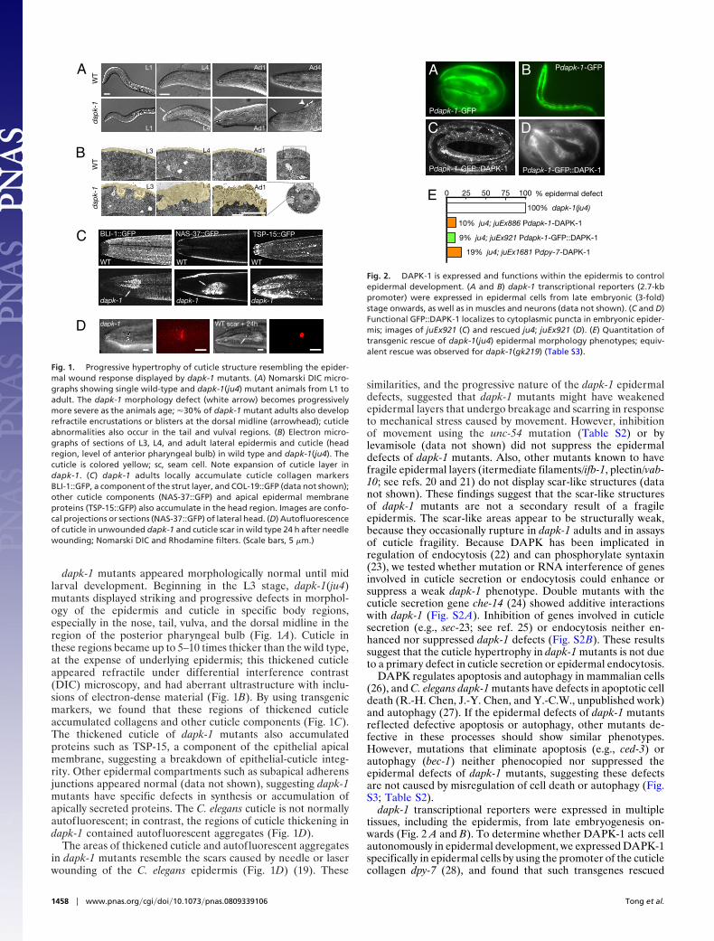
dapk-1 transcriptional reporters were expressed in multipletissues, including the epidermis, from late embryogenesis on-wards (Fig. 2 A and B). To determine whether DAPK-1 acts cellautonomously in epidermal development, we expressed DAPK-1specifically in epidermal cells by using the promoter of the cuticlecollagen dpy-7 (28), and found that such transgenes rescued
A L1 L4 Ad1
B
L3 L4
L3
WT
dapk-1
BLI-1::GFP
L3
L1 L4
L4
A1
Ad1
C
sc
sc
WT
dap
k-1
WT
dap
k-1
D dapk-1
NAS-37::GFP TSP-15::GFP
WT
dapk-1
WT
dapk-1
L4
Ad1L4 A
A
WT scar + 24h
Ad1
Ad4
Ad4
Fig. 1. Progressive hypertrophy of cuticle structure resembling the epider-mal wound response displayed by dapk-1 mutants. (A) Nomarski DIC micro-graphs showing single wild-type and dapk-1(ju4) mutant animals from L1 toadult. The dapk-1 morphology defect (white arrow) becomes progressivelymore severe as the animals age; �30% of dapk-1 mutant adults also developrefractile encrustations or blisters at the dorsal midline (arrowhead); cuticleabnormalities also occur in the tail and vulval regions. (B) Electron micro-graphs of sections of L3, L4, and adult lateral epidermis and cuticle (headregion, level of anterior pharyngeal bulb) in wild type and dapk-1(ju4). Thecuticle is colored yellow; sc, seam cell. Note expansion of cuticle layer indapk-1. (C) dapk-1 adults locally accumulate cuticle collagen markersBLI-1::GFP, a component of the strut layer, and COL-19::GFP (data not shown);other cuticle components (NAS-37::GFP) and apical epidermal membraneproteins (TSP-15::GFP) also accumulate in the head region. Images are confo-cal projections or sections (NAS-37::GFP) of lateral head. (D) Autofluorescenceof cuticle in unwounded dapk-1 and cuticle scar in wild type 24 h after needlewounding; Nomarski DIC and Rhodamine filters. (Scale bars, 5 �m.)
10% ju4; juEx886 Pdapk-1-DAPK-1
Pdapk-1-GFP
9% ju4; juEx921 Pdapk-1-GFP::DAPK-1
19% ju4; juEx1681 Pdpy-7-DAPK-1
Pdapk-1-GFP::DAPK-1
A
E100% dapk-1(ju4)
25 50 75 1000
B Pdapk-1-GFP
C D
Pdapk-1-GFP::DAPK-1
% epidermal defect
Fig. 2. DAPK-1 is expressed and functions within the epidermis to controlepidermal development. (A and B) dapk-1 transcriptional reporters (2.7-kbpromoter) were expressed in epidermal cells from late embryonic (3-fold)stage onwards, as well as in muscles and neurons (data not shown). (C and D)Functional GFP::DAPK-1 localizes to cytoplasmic puncta in embryonic epider-mis; images of juEx921 (C) and rescued ju4; juEx921 (D). (E) Quantitation oftransgenic rescue of dapk-1(ju4) epidermal morphology phenotypes; equiv-alent rescue was observed for dapk-1(gk219) (Table S3).
1458 � www.pnas.org�cgi�doi�10.1073�pnas.0809339106 Tong et al.
dapk-1 epidermal defects (Fig. 2E and Table S3). Within epi-dermal cells, functional GFP::DAPK-1 localized to cytoplasmicpuncta of unknown identity (Fig. 2 C and D). These resultssuggest that DAPK-1 acts independently of cell death pathwaysand autonomously in the epidermis.
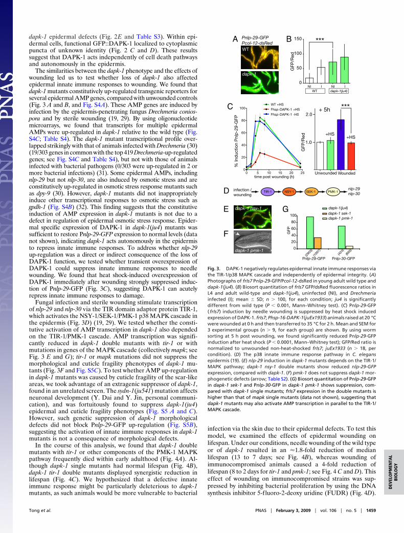
The similarities between the dapk-1 phenotype and the effects ofwounding led us to test whether loss of dapk-1 also affectedepidermal innate immune responses to wounding. We found thatdapk-1 mutants constitutively up-regulated transgenic reporters forseveral epidermal AMP genes, compared with unwounded controls(Fig. 3 A and B, and Fig. S4A). These AMP genes are induced byinfection by the epidermis-penetrating fungus Drechmeria conios-pora and by sterile wounding (19, 29). By using oligonucleotidemicroarrays, we found that transcripts for multiple epidermalAMPs were up-regulated in dapk-1 relative to the wild type (Fig.S4C; Table S4). The dapk-1 mutant transcriptional profile over-lapped strikingly with that of animals infected with Drechmeria (30)(19/303 genes in common with the top 419 Drechmeria-up-regulatedgenes; see Fig. S4C and Table S4), but not with those of animalsinfected with bacterial pathogens (0/303 were up-regulated in 2 ormore bacterial infections) (31). Some epidermal AMPs, includingnlp-29 but not nlp-30, are also induced by osmotic stress and areconstitutively up-regulated in osmotic stress response mutants suchas dpy-9 (30). However, dapk-1 mutants did not inappropriatelyinduce other transcriptional responses to osmotic stress such asgpdh-1 (Fig. S4B) (32). This finding suggests that the constitutiveinduction of AMP expression in dapk-1 mutants is not due to adefect in regulation of epidermal osmotic stress response. Epider-mal specific expression of DAPK-1 in dapk-1(ju4) mutants wassufficient to restore Pnlp-29-GFP expression to normal levels (datanot shown), indicating dapk-1 acts autonomously in the epidermisto repress innate immune responses. To address whether nlp-29up-regulation was a direct or indirect consequence of the loss ofDAPK-1 function, we tested whether transient overexpression ofDAPK-1 could suppress innate immune responses to needlewounding. We found that heat shock-induced overexpression ofDAPK-1 immediately after wounding strongly suppressed induc-tion of Pnlp-29-GFP (Fig. 3C), suggesting DAPK-1 can acutelyrepress innate immune responses to damage.
Fungal infection and sterile wounding stimulate transcriptionof nlp-29 and nlp-30 via the TIR domain adaptor protein TIR-1,which activates the NSY-1/SEK-1/PMK-1 p38 MAPK cascade inthe epidermis (Fig. 3D) (19, 29). We tested whether the consti-tutive activation of AMP transcription in dapk-1 also dependedon the TIR-1/PMK-1 cascade. AMP transcription was signifi-cantly reduced in dapk-1 double mutants with tir-1 or withmutations in genes of the MAPK cascade (collectively mapk; seeFig. 3 E and G); tir-1 or mapk mutations did not suppress themorphological and cuticle fragility phenotypes of dapk-1 mu-tants (Fig. 3F and Fig. S5C). To test whether AMP up-regulationin dapk-1 mutants was caused by cuticle fragility of the scar-likeareas, we took advantage of an extragenic suppressor of dapk-1,found in an unrelated screen. The sydn-1(ju541) mutation affectsneuronal development (Y. Dai and Y. Jin, personal communi-cation), and was fortuitously found to suppress dapk-1(ju4)epidermal and cuticle fragility phenotypes (Fig. S5 A and C).However, such genetic suppression of dapk-1 morphologicaldefects did not block Pnlp-29-GFP up-regulation (Fig. S5B),suggesting the activation of innate immune responses in dapk-1mutants is not a consequence of morphological defects.
In the course of this analysis, we found that dapk-1 doublemutants with tir-1 or other components of the PMK-1 MAPKpathway frequently died within early adulthood (Fig. 4A). Al-though dapk-1 single mutants had normal lifespan (Fig. 4B),dapk-1 tir-1 double mutants displayed synergistic reduction inlifespan (Fig. 4C). We hypothesized that a defective innateimmune response might be particularly deleterious to dapk-1mutants, as such animals would be more vulnerable to bacterial
infection via the skin due to their epidermal defects. To test thismodel, we examined the effects of epidermal wounding onlifespan. Under our conditions, needle wounding of the wild typeor of dapk-1 resulted in an �1.8-fold reduction of medianlifespan (13 to 7 days; see Fig. 4B), whereas wounding ofimmunocompromised animals caused a 4-fold reduction oflifespan (8 to 2 days for tir-1 and pmk-1; see Fig. 4 C and D). Thiseffect of wounding on immunocompromised strains was sup-pressed by inhibiting bacterial proliferation by using the DNAsynthesis inhibitor 5-f luoro-2-deoxy uridine (FUDR) (Fig. 4D).
0
20
40
60
80
100
dapk-1(ju4)dapk-1 sek-1 dapk-1 pmk-1
GFP
Pnlp-29-GFP Pnlp-30-GFP(29
03)
(1097
)(42
4)(66
5)
A B
dapk-1
WT
Pnlp-29-GFPPcol-12-dsRed
E G
GFP
/Red
dapk-1 nsy-1
TIR-1 NSY-1 SEK-1 PMK-1D infection /wounding
nlp-29nlp-30
F
dapk-1 pmk-1
WT +HS
Phsp-DAPK-1 +HS
Phsp-DAPK-1 -HS
C
0
50
100
150 ***
NI I NI IWT dapk-1(ju4)
Unwounded Wounded
2.0
1.0
Phsp-DAPK-1 +HS
Phsp-DAPK-1 -HS
0 5 10 15 20 250
20
40
60
80
100
GFP
/Red +HS
+HS
***+ 5h
% In
duc
tion
Pnl
p-2
9-G
FP
time post wounding (h)
Fig. 3. DAPK-1 negatively regulates epidermal innate immune responses viathe TIR-1/p38 MAPK cascade and independently of epidermal integrity. (A)Photographs of frIs7 Pnlp-29-GFP/Pcol-12-dsRed in young adult wild type anddapk-1(ju4). (B) Biosort quantitation of frIs7 GFP/dsRed fluorescence ratios inL4 and adult wild-type and dapk-1(ju4), uninfected (NI), and Drechmeriainfected (I); mean � SD; n � 100, for each condition; ju4 is significantlydifferent from wild type (P � 0.001, Mann–Whitney test). (C) Pnlp-29-GFP( frIs7) induction by needle wounding is suppressed by heat shock inducedexpression of DAPK-1. frIs7; Phsp-16-DAPK-1(juEx1933) animals raised at 20 °Cwere wounded at 0 h and then transferred to 35 °C for 2 h. Mean and SEM for3 experimental groups (n � 9, for each group) are shown. By using wormsorting at 5 h post wounding, we found significantly reduced Pnlp-29-GFPinduction after heat shock (P � 0.0001, Mann–Whitney test); GFP/Red ratio isnormalized to unwounded non-heat-shocked frIs7; juEx1933 (n � 18, percondition). (D) The p38 innate immune response pathway in C. elegansepidermis (19). (E) nlp-29 induction in dapk-1 mutants depends on the TIR-1/MAPK pathway; dapk-1 nsy-1 double mutants show reduced nlp-29-GFPexpression, compared with dapk-1. (F) pmk-1 does not suppress dapk-1 mor-phogenetic defects (arrow; Table S2). (G) Biosort quantitation of Pnlp-29-GFPin dapk-1 sek-1 and Pnlp-30-GFP in dapk-1 pmk-1 shows suppression, com-pared with dapk-1 single mutants; frIs7 expression in the double mutants ishigher than that of mapk single mutants (data not shown), suggesting thatdapk-1 mutants may also activate AMP transcription in parallel to the TIR-1/MAPK cascade.
Tong et al. PNAS � February 3, 2009 � vol. 106 � no. 5 � 1459
DEV
ELO
PMEN
TAL
BIO
LOG
Y
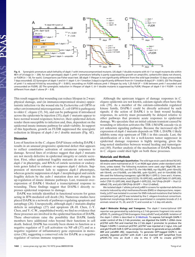
This result suggests that wounding can reduce lifespan in 2 ways:physical damage, and (in immunocompromised strains) oppor-tunistic infection via the wound site by Escherichia coli OP50 orother environmental microorganisms. E. coli OP50 is pathogenicto older C. elegans (33, 34), and can be pathogenic if introducedacross the epidermis by injection (35); dapk-1 mutants appear tohave normal wound responses; however, their epidermal defectsrender them susceptible to infection and, thus, dependent on theepidermal innate immune pathway for adult viability. In supportof this hypothesis, growth on FUDR suppressed the synergisticreduction in lifespan of dapk-1 tir-1 double mutants (Fig. 4E).
DiscussionLoss of function in the C. elegans DAP kinase ortholog DAPK-1results in an unusual progressive epidermal defect that appearsto reflect constitutive activation of cutaneous responses todamage. Several lines of evidence suggest that dapk-1 mutantsare not merely defective in epidermal integrity or cuticle secre-tion. First, other epidermal fragility mutants do not resembledapk-1 in phenotype, and RNAi of cuticle secretion or endocy-tosis genes failed to enhance or suppress dapk-1 defects. Sup-pression of movement fails to suppress dapk-1 phenotypes,whereas genetic suppression of dapk-1 morphological and cuticlefragility defects by the sydn-1 mutation does not abrogate itsup-regulation of innate immune pathways. Last, transient over-expression of DAPK-1 blocked a transcriptional response towounding. These findings suggest that DAPK-1 directly re-presses epidermal responses to damage.
DAPK was initially identified in functional screens for genesacting in IFN-mediated cell death (36). Numerous studies haveplaced DAPK in a network of pathways regulating apoptosis andautophagy (26). Unexpectedly, although dapk-1 mutants displaydefects in autophagy (27) and apoptosis (R.-H. Chen, J.-Y.Chen, and Y.-C.W., unpublished work), we find no evidence thatthese processes are involved in the epidermal function of DAPK.These observations raise the possibility that DAPK familymembers have additional roles beyond their functions in celldeath. Indeed, DAPK has recently been shown to function as anegative regulator of T cell activation via NF-�B (37) and as anegative regulator of inflammatory gene expression in mono-cytes (38), suggesting a conserved role for DAPK as a negativeregulator of various immune responses.
Although the upstream triggers of damage responses in C.elegans epidermis are not known, calcium signals often have thisrole (39). As a member of the calcium-calmodulin regulatedkinase family, DAPK-1 could be directly activated by suchsignals; if the action of DAPK-1 is to limit wound healingresponses, its activity must presumably be delayed relative toother pathways that promote acute responses to epidermaldamage. We speculate that an initial calcium transient caused bywounding or infection activates the TIR-1/MAPK cascade via asyet unknown signaling pathways. Because the increased nlp-29expression of dapk-1 mutants depends on TIR-1, DAPK-1 likelyinhibits some step upstream of TIR-1 in this cascade. Last, theidentification of a role for a well-known tumor suppressor inepidermal damage responses is highly intriguing, given thelong-noted similarities between wound healing and tumorigen-esis (40). Further analysis of the mechanism of DAPK functionin C. elegans could shed light on both these processes.
Materials and MethodsGenetics and Phenotypic Quantitation. The wild-type strain used is Bristol N2 (41).All strains were maintained at 25 °C on NGM agar plates under standard condi-tions, unless stated. The following mutations were used: atgr-18(gk378), bec-1(ok700), ced-2(e1752), ced-3(n717), che-14(e1960), nsy-1(ky397), pmk-1(km25),sek-1(km4), unc-51(e369), unc-54(e190), sydn-1(ju541), and tir-1(tm3036) (19).We used the following transgenes: cgEx198 [BLI-1::GFP] (J. Crew and J. Kramer,personal communication), kaIs12 [COL-19::GFP] (42), oxEx587 [NAS-37::GFP] (43),imEx1 [TSP-15::GFP] (44), kbIs5 [Pgpdh-1-GFP] (32), frIs7 [Pnlp-29-GFP � Pcol-12-dsRed] (19), and juEx1384 [Pnlp-30-GFP � Pcol-12-dsRed].
We isolated dapk-1 alleles ju4 and ju469 in screens for epidermal-defectivemutants induced by ethyl methanesulfonate (EMS) or diepoxyoctane, respec-tively; ju557 was isolated in a screen for EMS-induced mutations that failed tocomplement ju4; gk219 was isolated by the C. elegans knockout consortium.Epidermal morphology defects were quantitated in complete broods of 2–6animals raised at 15, 20, and 25 °C and scored as 1–2 day-old adults.
dapk-1 Molecular Biology and Transgenes. The dapk-1 transcriptional GFPreporter was generated by subcloning a 2.7-kb genomic fragment into vectorpPD95.75, yielding pCZ764 (transgenic lines juEx927 and juEx928). Isolation ofthe dapk-1 cDNA is described in SI Methods. To express full-length DAPK-1under control of the 2.7-kb promoter, we digested the DAPK-1 cDNA clonepCZ763 with PstI and inserted the 2.7-kb dapk-1 promoter to produce thePdapk-1-DAPK-1 construct pCZ767; pCZ767 was injected at 2 ng/�L into ju4and gk219 with SUR-5::GFP as coinjection marker to generate arrays juEx886–889 and juEx890–892, respectively. To generate GFP-tagged DAPK-1, wepartially digested pCZ767 with EcoR I and inserted GFP (amplified frompPD95.75) into an EcoR I site in the 5� UTR to create pCZ770
A
2% tir-1
tir-1 dapk-1
sek-1
sek-1 dapk-1
0.3 pmk-1 D
6% dapk-1(ju4)
20 60 80 1000 40
pmk-1 dapk-1
% dead 48 h post L4
69
19
56
24
B WT
WT W
dapk-1
dapk-1 W
% s
urvi
val
E
C
% s
urvi
val
0 10 20 300
50
100
% s
urvi
val
pmk-1pmk-1 Wpmk-1 FUDRpmk-1 W FUDR
days
days
0 10 20 300
50
100
days
0 10 20 300
50
100
-FUDR
+FUDR
days
dapk-1tir-1dapk-1 tir-1
% s
urvi
val
pmk-1 dapk-1 + FUDR 33
pmk-1 dapk-1 + ampicillin45
*** *
***
***
***
*** P < 0.001
* P < 0.05
dapk-1tir-1dapk-1 tir-1
0 10 20 300
50
100
Fig. 4. Synergistic premature adult lethality of dapk-1 with immunocompromised mutants. (A) dapk-1 double mutants with tir-1 or mapk mutants die within48 h of L4 stage (n � 200, for each genotype); dapk-1; pmk-1 premature lethality is partly suppressed by growth on ampicillin, carbenicillin (data not shown),or FUDR (n � 50, for each). Comparisons use Fisher exact test. (B) dapk-1 lifespan is not significantly different from the wild type (median 12 days unwounded,7 days wounded). (C) Synergism of dapk-1 and tir-1: dapk-1; tir-1 (median 3 days) is significantly different from tir-1 (median 8 days) (P � 0.001). (D) The lifespanof pmk-1 is reduced 4-fold by wounding (P � 0.001); wounding on FUDR reduces pmk-1 lifespan by only 1.25-fold (P � 0.06 between pmk-1 wounded andunwounded on FUDR). (E) The synergistic reduction in lifespan of dapk-1; tir-1 double mutants is suppressed by FUDR; lifespan of dapk-1 tir-1 FUDR� is notdifferent from dapk-1 alone (P � 0.9).
1460 � www.pnas.org�cgi�doi�10.1073�pnas.0809339106 Tong et al.
Pdapk-1-GFP::DAPK-1; pCZ770 was injected at 1.5 ng/�L into ju4 and gk219with Pttx-3-RFP as coinjection marker to generate transgenes juEx921–923and juEx924–926, respectively. To express DAPK-1 in the epidermis, we am-plified the 305 bp dpy-7 promoter (28), and cloned it into pCZ763 to createPdpy-7-DAPK-1 (pCZ766); pCZ766 was injected at 1 ng/�L into wild-typeanimals with Pttx-3-RFP as coinjection marker to generate transgenesjuEx1681 and juEx1682. dapk-1 cDNA was cloned into the hsp-16 vectorpPD49.78 (Fire lab vector kit) to create pCZ763; pCZ763 was injected at 3 ng/�Lwith the coinjection marker Pttx-3-RFP to create juEx1933.
Electron Microscopy. dapk-1(ju4) animals were grown at 25 °C and preparedfor electron microscopy as previously described (20). We sectioned 1 L3, 1 L4,and 2 adults from the tip of the nose to the anterior end of the gonad. Imagesin Fig. 1 are from the region of the anterior pharyngeal bulb.
Pnlp-29-GFP Scoring and Worm Sorting. Fluorescent worm sorting by using theCopas Biosort was performed as described (19), on synchronized populationsgrown at 20 °C. For quantitative analysis, we selected events with TOF 250–800 and GFP 15–250. To score frIs7 induction in the heat-shock time course (Fig.3C), we wounded young adults and scored fluorescence by using a LeicaMZFLIII fluorescence dissection microscope and long-pass GFP filter. Eachanimal was scored as uninduced (orange or yellow-orange) or induced (yel-low, yellow-green, and green) at each time point to estimate the percentageinduction in a population. Photomicrographs in Fig. 3 and Fig. S4 were takenby using a Zeiss Axioplan imaging microscope and Axiocam color camera, 40�objective, Zeiss GFP long-pass filters and 250 ms exposure time. Images werenot processed further.
Lethality, Lifespan, and Wounding Experiments. Premature lethality was mea-sured by picking L4s and scoring viability 48 h later. Aging experiments wereperformed at 20 °C; n � 50 for each condition. We picked L4s to individualplates and transferred them until no further progeny were produced. Animalswere scored daily until death, defined as failure to respond to touch. Kaplan–Meier survival curves were compared by using the Mantel-Cox test (GraphpadPrism). We wounded animals with single stabs of a microinjection needle tothe posterior body 24 h after the L4 stage, as previously described (19). Toprevent bacterial proliferation in lifespan assays, we transferred day 1 adultsto plates containing 50 �g/mL FUDR immediately before wounding. Suppres-sion of premature lethality was also tested by using 50 �g/mL ampicillin or 50mM carbenicillin, as described (33).
ACKNOWLEDGMENTS. We thank Y. Dai and Y. Jin for sharing unpublishedresults on sydn-1; J. Crew and J. Kramer (Northwestern University, Chicago) forBLI-1::GFP; O. Zugasti (California Institute for Regenerative Medicine, SanFrancisco) for the nlp-30 array; our laboratories for advice and comments; Y.Duverger for worm sorting, by using facilities of the Marseille-Nice genopole.Some strains were provided by the Caenorhabditis Genetics Center (Universityof Minnesota, Minneapolis) funded by the National Institutes of HealthNational Center for Research Resources. The Ewbank lab is a Fondation pourla Recherche Medicale Equipe Labellisee and is supported by Institut Nationalde la Sante et de la Recherche Medicale, the Centre National de la RechercheScientifique, and the Agence Nationale pour la Recherche. This work wassupported by National Institutes of Health Grant GM54657 (to A.D.C.). Y.-C.W.was supported by the National Science Council, Taiwan. Collaborative workwas supported by United States National Science Foundation Award OISE0726131 (to A.D.C. and J.J.E.) and a French Foreign Ministry award from theFrance Berkeley Fund.
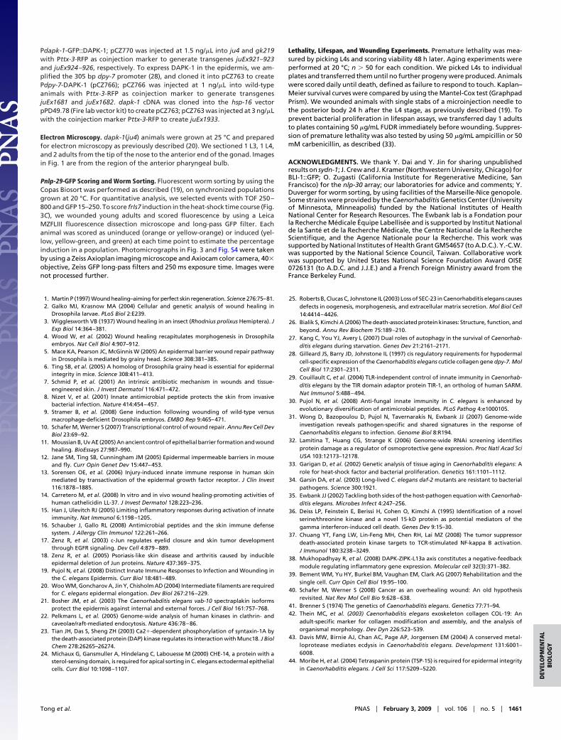
1. Martin P (1997) Wound healing–aiming for perfect skin regeneration. Science 276:75–81.2. Galko MJ, Krasnow MA (2004) Cellular and genetic analysis of wound healing in
Drosophila larvae. PLoS Biol 2:E239.3. Wigglesworth VB (1937) Wound healing in an insect (Rhodnius prolixus Hemiptera). J
Exp Biol 14:364–381.4. Wood W, et al. (2002) Wound healing recapitulates morphogenesis in Drosophila
embryos. Nat Cell Biol 4:907–912.5. Mace KA, Pearson JC, McGinnis W (2005) An epidermal barrier wound repair pathway
in Drosophila is mediated by grainy head. Science 308:381–385.6. Ting SB, et al. (2005) A homolog of Drosophila grainy head is essential for epidermal
integrity in mice. Science 308:411–413.7. Schmid P, et al. (2001) An intrinsic antibiotic mechanism in wounds and tissue-
engineered skin. J Invest Dermatol 116:471–472.8. Nizet V, et al. (2001) Innate antimicrobial peptide protects the skin from invasive
bacterial infection. Nature 414:454–457.9. Stramer B, et al. (2008) Gene induction following wounding of wild-type versus
macrophage-deficient Drosophila embryos. EMBO Rep 9:465–471.10. Schafer M, Werner S (2007) Transcriptional control of wound repair. Annu Rev Cell Dev
Biol 23:69–92.11. Moussian B, Uv AE (2005) An ancient control of epithelial barrier formation and wound
healing. BioEssays 27:987–990.12. Jane SM, Ting SB, Cunningham JM (2005) Epidermal impermeable barriers in mouse
and fly. Curr Opin Genet Dev 15:447–453.13. Sorensen OE, et al. (2006) Injury-induced innate immune response in human skin
mediated by transactivation of the epidermal growth factor receptor. J Clin Invest116:1878–1885.
14. Carretero M, et al. (2008) In vitro and in vivo wound healing-promoting activities ofhuman cathelicidin LL-37. J Invest Dermatol 128:223–236.
15. Han J, Ulevitch RJ (2005) Limiting inflammatory responses during activation of innateimmunity. Nat Immunol 6:1198–1205.
16. Schauber J, Gallo RL (2008) Antimicrobial peptides and the skin immune defensesystem. J Allergy Clin Immunol 122:261–266.
17. Zenz R, et al. (2003) c-Jun regulates eyelid closure and skin tumor developmentthrough EGFR signaling. Dev Cell 4:879–889.
18. Zenz R, et al. (2005) Psoriasis-like skin disease and arthritis caused by inducibleepidermal deletion of Jun proteins. Nature 437:369–375.
19. Pujol N, et al. (2008) Distinct Innate Immune Responses to Infection and Wounding inthe C. elegans Epidermis. Curr Biol 18:481–489.
20. Woo WM, Goncharov A, Jin Y, Chisholm AD (2004) Intermediate filaments are requiredfor C. elegans epidermal elongation. Dev Biol 267:216–229.
21. Bosher JM, et al. (2003) The Caenorhabditis elegans vab-10 spectraplakin isoformsprotect the epidermis against internal and external forces. J Cell Biol 161:757–768.
22. Pelkmans L, et al. (2005) Genome-wide analysis of human kinases in clathrin- andcaveolae/raft-mediated endocytosis. Nature 436:78–86.
23. Tian JH, Das S, Sheng ZH (2003) Ca2�-dependent phosphorylation of syntaxin-1A bythe death-associated protein (DAP) kinase regulates its interaction with Munc18. J BiolChem 278:26265–26274.
24. Michaux G, Gansmuller A, Hindelang C, Labouesse M (2000) CHE-14, a protein with asterol-sensing domain, is required for apical sorting in C. elegans ectodermal epithelialcells. Curr Biol 10:1098–1107.
25. Roberts B, Clucas C, Johnstone IL (2003) Loss of SEC-23 in Caenorhabditis elegans causesdefects in oogenesis, morphogenesis, and extracellular matrix secretion. Mol Biol Cell14:4414–4426.
26. Bialik S, Kimchi A (2006) The death-associated protein kinases: Structure, function, andbeyond. Annu Rev Biochem 75:189–210.
27. Kang C, You YJ, Avery L (2007) Dual roles of autophagy in the survival of Caenorhab-ditis elegans during starvation. Genes Dev 21:2161–2171.
28. Gilleard JS, Barry JD, Johnstone IL (1997) cis regulatory requirements for hypodermalcell-specific expression of the Caenorhabditis elegans cuticle collagen gene dpy-7. MolCell Biol 17:2301–2311.
29. Couillault C, et al. (2004) TLR-independent control of innate immunity in Caenorhab-ditis elegans by the TIR domain adaptor protein TIR-1, an ortholog of human SARM.Nat Immunol 5:488–494.
30. Pujol N, et al. (2008) Anti-fungal innate immunity in C. elegans is enhanced byevolutionary diversification of antimicrobial peptides. PLoS Pathog 4:e1000105.
31. Wong D, Bazopoulou D, Pujol N, Tavernarakis N, Ewbank JJ (2007) Genome-wideinvestigation reveals pathogen-specific and shared signatures in the response ofCaenorhabditis elegans to infection. Genome Biol 8:R194.
32. Lamitina T, Huang CG, Strange K (2006) Genome-wide RNAi screening identifiesprotein damage as a regulator of osmoprotective gene expression. Proc Natl Acad SciUSA 103:12173–12178.
33. Garigan D, et al. (2002) Genetic analysis of tissue aging in Caenorhabditis elegans: Arole for heat-shock factor and bacterial proliferation. Genetics 161:1101–1112.
34. Garsin DA, et al. (2003) Long-lived C. elegans daf-2 mutants are resistant to bacterialpathogens. Science 300:1921.
35. Ewbank JJ (2002) Tackling both sides of the host-pathogen equation with Caenorhab-ditis elegans. Microbes Infect 4:247–256.
36. Deiss LP, Feinstein E, Berissi H, Cohen O, Kimchi A (1995) Identification of a novelserine/threonine kinase and a novel 15-kD protein as potential mediators of thegamma interferon-induced cell death. Genes Dev 9:15–30.
37. Chuang YT, Fang LW, Lin-Feng MH, Chen RH, Lai MZ (2008) The tumor suppressordeath-associated protein kinase targets to TCR-stimulated NF-kappa B activation.J Immunol 180:3238–3249.
38. Mukhopadhyay R, et al. (2008) DAPK-ZIPK-L13a axis constitutes a negative-feedbackmodule regulating inflammatory gene expression. Molecular cell 32(3):371–382.
39. Bement WM, Yu HY, Burkel BM, Vaughan EM, Clark AG (2007) Rehabilitation and thesingle cell. Curr Opin Cell Biol 19:95–100.
40. Schafer M, Werner S (2008) Cancer as an overhealing wound: An old hypothesisrevisited. Nat Rev Mol Cell Bio 9:628–638.
41. Brenner S (1974) The genetics of Caenorhabditis elegans. Genetics 77:71–94.42. Thein MC, et al. (2003) Caenorhabditis elegans exoskeleton collagen COL-19: An
adult-specific marker for collagen modification and assembly, and the analysis oforganismal morphology. Dev Dyn 226:523–539.
43. Davis MW, Birnie AJ, Chan AC, Page AP, Jorgensen EM (2004) A conserved metal-loprotease mediates ecdysis in Caenorhabditis elegans. Development 131:6001–6008.
44. Moribe H, et al. (2004) Tetraspanin protein (TSP-15) is required for epidermal integrityin Caenorhabditis elegans. J Cell Sci 117:5209–5220.
Tong et al. PNAS � February 3, 2009 � vol. 106 � no. 5 � 1461
DEV
ELO
PMEN
TAL
BIO
LOG
Y