Missing anterior teeth: orthodonticclosure and transplantation asviable options to conventionalreplacementsARILD STENVIK & BJORN U. ZACHRISSON
Introduction
Traditional replacement options for missing maxillary
incisors include implants and partial dentures.
Although less well known among endodontists and
general dentists, orthodontic space closure and auto-
transplantation of developing premolars may, however,
constitute relevant alternatives.
The challenge in treating patients with missing max-
illary incisors and concomitant malocclusion is how to
achieve the best esthetic and functional results, particu-
larly in the long term (1). Although data for survival of
single tooth implants is favorable, over time there may be
significant problems associated with their use in the
esthetic zone, such as infraocclusion (2, 3), gingival
retraction with root exposure, and darkening of the
overlying gingiva due to resorption of the buccal bone
plate (4). Autotransplantation of teeth and orthodontic
space closure represent viable biological approaches for
replacement of incisors because of the permanence of the
result, particularly in growing individuals.
Treatment decisions for young people with missing
incisors should be based on a comprehensive assessment
that includes several factors (1), and the ultimate
treatment plan should be dictated by the diagnosis and
not by a preferred methodology. From 2000 to 2003,
the authors published a series of articles in the American
Journal of Orthodontics and Dentofacial Orthopedics on
the outcome of tooth transplantation, and of orthodontic
space closure in the management of missing teeth (5–9).
It is clear from the debate that followed (3, 10–16) that
there are conflicting opinions, pointing to a need for
further information and research in this area.
The purpose of this article is to summarize the
authors’ experience over the past 40 years with
transplantation and orthodontic space closure in the
treatment of missing incisors. There are two main
reasons for missing incisors in young people: traumatic
injuries and congenital absences. Because the nature of
the problems differs in these two situations, the
management with regards to the two types of missing
teeth will be discussed separately.
Loss of incisors from traumaticinjuries
Traumatic injuries may affect one or more maxillary
incisors as well as other oral tissues. The treatment plan in
each situation will depend on factors such as the number
of missing teeth, the status of the remaining
teeth, possible concerns about the occlusion, space
availability, age, facial morphology, growth pattern, and
tooth morphology (1). From a preventative perspective,
persons with protruding incisors are at risk for
dental injuries (17) and may therefore need orthodontic
treatment irrespective of whether teeth are missing
or not.
Indications for autotransplantation
More than 30 years ago, Slagsvold & Bjercke (18)
established a method of transplanting teeth with
41
Endodontic Topics 2006, 14, 41–50All rights reserved
Copyright r Blackwell Munksgaard
ENDODONTIC TOPICS 20081601-1538
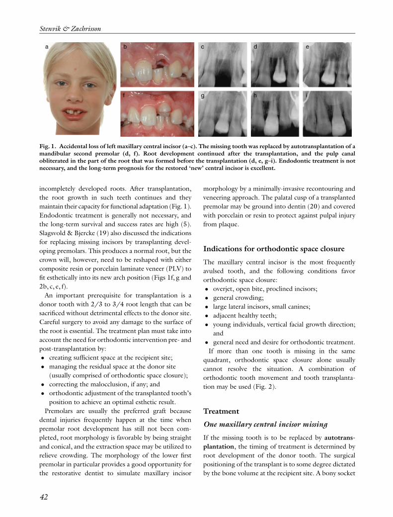
incompletely developed roots. After transplantation,
the root growth in such teeth continues and they
maintain their capacity for functional adaptation (Fig. 1).
Endodontic treatment is generally not necessary, and
the long-term survival and success rates are high (5).
Slagsvold & Bjercke (19) also discussed the indications
for replacing missing incisors by transplanting devel-
oping premolars. This produces a normal root, but the
crown will, however, need to be reshaped with either
composite resin or porcelain laminate veneer (PLV) to
fit esthetically into its new arch position (Figs 1f, g and
2b, c, e, f).
An important prerequisite for transplantation is a
donor tooth with 2/3 to 3/4 root length that can be
sacrificed without detrimental effects to the donor site.
Careful surgery to avoid any damage to the surface of
the root is essential. The treatment plan must take into
account the need for orthodontic intervention pre- and
post-transplantation by:
� creating sufficient space at the recipient site;
� managing the residual space at the donor site
(usually comprised of orthodontic space closure);
� correcting the malocclusion, if any; and
� orthodontic adjustment of the transplanted tooth’s
position to achieve an optimal esthetic result.
Premolars are usually the preferred graft because
dental injuries frequently happen at the time when
premolar root development has still not been com-
pleted, root morphology is favorable by being straight
and conical, and the extraction space may be utilized to
relieve crowding. The morphology of the lower first
premolar in particular provides a good opportunity for
the restorative dentist to simulate maxillary incisor
morphology by a minimally-invasive recontouring and
veneering approach. The palatal cusp of a transplanted
premolar may be ground into dentin (20) and covered
with porcelain or resin to protect against pulpal injury
from plaque.
Indications for orthodontic space closure
The maxillary central incisor is the most frequently
avulsed tooth, and the following conditions favor
orthodontic space closure:
� overjet, open bite, proclined incisors;
� general crowding;
� large lateral incisors, small canines;
� adjacent healthy teeth;
� young individuals, vertical facial growth direction;
and
� general need and desire for orthodontic treatment.
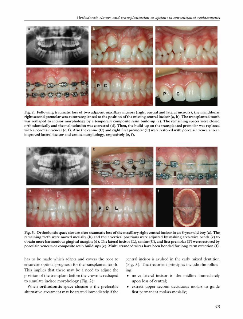
If more than one tooth is missing in the same
quadrant, orthodontic space closure alone usually
cannot resolve the situation. A combination of
orthodontic tooth movement and tooth transplanta-
tion may be used (Fig. 2).
Treatment
One maxillary central incisor missing
If the missing tooth is to be replaced by autotrans-
plantation, the timing of treatment is determined by
root development of the donor tooth. The surgical
positioning of the transplant is to some degree dictated
by the bone volume at the recipient site. A bony socket
Fig. 1. Accidental loss of left maxillary central incisor (a–c). The missing tooth was replaced by autotransplantation of amandibular second premolar (d, f). Root development continued after the transplantation, and the pulp canalobliterated in the part of the root that was formed before the transplantation (d, e, g–i). Endodontic treatment is notnecessary, and the long-term prognosis for the restored ‘new’ central incisor is excellent.
Stenvik & Zachrisson
42
has to be made which adapts and covers the root to
ensure an optimal prognosis for the transplanted tooth.
This implies that there may be a need to adjust the
position of the transplant before the crown is reshaped
to simulate incisor morphology (Fig. 2).
When orthodontic space closure is the preferable
alternative, treatment may be started immediately if the
central incisor is avulsed in the early mixed dentition
(Fig. 3). The treatment principles include the follow-
ing:
� move lateral incisor to the midline immediately
upon loss of central;
� extract upper second deciduous molars to guide
first permanent molars mesially;
Fig. 2. Following traumatic loss of two adjacent maxillary incisors (right central and lateral incisors), the mandibularright second premolar was autotransplanted to the position of the missing central incisor (a, b). The transplanted toothwas reshaped to incisor morphology by a temporary composite resin build-up (c). The remaining spaces were closedorthodontically and the malocclusion was corrected (d). Then, the build-up on the transplanted premolar was replacedwith a porcelain veneer (e, f). Also the canine (C) and right first premolar (P) were restored with porcelain veneers to animproved lateral incisor and canine morphology, respectively (e, f).
Fig. 3. Orthodontic space closure after traumatic loss of the maxillary right central incisor in an 8-year-old boy (a). Theremaining teeth were moved mesially (b) and their vertical positions were adjusted by making arch-wire bends (c) toobtain more harmonious gingival margins (d). The lateral incisor (L), canine (C), and first premolar (P) were restored byporcelain veneers or composite resin build-ups (e). Multi-stranded wires have been bonded for long-term retention (f).
Orthodontic closure and transplantation as options to conventional replacements
43
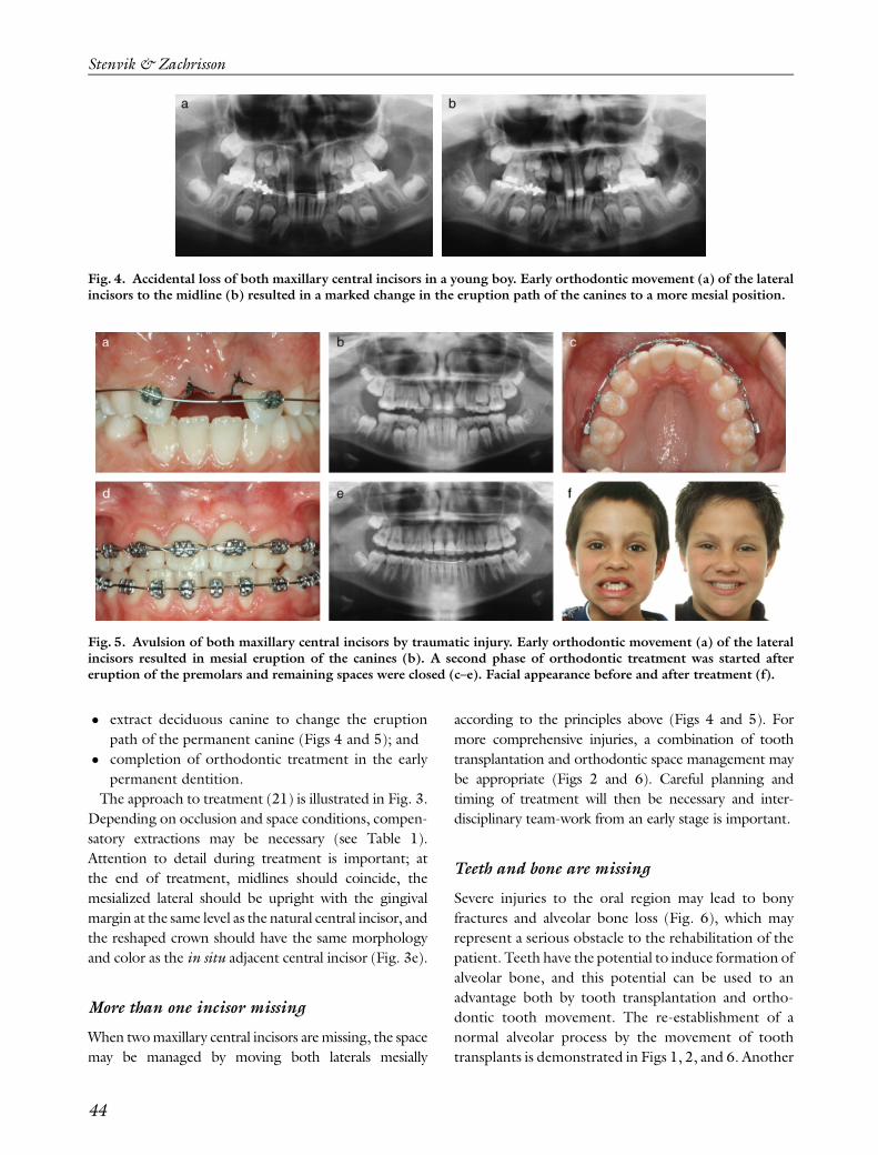
� extract deciduous canine to change the eruption
path of the permanent canine (Figs 4 and 5); and
� completion of orthodontic treatment in the early
permanent dentition.
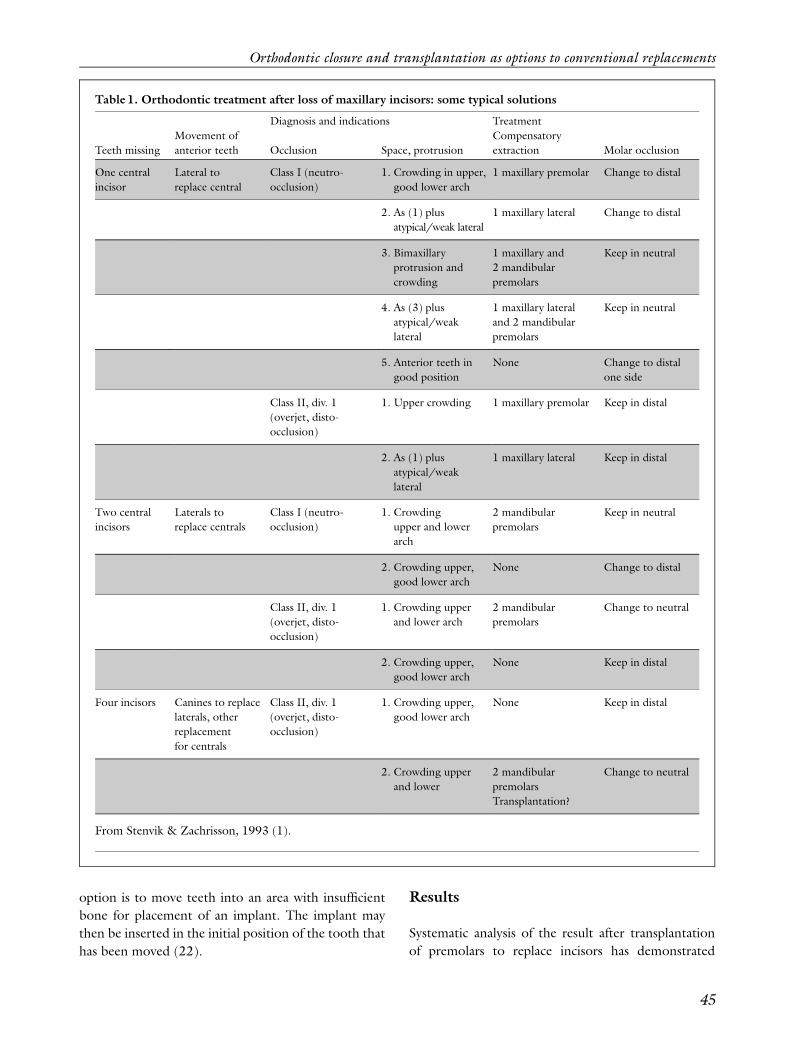
The approach to treatment (21) is illustrated in Fig. 3.
Depending on occlusion and space conditions, compen-
satory extractions may be necessary (see Table 1).
Attention to detail during treatment is important; at
the end of treatment, midlines should coincide, the
mesialized lateral should be upright with the gingival
margin at the same level as the natural central incisor, and
the reshaped crown should have the same morphology
and color as the in situ adjacent central incisor (Fig. 3e).
More than one incisor missing
When two maxillary central incisors are missing, the space
may be managed by moving both laterals mesially
according to the principles above (Figs 4 and 5). For
more comprehensive injuries, a combination of tooth
transplantation and orthodontic space management may
be appropriate (Figs 2 and 6). Careful planning and
timing of treatment will then be necessary and inter-
disciplinary team-work from an early stage is important.
Teeth and bone are missing
Severe injuries to the oral region may lead to bony
fractures and alveolar bone loss (Fig. 6), which may
represent a serious obstacle to the rehabilitation of the
patient. Teeth have the potential to induce formation of
alveolar bone, and this potential can be used to an
advantage both by tooth transplantation and ortho-
dontic tooth movement. The re-establishment of a
normal alveolar process by the movement of tooth
transplants is demonstrated in Figs 1, 2, and 6. Another
Fig. 4. Accidental loss of both maxillary central incisors in a young boy. Early orthodontic movement (a) of the lateralincisors to the midline (b) resulted in a marked change in the eruption path of the canines to a more mesial position.
Fig. 5. Avulsion of both maxillary central incisors by traumatic injury. Early orthodontic movement (a) of the lateralincisors resulted in mesial eruption of the canines (b). A second phase of orthodontic treatment was started aftereruption of the premolars and remaining spaces were closed (c–e). Facial appearance before and after treatment (f).
Stenvik & Zachrisson
44
option is to move teeth into an area with insufficient
bone for placement of an implant. The implant may
then be inserted in the initial position of the tooth that
has been moved (22).
Results
Systematic analysis of the result after transplantation
of premolars to replace incisors has demonstrated
Table 1. Orthodontic treatment after loss of maxillary incisors: some typical solutions
Teeth missing
Movement of
anterior teeth
Diagnosis and indications Treatment
Occlusion Space, protrusion
Compensatory
extraction Molar occlusion
One central
incisor
Lateral to
replace central
Class I (neutro-
occlusion)
1. Crowding in upper,
good lower arch
1 maxillary premolar Change to distal
2. As (1) plus
atypical/weak lateral
1 maxillary lateral Change to distal
3. Bimaxillary
protrusion and
crowding
1 maxillary and
2 mandibular
premolars
Keep in neutral
4. As (3) plus
atypical/weak
lateral
1 maxillary lateral
and 2 mandibular
premolars
Keep in neutral
5. Anterior teeth in
good position
None Change to distal
one side
Class II, div. 1
(overjet, disto-
occlusion)
1. Upper crowding 1 maxillary premolar Keep in distal
2. As (1) plus
atypical/weak
lateral
1 maxillary lateral Keep in distal
Two central
incisors
Laterals to
replace centrals
Class I (neutro-
occlusion)
1. Crowding
upper and lower
arch
2 mandibular
premolars
Keep in neutral
2. Crowding upper,
good lower arch
None Change to distal
Class II, div. 1
(overjet, disto-
occlusion)
1. Crowding upper
and lower arch
2 mandibular
premolars
Change to neutral
2. Crowding upper,
good lower arch
None Keep in distal
Four incisors Canines to replace
laterals, other
replacement
for centrals
Class II, div. 1
(overjet, disto-
occlusion)
1. Crowding upper,
good lower arch
None Keep in distal
2. Crowding upper
and lower
2 mandibular
premolars
Transplantation?
Change to neutral
From Stenvik & Zachrisson, 1993 (1).
Orthodontic closure and transplantation as options to conventional replacements
45
long-term (425 years) survival rates of more than 90%
(23, 24). Publications from teams in Scandinavia (25)
and Japan (26) contain examples of premolars success-
fully transplanted to replace incisors. We found that
compared with natural incisors, the overall status of the
transplanted premolars and surrounding tissues is similar
(6). The esthetic results were also generally satisfactory
(7), particularly when adjunctive orthodontic treatment
ensured optimal positioning of the transplant (Fig. 2b–f).
When patients expressed dissatisfaction, the reason
usually was related to the suboptimal appearance of the
composite resin build-ups being made to reshape the
lateral incisor to the central incisor shape and color.
Teamwork with the restorative dentist is therefore
important, and meticulous attention to detail should be
exercised at all stages of treatment.
Congenitally missing maxillary lateralincisors
Indications for orthodontic space closure
Several factors should be considered in the assessment
of indications for orthodontic space closure, including
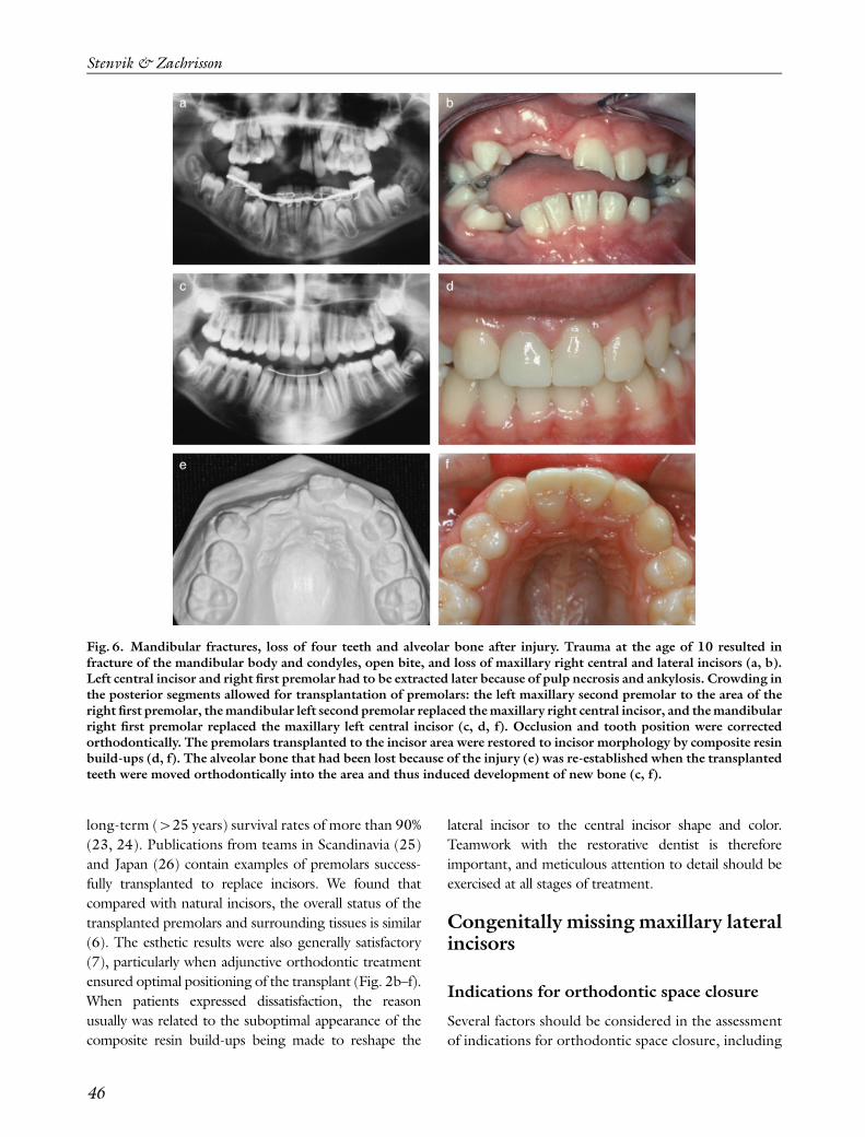
Fig. 6. Mandibular fractures, loss of four teeth and alveolar bone after injury. Trauma at the age of 10 resulted infracture of the mandibular body and condyles, open bite, and loss of maxillary right central and lateral incisors (a, b).Left central incisor and right first premolar had to be extracted later because of pulp necrosis and ankylosis. Crowding inthe posterior segments allowed for transplantation of premolars: the left maxillary second premolar to the area of theright first premolar, the mandibular left second premolar replaced the maxillary right central incisor, and the mandibularright first premolar replaced the maxillary left central incisor (c, d, f). Occlusion and tooth position were correctedorthodontically. The premolars transplanted to the incisor area were restored to incisor morphology by composite resinbuild-ups (d, f). The alveolar bone that had been lost because of the injury (e) was re-established when the transplantedteeth were moved orthodontically into the area and thus induced development of new bone (c, f).
Stenvik & Zachrisson
46
amount of crowding and spacing, size and shape of the
teeth, and the occlusal relationships. Transplantation of
teeth is rarely indicated for absent lateral incisors. The
indications for space closure are summarized by Rosa &
Zachrisson (27, 28):
� a tendency toward maxillary crowding;
� a well-balanced profile and normally inclined
incisors;
� canines and premolars of similar size;
� dentoalveolar protrusion;
� Class II molar occlusion; and
� mandibular crowding or protrusion.
Mesial drift of the canines during eruption frequently
takes place, and this may represent an additional
indication because the distribution of spaces may
prevent replacement of lateral incisors by restorative
methods. If orthodontic measures need to be initiated
anyway, spaces may just as well be closed by mesial
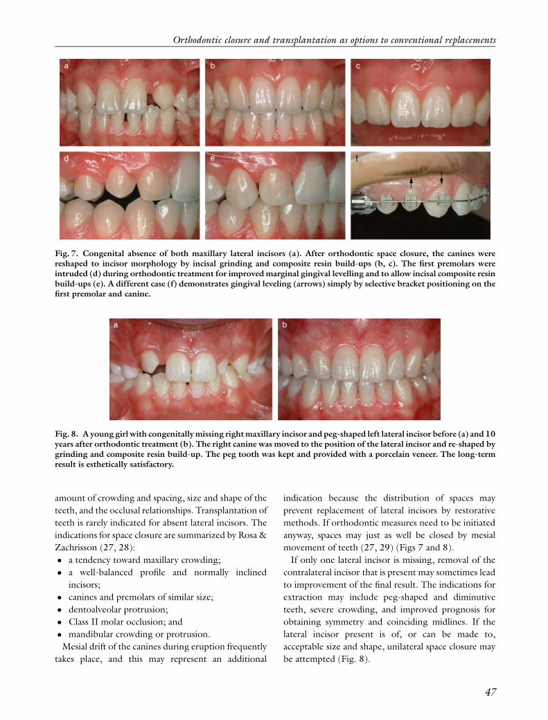
movement of teeth (27, 29) (Figs 7 and 8).
If only one lateral incisor is missing, removal of the
contralateral incisor that is present may sometimes lead
to improvement of the final result. The indications for
extraction may include peg-shaped and diminutive
teeth, severe crowding, and improved prognosis for
obtaining symmetry and coinciding midlines. If the
lateral incisor present is of, or can be made to,
acceptable size and shape, unilateral space closure may
be attempted (Fig. 8).
Fig. 7. Congenital absence of both maxillary lateral incisors (a). After orthodontic space closure, the canines werereshaped to incisor morphology by incisal grinding and composite resin build-ups (b, c). The first premolars wereintruded (d) during orthodontic treatment for improved marginal gingival levelling and to allow incisal composite resinbuild-ups (e). A different case (f) demonstrates gingival leveling (arrows) simply by selective bracket positioning on thefirst premolar and canine.
Fig. 8. A young girl with congenitally missing right maxillary incisor and peg-shaped left lateral incisor before (a) and 10years after orthodontic treatment (b). The right canine was moved to the position of the lateral incisor and re-shaped bygrinding and composite resin build-up. The peg tooth was kept and provided with a porcelain veneer. The long-termresult is esthetically satisfactory.
Orthodontic closure and transplantation as options to conventional replacements
47
Treatment
Guidelines for management of orthodontic space
closure for missing lateral incisors were proposed by
Tuverson in 1970 (29), and are still valid. Successful
treatment of patients with missing lateral incisors
requires attention to a number of details:
� The difference in crown torque (inclination)
between the lateral incisor and canine: sufficient
lingual root torque of the canine is important (Fig.
8), otherwise it may look unnatural in the position
of the lateral incisor.
� The size difference between lateral incisors, canines,
and premolars: this may result in ‘lateral incisors’
appearing too wide and long and ‘canines’ that are
too small and narrow. This may be prevented by
extrusion and grinding of the canines (20), and
intrusion of premolars and composite resin build-
up of the labial cusp (27) (Fig. 7d, e). By selective
bracket placement, the desired movement can be
‘built’ into the orthodontic appliance (Fig. 7f).
� Functional occlusion: long-term follow-up studies
demonstrate no functional occlusion or TMJ
problems in space closure patients (30, 31).
Apparently the presence or absence of a canine rise
is not related to periodontal status, and there is no
evidence that a Class I canine relationship is the
preferred mode of treatment.
� Contouring of canines/color: with today’s techni-
ques in esthetic restorative dentistry, it is possible to
transform a maxillary canine such that it has a
naturally looking lateral incisor morphology (Figs
2, 3, 7 and 8). This may include orthodontic
extrusion for gingival leveling, incisal grinding and
interproximal stripping, composite resin ‘corners’,
vital bleaching of yellowish canines, and PLVs.
� Retention and relapse: there is usually a marked
tendency for reopening of spaces in the anterior
maxilla, and therefore long-term retention with
bonded multi-stranded wire retainers over four to
six teeth should routinely be applied (Fig. 3f).
Clinical implications
Only a few systematic studies examining the results of
space closure for missing maxillary lateral incisors are
available (8, 30, 31). The results are encouraging, both
from a biological and functional perspective. In addition,
a number of treated patients have been presented in case
reports (20, 27–29, 32–34), demonstrating that ex-
cellent esthetic results may be obtained. The most
common arguments against orthodontic space closure
have been related to the esthetic result and the potential
lack of a canine-protected occlusion during function.
These challenges can now be overcome by attention to
detail in planning and treatment as discussed above.
Space conditions and occlusal relationships may
sometimes make closure of all spaces unrealistic. An
alternative may then be to open space in the posterior
segments for single-tooth implants or bridges. This
approach will have the same biological advantages in
the anterior part of the maxilla as normal space closure
and may provide better long-term stability. Restora-
tions in the posterior area of the maxilla do not have to
meet the same strict esthetic requirements as those in
the anterior area. If implants are selected as the
replacements for the missing teeth, they will get a
favorable axial loading. As discussed elsewhere (4), a
critical factor in implant therapy is the long-term
appearance of the soft tissue surrounding the replace-
ment, such as the risk for gingival recession, dark
margins, and lack of satisfactory gingival papilla
contour. In the anterior maxilla, such outcomes may
be detrimental to the overall esthetic result, but they are
much less troublesome in the posterior areas.
So far no studies are available examining the overall
costs of orthodontic space closure compared with other
alternatives from a long-term perspective. The validity
of prospective studies is complicated by the continuous
introduction of new methods and technology. This
applies both to the restorative and orthodontic
disciplines. As an example, space management in
orthodontics may in the future be facilitated by the
use of mini-screws, providing absolute anchorage.
Discussion and conclusion
The most evident advantage of replacing missing
incisors with the patient’s own teeth, either by
transplantation or orthodontic space closure, is the
permanence and biological compatibility of the treat-
ment result. At the end of treatment, normal gingival
tissues and papillae will surround all teeth. Even when
PLVs are needed in young patients, such restorations
can be made shortly after the surgical and orthodontic
treatment. Only a minimal amount of enamel is
removed during preparation and there is little risk of
Stenvik & Zachrisson
48
pulp damage. This is important because the majority of
patients presenting with missing incisors due to trauma
or agenesis are children and adolescents. If treatment is
postponed or spaces are opened, young patients have to
wait until completion of facial growth before receiving
their final restoration. During the interim period,
which may last several years, the patient will have to
wear either a removable or fixed resin-bonded tempor-
ary prosthesis.
Evidence-based clinical practice for the management
of missing anterior teeth should be established from
data on the functional and esthetic clinical result of
various treatment modalities, as well as costs in the
long-term. No such data are currently available. One of
the reasons may be the difficulties in collecting samples
of sufficient size for follow-up and analysis. The
frequency of missing maxillary lateral incisors is about
1%, and the incidence of dental injuries is o2%
annually, of which only 3–4% may be considered to be
severe. Optimal results also require integrated inter-
disciplinary teamwork, which further complicates the
collection of research data.
Even if solid comparative research data for the different
replacement methods so far are not available, our
experience indicates that orthodontic space closure and
transplantation of teeth can produce treatment results
that are almost indistinguishable from an intact dentition.
The goal should be that patients who have received
treatment for missing teeth will have treatment results
that are indistinguishable from normal appearance. A
prerequisite is that the therapy is based on a complete
diagnosis, that the indications for the selected approach
are present, and that attention to detail throughout
treatment is exercised by all involved in the treatment.
References
1. Stenvik A, Zachrisson BU. Orthodontic closure andtransplantation in the treatment of missing anteriorteeth. Endod Dent Traumatol 1993: 9: 45–52.
2. Bernard JO, Schatz JP, Christou P, Belser U, Kiliaridis S.Long-term vertical changes of the maxillary anteriorteeth adjacent to single implants in young and matureadults. A retrospective study. J Clin Periodontol 2004:31: 1024–1028.
3. Jemt T, Ahlberg G, Henriksson K, Bondevik O.Changes of anterior clinical crown height in patientsprovided with single implant restorations after morethan 15 years of follow-up. Int J Prosthodont 2006: 19:455–461.
4. Zachrisson BU, Stenvik A. Single implants – optimaltherapy for missing lateral incisors? (Readers Forum).Am J Orthod Dentofac Ortop 2004: 126: 13A–15A.
5. Czochrowska EM, Stenvik A, Bjercke B, ZachrissonBU. Outcome of tooth transplantation: survival andsuccess rates 17–41 years posttreatment. Am J OrthodDentofac Orthop 2002: 121: 110–119.
6. Czochrowska EM, Stenvik A, Album B, Zachrisson BU.Autotransplantation of premolars to replace maxillaryincisors. A comparison with natural incisors. Am JOrthod Dentofac Orthop 2000: 118: 592–600.
7. Czochrowska EM, Stenvik A, Zachrisson BU. Theesthetic outcome of autotransplanted premolars repla-cing maxillary incisors. Dent Traumatol 2002: 18:237–245.
8. Czochrowska EM, Skaare AB, Stenvik A, ZachrissonBU. Outcome of orthodontic space closure with onemaxillary incisor missing. Am J Orthod DentofacOrthoped 2003: 123: 597–603.
9. Czochrowska EM, Semb G, Stenvik A. Non-prostho-dontic management of alveolar clefts with 2 incisorsmissing on the cleft side. A report of 5 cases. Am JOrthod Dentofac Orthop 2002: 122: 587–592.
10. Antosz M. Space closure for a missing maxillary incisor(Readers Forum). Am J Orthod Dentofac Orthop 2003:124: 18A–19A.
11. Stenvik A. Why are orthodontists in the United Statesreluctant to include the autotransplantation of perma-nent teeth when planning treatment for patients with 1or 2 missing teeth (‘Ask us’). Am J Orthod DentofacOrthop 2003: 123: 18A.
12. Turpin DL. Treatment of missing lateral incisors(Editorial). Am J Orthod Dentofac Orthop 2004: 125:129.
13. Tuverson DL. Close space to treat missing lateralincisors (Readers Forum). Am J Orthod DentofacOrthop 2004: 125: 17A.
14. Hang WM. Treatment of missing lateral incisors(Readers Forum). Am J Orthod Dentofac Orthop2004: 125: 18A–19A.
15. Wilson TG, Ding TA. Optimal therapy for missinglateral incisors? (Readers Forum). Am J OrthodDentofac Orthop 2004: 126: 22A–23A.
16. Waldron JM. Substitution treatment for missing lateralincisors (Readers Forum). Am J Orthod DentofacOrthop 2004: 126: 21A–22A.
17. Artun J, Behbehani F, Al-Jame B, Kerosuo H. Incisortrauma in an adolescent Arab population: prevalence,severity, and occlusal risk factors. Am J OrthodDentofacial Orthop 2005: 128: 347–352.
18. Slagsvold O, Bjercke B. Autotransplantation ofpremolars with partly formed roots. A radiographicstudy of root growth. Am J Orthod 1974: 66: 355–366.
19. Slagsvold O, Bjercke B. Applicability of autotransplan-tation in cases of missing upper anterior teeth. Am JOrthod 1978: 74: 410–421.
20. Thordarson A, Zachrisson BU, Mjor IA. Remodellingof canines to the shape of lateral incisors by grinding: a
Orthodontic closure and transplantation as options to conventional replacements
49
long-term clinical and radiographic examination. Am JOrthod Dentofac Orthop 1991: 100: 123–132.
21. Zachrisson BU. Improving results in cases with maxillaryincisors missing. Am J Orthod 1978: 73: 274–289.
22. Zachrisson BU. Implant site development by horizontaltooth movement. World J Orthod 2003: 4: 266–272.
23. Kristerson L, Lagerstrom L. Autotransplantation ofteeth in cases with agenesis or traumatic loss of maxillaryincisors. Eur J Orthod 1991: 13: 486–492.
24. Kugelberg R, Tegsjo U, Malmgren O. Autotransplan-tation of 45 teeth to the upper incisor region inadolescents. Swed Dent J 1994: 18: 165–172.
25. Andreasen JO, Paulsen HU, Yu Z, Bayer T, SchwartzO. A long-term study of 370 autotransplanted pre-molars. Part II. Tooth survival and pulp healingsubsequent to transplantation. Eur J Orthod 1990:12: 14–24.
26. Tsukiboshi M. Autotransplantation of teeth: require-ments for predictable success. Dent Traumatol 2002:18: 157–180.
27. Rosa M, Zachrisson BU. Integrating esthetic dentistryand space closure in patients with missing maxillaryincisors. J Clin Orthod 2001: 35: 221–234.
28. Zachrisson BU. Improving the esthetic outcome ofcanine substitution for missing maxillary lateral incisors.World J Orthod 2007: 8: 72–79.
29. Tuverson DL. Orthodontic treatment using canines inplace of missing lateral incisors. Am J Orthod 1970: 58:109–127.
30. Nordquist GG, McNeill RW. Orthodontic vs. restora-tive treatment of the congenitally abscent lateral incisor.Long-term periodontal and occlusal evaluation. JPeriodontol 1975: 46: 139–143.
31. Robertsson S, Mohlin B. The congenitally missingupper lateral incisor. A retrospective study of ortho-dontic space closure versus restorative treatment. Eur JOrthod 2000: 22: 697–710.
32. Senty EL. The maxillary cuspid and missing lateralincisors: esthetics and occlusion. Angle Orthod 1976:46: 365–371.
33. Miller WB, McLendon WJ, Hines FB. Two treatmentapproaches for missing or peg-shaped maxillary lateralincisors: a case study on identical twins. Am J OrthodDentofac Orthop 1987: 92: 249–256.
34. Sabri R. Management of missing maxillary lateralincisors. J Am Dent Assoc 1999: 130: 80–84.
Stenvik & Zachrisson
50