2/18/2014
1
Foundations of Public Health Immunology
Hypersensitivity
Type I hypersensitivity reactions can be caused by a variety of allergens
Objectives
• Describe the hypersensitivity reaction to antigen exposure
• Identify and explain the similarities and differences in the mechanism of the four types of hypersensitivity reactions
• Identify selected disorders for each type of hypersensitivity reaction (selected disorders of autoimmunity)
• Identify and explain the mechanisms of sepsis
Hypersensitivity
• Normally beneficial immune responses that occur in an exaggerated or inappropriate form• Results in inflammation, tissue damage or other
problems known as immunopathology
• Hypersensitivity reaction only occur on the second or subsequent exposure to an allergen• The host must first be sensitized to the allergen!!
Hypersensitivity
• Four types of hypersensitivity diseases
• Mechanism of immune injury is different in each type
Characteristics of Hypersensitivity Type I: Immediate Hypersensitivity• Also know as:
• Allergy
• Atopy
• Anaphylaxis• Most severe form of Type I reaction
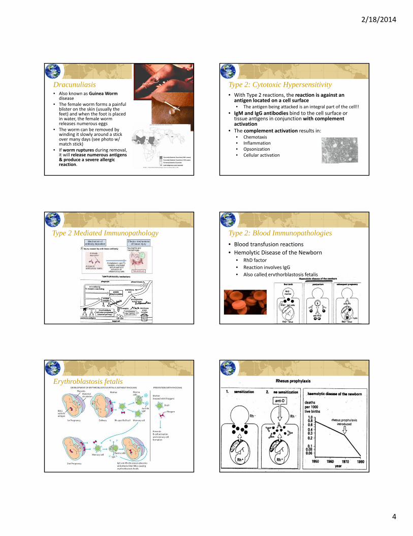
• Examples of Type I reactions
• Asthma
• Hay fever
• Some food and drug allergies
2/18/2014
2
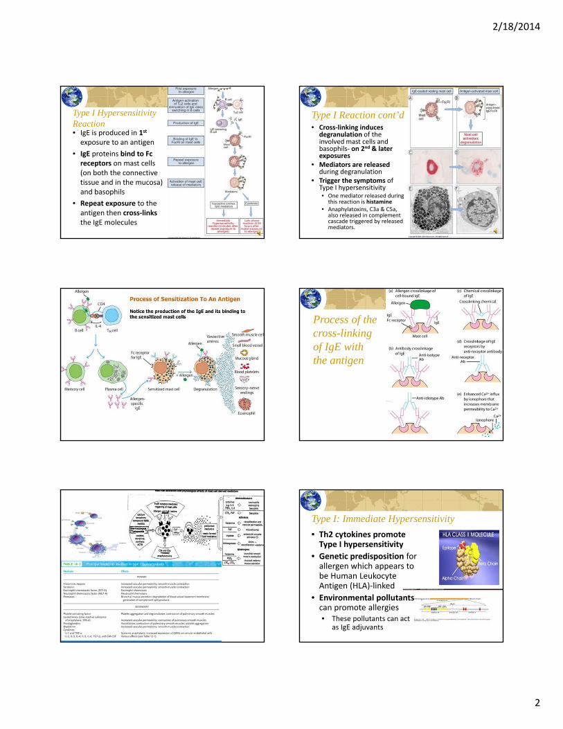
Type I Hypersensitivity Reaction• IgE is produced in 1st
exposure to an antigen
• IgE proteins bind to Fc receptors on mast cells (on both the connective tissue and in the mucosa) and basophils
• Repeat exposure to the antigen then cross‐linksthe IgE molecules
Type I Reaction cont’d• Cross‐linking induces
degranulation of the involved mast cells and basophils‐ on 2nd & later exposures
• Mediators are released during degranulation
• Trigger the symptoms of Type I hypersensitivity• One mediator released during
this reaction is histamine• Anaphylatoxins, C3a & C5a,
also released in complement cascade triggered by released mediators.
Process of the cross-linking of IgE with the antigen
Type I: Immediate Hypersensitivity
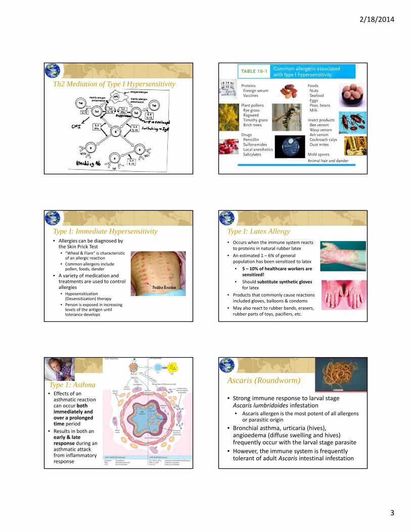
• Th2 cytokines promote Type I hypersensitivity
• Genetic predisposition for allergen which appears to be Human Leukocyte Antigen (HLA)‐linked
• Environmental pollutants can promote allergies• These pollutants can act
as IgE adjuvants
2/18/2014
3
Th2 Mediation of Type I Hypersensitivity
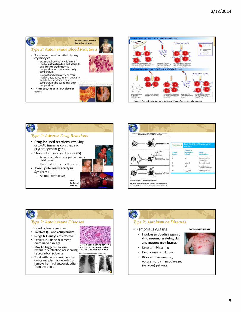
Type I: Immediate Hypersensitivity• Allergies can be diagnosed by
the Skin Prick Test• “Wheal & Flare” is characteristic
of an allergic reaction
• Common allergens include pollen, foods, dander
• A variety of medication and treatments are used to control allergies• Hyposensitization
(Desensitization) therapy
• Person is exposed in increasing levels of the antigen until tolerance develops
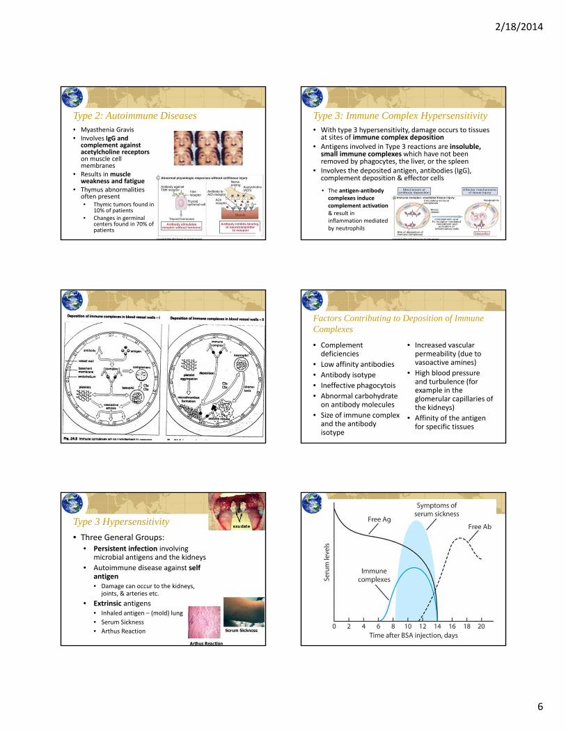
Type I: Latex Allergy• Occurs when the immune system reacts
to proteins in natural rubber latex
• An estimated 1 – 6% of general population has been sensitized to latex
• 5 – 10% of healthcare workers are sensitized!
• Should substitute synthetic gloves for latex
• Products that commonly cause reactions included gloves, balloons & condoms
• May also react to rubber bands, erasers, rubber parts of toys, pacifiers, etc.
Type 1: Asthma• Effects of an asthmatic reaction can occur both immediately and over a prolonged time period
• Results in both an early & late response during an asthmatic attack from inflammatory response
Ascaris (Roundworm)
• Strong immune response to larval stage Ascaris lumbridoides infestation• Ascaris allergen is the most potent of all allergens
or parasitic origin
• Bronchial asthma, urticaria (hives), angioedema (diffuse swelling and hives) frequently occur with the larval stage parasite
• However, the immune system is frequently tolerant of adult Ascaris intestinal infestation
2/18/2014
4
Dracunuliasis• Also known as Guinea Worm
disease• The female worm forms a painful
blister on the skin (usually the feet) and when the foot is placed in water, the female worm releases numerous eggs
• The worm can be removed by winding it slowly around a stick over many days (see photo w/ match stick)
• If worm ruptures during removal, it will release numerous antigens & produce a severe allergic reaction.
Type 2: Cytotoxic Hypersensitivity• With Type 2 reactions, the reaction is against an antigen located on a cell surface• The antigen being attacked is an integral part of the cell!!
• IgM and IgG antibodies bind to the cell surface or tissue antigens in conjunction with complement activation
• The complement activation results in:• Chemotaxis• Inflammation• Opsonization• Cellular activation
Type 2 Mediated Immunopathology Type 2: Blood Immunopathologies
• Blood transfusion reactions
• Hemolytic Disease of the Newborn• RhD factor
• Reaction involves IgG
• Also called erythorblastosis fetalis
Erythroblastosis fetalis
2/18/2014
5
Type 2: Autoimmune Blood Reactions• Spontaneous reactions that destroy
erythrocytes• Warm antibody hemolytic anemia
involve autoantibodies that attach to and destroy erythrocytes at temperatures above normal body temperature
• Cold antibody hemolytic anemia involve autoantibodies that attach to and destroy erythrocytes at temperatures below normal body temperature
• Thrombocytopenia (low platelet count)
Bleeding under the skin due to low platelets.
Type 2: Adverse Drug Reactions• Drug‐induced reactions involving drug‐Ab immune complex and erythrocyte antigens
• Steven‐Johnson Syndrome (SJS)• Affects people of all ages, but more
child cases• If untreated, can result in death
• Toxic Epidermal NecrolysisSyndrome• Another form of SJS
Toxic Epidermal Necrosis
Type 2: Autoimmune Diseases• Goodpasture’s syndrome• Involves IgG and complement• Lungs & kidneys are effected• Results in kidney basement membrane damage
• May be triggered by viral respiratory infections or inhaling hydrocarbon solvents
• Treat with immunosuppressive drugs and plasmapheresis (to remove harmful autoantibodies from the blood)
Type 2: Autoimmune Diseases
• Pemphigus vulgaris
• Involves antibodies against chromosome proteins, skin and mucous membranes
• Results in blistering
• Exact cause is unknown
• Disease is uncommon, occurs mostly in middle‐aged (or older) patients
2/18/2014
6
Type 2: Autoimmune Diseases• Myasthenia Gravis• Involves IgG and
complement against acetylcholine receptorson muscle cell membranes
• Results in muscle weakness and fatigue
• Thymus abnormalities often present• Thymic tumors found in
10% of patients• Changes in germinal
centers found in 70% of patients
Type 3: Immune Complex Hypersensitivity• With type 3 hypersensitivity, damage occurs to tissues at sites of immune complex deposition
• Antigens involved in Type 3 reactions are insoluble, small immune complexeswhich have not been removed by phagocytes, the liver, or the spleen
• Involves the deposited antigen, antibodies (IgG), complement deposition & effector cells
• The antigen‐antibody complexes induce complement activation & result in inflammation mediated by neutrophils
Factors Contributing to Deposition of Immune Complexes
• Complement deficiencies
• Low affinity antibodies
• Antibody isotype
• Ineffective phagocytois
• Abnormal carbohydrate on antibody molecules
• Size of immune complex and the antibody isotype
• Increased vascular permeability (due to vasoactive amines)
• High blood pressure and turbulence (for example in the glomerular capillaries of the kidneys)
• Affinity of the antigen for specific tissues
• Three General Groups:• Persistent infection involving
microbial antigens and the kidneys
• Autoimmune disease against self antigen• Damage can occur to the kidneys,
joints, & arteries etc.
• Extrinsic antigens• Inhaled antigen – (mold) lung
• Serum Sickness
• Arthus Reaction
Type 3 Hypersensitivity
2/18/2014
7
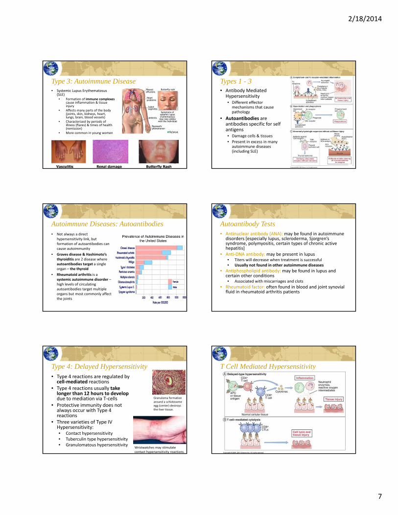
Type 3: Autoimmune Disease• Systemic Lupus Erythematosus
(SLE)• Formation of immune complexes
cause inflammation & tissue injury
• Affects many parts of the body (joints, skin, kidneys, heart, lungs, brain, blood vessels)
• Characterized by periods of illness (flares) & times of health (remission)
• More common in young women
Types 1 - 3• Antibody Mediated
Hypersensitivity• Different effector
mechanisms that cause pathology
• Autoantibodies are antibodies specific for self antigens• Damage cells & tissues
• Present in excess in many autoimmune diseases (including SLE)
Autoimmune Diseases: Autoantibodies• Not always a direct
hypersensitivity link, but formation of autoantibodies can cause autoimmunity
• Graves disease & Hashimoto’s thyroiditis are 2 disease where autoantibodies target a single organ – the thyroid
• Rheumatoid arthritis is a systemic autoimmune disorder –high levels of circulating autoantibodies target multiple organs but most commonly affect the joints
Autoantibody Tests• Antinuclear antibody (ANA): may be found in autoimmune
disorders [especially lupus, scleroderma, Sjorgren’ssyndrome, polymyositis, certain types of chronic active hepatitis]
• Anti‐DNA antibody: may be present in lupus• Titers will decrease when treatment is successful• Usually not found in other autoimmune diseases
• Antiphospholipid antibody: may be found in lupus and certain other conditions• Associated with miscarriages and clots
• Rheumatoid factor: often found in blood and joint synovial fluid in rheumatoid arthritis patients
Type 4: Delayed Hypersensitivity• Type 4 reactions are regulated by cell‐mediated reactions
• Type 4 reactions usually take longer than 12 hours to develop due to mediation via T‐cells
• Protective immunity does not always occur with Type 4 reactions
• Three varieties of Type IV Hypersensitivity:• Contact hypersensitivity• Tuberculin type hypersensitivity• Granulomatous hypersensitivity
Granuloma formation around a schistosomeegg (center) destroys the liver tissue.
Wristwatches may stimulate contact hypersensitivity reactions.
T Cell Mediated Hypersensitivity
2/18/2014
8
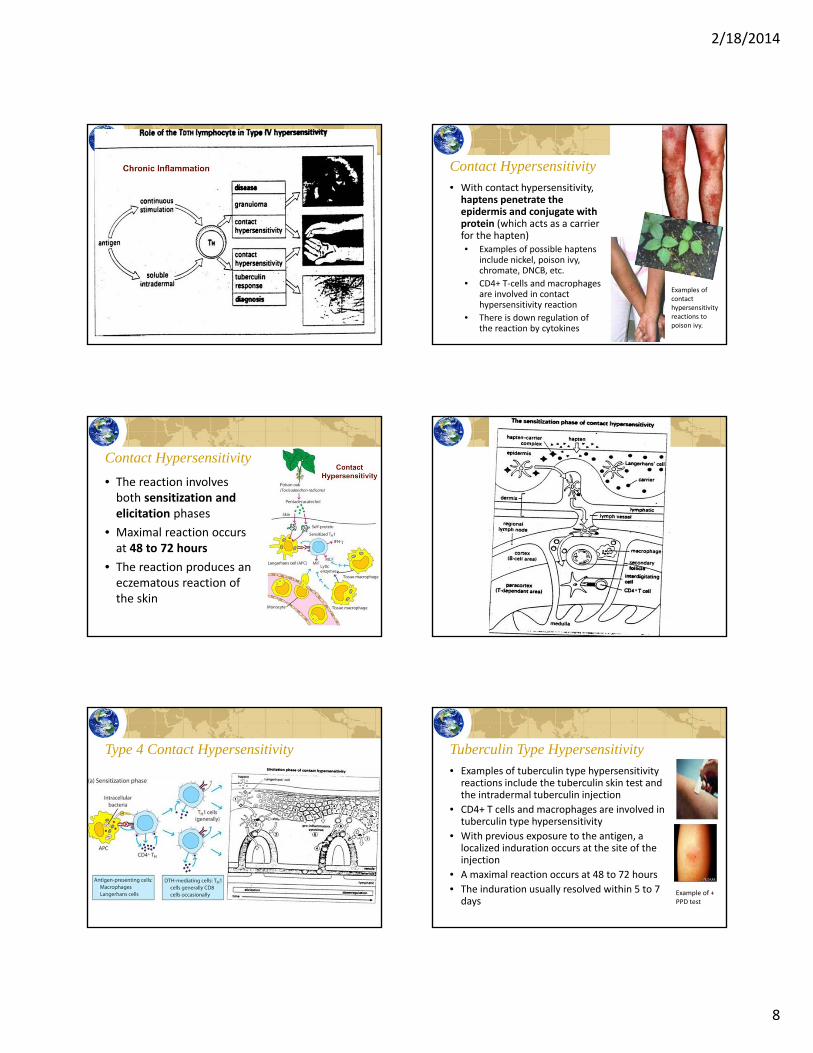
Contact Hypersensitivity• With contact hypersensitivity, haptens penetrate the epidermis and conjugate with protein (which acts as a carrier for the hapten)• Examples of possible haptens
include nickel, poison ivy, chromate, DNCB, etc.
• CD4+ T‐cells and macrophages are involved in contact hypersensitivity reaction
• There is down regulation of the reaction by cytokines
Examples of contact hypersensitivity reactions to poison ivy.
Contact Hypersensitivity
• The reaction involves both sensitization and elicitation phases
• Maximal reaction occurs at 48 to 72 hours
• The reaction produces an eczematous reaction of the skin
Type 4 Contact Hypersensitivity Tuberculin Type Hypersensitivity• Examples of tuberculin type hypersensitivity reactions include the tuberculin skin test and the intradermal tuberculin injection
• CD4+ T cells and macrophages are involved in tuberculin type hypersensitivity
• With previous exposure to the antigen, a localized induration occurs at the site of the injection
• A maximal reaction occurs at 48 to 72 hours
• The induration usually resolved within 5 to 7 days
Example of + PPD test
2/18/2014
9
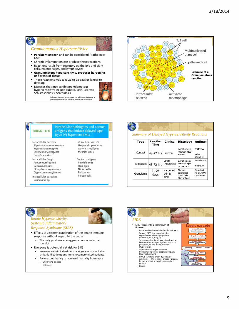
Granulomatous Hypersensitivity• Persistent antigen and can be considered “Pathologic CMI”
• Chronic inflammation can produce these reactions• Reactions result from secretory epithelioid and giant cells, macrophages, and lymphocytes
• Granulomatous hypersensitivity produces hardening or fibrosis of tissue
• These reactions may take 21 to 28 days or longer to develop
• Diseases that may exhibit granulomatous hypersensitivity include Tuberculosis, Leprosy, Schistosomiasis, Sarcoidosis
Enlarged liver and spleen occurs in schistosomiasis due to granuloma formation, blocking abdominal circulation.
Summary of Delayed Hypersensitivity Reactions
Innate Hypersensitivity: Systemic Inflammatory Response Syndrome (SIRS)• Effects of a systemic activation of the innate immune response without regard to the cause• The body produces an exaggerated response to the
stimulus
• Everyone is potentially at risk for SIRS• However, certain individuals are at greater risk including
critically ill patients and immunocompromised patients
• Factors contributing to increased mortality from sepsis• underlying disease
• older age
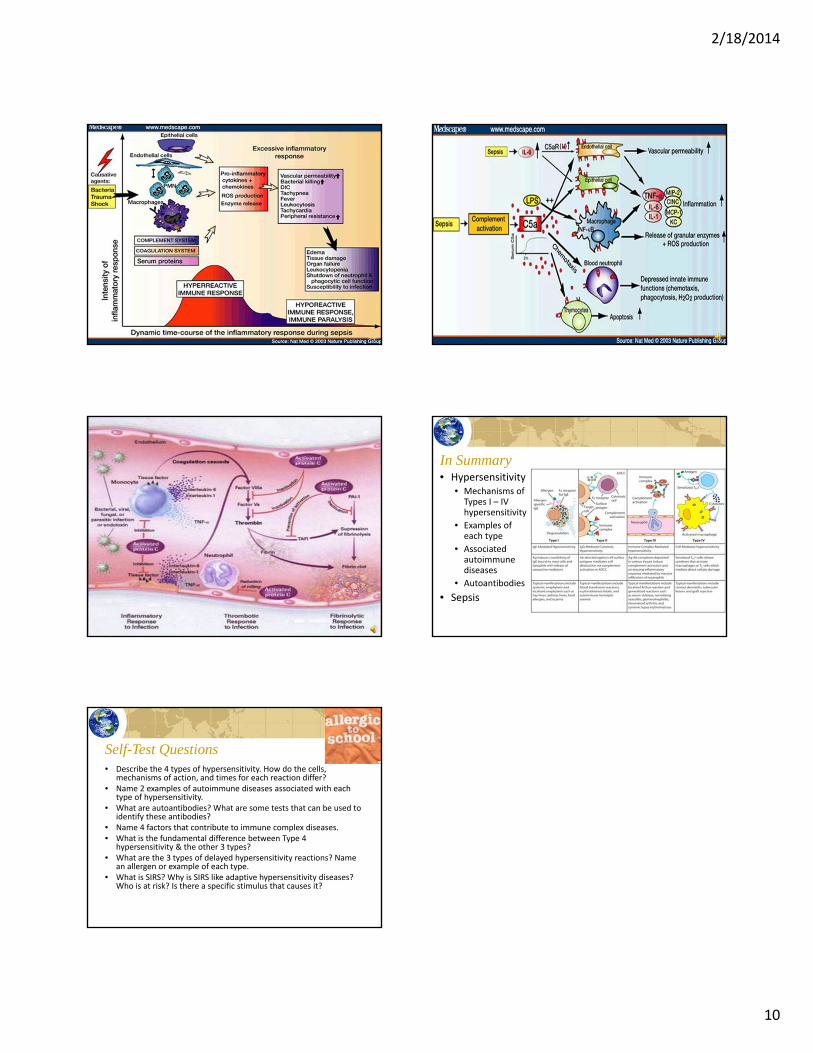
SIRS• SIRS represents a continuum of
disease:• Bacteremia – bacteria in the bloodstream• Sepsis – SIRS due to an infection
regardless of infecting organism (bacterial, viral, fungal)
• Severe sepsis – Sepsis associated with at least one acute organ dysfunction, poor perfusion, or low blood pressure (hypotension)
• Septic shock – Sepsis‐induced hypotension persists despite adequate fluid replacement
• MODS (Multiple organ dysfunction syndrome) – Presence of altered function in two or more organs in an acutely ill patient
• Death
2/18/2014
10
In Summary• Hypersensitivity
• Mechanisms of Types I – IV hypersensitivity
• Examples of each type
• Associated autoimmune diseases
• Autoantibodies
• Sepsis
Self-Test Questions• Describe the 4 types of hypersensitivity. How do the cells,
mechanisms of action, and times for each reaction differ?• Name 2 examples of autoimmune diseases associated with each
type of hypersensitivity.• What are autoantibodies? What are some tests that can be used to
identify these antibodies?• Name 4 factors that contribute to immune complex diseases. • What is the fundamental difference between Type 4
hypersensitivity & the other 3 types?• What are the 3 types of delayed hypersensitivity reactions? Name
an allergen or example of each type.• What is SIRS? Why is SIRS like adaptive hypersensitivity diseases?
Who is at risk? Is there a specific stimulus that causes it?