Corneal graft survival and intraocular pressure control after Descemet stripping automated
endothelial keratoplasty in eyes with pre-existing glaucoma
Desmond QUEK1, Tina WONG1,2, Donald TAN1,2, Jodhbir MEHTA1,2,3
1Singapore National Eye Centre and Singapore Eye Research Institute2Department of Ophthalmology, Yong Loo Lin School of Medicine, National University of Singapore
3Clinical Sciences, Duke-NUS Graduate Medical School
The authors have no financial interest in the subject matter of this e-poster
Singapore Eye Research InstituteSingapore National Eye Centre
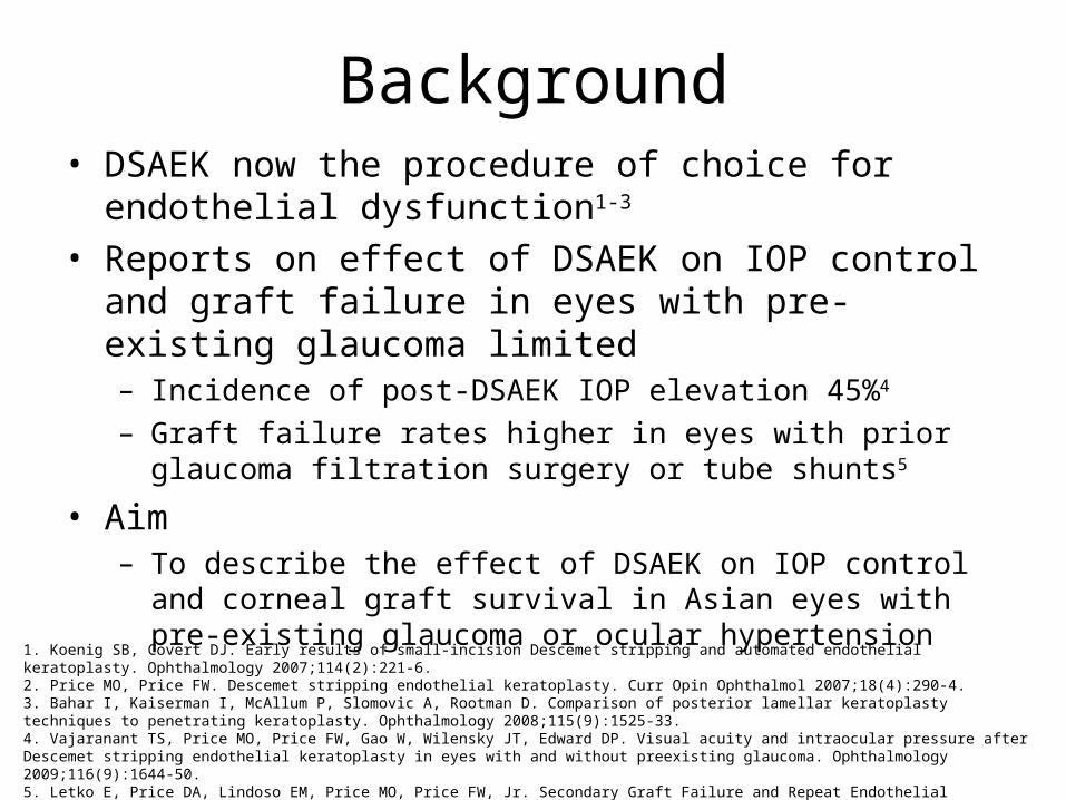
Background• DSAEK now the procedure of choice for endothelial
dysfunction1-3
• Reports on effect of DSAEK on IOP control and graft failure in eyes with pre-existing glaucoma limited– Incidence of post-DSAEK IOP elevation 45%4
– Graft failure rates higher in eyes with prior glaucoma filtration surgery or tube shunts5
• Aim– To describe the effect of DSAEK on IOP control and corneal graft
survival in Asian eyes with pre-existing glaucoma or ocular hypertension
1. Koenig SB, Covert DJ. Early results of small-incision Descemet stripping and automated endothelial keratoplasty. Ophthalmology 2007;114(2):221-6.2. Price MO, Price FW. Descemet stripping endothelial keratoplasty. Curr Opin Ophthalmol 2007;18(4):290-4.3. Bahar I, Kaiserman I, McAllum P, Slomovic A, Rootman D. Comparison of posterior lamellar keratoplasty techniques to penetrating keratoplasty. Ophthalmology 2008;115(9):1525-33.4. Vajaranant TS, Price MO, Price FW, Gao W, Wilensky JT, Edward DP. Visual acuity and intraocular pressure after Descemet stripping endothelial keratoplasty in eyes with and without preexisting glaucoma. Ophthalmology 2009;116(9):1644-50.5. Letko E, Price DA, Lindoso EM, Price MO, Price FW, Jr. Secondary Graft Failure and Repeat Endothelial Keratoplasty after Descemet Stripping Automated Endothelial Keratoplasty. Ophthalmology 2010 Sep 22 [Epub ahead of print].
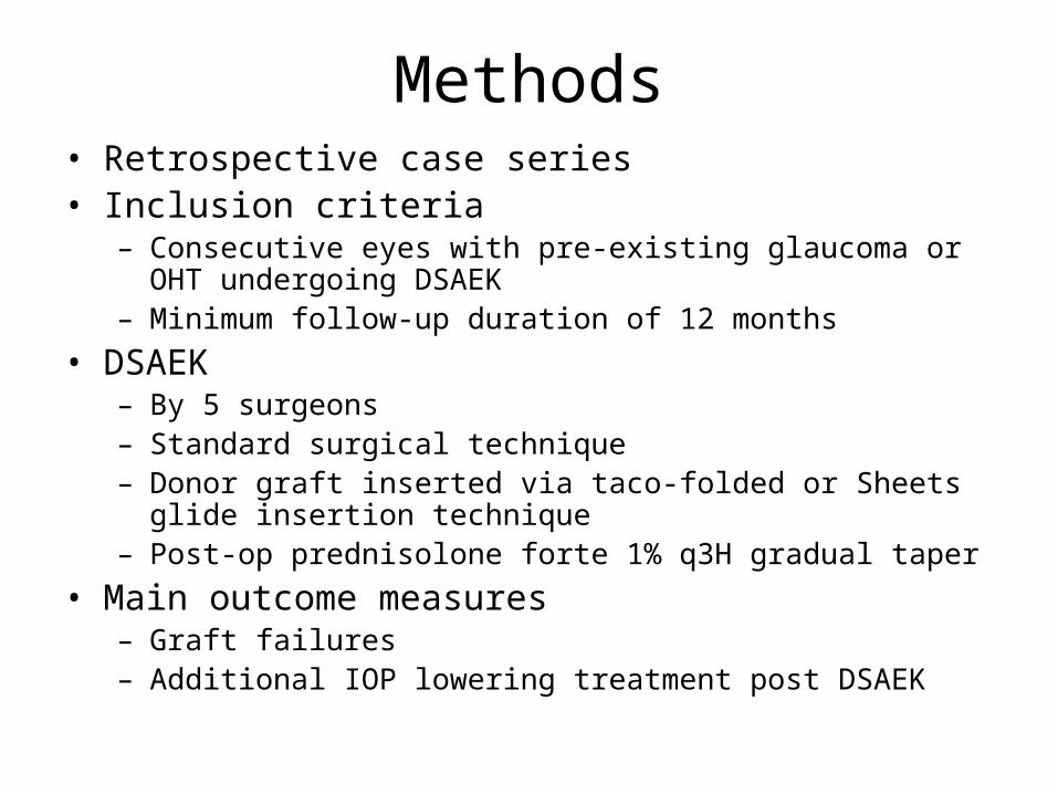
Methods• Retrospective case series• Inclusion criteria
– Consecutive eyes with pre-existing glaucoma or OHT undergoing DSAEK
– Minimum follow-up duration of 12 months• DSAEK
– By 5 surgeons– Standard surgical technique– Donor graft inserted via taco-folded or Sheets glide insertion
technique– Post-op prednisolone forte 1% q3H gradual taper
• Main outcome measures– Graft failures– Additional IOP lowering treatment post DSAEK
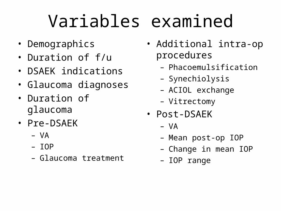
Variables examined• Demographics• Duration of f/u• DSAEK indications• Glaucoma diagnoses• Duration of glaucoma• Pre-DSAEK
– VA– IOP– Glaucoma treatment
• Additional intra-op procedures– Phacoemulsification– Synechiolysis– ACIOL exchange– Vitrectomy
• Post-DSAEK– VA– Mean post-op IOP– Change in mean IOP– IOP range
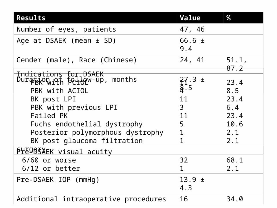
Pre-DSAEK visual acuity6/60 or worse6/12 or better
321
68.12.1
Pre-DSAEK IOP (mmHg) 13.9 ± 4.3
Additional intraoperative procedures 16 34.0
Indications for DSAEKPBK with PCIOLPBK with ACIOLBK post LPIPBK with previous LPIFailed PKFuchs endothelial dystrophyPosterior polymorphous dystrophyBK post glaucoma filtration surgery
11411311511
23.48.523.46.423.410.62.12.1
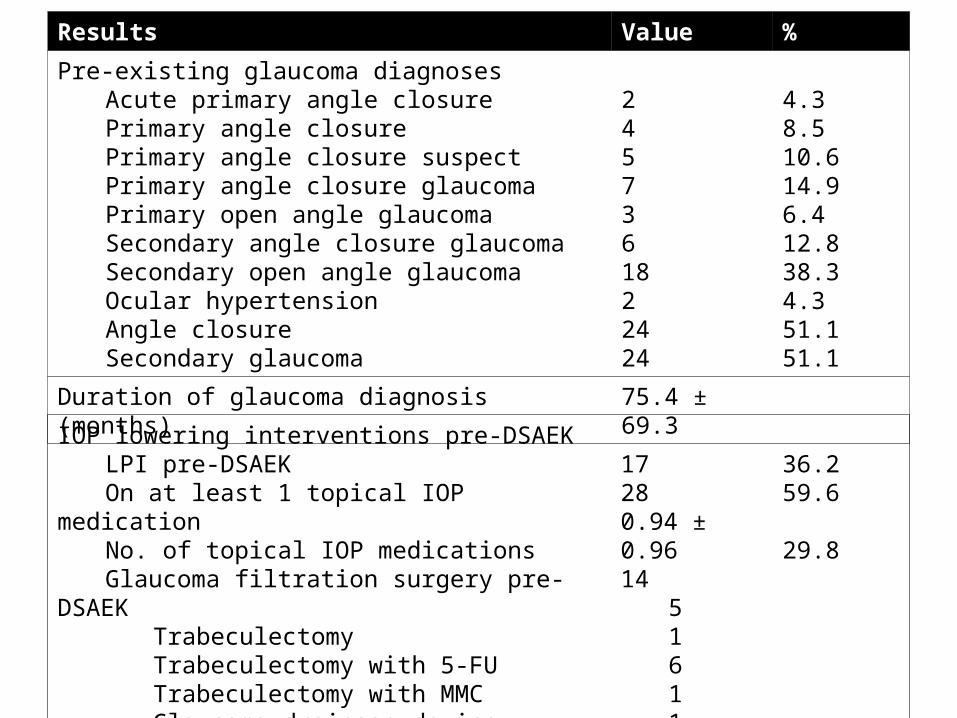
Results Value %
Number of eyes, patients 47, 46
Age at DSAEK (mean ± SD) 66.6 ± 9.4
Gender (male), Race (Chinese) 24, 41 51.1, 87.2
Duration of follow-up, months 27.3 ± 8.5
Results Value %
Pre-existing glaucoma diagnosesAcute primary angle closurePrimary angle closurePrimary angle closure suspectPrimary angle closure glaucomaPrimary open angle glaucomaSecondary angle closure glaucomaSecondary open angle glaucomaOcular hypertensionAngle closureSecondary glaucoma
2457361822424
4.38.510.614.96.412.838.34.351.151.1
Duration of glaucoma diagnosis (months) 75.4 ± 69.3
IOP lowering interventions pre-DSAEKLPI pre-DSAEKOn at least 1 topical IOP medicationNo. of topical IOP medications Glaucoma filtration surgery pre-DSAEK
TrabeculectomyTrabeculectomy with 5-FUTrabeculectomy with MMCGlaucoma drainage deviceTrabeculectomy + GDD
17280.94 ± 0.9614
51611
36.259.6
29.8
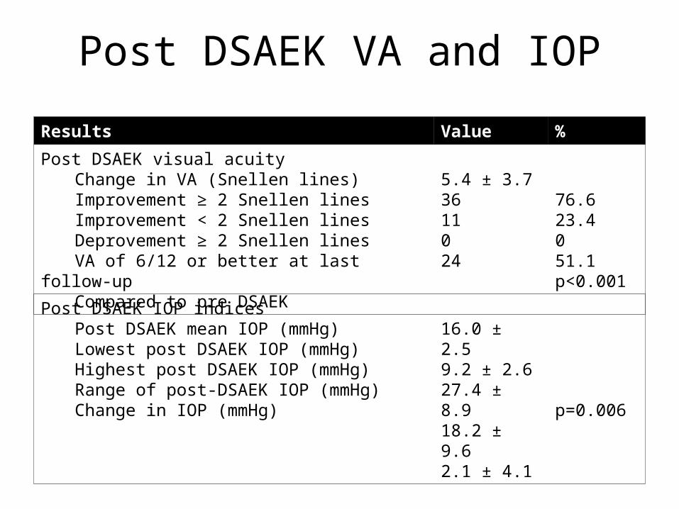
Results Value %
Post DSAEK visual acuityChange in VA (Snellen lines)Improvement ≥ 2 Snellen linesImprovement < 2 Snellen linesDeprovement ≥ 2 Snellen linesVA of 6/12 or better at last follow-upCompared to pre DSAEK
5.4 ± 3.73611024
76.623.4051.1p<0.001
Post DSAEK VA and IOP
Post DSAEK IOP indicesPost DSAEK mean IOP (mmHg)Lowest post DSAEK IOP (mmHg)Highest post DSAEK IOP (mmHg)Range of post-DSAEK IOP (mmHg)Change in IOP (mmHg)
16.0 ± 2.59.2 ± 2.627.4 ± 8.918.2 ± 9.62.1 ± 4.1 p=0.006
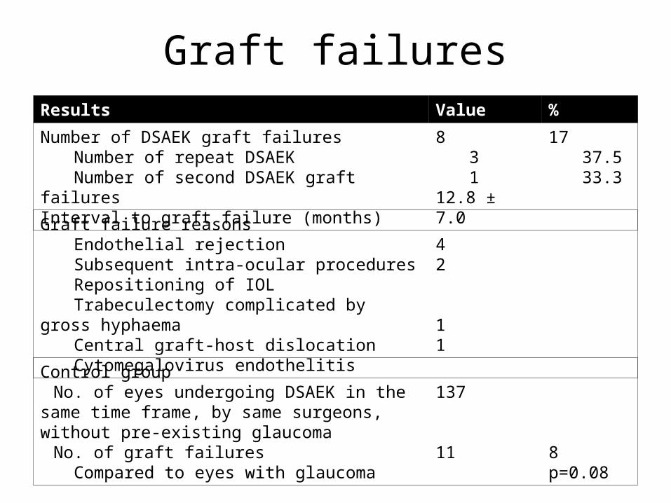
Graft failuresResults Value %
Number of DSAEK graft failuresNumber of repeat DSAEKNumber of second DSAEK graft failures
Interval to graft failure (months)
831
12.8 ± 7.0
1737.533.3
Graft failure reasonsEndothelial rejectionSubsequent intra-ocular proceduresRepositioning of IOLTrabeculectomy complicated by gross hyphaemaCentral graft-host dislocationCytomegalovirus endothelitis
42
11
Control groupNo. of eyes undergoing DSAEK in the same time
frame, by same surgeons, without pre-existing glaucoma
No. of graft failuresCompared to eyes with glaucoma
137
11 8p=0.08
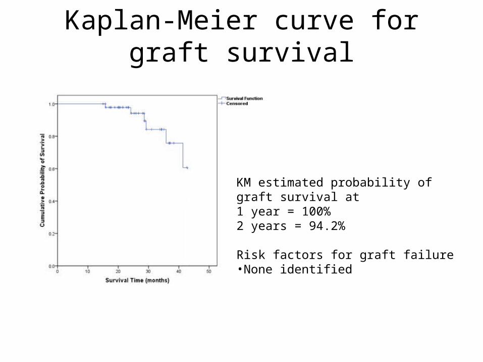
Kaplan-Meier curve for graft survival
KM estimated probability of graft survival at1 year = 100%2 years = 94.2%
Risk factors for graft failure•None identified
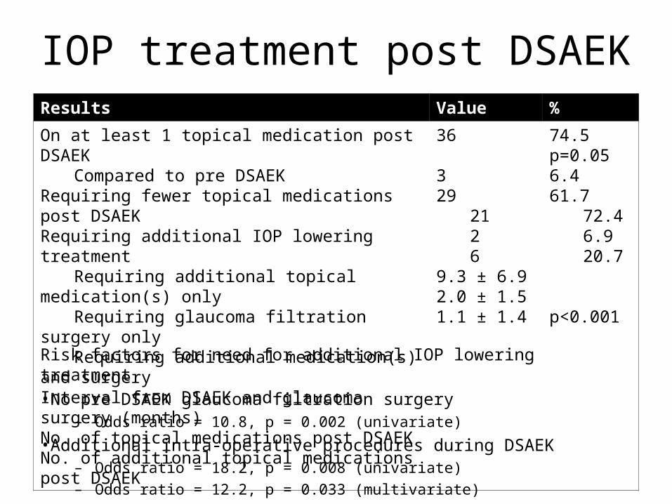
IOP treatment post DSAEKResults Value %
On at least 1 topical medication post DSAEKCompared to pre DSAEK
Requiring fewer topical medications post DSAEKRequiring additional IOP lowering treatment
Requiring additional topical medication(s) onlyRequiring glaucoma filtration surgery onlyRequiring additional medication(s) and surgery
Interval from DSAEK and glaucoma surgery (months)No. of topical medications post DSAEKNo. of additional topical medications post DSAEK
36
329
2126
9.3 ± 6.92.0 ± 1.51.1 ± 1.4
74.5p=0.056.461.7
72.46.920.7
p<0.001
Risk factors for need for additional IOP lowering treatment•No pre DSAEK glaucoma filtration surgery
– Odds ratio = 10.8, p = 0.002 (univariate)
•Additional intra-operative procedures during DSAEK– Odds ratio = 18.2, p = 0.008 (univariate)– Odds ratio = 12.2, p = 0.033 (multivariate)
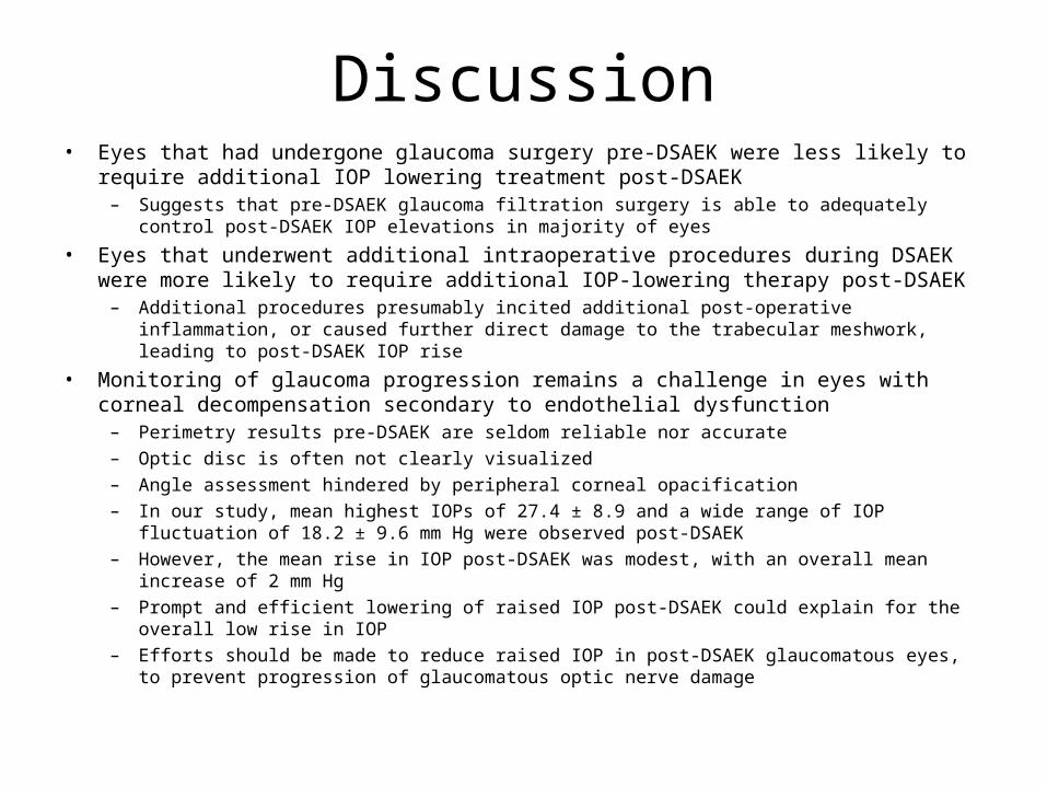
Discussion• Eyes that had undergone glaucoma surgery pre-DSAEK were less likely to require additional
IOP lowering treatment post-DSAEK– Suggests that pre-DSAEK glaucoma filtration surgery is able to adequately control post-DSAEK IOP
elevations in majority of eyes
• Eyes that underwent additional intraoperative procedures during DSAEK were more likely to require additional IOP-lowering therapy post-DSAEK– Additional procedures presumably incited additional post-operative inflammation, or caused further
direct damage to the trabecular meshwork, leading to post-DSAEK IOP rise
• Monitoring of glaucoma progression remains a challenge in eyes with corneal decompensation secondary to endothelial dysfunction– Perimetry results pre-DSAEK are seldom reliable nor accurate– Optic disc is often not clearly visualized– Angle assessment hindered by peripheral corneal opacification– In our study, mean highest IOPs of 27.4 ± 8.9 and a wide range of IOP fluctuation of 18.2 ± 9.6 mm
Hg were observed post-DSAEK– However, the mean rise in IOP post-DSAEK was modest, with an overall mean increase of 2 mm Hg– Prompt and efficient lowering of raised IOP post-DSAEK could explain for the overall low rise in IOP– Efforts should be made to reduce raised IOP in post-DSAEK glaucomatous eyes, to prevent
progression of glaucomatous optic nerve damage
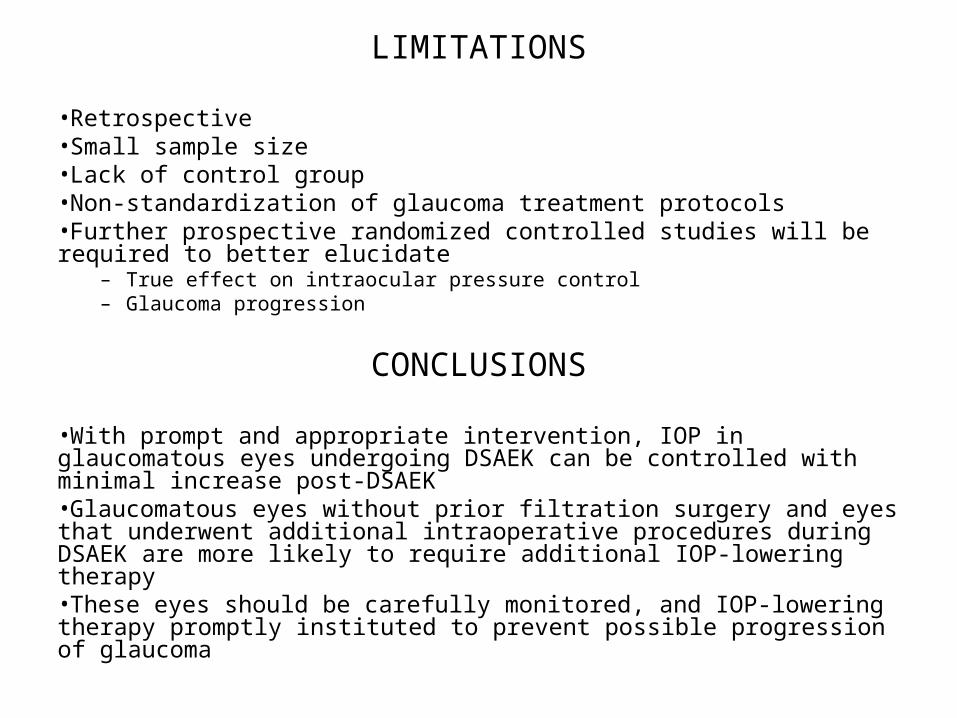
LIMITATIONS
•Retrospective •Small sample size•Lack of control group•Non-standardization of glaucoma treatment protocols•Further prospective randomized controlled studies will be required to better elucidate
– True effect on intraocular pressure control– Glaucoma progression
CONCLUSIONS
•With prompt and appropriate intervention, IOP in glaucomatous eyes undergoing DSAEK can be controlled with minimal increase post-DSAEK•Glaucomatous eyes without prior filtration surgery and eyes that underwent additional intraoperative procedures during DSAEK are more likely to require additional IOP-lowering therapy•These eyes should be carefully monitored, and IOP-lowering therapy promptly instituted to prevent possible progression of glaucoma