OPEN
ORIGINAL ARTICLE
Brain antibodies in the cortex and blood of people withschizophrenia and controlsLJ Glass1,2, D Sinclair1,2,3, D Boerrigter1,2, K Naude1,2, SJ Fung1,2,3, D Brown4,5, VS Catts1,2,3, P Tooney6, M O’Donnell3, R Lenroot1,2,3,C Galletly7,8, D Liu7,9, TW Weickert1,2,3 and C Shannon Weickert1,2,3
The immune system is implicated in the pathogenesis of schizophrenia, with elevated proinflammatory cytokine mRNAs found inthe brains of ~ 40% of individuals with the disorder. However, it is not clear if antibodies (specifically immunoglobulin-γ (IgG)) canbe found in the brain of people with schizophrenia and if their abundance relates to brain inflammatory cytokine mRNA levels.Therefore, we investigated the localization and abundance of IgG in the frontal cortex of people with schizophrenia and controls,and the impact of proinflammatory cytokine status on IgG abundance in these groups. Brain IgGs were detected surrounding bloodvessels in the human and non-human primate frontal cortex by immunohistochemistry. IgG levels did not differ significantlybetween schizophrenia cases and controls, or between schizophrenia cases in ‘high’ and ‘low’ proinflammatory cytokine subgroups.Consistent with the existence of IgG in the parenchyma of human brain, mRNA and protein of the IgG transporter (FcGRT) werepresent in the brain, and did not differ according to diagnosis or inflammatory status. Finally, brain-reactive antibody presence andabundance was investigated in the blood of living people. The plasma of living schizophrenia patients and healthy controlscontained antibodies that displayed positive binding to Rhesus macaque cerebellar tissue, and the abundance of these antibodieswas significantly lower in patients than controls. These findings suggest that antibodies in the brain and brain-reactive antibodies inthe blood are present under normal circumstances.
Translational Psychiatry (2017) 7, e1192; doi:10.1038/tp.2017.134; published online 8 August 2017
INTRODUCTIONThere is increasing evidence of immune abnormalities in peoplewith schizophrenia. In the blood, increased concentration ofcytokines, particularly interferon (IFN)-γ, interleukin (IL)-1β, solubleIL-2 receptor (sIL-2R), IL-6, IL-12, transforming growth factor (TGF)-β and tumor necrosis factor (TNF)-α, are found in people withschizophrenia when compared to controls.1,2 In the brain,specifically dorsolateral prefrontal cortex (DLPFC), increased mRNAexpression of IL-6, IL-1β and IL-8 cytokines can be found in somepeople with schizophrenia.3–6 Transcript levels of various immuneregulators and their chaperone proteins are also altered in theprefrontal cortex of subjects with schizophrenia.7,8 Antipsychoticmedications can have immunomodulatory effects,9–11 often low-ering cytokine levels in addition to alleviating positive symptomsof schizophrenia. However, blood levels of IL-1β, IL-6, IL-12, IFN-γ,TNF-α, sIL-2R and TGF-β have been found to be elevated inunmedicated first-episode psychosis1,9,12 and chronically medi-cated patients,13,14 indicating that antipsychotic treatment neithersolely explains, nor completely remediates, immune activation inschizophrenia.To date, it is unclear whether antibodies play a role in immune
dysregulation in schizophrenia. The T-cell-produced cytokinesactivate B cells to switch from producing weakly bindingimmunoglobulin-μ to the highly specified immunoglobulin-γ
(IgG). Playing an integral part in the secondary immune response,IgG antibodies bind complement, facilitate phagocytosis throughopsonization, and direct cytotoxic activities of natural killer cells.15
In peripheral blood, elevated B-cell and reduced T-cell popula-tions have been found in schizophrenia.16–18 In fact, matureB cells numbers appear to normalize in some schizophreniapatients whose clinical state has improved with antipsychotictreatment.17,19 These observations suggest that immune dysregu-lation in schizophrenia may include an underlying component ofB-cell pathology.Antibodies in schizophrenia pertaining to brain pathology are
likely to recognize brain antigens (brain-reactive) and should bepresent within the brain itself. Brain-reactive antibodies are knownto be present in the blood in health20 and psychiatric disease,20–26
and may reflect antibody-related immune pathology in schizo-phrenia. Antibodies from blood have been shown to bind tomonkey and human brain tissue antigens.21,22 More specifically,antibodies targeting N-methyl-D-aspartate receptors (NMDAR) arefound in the cerebrospinal fluid and serum of people with NMDARencephalitis, who exhibit schizophrenia-like symptoms includingpsychosis and cognitive impairments,27,28 and in some peopleexperiencing first-episode psychosis.29 In people with schizophre-nia, there are serum antibodies targeting other neurotransmitterreceptors (for example, muscarinic cholinergic receptor 1, opioid
1Schizophrenia Research Laboratory, Sydney, NSW, Australia; 2Neuroscience Research Australia, Sydney, NSW, Australia; 3School of Psychiatry, University of New South Wales,Sydney, NSW, Australia; 4St Vincent’s Centre for Applied Medical Research, St Vincent's Hospital, Sydney, NSW, Australia; 5ICPMR, Westmead Hospital, Westmead, NSW, Australia;6School of Biomedical Sciences and Pharmacy, University of Newcastle, Newcastle, NSW, Australia; 7Discipline of Psychiatry, Adelaide University, Adelaide, SA, Australia; 8RamsayHealth Care, Adelaide, SA, Australia and 9Northern Adelaide Local Health Network, Adelaide, SA, Australia. Correspondence: Professor C Shannon Weickert, SchizophreniaResearch Laboratory, Neuroscience Research Australia, Barker Street, Randwick, NSW 2031, Australia.E-mail: [email protected] 1 December 2016; revised 9 May 2017; accepted 9 May 2017
Citation: Transl Psychiatry (2017) 7, e1192; doi:10.1038/tp.2017.134
www.nature.com/tp
receptor-μ and serotonin receptor-1A receptors),23–25 heat-shockproteins30–32 and glyceraldehyde 3-phosphate dehydrogenase26
(GAPDH). However, whether the human brain has an appreciableamount of IgGs, and whether their abundance is altered inschizophrenia, is unknown.The presence of IgGs in the brain, particularly the healthy brain,
is more plausible if mechanisms exist in the brain to allow theirinflux and efflux. The Fc fragment neonatal receptor (FcRN) isfound in the choroid plexus and microvascular endothelial cells33
and facilitates the transit of IgG across the luminal surface.34 It iscomposed of a heavy chain, Fc region of IgG-targeting receptortransporter (FcGRT), and a light chain, β-2-microglobulin.35 Actingin a pH-dependent manner, FcRN binds to IgG at an acidic pH andreleases it at a neutral pH.36,37 As a result, the expression of FcGRTmay influence the abundance of antibodies in the brain inpsychiatric disease.Therefore, in this study we aimed to (1) determine the presence
of IgGs in the postmortem brains of people with schizophreniaand controls with a focus on the DLPFC and orbitofrontal cortex(OFC); (2) compare IgG levels between people with schizophreniaand controls previously categorised as ‘high inflammation’ or ‘lowinflammation’ based on proinflammatory cytokine levels;3–5 (3)compare the abundance of FcGRT mRNA and protein between theaforementioned groups; and (4) assess the prevalence of brain-reactive antibodies in the plasma of a cohort of livingschizophrenia patients and controls.
MATERIALS AND METHODSTissue and bloodHuman brain tissue. Post-mortem brain tissue samples (n= 79 individuals)were obtained from the New South Wales Tissue Resource Centre. OFCtissue from the medial gyrus rectus to include BA11 (between the branchesof the orbital sulcus)38 was obtained from individuals with schizophrenia(n=38) and controls (n= 38) and was cryostat sectioned in the coronalplane with sections mounted onto glass slides (Supplementary Table S1).Chunks of pulverized DLPFC gray matter (40 mg) from BA46 (middlefrontal gyrus)39 was obtained from individuals with schizophrenia (n= 37)and controls (n=37; Supplementary Table S2). Sample sizes were chosenbased on variance observed in previous studies, with a sample size ofn= 74 expected to have 480% power to detect an effect size d=0.25.39,40
This study was carried out in accordance with the latest version of theDeclaration of Helsinki after review by the Human Research EthicsCommittee at the UNSW (HREC #12435).
Rhesus macaque brain tissue. Fresh frozen frontal perfused rhesusmacaque (Macaca mulatta, n=7) cortex (containing the principal sulcus)was cryostat sectioned in the coronal plane. All research procedures withnon-human primates from the National Institutes of Mental Health (NIMH,USA) and were carried out in adherence to the regulations of the U.S.Animal Welfare Act (USDA, 1990) and Public Health Service Policies (PHS,2002), in accordance with the ILAR ‘Guide for the Care and Use ofLaboratory Animals’, and are described in Fung et al.41 The study wasperformed under an Animal Study Protocol approved by the NIMH AnimalCare and Use Committee.
Human serum and plasma. Samples were obtained from a living cohort ofcontrols (n= 73) and people with schizophrenia (n= 94)42 (SupplementaryTable S3). Patients were matched to healthy controls based on gender andage within 5 years. Informed consent was obtained in accordance with aprotocol approved by the University of New South Wales (UNSW) and theSouth Eastern Sydney, Illawarra Area Health Service Human Research EthicsCommittees (HREC #07259, HREC #07121), and the Queen ElizabethHospital Human Ethics Committee (SA; HREC # 8222 6841). To prepareserum, whole blood was collected in SST tubes (BD Biosciences, FranklinLakes, NJ, USA), incubated at room temperature (RT) for 30 min,centrifuged at 2000 g for 5 min at 4 °C. To prepare plasma, whole bloodwas collected in EDTA tubes (BD Biosciences), centrifuged at 1200 g for15 min at 4 °C. The resulting serum, or plasma, was transferred to lowbinding tubes and stored at − 80 °C.
ImmunohistochemistryImmunohistochemistry to detect endogenous IgG in human OFC and rhesusmacaque PFC. Human postmortem OFC sections from schizophreniacases and controls, or rhesus macaque PFC, were thawed (RT for 20 min),fixed with 4% paraformaldehyde, washed (3 × PBS, 5 min) and submergedin 3:1 100% methanol in 3% H2O2 for 20 min at RT to block endogenousperoxidases. For human OFC, tissue was washed and blocked overnightwith 10% normal rabbit serum (S-5000, Vector Laboratories, Peterborough,UK). For rhesus macaque PFC, tissue was blocked for 1 h at RT with 10%normal goat serum (S-1000, Vector Laboratories) and incubated overnightwith mouse anti-monkey IgG primary (1:500, 4700-01, Southern Biotech,Birmingham, AL, USA). The next day, tissue was washed as above andincubated for 1 h at RT with (for human OFC) biotinylated rabbit anti-human IgG ‘secondary’ antibody preabsorbed against mouse (1:200,Ab7159, Abcam, Cambridge, UK) or (for rhesus macaque PFC) biotinylatedgoat anti-mouse IgG (1:500, BA9200, Vector Laboratories). After washingagain, the tissue was incubated for 1 h at RT with avidin-biotin-peroxidasecomplex (VectaStain ABC kit, PK-4000, Vector Laboratories). Then 3’3-diaminobenzidine (DAB, 12 mM final concentration in PBS with 3% H2O2)was applied to the tissue for 3 min, before Nissl counterstaining (3 minexposure with 0.002% thionin). Images were taken with a Nikon Eclipse i80microscope (Nikon, Tokyo, Japan) using a × 20 objective, and with contrastenhanced with ImageJ (v1.50.e, NIH, Bethesda, MD, USA).
Human OFC fluorescent immunohistochemistry. Fresh frozen OFC sectionsfrom people with schizophrenia (n= 9) and healthy controls (n=9) werefixed, washed and blocked with 10% normal goat serum and 10% donkeyserum (Jackson Immunoresearch Laboratories, Baltimore, MD, USA) indiluent for 1 h at RT. Tissue was then incubated overnight at 4 °C withrabbit anti-collagen IV (1:5000, AB6586, Abcam), biotinylated goat anti-human IgG (1:200, AB97168, Abcam), and mouse anti-neuronal nuclei(1:1000, mAB377, Chemicon International, Australia) primary antibodies.The following day tissue was washed as above and incubated in the darkwith goat anti-rabbit IgG AlexaFluor 405 preabsorbed against chicken, cow,horse, human, mouse, pig and rat (1:500, AB175654, Abcam), streptavidinAlexaFluor 647 (1:1000, S21374, Life Technologies, Eugene, OR, USA) anddonkey anti-mouse IgG AlexaFluor 488 preabsorbed against chicken, cow,goat, human, rabbit, rat, and sheep (1:500, AB150109, Abcam) for 1 h at 4 °C. Tissue was washed twice in PBS and then for 5 min in 10 μM acridineorange hemi(zinc chloride) (Ab146348, Abcam) in PBS at RT. Slides werewashed twice in 5 mM cupric sulfate and 50 mM ammonium acetatesolution for 15 min at RT to quench autofluorescence. Tissue was thenmounted with fluorescent-friendly immersion oil (Citifluor AF1 anti-fadent,ProSciTech, Thuringowa, QLD, Australia) and slide edges were sealed withnail polish. Z-stack spectral images were captured using a Nikon Eclipse 90ilaser-scanning microscope and subjected to blind unmixing. Images weretaken at × 40 and contrast and brightness enhanced using ImageJ.
Immunohistochemistry using human serum from living people as a ‘primary’antibody. Pooled serum from live individuals with schizophrenia (n= 10),or pooled serum from healthy controls (n= 10) was diluted (1:150, 1:300,1:700) to be used as ‘primary’ antibodies. Perfused Rhesus macaquecerebellar sections43 were used to so that brain-reactive IgG in the bloodcould be detected without IgG in the blood vessels and/or brainconfounding the results. Sections were treated the same as the Rhesusmacaque PFC above except that pooled serum was used as the ‘primary’antibody and the secondary antibody was goat anti-human (1:250).
Quantitative real-time PCRRNA was extracted and cDNA synthesized from DLPFC tissue of peoplewith schizophrenia (n=37) and healthy controls (n=37) as previouslydescribed in Weickert et al.39 Transcript levels were measured by qPCRusing Applied Biosystems’ Prism 7900HT Real-time PCR system (Foster City,CA, USA). A pre-designed Taqman gene expression assay from AppliedBiosystems (Foster City, CA, USA) was used for FCGRT (Hs01108967_m1),normalized to the geometric mean of four housekeeper genes; β-actin(Hs99999903_m1), GAPDH (Hs99999905_m1), TATA box binding protein(Hs00427620_m1), and ubiquitin C (Hs00824723_m1) that did not vary inexpression between diagnostic groups.39
Western blottingHuman DLPFC western blotting. For endogenous IgG detection, DLPFCtissue from schizophrenia cases (n= 37) and controls (n= 37) was
Brain antibodies in the cortex and bloodLJ Glass et al
2
Translational Psychiatry (2017), 1 – 9
homogenized in buffer (50% 0.1 M Tris Buffer pH7.5, 50% glycerol, proteaseinhibitor cocktail 1:100 and aprotinin 1:1 600) and 10 μg of each sampleelectrophoresed for 75 min at 120 V on a 10% bis-tris polyacrylamide gelsalongside a molecular weight ladder (Precision Plus, BioRad Laboratories,Hercules, CA, USA) and a pooled internal control (IC) sample. Proteins weretransferred onto nitrocellulose membranes (BioRad) at 100 V for 2 h, andthen blocked for 2 h at 4 °C in 5% skim milk in Tris-buffered saline (TBS)containing 0.1% Tween-20 (TBST). As this assay was to detect IgG in thebrain blots were left in TBS at 4 °C for 1–2 nights without a primary. Blotswere incubated with horse radish peroxidase (HRP) conjugated goat anti-human IgG secondary antibody (1:5000; #PA1-28829, Pierce antibodies,Rockford, IL, USA) for 1 h at RT. Immunoreactive bands were detectedusing the enhanced chemiluminesence (ECL) detection kit (AmershamBiosciences, Piscataway, NJ, USA) and were exposed to ECL Hyperfilm(Amersham Biosciences). Membranes were then stripped (stripping buffer25 mM glycine, 1.5% SDS, pH2.0) and reprobed with mouse anti-β-actinprimary antibody (1:10 000; MAB1501, Merck Millipore, Billerica, MA, USA)and HRP conjugated goat anti-mouse secondary antibody (1:5000; AP124P,Merck Millipore). Immunoreactive band intensities were normalized to theintensity of the β-actin band in the same lane and the IC (27.75% interblotvariability) from the same gel. Samples were run in duplicate, in separateexperimental runs and averaged, and quantified with Image J.FcGRT protein was quantified by western blot as described above but
using Odyssey detection (LI-COR Biosciences, Lincoln, NE, USA). Proteinswere transferred onto Immobolin-FL PVDF membrane (IPFL20200, MerckMillipore), blocked with LI-COR TBS blocking buffer and probed with rabbitanti-FcGRT IgG primary antibody (H-274; 1:200, sc-66892, Santa Cruz,Dallas, TX, USA) and the same mouse anti-β-actin as above, and pairedwith IRDye 800 CW donkey anti-rabbit IgG (1:15 000, 925-32213, LI-COR)and IRDye 680 RD donkey anti-mouse (1:10 000, 925-68072, LI-COR)secondary antibodies respectively. Bands were visualized using theOdyssey scanner (LI-COR) and quantified with Image Studio Lite software(LI-COR).
Western blotting using pooled human serum as the ‘primary’ antibody.Brain-reactive IgG in the serum of living schizophrenia cases (n=10) andcontrols (n=10) was detected by Western blot using the ECL detection kit(Amersham Biosciences) and 10 μg of protein from homogenized rhesusmacaque cerebellar tissue. Blots were incubated overnight with the serum‘primary’ antibody (1:200 1% skim milk in TBST). The following day, blotswere incubated with HRP conjugated goat anti-human secondary (1:5000;#PA1-28829, Pierce) before ECL detection as above.
Indirect immunofluorescence for plasma brain-reactive antibodies. Plasmasamples from living people with schizophrenia (n=94) and living healthycontrols (n= 72) were diluted 1:10 in PBS containing 0.1% Tween-20 (PBST)and applied to BIOCHIP Slides (EuroImmun, Lübeck, Germany) using thetiterplane technique for 30 min at RT. BIOCHIP Slides contained 10 reactionfields each with 4 substrates, one of which was primate cerebellum.BIOCHIP Slides were rinsed and then immersed in PBST for 5 min beforeincubation with fluorescein labeled anti-human globulin for 30 min at RT.BIOCHIP Slides were rinsed and immersed again in PBST for 5 min and thenmounted so that reaction fields were embedded in glycerol/PBS, as permanufacturer’s instructions. A Nikon Eclipse 90i laser-scanning microscopewith × 20 objective lens and NIS Elements software were used to examineand image BIOCHIP Slides. An IC (reaction field with pooled plasmafrom 10 controls and 10 schizophrenia patients) and manufacturersupplied negative control (1:10 in PBST) were included for each roundof analysis. Brain-reactive IgGs were considered present in the plasmaif the pixel intensity of primate cerebellar staining was greater thantwo standard deviations from the mean pixel intensity of the negativecontrols.
Data analysesAll analyses were performed using the Statistical Package for SocialSciences (version 22, IBM, Armonk, NY, USA) or GraphPad Prism (version6.04 La Jolla, CA, USA). To achieve normal distribution average IgG levelswere square root transformed (postmortem brain and plasma cohorts), andbrain FcGRT mRNA/protein levels were log transformed. Grubbs testsyielded no outliers for brain IgG, FcGRT protein or FcGRT mRNA or bloodIgG. Inflammatory subgroups were previously classified based on themRNA expression of four inflammatory cytokines identified through two-step recursive clustering as described in Fillman et al.3 Due to low samplesize (n=4) the control high inflammation subgroup was excluded for the
analysis according to inflammatory subgroups. Demographic variables(age at death, freezer months, pH, postmortem interval (PMI) and mRNAintegrity number) were included as covariates in analyses of groupdifferences if they were significantly correlated with the variable ofinterest, as determined by Pearson’s correlation. In the absence of suchcorrelations, Student’s t-tests or one-way ANOVAs were used, followed byFischer’s LSD post hoc tests if Po0.05. Levene’s test was used to determinehomogeneity of variance between groups and when required the statisticadjusted for unequal variances (n= 1 test) reported. To determine whetherinflammatory history in the week before death, or cause of death,influenced IgG abundance in the cortex of schizophrenia cases or controls,factorial ANOVAs with diagnosis and inflammatory history (yes/no) orcause of death (cardiac complications—yes/no) were used. For cause ofdeath, individuals who died of cardiac complications were compared tothose who died of other causes because sample sizes for other causes ofdeath (respiratory (n= 3), suicide (n= 0), other (n= 4)) within diagnosticgroups were too small for meaningful comparison. Chi-squared test wasused to compare the incidence of plasma brain-reactive IgG positivity inschizophrenia and control groups.
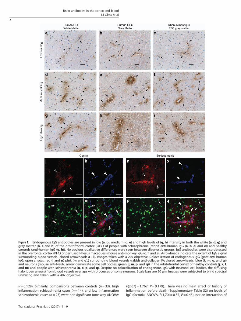
RESULTSIgG is present in the orbitofrontal cortex of controls and peoplewith schizophreniaIgG was detected by immunohistochemistry in the OFC of peoplewith schizophrenia (n= 38, Figure 1a, b, d and e) and controls(n= 38, Figure 1g and h) as indicated by a diffuse brown DABreaction product. A darker halo of brown reaction product wasoften visible surrounding many blood vessels (Figure 1a–iarrowheads). The immunoreactivity appeared to radiate outwardsfrom the blood vessel. The degree of signal extension into thebrain parenchyma varied between individuals irrespective ofdiagnosis and even from blood vessel to blood vessel within thesame brain (Figure 1a–i).To exclude the possibility that IgG immunoreactivity in the
human OFC was artifact arising from tissue degradation ordiffusion of residual blood components into the tissue withprolonged PMI, we performed the same immunohistochemistryusing saline perfused PFC of Rhesus macaques and an anti-rhesusIgG antibody (n= 7, Figure 1c, f, and i). As with the human OFC,the DAB signal from the IgG immunoreactivity was found in andsurrounding various blood vessels, dissipating into the brainparenchyma. Immunoreactivity was absent in the “no secondary”human, and primate no primary, control sections (SupplementaryFigure S1).
IgG associates mainly with blood vesselsImmunoreactivity (red) indicating presence of endogenous IgGs(Figure 1j and n) was closely associated with collagen IV-positive(blue) blood vessels (Figure 1k and o) in both controls (n= 9) andpeople with schizophrenia (n= 9, Figure 1j-m and n-q respec-tively). A diffuse halo of IgG staining radiated from some, but notall, blood vessels. IgG signal varied in intensity in the parenchymabetween individuals. Cell bodies (green) of neurons (Figure 1l andp), did not appear to be directly associated with IgG signal,however the halo of IgG appeared to overlap with the processesof some neurons. These processes were often adjacent to IgGpositive blood vessels. None of the no primary control slides hadimmunoreactive signal (Supplementary Figure S2).
IgG levels in human prefrontal cortex do not differ significantlybetween diagnostic or inflammatory groupsAn immunoreactive band at the weight consistent with that of theIgG heavy chain (50kDa) was detected in all humans tested(n= 74). The abundance of IgG was not different when comparingschizophrenia cases and controls (t(72) =− 0.991, P= 0.325), orthose with high inflammation (n= 18) compared with lowinflammation regardless of diagnosis (n= 56; t(72) = -1.541,
Brain antibodies in the cortex and bloodLJ Glass et al
3
Translational Psychiatry (2017), 1 – 9
P= 0.128). Similarly, comparisons between controls (n= 33), highinflammation schizophrenia cases (n= 14), and low inflammationschizophrenia cases (n= 23) were not significant (one-way ANOVA:
F(2,67) = 1.767, P= 0.179). There was no main effect of history ofinflammation before death (Supplementary Table S2) on levels ofIgG (factorial ANOVA, F(1,70) = 0.57, P= 0.45), nor an interaction of
Figure 1. Endogenous IgG antibodies are present in low (a, b), medium (d, e) and high levels of (g, h) intensity in both the white (a, d, g) andgray matter (b, e and h) of the orbitofrontal cortex (OFC) of people with schizophrenia (rabbit anti-human IgG (a, b, d, and e)) and healthycontrols (anti-human IgG (g, h)). No obvious qualitative differences were seen between diagnostic groups. IgG antibodies were also detectedin the prefrontal cortex (PFC) of perfused Rhesus macaques (mouse anti-monkey IgG (c, f, and i)). Arrowheads indicate the extent of IgG signalsurrounding blood vessels (closed arrowheads a - i). Images taken with a 20x objective. Colocalization of endogenous IgG (goat anti-humanIgG; open arrows, red (j and n) pink (m and q),) surrounding blood vessels (rabbit anti-collagen IV; closed arrowheads; blue (k, m, o, and q))and neurons (mouse anti-NeuN; arrow demarcate some cell bodies, green (l,m, p, and q)) in the orbitofrontal cortex of healthy controls (j, k, l,and m) and people with schizophrenia (n, o, p, and q). Despite no colocalization of endogenous IgG with neuronal cell bodies, the diffusinghalo (open arrows) from blood vessels overlaps with processes of some neurons. Scale bars are 50 μm. Images were subjected to blind spectralunmixing and taken with a 40x objective.
Brain antibodies in the cortex and bloodLJ Glass et al
4
Translational Psychiatry (2017), 1 – 9

history of inflammation before death with diagnosis (P= 0.74).Similarly, there was no main effect of cause of death (cardiaccomplications) on levels of IgG (factorial ANOVA, F(1,70) = 0.001,P= 0.97), nor an interaction of cause of death with diagnosis(P= 0.94).
FcGRT levels in human dorsolateral prefrontal cortex do not differbetween diagnostic or inflammatory groupsFcGRT mRNA (Figure 2c) and protein (Figure 2d and e) in theDLPFC were investigated by qPCR and western blotting respec-tively. FcGRT mRNA expression significantly correlated with PMI(r=− 0.281, P= 0.015), but not brain tissue pH. Normalized FcGRTmRNA levels did not differ between individuals with schizophreniaand controls (ANCOVA, covarying for PMI, F(1,71) = 0.213,P= 0.646), between high inflammation and low inflammationgroups overall (ANCOVA, covarying for PMI, F(1,71) = 2.985,P= 0.88) or among high inflammation schizophrenia cases, lowinflammation schizophrenia cases and controls (Figure 2c;ANCOVA, covarying for PMI, F(2,66) = 0.745, P= 0.478].Probing for FcGRT protein by western blot, we detected a
prominent immunoreactive band at approximately 50 kDa, slightlylarger than the expected molecular weight of FcGRT at 40 kDa(Figure 2d). Intensity of this FcGRT immunoreactive band did notcorrelate with any demographic variables, and the intensity ofFcGRT (FcGRT/ β-actin) did not differ between diagnostic groups(t(72) =− 1.43, P= 0.159) or inflammatory groups (t(72) = 0.99,P= 0.32). We did not detect a significant difference in FcGRTprotein levels among high inflammation schizophrenia cases,low inflammation schizophrenia cases and controls (Figure 2e;F(2,67) = 0.93, P= 0.40).
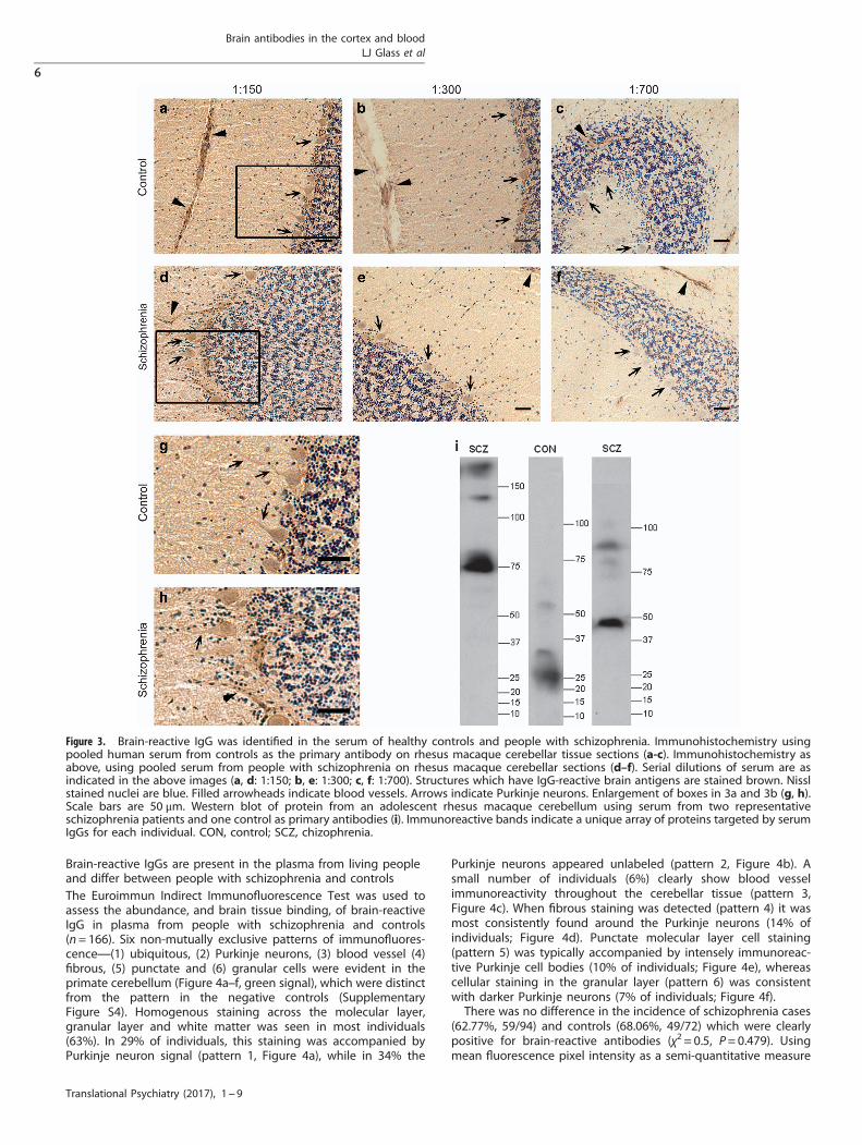
Brain-reactive IgG are present in the serum of living schizophreniapatients and living healthy controlsBrain-reactive IgG were detected using pooled serum samplesfrom a cohort of living healthy controls (Figure 3a-c and g) andliving people with schizophrenia (Figure 3d–f and h) as a primaryantibody to Rhesus macaque cerebellum sections processed forDAB IHC. The DAB signal product decreased in a serumconcentration-dependent manner (Figure 3a–f and h). Immuno-reactivity observed at a 1:700 serum dilution (Figure 3c and f)was indistinguishable from the control slide (SupplementaryFigure S3). Purkinje neurons apical dendrites were visible athigher serum concentrations, 1:150 (Figure 3a and d, arrows inFigure 3g and h) and 1:300 (Figure 3b and e). Dendrites were notalways distinguishable due to the diffuse molecular layer staining.The Purkinje neuron cell bodies (Figure 3a–f arrows) were visiblein all sections. Consistent staining of blood vessels was seen in allsections (Figure 3a–f arrowheads) including the no serum control(Supplementary Figure S3). Cross reactivity of monkey tissue withthe anti-human secondary antibody alone was also evident inlight brown fibrous staining throughout the tissue (SupplementaryFigure S3). We did not find any qualitative difference in stainingintensity obtained with control (Figure 3a-c and g) or patient(Figure 3d–f and h) serum.
Brain-reactive serum IgG from living people recognize uniqueproteinsA western blot in which Rhesus macaque cerebellar protein wasprobed with serum from individual controls and schizophreniacases confirmed the presence of brain-reactive IgG in serum fromliving people. For each serum sample, multiple immunoreactivebands of molecular weights from 25 to 4150 kDa were identified(Figure 3i). Immunoreactive bands recognized by serum from eachindividual displayed a unique pattern and intensity.
Figure 2. An anti-IgG immunoreactive band of 50 kDa (a) was foundusing western blotting on dorsolateral prefrontal cortex homo-genate of all humans studied (n= 74). The intensity of the IgG variedfrom one human brain to another while the level of β-actin (at42 kDa) was of similar abundance. IgG abundance did not differ bydiagnosis of inflammatory subgroup (b) Horizontal bars representgroup means. Expression of FcGRT mRNA was comparable betweenhigh and low inflammation schizophrenia cases and controls (c)Representative western blot probed for FcGRT protein in the humanDLPFC (d) Protein levels of FcGRT in the DLPFC did not differbetween high inflammation schizophrenia cases, low inflammationschizophrenia cases or controls (e) Horizontal bars representgroup means.
Brain antibodies in the cortex and bloodLJ Glass et al
5
Translational Psychiatry (2017), 1 – 9
Brain-reactive IgGs are present in the plasma from living peopleand differ between people with schizophrenia and controlsThe Euroimmun Indirect Immunofluorescence Test was used toassess the abundance, and brain tissue binding, of brain-reactiveIgG in plasma from people with schizophrenia and controls(n= 166). Six non-mutually exclusive patterns of immunofluores-cence—(1) ubiquitous, (2) Purkinje neurons, (3) blood vessel (4)fibrous, (5) punctate and (6) granular cells were evident in theprimate cerebellum (Figure 4a–f, green signal), which were distinctfrom the pattern in the negative controls (SupplementaryFigure S4). Homogenous staining across the molecular layer,granular layer and white matter was seen in most individuals(63%). In 29% of individuals, this staining was accompanied byPurkinje neuron signal (pattern 1, Figure 4a), while in 34% the
Purkinje neurons appeared unlabeled (pattern 2, Figure 4b). Asmall number of individuals (6%) clearly show blood vesselimmunoreactivity throughout the cerebellar tissue (pattern 3,Figure 4c). When fibrous staining was detected (pattern 4) it wasmost consistently found around the Purkinje neurons (14% ofindividuals; Figure 4d). Punctate molecular layer cell staining(pattern 5) was typically accompanied by intensely immunoreac-tive Purkinje cell bodies (10% of individuals; Figure 4e), whereascellular staining in the granular layer (pattern 6) was consistentwith darker Purkinje neurons (7% of individuals; Figure 4f).There was no difference in the incidence of schizophrenia cases
(62.77%, 59/94) and controls (68.06%, 49/72) which were clearlypositive for brain-reactive antibodies (χ2 = 0.5, P= 0.479). Usingmean fluorescence pixel intensity as a semi-quantitative measure
Figure 3. Brain-reactive IgG was identified in the serum of healthy controls and people with schizophrenia. Immunohistochemistry usingpooled human serum from controls as the primary antibody on rhesus macaque cerebellar tissue sections (a-c). Immunohistochemistry asabove, using pooled serum from people with schizophrenia on rhesus macaque cerebellar sections (d–f). Serial dilutions of serum are asindicated in the above images (a, d: 1:150; b, e: 1:300; c, f: 1:700). Structures which have IgG-reactive brain antigens are stained brown. Nisslstained nuclei are blue. Filled arrowheads indicate blood vessels. Arrows indicate Purkinje neurons. Enlargement of boxes in 3a and 3b (g, h).Scale bars are 50 μm. Western blot of protein from an adolescent rhesus macaque cerebellum using serum from two representativeschizophrenia patients and one control as primary antibodies (i). Immunoreactive bands indicate a unique array of proteins targeted by serumIgGs for each individual. CON, control; SCZ, chizophrenia.
Brain antibodies in the cortex and bloodLJ Glass et al
6
Translational Psychiatry (2017), 1 – 9

of IgG abundance, plasma from live schizophrenia patientscontained slightly, but significantly, lower brain-reactive IgG levelsthan that of healthy living controls (Figure 4g; t(128.6) =− 2.377,P= 0.019 adjusting for unequal variance). IgG abundance did notcorrelate with plasma storage freezer time (n= 166, r= 0.103,P= 0.189), or schizophrenia patient daily chlorpromazine equiva-lent dose (n= 94, r=− 0.59, P= 0.572).
DISCUSSIONIn this study, we found evidence for IgGs in the adult humancortex, particularly in diffuse patterns surrounding blood vesselsbut extending into brain parenchyma in both controls and people
with schizophrenia. To our knowledge, we are the first to findevidence of, and to quantify, IgG within the normal human brain.In support of the human brain’s capacity for IgG movement acrossthe blood brain barrier (BBB), we detected the IgG transporter(FcGRT), in brain at both the mRNA and protein levels. Contrary toour expectations, we failed to detect differences in the abundanceof IgGs, FcGRT protein or FcGRT mRNA in the brains of people withschizophrenia compared with healthy controls. This suggests thatIgG is normally present in, and actively effluxed from, the brain.Brain-reactive antibodies were also detected in serum of livingpeople, and appeared to target a range of neural proteins. We didnot observe differences in the incidence of plasma brain-reactiveantibody-positivity between schizophrenia cases and controlsfrom a cohort of living people, but semi-quantitative analysissuggested decreased levels of plasma brain-reactive IgGs inschizophrenia. Overall, we found that all individuals had IgG in thebrain, with equivalent abundance in schizophrenia cases andcontrols, even when taking elevated proinflammatory cytokines4
into account.One main limitation of our study is that many results are
derived from postmortem brain (Figures 1, 2, 3, 4). However it isunlikely that the patterns of IgG in the brain observed in this studyare artifacts associated with long PMI or the presence of residualblood components in cortical blood vessels. The diffuse pattern ofIgG staining around the blood vessel in the cortex of humans wasalso seen in saline perfused rhesus macaques with very short PMI.This pattern is consistent with that observed in the saline perfusedrodent brain,44,45 as is the endothelial cell immunoreactivity weobserved.46 These results from three mammalian species supportsour use of immunohistochemical methods to investigate IgG inthe brain of people with schizophrenia and controls, and indicatethe suitability of monkeys and rodents as animal models for futurestudies.IgG may enter the mammalian brain from the blood by crossing
the BBB, or be produced by B cells that have transmigrated fromthe blood into the brain perivascular space.28,47,48 Transienthypertension from elevated adrenalin49 or stress-induced proin-flammatory cytokines, which compromise endothelium junctionintegrity50 can also facilitate IgG entrance from the blood. Thus,we hypothesized that individuals with schizophrenia withincreased peripheral and brain proinflammatory cytokines (highinflammation)1,4 may have elevated endogenous brain IgG due tochanges in the BBB.51 Contrary to this hypothesis, we were unableto detect a difference in brain IgG levels between high and lowinflammation subgroups, or schizophrenia cases compared to
Figure 4. Plasma brain-reactive antibodies from a live patient cohortof people with schizophrenia (n= 94) and controls (n= 72) resultedin six different patterns of fluorescence (green) when applied to theprimate cerebellar tissue of the Euroimmun Indirect Immunofluor-escence Test: equivalent intensity in Purkinje neurons and molecularand granular layers (29%, 48/166) (a) low Purkinje neuron intensity(34%, 56/166) (b), distinctive blood vessels (6%, 10/166) (c), brightring around Purkinkje neurons with fibers throughout (14%, 23/166)(d), bright Purkinje neurons with punctate molecular layer cells(10%, 17/166) (e) and granular layer cells and bright molecular layer(7%, 12/166) (f). Images taken at 20x magnification. Scale bars are50μm. Antibody fluorescence intensity was lower in people withschizophrenia than controls, t(128.6)=− 2.377, P= 0.019 whenadjusted for unequal variance (Levene’s test: F(1,164)= 5.877,P= 0.016) as denoted by asterisk (g). Solid horizontal bars representgroup means. Dashed line indicates the average IgG intensity acrossthe no plasma controls (g). Brain-reactive IgG was consideredpresent if intensity of staining with plasma was greater than twostandard deviations (dotted lines, g) above the average negativecontrols (dashed line; g). Arrows indicate Purkinje neurons. Mol,molecular layer; Gr, granular layer.
Brain antibodies in the cortex and bloodLJ Glass et al
7
Translational Psychiatry (2017), 1 – 9

controls. Instead, our results suggest that there is a quantifiablelevel of IgG in the healthy brain where they may contribute tonormal functioning. Further testing of this idea could includeassessing the impact of changing brain IgG levels on Fcγ-receptorabundance and function.In our study, we found that over 60% of plasma from living
people meet our criteria for positive immunostaining of braintissue regardless of diagnosis, adding further support to thehypothesis that brain-reactive antibodies are ubiquitously foundin human blood regardless of disease.20,52 Such a high rate ofantibody-positivity contrasts with prior reports of relatively lowerincidence of brain-reactive antibodies in schizophrenia studies. Anunderlying cause of discrepancy may be that these studies focuson antibodies targeting certain pre-selected antigens in theblood22,24 and therefore fail to analyze the plethora of other brain-reactive antibodies that may be present. That being said, unlikeprevious studies, ours did not rule out the binding of antibodies toantigens common to other organs. Future studies should considerpreabsorbing samples to remove IgGs which target peripheralantigens53,54 as this would circumvent this issue.Although our study does not provide evidence of widespread
antibody dysregulation in the schizophrenia brain, there iscompelling evidence for deleterious effects caused by antibodiesin the brain. Anti-NMDAR antibodies in NMDAR encephalitis andbrain-reactive antibodies in neuropsychiatric SLE, play a causativerole in psychiatric symptoms of both disorders.55,56 Cultured cellstreated with cerebrospinal fluid IgG from NMDAR encephalitispatients displayed decreased numbers of NMDARs on postsynap-tic dendrites in vitro,28 but only impair behavior in mice with BBBdysfunction.27 Since comparable levels of anti-NMDAR antibodiesare seen in the blood of schizophrenia patients and healthycontrols,27 coincident BBB dysfunction may be required forantibody mediated pathology in schizophrenia. Importantly, whilewe did not detect widespread antibody-related abnormalities inthe postmortem schizophrenia brain, or blood from living peoplewith schizophrenia, our methods were inappropriate to investi-gate subtle dysregulation caused by antibodies targeting parti-cular proteins.Our work indicates that IgGs may be found in the brain under
normal conditions. Measurement of the relative abundances ofstructurally different IgGs in the blood and brain of control caseswould aid in determining the normal composition of the brain IgGpopulation and provide initial insights into their actions. A focuson the origin, regulation, and function of brain IgG in futurestudies could aid in establishing the effects of IgG in the brain.Finally, the possibility of greater access to the brain of IgGssuggests that the psychiatric and neurological consequences ofmonoclonal antibody therapies may be more extensive thancurrently anticipated, and this requires further consideration.
CONFLICT OF INTERESTCSW is a member of an Advisory Board for Lundbeck Australia Pty Ltd. The otherauthors declare no conflict of interest.
ACKNOWLEDGMENTSTissues were received from the New South Wales Tissue Resource Centre at theUniversity of Sydney, which is supported by the National Health and MedicalResearch Council (NHMRC) of Australia, Schizophrenia Research Institute, NationalInstitute of Alcohol Abuse and Alcoholism (NIH (NIAAA) R24AA012725). CSW issupported by Schizophrenia Research Institute (utilizing infrastructure funding fromthe NSW Ministry of Health and the Macquarie Group Foundation), the University ofNew South Wales, and Neuroscience Research Australia. CSW is a recipient of anNHMRC Principle Research Fellowship (#1117079). DS is a recipient of an NHMRC CJMartin Early Career Research Fellowship (#1072878). The serum collection and subjectcharacterization aspect of our project was supported by the NHMRC (#568807).
REFERENCES1 Miller BJ, Buckley P, Seabolt W, Mellor A, Kirkpatrick B. Meta-analysis of cytokine
alterations in schizophrenia: clinical status and antipsychotic effects. Biol Psy-chiatry 2011; 70: 663–671.
2 Fillman SG, Weickert TW, Lenroot RK, Catts SV, Bruggemann JM, Catts VS et al.Elevated peripheral cytokines characterize a subgroup of people with schizo-phrenia displaying poor verbal fluency and reduced Broca's area volume. MolPsychiatry 2016; 21: 1090–1098.
3 Fillman SG, Cloonan N, Catts VS, Miller LC, Wong J, McCrossin T et al. Increasedinflammatory markers identified in the dorsolateral prefrontal cortex of indivi-duals with schizophrenia. Mol Psychiatry 2013; 18: 206–214.
4 Fillman SG, Sinclair D, Fung SJ, Webster MJ, Shannon Weickert C. Markers ofinflammation and stress distinguish subsets of individuals with schizophrenia andbipolar disorder. Transl Psychiatry 2014; 4: e365.
5 Zhang Y, Catts VS, Sheedy D, McCrossin T, Kril JJ, Shannon Weickert C. Corticalgrey matter volume reduction in people with schizophrenia is associated withneuro-inflammation. Transl Psychiatry 2016; 6: e982.
6 Volk DW, Chitrapu A, Edelson JR, Roman KM, Moroco AE, Lewis DA. Molecularmechanisms and timing of cortical immune activation in schizophrenia. Am JPsychiatry 2015; 172: 1112–1121.
7 Arion D, Unger T, Lewis DA, Levitt P, Mirnics K. Molecular evidence for increasedexpression of genes related to immune and chaperone function in the prefrontalcortex in schizophrenia. Biol Psychiatry 2007; 62: 711–721.
8 Saetre P, Emilsson L, Axelsson E, Kreuger J, Lindholm E, Jazin E. Inflammation-related genes up-regulated in schizophrenia brains. BMC Psychiatry 2007; 7: 46.
9 de Witte L, Tomasik J, Schwarz E, Guest PC, Rahmoune H, Kahn RS et al. Cytokinealterations in first-episode schizophrenia patients before and after antipsychotictreatment. Schizophr Res 2014; 154: 23–29.
10 Song C, Lin A, Kenis G, Bosmans E, Maes M. Immunosuppressive effects of clo-zapine and haloperidol: enhanced production of the interleukin-1 receptorantagonist. Schizophr Res 2000; 42: 157–164.
11 Leykin I, Mayer R, Shinitzky M. Short and long-term immunosuppressive effects ofclozapine and haloperidol. Immunopharmacol 1997; 37: 75–86.
12 Di Nicola M, Cattaneo A, Hepgul N, Di Forti M, Aitchison KJ, Janiri L et al. Serumand gene expression profile of cytokines in first-episode psychosis. Brain BehavImmun 2013; 31: 90–95.
13 Borovcanin M, Jovanovic I, Radosavljevic G, Djukic Dejanovic S, Bankovic D,Arsenijevic N et al. Elevated serum level of type-2 cytokine and low IL-17 in firstepisode psychosis and schizophrenia in relapse. J Psychiatr Res 2012; 46:1421–1426.
14 Al-Hakeim HK, Al-Rammahi DA, Al-Dujaili AH. IL-6, IL-18, sIL-2R, and TNFalphaproinflammatory markers in depression and schizophrenia patients who are freeof overt inflammation. J Affect Disord 2015; 182: 106–114.
15 Owen JA, Punt J, Stranford SA, Jones PP. Kuby Immunology. WH Freeman:New York, 2013.
16 Printz DJ, Strauss DH, Goetz R, Sadiq S, Malaspina D, Krolewski J et al. Elevation ofCD5+ B lymphocytes in schizophrenia. Biol Psychiatry 1999; 46: 110–118.
17 Steiner J, Jacobs R, Panteli B, Brauner M, Schiltz K, Bahn S et al. Acute schizo-phrenia is accompanied by reduced T cell and increased B cell immunity. Eur ArchPsychiatry Clin Neurosci 2010; 260: 509–518.
18 Riedel M, Spellmann I, Schwarz MJ, Strassnig M, Sikorski C, Moller HJ et al.Decreased T cellular immune response in schizophrenic patients. J Psychiatr Res2007; 41: 3–7.
19 Maino K, Gruber R, Riedel M, Seitz N, Schwarz M, Muller N. T- and B-lymphocytesin patients with schizophrenia in acute psychotic episode and the course of thetreatment. Psychiatry Res 2007; 152: 173–180.
20 Levin EC, Acharya NK, Han M, Zavareh SB, Sedeyn JC, Venkataraman V et al. Brain-reactive autoantibodies are nearly ubiquitous in human sera and may be linked topathology in the context of blood-brain barrier breakdown. Brain Res 2010; 1345:221–232.
21 Margari F, Petruzzelli MG, Mianulli R, Toto M, Pastore A, Bizzaro N et al. Anti-brainautoantibodies in the serum of schizophrenic patients: a case-control study.Psychiatry Res 2013; 210: 800–805.
22 Dahm L, Ott C, Steiner J, Stepniak B, Teegen B, Saschenbrecker S et al.Seroprevalence of autoantibodies against brain antigens in health and disease.Ann Neurol 2014; 76: 82–94.
23 Borda T, Gomez R, Berria MI, Sterin-Borda L. Antibodies against astrocyte M1 andM2 muscarinic cholinoceptor from schizophrenic patients' sera. Glia 2004; 45:144–154.
24 Tanaka S, Matsunaga H, Kimura M, Tatsumi K, Hidaka Y, Takano T et al. Auto-antibodies against four kinds of neurotransmitter receptors in psychiatric dis-orders. J Neuroimmunol 2003; 141: 155–164.
25 Jones AL, Mowry BJ, McLean DE, Mantzioris BX, Pender MP, Greer JM. Elevatedlevels of autoantibodies targeting the M1 muscarinic acetylcholine receptor and
Brain antibodies in the cortex and bloodLJ Glass et al
8
Translational Psychiatry (2017), 1 – 9

neurofilament medium in sera from subgroups of patients with schizophrenia.J Neuroimmunol 2014; 269: 68–75.
26 Delunardo F, Soldati D, Bellisario V, Berry A, Camerini S, Crescenzi M et al. Anti-GAPDH autoantibodies as a pathogenic determinant and potential biomarker ofneuropsychiatric diseases. Arthritis Rheumatol 2016; 68: 2708–2716.
27 Hammer C, Stepniak B, Schneider A, Papiol S, Tantra M, Begemann M et al.Neuropsychiatric disease relevance of circulating anti-NMDA receptor auto-antibodies depends on blood-brain barrier integrity. Mol Psychiatry 2014; 19:1143–1149.
28 Dalmau J, Gleichman AJ, Hughes EG, Rossi JE, Peng X, Lai M et al. Anti-NMDA-receptor encephalitis: case series and analysis of the effects of antibodies. LancetNeurol 2008; 7: 1091–1098.
29 Ezeoke A, Mellor A, Buckley P, Miller B. A systematic, quantitative review of bloodautoantibodies in schizophrenia. Schizophr Res 2013; 150: 245–251.
30 Schwarz MJ, Riedel M, Gruber R, Muller N, Ackenheil M. Autoantibodies against60-kDa heat shock protein in schizophrenia. Eur Arch Psychitry Clin Neurosci 1998;248: 282–288.
31 Kim JJ, Lee SJ, Toh KY, Lee CU, Lee C, Paik IH. Identification of antibodies to heatshock proteins 90 kDa and 70 kDa in patients with schizophrenia. Schizophr Res2001; 52: 127–135.
32 Kilidireas K, Sadiq SA, Gorig AD, Latov N, Strauss DH, Hashim GA et al. Antibodiesto the human 60 kDa heat-shock protein in patients with schizophrenia. TheLancet 1992; 340: 569–572.
33 Schlachetzki F, Zhu CN, Pardridge WM. Expression of the neonatal Fc receptor(FcRn) at the blood-brain barrier. J Neurochem 2002; 81: 203–206.
34 Zhang Y, Pardridge WM. Mediated efflux of IgG molecules from brain to bloodacross the blood-brain barrier. J Neuroimmunol 2001; 114: 168–172.
35 Simister NE, Mostov KE. An Fc receptor structurally related to MHC class I anti-gens. Nature 1989; 337: 184–187.
36 Rodewald R. pH-dependent binding of immunoglobulins to intestinal cells of theneonatal rat. J Cell Biol 1976; 71: 666–669.
37 Raghavan M, Bonagura VR, Morrison SL, Bjorkman PJ. Analysis of the Ph-dependence of the neonatal Fc receptor immunoglobulin-G interaction usingantibody and receptor variant. Biochemistry 1995; 34: 14649–14657.
38 Joshi D, Catts VS, Olaya JC, Shannon Weickert C. Relationship between soma-tostatin and death receptor expression in the orbital frontal cortex in schizo-phrenia: a postmortem brain mRNA study. Npj Schizophr 2015; 1: 14004.
39 Weickert CS, Sheedy D, Rothmond DA, Dedova I, Fung S, Garrick T et al. Selectionof reference gene expression in a schizophrenia brain cohort. Aust N Z J Psychiatry2010; 44: 59–70.
40 Sinclair D, Fillman SG, Webster MJ, Weickert CS. Dysregulation of glucocorticoidreceptor co-factors FKBP5, BAG1 and PTGES3 in prefrontal cortex in psychoticillness. Sci Rep 2013; 3: 3539.
41 Fung SJ, Joshi D, Allen KM, Sivagnanasundaram S, Rothmond DA, Saunders R et al.Developmental patterns of doublecortin expression and white matter neurondensity in the postnatal primate prefrontal cortex and schizophrenia. PLoS One2011; 6: e25194.
42 Weinberg D, Lenroot R, Jacomb I, Allen K, Bruggemann J, Wells R et al. CognitiveSubtypes of schizophrenia characterized by differential brain volumetric reduc-tions and cognitive decline. JAMA Psychiatry 2016; 73: 1251–1259.
43 Richards AB, Morris RW, Ward S, Schmitz S, Rothmond DA, Noble PL et al.Gonadectomy negatively impacts social behavior of adolescent male primates.Horm Behav 2009; 56: 140–148.
44 Yoshimi K, Woo M, Son Y, Baudry M, Thompson RF. IgG-immunostaining in theintact rabbit brain: variable but significant staining of hippocampal and cerebellarneurons with anti-IgG. Brain Res 2002; 956: 53–66.
45 Lunnon K, Teeling JL, Tutt AL, Cragg MS, Glennie MJ, Perry VH. Systemic inflam-mation modulates Fc receptor expression on microglia during chronic neurode-generation. J Immunol 2011; 186: 7215–7224.
46 Hazama GI, Yasuhara O, Morita H, Aimi Y, Tooyama I, Kimura H. Mouse brain IgG-like immunoreactivity: strain-specific occurrence in microglia and biochemicalidentification of IgG. J Comp Neurol 2005; 492: 234–249.
47 Alter A, Duddy M, Hebert S, Biernacki K, Prat A, Antel JP et al. Determinants ofhuman B cell migration across brain endothelial cells. J Immunol 2003; 170:4497–4505.
48 Lovato L, Willis SN, Rodig SJ, Caron T, Almendinger SE, Howell OW et al. Related Bcell clones populate the meninges and parenchyma of patients with multiplesclerosis. Brain 2011; 134(Pt 2): 534–541.
49 Kuang F, Wang BR, Zhang P, Fei LL, Jia Y, Duan XL et al. Extravasation of blood-borne immunoglobulin G through blood-brain barrier during adrenaline-inducedtransient hypertension in the rat. Int J Neurosci 2004; 114: 575–591.
50 Oshima T, Laroux FS, Coe LL, Morise Z, Kawachi S, Bauer P et al. Interferon-gammaand interleukin-10 reciprocally regulate endothelial junction integrity and barrierfunction. Microvasc Res 2001; 61: 130–143.
51 Uranova NA, Zimina IS, Vikhreva OV, Krukov NO, Rachmanova VI, Orlovskaya DD.Ultrastructural damage of capillaries in the neocortex in schizophrenia. World JBiol Psychiatry 2010; 11: 567–578.
52 Nagele EP, Han M, Acharya NK, DeMarshall C, Kosciuk MC, Nagele RG. Natural IgGautoantibodies are abundant and ubiquitous in human sera, and their number isinfluenced by age, gender, and disease. PLoS One 2013; 8: e60726.
53 Jarius S, Probst C, Borowski K, Franciotta D, Wildemann B, Stoecker W et al.Standardized method for the detection of antibodies to aquaporin-4 based on ahighly sensitive immunofluorescence assay employing recombinant target anti-gen. J Neurol Sci 2010; 291: 52–56.
54 Lennon Vanda A, Wingerchuk Dean M, Kryzer Thomas J, Pittock Sean J, Lucchi-netti Claudia F, Fujihara Kazuo, Nakashima Ichiro, Weinshenker Brian G A serumautoantibody marker of neuromyelitis optica: distinction from multiple sclerosis.The Lancet 2004; 364: 2106–2112.
55 Tin SK, Xu Q, Thumboo J, Lee LY, Tse C, Fong KY. Novel brain reactive auto-antibodies: prevalence in systemic lupus erythematosus and association withpsychoses and seizures. J Neuroimmunol 2005; 169: 153–160.
56 Dalmau J, Lancaster E, Martinez-Hernandez E, Rosenfeld MR, Balice-Gordon R.Clinical experience and laboratory investigations in patients with anti-NMDARencephalitis. Lancet Neurol 2011; 10: 63–74.
This work is licensed under a Creative Commons Attribution 4.0International License. The images or other third party material in this
article are included in the article’s Creative Commons license, unless indicatedotherwise in the credit line; if the material is not included under the Creative Commonslicense, users will need to obtain permission from the license holder to reproduce thematerial. To view a copy of this license, visit http://creativecommons.org/licenses/by/4.0/
© The Author(s) 2017
Supplementary Information accompanies the paper on the Translational Psychiatry website (http://www.nature.com/tp)
Brain antibodies in the cortex and bloodLJ Glass et al
9
Translational Psychiatry (2017), 1 – 9