Hindawi Publishing Corporation BioMed Research International Volume 2013, Article ID 974580, 10 pages http://dx.doi.org/10.1155/2013/974580 Research Article In Vitro Wound Healing Potential and Identification of Bioactive Compounds from Moringa oleifera Lam Abubakar Amali Muhammad, 1 Nur Aimi Syarina Pauzi, 1 Palanisamy Arulselvan, 1 Faridah Abas, 2,3 and Sharida Fakurazi 1,4 1 Laboratory of Vaccines and Immunotherapeutics, Institute of Bioscience, Universiti Putra Malaysia, 43400, Serdang, Selangor, Malaysia 2 Department of Food Science, Faculty of Food Science and Technology, Universiti Putra Malaysia, 43400, Serdang, Selangor, Malaysia 3 Laboratory of Natural Products, Institute of Bioscience, Universiti Putra Malaysia, 43400, Serdang, Selangor, Malaysia 4 Department of Human Anatomy, Faculty of Medicine and Health Sciences, and Laboratory of Vaccines and Immunotherapeutics, Institute of Bioscience, Universiti Putra Malaysia, 43400 Serdang, Selangor, Malaysia Correspondence should be addressed to Sharida Fakurazi; [email protected] Received 2 August 2013; Revised 13 November 2013; Accepted 26 November 2013 Academic Editor: Leonid Medved Copyright © 2013 Abubakar Amali Muhammad et al. is is an open access article distributed under the Creative Commons Attribution License, which permits unrestricted use, distribution, and reproduction in any medium, provided the original work is properly cited. Moringa oleifera Lam. (M. oleifera) from the monogeneric family Moringaceae is found in tropical and subtropical countries. e present study was aimed at exploring the in vitro wound healing potential of M. oleifera and identification of active compounds that may be responsible for its wound healing action. e study included cell viability, proliferation, and wound scratch test assays. Different solvent crude extracts were screened, and the most active crude extract was further subjected to differential bioguided fractionation. Fractions were also screened and most active aqueous fraction was finally obtained for further investigation. HPLC and LC-MS/MS analysis were used for identification and confirmation of bioactive compounds. e results of our study demonstrated that aqueous fraction of M. oleifera significantly enhanced proliferation and viability as well as migration of human dermal fibroblast (HDF) cells compared to the untreated control and other fractions. e HPLC and LC-MS/MS studies revealed kaempferol and quercetin compounds in the crude methanolic extract and a major bioactive compound Vicenin-2 was identified in the bioactive aqueous fraction which was confirmed with standard Vicenin-2 using HPLC and UV spectroscopic methods. ese findings suggest that bioactive fraction of M. oleifera containing Vicenin-2 compound may enhance faster wound healing in vitro. 1. Introduction Skin is one of the largest organs in human body and serves as a protective barrier to external noxious agents including microorganisms. is protective barrier could be disrupted due to injuries or wounds, hence, the need to maintain its structural integrity. Wound results in the loss of continuity of epithelium in the skin with or without the loss of underlying connective tissue [1]. Wound healing is a complex process of restoring impaired cells and tissues back to their normal state and occurs as a cellular response to injury and involves activation of fibroblast, endothelial cells, and macrophages [2]. Wound healing also involves a well-orchestrated inte- gration of biological and molecular events of cell migration, cell proliferation, and extracellular matrix (ECM) deposition [3]. During wound healing process, growth factors released from fibroblasts, macrophages, neutrophils, keratinocytes, and endothelial cells influence all phases of wound healing and occur by providing signals for various cellular activities [4]. Management of chronic wound involves the use of antibi- otics, anti-inflammatory agents, or combination of both, but some of these drugs are associated with unwanted side effects [5] hence the need for other alternatives without producing toxicity. Moringa oleifera Lam is the most widely cultivated species of a monogeneric family Moringaceae. It is native to India, Pakistan, Bangladesh, and Afghanistan, utilized by ancient Romans, Greeks, and Egyptians; now distributed

Welcome message from author
This document is posted to help you gain knowledge. Please leave a comment to let me know what you think about it! Share it to your friends and learn new things together.
Transcript
Hindawi Publishing CorporationBioMed Research InternationalVolume 2013, Article ID 974580, 10 pageshttp://dx.doi.org/10.1155/2013/974580
Research ArticleIn Vitro Wound Healing Potential and Identification ofBioactive Compounds from Moringa oleifera Lam
Abubakar Amali Muhammad,1 Nur Aimi Syarina Pauzi,1 Palanisamy Arulselvan,1
Faridah Abas,2,3 and Sharida Fakurazi1,4
1 Laboratory of Vaccines and Immunotherapeutics, Institute of Bioscience, Universiti Putra Malaysia, 43400,Serdang, Selangor, Malaysia
2 Department of Food Science, Faculty of Food Science and Technology, Universiti Putra Malaysia, 43400, Serdang, Selangor, Malaysia3 Laboratory of Natural Products, Institute of Bioscience, Universiti Putra Malaysia, 43400, Serdang, Selangor, Malaysia4Department of Human Anatomy, Faculty of Medicine and Health Sciences, and Laboratory of Vaccines andImmunotherapeutics, Institute of Bioscience, Universiti Putra Malaysia, 43400 Serdang, Selangor, Malaysia
Correspondence should be addressed to Sharida Fakurazi; [email protected]
Received 2 August 2013; Revised 13 November 2013; Accepted 26 November 2013
Academic Editor: Leonid Medved
Copyright © 2013 Abubakar Amali Muhammad et al. This is an open access article distributed under the Creative CommonsAttribution License, which permits unrestricted use, distribution, and reproduction in any medium, provided the original work isproperly cited.
Moringa oleifera Lam. (M. oleifera) from the monogeneric family Moringaceae is found in tropical and subtropical countries. Thepresent study was aimed at exploring the in vitro wound healing potential of M. oleifera and identification of active compoundsthat may be responsible for its wound healing action.The study included cell viability, proliferation, and wound scratch test assays.Different solvent crude extracts were screened, and the most active crude extract was further subjected to differential bioguidedfractionation. Fractions were also screened and most active aqueous fraction was finally obtained for further investigation.HPLC and LC-MS/MS analysis were used for identification and confirmation of bioactive compounds. The results of our studydemonstrated that aqueous fraction ofM. oleifera significantly enhanced proliferation and viability as well as migration of humandermal fibroblast (HDF) cells compared to the untreated control and other fractions. The HPLC and LC-MS/MS studies revealedkaempferol and quercetin compounds in the crudemethanolic extract and amajor bioactive compoundVicenin-2 was identified inthe bioactive aqueous fraction which was confirmed with standard Vicenin-2 using HPLC and UV spectroscopic methods. Thesefindings suggest that bioactive fraction ofM. oleifera containing Vicenin-2 compound may enhance faster wound healing in vitro.
1. Introduction
Skin is one of the largest organs in human body and servesas a protective barrier to external noxious agents includingmicroorganisms. This protective barrier could be disrupteddue to injuries or wounds, hence, the need to maintain itsstructural integrity. Wound results in the loss of continuity ofepithelium in the skin with or without the loss of underlyingconnective tissue [1]. Wound healing is a complex processof restoring impaired cells and tissues back to their normalstate and occurs as a cellular response to injury and involvesactivation of fibroblast, endothelial cells, and macrophages[2]. Wound healing also involves a well-orchestrated inte-gration of biological and molecular events of cell migration,
cell proliferation, and extracellular matrix (ECM) deposition[3]. During wound healing process, growth factors releasedfrom fibroblasts, macrophages, neutrophils, keratinocytes,and endothelial cells influence all phases of wound healingand occur by providing signals for various cellular activities[4].Management of chronic wound involves the use of antibi-otics, anti-inflammatory agents, or combination of both, butsome of these drugs are associated with unwanted side effects[5] hence the need for other alternatives without producingtoxicity.
Moringa oleifera Lam is the most widely cultivatedspecies of a monogeneric family Moringaceae. It is nativeto India, Pakistan, Bangladesh, and Afghanistan, utilizedby ancient Romans, Greeks, and Egyptians; now distributed
2 BioMed Research International
in many countries of the tropics and subtropics [6]. Theedible parts of plant have been employed traditionally forskin diseases, rheumatism, anemia, cholera, and other ail-ment. The plant has an impressive range of medicinal useswith high nutritional value and serves as a good sourceof proteins, vitamins, beta-carotene, amino acid, and vari-ous phenolics [7]. It was reported to possess antimicrobialaction [8], anti-inflammatory properties [9], antidiabetic[10, 11], antioxidant [12], and anticancer properties [13,14]. It has also been reported to possess hepatoprotectiveactivity through antioxidant nature [15–17]. Evaluation ofvarious plant products according to their traditional uses andmedicinal value based on their therapeutic efficacy leads tothe discovery of newer and cost-effective drugs for treatingvarious ailments. This forms the basis for selection of M.oleifera as a potential wound healing agent. To the bestof our knowledge, the in vitro wound healing potential ofthis plants extracts or bioactive compounds has not beenreported previously. Therefore, this study was aimed atexploring the potential of M. oleifera in enhancing woundhealing and identification of putative bioactive compoundsresponsible for the wound healing action through in vitroprocess.
2. Materials and Methods
2.1. Chemicals and Reagents. All chemicals used were ofanalytical and HPLC grade (Merck, Germany). Methanol,ethanol, n-hexane, dichloromethane, ethyl acetate, and n-butanol were used for extraction and separation of com-pounds. Formic acid (Fisher, Loughborough, UK) was usedas buffer. Cell culture fibroblast medium, fetal bovine serum,antibiotics, trypsin, trypsin neutralizing solution, MTT pow-der, and PBS, were all from Science Cell Research Laborato-ries (Carlsbad, USA). Commercial standard Vicenin-2 andbioactive compound were purchased from Haihang indus-trial company, Beijing, China. C
18column (3 𝜇m, 150mm ×
2.1mm) was used for the separation and served as thestationary phase, while methanol and water were used asmobile phase.
2.2. Preparation of Plant Material and Extraction. The leavesof M. oleifera Lam (MO) were collected from Serdang City,Selangor, Malaysia, and were authenticated by Mr. Abdul-Ghaffar Othman from the Farm Unit of Faculty of Agri-culture, Universiti Putra Malaysia (UPM), in October 2010.The air-dried powdered leaves of M. oleifera (500 g) wereextracted successively with different solvents consisting ofmethanol, ethanol, and water to determine the most activesolvent crude extract in vitro (at least three times for eachsolvent until the extract was exhausted) with the help of ashaker (Lab Tech shaker, model; LSI-3016R, Korea) set at25∘C.The extracts were then filtered and evaporated throughrotary evaporator at 25∘C (Buchi, Switzerland) to drynessunder reduced pressure and further freeze dried with freezedrier (Virtis Bench Top K, United States) and the yield ofeach dried extracts was calculated and stored at −20∘C untilrequired for further use.
2.3. Differential Fractionation of the Crude Extract ofMethanol. The active methanolic extract was chosen basedon the in vitro screening results which consisted of invitro wound scratch test assay and fibroblast proliferationassays in which all the three solvent crude extracts werescreened to get the most active solvent crude extract,the bioactive methanolic crude extract, then was furthersubjected to differential bioguided fractionation using thein vitro screening procedures, and based on the results,aqueous fraction from the crude methanolic extract wasthe most active fraction. The most active methanolic crudeextract was subjected further to differential fractionationusing n-hexane, dichloromethane, ethyl acetate, n-butanol,and water. The methanoliccrude extract was redissolved inmethanol : water (25 : 75) and partitioned against equivalentvolume each of n-hexane, dichloromethane, ethyl acetate,and n-butanol and five fractions were obtained with respec-tive solvents. The fractions were freeze dried and storedappropriately in −20∘C until required for further study [18,19].
2.4. HPLC Analysis. HPLC-DAD qualitative analyses wereperformed to determine and compare the general profilingof the compounds present in the most active crude extractof methanol and its aqueous fraction using Agilent Tech-nologies (Santa Clara, USA) attached to a computer system.Gradient elution was carried out with methanol (solvent A)and water (solvent B) with 0.1% formic acid. C
18column
was used for separation of active compounds at a flow rateof 0.8mL/min. The extracts and fractions were prepared atconcentration of 10,000𝜇g/g of the sample injection volumebeing 10 𝜇L. The gradient program commenced from 5 : 95(v/v) until 100 : 0 (v/v) of A : B over 65min. UV detector wasset 254 nm and the peak areas were generated automaticallyby computer using Agilent software.
2.5. LC-MS/MS Analysis. Mass spectra were acquired usingThermo Finnegan model LCQ (San Jose, CA) ion trap massspectrometer equipped with an ESI source. The instrumentwas coupled to a surveyor diode array detector (DAD) (200–600 nm; 5mm band width) and surveyor autosampler. Thehyphenated system was supported with an X caliber 1.2 andmass frontier 5.0 software. Analytes separation was carriedout on a Hipersil Gold C
18column (3 𝜇m, 150mm × 2.1mm)
with a gradient mobile phase containing methanol (solventA) and water (solvent B), each containing 0.1% formic acid.The gradient program commenced from 10 : 90(v/v) to 100 : 0(v/v) of A : B over 65mins with a flow rate of 250𝜇L/min.The –ve ion mass spectra were obtained from LCQ DECAESI/MS detector on full scan mode (50–1000 amu) at ascan rate of 0.5Hz and the capillary temperature was setto 275∘C. A data dependent program was used in theliquid chromatography-tandem mass spectrometry analy-sis, so that the most abundant ions in each scan wereselected and subjected to MS/MS analysis. The collisioninduced dissociation (CID) energy was adjusted to 35%[19].
BioMed Research International 3
2.6. Quantification and Confirmation ofBioactive Compound Vicenin-2
2.6.1. HPLCMethod. HPLC was performed on a Jasco HPLC(Tokyo, Japan) consisting of a Jasco pump (PU-2086 Plus)and a Jasco UV detector model UV-2077 Plus linked byJasco LC-Net II/ADC interface box and a software attachedto a computer system. Analytical system was carried out ona C18
column. One mg of aqueous fraction of M. oleiferawas weighed and dissolved in 1mL 50 : 50 methanol : waterand sonicated to ensure complete dissolution, this was thenfiltered through 0.45 𝜇m membrane filter paper (Whatman,GEHealthcare).The reference compound Vicenin-2 was alsoequally weighed and dissolved similar to aqueous fraction.Samples were stored at 4∘C prior to analysis.
HPLC separation/confirmation of standard Vicenin-2and aqueous fraction was performed at 254 nm. The flowrate and injection volume of the samples were 0.8mL/minand 20𝜇L. The chromatographic peaks of the analytes wereconfirmed by comparing their retention time andUV spectrawith those of the reference standards Vicenin-2. The analysiswas carried out at temperature of 25∘C.
2.6.2. UV/Vis Spectroscopic Analysis. The absorption spec-trum of a sample can be measured as a plot of the absorbanceof a sample as a function of wavelength.This plot can be usedto identify an unknown sample since some compounds havecharacteristic absorption spectra. The UV-Vis spectroscopicmeasurements were made with a double-beam spectropho-tometer (Lambda 35; Perkin Elmer) to obtain maximumabsorption spectrum. The spectra were recorded between220 and 600 nm. For the sample preparation, approximately3mL of the standard Vicenin-2 contains 0.003M in distilledwater and sample containing 0.003M of aqueous fraction indistilled water placed in a 10mm cuvette. In this experiment,we used distilled water as a reference.
2.7. In Vitro Biological Experim ent
2.7.1. In Vitro Screening of Different SolventCrude Extracts and Fractions
Cell Culture. Human dermal fibroblast (HDF) cells wereobtained from American Type Culture Collection (ATCC,Manassas, VA, USA, CRL-2301) and thawed as well asmaintained according to the ATCC protocol [20]. Cells werecultured in fibroblast media premixed with 2% fetal bovineserum (FBS), growth supplements, and antibiotics consistingof L-glutamine 15mmol/L, streptomycin 100 𝜇g/mL, andpenicillin 100U/Ml and incubated in 5%CO
2and 37∘C. Cells
at 80%–90% confluence were used for seeding and treatmentthroughout the experiment.
2.7.2. (3-(4,5-Dimethylthiazol-2-yl)-2,5-diphenyl TetrazoliumBromide (MTT)) Colorimetric Assays [21]. HDF cells wereseeded into 96-well plate in triplicate at a density of 5× 10⁵ (in100 𝜇L medium) per well and grown for 24 hrs. The medium
was replaced with serial dilutions of aqueous fraction of thecrude methanolic extract at 12.5, 25, 50, 100, 200, 400, and800𝜇g/mL and the plates were incubated for 72 hrs. 10 𝜇Lof MTT reagent was then added to each of the wells andincubated for another 3 hrs. The purple formazan formedwas solubilzed by adding 100 𝜇L dimethyl sulfoxide (DMSO)to all the wells including control (without any treatment),then swirled gently to mix well and this was then kept inthe dark place at room temperature for about 30mins. ELISAmicroplate readerwas used to read absorbance at 570 nmwithreference of 630 nm. Graph of absorbance against number ofcells was plotted to determine the HDF cells proliferation asper the standard methods.
2.7.3. Fibroblast Proliferation and Viability Assay. Briefly,HDF cells were seeded on 24-well plates in triplicates and in-cubated at 37∘C until confluent, and serial dilutions of eachcrude extracts (methanol, ethanol, and water) were added tocells and without any treatment served as control incubatedfor 24 hrs. The confluent of HDF cells were then releasedfrom the wells with trypsin and trypsin neutralizing solution(TNS) to count the cells with a hemocytometer. Typan bluedye (0.4%) was used for staining to differentiate betweendead and viable cells because is a convenient, reliable, andinexpensive assay that makes faster distinction between deadand live cells [22]. The procedure was also repeated at 48and 72 hrs. The number of cells obtained from the countcorresponds to 𝑛× 104 cells per milliliter of suspension.Experiments were performed in triplicate, and each samplewas counted twice and the average reading was taken. Datawere recorded and analyzed statistically using SPSS version17.0 from IBM.
2.7.4. Wound Scratch Assay Test. This experiment was per-formed according to the previously reported and standard-ized protocol [23]. HDF cells were seeded in a 24-wellplates at a concentration of 3 × 10⁵ cells/mL cultured in afibroblast media containing 5% FBS and grown to confluentcell monolayer.Themedia were pipetted out and discarded, asmall area was then scratched using 200 𝜇L pipette tip, andthe cells were then rinsed with PBS to remove the loosendebris of the cells. Fibroblast media with serial dilution ofdifferent concentration of methanolic extract at 3.125, 6.25,12.5, 25, 50, and 100 𝜇g/mL were replaced and the plates wereincubated at 37∘C and 5% CO
2. The distance between two
layers of cells which was scratched by pipette tip was theninspectedmicroscopically at 0, 24, 48, and 72 hrs, respectively.As the HDF cells migrate to fill the scratched area, andimages were captured digital camera attached to microscopeand computer system. The experiments were performed intriplicate and data were analyzed using Corel draw graphicssuite x6 software.
2.8. Screening of Different Fractions from Crude Extract ofMethanol. A bioguided assay fractionation was performedin order to obtain the most active fraction that could befurther analyzed usingHPLCandLC-MSmethods to identifythe bioactive compounds present in the fraction. A total of
4 BioMed Research International
02468
101214161820
Methanol Water Ethanol
Perc
enta
ge y
ield
Crude extracts
(a)
0
0.5
1
1.5
2
2.5
3
3.5
4
Aqueous Ethylacetate
Hexane Dichloro-methane
Butanol
Perc
enta
ge y
ield
Fractions
(b)
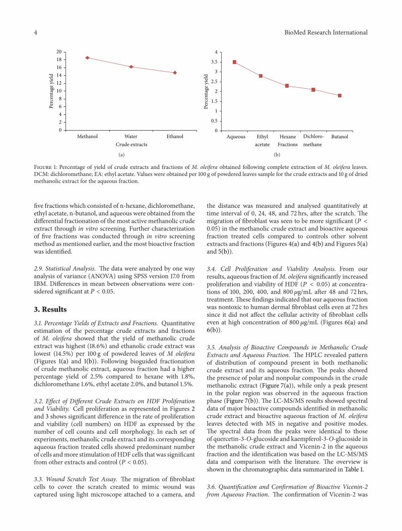
Figure 1: Percentage of yield of crude extracts and fractions of M. oleifera obtained following complete extraction of M. oleifera leaves.DCM: dichloromethane; EA: ethyl acetate. Values were obtained per 100 g of powdered leaves sample for the crude extracts and 10 g of driedmethanolic extract for the aqueous fraction.
five fractions which consisted of n-hexane, dichloromethane,ethyl acetate, n-butanol, and aqueous were obtained from thedifferential fractionation of the most active methanolic crudeextract through in vitro screening. Further characterizationof five fractions was conducted through in vitro screeningmethod as mentioned earlier, and the most bioactive fractionwas identified.
2.9. Statistical Analysis. The data were analyzed by one wayanalysis of variance (ANOVA) using SPSS version 17.0 fromIBM. Differences in mean between observations were con-sidered significant at 𝑃 < 0.05.
3. Results
3.1. Percentage Yields of Extracts and Fractions. Quantitativeestimation of the percentage crude extracts and fractionsof M. oleifera showed that the yield of methanolic crudeextract was highest (18.6%) and ethanolic crude extract waslowest (14.5%) per 100 g of powdered leaves of M oleifera(Figures 1(a) and 1(b)). Following bioguided fractionationof crude methanolic extract, aqueous fraction had a higherpercentage yield of 2.5% compared to hexane with 1.8%,dichloromethane 1.6%, ethyl acetate 2.0%, and butanol 1.5%.
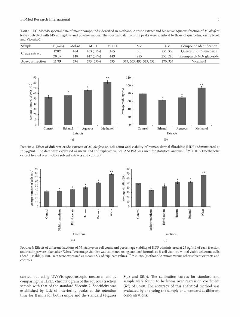
3.2. Effect of Different Crude Extracts on HDF Proliferationand Viability. Cell proliferation as represented in Figures 2and 3 shows significant difference in the rate of proliferationand viability (cell numbers) on HDF as expressed by thenumber of cell counts and cell morphology. In each set ofexperiments, methanolic crude extract and its correspondingaqueous fraction treated cells showed predominant numberof cells andmore stimulation ofHDF cells that was significantfrom other extracts and control (𝑃 < 0.05).
3.3. Wound Scratch Test Assay. The migration of fibroblastcells to cover the scratch created to mimic wound wascaptured using light microscope attached to a camera, and
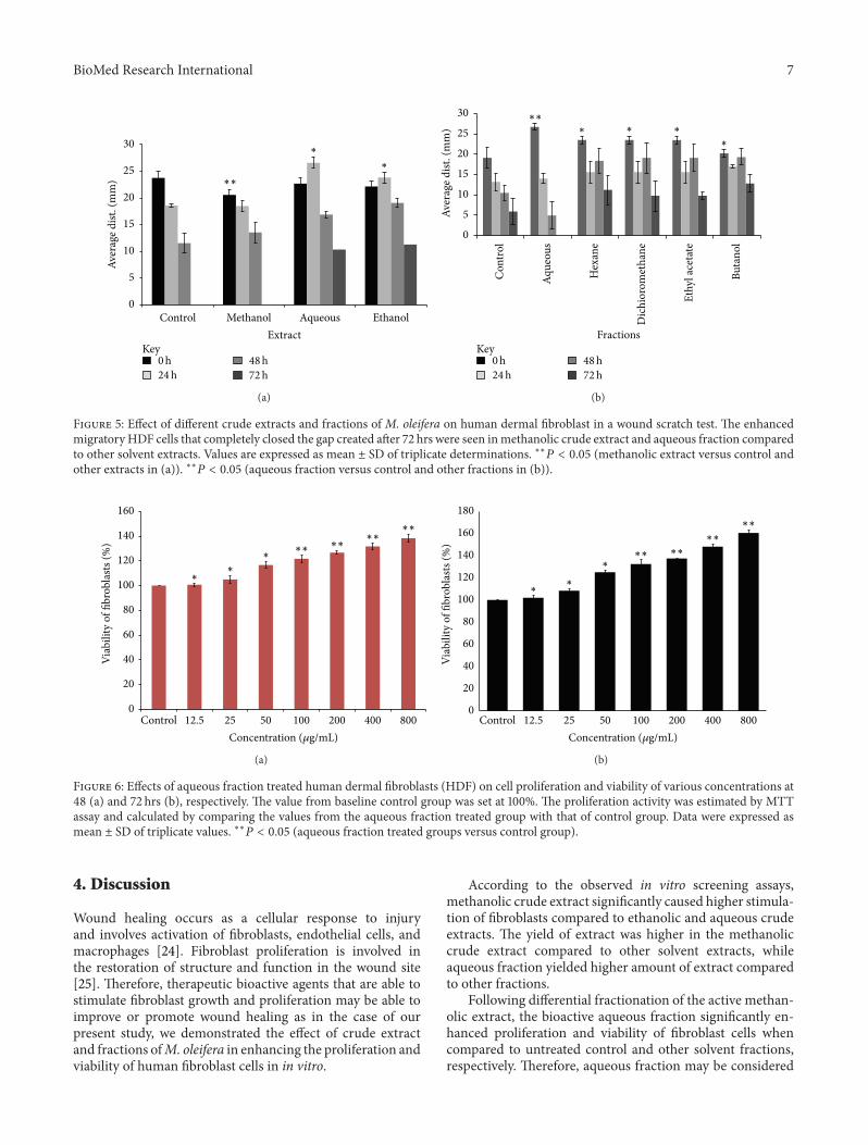
the distance was measured and analysed quantitatively attime interval of 0, 24, 48, and 72 hrs, after the scratch. Themigration of fibroblast was seen to be more significant (𝑃 <0.05) in the methanolic crude extract and bioactive aqueousfraction treated cells compared to controls other solventextracts and fractions (Figures 4(a) and 4(b) and Figures 5(a)and 5(b)).
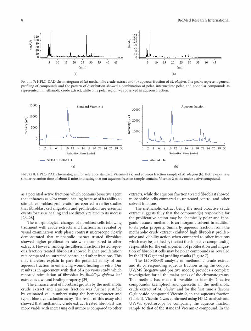
3.4. Cell Proliferation and Viability Analysis. From ourresults, aqueous fraction ofM. oleifera significantly increasedproliferation and viability of HDF (𝑃 < 0.05) at concentra-tions of 100, 200, 400, and 800 𝜇g/mL after 48 and 72 hrs,treatment.These findings indicated that our aqueous fractionwas nontoxic to human dermal fibroblast cells even at 72 hrssince it did not affect the cellular activity of fibroblast cellseven at high concentration of 800𝜇g/mL (Figures 6(a) and6(b)).
3.5. Analysis of Bioactive Compounds in Methanolic CrudeExtracts and Aqueous Fraction. The HPLC revealed patternof distribution of compound present in both methanoliccrude extract and its aqueous fraction. The peaks showedthe presence of polar and nonpolar compounds in the crudemethanolic extract (Figure 7(a)), while only a peak presentin the polar region was observed in the aqueous fractionphase (Figure 7(b)). The LC-MS/MS results showed spectraldata of major bioactive compounds identified in methanoliccrude extract and bioactive aqueous fraction of M. oleiferaleaves detected with MS in negative and positive modes.The spectral data from the peaks were identical to thoseof quercetin-3-O-glucoside and kaempferol-3-O-glucoside inthe methanolic crude extract and Vicenin-2 in the aqueousfraction and the identification was based on the LC-MS/MSdata and comparison with the literature. The overview isshown in the chromatographic data summarized in Table 1.
3.6. Quantification and Confirmation of Bioactive Vicenin-2from Aqueous Fraction. The confirmation of Vicenin-2 was
BioMed Research International 5
Table 1: LC-MS/MS spectral data of major compounds identified in methanolic crude extract and bioactive aqueous fraction of M. oleiferaleaves detected with MS in negative and positive modes. The spectral data from the peaks were identical to those of quercetin, kaempferol,and Vicenin-2.
Sample RT (min) Mol⋅wt M −H M + H MZ UV Compound identification
Crude extract 17.82 464 463 (35%) 465 301 255, 350 Quercetin-3-O-glucoside20.89 448 447 (35%) 449 285 255, 240 Kaempferol-3-O- glucoside
Aqueous fraction 12.79 594 593 (35%) 595 575, 503, 493, 525, 353. 270, 335 Vicenin-2
0102030405060708090
Aver
age n
umbe
r of c
ells×105
Control Ethanol Aqueous MethanolExtracts
∗
∗
∗∗
(a)
0
20
40
60
80
100
120
Control Ethanol Aqueous Methanol
Aver
age v
iabi
lity
(%)
Extracts
∗∗
∗∗
(b)
Figure 2: Effect of different crude extracts of M. oleifera on cell count and viability of human dermal fibroblast (HDF) administered at12.5 𝜇g/mL. The data were expressed as mean ± SD of triplicate values. ANOVA was used for statistical analysis. ∗∗𝑃 < 0.05 (methanolicextract treated versus other solvent extracts and control).
0102030405060708090
Fractions
Avea
ge n
umbe
r of c
ells×105
Con
trol
Dic
hior
omet
hane
Ethy
l ace
tate
Hex
ane
Buta
nol
Wat
er
∗
∗∗
∗
∗∗
(a)
01020304050607080
Aver
age v
iabi
lity
(%)
Fractions
Con
trol
Dic
hior
omet
hane
Ethy
l ace
tate
Hex
ane
Buta
nol
Wat
er
∗∗
∗
∗
∗∗
(b)
Figure 3: Effects of different fractions ofM. oleifera on cell count and percentage viability of HDF administered at 25 𝜇g/mL of each fractionand readings were taken after 72 hrs. Percentage viability was estimated using standard formula as % cell viability = total viable cells/total cells(dead + viable) × 100. Data were expressed as mean± SD of triplicate values. ∗∗𝑃 < 0.05 (methanolic extract versus other solvent extracts andcontrol).
carried out using UV/Vis spectroscopic measurement bycomparing the HPLC chromatogram of the aqueous fractionsample with that of the standard Vicenin-2. Specificity wasestablished by lack of interfering peaks at the retentiontime for 11mins for both sample and the standard (Figures
8(a) and 8(b)). The calibration curves for standard andsample were found to be linear over regression coefficient(𝑅2) of 0.988. The accuracy of this analytical method wasevaluated by analyzing the sample and standard at differentconcentrations.

6 BioMed Research International
(i)
(ii)
(iv)
(iii)
0hr 24hr 48hr 72hr
(a)
(i)
(ii)
(iv)
(v)
(vi)
(iii)
0hr 24hr 48hr 72hr
(b)
Figure 4: Digital image showing the effect of different fractions of M. oleifera on human dermal fibroblast migration in a wound scratchtest assay: (a) (i): control without treatment; (ii): methanol; (iii): ethanol; (iv): aqueous extracts; (b) (i): control; (ii): n-hexane; (iii):dichloromethane; (iv): ethyl acetate; (v): n-butanol; (vi): aqueous fractions. A confluent monolayer of human dermal fibroblast (HDF) wasscratched using a sterilised 200𝜇L pipette tip. Different fractions were applied as treatments to the wounded (open gap) and fibroblast mediaserved as control. Migration of fibroblast cells were captured andmeasured using light microscope attached to a digital camera.Magnification(4x).
BioMed Research International 7
0
5
10
15
20
25
30
Control Methanol Aqueous Ethanol
Aver
age d
ist. (
mm
)
Extract
∗
∗
∗∗
Key0h24h
48h72h
(a)
0
5
10
15
20
25
30
Aver
age d
ist. (
mm
)
Fractions
Con
trol
Aque
ous
Hex
ane
Dic
hior
omet
hane
Ethy
l ace
tate
Buta
nol
∗ ∗
∗
∗∗∗
Key0h24h
48h72h
(b)
Figure 5: Effect of different crude extracts and fractions of M. oleifera on human dermal fibroblast in a wound scratch test. The enhancedmigratory HDF cells that completely closed the gap created after 72 hrs were seen inmethanolic crude extract and aqueous fraction comparedto other solvent extracts. Values are expressed as mean ± SD of triplicate determinations. ∗∗𝑃 < 0.05 (methanolic extract versus control andother extracts in (a)). ∗∗𝑃 < 0.05 (aqueous fraction versus control and other fractions in (b)).
0
20
40
60
80
100
120
140
160
Control 12.5 25 50 100 200 400 800
Viab
ility
of fi
brob
lasts
(%)
Concentration (𝜇g/mL)
∗
∗∗
∗∗∗∗
∗∗∗∗
(a)
0
20
40
60
80
100
120
140
160
180
Control 12.5 25 50 100 200 400 800
Viab
ility
of fi
brob
lasts
(%)
Concentration (𝜇g/mL)
∗∗
∗∗∗ ∗∗
∗∗
∗∗
(b)
Figure 6: Effects of aqueous fraction treated human dermal fibroblasts (HDF) on cell proliferation and viability of various concentrations at48 (a) and 72 hrs (b), respectively. The value from baseline control group was set at 100%. The proliferation activity was estimated by MTTassay and calculated by comparing the values from the aqueous fraction treated group with that of control group. Data were expressed asmean ± SD of triplicate values. ∗∗𝑃 < 0.05 (aqueous fraction treated groups versus control group).
4. Discussion
Wound healing occurs as a cellular response to injuryand involves activation of fibroblasts, endothelial cells, andmacrophages [24]. Fibroblast proliferation is involved inthe restoration of structure and function in the wound site[25]. Therefore, therapeutic bioactive agents that are able tostimulate fibroblast growth and proliferation may be able toimprove or promote wound healing as in the case of ourpresent study, we demonstrated the effect of crude extractand fractions ofM. oleifera in enhancing the proliferation andviability of human fibroblast cells in in vitro.
According to the observed in vitro screening assays,methanolic crude extract significantly caused higher stimula-tion of fibroblasts compared to ethanolic and aqueous crudeextracts. The yield of extract was higher in the methanoliccrude extract compared to other solvent extracts, whileaqueous fraction yielded higher amount of extract comparedto other fractions.
Following differential fractionation of the active methan-olic extract, the bioactive aqueous fraction significantly en-hanced proliferation and viability of fibroblast cells whencompared to untreated control and other solvent fractions,respectively. Therefore, aqueous fraction may be considered
8 BioMed Research International
(mAU
)
120100
80604020
05 10 15 20 25 30 35 40 45
(min)
(a)
(mAU
)
175150125100
755025
05 10 15 20 25 30 35 40 45
(min)
(b)
Figure 7: HPLC-DAD chromatogram of (a) methanolic crude extract and (b) aqueous fraction of M. oleifera. The peaks represent generalprofiling of compounds and the pattern of distribution showed a combination of polar, intermediate polar, and nonpolar compounds asrepresented in methanolic crude extract, while only polar region was observed in aqueous fraction.
0 2 4 6 8 10 12 14 16 18 20 22 24 26 28 30
0
5000
10000
15000
Retention time (min)
Standard Vicenin-2
Inte
nsity
(𝜇V
)
STDABU500-CH4
(a)
0 2 4 6 8 10 12 14 16 18 20 22 24 26 28 30
0
10000
20000
30000
Retention time (min)
Abu 5-CH4
Aqueous fraction
Inte
nsity
(𝜇V
)
(b)
Figure 8: HPLC-DAD chromatogram for reference standard Vicenin-2 (a) and aqueous fraction sample ofM. oleifera (b). Both peaks havesimilar retention time of about 11mins indicating that our aqueous fraction sample contains Vicenin-2 as the major active compound.
as a potential active fractions which contains bioactive agentthat enhances in vitro wound healing because of its ability tostimulate fibroblast proliferation as reported in earlier studiesthat fibroblast cell migration and proliferation are essentialevents for tissue healing and are directly related to its success[26–28].
The morphological changes of fibroblast cells followingtreatment with crude extracts and fractions as revealed byvisual examination with phase contrast microscope clearlydemonstrated that methanolic extract treated fibroblastshowed higher proliferation rate when compared to otherextracts. However, among the different fractions tested, aque-ous fraction treated fibroblast showed higher proliferationrate compared to untreated control and other fractions. Thismay therefore explain in part the potential ability of ouraqueous fraction in enhancing wound healing in vitro. Ourresults is in agreement with that of a previous study whichreported stimulation of fibroblast by Buddleja globosa leafextract as a wound healing property [29].
The enhancement of fibroblast growth by the methanoliccrude extract and aqueous fraction was further justifiedby estimated cell numbers using the hemocytometer andtypan blue dye exclusion assay. The result of this assay alsoshowed that methanolic crude extract treated fibroblast wasmore viable with increasing cell numbers compared to other
extracts, while the aqueous fraction treated fibroblast showedmore viable cells compared to untreated control and othersolvent fractions.
The methanolic extract being the most bioactive crudeextract suggests fully that the compound(s) responsible forthe proliferative action may be chemically polar and inor-ganic because methanol is an inorganic solvent in additionto its polar property. Similarly, aqueous fraction from themethanolic crude extract exhibited high fibroblast prolifer-ative and viability action when compared to other fractionswhichmay be justified by the fact that bioactive compound(s)responsible for the enhancement of proliferation and migra-tion of fibroblast cells may be polar compounds as revealedby the HPLC general profiling results (Figure 7).
The LC-MS/MS analysis of methanolic crude extractand its corresponding aqueous fraction using the coupledUV/MS (negative and positive modes) provides a completeinvestigation for all the major peaks of the chromatograms.This method has made it possible to identify 2 activecompounds: kaempferol and quercetin in the methanoliccrude extract of M. oleifera and for the first time a flavoneC-glucoside compound Vicenin-2, in the aqueous fraction(Table 1). Vicenin-2 was confirmed using HPLC analysis andUV/Vis spectroscopy by comparing the aqueous fractionsample to that of the standard Vicenin-2 compound. In the
BioMed Research International 9
HPLC analysis, it was observed that both the sample and thestandard had a similar retention time of 11mins and this wasfurther evaluated by spectroscopic measurement. The bandsthat appeared in the aqueous fraction sample remain between272 and 338 nm similar to that of the standard Vicenin-2throughout the reaction period. This result suggested thatboth the sample and standard have the same characteristicUV-Vis absorption spectrum.
The identified bioactive compounds in the methanoliccrude extract and aqueous fraction of M. oleifera may serveas a lead in the drug discovery and development of a woundhealing agent that may enhance wound healing. These activecompounds belong to the group of flavonoid compounds,this group of compounds is known to be beneficial inwound healing treatment, and this has been linked to theirantioxidant property [30, 31].
5. Conclusion
Our study clearly demonstrated the activity of the crudeextracts and fractions of M. oleifera in enhancing prolifer-ation and viability of HDF cells in in vitro. The bioactivecompounds present in aqueous fraction of this plant crudeextract have been successfully identified and confirmed byHPLC and LC-MS/MS using reference compound which wasprocured from commercial source. Aqueous fraction of M.oleifera may therefore be a lead in the drug discovery ofwound healing active agent. Based on these fruitful findings,further preclinical investigation of the aqueous bioactivefraction and identified bioactive compounds are currentlybeing conducted by our research group.
Conflict of Interests
The authors declare that they have no conflict of interests.
Acknowledgment
This study was supported by University research GrantScheme (RUGS) of Universiti Putra Malaysia (UPM Grantnos.: 9300379 and 9366700).
References
[1] B. P. Nagori and R. Solanki, “Role of medicinal plants in woundhealing,” Research Journal of Medicinal Plant, vol. 5, no. 4, pp.392–405, 2011.
[2] J. C. W. Tam, K. M. Lau, C. L. Liu et al., “The in vivo and invitro diabetic wound healing effects of a 2-herb formula and itsmechanisms of action,” Journal of Ethnopharmacology, vol. 134,no. 3, pp. 831–838, 2011.
[3] L. Pradhan, C. Nabzdyk, N. D. Andersen, F. W. LoGerfo, andA. Veves, “Inflammation and neuropeptides: the connection indiabetic wound healing,” Expert Reviews inMolecularMedicine,vol. 11, no. 2, pp. 1–25, 2009.
[4] M. Yang, L. Sheng, T. R. Zhang, andQ. Li, “Stem cell therapy forlower extremity diabetic ulcers. Where do we stand?” BioMedResearch International, vol. 2013, Article ID 462179, 8 pages,2013.
[5] B. S. Nayak, M. R. Marshall, and G. Isitor, “Wound healingpotential of ethanolic extract of Kalanchoe pinnata lam. leaf-apreliminary study,” Indian Journal of Experimental Biology, vol.48, no. 6, pp. 572–576, 2010.
[6] B. S. Biswas, A. Chowdhury, J. Das, A. Roy, and S. Z. Hosen,“Pharmacological potentials ofMoringa oleifera lam. A review,”International Journal of Pharmaceutical Science Research, vol. 3,pp. 305–310, 2012.
[7] F. Anwar, S. Latif, M. Ashraf, and A. H. Gilani, “Moringaoleifera: a food plant with multiple medicinal uses,” Phytother-apy Research, vol. 21, no. 1, pp. 17–25, 2007.
[8] T. R. Prashith Kekuda, N. Mallikarjun, D. Swathi, K. V. Nayana,M. B. Aiyar, and T. R. Rohini, “Antibacterial and antifungalefficacy of steam distillate of Moringa oleifera lam,” Journal ofPharmaceutical Sciences and Research, vol. 2, no. 1, pp. 34–37,2010.
[9] S. G. Mahajan, R. G. Mali, and A. A. Mehta, “Protective effectof ethanolic extract of seeds of Moringa oleifera lam. againstinflammation associated with development of arthritis in rats,”Journal of Immunotoxicology, vol. 4, no. 1, pp. 39–47, 2007.
[10] D. Jaiswal, P. Kumar Rai, A. Kumar, S. Mehta, and G. Watal,“Effect of Moringa oleifera lam. leaves aqueous extract therapyon hyperglycemic rats,” Journal of Ethnopharmacology, vol. 123,no. 3, pp. 392–396, 2009.
[11] C. Edoga, O. Njoku, E. Amadi, and J. Okeke, “Blood sugar low-ering effect ofMoringa oleifera lam in albino rats,” InternationalJournal of Science and Technology, vol. 3, no. 1, 2013.
[12] B. Sultana, F. Anwar, and M. Ashraf, “Effect of extraction sol-vent/technique on the antioxidant activity of selectedmedicinalplant extracts,”Molecules, vol. 14, no. 6, pp. 2167–2180, 2009.
[13] M. Parvathy and A. Umamaheshwari, “Cytotoxic effect ofMoringa oleifera leaf extracts on human multiple myeloma celllines,” Trends in Medical Research, vol. 2, no. 1, pp. 44–50, 2007.
[14] S. Sreelatha, A. Jeyachitra, and P. R. Padma, “Antiproliferationand induction of apoptosis by Moringa oleifera leaf extract onhuman cancer cells,” Food and Chemical Toxicology, vol. 49, no.6, pp. 1270–1275, 2011.
[15] S. Fakurazi, U. Nanthini, and I. Hairuszah, “Hepatoprotec-tive and antioxidant action of Moringa oleifera lam. againstsacetaminophen induced hepatoxicity in rats,” InternationalJournal of Pharmacology, vol. 4, no. 4, pp. 270–275, 2008.
[16] S. Fakurazi, S. A. Sharifudin, and P. Arulselvan, “Moringaoleifera hydroethanolic extracts effectively alleviate acetamino-phen-induced hepatotoxicity in experimental rats through theirantioxidant nature,”Molecules, vol. 17, no. 7, pp. 8334–8350, 2012.
[17] S. A. Sharifudin, S. Fakurazi, M. T. Hidayat, I. Hairuszah, M.Aris Mohd Moklas, and P. Arulselvan, “Therapeutic potentialof Moringa oleifera extracts against acetaminophen-inducedhepatotoxicity in rats,” Pharmaceutical Biology, vol. 51, no. 3, pp.279–288, 2013.
[18] M. Nisar, S. Khan, A. Dar, W. Rehman, R. Khan, and I. Jan,“Antidepressant screening and flavonoids isolation from Ere-mostachys laciniata (L) bunge,”African Journal of Biotechnology,vol. 10, no. 9, pp. 1696–1699, 2011.
[19] http://www.atcc.org/Products/Cells%20and%20Microorgan-isms/Cell%20Lines.aspx.
[20] N. H. Noor Hashim, F. Abas, K. Shaari, and N. H. Lajis,“LC-DAD-ESIMS/MS characterization of antioxidant and anti-cholinesterase constituents present in the active fraction fromPersicaria hydropiper,” LWT-Food Science and Technology, vol.46, no. 2, pp. 468–476, 2012.
10 BioMed Research International
[21] A. U. Kura, A. Ali, S. HasanHussein,M. Z. Hussein, S. Fakurazi,and P. Arulselvan, “Development of a controlled-release anti-parkinsonian nanodelivery system using levodopa as the activeagent,” International Journal of Nanomedicine, vol. 8, article 1103,2013.
[22] M. C. Phelan and J. Wiley, “Basic techniques for mammaliancell tissue culture,” Current Protocol in Cell Biology, vol. 36, no.1, pp. 1–11, 2007.
[23] C.-C. Liang, A. Y. Park, and J.-L. Guan, “In vitro scratchassay: a convenient and inexpensive method for analysis of cellmigration in vitro,” Nature Protocols, vol. 2, no. 2, pp. 329–333,2007.
[24] R. A. F. Clark, “Fibrin and wound healing,” Annals of the NewYork Academy of Sciences, vol. 936, no. 1, pp. 355–367, 2001.
[25] A. Y. Mensah, J. Sampson, P. J. Houghton et al., “Effects ofBuddleja globosa leaf and its constituents relevant to woundhealing,” Journal of Ethnopharmacology, vol. 77, no. 2-3, pp. 219–226, 2001.
[26] L. Hakkinen, V.-J. Uitto, and H. Larjava, “Cell biology ofgingival wound healing,” Periodontology, vol. 24, no. 1, pp. 127–152, 2000.
[27] W. Posten, D. A. Wrone, J. S. Dover, K. A. Arndt, S. Silapunt,and M. Alam, “Low-level laser therapy for wound healing:mechanism and efficacy,” Dermatologic Surgery, vol. 31, no. 3,pp. 334–340, 2005.
[28] B. G. Fernanda, N. P. Taisa, S. T. Ana Paula, S. B. Vanderlei, H.Josimeri, and A. D. Carlos, “In vitro wound healing improve-ment by low-level laser therapy application in cultured gingivalfibroblasts,” International Journal of Dentistry, vol. 2012, ArticleID 719452, 6 pages, 2012.
[29] S. Lodhi, R. S. Pawar, A. P. Jain, and A. K. Singhai, “Woundhealing potential of Tephrosia purpurea (Linn.) Pers. in rats,”Journal of Ethnopharmacology, vol. 108, no. 2, pp. 204–210, 2006.
[30] M. P. Jorge, C. Madjarof, A. L. T. G. Ruiz et al., “Evaluation ofwound healing properties of Arrabidaea chica verlot extract,”Journal of Ethnopharmacology, vol. 118, no. 3, pp. 361–366, 2008.
[31] S. Ambiga, R. Narayanan, D. Gowri, D. Sukumar, and S.Madhavan, “Evaluation of wound healing activity of flavonoidsfrom Ipomoea carnea jacq,” Ancient Science of Life, vol. 26, no.3, p. 45, 2007.
Submit your manuscripts athttp://www.hindawi.com
PainResearch and TreatmentHindawi Publishing Corporationhttp://www.hindawi.com Volume 2014
The Scientific World JournalHindawi Publishing Corporation http://www.hindawi.com Volume 2014
Hindawi Publishing Corporationhttp://www.hindawi.com
Volume 2014
ToxinsJournal of
VaccinesJournal of
Hindawi Publishing Corporation http://www.hindawi.com Volume 2014
Hindawi Publishing Corporationhttp://www.hindawi.com Volume 2014
AntibioticsInternational Journal of
ToxicologyJournal of
Hindawi Publishing Corporationhttp://www.hindawi.com Volume 2014
StrokeResearch and TreatmentHindawi Publishing Corporationhttp://www.hindawi.com Volume 2014
Drug DeliveryJournal of
Hindawi Publishing Corporationhttp://www.hindawi.com Volume 2014
Hindawi Publishing Corporationhttp://www.hindawi.com Volume 2014
Advances in Pharmacological Sciences
Tropical MedicineJournal of
Hindawi Publishing Corporationhttp://www.hindawi.com Volume 2014
Medicinal ChemistryInternational Journal of
Hindawi Publishing Corporationhttp://www.hindawi.com Volume 2014
AddictionJournal of
Hindawi Publishing Corporationhttp://www.hindawi.com Volume 2014
Hindawi Publishing Corporationhttp://www.hindawi.com Volume 2014
BioMed Research International
Emergency Medicine InternationalHindawi Publishing Corporationhttp://www.hindawi.com Volume 2014
Hindawi Publishing Corporationhttp://www.hindawi.com Volume 2014
Autoimmune Diseases
Hindawi Publishing Corporationhttp://www.hindawi.com Volume 2014
Anesthesiology Research and Practice
ScientificaHindawi Publishing Corporationhttp://www.hindawi.com Volume 2014
Journal of
Hindawi Publishing Corporationhttp://www.hindawi.com Volume 2014
Pharmaceutics
Hindawi Publishing Corporationhttp://www.hindawi.com Volume 2014
MEDIATORSINFLAMMATION
of
Related Documents