6/16/2016 1 INTRODUCTION TO MICROBIOLOGY CHAPTERS 1, 3, & 9 (PGS. 240-253) CH 1 2 Unit 1 Objectives: 1. Describe current microbiological research practices. 2. Describe the 5 major types of microbes. 3. Explain taxonomy and scientific naming. 4. Describe different types of stains and microbial preparation for microscope viewing. 5. Identify and use the light microscope. Microbiology 101 SO…what is microbiology? What is a microbe? 2 dimensions of microbiology: 1. Types of microbes 2. What microbiologists do 3 Roles of Microbes Pathogens Food chain ◦ Autotrophs ◦ Decomposers Digestive Foods and fermentation Antibiotics Biotechnology Bioremediation Disease Research Why Use Microbes in Research? 1. Size/ Structure 2. Large populations 3. Rapid Growth Rate 4. Research Benefits ◦ Vaccines ◦ Antibiotics Health-Related Fields of Study Immunology Epidemiology Etiology Bioremediation

Welcome message from author
This document is posted to help you gain knowledge. Please leave a comment to let me know what you think about it! Share it to your friends and learn new things together.
Transcript
6/16/2016
1
INTRODUCTION TO MICROBIOLOGY
CHAPTERS 1, 3, & 9 (PGS. 240-253)
CH 1 2
Unit 1 Objectives:
1. Describe current microbiological research practices.
2. Describe the 5 major types of microbes.
3. Explain taxonomy and scientific naming.
4. Describe different types of stains and microbial
preparation for microscope viewing.
5. Identify and use the light microscope.
Microbiology 101
SO…what is microbiology?
What is a microbe?
2 dimensions of microbiology:
1. Types of microbes
2. What microbiologists do
3
Roles of Microbes
Pathogens
Food chain
◦ Autotrophs
◦ Decomposers
Digestive
Foods and fermentation
Antibiotics
Biotechnology
Bioremediation
Disease Research
Why Use Microbes in Research?
1. Size/ Structure
2. Large populations
3. Rapid Growth Rate
4. Research Benefits
◦ Vaccines
◦ Antibiotics
Health-Related Fields of Study
Immunology
Epidemiology
Etiology
Bioremediation

6/16/2016
2
Fields of Study using Application
Infection Control
Chemotherapy
Industrial Microbiology
Biotechnology
Brain Check…
1. How can microorganisms be beneficial?
2. What is the difference between epidemiology and etiology?
3. Name 5 bacterial diseases.
4. Name 5 viral diseases.
The Kingdoms of Life Terms to know
Prokaryotic: cells that DO NOT have a nucleus
Eukaryotic: cells that DO have a nucleus
Autotroph: organisms that produce their own food using sunlight
Chemotroph: organisms that consume inorganic or organic
substances for nutrition
Heterotroph: organisms that have to consume other organisms for
food (carnivores, omnivores, herbivores)
10
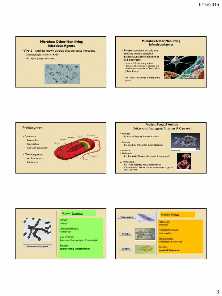
General Categories of Microbes 1. Non-living infectious particles: Viruses, Viroids, & Prions
2. Prokaryotes—NO NUCLEUS: Bacteria
• Unicellular (single-celled) microbes
3. Eukaryotes—HAVE A NUCLEUS: Protists, Fungi, & Animals
• Unicellular OR multicellular (many-celled) microbes
• 5 major categories: viruses, bacteria, protists, fungi, & animals
11
Viruses
Nonliving because it doesn’t display
characteristics of life until it has a host
Simple structure
◦ Capsid
◦ Nucleic acid
Smaller relatives
◦ Viroids
◦ Prions
6/16/2016
3
Microbes: Other Non-living
Infectious Agents
Viroid—smallest known particle that can cause infections
◦ Circular, single strands of RNA
◦ No capsid (no protein coat)
Microbes: Other Non-living
Infectious Agents
Prions—proteins that do not have any nucleic acids, but instead cause other proteins to fold incorrectly.
◦ responsible for many animal diseases like mad cow disease and the human equivalent Creutzfeldt-Jakob disease.
◦ ex. Kuru—occurred in many tribal
places
Prokaryotes
Structure
◦ No nucleus
◦ Organelles
◦ Cell wall (optional)
Two Kingdoms:
◦ Archaebacteria
◦ Eubacteria
15
Protists, Fungi, & Animals
(Eukaryotic Pathogens, Parasites & Carriers)
Protists ◦ Ex. African Sleeping Sickness & Malaria
Protists ◦ Ex. Candida, Aspergillus, & Cryptococcus
Animals:
1. Helminths ◦ Ex. Parasitic Worms (flat, round, & segmented)
2. Arthropods ◦ Ex. Ticks, insects, fleas, mosquitoes
◦ Cause/transmit disease in their microscopic stages or act as carriers
Kingdom: Eubacteria
Cell type :
prokaryotic
Unicellular/Multicellular:
All unicellular
Mode of Nutrition:
Autotrophic, Chemoautrophic, & Heterotrophic
Examples:
Staphylococcus & Mycobacterium Streptococcus pyogenes
Kingdom: Protista
Type of cells:
eukaryotic
Unicellular/Multicellular:
most unicellular
Mode of Nutrition:
Heterotrophic & Autotrophic
Examples:
Amoeba & Paramecium
Paramecium
Amoeba
Euglena

6/16/2016
4
Kingdom: Fungi
Type of cells:
eukaryotic
Unicellular/Multicellular:
Mostly multicellular
Mode of Nutrition:
Heterotrophic
Examples:
Yeast & molds
Mushroom
Yeast
Molds
Kingdom: Animalia
Type of cells:
eukaryotic
Unicellular/Multicellular:
multicellular
Mode of Nutrition:
Heterotrophic
Examples:
Arthropods (insects) & Helminths (worms)
Germ Theory
Theory definition:
◦ Microorganisms can invade other
organisms and cause disease
Important Contributors:
◦ Koch
◦ Semmelweiss
◦ Lister
◦ Jenner
◦ Pasteur
Identification of the Pathogen: Koch’s Postulates
Importance: criteria established by Robert Koch to identify the
causative agent of a particular disease:
1. The bacteria must be present in every case of the disease.
2. The bacteria must be isolated from the host with the disease
and grown in pure culture.
3. The specific disease must be reproduced when a pure culture
of the bacteria is inoculated into a healthy susceptible host.
4. The bacteria must be recoverable from the experimentally
infected host.
22
Aseptic Technique
Importance: handwashing and personal
hygiene while treating patients was identified
as necessary when treating patients
Semmelweiss: work demonstrated that
hand-washing could drastically reduce the
number of women dying after childbirth
Joseph Lister: father of aseptic technique in
surgery
23
Immunology
Edward Jenner
◦ pioneer of smallpox vaccine, the
world's first vaccine.
◦ "the father of immunology”
Louis Pasteur
◦ developed pasteurization
◦ created the Rabies vaccine

6/16/2016
5
Brain Check
1. What are the 5 major groups of microbes?
2. What role do animals play in microbial diseases?
3. What is the germ theory? Can you name one scientist that
made a contribution to this theory?
25
MICROSCOPY & STAINING
CH 3
Metric Units
Microbe sizes – μm to nm
Cell Theory
Theory definition:
1. All living things are made of cells.
2. Cells are the basic units of structure
and function in living things.
3. Living cells come only from other
living cells.
Important Contributors:
◦ Hooke
◦ von Leeuwenhoek
History of the Microscope
Robert Hooke
Used the compound microscope to study cork and came up
with the name “cells”
He drew this with a
QUILL pen…real
talk!
History of the Microscope Anton von Leeuwenhoek
First person to witness (and record) looking at a live cell under a microscope…called them animacules
He made over 500 different types of microscopes
FYI: It contained one lens and used only natural light to view objects
6/16/2016
6
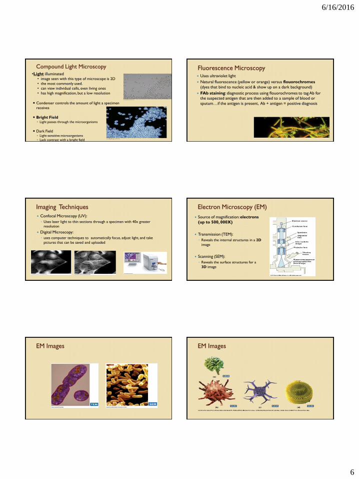
Compound Light Microscopy •Light illuminated
• image seen with this type of microscope is 2D
• the most commonly used.
• can view individual cells, even living ones
• has high magnification, but a low resolution
Condenser controls the amount of light a specimen
receives
Bright Field ◦ Light passes through the microorganisms
Dark Field ◦ Light-sensitive microorganisms
◦ Lack contrast with a bright field
Fluorescence Microscopy Uses ultraviolet light
Natural fluorescence (yellow or orange) versus flouorochromes
(dyes that bind to nucleic acid & show up on a dark background)
FAb staining: diagnostic process using flouorochromes to tag Ab for
the suspected antigen that are then added to a sample of blood or
sputum…if the antigen is present, Ab + antigen = positive diagnosis
Imaging Techniques
Confocal Microscopy (UV):
◦ Uses laser light to thin sections through a specimen with 40x greater
resolution
Digital Microscopy:
◦ uses computer techniques to automatically focus, adjust light, and take
pictures that can be saved and uploaded
Electron Microscopy (EM)
Source of magnification: electrons
(up to 500, 000X)
Transmission (TEM):
◦ Reveals the internal structures in a 2D
image
Scanning (SEM):
◦ Reveals the surface structures for a
3D image
EM Images EM Images

6/16/2016
7
Light Microscope Specimen Preparation:
Wet Mounts
Wet Mounts
◦ Helps show motility in
microorganisms
◦ Shows if specimen are even
present
◦ fresh cultures must be used
for maximum motility
◦ NO STAINS!!
Light Microscope Specimen Preparation:
Smears
◦ Allow you to apply stains (dye)
to cells which are usually
colorless
◦ Steps:
1. Placement of cells
2. Air drying
3. Heat fixation (flame or
warmer)
4. Apply stain
Staining Principles
What is a stain?
◦ A stain, or dye, is a molecule that can bind to cellular structure and give it
color…because cytoplasm is clear
Why do we use them?
1. To help investigate major groups
2. Examine structural and chemical differences in cells
3. Look at parts of the cell
Staining Principles
Basic stains:
◦ most commonly used
◦ Ex. Methylene blue & crystal violet
Acidic stains:
◦ Attracted to certain cell parts
◦ Ex. Eosin (dark red) & picric acid (yellow)
Staining Principles
1. Simple Stains:
◦ Uses a single dye
◦ Reveals basic cell structures and arrangements
◦ Ex. Methylene blue, crystal violet, & carbolfuchsin
2. Differential Stains: Gram & Ziehl-Neelsen
◦ Using 2 or more dyes
◦ Distinguishes between 2 kinds of organisms or 2 different parts of it
3. Special Stains: Flagellar & Schaffer-Fulton
Gram Stain
Technique
Significance
◦ Cell wall anatomy
◦ Diagnosis

6/16/2016
8
Ziehl-Neelsen Acid Fast Stain
Acid Fast Bacteria
◦ Identifies Mycobacterium (tuberculosis & leprosy)
◦ Stain red because of the lipids in their cell membranes
Flagellar staining
Detects motility
Metal staining
It’s difficult so don’t worry about
doing it
Schaeffer-Fulton staining
Detects endospores
Medical significance
Brain Check
1. What type of staining separates bacteria into 2 major groups?
2. What type of staining is important for medical
diagnosis/treatment?
3. Name a difference between a light microscope and an electron
microscope.
46
Tools of Classification
I. Classification
What is classification?
Grouping of organisms based on similarities.
Why classify?
1. makes information easier to manage and use
2. It shows relationships between living things.
Carolus Linnaeus (1700’s):
came up with the 2-part naming system that is used today for living things called binomial nomenclature (scientific naming).

6/16/2016
9
Taxonomy def: The science of classifying living things.
Taxonomist def: a scientist who studies classification
Levels of Classification (Taxonomic Categories – Taxa)
1. Domain “Most broad” 2. Kingdom 3. Phylum (Division for plants) 4. Class 5. Order 6. Family 7. Genus 8. Species “Most specific”
1.
2.
3.
4.
5.
6.
7.
8.
Write down this Memory Aid:
Scientists DO NOT use common names of organisms !!!
*Common names are the everyday name of an organism.
Reason #1:
The names are not very descriptive (which can be confusing in naming the organisms)
Reason #2: Some organisms have more than one common name
Scientific Names…
An organism has only one scientific name.
common dog
Canus familiaris
wolf
Canus lupus
Walking Pneumonia Mycoplasma
pneumoniae
Staphylococcus aureus
Staph
*Rules for writing scientific names*
1. The Genus is written first and the species is written second.
Ex: Homo (Genus) sapiens (species) = Homo sapiens
2. The first letter in the first word (Genus) is capitalized, and the
second word is written lower case.
Ex: Homo sapiens
3. The scientific name must be underlined OR written in italics.
Ex: Homo sapiens Homo sapiens
Lontra canadensis/Enhydra lutris
River otter/Sea otter
6/16/2016
10
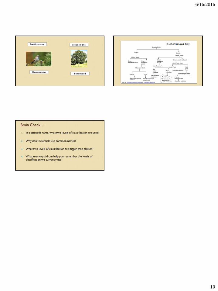
English sparrow
House sparrow
Sycamore tree
buttonwood
56
Brain Check…
1. In a scientific name, what two levels of classification are used?
2. Why don’t scientists use common names?
3. What two levels of classification are bigger than phylum?
4. What memory aid can help you remember the levels of classification we currently use?
Related Documents