Viruses and Prions Chapter 14

Viruses and Prions
Jan 01, 2016
Viruses and Prions. Chapter 14. 14.1 Structure and Classification of Animal Viruses. Structure DNA or RNA genome Double stranded (ds) or single stranded (ss) Surrounded by a capsid (protein coat) The nucleic acid and capsid are termed nucleocapsid Some viruses have an envelope - PowerPoint PPT Presentation
Welcome message from author
This document is posted to help you gain knowledge. Please leave a comment to let me know what you think about it! Share it to your friends and learn new things together.
Transcript

Viruses and PrionsChapter 14
14.1 Structure and Classification of Animal VirusesStructure
DNA or RNA genome
Double stranded (ds) or single stranded (ss)
Surrounded by a capsid (protein coat)
The nucleic acid and capsid are termed nucleocapsid
Some viruses have an envelope
The envelope is a phospholipid bilayer membrane that was obtained from the cell in which the virus arose
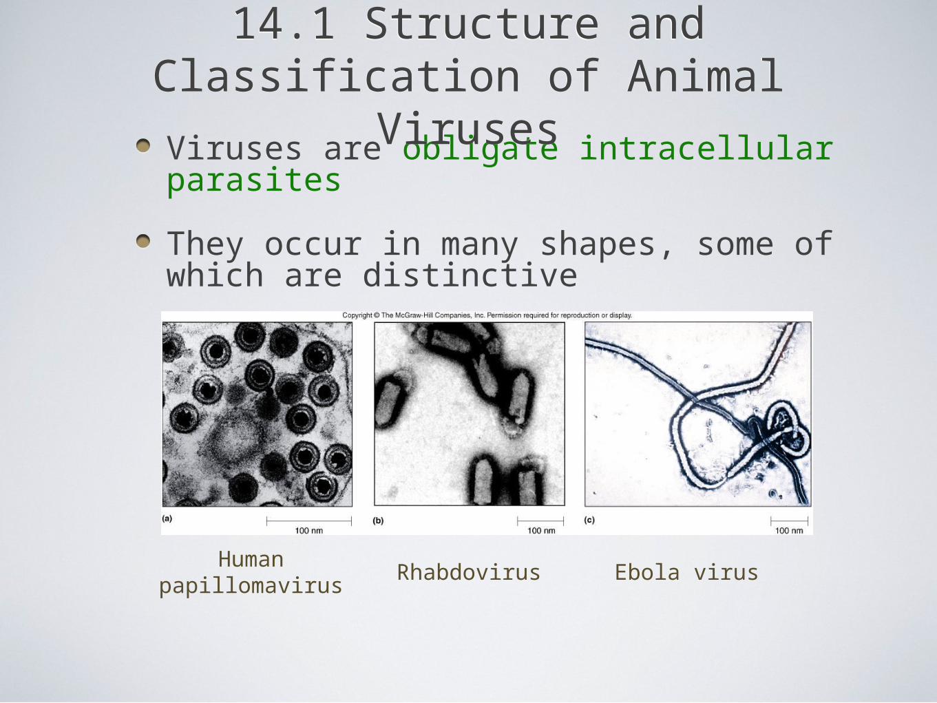
Viruses are obligate intracellular parasites
They occur in many shapes, some of which are distinctive
14.1 Structure and Classification of Animal Viruses
14.1 Structure and Classification of Animal Viruses
Humanpapillomavirus
Rhabdovirus Ebola virus
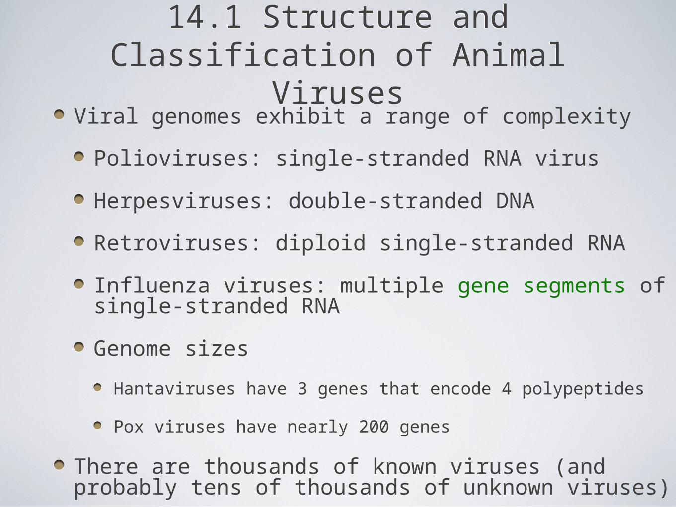
Viral genomes exhibit a range of complexity
Polioviruses: single-stranded RNA virus
Herpesviruses: double-stranded DNA
Retroviruses: diploid single-stranded RNA
Influenza viruses: multiple gene segments of single-stranded RNA
Genome sizes
Hantaviruses have 3 genes that encode 4 polypeptides
Pox viruses have nearly 200 genes
There are thousands of known viruses (and probably tens of thousands of unknown viruses)
14.1 Structure and Classification of Animal Viruses
14.1 Structure and Classification of Animal Viruses
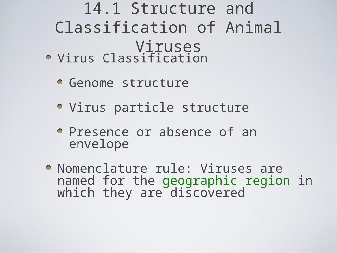
Virus Classification
Genome structure
Virus particle structure
Presence or absence of an envelope
Nomenclature rule: Viruses are named for the geographic region in which they are discovered
14.1 Structure and Classification of Animal Viruses
14.1 Structure and Classification of Animal Viruses
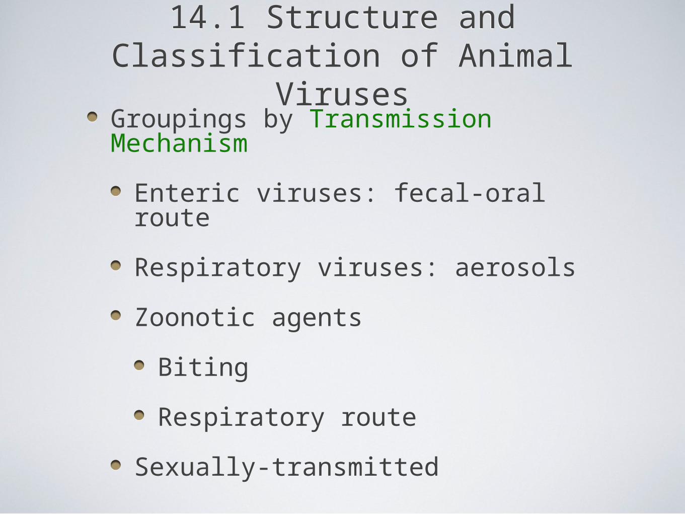
Groupings by Transmission Mechanism
Enteric viruses: fecal-oral route
Respiratory viruses: aerosols
Zoonotic agents
Biting
Respiratory route
Sexually-transmitted
14.1 Structure and Classification of Animal Viruses
14.1 Structure and Classification of Animal Viruses
14.2 Interactions of Animal Viruses and Their Hosts
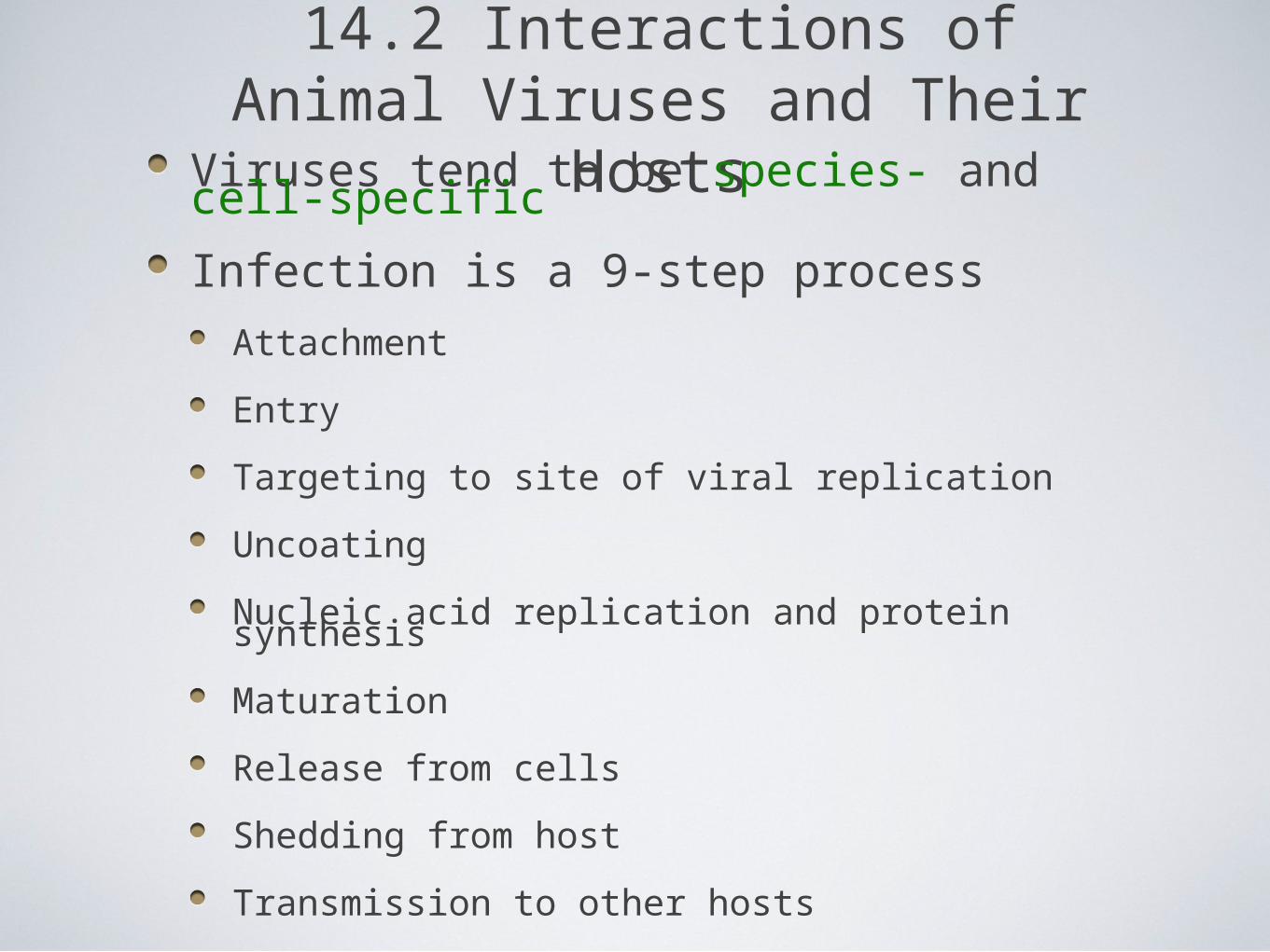
Viruses tend to be species- and cell-specific
Infection is a 9-step process
Attachment
Entry
Targeting to site of viral replication
Uncoating
Nucleic acid replication and protein synthesis
Maturation
Release from cells
Shedding from host
Transmission to other hosts
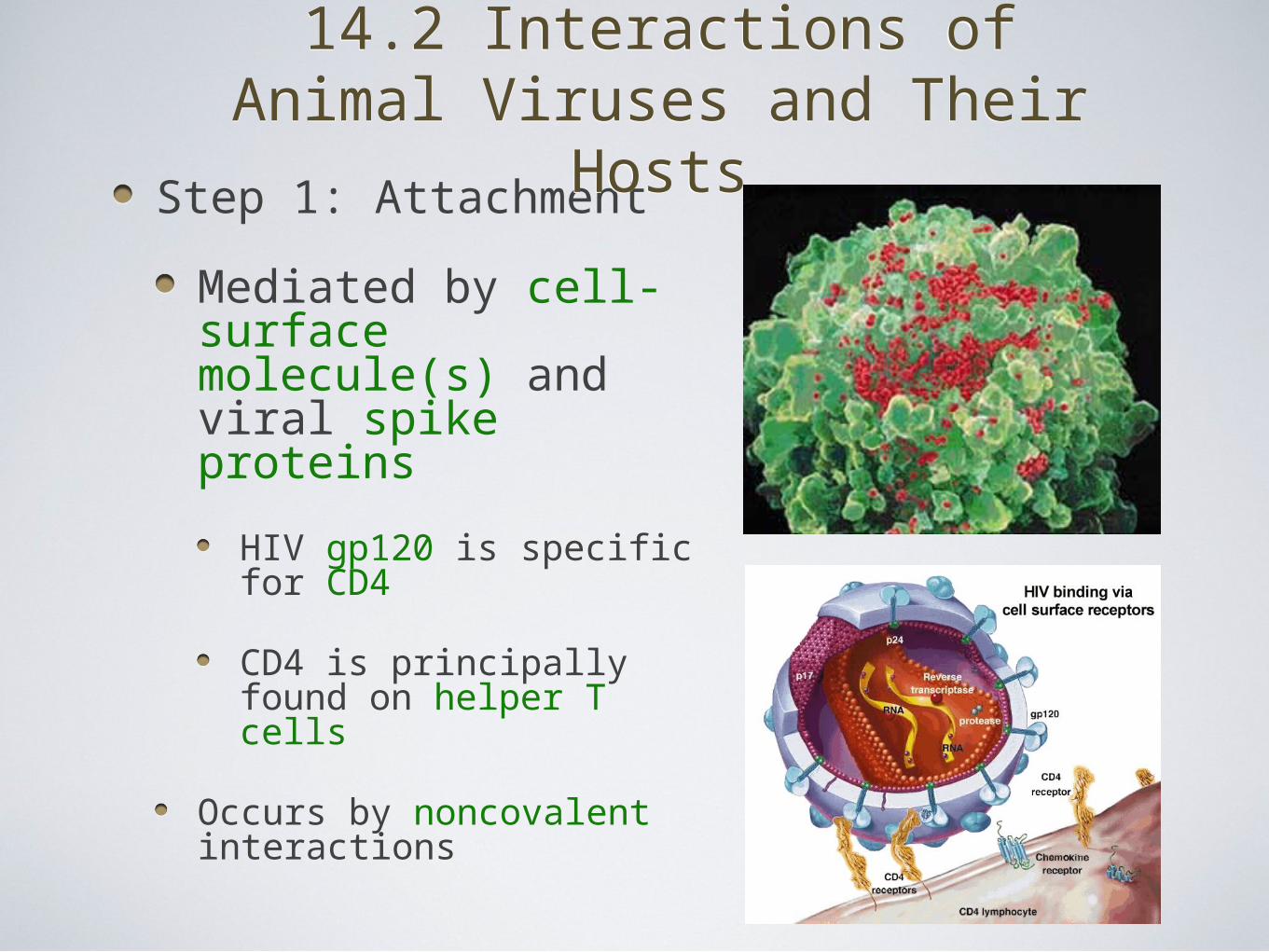
Step 1: Attachment
Mediated by cell-surface molecule(s) and viral spike proteins
HIV gp120 is specific for CD4
CD4 is principally found on helper T cells
Occurs by noncovalent interactions
14.2 Interactions of Animal Viruses and Their Hosts
14.2 Interactions of Animal Viruses and Their Hosts
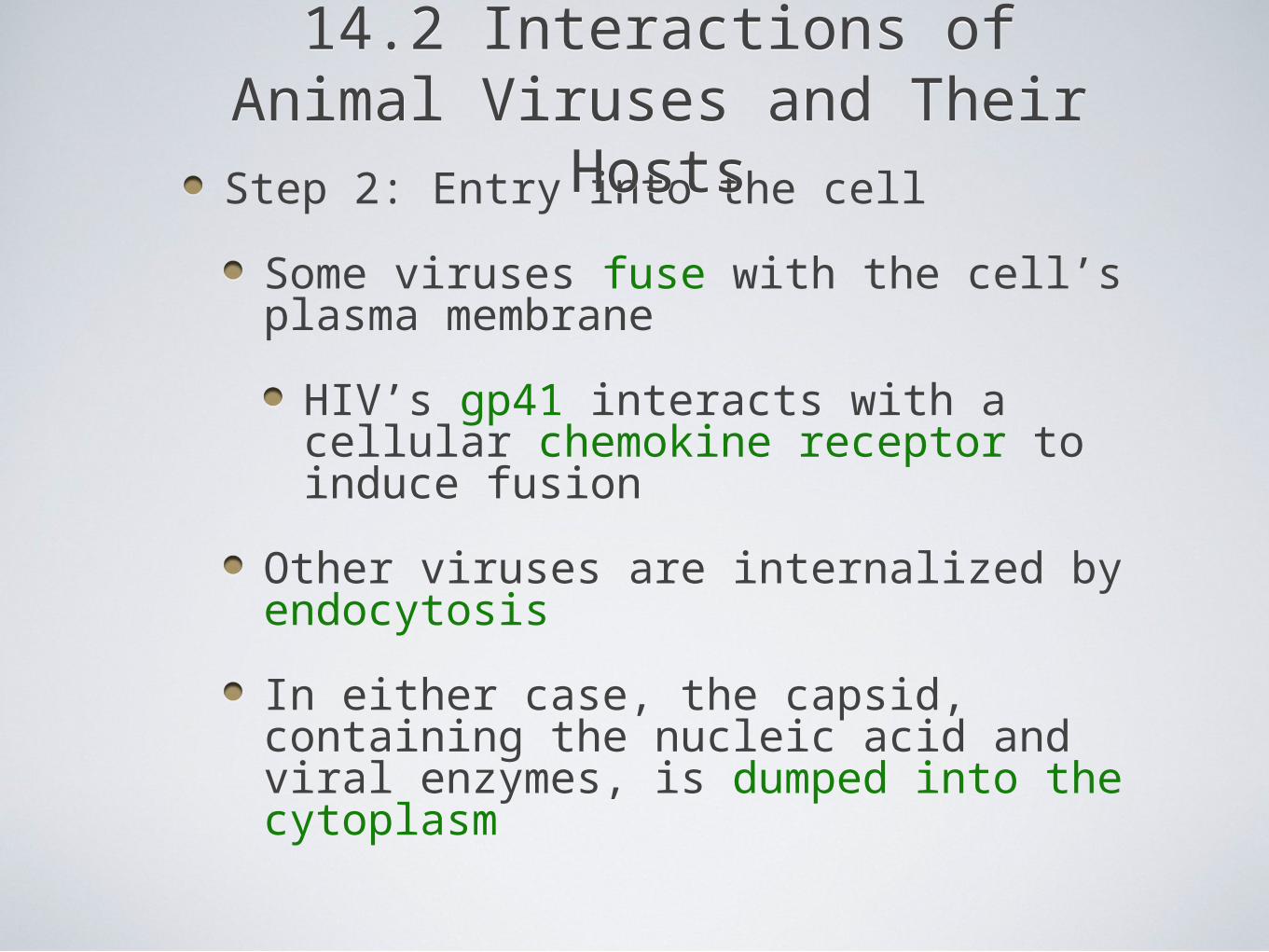
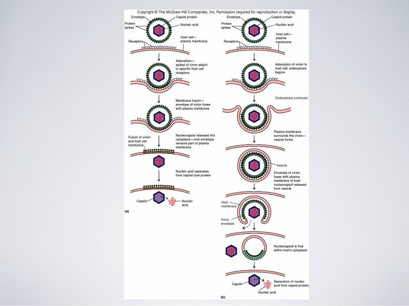
Step 2: Entry into the cell
Some viruses fuse with the cell’s plasma membrane
HIV’s gp41 interacts with a cellular chemokine receptor to induce fusion
Other viruses are internalized by endocytosis
In either case, the capsid, containing the nucleic acid and viral enzymes, is dumped into the cytoplasm
14.2 Interactions of Animal Viruses and Their Hosts
14.2 Interactions of Animal Viruses and Their Hosts
14.2 Interactions of Animal Viruses and Their Hosts
14.2 Interactions of Animal Viruses and Their Hosts
Step 3: Targeting to the site of viral replication
Most DNA viruses replicate in the nucleus
Most RNA viruses replicate in the cytoplasm
Some viruses integrate their dsDNA into the host cell’s genome (i.e., chromosomes)
Some viruses copy their RNA into dsDNA, which is then integrated into the host cell’s genome
14.2 Interactions of Animal Viruses and Their Hosts
14.2 Interactions of Animal Viruses and Their Hosts
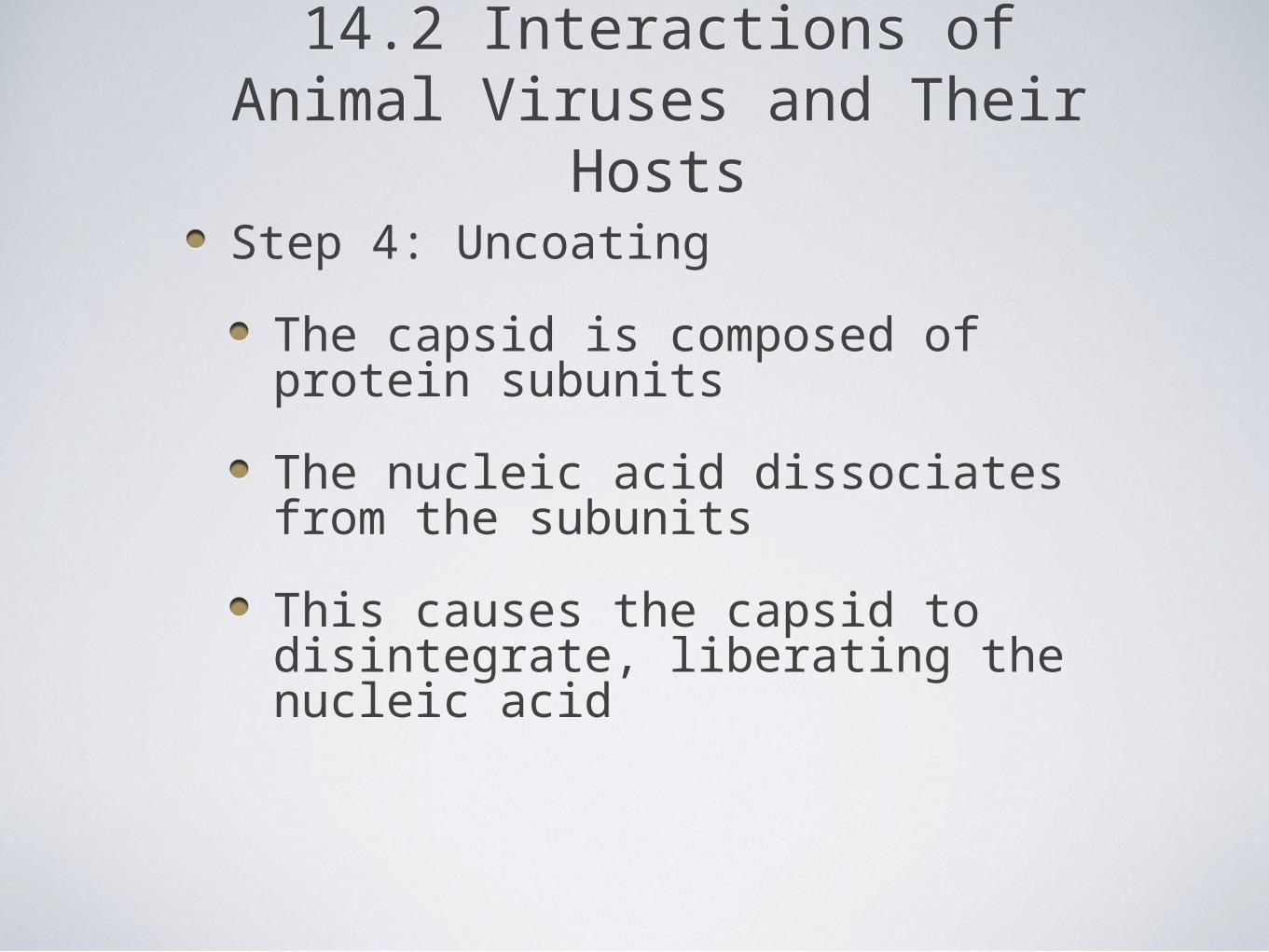
Step 4: Uncoating
The capsid is composed of protein subunits
The nucleic acid dissociates from the subunits
This causes the capsid to disintegrate, liberating the nucleic acid
14.2 Interactions of Animal Viruses and Their Hosts
14.2 Interactions of Animal Viruses and Their Hosts
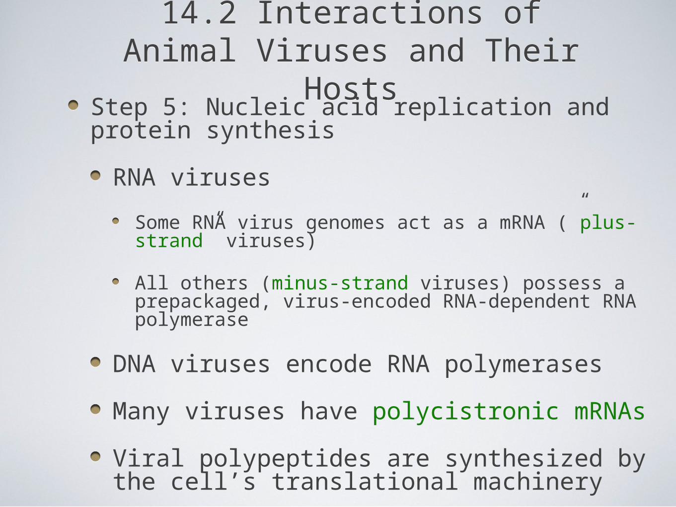
Step 5: Nucleic acid replication and protein synthesis
RNA viruses
Some RNA virus genomes act as a mRNA (”plus-strand” viruses)
All others (minus-strand viruses) possess a prepackaged, virus-encoded RNA-dependent RNA polymerase
DNA viruses encode RNA polymerases
Many viruses have polycistronic mRNAs
Viral polypeptides are synthesized by the cell’s translational machinery
14.2 Interactions of Animal Viruses and Their Hosts
14.2 Interactions of Animal Viruses and Their Hosts
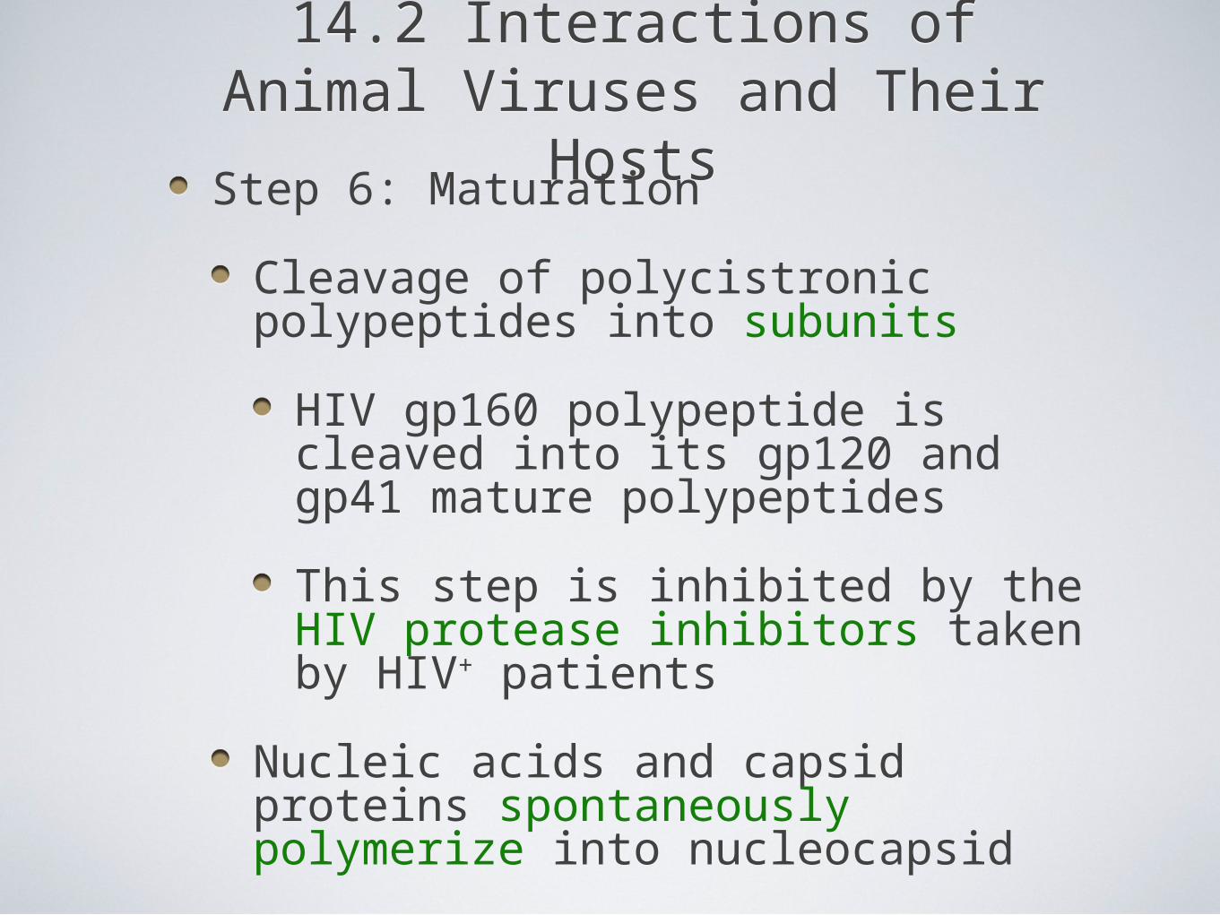
Step 6: Maturation
Cleavage of polycistronic polypeptides into subunits
HIV gp160 polypeptide is cleaved into its gp120 and gp41 mature polypeptides
This step is inhibited by the HIV protease inhibitors taken by HIV+ patients
Nucleic acids and capsid proteins spontaneously polymerize into nucleocapsid
14.2 Interactions of Animal Viruses and Their Hosts
14.2 Interactions of Animal Viruses and Their Hosts
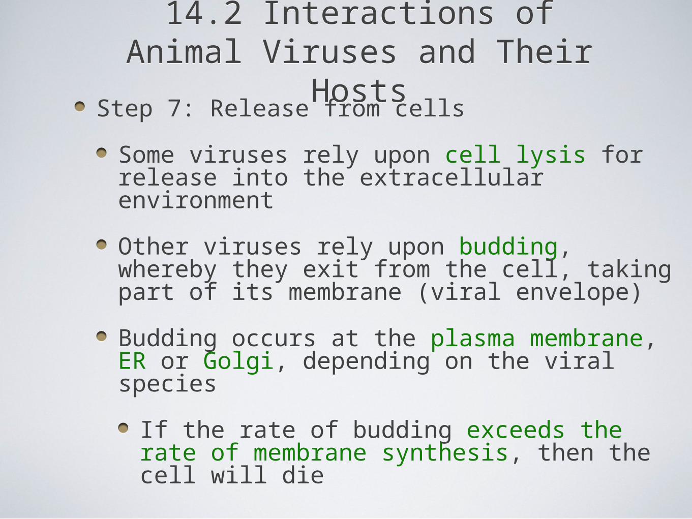
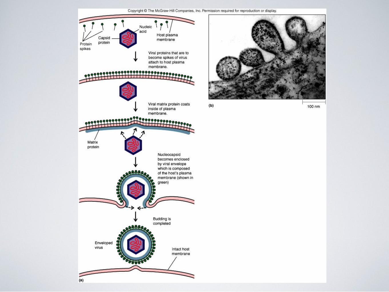
Step 7: Release from cells
Some viruses rely upon cell lysis for release into the extracellular environment
Other viruses rely upon budding, whereby they exit from the cell, taking part of its membrane (viral envelope)
Budding occurs at the plasma membrane, ER or Golgi, depending on the viral species
If the rate of budding exceeds the rate of membrane synthesis, then the cell will die
Step 8: Shedding from the host
Viruses must leave the infected host to infect other hosts
Shedding can be a minor event (such as cold viruses) or a catastrophic event (such as hemorrhagic fever viruses)
Step 9: Transmission to other hosts
Transmission routes usually reflect the sites of infection for viruses (e.g., respiratory, GI, STD)
14.2 Interactions of Animal Viruses and Their Hosts
14.2 Interactions of Animal Viruses and Their Hosts
14.2 Interactions of Animal Viruses and Their Hosts
14.2 Interactions of Animal Viruses and Their Hosts
Persistent infections
Latent - periods of inactivation and activation (e.g., herpesviruses); usually limited pathology
Chronic - infectious virus can be detected for years or decades with little discernible pathology, but can eventually lead to disease (e.g., hepatitis B and C viruses)
Slow infections - short period of acute infection (weeks) followed by the apparent disappearance of virus for months or years, with pathology ensuing (e.g., HIV)
14.3 Viruses and Human TumorsTumor viruses drive cell proliferation
Several mechanisms account for this phenomenon
Viral oncogenes that stimulate cell proliferation
Viral DNA integrates adjacent to genes that drive cell division
Expression of the viral genes leads to aberrant expression of the cellular gene
Some viruses encode growth factors that stimulate cellular proliferation
Epstein-Barr virus encodes viral interleukin-10 that causes B cell proliferation, leading to Burkitt’s lymphoma
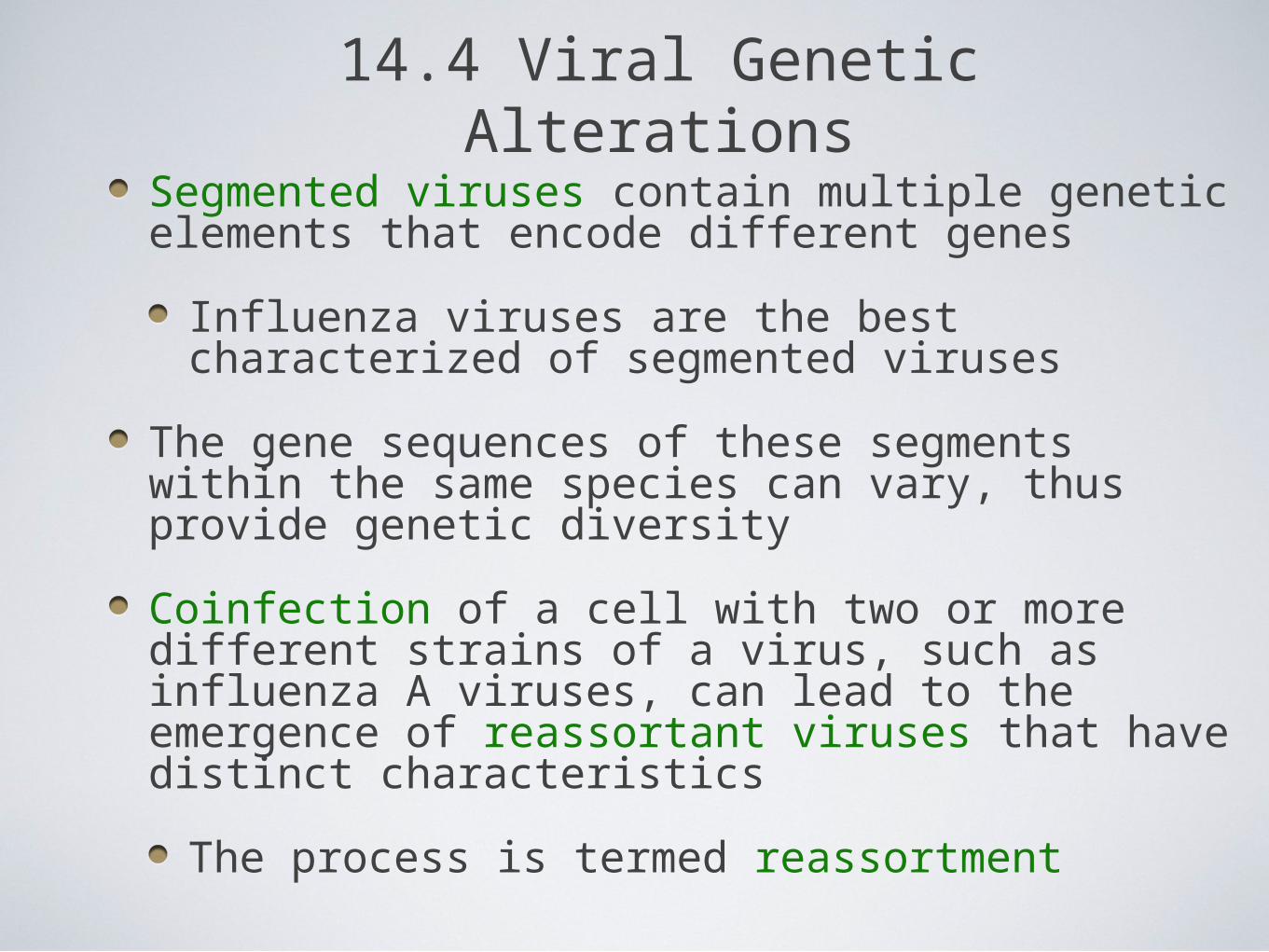
14.4 Viral Genetic AlterationsSegmented viruses contain multiple genetic elements that encode different genes
Influenza viruses are the best characterized of segmented viruses
The gene sequences of these segments within the same species can vary, thus provide genetic diversity
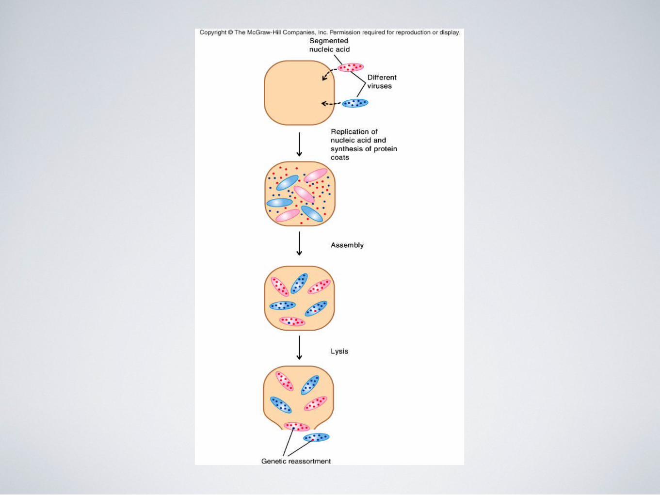
Coinfection of a cell with two or more different strains of a virus, such as influenza A viruses, can lead to the emergence of reassortant viruses that have distinct characteristics
The process is termed reassortment
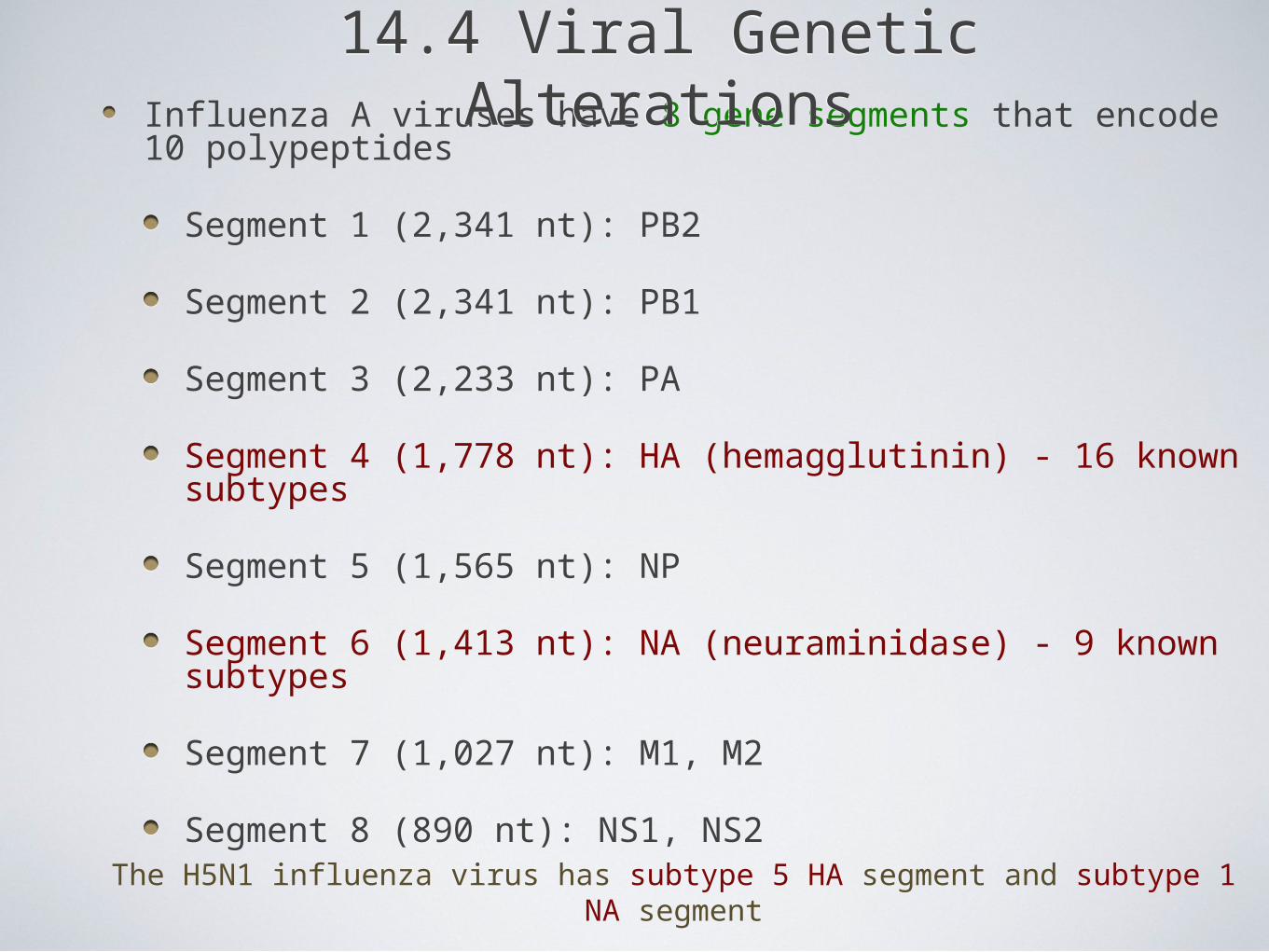
Influenza A viruses have 8 gene segments that encode 10 polypeptides
Segment 1 (2,341 nt): PB2
Segment 2 (2,341 nt): PB1
Segment 3 (2,233 nt): PA
Segment 4 (1,778 nt): HA (hemagglutinin) - 16 known subtypes
Segment 5 (1,565 nt): NP
Segment 6 (1,413 nt): NA (neuraminidase) - 9 known subtypes
Segment 7 (1,027 nt): M1, M2
Segment 8 (890 nt): NS1, NS2
14.4 Viral Genetic Alterations14.4 Viral Genetic Alterations
The H5N1 influenza virus has subtype 5 HA segment and subtype 1 NA segment
14.5 Methods Used to Study Viruses
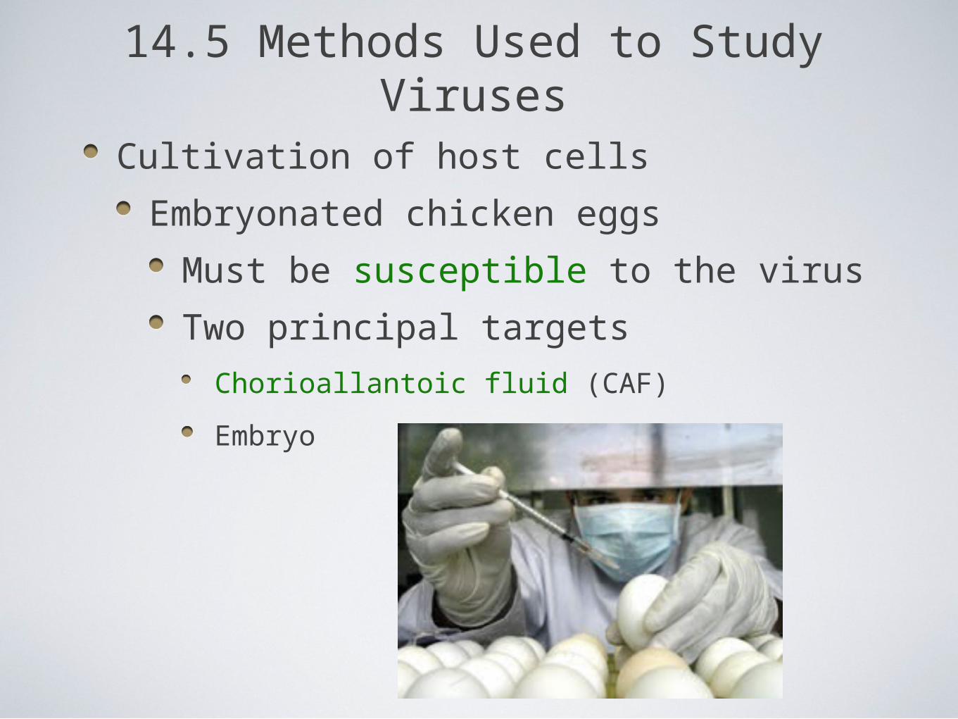
Cultivation of host cells
Embryonated chicken eggs
Must be susceptible to the virus
Two principal targets
Chorioallantoic fluid (CAF)
Embryo
14.5 Methods Used to Study Viruses
14.5 Methods Used to Study Viruses
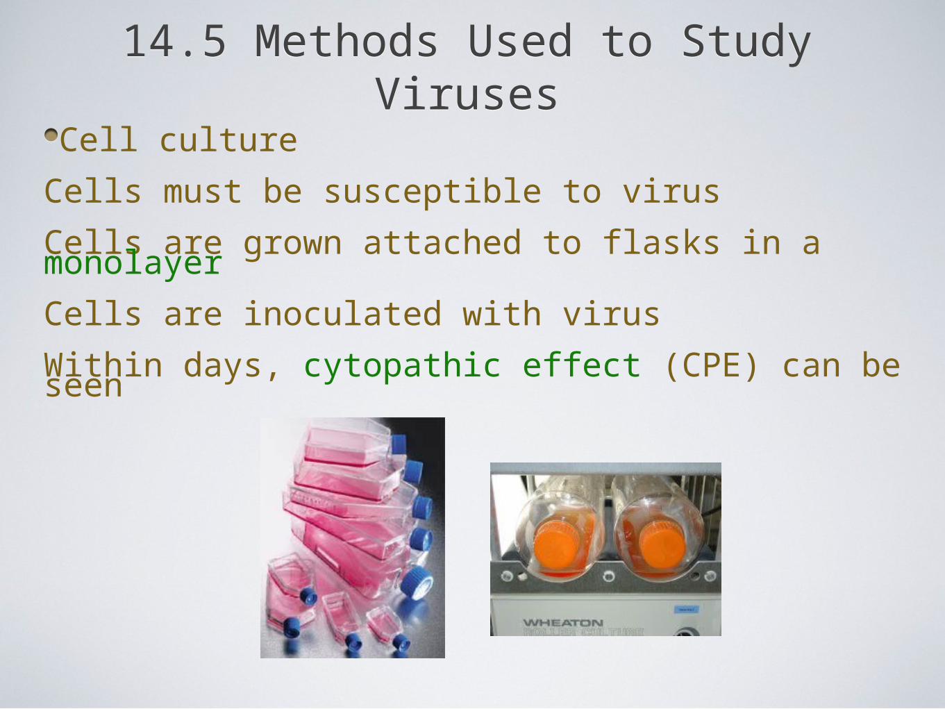
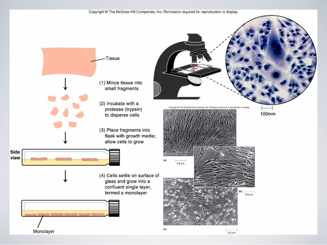
Cell culture
Cells must be susceptible to virus
Cells are grown attached to flasks in a monolayer
Cells are inoculated with virus
Within days, cytopathic effect (CPE) can be seen
14.7 Other Infectious AgentsPrions
Proteinaceous infectious particle
Cause spongiform encephalopathies
Characteristics
They contain no nucleic acids
They are a normal cellular protein (PrPc) that has misfolded into a pathogenic protein
The prion protein “replicates” itself by causing copies of the normal protein to misfold into the prion protein
Diseases
Creutzfeldt-Jakob (New Variant CJ from “mad” cows)
Kuru (religious consumption of brains from deceased)
Chronic wasting disease (elk, deer, moose)
Related Documents