S P E C T P E T M R C T E C HO C l i n i c a l P r o t o c o l s Q u a l i t y M a n a g e m e n t International Conference on Integrated Medical Imaging in Cardiovascular Diseases (IMIC 2013) 30 September – 4 October 2013 Vienna International Centre Vienna, Austria A p p r o p r i a t e U s e C r i t e r i a CN-202 @ ABSTRACTS

Welcome message from author
This document is posted to help you gain knowledge. Please leave a comment to let me know what you think about it! Share it to your friends and learn new things together.
Transcript
SP
EC
T PET MR CT ECHO
Clinical Protocols Quality Management
International Conference onIntegrated Medical Imaging in Cardiovascular Diseases(IMIC 2013)30 September – 4 October 2013Vienna International CentreVienna, Austria
Appropriate Use Criteria
CN-202
@@
Inte
rnatio
nal C
onfe
rence o
n In
tegra
ted M
edic
al Im
agin
g
in C
ard
iova
scula
r Dise
ase
s (IMIC
2013)
1
International Atomic Energy Agency
IAEA-CN-202Vienna International Centre
P.O. Box 100Wagramer Strasse 51400 Vienna, AustriaTel.: +43 1 2600 (0)Fax: +43 1 26007
Email: offi [email protected]
IAEA conference web site:http://www-pub.iaea.org/iaeameetings/
13-3
2851
ABSTRACTS
@Organized by the
In cooperation with the
Asian Regional Cooperative Council for Nuclear Medicine
Australian and New Zealand Society of Nuclear Medicine
European Associacion of Nuclear Medicine
World Federation of Nuclear Medicine and Biology
European Society of Radiology
International Society of Radiology
The material in this book has been supplied by the authors and has not been edited. The views expressed remain the responsibility of the named authors and do not necessarily refl ect those of the government of the designating Member State(s). The IAEA cannot be held responsible for any material reproduced in this book.
SP
EC
T PET MR CT ECHO
Clinical Protocols Quality Management
International Conference onIntegrated Medical Imaging in Cardiovascular Diseases(IMIC 2013)30 September – 4 October 2013Vienna International CentreVienna, Austria
Appropriate Use Criteria
CN-202
@@
Inte
rnatio
nal C
onfe
rence o
n In
tegra
ted M
edic
al Im
agin
g
in C
ard
iova
scula
r Dise
ase
s (IMIC
2013)
1
International Atomic Energy Agency
IAEA-CN-202Vienna International Centre
P.O. Box 100Wagramer Strasse 51400 Vienna, AustriaTel.: +43 1 2600 (0)Fax: +43 1 26007
Email: offi [email protected]
IAEA conference web site:http://www-pub.iaea.org/iaeameetings/
13-3
2851
ABSTRACTS
International Conference on
Integrated Medical Imaging in Cardiovascular Diseases
(IMIC 2013)
Vienna International Centre
Vienna, Austria
30 September – 4 October 2013
BOOK OF ABSTRACTS
IAEA-CN-202
i
CONTENTS
MEMBER STATE EXPERIENCE WITH S.P.E.C.T., P.E.T., ECHOCARDIOGRAPHY, C.T. AND M.R.I. IN THE MANAGEMENT OF C.V.D.s
IAEA-CN-202/103…………………………………………………………………………… 3
Cardiac CT Patient Dose in Algeria: First Results
N. Khelassi-Toutaoui, A. Benali, A. Toutaoui, Z. Brahimi, et al.
IAEA-CN-202/104…………………………………………………………………………… 5
A Study of 4 Minutes versus 6 Minutes Protocol for Pharmacological Stress Testing Using Adenosine for Myocardial Perfusion Imaging and Comparison with Echocardiography Findings
G. Malhotra, K. Panchal, M. V. Reddy, R. V. Asopa, et al.
IAEA-CN-202/106…………………………………………………………………………… 7
Correlation & Agreement of Measurements of Left Ventricular Ejection Fraction by Radionuclide Methods & Echocardiogram
F. Alam, R. Hossain, A. K. Sarker
IAEA-CN-202/108…………………………………………………………………………...10
Fluordeoxyglucose in the Assessment and Therapy Control of an Atrial Angiosarcoma
M. J. Jofré Manieu
IAEA-CN-202/109………………………………………………………………………….. 13
Application of SPECT-CT in Patients with Suspicion of a Vascular Graft Infection
I. Kostadinova, S. Ivanova, M. Garcheva
IAEA-CN-202/111………………………………………………………………………….. 14
FDG Uptake in Large Arteries, Comparison between Genders and Correlation with Aging
G. Bural, D. Torigian, M. Houseni, A. Alavi
IAEA-CN-202/112………………………………………………………………………….. 15
Implications of Persistent ST Segment Elevation in Q-Wave Anteroseptal Myocardial Infarction – Correlation with Myocardial Perfusion Gated SPECT
P. Subramanyam, S. S. Palaniswamy
ii
IAEA-CN-202/113………………………………………………………………………….. 16
Is Exercise Induced ST Depression an Accurate Indicator of Viability in Infarct-Related Artery? - A Myocardial Perfusion SPECT Study
P. Subramanyam, S. S. Palaniswamy
IAEA-CN-202/114………………………………………………………………………….. 17
Influence of Attenuation Correction on the Interpretation of Myocardial Perfusion Images
M. Garcheva, I. Kostadinova
IAEA-CN-202/115………………………………………………………………………….. 18
Dynamics of Myocardial Perfusion with MIBI in Patients with Coronary Heart Disease and Post-Infarction Cardiosclerosis After Stem Cell Therapy
A. Dustov, U. Kurbanov, M. Mirshahi
IAEA-CN-202/116…………………………………………………………………………..20
Early Diastolic Dysfunction Detection by 16-bin Gated MPI SPECT in End Stage Liver Disease Patients with Normal Myocardial Perfusion Undergoing Pre-Transplant Cardiac Evaluation
S. J. Gandhi, S. L. G. Praveen, S. Padma, S. S. Palaniswamy
IAEA-CN-202/117………………………………………………………………………….. 22
Can MPI SPECT Predict Abnormalities on Carotid / Lower Limb Doppler USG for Extra Coronary Atherosclerotic Disease
S. J. Gandhi, S. L. G. Praveen, S. Padma, S. S. Palaniswamy
IAEA-CN-202/118………………………………………………………………………….. 24
Role of Myocardial Perfusion SPECT in the Prediction of Post-Surgery Recovery of Ischemic Mitral Regurgitation in Patients with Severe LV Dysfunction
S. S. Palaniswamy, S. Padma
IAEA-CN-202/119………………………………………………………………………….. 25
The Correlation between LVEF Gated-SPECT with Hemodialysis, Calcium-Phosphorus Product and Parathyroid Hormone in Patients with Chronic Kidney Diseases
D. Nariman, E. Purnomo
iii
IAEA-CN-202/120………………………………………………………………………….. 26
Correlation between Mean Platelet Volume and Myocardial Perfusion SPECT Parameters
A. S. Doğan, E. İzgi, M. E. Erkan, M. Aşık, et al.
IAEA-CN-202/121………………………………………………………………………….. 27
Evaluation of Gated SPECT MIBI Functional and Perfusion Abnormalities in Patients with Previous Inferior Myocardial Infarction
D. Sobic Saranovic, Z. Petrasinovic, V. Artiko, V. Obradovic
IAEA-CN-202/122………………………………………………………………………….. 28
Possibilities of Echocardiography in Estimation of Cardiotoxicity of Chemotherapy in Cancer Patients
O. Solodyannikova, N. Golovko, L. Shevchuk
IAEA-CN-202/123………………………………………………………………………….. 30
Comparison of Left Ventricular Ejection Fraction between Gated Myocardial Perfusion SPECT/CT and Echocardiography in CHD patients
X. Liu, J. Yuan, C. Liu, J. J. Zaknun
IAEA-CN-202/125………………………………………………………………………….. 31
Does the Thallium Defect Pattern in Myocardial Perfusion Scintigraphy Depict the Level of Stenoses in Single Vessel CAD Involving the Left Anterior Descending Artery
V. Agarwal
IAEA-CN-202/126………………………………………………………………………….. 32
Myocardial Infarct in Young Patients: SPECT MPI Findings
S. Rahabi, M. Habbeche, I. Ghedbane, A. Khelifa, et al.
IAEA-CN-202/127………………………………………………………………………….. 33
Brain Natriuretic Peptide Correlation Study with SPECT Stress and Rest MPI Findings
S. E. Bouyoucef, S. Rahabi, M. Habbeche, I. Ghedbane, et al.
IAEA-CN-202/129………………………………………………………………………….. 34
Proportion of Patients with QRS Greater than 120 msec and Normal Phase Gated SPECT MPI Image
S. Merlano Gaitan, E. Rodriguez, R. Murgueitio
iv
IAEA-CN-202/130………………………………………………………………………….. 35
Evaluation of Myocardial Viability with Myocardial Perfusion Imaging in Patients with Left Ventricular Aneurysms
C. A. Stan, C. Chirion, D. A. Stanescu, D. C. Calin, et al.
IAEA-CN-202/133………………………………………………………………………….. 36
Characteristics of Perfusion Defects, Left Ventricular Function and Prognostic Value of Early Dipyridamole Tc99m Sestamibi Gated SPECT MPI in Post-Myocardial Infarction Patients without Primary Angioplasty
H. N. Ha, L. P. Vu, H. M. Le
IAEA-CN-202/134………………………………………………………………………….. 38
13N-ammonia Cardiac PET/CT and Coronary CT Angiography
G. Estrada, J. Altamirano
IAEA-CN-202/136………………………………………………………………………….. 39
Evaluation of a Single Utilization of Pulmonary Perfusion Scintigraphy in Patients with Suspected Pulmonary Embolism
P.S. Choudhury, A. K. Chaturvedi, S. A. Rao, N. Watanabe
IAEA-CN-202/138………………………………………………………………………….. 41
Diagnostic Reference Levels for Adult Postero-Anterior Chest X-ray Examination in Kampala, Uganda - A Precursor for the National Diagnostic Reference Level
R. Byanyima Kusaba
IAEA-CN-202/140………………………………………………………………………….. 42
Indian Experience with Ultrafast Solid State Detector Dedicated Cardiac Camera in Myocardial Perfusion Imaging (MPI)
G. Priyanka, L. Vikram
IAEA-CN-202/141………………………………………………………………………….. 43
The Incremental Prognostic Value of SPECT-MPI in Predicting Cardiovascular Events in Patients with Metabolic Syndrome with Suspected CAD
A. F. Apostol
v
IAEA-CN-202/142………………………………………………………………………….. 44
Referral Physicians' Indications for Myocardial Perfusion Scintigraphy
B. Caliskan
IAEA-CN-202/143………………………………………………………………………….. 45
Phase Analysis by Gated Tc-99m Sestamibi SPECT for Left Ventricular Dyssynchrony: Impact of Myocardial Ischemia
C. Tinoco Mesquita, S. Garcia, J. Cunha de Azevedo, A. Ribeiro Nogueira Oliveira, et al.
IAEA-CN-202/144………………………………………………………………………….. 46
Correlation between Stress and Rest Left Ventricular Ejection Fraction in Gated Single Photon Emission Computed Tomography (SPECT) with the Extent and Severity of Perfusion Abnormalities
A. P. Quinon
IAEA-CN-202/146………………………………………………………………………….. 47
Positron Emission Tomography versus Cardiac Magnetic Resonance Imaging for the Assessment of Myocardial Viability: A Meta-Analysis
I. S. Bandong
IAEA-CN-202/147………………………………………………………………………….. 48
PET/CT: Uptake Pattern in Prosthetic Heart Valves
J. I. Arma, A. Mollerach, L. Paganini, V. Jager
IAEA-CN-202/148………………………………………………………………………….. 50
Utility of Myocardial Perfusion Studies for the Re-stratification of Risk of Ischemic Heart Disease and its Frequent Risk Factors
C. Arroyo Castelán, J. Z. García, R. M. Cuaxospa
IAEA-CN-202/149………………………………………………………………………….. 52
Clinical Experience and Applications of Simultaneous PET/MR Imaging in Cardiology
I. Cho
vi
IAEA-CN-202/150………………………………………………………………………….. 53
A Discordance Results between Myocardial Perfusion Imaging with Angiography: A Case Report
A. Elliyanti, A. H. S. Kartamihardja
IAEA-CN-202/152………………………………………………………………………….. 55
Myocardial Perfusion Scintigraphy and CT Coronary Angiography in the Evaluation of Coronary Artery Disease
P. Orellana Briones, A. Haeger, R. Jaimovich, R. Baeza
IAEA-CN-202/153………………………………………………………………………….. 56
Impact of stress induced 201Tl gated MPS on LV dyssynchrony by phase analysis
M. Parmar
IAEA-CN-202/154………………………………………………………………………….. 58
TC99m MIBI SPECT, Single Injection Dual Screening of IHD (Silent Myocardial Infarction) and Peripheral Arterial Disease (PAD) in Diabetes Mellitus
R. Rasheed
IAEA-CN-202/155………………………………………………………………………….. 60
Capacity of Steady-State CMR Imaging Sequence for Myocardial Necrosis Detection
M. Niella, D. Fino, F. Gonzalez, A. A. Astesiano, et al.
IAEA-CN-202/156………………………………………………………………………….. 62
Influence of a Low-Carbohydrate Diet on the Assessment of Myocardial Viability with 18F- Fluorodeoxyglucose PET: Comparison with the Euglycemic Hyperinsulinemic Clamp
F. Rodrigues Filho, J. Soares Jr, M. Izaki, M. C. P. Giorgi, et al.
IAEA-CN-202/159………………………………………………………………………….. 64
Role of [13N] NH3-[18F] FDG PET/CT in Predicting the Outcome in Patients with Coronary Artery Disease
A. Sood
vii
IAEA-CN-202/161………………………………………………………………………….. 66
Prognostic Value of Thallium ECG-gated Myocardial Perfusion Single-Photon Emission Computed Tomography (SPECT) Left Ventricular Parameters in Patients with Suspected Coronary Artery Disease
I. S. Bandong
IAEA-CN-202/165………………………………………………………………………….. 67
Myocardial Perfusion Scan (MPS) Results in Asymptomatic Patients with Coronary Artery Disease (CAD) Risk Factors
R. Yudistiro
IAEA-CN-202/166………………………………………………………………………….. 68
99mTc-Sestamibi Exercise SPECT and Follow-up in Patients Able to Reach 10 Metabolic Equivalents of Task (METs)
J. S. González
IAEA-CN-202/167………………………………………………………………………….. 70
Feasibility of ECG-Gated at Myocardial Perfusion Studies: Is It Worthy to ECG-Gated at Both Stress and Rest
E. Lemos Pereira, L. Oliveira, M. Fernandes, P. Colarinha
IAEA-CN-202/168………………………………………………………………………….. 72
The Place of Cardiac Computed Tomography Angiography (CCTA) in Cardiac Pathology
M. Mouloudi, T. Bassaid, N. Kriou, N. Boubendir, et al.
IAEA-CN-202/170………………………………………………………………………….. 73
Preparation and Preclinical Evaluation of [64Cu]Cu(II)-PTSM: A Tracer for Myocardial Perfusion
J. C. Manrique-Arias, V. M. Lara-Camacho, M. Avila-Garcia, A. Flores-Moreno, et al.
IAEA-CN-202/171………………………………………………………………………….. 75
Evaluation of Left Ventricular Function in Mali using a Radionuclide Method: Application, Methodology and Normal Values
S. Sidibé, S. Traoré, R. Diakité
viii
IAEA-CN-202/172………………………………………………………………………….. 76
Gated SPECT Functional Assessment in Obese Patients
A. Puente, J. Morales, G. Meléndez
IAEA-CN-202/174………………………………………………………………………….. 77
Rest Thallium-201/Stress Technetium-99m Sestamibi Dual-Isotope Myocardial Perfusion Single-Photon Emission Computed Tomography in Detecting of Chronic Coronary Artery Disease
P. K. Huynh, L. C. Vu, X. Q. Truong, C. X. Nguyen
IAEA-CN-202/175………………………………………………………………………….. 78 Sodium Bicarbonate Augmented 201Tl Rest/Redistribution Myocardial Perfusion SPECT in Detecting Myocardial Viability
K. Khan, A. Jielani, M. Ayub, N. Z. Abbasi, et al.
IAEA-CN-202/177………………………………………………………………………….. 79
Gated SPECT Imaging for Evaluation of Coronary Artery Disease - Experience in a Developing Country
S. Afroz
IAEA-CN-202/178………………………………………………………………………….. 80
Challenges in Nuclear Cardiology in Senegal
O. Diop, S. Seck-Gassama, E. H. L. Bathily, G. A. Kabre, et al.
IAEA-CN-202/179………………………………………………………………………….. 82
Evaluation of Coronary Artery Disease Using Stress ECG and Myocardial Perfusion Imaging in a Developing Country
M. Mbodj, O. Diop, B. Ndong, E. H. L. Bathily, et al.
IAEA-CN-202/180………………………………………………………………………….. 83
Experience with Performing Myocardial Perfusion Imaging (MPI) in Ischemic Heart Disease in Senegal
S. Seck-Gassama, O. Diop, B. Ndong, E. H. L. Bathily, et al.
IAEA-CN-202/183………………………………………………………………………….. 84
Nuclear Cardiology Status in Myanmar
K. Myint
ix
IAEA-CN-202/184………………………………………………………………………….. 85
Prognostic Value of Myocardial Perfusion Test with 99mTc-MIBI in Symptomatic and Asymptomatic Patients with Coronary Risk Factors
G. Fadragas
IAEA-CN-202/186………………………………………………………………………….. 86
Usefulness of Assessment of Coronary Reserve Flow in a Patient with Coronary Multivessel Disease
M. Mera, M. Montecinos Santibanez, S. Lapresa, M. Casal, et al.
IAEA-CN-202/187………………………………………………………………………….. 88
18F-FDG PET-CT Imaging for the Evaluation of Small- and Medium-size Vessel Vasculitis
S. Voo, M. Kemna, P. van Paassen, J. W. Cohen-Tervaert, et al.
IAEA-CN-202/188………………………………………………………………………….. 89
Comparison of Myocardial Perfusion SPECT Acquired with Linear Energy High Resolution Collimators (LEHR) and SMARTZOOM Collimators, Automatic Quantitative Software Differences
M. Havel, O. Kraft
IAEA-CN-202/189………………………………………………………………………….. 91
Combination of Myocardial Perfusion SPECT with Calcium Score Measurement Improved the Detection of Ischemic Cardiomyopathy
L. Henzlová, M. Kamínek, I. Metelková, M. Budíková, et al.
IAEA-CN-202/190………………………………………………………………………….. 92
Myocardial SPECT CT: Evaluation of Calcium Score during Perfusion SPECT Adds Diagnostic and Prognostic Data
A. Y. Chambi Cotrado
IAEA-CN-202/191…………………………………………………………………………. 94
Evaluation of Cardiac Synchronism by GATED SPECT: Impact of Myocardial Fibrosis
C. C. Wiefels, S. Garcia, J. Cunha de Azevedo, A. Ribeiro Nogueira Oliveira, et al.
x
IAEA-CN-202/192…………………………………………………………………………. 95
Predicting Variables of Abnormal Myocardial Perfusion (MP) in Asymptomatic Patients
F. Faccio
IAEA-CN-202/194…………………………………………………………………………. 96
Phantom Study of Myocardial Perfusion SPECT-CT
C. J. Trauernicht, T. Kotze, R. Steyn, J. Boniaszczuk
IAEA-CN-202/195…………………………………………………………………………. 97
Efficacy of Full Fat Milk versus Diluted Lemon Juice to Reduce Interfering Infra-Cardiac Activity of Tc-99m Sestamibi during Myocardial Perfusion Imaging
K. Purbhoo
IAEA-CN-202/196…………………………………………………………………………. 98
Changes in Myocardial Perfusion in Patients Undergoing Cardiac Resynchronization
C. Martínez, G. Solis, D. López, A. Puente
IAEA-CN-202/198…………………………………………………………………………. 99
Cardiac Adrenergic Innervation Imaging Assessed with SPECT: Imaging Beyond Planar Cardiac Adrenergic Innervation Scan
D. Vajauskas, A. E. Tamošiūnas
IAEA-CN-202/199………………………………………………………………………… 101
Abnormalities of Myocardial FDG Uptake during Routine Oncology Positron Emission Tomography Studies
Y. Kmetyuk, O. Moskalets, O. Bondaruk, A. Ashykhmin
IAEA-CN-202/200………………………………………………………………………… 103
F18-FDG PET/CT in Assessing Myocardial Viability: First Kuwait Experience
A. Al-Shammari, R. Ashkanani, S. Alabsi, S. Alenezi, et al.
IAEA-CN-202/202………………………………………………………………………… 105
Prone Myocardial Perfusion SPECT Additional Imaging: Review of Results vs. Supine in the Interpretation
I. Berrocal Gamboa, C. Fonseca, M. Torres, J. Salas, et al.
xi
IAEA-CN-202/203………………………………………………………………………… 107
Myocardial Perfusion Imaging with Half the Radiation Dose using Conventional Gamma Camera
N. Zafrir
IAEA-CN-202/204………………………………………………………………………… 109
Usefulness of the Study of Myocardial Perfusion with Dipyridamole in Clinical Practice
A. A. Chaname Abad
IAEA-CN-202/205………………………………………………………………………… 111
Myocardial Perfusion Imaging in Patients with Diabetes Mellitus
M. R. Mititelu, C. Mazilu
IAEA-CN-202/206………………………………………………………………………… 112
Usefulness of SPECT-CT in Myocardial Perfusion Studies
R. Jaimovich, P. Orellana, J. C. Quintana, A. Haeger, et al.
IAEA-CN-202/214………………………………………………………………………… 114
Assessment of Suspected Coronary Arterial Disease using Myocardial Perfusion Scintigraphy (SPECT) Combined with Multi-Detector Computed Tomography in a Brazilian Population
R. W. Lopes, I. M. F. Pinto, L. E. Mastrocola, F. B. P. Alves
IAEA-CN-202/215………………………………………………………………………… 115
Importance of Adequate Quality Control of Left Ventricular Boundaries with Commercial Software in Gated Myocardial Perfusion SPECT: Analysis of Patients with Ventricular Dilation and Myocardial Infarction
P. Zhindon Pacurucu
IAEA-CN-202/216………………………………………………………………………… 117
Assessment of Peripheral Neuropathy and Microalbuminuria in Coronary Asymptomatic Patients with Diabetes Mellitus Type 2-Related with Myocardial Stress Perfusion Abnormalities
L. T. D. P. Massardo
xii
IAEA-CN-202/217………………………………………………………………………… 119
FDG –PET/CT for Assessment of Abdominal Aortic Infection with and without Graft: Experience in a Single Tertiary Medical Center
H. Shacham Suchman, A. Aharon, V. Kulikov, E. Even-Sapir
IAEA-CN-202/218………………………………………………………………………… 121
Myocardial Viability: Comparison between Studies Thallium/SPECT and FDG/PET
J. Gómez Garibo, J. Serna, S. Paredes
IAEA-CN-202/219………………………………………………………………………… 122
Artefacts in SPECT Myocardial Perfusion
M. Zdraveska Kochovska, V. Majstorov, D. Pop Gjorceval, M. Vavlukis, et al.
IAEA-CN-202/220………………………………………………………………………… 123
Value of Gated SPECT Myocardial Perfusion Imaging with and without Attenuation Correction in Diagnosis of Coronary Artery Disease
H. Le Ngoc, H. Le Manh
IAEA-CN-202/221………………………………………………………………………… 124
The Value of Tc-99m MIBI SPECT during Nitrate Administration in Assessment of Viable Myocardium in Patients with Dilated Cardiomyopathy
A. Jakubović Čičkušić, B. Izić, M. Sulejmanović
IAEA-CN-202/223………………………………………………………………………… 125
The Use of Atropin in Exercise Myocardial Perfusion Scintigraphy: Can It Improve the Study Diagnosis Accuracy
I. Garty, J. Schurman
IAEA-CN-202/224………………………………………………………………………… 126
Value of Normal Myocardial Perfusion Images Compared with Non-coronary Stenosis Determined by Coronary Angiographic for One Year Prognosis
L. O. Cabrera Rodriguez, A. Peix, K. Padrón, R. Carrillo, et al.
xiii
IAEA-CN-202/225………………………………………………………………………… 127
Is Mechanical Dyssynchrony Related to Myocardial Viability: Simultaneous Evaluation with Gated-Single Photon Emission Computed Tomography
K. Padrón, L. O. Cabrera Rodriguez, A. Peix, J. A. García, et al.
IAEA-CN-202/226………………………………………………………………………… 129
Case Report: Gated Tetrofosmine Myocardial Perfusion Image Showing Light Ischemia in Presence of Multi-Vessel CAD
M. A. Pineda Tovar, A. Ortega Ramirez, J. S. Valencia Sánchez, M. Calderón Abbo, et al.
IAEA-CN-202/228………………………………………………………………………… 131
MRI: Diffusion Tensor Imaging using a Bi-Exponential Model
M. Makoso Baduka, F. Peeters, V. Lukanda Mwamba, V. Tsimba Tsasa, et al.
IAEA-CN-202/229………………………………………………………………………… 133
Coronary Computed Tomography Angiography as a Gatekeeper for Invasive Angiography in Patients with Mild Ischemia by SPECT-MPI in a Developing Country
R. J. Cerci, M. Zapparoli, F. R. Farias, S. S. Zier, et al.
IAEA-CN-202/231………………………………………………………………………… 135
False Positive in Myocardial Perfusion SPECT Due to Left Bundle Branch Block Artefact
A. C. Jiménez, A. Alfaro, G. Castro, K. Calvo
IAEA-CN-202/232………………………………………………………………………… 137
Cardiac and Vascular Effects of Chronic Cocaine Abuse in Young Asymptomatic Subjects
M. Kapitán Otero, A. Negrin, D. Bia, M. Lujambio, et al.
IAEA-CN-202/234………………………………………………………………………… 139
Identification of Cardiac High Risk Female Patient Profile
A. Oliveira, P. Crisóstomo, M. Cardona, B. Reis, et al.
IAEA-CN-202/235………………………………………………………………………… 140
Assessment of Attenuation Correction Effects in Image Quality of Myocardial Perfusion Scintigraphies
A. Oliveira, P. Crisóstomo, M. Cardona, J. Azevedo, et al.
xiv
IAEA-CN-202/236………………………………………………………………………… 142
Breast Attenuation Impact on Anatomic Functional Correlation of Myocardial Perfusion Scintigraphy and Coronary Angiography
A. Oliveira, N. Monerat, M. Cardona, P. Crisóstomo, et al.
IAEA-CN-202/237………………………………………………………………………… 144
Myocardial Viability Studies with PET-FDG Associated with Multislice CT Coronary Angiography: Initial Experience in Uruguay
F. Mut, E. Besada, A. Beltrán, A. Panzacchi, et al.
IAEA-CN-202/239………………………………………………………………………… 145
Evaluation of Left Ventricle Ejection Fraction in Oncologic Patients; Comparison of Resting Planar Radionuclide Ventriculography and Resting Gated Myocardial Perfusion SPECT Techniques
A. N. K. Al-Ibraheem, Z. Alrababa
IAEA-CN-202/240………………………………………………………………………… 146
A Comparative Analysis of Myocardial Perfusion on Gated SPECT versus Coronary Atherosclerosis and Calcium Score on 64-Slice CT
P. Mohan, U. Kaul, R. Gupta, H. Mahajan
IAEA-CN-202/241………………………………………………………………………… 147
Contribution of Myocardial Perfusion Imaging in Cardiovascular Disease in Countries with Limited Resources
I. Tahirou, I. D. J. Moussa, A. Ada, I. Djeomboro, et al.
IAEA-CN-202/244………………………………………………………………………… 148
Imaging-based Cancer Patient Management by Using US and MRI in Oncologic Imaging of Cardiovascular System
E. Slobina, N. Maroz-Vadalazhskaya
IAEA-CN-202/245………………………………………………………………………… 149
Metabolic Syndrome Indicates Larger Stress Defect Score on GATED SPECT
M. E. Erkan, M. Aşık, A. Yılmaz, M. Z. Yılmaztekin, et al.
xv
IAEA-CN-202/246………………………………………………………………………… 150
Relationship between Insulin Resistance and Myocardial Perfusion SPECT Parameters
M. Aşık, M. E. Erkan, A. Yılmaz, M. Z. Yılmaztekin, et al.
IAEA-CN-202/247………………………………………………………………………… 151
Tc-99m Setamibi Detects Carbon Monoxide Poisoning
M. E. Erkan, M. Aşık, A. Sarıtaş, S. Çolakoğlu, et al.
IAEA-CN-202/248………………………………………………………………………… 152
MRI and Cardiac Ultrasound Utilities in Patients with Acute Myocardial Infarction and No-Reflow Phenomena Early after Invasive Coronary Reperfusion
N. Maroz-Vadalazhskaya, O. L. Polonetcki, E. L. Slobina, R. A. Sakovitch
IAEA-CN-202/249………………………………………………………………………… 153
Characterization of Patients Identified as High Risk in Gated SPECT Myocardial Perfusion Imaging
A. Amin, M. Agolti, A. Rodriguez
IAEA-CN-202/250………………………………………………………………………… 155
The Role of CT in Diagnosing Congenital Heart Diseases in Iraq
M. H. G. Baghdadi
IAEA-CN-202/251………………………………………………………………………… 157
The Acute in Hospital Outcomes of PCI in Ibn-Albetar Hospital
A. S. A. Al-Breesam
IAEA-CN-202/252………………………………………………………………………… 158
Index of Quantification of Volume Differences in 3D SPECT Stress / Rest Images
S. Synefia
IAEA-CN-202/256………………………………………………………………………… 160
Myocardial Perfusion Abnormality on SPECT MPI and Abdominal Aortic Calcification Detected by DXA
A. Klaipetch, S. Namwongprom, T. Kaewchur, W. Teeyasoontranon, et al.
xvi
IAEA-CN-202/257………………………………………………………………………… 162
Correlation of Myocardial Perfusion Imaging and Computed Tomography Coronary Angiography for the Assessment of Coronary Arteries Disease: SQUH Experience
S. S. H. Hussein, H. Al Dhuhli, A. K. Maher Ali
IAEA-CN-202/259………………………………………………………………………… 164
Conventional Angiography Imaging of Post-traumatic Pseudoaneurysm of the Superficial Femoral Artery
M. Dereli
IAEA-CN-202/260………………………………………………………………………… 165
Prospect of Cardiac Imaging Necessity in B. P. Koirala Memorial Cancer Hospital
L. N. Singh, M. Tamang, R. Shaha Chaudhary
IAEA-CN-202/263………………………………………………………………………… 167
Using Non-Invasive Assays to Improve Detection of Oxidized Low Density Lipoproteins (OxLDL) in Atherosclerosis
B. C. Bui, C. C. Gia, M. A. H Nguyen, S. T. Vo, et al.
IAEA-CN-202/264………………………………………………………………………… 168
Patient Peak Skin Dose and Dose Area Product from Interventional Cardiology Procedures
E. M. Antar, I. Duhaini, S. Manaa, H. Al Naemi
USE OF HYBRID IMAGING OR INTEGRATED IMAGING IN C.V.D. MANAGEMENT
IAEA-CN-202/132………………………………………………………………………… 173
Comparison of Left Ventricular Ejection Fraction in Heart Failure by Echocardiography and Radionuclide Ventriculography
R. A. Rasata
IAEA-CN-202/135………………………………………………………………………… 174
Multimodality Imaging for Assessment of Heart Failure Patients in a PET/CT Center: 13N-ammonia, 18FDG, Coronary CT Angiography and Calcium Score
J. Altamirano, G. Estrada
xvii
IAEA-CN-202/139………………………………………………………………………… 175
Radionuclide Ventriculography in the Evaluation of Cardiotoxicity by Chemotherapy
F. Z. T. Huber, W. E. F. M. Alves, M. J. Santos, E. T. Rocha
IAEA-CN-202/145………………………………………………………………………… 177
Acceptance Testing of a Hybrid Operating Room in the Philippines
A. F. Samiling, A. Morales, Jr.
IAEA-CN-202/162………………………………………………………………………… 178
Automated Diagnosis of Nuclear Cardiac Images Using Advanced Digital Image Processing Techniques
H. K. A. Mahmoud
IAEA-CN-202/173………………………………………………………………………… 179
Evaluation of Integrated Medical Imaging for Congenital Aorto-Cardiac Anomalies in Tertiary Referral Centers in Sri Lanka (with Special Emphasis on Paediatric CT and Radiation Exposure)
A. S. Pallewatte
IAEA-CN-202/176………………………………………………………………………… 181
Potential Benefits of Equilibrium Radionuclide Angiography in HIV-related Cardiac Disorders
Z. Jawa, A. Isa, S. Lawal
IAEA-CN-202/201………………………………………………………………………… 182
Correlation of Myocardial Blood Flow and Flow Reserve to Anatomical Lesions using Hybrid PET/CT
H. Bom, H. S. Kim, S. G. Cho
IAEA-CN-202/238………………………………………………………………………… 183
Establishing Nuclear Cardiology Practice with Integrated Imaging in Mauritius and the Difficulties Encountered
A. S. Naojee
xviii
IAEA-CN-202/243………………………………………………………………………… 184
Value of Hybrid Imaging-Based Attenuation Correction in the Diagnostic Work-up of Myocardial Perfusion Scintigraphy
M. F. Bozkurt, E. L. Ergün, Ö. Uğur
IAEA-CN-202/253………………………………………………………………………… 186
Perfusion Scintigraphy and Hybrid Imaging in the Diagnosis of Pulmonary Embolism in Pregnancy
N. Ben Rais
IAEA-CN-202/254………………………………………………………………………… 189
Manufacturing and Integrated Medical Imaging of High Specific Activity [Sn117m]-Annexin in Cardiovascular Disease
N. R. Stevenson, G. Gonzales, J. Simon, S. Srivastava, et al.
IAEA-CN-202/261………………………………………………………………………… 191
Update of Integrated Medical Imaging in Cardiovascular Diseases
M. A. A. Abu Saiba
RADIOPHARMACEUTICAL PRODUCTION USING CYCLOTRONS AND RADIONUCLIDE GENERATORS – INCLUDING GOOD MANUFACTURING PRACTICE AND QUALITY ASSURANCE ASPECTS – WITH SPECIAL REFERENCE TO IMAGING AGENTS FOR C.V.D.s
IAEA-CN-202/105………………………………………………………………………… 195
Production & Quality Assurance of 13 NH3 Ammonia in a Busy PET –CT Centre for Myocardial Viability Studies
A. Malhotra, K. Praveen, C. D. Patel, B. S. Pathania
IAEA-CN-202/181………………………………………………………………………… 197
Challenges in Radiopharmacy in Nuclear Cardiology in Senegal
R. S. Senghor, S. Seck-Gassama, M. Mbodj
xix
IAEA-CN-202/182………………………………………………………………………… 199
Performance of the Ultratechnekow Generator in Nuclear Cardiology in Senegal
R. S. Senghor, L. Dieng, M. Diarra, S. Seck-Gassama
IAEA-CN-202/242………………………………………………………………………… 201
TAEA-PAF Produced Radioisotopes and Radiopharmaceuticals for Cardiovascular Diseases
E. H. Has, A. Tanrikut, A. N. Yuksel, S. Unal, et al.
ISSUES OF MEDICAL PHYSICS, INSTRUMENTATION AND IMAGE PROCESSING AND ANALYSIS RELATED TO C.V.D. IMAGING
IAEA-CN-202/212………………………………………………………………………… 203
Sn117m-DOTA-Annexin as a Novel Vulnerable Plaque Tracer: First Time in Human Trials
R. Jaimovich, P. Orellana, L. Marine, A. Krammer, et al.
QUALITY MANAGEMENT, QUALITY CONTROL, QUALITY ASSURANCE AND AUDITS IN INTEGRATED MEDICAL IMAGING AND NUCLEAR MEDICINE PRACTICE
IAEA-CN-202/102………………………………………………………………………… 207
Patient Dose Assessment in Interventional Cardiology Procedures in Algeria
N. Khelassi-Toutaoui, A. Merad, A. Toutaoui, Z. Brahimi, et al.
IAEA-CN-202/131………………………………………………………………………… 208
Improvement of Nuclear Cardiology in Morocco
N. Ben Rais
IAEA-CN-202/158………………………………………………………………………… 210
Image Quality and Radiation Dose using Low Tube Kv in ECG Gated 64 Slice CT Coronary Angiography
D. H. Salama, A. M. Badawi
xx
IAEA-CN-202/163………………………………………………………………………… 211
Miocardial Quantitative Gated SPECT in Optimization of Device and Patient Selection
A. Hovhannesyan, D. Ohanyan, A. Khachatryan
IAEA-CN-202/169………………………………………………………………………… 213
Pulmonary Embolism: Diagnostic Strategies
S. Moussouni, T. Bassaide, H. Mahmoudi, N. Merzougui, et al.
IAEA-CN-202/193………………………………………………………………………… 214
Estimation of Effective Doses for Patients in Interventional Cardiology
A. Skopljak-Beganović, B. Hanić, A. Beganović, M. Gazdić-Šantić, et al.
IAEA-CN-202/197………………………………………………………………………… 215
Appropriate Use Criteria for Cardiac Radionuclide Imaging - Compliance and Relationship to Clinical Impact
A. Prata, A. I. Santos, I. Henriksson, H. Pereira
IAEA-CN-202/207………………………………………………………………………… 216
Comparison of Ejection Fraction Left Ventricular Obtained by Method Manual and Automatic in the Nuclear Medicine Institute Sucre-Bolivia
E. Huanca Sardinas, M. R. Vásquez Ibáñez, A. Zambrana Zelada, M. Torrez Cabero
IAEA-CN-202/208………………………………………………………………………… 217
Efficiency of In-Vivo Labeling of RBCs for MUGA Scans at Ibadan, Oyo-State, Nigeria
I. Lawal, Y. Onimode, B.O.A. Osifo
IAEA-CN-202/210………………………………………………………………………… 218
Validation of LVEFs from Gated Cardiac Blood Pool Studies at Ibadan
Y. A. Onimode, B. O. A. Osifo
IAEA-CN-202/211………………………………………………………………………… 219
Pattern of Clinician Referral of Patients for Gated Cardiac Blood Pool Studies
Y. A. Onimode, M. D. T. Vangu, B. O. A. Osifo
xxi
IAEA-CN-202/222………………………………………………………………………… 220
The Challenge of Diagnosing Coronary Heart Diseases (CHD) in Women: the Special Role of ECG-Gated SPECT Myocardial Scintigraphy (MS)
I. Garty
IAEA-CN-202/227………………………………………………………………………… 222
18F-FDG Cardiac PET: Good or Bad Patient Preparation for Good Image Quality - How to Know It in Advance
S. A. Rogan, A. Balenovic
IAEA-CN-202/255………………………………………………………………………… 223
Quality Management Audit in Nuclear Medicine Practices (QUANUM) Experience at King Chulalongkorn Memorial Hospital
U. Vutrapongwatana, T. Chaiwatanarat, A. Krisanachinda, S. Sirisalipoch, et al.
IAEA-CN-202/262………………………………………………………………………… 225
Drafting and Implementing a Procedure Guideline for Investigation of Hibernating Myocardium in a Resource-Constrained Setting
A. Doruyter, A. Ellmann, J. Warwick, J. Holness
RADIATION PROTECTION FOR PERSONNEL AND DOSE REDUCTION FOR PATIENT
IAEA-CN-202/137………………………………………………………………………… 229
Radiation Protection for Personnel during Chronic Total Occlusion Recanalization
L. Price, A. Pascoal
IAEA-CN-202/209………………………………………………………………………… 231
Safety and Risk Assessment for Personnel and Patients in In-Vivo Labeling for MUGA Scans at Ibadan, Oyo-State Nigeria
I. Lawal, Y. Onimode, B.O.A. Osifo

1
1

2
2
MEMBER STATE EXPERIENCE WITH SPECT, PET, ECHOCARDIOGRAPHY, CT AND MRI IN THE MANAGEMENT OF CVDs
3
3
IAEA-CN-202/103
Cardiac CT Patient Dose in Algeria: First Results
N. Khelassi-Toutaouia, A. Benalib, A. Toutaouia, Z. Brahimia, A. Maachoua, cR. Boughrarouc
aDépartement de physique médicale, Centre de Recherche Nucléaire d’Alger, 2 Bd
Frantz Fanon BP 399 Alger RP, Algérie bUniversité des Sciences et de la Technologie Houari Boumediene, BP 32 El Alia
16111 Bab Ezzouar, Alger, Algérie cService d’Imagerie Médicale, Centre Hospitalier Universitaire Lamine Debaghine,
Boulevard Saïd Taouti, Bab El Oued, Alger, Algérie
E-mail Address of Main Author: [email protected]
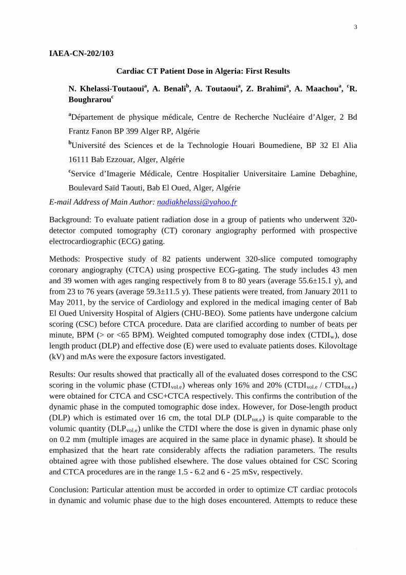
Background: To evaluate patient radiation dose in a group of patients who underwent 320-detector computed tomography (CT) coronary angiography performed with prospective electrocardiographic (ECG) gating.
Methods: Prospective study of 82 patients underwent 320-slice computed tomography coronary angiography (CTCA) using prospective ECG-gating. The study includes 43 men and 39 women with ages ranging respectively from 8 to 80 years (average 55.6±15.1 y), and from 23 to 76 years (average 59.3±11.5 y). These patients were treated, from January 2011 to May 2011, by the service of Cardiology and explored in the medical imaging center of Bab El Oued University Hospital of Algiers (CHU-BEO). Some patients have undergone calcium scoring (CSC) before CTCA procedure. Data are clarified according to number of beats per minute, BPM (> or <65 BPM). Weighted computed tomography dose index (CTDIw), dose length product (DLP) and effective dose (E) were used to evaluate patients doses. Kilovoltage (kV) and mAs were the exposure factors investigated.
Results: Our results showed that practically all of the evaluated doses correspond to the CSC scoring in the volumic phase (CTDIvol.e) whereas only 16% and 20% (CTDIvol.e / CTDItot.e) were obtained for CTCA and CSC+CTCA respectively. This confirms the contribution of the dynamic phase in the computed tomographic dose index. However, for Dose-length product (DLP) which is estimated over 16 cm, the total DLP (DLPtot.e) is quite comparable to the volumic quantity (DLPvol.e) unlike the CTDI where the dose is given in dynamic phase only on 0.2 mm (multiple images are acquired in the same place in dynamic phase). It should be emphasized that the heart rate considerably affects the radiation parameters. The results obtained agree with those published elsewhere. The dose values obtained for CSC Scoring and CTCA procedures are in the range 1.5 - 6.2 and 6 - 25 mSv, respectively.
Conclusion: Particular attention must be accorded in order to optimize CT cardiac protocols in dynamic and volumic phase due to the high doses encountered. Attempts to reduce these

4
4
doses should be considered before each use of coronary CT based on patient morphology and the clinical indications.
5
5
IAEA-CN-202/104
A Study of 4 Minutes versus 6 Minutes Protocol for Pharmacological Stress Testing Using Adenosine for Myocardial Perfusion Imaging and Comparison with
Echocardiography Findings
G. Malhotraa, K. Panchala, M. V. Reddyb, R. V. Asopaa, M. G. R. Rajana
a Radiation Medicine Centre, Bhabha Atomic Research Centre, TMC Annexe, Jerbai Wadia Road, Parel, Mumbai, India b Western Railways Jagjivan Ram Hospital, Mumbai, India
E-mail Address of Main Author: [email protected]
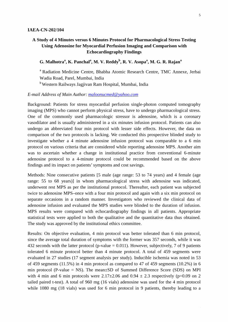
Background: Patients for stress myocardial perfusion single-photon computed tomography imaging (MPS) who cannot perform physical stress, have to undergo pharmacological stress. One of the commonly used pharmacologic stressor is adenosine, which is a coronary vasodilator and is usually administered in a six minutes infusion protocol. Patients can also undergo an abbreviated four min protocol with lesser side effects. However, the data on comparison of the two protocols is lacking. We conducted this prospective blinded study to investigate whether a 4 minute adenosine infusion protocol was comparable to a 6 min protocol on various criteria that are considered while reporting adenosine MPS. Another aim was to ascertain whether a change in institutional practice from conventional 6-minute adenosine protocol to a 4-minute protocol could be recommended based on the above findings and its impact on patients’ symptoms and cost savings.
Methods: Nine consecutive patients [5 male (age range: 53 to 74 years) and 4 female (age range: 55 to 68 years)] in whom pharmacological stress with adenosine was indicated, underwent rest MPS as per the institutional protocol. Thereafter, each patient was subjected twice to adenosine MPS–once with a four min protocol and again with a six min protocol on separate occasions in a random manner. Investigators who reviewed the clinical data of adenosine infusion and evaluated the MPS studies were blinded to the duration of infusion. MPS results were compared with echocardiography findings in all patients. Appropriate statistical tests were applied to both the qualitative and the quantitative data thus obtained. The study was approved by the institutional ethics committee.
Results: On objective evaluation, 4 min protocol was better tolerated than 6 min protocol, since the average total duration of symptoms with the former was 357 seconds, while it was 432 seconds with the latter protocol (p-value = 0.011). However, subjectively, 7 of 9 patients tolerated 6 minute protocol better than 4 minute protocol. A total of 459 segments were evaluated in 27 studies (17 segment analysis per study). Inducible ischemia was noted in 53 of 459 segments (11.5%) in 4 min protocol as compared to 47 of 459 segments (10.2%) in 6 min protocol (P-value = NS). The mean±SD of Summed Difference Score (SDS) on MPI with 4 min and 6 min protocols were 2.17±2.06 and 0.94 ± 2.3 respectively (p=0.09 on 2 tailed paired t-test). A total of 960 mg (16 vials) adenosine was used for the 4 min protocol while 1080 mg (18 vials) was used for 6 min protocol in 9 patients, thereby leading to a

6
6
potential cost saving of 11.1% in the former protocol. While echocardiography was normal in 2 of 9 patients, MPS was normal in 7 of 9 patients. Thus MPS proved to be an effective gatekeeper for coronary angiography by virtue of its high negative predictive value. 2 patients with abnormal findings on MPS also showed corresponding wall motion abnormalities on echocardiography but MPS detected additional areas as well.
Conclusion: MPI with 4 min adenosine infusion was found to be non-inferior to the 6 min protocol and potentially cost effective. Therefore, it can be recommended for routine use in our institution. Due to contrasting outcome of subjective and objective criteria, it cannot be implicated with certainty that abbreviated 4 min infusion protocol is better tolerated than the conventionally recommended 6-min protocol.
7
7
IAEA-CN-202/106
Correlation & Agreement of Measurements of Left Ventricular Ejection Fraction by Radionuclide Methods & Echocardiogram
F. Alam, R. Hossain, A. K. Sarker
Institute of Nuclear Medicine, Dhaka, Bangladesh
E-mail Address of Main Author: [email protected]
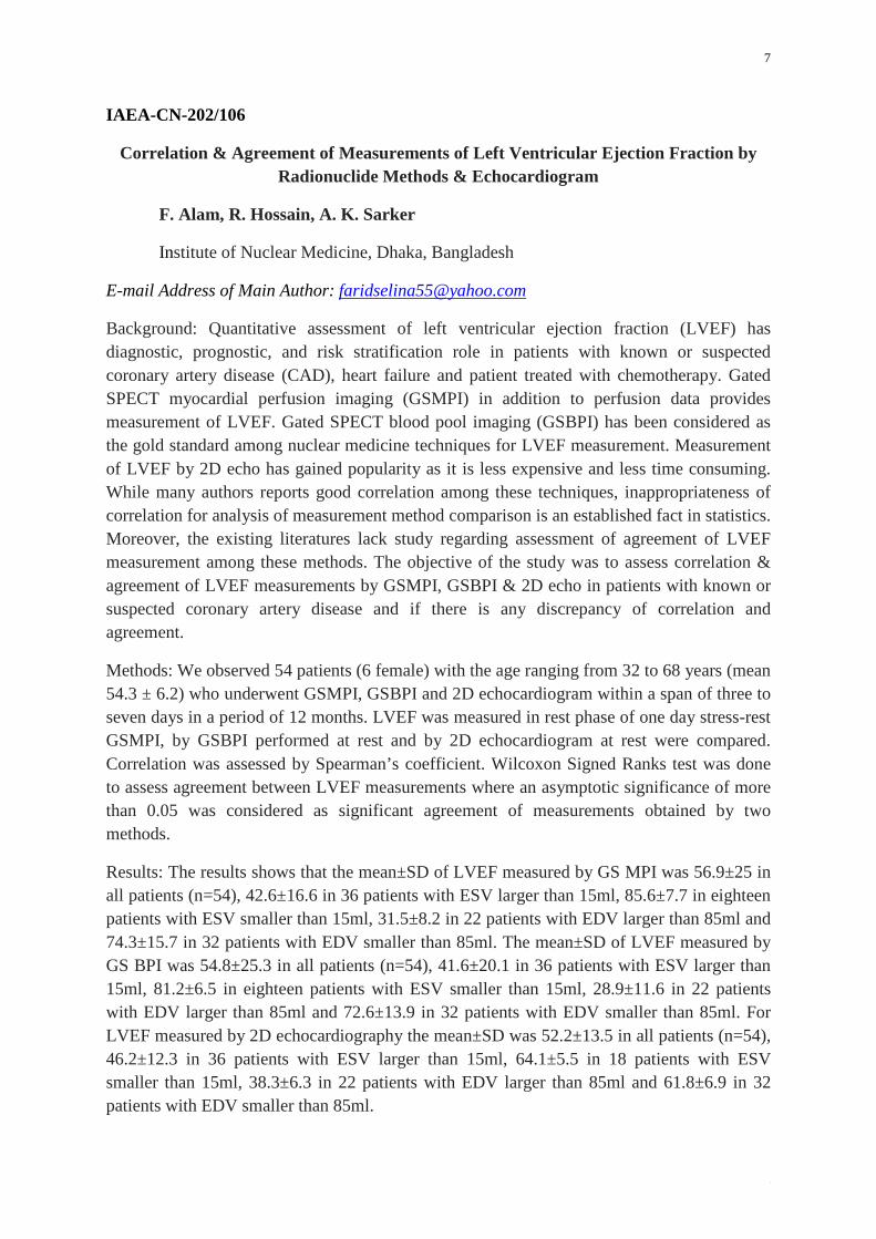
Background: Quantitative assessment of left ventricular ejection fraction (LVEF) has diagnostic, prognostic, and risk stratification role in patients with known or suspected coronary artery disease (CAD), heart failure and patient treated with chemotherapy. Gated SPECT myocardial perfusion imaging (GSMPI) in addition to perfusion data provides measurement of LVEF. Gated SPECT blood pool imaging (GSBPI) has been considered as the gold standard among nuclear medicine techniques for LVEF measurement. Measurement of LVEF by 2D echo has gained popularity as it is less expensive and less time consuming. While many authors reports good correlation among these techniques, inappropriateness of correlation for analysis of measurement method comparison is an established fact in statistics. Moreover, the existing literatures lack study regarding assessment of agreement of LVEF measurement among these methods. The objective of the study was to assess correlation & agreement of LVEF measurements by GSMPI, GSBPI & 2D echo in patients with known or suspected coronary artery disease and if there is any discrepancy of correlation and agreement.
Methods: We observed 54 patients (6 female) with the age ranging from 32 to 68 years (mean 54.3 ± 6.2) who underwent GSMPI, GSBPI and 2D echocardiogram within a span of three to seven days in a period of 12 months. LVEF was measured in rest phase of one day stress-rest GSMPI, by GSBPI performed at rest and by 2D echocardiogram at rest were compared. Correlation was assessed by Spearman’s coefficient. Wilcoxon Signed Ranks test was done to assess agreement between LVEF measurements where an asymptotic significance of more than 0.05 was considered as significant agreement of measurements obtained by two methods.
Results: The results shows that the mean±SD of LVEF measured by GS MPI was 56.9±25 in all patients (n=54), 42.6±16.6 in 36 patients with ESV larger than 15ml, 85.6±7.7 in eighteen patients with ESV smaller than 15ml, 31.5±8.2 in 22 patients with EDV larger than 85ml and 74.3±15.7 in 32 patients with EDV smaller than 85ml. The mean±SD of LVEF measured by GS BPI was 54.8±25.3 in all patients (n=54), 41.6±20.1 in 36 patients with ESV larger than 15ml, 81.2±6.5 in eighteen patients with ESV smaller than 15ml, 28.9±11.6 in 22 patients with EDV larger than 85ml and 72.6±13.9 in 32 patients with EDV smaller than 85ml. For LVEF measured by 2D echocardiography the mean±SD was 52.2±13.5 in all patients (n=54), 46.2±12.3 in 36 patients with ESV larger than 15ml, 64.1±5.5 in 18 patients with ESV smaller than 15ml, 38.3±6.3 in 22 patients with EDV larger than 85ml and 61.8±6.9 in 32 patients with EDV smaller than 85ml.
8
8
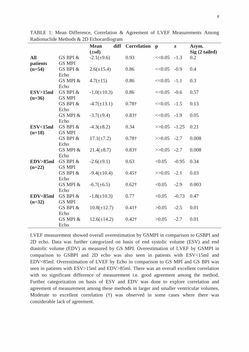
TABLE 1: Mean Difference, Correlation & Agreement of LVEF Measurements Among Radionuclide Methods & 2D Echocardiogram Mean diff
(±sd) Correlation p z Asym.
Sig (2 tailed) All patients (n=54)
GS BPI & GS MPI
-2.1(±9.6) 0.93 <<0.05 -1.3 0.2
GS BPI & Echo
2.6(±15.4) 0.86 <<0.05 -0.9 0.4
GS MPI & Echo
4.7(±15) 0.86 <<0.05 -1.1 0.3
ESV>15ml (n=36)
GS BPI & GS MPI
-1.0(±10.3) 0.86 <<0.05 -0.6 0.57
GS BPI & Echo
-4.7(±13.1) 0.78† <<0.05 -1.5 0.13
GS MPI & Echo
-3.7(±9.4) 0.83† <<0.05 -1.9 0.05
ESV<15ml (n=18)
GS BPI & GS MPI
-4.3(±8.2) 0.34 >>0.05 -1.25 0.21
GS BPI & Echo
17.1(±7.2) 0.78† >>0.05 -2.7 0.008
GS MPI & Echo
21.4(±8.7) 0.83† >>0.05 -2.7 0.008
EDV>85ml (n=22)
GS BPI & GS MPI
-2.6(±9.1) 0.63 <0.05 -0.95 0.34
GS BPI & Echo
-9.4(±10.4) 0.45† >>0.05 -2.1 0.03
GS MPI & Echo
-6.7(±6.5) 0.62† <0.05 -2.9 0.003
EDV<85ml (n=32)
GS BPI & GS MPI
-1.8(±10.3) 0.77 <0.05 -0.73 0.47
GS BPI & Echo
10.8(±12.7) 0.41† >0.05 -2.5 0.01
GS MPI & Echo
12.6(±14.2) 0.42† >0.05 -2.7 0.01
LVEF measurement showed overall overestimation by GSMPI in comparison to GSBPI and 2D echo. Data was further categorized on basis of end systolic volume (ESV) and end diastolic volume (EDV) as measured by GS MPI. Overestimation of LVEF by GSMPI in comparison to GSBPI and 2D echo was also seen in patients with ESV<15ml and EDV<85ml. Overestimation of LVEF by Echo in comparison to GS MPI and GS BPI was seen in patients with ESV>15ml and EDV>85ml. There was an overall excellent correlation with no significant difference of measurement i.e. good agreement among the method. Further categorization on basis of ESV and EDV was done to explore correlation and agreement of measurement among these methods in larger and smaller ventricular volumes. Moderate to excellent correlation (†) was observed in some cases where there was considerable lack of agreement.

9
9
Conclusion: Our findings demonstrate that significant overall agreement exists in measurement of LVEF by radionuclide methods and 2D echocardiogram. The agreement is variable among different LV volume categories.
10
10
IAEA-CN-202/108
Fluordeoxyglucose in the Assessment and Therapy Control of an Atrial Angiosarcoma
M. J. Jofré Manieu
Hospital Militar de Santiago, Av Larrain 9100, La Reina, Santiago-Chile
E-mail Address of Main Author: [email protected]
Background: In general, the role of Fluordeoxyglucose (FDG) in sarcomas is not well defined and there is not enough experience due to their great heterogeneity and relative low prevalence, and also because FDG may have variable uptake are not included in most oncological guidelines in adults. Primary cardiac neoplasms are relatively rare entities being angiosarcomas of the heart and pericardium the most common; they have a very aggressive behavior, rapidly invade adjacent soft tissues and present a high frequency of early metastatic spread. The presentation is diverse and nonspecific and the prognosis is bad (<1 year of mean survival) being more frequent at the right side. Histological type does not affect prognosis, but longer survivals are associated with left sided lesions and distal spread. The management includes surgical excision with curative intention in no disseminated cases plus chemo and radiotherapy. Several anatomic cardiac imaging are commonly employed but only few case reports using PET or PET-CT with FDG for staging with avid lesions in situ, and another for initial therapy response.
Methods: Clinical Case: We present a 49 year old female with a right atrial angiosarcoma recently subjected to surgery (macroscopically R0) and pacemaker implantation. The patient was evaluated with PET-FDG that only showed a mild cervical uptake in a thyroid nodule confirmed with ultrasound posteriorly. Seven months later, a cardiac recurrence depicted by a control FDG scan after finishing the last cycle of chemotherapy; she also received local radiotherapy. Five months later, she presented a new focal mediastinal increased uptake as well as another prevertebral focal increased uptake at T7 level. More cycles of chemo were added until FDG was negative. A few months later PET scan confirmed a mild FDG avid prevertebral lesion at T9-T10 level and a new mediastinal recurrence, continuing with chemotherapy until PET scan was negative again. Three months later, a new cardiac FDG uptake was demonstrated receiving then Talidomide.
In the next year, 35 months after initial diagnosis, FDG demonstrated a cardiac lesion, hepatomegaly and multiple liver lesions with intense FDG uptake. The patient was always asymptomatic after initial surgery and only presented abdominal discomfort in the late period. She died 39 months after the diagnosis, due to hemorrhagic stroke and brain metastasis from her primary atrial sarcoma.
Results: Echocardiograms showed pericardial effusion when local recurrence was observed, CT scans demonstrated liver and mediastinal lesions in the advanced stages, bone scans were negative in 3 opportunities and all were concordant with FDG scans.
11
11
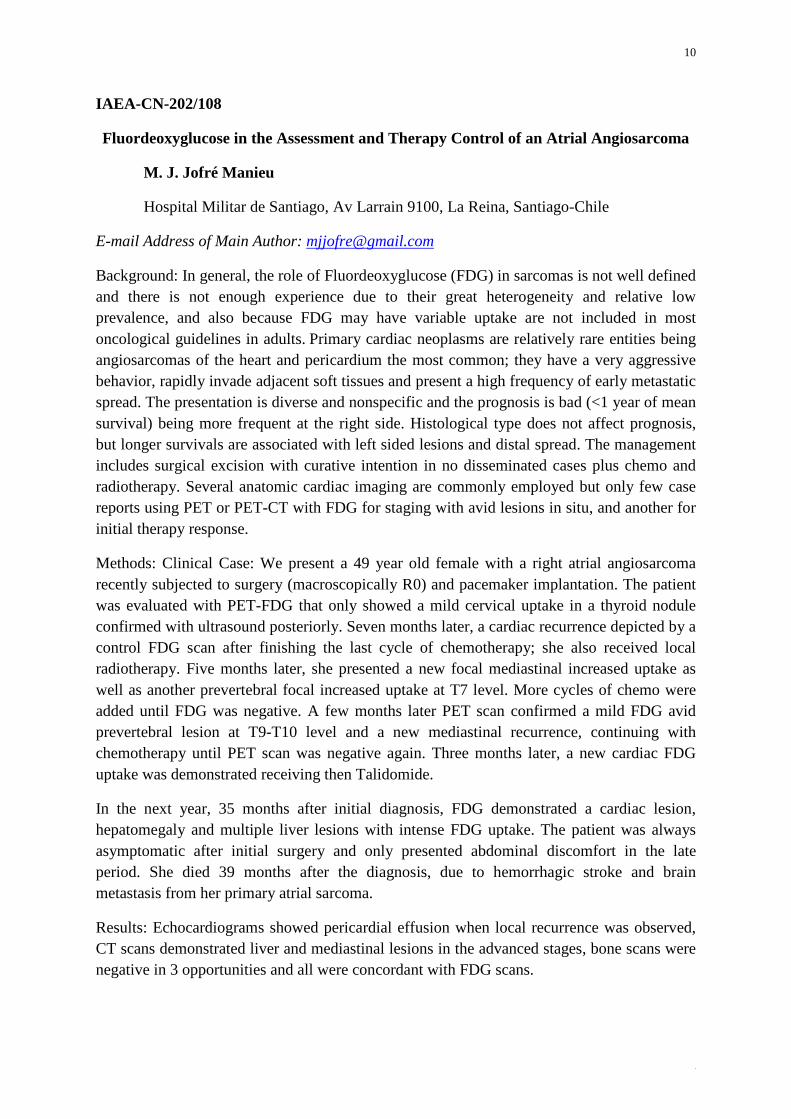
Conclusion: Cardiac angiosarcomas, despite their low prevalence, could be evaluated with FDG a) for staging, appearing to have a good prognostic value; b) to select the best therapy and c) to perform close follow-up of the different therapeutic modalities as is shown in our patient, who presented a longer survival than reported in the literature (most works around 6 months). In her case, the therapy control was performed mainly metabolically with FDG that was able to demonstrate local recurrence and distal spread.
(A) (B)
FIG. 1: (A) FDG-PET projection of the first mediastinal recurrence 1 year after right atrial angiosarcoma diagnosis and (B) 6 months later, after chemotherapy.
12
12
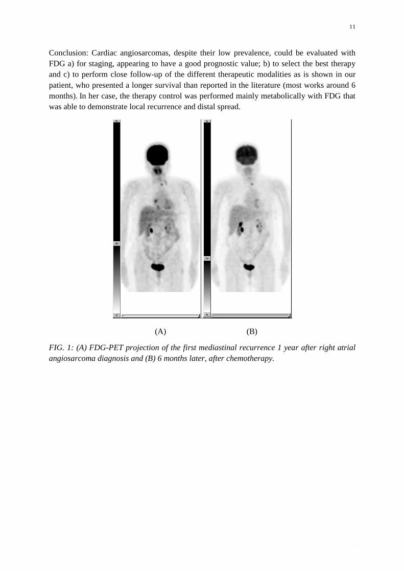
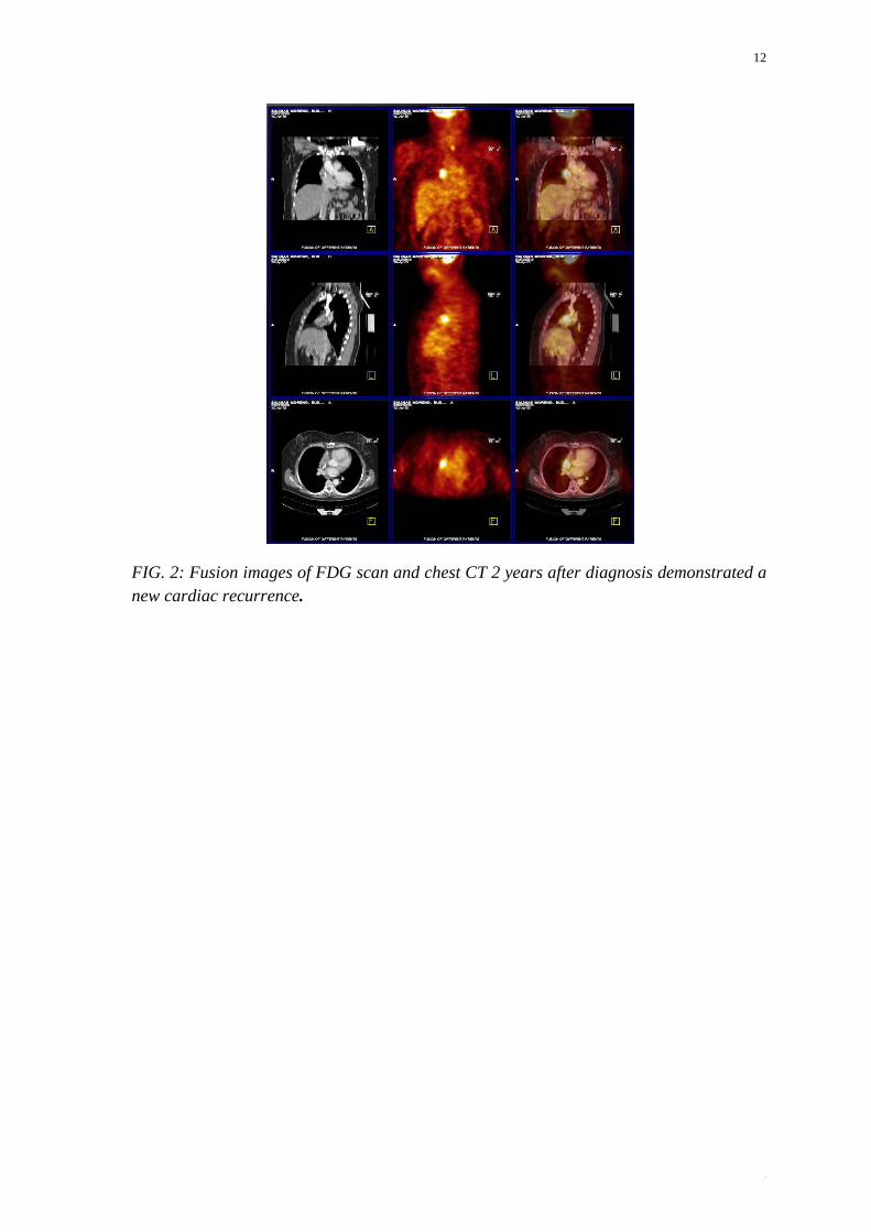
FIG. 2: Fusion images of FDG scan and chest CT 2 years after diagnosis demonstrated a new cardiac recurrence.
13
13
IAEA-CN-202/109
Application of SPECT-CT in Patients with Suspicion of a Vascular Graft Infection
I. Kostadinova, S. Ivanova, M. Garcheva
Medical University, Str.Zdrave 2, Sofia, Bulgaria
E-mail Address of Main Author: [email protected]
Background: Vascular graft infection is a rare, but serious complication with a high rate of morbidity and mortality. Labeled leucocytes imaging could ensure its prompt and specific visualization for an efficient treatment. The aim of the study was to apply new hybrid imaging SPECT-CT in patients with suspicion of a vascular graft infection.
Methods: We have examined 21 patients (Pts), aged 60-80 years, for a period of 4 years of whom 14 were with aortobifemoral, 2 with femorofemaral, 2 with axillofemoral and 3 with ileofemoral bypass. Fourteen of the patients were still under the antibiotic therapy, not enough clinically influenced by it. We have used “in vitro” labeled leukocytes with 99mTc-HMPAO (370MBq) and the following protocol of investigation: static/whole body SPECT-CT/low dose CT/, 2h p.i.
Results: The results of all patients were verified microbiologically, by surgery or by follow-up. An index of accumulation (IA) was used for an objective quantification of the results and for evaluation of the activity of the infection process. IA was calculated as the activity of the suspected area divided by the activity of the contralateral area, after both were normalized to the background activity. The infection was ruled out when IA was below 1.1 (in 2 Pts), low grade of activity was considered at IA of 1.1-1.3 (in 11 Pts) and active infection at IA above 1.3 (in 8 Pts). Using CT, exact localization of the infection was possible, differentiating infection along the vascular graft and/or infection in the soft tissues adjacent to the graft (in 4/21 Pts) as well as normal distribution of the labeled leucocytes in the bone marrow when the vascular graft was superimposed (in 2/21 Pts). In two of the patients additional fistula was visualized and in one a perigraft abscess. In summary, there were 2 false negative results (one of the Pts was still and another had recent antibiotic therapy), 1 false positive (due to non-infectious reaction to the graft), 16 true positive and 2 true negative results, yielding a sensitivity of 88.9%, specificity of 66.7% and accuracy of 85.7%. For 30% of the patients SPECT-CT contributed important additional information, which changed the therapy.
Conclusion: In conclusion, we suggest that combining hybrid imaging with quantitative criteria for evaluation of the activity of the vascular graft infection ensures exact localization and measurement of the activity of the infectious process. In addition, collected information for the state of the surrounding tissues can influence the therapeutic strategy.
14
14
IAEA-CN-202/111
FDG Uptake in Large Arteries, Comparison between Genders and Correlation with Aging
G. Bural, D. Torigian, M. Houseni, A. Alavi
Izmir Katip Çelebi University, Atatürk Research and Training Hospital, Department of Nuclear Medicine, Basin Sitesi, Izmir, Turkey
E-mail Address of Main Author: [email protected]
Background: FDG-PET can detect, localize, and quantify degree of inflammatory change in the arterial wall due to early atherosclerosis. Our aim was thus to quantify and compare inflammatory atherosclerotic changes in the large arteries via FDG-PET as a function of age and gender.
Methods: We evaluated the presence of arterial wall FDG uptake in 138 subjects (58 men, 80 women; 5-74 years) who had a whole-body FDG-PET for assessment of non-cardiovascular disorders. Subjects were initially grouped according to gender, then by age (below or above 50) with at least 25 subjects per group. We measured the mean SUV of each segment, and determined differences of SUVs in large arteries based on gender. We recorded FDG uptake in ascending, arch, descending, abdominal aortic segments, also in iliac and femoral arteries, and calculated and compared per cent (%) of segments showing FDG uptake in four groups.
Results: Mean SUVs in visible arterial segments between genders did not reveal any statistically significant difference for the entire group. Percentage of visible arterial segments with FDG uptake was higher in older subjects (79% for men, 79% for women) compared to younger ones (63% for men and 54% for women) for both genders (p<0.05). In younger subjects it was higher for men (63%) compared to women (54%) (p= 0.05).
Conclusion: The severity of the inflammatory atherosclerotic process as measured by SUVs in large arteries between genders did not change in younger and older subjects. Prevalence of segments with FDG uptake was increasing with age for both genders. In younger subjects prevalence of segments with FDG uptake in men was higher than in women.
15
15
IAEA-CN-202/112
Implications of Persistent ST Segment Elevation in Q-Wave Anteroseptal Myocardial Infarction: Correlation with Myocardial Perfusion Gated SPECT
P. Subramanyam, S. S. Palaniswamy
Amrita Institute of Medical Sciences, Ponekkara, Cochin, Kerala, India
E-mail Address of Main Author: [email protected]
Background: Electrocardiographic (ECG) ST segment elevation that persists 2 or more weeks following Q wave myocardial infarction has been associated with worse prognosis due to 'ventricular aneurysm' and absence of myocardial viability. Regional systolic dysfunction may reflect either viable myocardium or scar. We hypothesized that patients with persistent ST segment elevation after Q wave infarction might demonstrate salvageable myocardium in the infarct region. We attempted to study whether persistent ST segment elevation in Q wave anteroseptal myocardial infarction (QASMI) represents presence or absence of ischemia/ viability in LAD territory by myocardial perfusion gated SPECT (MPSPECT).
Methods: 135 QASMI patients (M:F 108:27, age 41-75 yrs, mean 56 + 7 yrs) referred for risk stratification were retrospectively analysed. QASMI patients > 1 month and resting ECG showing sinus rhythm, QRS < 120 ms, Q waves in at least 2 contiguous precordial leads with ST segment elevation of > 1.5 mm were included. All underwent same day stress (TMT/ pharmacological stress) rest gated MPSPECT on a dual head variable angle gamma camera. Images were visually interpreted and analysed using 16 segment myocardial model for LAD ischemia. Presence of reversible perfusion defects indicated ischemia while > 40 % MIBI uptake and myocardial systolic wall thickening in gated study indicated viability.
Results: Patients were categorized into Group I (showing LAD viability + ischemia 77/135 patients 57%) and Group II (showing no LAD viability 58/135 patients 43%).Group I was further subdivided to I A & I B based on presence and absence of associated ischemia. Group I patients showed mean viable 6 + 0.5 segments and ischemic 3 + 0.2 segments. Group II patients had only 3 + 0.2 viable segments. Group I A patients presented with mean 6 + 0.4 viable and ischemic 3 + 0.5 segments. Patients under Group I B subgroup had mean 5 + 0.7 viable segments. There was statistically significant difference between rest LVEF of Group I & Group II (45 + 5 vs 30 + 4).
Conclusion: Persistent ST elevation in QASMI patients implies that there can be underlying LAD ischemia and viability thereby prompting further investigation in this subgroup.

16
16
IAEA-CN-202/113
Is Exercise Induced ST Depression an Accurate Indicator of Viability in Infarct-Related Artery? - A Myocardial Perfusion SPECT Study
P. Subramanyam, S. S. Palaniswamy
Amrita Institute of Medical Sciences, Ponekkara, Cochin, Kerala, India
E-mail Address of Main Author: [email protected]
Background: While exercise induced ST elevation is linked to presence of myocardial ischemia, residual viability, regional wall motion abnormalities & sympathetic overactivity, there are limited literature on the importance of Exercise induced ST Depression (ExST Dep) in opposite leads associated with infarct lead ST elevation & its clinical significance. We attempted to determine whether ExST Dep (defined as ST depression in ECG lead opposite to lead showing ST segment elevation) is associated with residual viability in infarct related artery in patients with Myocardial Infarction (MI).
Methods: 46 patients referred for Post MI risk stratification with MPI between Jan 06 - 07 were retrospectively analysed. Inclusion criteria were Q wave MI, more than 1 mm ST depression at 80 m sec after J point in infarct related leads. Patients with LBBB or RBBB, LVH, Diabetes, on drugs affecting ST segment were excluded. Patients underwent same day stress rest gated Myocardial Perfusion SPECT (MP SPECT). As part of stress MP SPECT, patients underwent symptom limited Treadmill Test (TMT) & TMT ECG findings were analyzed for the presence of ST elevation, ExST Dep in non-infarct related leads. Stress, rest images were interpreted using a 17-segment model & Summed stress, rest & difference scores were calculated. Coronary angiogram correlation was available for all pts.
Results: Reciprocal ExST Dep associated with ST elevation in infarct ECG leads were present in 57% (Group A 26/43 patients) & not in 43 % (Group B 20/43 pts). There is no significant difference between Group A & B pts in terms of ST segment elevation & TMT exercise data. Out of 43 patients, 24 had Anterior, 14 Inferior & 5 Lateral MI. Mean number of ischaemic & or viable segments detected by MP SPECT in group A & B patients in LAD territory are 4.5 + 0.8 & 2.5 + 0.5 (p < 0.01) respectively. MP SPECT showed significant residual viability (at least 50% of myocardial segments) in infarct related artery in 88.5% of Gp A pts (23/26 pts) when compared to only 4/20 patients 20% of Gp B patients. The overall sensitivity, specificity & accuracy of reciprocal ExST Dep associated with exercise induced ST elevation towards the residual viability detection were 88%, 100%, 90% respectively.
Conclusion: Presence of reciprocal ExST Dep in non-infarcted leads associated with exercise induced ST elevation in infarct related ECG leads during a TMT indicate residual viability within the infarct related artery.
17
17
IAEA-CN-202/114
Influence of Attenuation Correction on the Interpretation of Myocardial Perfusion Images
M. Garcheva, I. Kostadinova
Medical University-Sofia, 2,”Zdrave”str. Sofia 1431, Bulgaria
E-mail Address of Main Author: [email protected]
Background: The main application of SPECT-CT in Nuclear Cardiology is for attenuation correction trough fast creation of attenuation maps. The aim of the study was to determine the influence of attenuation correction on the interpretation of myocardial perfusion images (MPI).
Methods: Thirty patients (aged 35-78) were examined by stress/rest MPI with Tc-99m tetrofosmin on SPECT-CT Siemens Symbia 2T. The attenuation corrected images (AC) were compared to the uncorrected images (NAC) by visual analysis. A special data base was used for quantification. All patients had known coronary anatomy, examined within one month by invasive, or computed coronary angiography. Fifteen patients had previous revascularization: operative or invasive. All low-dose CT scans were verified for additional findings.
Results: The coronary anatomy demonstrated 10 significant LAD (left anterior descending artery) stenoses, 12 RCA (right coronary artery) and 12 rCx (circumflex artery) stenoses. Perfusion abnormalities on AC, NAC or both were found in 23 patients. Differences in interpretation between AC and NAC were found in 16 patients (53%). The overall detection rate of the involved vascular territories increased from 27/34 (76%) for NAC to 32/34 (94%) for AC images with significant hypoperfusion in more than one vessel territory found in 7 patients according to AC and in 4 patients- according to NAC. Differences occurred also in the detection and extent evaluation of viable myocardium: in 7 territories on AC versus 2 territories on NAC concerning the decision for revascularization. The quantitative analysis identified insignificant differences in summed stress score (SSS) between AC and NAC (3.26±4.36 versus 3.3±4.81, p>0.5. In 1 patient (3.3%) an unexepected lung mass was found on the low-dose CT scan. After the application of attenuation correction, the therapeutic decision was changed in 7/16 patients (43%) with different AC/NAC interpretation and 30% from all patients with abnormalities.
Conclusion: The attenuation correction of MPI contributes to improved diagnostic accuracy, which reflects on the patients’ risk stratification and management.
18
18
IAEA-CN-202/115
Dynamics of Myocardial Perfusion with MIBI in Patients with Coronary Heart Disease and Post-Infarction Cardiosclerosis after Stem Cell Therapy
A. Dustova, U. Kurbanovb, M. Mirshahic
a Institute of Gastroenterology of Tajikistan Academy of Medical Science, Dushanbe, Tajikistan b Dept. of Surgical diseases and Stem cell laboratory, Avicenna Tajik State Medical University c UMRS 872, INSERM, Paris VI Faculty of Medicine, Paris, France
E-mail Address of Main Author: [email protected]
Background: To study efficacy and safety of autologous bone marrow stem cell therapy for the tissue genesis and neoangiogenesis in ischemic parts of heart in patients with coronary heartdisease (CHD) and post-infarction cardiosclerosis after macro focal myocardial infarction.
Methods: 30 patients with a diagnosis of ischemic heart diseases, myocardial infarction (deferred Q-myocardial infarction without significant complications barred from 3 to 6 months) were selected. In some of them, coronary angiography revealed severe coronary artery pathology: left coronary artery trunk (4 cases) and 3 vascular lesions (7 cases). Among these patients, 15 of them were treated with the standard protocol of treatment (control group), the 15 others were transplanted with their bone marrow stem cells. Stem cells CD133 were isolated from mononuclear cells by density gradient centrifugation using Ficoll, followed by immuno-magnetic separation. Isolated cells of patients with coronary artery disease were injected in intra-arterial into the coronary arteries under angiography in the average dose of 5 ml of suspension containing 0,8-1,5 million cells. We carried out myocardial scintigraphy using Tc99m with the ligand methoxyisobutylisonitrile (MIBI) in order to evaluate the dynamics of myocardial perfusion in all patients with coronary heart disease and post-infarction cardiosclerosis before and after cell therapy.
Results: Results of clinical examination of patients revealed an improvement after 3 months and 6 months in both groups of patients. Radionuclide investigations of heart according to data from myocardial scintigraphy using Tc99m with MIBI among patients with coronary heart disease (CHD) and post-infarction cardiosclerosis before and after cell therapy were performed. Data shown a cicatricial change in the general area in average was equal to 25%. Viable myocardium was detected in the region of lateral and posterior walls (apical segments), posterior interventricular septum (basal and medial segments), and apex, neighboring the leading wall of left ventricle of heart. Radionuclide investigations of heart in patients after 1, 3, 6 and 14 months of cell therapy showed considerable decrease in stable perfusion defect, as monitored by indicators as well as after 6 months compared to initial indicators values. It was also shown that one-time transplantation of autologous mononuclear cells of bone marrow has a positive effect on dynamics of stable and transient perfusion

19
19
defect according to scintigraphic diagnostics with Tс-99m in patients with CHD and post-infarction cardiosclerosis during a 3 and 6 month monitoring periods following cell therapy.
Conclusion: Our method of stem cell transplantation is safe and does not increase mortality as a consequence of heart disease. Treatment of autologous stem cells significantly improved key indicators of heart hemodynamic.
20
20
IAEA-CN-202/116
Early Diastolic Dysfunction Detection by 16 bin Gated MPI SPECT in End Stage Liver Disease Patients with Normal Myocardial Perfusion Undergoing Pre-Transplant
Cardiac Evaluation
S. J. Gandhi, S. L. G. Praveen, S. Padma, S. S. Palaniswamy
Department of Nuclear Medicine & PET CT, Amrita Institute of Medical Sciences, Ponekkara, Kochi, Kerala, India
E-mail Address of Main Author: [email protected]
Background: End Stage Liver Disease (ESLD) patientsare assumed to have normal cardiac function based on normal/supranormal Echo LVEF. But they can have LV hypertrophy/dilatation, systolic & diastolic dysfunction pointing towards cirrhotic cardiomyopathy. We evaluated early diastolic dysfunction (EDD) in ESLD patientswith normal perfusion on MPI using 16 bin gated SPECT quantitative parameters.
Methods: 42 ESLD patients (Jan 09-Sept 12) undergoing gated (16 bin) stress MPI & rest Echo as part of pretransplant workup, were retrospectively studied. All underwent stress (pharmacological / physical) MPI, single day stress-rest (9:27 mCi of 99mTc –MIBI) protocol. Stress, rest MPI SPECT studies were evaluated for inducible ischemia. Different quantitative parameters like EDV, ESV, LVEF, PFR, TPFR, MFR3 etc. were obtained using 4DM SPECT software and compared with normal database. Statistical analysis was done with unpaired t-test.
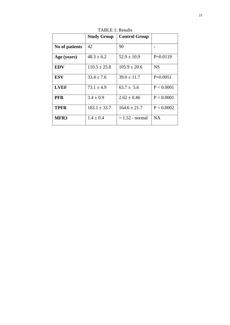
Results: Higher LVEF and lower ESV values in ESLD group suggested normal/supranormal systolic function. EDV values showed no statistically significant difference. Significantly higher TPFR and lower MFR3 (<1.52) values observed in ESLD group. Higher PFR values found in ESLD group, a contradictory finding is explained by younger patients in study group. Diastolic dysfunction was detected in 24 patients by MPI and in 18 by Echo, suggesting higher sensitivity of MPI.
Conclusion: Quantitative parameters like TPFR and MFR3 by 16 bin gated MPI are helpful in detecting EDD, earlier than Echo in ESLD patients with normal perfusion & preserved systolic function. PFR is not an accurate parameter as it is age dependent.
21
21
TABLE 1: Results Study Group Control Group
No of patients 42 90 -
Age (years) 48.3 ± 6.2 52.9 ± 10.9 P=0.0119
EDV 110.5 ± 25.8 105.9 ± 20.6 NS
ESV 33.4 ± 7.6 39.0 ± 11.7 P=0.0051
LVEF 73.1 ± 4.9 63.7 ± 5.6 P < 0.0001
PFR 3.4 ± 0.9 2.62 ± 0.46 P < 0.0001
TPFR 183.1 ± 33.7 164.6 ± 21.7 P = 0.0002
MFR3 1.4 ± 0.4 > 1.52 - normal NA
22
22
IAEA-CN-202/117
Can MPI SPECT Predict Abnormalities on Carotid / Lower Limb Doppler USG for Extra Coronary Atherosclerotic Disease
S. J. Gandhi, S. L. G. Praveen, S. Padma, S. S. Palaniswamy
Department of Nuclear Medicine & PET CT, Amrita Institute of Medical Sciences, Ponekkara, Kochi, Kerala, India
E-mail Address of Main Author: [email protected]
Background: Extra-coronary atherosclerotic vascular disease (peripheral arterial disease (PAD) and carotid artery disease) share common risk factors with coronary artery disease. There is high incidence of asymptomatic extra-coronary atherosclerotic disease in patient with myocardial ischemia on SPECT MPI. The aim was to correlate ischemic severity on quantitative SPECT MPI & presence of extra-coronary atherosclerotic disease on carotid /lower limb arterial Doppler USG.
Methods: Asymptomatic patients who underwent stress (physical/pharmacological) myocardial perfusion imaging (MPI SPECT) and also lower limb or neck arterial Doppler USG or both for diagnosis of PAD during the year 2012 were included in this retrospective analysis. Patients with prior history of myocardial infarction were excluded from study. 28 patients were found to be appropriate for evaluation. Doppler USG findings were compared with qualitative & quantitative SPECT-MPI findings. SPECT-MPI images were acquired using Siemens gamma camera (E-CAM). Commercially available Corridor 4DM-SPECT software was used for evaluating quantitative parameters. Coronary angiogram (CAG) correlation was also available for MPI SPECT positive patients.
Results: 28 patients (Male: Female - 16:12, Age range 48-74 yrs, median: 64 yrs) were considered for evaluation. All patients were asymptomatic and had no prior history of myocardial infarction. 15/28 (54%) patients had normal MPI; all of these patients had normal Doppler USG findings. Negative predictive value of MPI SPECT for Doppler USG abnormalities was 100%. 13/28 (46%) patients had abnormal MPI SPECT findings (i.e. Mean Summed difference score: 4 & SVD - 6, DVD - 1, TVD - 5 on CAG). 8 of this 13 patients (62%) had significant abnormalities on Doppler USG also. 5/13 patients (38%) had normal Doppler USG findings. Positive predictive value of MPI for Doppler USG abnormalities was 62%. Interestingly 2/8 patients with neck Doppler USG abnormality developed cerebral infarct also subsequently.
Conclusion: Myocardial ischemia on SPECT MPI is an independent predictor of Extra-coronary atherosclerotic disease with very high negative predictive value & reasonably good positive predictive value.

23
23
TABLE 1: Results
Doppler positive
Doppler negative
Total
MPI positive 8 5 13
MPI negative
0 15 15
Total 8 20 28
24
24
IAEA-CN-202/118
Role of Myocardial Perfusion SPECT in the Prediction of Post-Surgery Recovery of Ischemic Mitral Regurgitation in Patients with Severe LV Dysfunction
S. S. Palaniswamy, S. Padma
Amrita Insitute of Medical Sciences, Cochin, India
E-mail Address of Main Author: [email protected]
Background: Functional Mitral Regurgitation (MR) occurs with a structurally normal valve as a complication of systolic left ventricular dysfunction precipitated by ischaemic heart disease. Even moderate ischemic MR has a negative prognosis with impaired left ventricular function when treated by CABG. PET studies have proved that presence of myocardial scar is a poor prognostic indicator of surgical outcome of ischaemic MR. Our aim was to analyze the prognostic impact of ischaemia & myocardial viability assessed by stress Myocardial Perfusion Imaging (MPI) in ischemic MR patients with LV dysfunction undergoing CABG & Mitral Valve Repair (MVR).
Methods: 18 patients (M: F 13:5 pts, age range 55 - 72 yrs mean 52 + 11 yrs) diagnosed with ischaemic MR + MI who underwent CABG were retrospectively analyzed. Preoperatively, all patients had their resting LV function & MR severity assessed by trans-thoracic echocardiography. Severity of MR was graded as mild (1, 2) & severe (3 or 4+).
They also underwent same day 99mTc MIBI Exercise (TMT / Adenosine 11: 7 patients) MPI preoperatively. A 20-segment myocardial model was used for quantitation. No. of infarcted, ischaemic & viable myocardial segments were quantified by Stress Summed, Rest & Difference scores (SSS, SRS & SDS). 11/18 patients underwent CABG with MVR & remaining 7 had only CABG. All patients had at least 6 months (mean 10 + 4 months) follow-up. Postoperative echocardiography was routinely performed by 6th month for LV EF & MR assessment.
Results: 2/18 pts expired during follow up and both showed larger infarct (scar 11 segments) & persistent severe MR. Out of 11 pts who underwent CABG & MVR, 6 pts showed significant improvement in both LVEF & MR score post surgery. There is a significant difference of scar (9.9 + 4.5 Vs 6 + 3.5 P<0.01), ischemic (2.5 + 2.3 Vs 5 + 4.5 P<0.01) & viable (9.9 + 4.5 Vs 6 + 3.5 P<0.01) segments between patients showing improvement & no improvement. Interestingly 3/7 patients who underwent only CABG (No MVR) showed an improvement in postoperative MR score had viable & reversible ischaemic but no scar myocardium.
Conclusion: Presence of viable & ischemic myocardium is a better prognostic marker than scarred myocardium in patients with ischemic MR undergoing CABG & MVR. Stress MPI is a useful investigation in assessing & prognosticating ischemic MR prior to surgery.
25
25
IAEA-CN-202/119
The Correlation between LVEF Gated-SPECT with Hemodialysis, Calcium-Phosphorus Product and Parathyroid Hormone in Patients with Chronic Kidney
Diseases D. Nariman, E. Purnomo
Department of Nuclear Medicine, Gatot Subroto Central Army Hospital, Jakarta, Indonesia
E-mail Address of Main Author: [email protected]
Background: In chronic kidney diseases patients who perform routine hemodialysis may occur metabolic disorders of calcium, phosphorus and vitamin D, cardiovascular disorders, hematology, and other. Cardiovascular events are a major cause of mortality in patients with chronic kidney diseases. Metabolic disorders of calcium, phosphorus and vitamin D can lead to impaired vascular tissue and secondary hyperparathyroidism. It can also cause cardiovascular problems. Cardiovascular disorder such as left ventricular ejection fraction was evaluated by nuclear medicine techniques Gated-SPECT. Assessment of left ventricular ejection fraction was associated with the number of hemodialysis has been done, calcium-phosphorus product and parathyroid hormone levels. The aim of this study was to correlate between left ventricular ejection fractions Gated-SPECT with the number of hemodialysis, the levels of calcium-phosphorus product, and parathyroid hormone levels.
Methods: The data was taken as a retrospective study. Subjects consisted of thirteen patients with chronic kidney diseases (8 men and 5 women) who underwent routine hemodialysis for more than a year. Routine hemodialysis was done between 172 to 1236 times. The age range was between 43 to 67 years old. The causes of chronic kidney diseases were diabetic nephropathy three subjects and ten subject’s glomerulonephritis. Parathyroid hormone levels between 83.07 to 1096 pg / mL, (more than 65 pg / mL). Calcium ion levels between 3.69 to 4.93 mg / dL. Total calcium levels between 7.68 to 10.25 mg / dL. Serum phosphorus levels between 6 to 11.8 mg / dL. The Left ventricle ejection fraction with Gated-SPECT technique was between 35 to 58%. Clinically, the result of the calcium-phosphorus product is important as an indicator progression of calcification soft tissue and vascular.
Results: Data processing is done by using a computer. Correlation is a method to find the relationship between two numerical variables. Pearson correlation coefficient (r) is divided into five levels, a good correlation when r> 0.80, and the moderate correlation when r between 0.60 to 0.79. There is a moderate correlation between LVEF Gated-SPECT with the number of hemodialysis has been done (r = -0.735, p = 0.004). But there is no correlation between LVEF with parathyroid hormone levels (r = -0.032, p = 0.917) and LVEF with calcium-phosphorus product (r = -0.019, p = 0.95).
Conclusion: From this study there was a moderate correlation between LVEF Gated-SPECT with the number of hemodialysis has been done in patient with chronic kidney diseases. It does not show correlation with parathyroid hormone levels and also with the results of the calcium-phosphorus product. Further investigation with more of subjects is needed to improve its accuracy.
Keywords: Gated-SPECT, Tc-99m tetrofosmin, routine hemodialysis, parathyroid hormone, Calcium-PhosphorousProducts.
26
26
IAEA-CN-202/120
Correlation between Mean Platelet Volume and Myocardial Perfusion SPECT Parameters
A. S. Doğan, E. İzgi, M. E. Erkan, M. Aşık, A. Yılmaz, M. Z. Yılmaztekin, A. N. Korkmaz, M. Yıldırım
Düzce University, School of Medicine, Department of Nuclear Medicine, Beçiyörükler köyü, Konuralp, Düzce, Turkey
E-mail Address of Main Author: [email protected]
Background: Mean platelet volume (MPV) is previously reported to be useful in predicting coronary artery disease and related morbidity and mortality. The purpose of this study is to correlate myocardial perfusion scintigraphic findings with mean platelet volume (MPV), which is a simple and reliable parameter of platelet function in patients with suspected coronary heart disease.
Methods: In this retrospective study, 344 patients who underwent myocardial perfusion scintigraphy in our clinic between 2009-2012 were included. The study group consisted of 139 men and 205 women (mean age: 56.67 ± 11.16). Based on the scintigraphic findings, patients were divided into three groups as normal, suspicious for ischemia and significant ischemia. The results of MPV and the parameters obtained from GATED myocardial perfusion SPECT scintigraphy, such as stress ejection fraction, end-systolic volumes and end-diastolic volumes were correlated.
Results: Ninety-six of the patients (27,9%) were normal, 98 (28,5%) were suspicious for ischemia and 150 (43,6%) showed significant ischemia. MPV values were found to be 8,43±1,19 in normal group, 8,10±1,34 in suspicious ischemic group, 8,06±1,23 in significant ischemic group. There was no significant difference between scintigraphic findings and MPV results. (p=0,060)
Conclusion: In our study, we found no correlation between MPV, which is considered to be a risk factor for cardiovascular disease and parameters of GATED myocardial perfusion SPECT scintigraphy.
27
27
IAEA-CN-202/121
Evaluation of Gated SPECT MIBI Functional and Perfusion Abnormalities in Patients with Previous Inferior Myocardial Infarction
D. Sobic Saranovic, Z. Petrasinovic, V. Artiko, V. Obradovic
Center for Nuclear Medicine, Clinical Center of Serbia, Visegradska 26, 11000 Belgrade, Serbia
E-mail Address of Main Author: [email protected]
Background: Gated SPECT MIBI (GSM) gives usefull information about the extent and severity of perfusion defects (PD) and global left ventricular (LV) function in patients with coronary artery disease.The aim of this study was to evaluate PD and LV function in patients with previous inferior myocardial infarction (IMI) with GSM.
Methods: This study included 43 patients (age 57±9, 33 males) withIMI, Q-wave on ECG and single-vessel coronary disease of right coronary artery on coronary angiography. All patients underwent a 2-day stress-rest GSM protocol with 99m-tehnetium metoxy-isobutile-isonitrile (MIBI). The assesment of PD was performed by polar maps. End-diastolic LV volume (EDV), end-systolic volume (ESV) and ejection fraction (EF) were determined by 4D-MSPECT software.
Results: In all enrolled patients with IMI, LV function parameters (EF, EDV, ESV) were not statistically different between rest and post-stress (EFr: 52±8% vs. EFps: 52±7%, EDVr137±29׃ ml vs. EDVps: 139±29 ml, ESVr65±20 ׃ ml vs. ESVps66±22׃ ml, p>0.05 respectively), while stress-PD value was higher but did not reach statistical significance (16±8% vs. 14±10%, p>0.05). In the subgroup with additonal ischemia to the fixed defect (n=21), post-stress PD value was statistically higher in comparison with PD value in rest study (PDr:14±7% vs. PDps: 20±7%, p<0.01). EFps was significantly decreased (EFr: 54±9 % vs. EFps: 50±7%, p<0.05) while ESVps was increased (ESVr: 61±22 ml vs. ESVps:71±22 ml, p<0.05) in comaprison to the rest value. There was not statistical differance between EDVr and EDVps (142±29 ml vs. 144±29 ml, p>0.05).
In the subgroup with fixed perfusion defect (n=22), there was not statistical significance between post-stress and rest parameters (EFr: 53±8 % vs. EFps: 54±9 %, EDVr: 133±29 ml vs. EDVps: 134±28 ml, ESVr: 62±22 ml vs. ESVps: 62±21 ml, p>0.05) as well as between PDr and PDps (14±7 % vs. 13±7 %, p>0.05).
Conclusions: MGS has important role in evaluation of PD and LV function in patients with IMI especially in patients with additional ischemia and transient post-ischemic LV dysfunction in who significantly decreased post stress EF is probably due to significantly increased ESV.
28
28
IAEA-CN-202/122
Possibilities of Echocardiography in Estimation of Cardiotoxicity of Chemotherapy in Cancer Patients
O. Solodiannikova, N. Golovko, L. Shevchuk
National Cancer Institute, Kyiv, Ukraine
E-mail Address of Main Author: [email protected]
Background: Recently the approach to cancer patients’ treatment substantially has changed, if previously cytotoxic drugs were used with short but intensive courses to achieve small increase in survival, then presently some chemotherapy types can last for years. It should be taken into account that cancer patients often have accompanying cardiac pathology, predisposition to it, and age-related changes of the cardiovascular system. Major cardiovascular side effects of anticancer therapy include QT prolongation and arrhythmia, ischemia and myocardial infarction, arterial hypertension, venous and arterial thromboembolia, cardiac dysfunction in the form of systolic and diastolic function disturbances and heart failure (HF). The aim of the study was to improve early preclinical diagnosis of cardiotoxic effects of chemotherapy in cancer patients.
Methods: We analyzed the National Cancer Institute archived data of case histories of 93 patients aged 21-70 years with cancer pathology, particularly lymphoma and breast cancer. All patients underwent ultrasonography, including transthoracic echocardiography, Doppler echocardiography (standard procedure). Analysis results’ were used to form two groups of patients, the structure of which is presented in the table.
TABLE 1: Distribution of Patients According to Diagnosis and Initial Cardiac Disease Groups Hodgkin's
lymphoma Non-Hodgkin's
lymphoma Breast cancer
1st group – patients with cardiac pathology
7 (46.66%) 21 (67.1%) 15 (55.5%)
2nd group – patients without signs of cardiac pathology
8 (53.33%) 30 (60.8%) 12 (44.4%)
All patients 15 51 27
For quantitative assessment of myocardial functional state following parameters were used: left ventricle (LV) linear dimensions and volume, endo-systolic (ESD, ESV), endo-diastolic (EDD, EDV), endo-systolic and endo-diastolic thickening of interventricular septum (IVS) and left ventricular posterior wall (PW), LV stroke volume (SV), LV ejection fraction (EF); linear dimensions of left atrium (LA) and aorta diameter (Ao). Doppler evaluation of flow velocities of transmitral, transtricuspid LV filling was performed.
29
29
Results: In patients with initial cardiac pathology, it was found worsening of primary cardiac disease, which often was accompanied by blood pressure increase, emergence of tachycardia, different kinds of arrhythmias, cardialgia. Echocardiography showed heart cavities marked expansion (left atrium ≥40 mm ± 2.2%, normally 38 ± 1); increase of EDD, EDV, ESD and ESV; presence of valvular disease (low, moderate, severe regurgitation, formation of combined, associated malformations). Moreover we found LV hypertrophy (135-140 ± 3.4%, normally myocardial mass index ≤120 g/m2 for men and women), lowed ejection fraction to 10% (before treatment all patients had preserved systolic cardiac function – EF 55.4 ± 3.6%; after therapy – EF 49.46 ± 2.3%). Diastolic dysfunction of 1st, 2nd types was identified.
In 2nd group (without cardiac disease) main clinical manifestations were tachycardia, sinus arrhythmia, in rare cases – blood pressure fall, emergence of cardialgia. Echocardiography showed slight decrease of EF – up to 10% (before treatment EF was 66.4 ± 2.2%, after – 57.2 ± 3.6%). Either atrial and ventricular dilatation or myocardial diastolic dysfunction was not observed.
Conclusions: 1) In patients with initial cardiac pathology significant changes of studied indices were observed, which may be due to age features of cardiodynamics and initially low compensatory myocardial reserve. 2) Analysis of echocardiographic data allowed identifying most characteristic features of cardiac pathology in cancer patients during chemotherapy. They include, first of all, left ventricle systolic dysfunction, and diastolic dysfunction of the 1st and 2nd types.
30
30
IAEA-CN-202/123
Comparison of Left Ventricular Ejection Fraction between Gated Myocardial Perfusion SPECT/CT and Echocardiography in CHD patients
X. Liu, J. Yuan, C. Liu, J. J. Zaknun
Department of Nuclear Medicine, Huashan Hospital, Fudan University 12 M. Urumqi Road, Shanghai 200040, China
E-mail Address of Main Author: [email protected]
Background: To compare left ventricular ejection fraction (LEVF) between 99mTc-MIBI Gated rest myocardial perfusion (MPS) SPECT and echocardiography in coronary heart disease (CHD) patients.
Methods: Patients were studied by rest gated MPS and echocardiography from department of cardiology. The patients had a history of CHD. Echocardiography was performed before MPS. Gated rest MPS was performed with 99mTc-sestamibi. Sestamibi was injected 925 MBq. Gated MPS acquisition was performed using dual head rotating gamma camera equipped with a low-energy, high-resolution collimator. All gated-SPECT acquisitions were reconstructed with an OSEM reconstruction method, with a postreconstruction 9-mm Gaussian filter and CT attenuation correction. An automated software 4DM was used to calculate rest LEVF. Paired t test was used to compare LVEF with MPS and echocardiography. All tests were two-tailed, and significance was set at P<0.05. Results were reported as the mean ± SD. All statistics were performed using SPSS 19.0 software
Results: Ten patients (mean 63.60 ± 12.65 years, six men and four women) were studied by MPS and echocardiography. Significant differences were found between these two groups ( t=-2.81, df=9, P=0.021). The LVEF of MPS was lower than echocardiography (41.00% ± 24.03% vs 49.80% ± 17.36%)
Conclusions: In the present study, the LVEF of MPS is lower than echocardiography in CHD patients. MPS may have higher sensitivity than echocardiography in the reduction of LVEF in CHD. Further studies with larger sample sizes are necessary.
31
31
IAEA-CN-202/125
Does the Thallium Defect Pattern in Myocardial Perfusion Scintigraphy Depict the Level of Stenoses in Single Vessel CAD Involving the Left Anterior Descending Artery
V. Agarwal
Medanta The Medicity Hospital, Sector 38, Gurgaon, India
E-mail Address of Main Author: [email protected]
Background: Coronary artery disease has shown massive progression in the last few decades, especially in Indian population and is increasingly being observed in the younger age group. Apart from the urgent intervention required in the patients who luckily report to the hospital in the 'golden hour', emphasis is being paid on the preventive measures, as well as on sensitive and easily available screening tests. Apart from being invasive, coronary angiography does not show the real effect on the myocardial blood flow, of the stenoses revealed. However, the myocardial perfusion scintigraphy shows the real ischemic burden on the myocardium in CAD patients and to correlate this reversible defect pattern and deduce some relationship with the level of stenoses, if possible; can be of good guidance to the clinicians, at least in single vessel disease patients.
Methods: We carried out this study in 326 patients out of 1200 patients in the study period of 7 months, who had SVD involving the left anterior descending based on recent CAG done in the same hospital. Of these, 154 patients had proximal stenoses (proximal to and including first major septal branch – as per SYNTAX scoring for coronary stenoses), 68 patients had mid stenoses (after S1 origin to ½ of distance up to apex) and 104 had distal stenoses (terminal portion of LAD). The MPI was done with dual isotope protocol in our department by injecting 3 mCi of Thallium-201 at rest and acquiring images 30-40 minutes later; and injecting 20 mCi of 99m-Tc-Sestamibi at peak exercise and acquiring images 45-60 minutes later. Five patterns of reversible perfusion defects were identified: type I (apex, anterior wall and septum), type II (apex and septum), type III (antero-septal wall), type IV (apex and anterior wall) and type V (apex).
Results: On correlating with the CAG findings available, type I, II and III were most commonly related to proximal stenoses (in 138 of 154), type IV with mid stenoses (in 54 of 68), whereas type V was related with distal stenoses (in 90 of 104). In the next phase of our study, we aim to validate our findings by reverse matching i.e the thallium reversible defect patterns to the CAG findings and hope to arrive at some statistically significant relation between the two.
Conclusion: Nevertheless, this study at least shows that there is indeed a relationship between the MPI defect pattern and level of stenoses in SVD patients; and once this relationship gets established in the 'validation population', hopefully this recognition will be useful for identifying patients with angina who are likely to have proximal LAD stenoses.
32
32
IAEA-CN-202/126
Myocardial Infarct in Young Patients: SPECT MPI Findings
S. Rahabi, M. Habbeche, I. Ghedbane, A. Khelifa, S. E. Bouyoucef
Department of Nuclear Medicine, CHU Bab El Oued, 16000, Algiers, Algeria
E-mail Address of Main Author: [email protected]
Background: Myocardial infarction (MI) mainly occurs in patients older than 50 but young men or women can also suffer with MI. Its incidence is not common but the disease carries a significant morbidity. The objectives of this study is to describes in young patients with confirmed myocardial infarct, the features of Myocardial perfusion imaging (MPI) and find out the parameters which may have predictive value during the follow up.
Methods: 53 patients under 50 years with myocardial infarct, 44 men and 9 women, have underwent a stress MPI within one two months after the MI. 8 patients do not have any risk factors meanwhile 45 have one or more. Smoking is the most frequent risk factor and was found in more than 65% of patients.
Results: All patients (53) have had abnormal stress MPI and only 17 had moderate abnormalities (defects and wall motions) touching less than 10% of all segments of myocardium. The majority of the patients had rather severe forms of MI with more than 10% segments abnormalities. Even more, 22% (14) of patients had severe myocardial infarction extended to more than 30% segments of the myocardium and 9 of the 14 patients were smokers. Viability studies have shown significant changes with enhanced nitrate test between stress score and rest score in 12 patients were the difference were above 10% of the total segments.
Conclusion: This study shows that MI in young patients the MI may be severe with severe MPI abnormalities. Outcomes are better when number of segments affected is under 10%. However, these abnormalities are to be correlated with the long terms outcomes in order to better determine their prognostic value.
33
33
IAEA-CN-202/127
Brain Natriuretic Peptide Correlation Study with SPECT Stress and Rest MPI Findings
S. E. Bouyoucef, S. Rahabi, M. Habbeche, I. Ghedbane, A. Khelifa, M. Tahmi
Department of Nuclear Medicine, CHU Bab El Oued, 16000, Algiers, Algeria
E-mail Address of Main Author: [email protected]
Background: Brain natriuretic peptide (BNP) is an amino acid peptide secreted mainly by the both the left atrial and ventricular myocytes. Elevation of BNP plasma concentration in symptomatic patient was found correlated with left ventricular dysfunction and heart failure. The purpose of this study is to compare the plasmatic BNP level before and after stress with SPECT myocardial perfusion findings including myocardial uptake and left ventricular volume.
Methods: Brain natriuretic peptide (BNP) levels were measured twice, before and after stress, in 68 patients with suspicion or known coronary heart disease who underwent ungated myocardial stress 99mTc-labelled tetrofosmine single-photon emission computed tomography.
Results: Myocardial uptake was normal in 27 patients and abnormal in 39 patients. In 27 patients with normal uptake, the mean BNP level was equal to 14.83 pg/ml (normal value 18.2 pg/ml) before stress and 16.45 pg/ml after the end of stress. 4 of 27 patients have had an elevated BNP level (> to 18.2 pg/ml). In the 39 patients with abnormal uptake, the mean BNP level was equal to 16.83 pg/ml before the stress and 17.52 pg/ml after the end of stress. In these 39 patients, no evident direct link was observed between the BNP level and myocardial ischemia, myocardial infarct and/or the extent of the abnormalities.
The left ventricular volume (LVV) was appreciated in ungated SPECT myocardial perfusion according both a visual estimation and an algorithm allowing a good repeatability of the measure. The left ventricle was non-dilated in 46 patients and all patients have a normal BNP level was normal, equal to 14.56 pg/ml (extremes 0.7 - 17 pg/ml). In opposite in the 22 patients with dilated ventricle, BNP level was superior to the normal value in 21 patients and normal in only one patient. The mean BNP level was clearly high, superior to 144 pg/ml (extremes 9 - 628 pg/ml).
34
34
IAEA-CN-202/129
Proportion of Patients with QRS Greater than 120 msec and Normal Phase Gated SPECT MPI Image
S. Merlano Gaitan, E. Rodriguez, R. Murgueitio
Fundacion A. Shaio Diagonal 115 A # 70C- 75 Bogotá – Colombia
E-mail Address of Main Author: [email protected]
Background: The most recent ACCF/AHA/HRS guidelines for device-based therapy update provide both expansion and limitation of the indications for cardiac resynchronization therapy (CRT). It refines the class I indication by confining it only to patients with left bundle branch block (LBBB) and QRS ≥150 msec. The patients with left bundle branch block but QRS duration only 120 to 149 msec and those with non-LBBB pattern and QRS ≥150 msec, now receive a Class IIa recommendation. On the other side of the spectrum, CRT is not recommended, for patients with NYHA Class I or II symptoms, non-LBBB pattern and QRS <150 msec; It is also known that 20 % to 40% of patients who receive CRT based on QRS duration do not improve so a comprehensive evaluation to identified patients with HF (heart failure) who might benefit is imperative. Phase analysis based on the first harmonic approximation has been validated for measuring LV dyssynchrony from spect MPI studies.
Methods: An analytical observational study was conducted to measure the proportion of subjects with QRS greater than 120 msec and normal phase image. Simultaneously, the percentage of patients with QRS less than 120 msec with dyssynchrony pattern in the phase image was measured. Using a non-probability sampling of consecutive cases we enrolled 67 patients with left ventricular ejection fraction (LVEF) ≤ 35% with guideline-directed medical therapy (GDMT) referred for 99mTC MIBI gated Spect (MPI) during 2011 and 2012. Using the Emory Tool Box (SyncTool; Emory University; Atlanta ,GA) we identified normal and dyssynchrony patterns with the phase image.
Results: A group of 67 consecutive patients (age 69 + 12 years 62% male ) with GMDT HF referred to GSPET 99mTc MIBI MPI were prospectively studied. A prevalence of 34% phase dyssynchrony pattern in patients with QRS less than 120 mseg was found. Among the group of patients with QRS greater than 120 mseg divided in two different groups: A. from 120 to 149 mseg and B. greater than 150 mseg the percentage of patients with Normal phase images were 36% and 20% respectively.
Conclusion: The prevalence or normal phase images among the group of patiens with GMDT HF and QRS greater than 120 mseg is high. (36% QRS 120-149 mseg 20% QRS greater than 150 mseg) Therefore it is point out that a significant percentage of patients with wide QRS (greater than 120 mseg) have NO evidence of mechanical dyssynchrony measured by the phase image obtained by the SPECT GATED MPI and may not be favoured by CRT. On the other hand, patients that may not fulfil the current ACC/AHA criteria could become eligible for resynchronization therapy base on phase image analysis.
35
35
IAEA-CN-202/130
Evaluation of Myocardial Viability with Myocardial Perfusion Imaging in Patients with Left Ventricular Aneurysms
C. A. Stana, C. Chiriona, D. A. Stanescua, D. C. Calinb, C. Ginghinab a Fundeni Clinical Institute, Dept. of Nuclear Medicine, Fundeni Street nr. 258, sector 2, 022328 Bucharest, Romania b Institute of Urgency for Cardiovascular Disease, Dept. of Cardiology 3, Fundeni Street nr. 258, sector 2, 022328 Bucharest, Romania
E-mail Address of Main Author: [email protected]
Background: Left ventricular aneurysms are among the most important complications that may occur after myocardial infarction. The prognostic can be fatal most often through complications which can arise from embolisms or rupture. The purpose of the study was to evaluate the myocardial viability with myocardial perfusion imaging in stress and rest conditions in patients with left ventricular aneurysms.
Methods: We evaluated a number of 6 male patients, between 54- 66 years, all of them with previous myocardial infarction and consecutive left ventricular aneurysms. We performed myocardial perfusion imaging in stress and rest conditions and correlated it with cardiac ultrasound and coronary angiography. Protocol for the myocardial perfusion imaging included upright bicycle exercise to an adequate workload (at least 85% of age-adjusted maximal predicted heart rate, or symptom-limited) to evaluate, late after discharge, risk stratification of post-myocardial infarction patients. The parameters include: reversible pathological uptake, myocardial viability, segmental kinetics, ejection fraction and orientated the cardiologist for interventional procedures or conservatory treatment.
Results: We found reversible pathological uptake in only one patient (16,6%), the rest of them (83,4%) had fixed uptake defects (no myocardial viability). The patients with myocardial necrosis had severe hypokinesia and diskinesia or akinesia including aneurysm area, and the ejection fraction was below 33% both in stress and rest conditions. In comparison with cardiac ultrasound, scintigraphy confirmed modification of parietal kinetics in almost all of the segments, but has found a lower ejection fraction both in stress and rest conditions. Coronary angiography was performed before scintigraphy and revealed different degrees of stenosis (above 70%) or occlusion in all the pacients. The decision for subsequent therapy based on the results of myocardial perfusion imaging, so only one patient was sent to myocardial revascularization, the rest followed conventional therapy.
Conclusion: Myocardial perfusion imaging was useful to evaluate the functional significance of coronary artery stenosis/occlusion revealed by coronary angiography to the myocardium and also to direct therapy to conventional or interventional methods.
36
36
IAEA-CN-202/133
Characteristics of Perfusion Defects, Left Ventricular Function and Prognostic Value of Early Dipyridamole Tc99m-sestamibi Gated SPECT MPI in Post-Myocardial
Infarction Patients without Primary Angioplasty
H. N. Ha, L. P. Vu, H. M. Le
Nuclear Medicine Department, Tran Hung Dao Hospital, No. 1 Tran Hung Dao Street, Hanoi, Vietnam
E-mail Address of Main Author: [email protected]
Background: The purpose of the study was to determine relationships between characteristics of myocardial perfusion defects, left ventricular systolic function on Gated SPECT Tc-99m sestamibi (MIBI) myocardial perfusion imaging (MPI) and patient prognosis after acute myocardial infarction (AMI) without primary angioplasty.
Methods: 139 post-myocardial infarction (MI) patients without primary angioplasty were underwent Tc99m-MIBI SPECT MPI using dipyridamole stress early after AMI at Heart Institute and Nuclear Medicine Department, Tran Hung Dao Hospital. The characteristics of MPI defects and left ventricular systolic function were analyzed and cardiac events including unstable angina (UA), coronary revascularization (CR), non-fatal myocardial reinfarction (MI), severe congestive heart failure (CHF) and cardiac death were followed in subsequent 2-year period.
Results: Reversible, mixed and fixed perfusion defects were detected in 63.9%, 18.5% and 17.6%, respectively in post-MI patients. In patient group with ESV≥70 ml, summed stress score (SSS) and summed rest score (SRS) were significantly higher in the group with rnd systolic volume (ESV)<70 ml (18.6 ± 5.02 and 15.5 ± 4.99 versus 14.4 ± 4.83 and 11.2 ± 4.63 (p<0.001). In patient group with ejectin fraction (EF) ≤40%, SSS and SRS were significantly higher compared to that of patient group with EF>40% (19.8 ± 4.36 and 17.1 ± 4.58 versus 15.5 ± 5.2 và 12.1 ± 4.85; p<0.001). There were correlation between SRS and SSS with EF (r = - 0.47, p < 0.001) và SSS (r = -0,44, p<0.001).
Patients with reversible defect and mixed defect had higher rate of cardiac events (43.4%) and CR (34.9%) in comparison to those with fixed defect (p < 0.01). SSS and SRS were higher in post-MI patient with cardiac event compared to those without these events (18.2 ± 5.3 versus 13.5 ± 3.65, p<0.001 and 14.7 ± 5.46 versus 11.0 ± 3.58, p<0.01). The reversible perfusion defects represented higher rate in patients with subsequent UA (92.1% versus 76.9%, p<0.05) and CR (95.8% versus 72.1%, p<0.01), respectively compared to post-MI stable patients whereas fixed defect rate was higher in group with CHF compared to patients without CHF (47.2% versus 6.1%, p<0.001). Patients with EF ≤ 40% had higher rate of CHF, cardiac death compared to those with EF>40% (p<0.05). In CHF patients, rates of severe and large defects were 97.1% and 76.8% in comparison with 88.2% and 70.7% respectively (p<0.05) in patients without CHF. 100% of subsequent fatal post-MI patients had large defects compared to 72% in non-fatal group.

37
37
Conclusion: In post-MI patients, fixed, reversible defects are frequently detected on SPECT MPI. The extent and severity of perfusion defects are significantly correlated to left ventricular volumes and ejection fraction. Myocardial perfusion defects, left ventricular systolic function on Gated SPECT MPI had prognostic value in post-MI patients without primary angioplasty.
38
38
IAEA-CN-202/134
13N-ammonia Cardiac PET/CT and Coronary CT Angiography
G. Estrada, J. Altamirano
C.T. Scanner del sur. Rafael Checa No. 3, col San Angel. Universidad Nacional Autónoma de México (UNAM). Unidad PET/CT ciclotrón. Edificio de Investigación de la Facultad de Medicina, Distrito Federal, México
E-mail Address of Main Author: [email protected]; [email protected]
Background: Catheter angiography has assumed the role of gold standard for vascular imaging; it is an invasive procedure, with 1.5 – 2% risk of significant morbidity and mortality, as well as high cost. With multi detector row computed tomography and scanners that produce 64 slices the studies are acquired faster and with more quality. Coronary CT angiography needs ECG gating to freeze cardiac motion. The objective is to recognize which patients will benefit from revascularization. In coronary CT angiography the quality of the images depends on the heart rate (less than 60 beats per minute). Beta-adrenergic receptor blockade is the first line treatment for reducing heart rate, it can be oral (50 -100 mg metoprolol) or intravenously (5-10 mg atenolol or 5-20 mg metoprolol) and depending on the particular case, one or the other was selected. 13N-ammonia has a half-life of 10 minutes, so its use is limited to a cyclotron facility, like ours. This study describes the potential use of these imaging modalities.
Methods: We performed a retrospective analysis of consecutive patients, who were referred to cardiac studies for clinical indications from January 2011 to February 2013. All images were acquired in a PET/CT 64 from Siemens, using LSO crystals. The studies were performed basal ammonia study followed by gated adenosine stress 13N-ammonia, using 20 mCi in each study.
Results: Only the 4.9% of all PET/CT patients were referred for a cardiac study. There were a total of 705 studies and 434 patients: 275 for PET/CT with 13N-ammonia, 355 for coronary CT, 54 for morphologic heart for nodal ablation, 17 for calcium score and 4 for viable tissue with 18FDG. Most of the ammonia patients had coronary CT. Results: In the cases where the 13N-ammonia and the coronary CT were performed together; only in 91% of these cases the result was concordant. 51.7% of patients with stenosis >50% had ischemia. 83% patients with atherosclerosis had ischemia. However, there is a small group of patients 7% with normal coronary arteries whose results are not concordant due to ischemia, detected only by 13N-ammonia.
Conclusion: The 13N-ammonia studies have high sensitivity compared with other modalities of studies. It helps to demonstrate ischemia even in patients with normal coronary arteries, which otherwise would not benefit from the treatment.
39
39
IAEA-CN-202/136
Evaluation of a Single Utilization of Pulmonary Perfusion Scintigraphy in Patients with Suspected Pulmonary Embolism
P. S. Choudhurya, A. K. Chaturvedia, S. A. Raoa, N. Watanabeb
a Rajiv Gandhi Cancer Institute & Research Center, New-Delhi, India
b Nuclear Medicine Section, Division of Human Health, IAEA, Vienna, Austria (formerly)
E-mail Address of Main Author: [email protected]
Background: Pulmonary Embolism (PE) is a serious but treatable hazard which requires fast and accurate diagnosis. Diagnostic approach emphasizes the importance of basic pathophysiologic concept that venous thromboembolism is a systemic disease process and PE is merely the respiratory manifestation of venous thrombo-embolism. Importance lies in the fact that it can be life threatening and thus a quick and reliable diagnosis should be achieved. The objective of this study (part of IAEA CRP) is to improve the diagnosis and management of patients with suspected acute pulmonary embolism by a simple radionuclide technique and to verify the role of Computed Tomography Pulmonary Angiography (CTPA) in these patients.
Methods: Patients presenting with suspicion of acute PE within 24 hours with a normal chest X-ray and capable of performing ventilation scintigraphy were included. Patients in renal failure and allergic to contrast media or with known PAH, AMI, unstable angina, R – L shunt, on thrombolytic therapy for > 3 days for any reason or who cannot be followed up for 6 – 12 months were excluded. Informed consent was obtained. Pre-test likelihood of disease was assessed by Well’s criteria. ECG, Doppler ultrasonography for lower limb DVT and D-dimer was also performed. Perfusion scintigraphy was performed with 99mTc MAA 111-185 MBq. Standard 6 views followed by SPECT were acquired with 800K/30 secs per projection with a dual head gamma camera and LEHR collimator. Ventilation study was performed with 99mTc DTPA aerosol in an aerosol generator. CTPA was acquired from aortic arch to dome of diaphragm after 15 sec Delay, 2 mm slices with Contrast volume of 100 ml at 4 ml / sec with dual slice CT scanner. The perfusion scans were interpreted independently by both PISAPED and revised PIOPED criteria. CTPA was interpreted using both soft tissue and lung window.
Results: Twenty three patients were recruited. There were 11 males and 12 females. Age range 23-81 years (mean 53 yrs) 91% had known malignancy 63% had high clinical probability & 33% had intermediate probability. Diagnosis of PE confirmed in 22 cases. The sensitivity and accuracy of CTPA was 91% & 96% respectively. The interpretation by PIOPED & PISAPED criteria showed a sensitivity of 75% & 90% with an accuracy of 73% and 86% respectively.
Conclusion: The above results show that lung scintigraphy can be used initially as screening to exclude or confirm PE and CTPA can be used in indeterminate scintigraphy findings or in

40
40
discordant clinical and scintigraphic probability, for confirmation. CTPA can be used as a first line investigation in cases of high clinical probability of PE. Perfusion only lung scan appears to be a reliable screening method in suspected acute PE and can guide management. Addition of SPECT improves the confidence levels for detection. Interpretation and performing ‘perfusion only’ studies can be much simpler without compromising diagnostic capability
41
41
IAEA-CN-202/138
Diagnostic Reference Levels for Adult Postero-Anterior Chest X-ray Examination in Kampala, Uganda - A Precursor for the National Diagnostic Reference Level
R. Byanyima Kusaba
Uganda Atomic Energy Council, P.O.Box 7044, Kampala, Uganda
E-mail Address of Main Author: [email protected]
Background: Diagnostic Reference Level(s) (DRLs) is a term used in medical imaging using ionizing radiation to indicate whether patient dose or administered activity (radioactive material) from a specified procedure is unusually high or low for that procedure. Hence, DRLs form a concise and powerful standard for optimizing radiation protection of patients.
Uganda lacks documented patient doses thus no National DRLs and yet there is widespread use of plain radiography for diagnostic purpose, the chest x-ray inclusive, yet it is the preliminary radiological investigation for patients suspected to have cardiac disease.
Methods: This will is an ongoing cross sectional descriptive study performed in Kampala the capital city of Uganda. Patients are weighed, a measuring tape used to verify the standard FFD of the radiography machine, and questionnaire filled with patients’ demographics, kVp, mAs and FSD. Report by John Le Heron on Tube output (mGy/mAs at 1 m) as a function of true kVp is done. The data analysis will be done using SPSS package.
Conclusion: The results will give the Entrance Surface Doses (ESDs) received by patients undergoing PA chest X-ray examinations in four (4) major public hospitals in Kampala. These will be compared with international reference dose values reported by the Commission for European Community (CEC), the International Atomic Energy Agency (IAEA).
42
42
IAEA-CN-202/140
Indian Experience with Ultrafast Solid State Detector Dedicated Cardiac Camera in Myocardial Perfusion Imaging (MPI)
G. Priyanka, L. Vikram
Jaslok Hospital and Research Centre, 15 Deshmukh Marg, Worli, Mumbai-26, India
E-mail Address of Main Author: [email protected]
Background: To report clinical experience for the first time in India of the Ultrafast Solid State dedicated cardiac camera in MPI and highlight its advantages and disadvantages in comparison with conventional gamma camera.
Methods: This study includes 4284 patients (Males-2443, Females-1841), who underwent MPI on the Ultrafast Solid State dedicated cardiac camera from its installation in February 2010 till February 2013. Of these, the first 20 patients underwent back-to-back MPI on the conventional dual head gamma camera and also on the solid state camera on the same day. Rest-stress protocol was used using 5mCi for the rest study and 15mCi for the stress study. Pharmacological stress was used in 20% of the patients. Images were acquired 40 minutes after rest injection and 20 minutes after stress injection. Acquisition time for the rest study was 5 minutes and for the stress study was 3 minutes. The entire rest-stress study was completed in 90-120 minutes. Interpretation was done using supplied software (myovation for Alycones and QGS/QPS). 16 frame gating was done for both studies. Two studies were done using dual isotope protocol with 201Tl (rest) and 99mTc-Tetrofosmin (stress).
Results: Excellent diagnostic quality images were obtainable with the solid state camera as compared to conventional dual head camera. There were no motion related artefacts. 16 frame gating and calculation of diastolic dysfunction was possible. Obese patients yielded diagnostic quality images at lower injected radio activities. Positioning was an issue with very obese patients. Dual isotope studies were possible without need for collimator change. Information regarding lung uptake of the tracer was not available with solid state detector due to small field of view. The images look slightly different compared to the conventional gamma camera.
Conclusion: Ultrafast Solid State dedicated cardiac camera permits fast acquisition at lesser injected radioactivity with lesser radiation exposure to patient, leading to greater patient comfort and compliance along with diagnostic quality images; resulting in high patient throughput along with time and cost savings.
43
43
IAEA-CN-202/141
The Incremental Prognostic Value of SPECT-MPI in Predicting Cardiovascular Events in Patients with Metabolic Syndrome with Suspected CAD
A. F. Apostol
Division of Nuclear Medicine, Philippine Heart Center, East Ave. Diliman, Quezon City 1100 Philippines
E-mail Address of Main Author: [email protected]
Background: Metabolic syndrome is a constellation of major cardiovascular risk factors: central obesity, insulin resistance, dyslipidemia and high blood pressure. All of which are associated with major adverse cardiovascular events (MACE). This study aims to determine the incremental prognostic value of SPECT MPI in predicting cardiovascular events in patients with metabolic syndrome with suspected coronary artery disease.
Methods: A total of 212 patients with one or more component factors for metabolic syndrome were studied. 143 patients had metabolic syndrome at study entry and the most common risk factor present is hypertension.
Results: Weak correlation was observed between the number of risk factors present and severity of perfusion and reversible defects (r=0.174, r=0.169). No significant correlation was observed between the severity of perfusion and reversible defects in patients with metabolic syndrome (p=0.184, p=0.254). The association of MACE with patients with metabolic syndrome also showed no significant correlation (p=0.492). However, with regards to the relationship of MACE and severity of perfusion and reversible defects, statistically significant correlation (p=0.000). In addition, the odds of developing MACE if at least 4 factors are present is about two- to three-fold high and 1.5x for 3 factors present as compared to those without metabolic syndrome.
Conclusion: SPECT MPI can provide additional diagnostic and prognostic data for patients presenting with risk factors for metabolic syndrome who are suspected to have coronary artery disease. Special emphasis can be given to those patients with defects more than 15% of the LV myocardium involved, for this subset of patients tends to develop more major cardiovascular events in the future, so as to institute early intervention and management.
44
44
IAEA-CN-202/142
Referral Physicians' Indications for Myocardial Perfusion Scintigraphy
B. Caliskan
Abant Izzet Baysal University, Medical Faculty, Department of Nuclear Medicine, Bolu, Turkey
E-mail Address of Main Author: [email protected]
Background: To investigate which clinical indications led to referrals for myocardial perfusion scintigraphy.
Methods: We retrospectively analysed all patients referred to our department for myocardial perfusion scintigraphy (MPS) between January 1st and December 31st 2012. Patients were characterized based on previous history of coronary artery disease (CAD), risk factors for CAD, age and gender. Clinical indications for MPS were sorted into five categories as follows: diagnosis of CAD; assessment of the impact of coronary stenosis, viability assessment; risk assessment (pre-operation evaluation of non-cardiac surgery); estimation of treatment effects (including patients with previous history of CAD).
Results: Overall, 516 patients were referred for MPS. Patient data was reviewed, and data from 430 patients, 224 women (52.1%) and 206 men (47.9%), with a mean age of 55±12 years were included in the study. Of the patients, 51.4% were hypertensive, 44.4% had a family history of CAD, 34.9% had dyslipidemia, 25.6%sufferred from diabetes mellitus, and 18.1% were smokers. Most of our patients were referred for MPS for diagnosis of CAD (293 patients, 68.1%), and most had a low probability of CAD (196 patients, 66.8%). Of the 293 patients, 71 patients (24.2%) had an intermediate probability and 26 patients (8%) had high probability of CAD. The other indications for MPS included: estimation of treatment effects (110 patients, 25.6%), assessment of the impact of coronary stenosis (14 patients, 3.3%), risk assessment (10 patients, 2.3%), and viability assessment (3 patients, 0.3%).
Conclusion: Based on our results, MPS is not used effectively in our region. Most of the indications, including pre-operation risk assessment, viability assessment, and assessment of functional stenosis, were rarely used. In a high proportion of cases, MPS was used inappropriately to evaluate patients with low probability of CAD.
45
IAEA-CN-202/143
Phase Analysis by Gated Tc-99m Sestamibi SPECT for Left Ventricular Dyssynchrony: Impact of Myocardial Ischemia
C. Tinoco Mesquita, S. Garcia, J. Cunha de Azevedo, A. Riberio Nogueira Oliveira, E. Tinoco Mesquita, L. Evelyn Amorim, L. Rocha Messias, A. Oliveira, T. Cordeiro, T. Camilo, P. Resplandes, S. Marina Ribeiro Miranda
Nuclear Medicine, Hospital Universitario Antonio Pedro, Universidade Federal Fluminense - Av. Marques do Parana 303, Centro, Niter6i, Rio de Janeiro, Brazil
Email Address of the Main Author: [email protected]
Background: In Gated SPECT myocardial perfusion imaging (MPI ) data are acquired after the rest and the stress injection to assess left ventricle (LV) function. Some studies suggest that stress-induced myocardial ischemia can cause dyssynchronous contraction in the ischemic region, leading to a deterioration in LV synchrony, especially with 20 I Tl, because of the short period to acquire post- stress images. The purpose of this study was to use Tc-99m sestamibi SPECT to investigate whether stress-induced myocardial ischemia is associated with LV mechanical dyssynchrony.
Methods: Enrolled in the study were 16 patients (mean age 57 years, 8 men) who were referred for dipyridamole stress and rest Tc-99m sestamibi gated SPECT MPI. The patients were divided into three groups: reversible defects - ischemia group (N = 5), fixed defects - infarct group (N = 5) and normal group (N = 6). LV dyssynchrony parameters were estimated by phase analysis package (SyncTool) of the Emory Cardiac Tool Box software, and compared between the stress and rest images.
Results: The phase analysis parameters evaluated showed no differences between the stress and rest in the main evaluation parameters: (A) SD - standard deviation: Ischemia Group p value = 0.28, Infarct Group p-value = 0.052 and Normal Group p-value = 0.086, (B) Band width - Bandwidth ( Ischemia Group p value = 0.21, Infarct Group p value = 0.1 , Normal Group value p = 0.09.
Conclusion: Phase analysis by gated Tc-99m sestamibi SPECT for left ventricular dyssynchrony seems to suffer little influence of myocardial ischemia. This observation suggests that phase analysis parameters are not a sensitive marker of myocardial ischemia.
46
IAEA-CN-202/144
Correlation between Stress and Rest Left Ventricular Ejection Fraction in Gated Single Photon Emission Computed Tomography (SPECT) with the Extent and Severity of
Perfusion Abnormalities
A. P. Quinon
Capitol University Medical City, Gusa National Highway, Cagayan de Oro City 9000, Philippines
Email Address of the Main Author: [email protected]
Background: Exercise-induced myocardial stunning has an impact on the left ventricular function which correlates with the prognosis in patients with coronary artery disease. The objective of the study is to compare rest and stress left ventricular ejection fraction (LVEF) stratified according to the extent and severity, summed stress score (SSS), of perfusion defects.
Methods: The study group comprises of 106 patients (61 +/- 11 years), with a history of ECG abnormalities, who underwent Tc-99m SestaMIBI rest-stress protocol. Patients are grouped based on their SSS score (0-3 normal, 4-8 abnormal, >9 severely abnormal, Groups 1-3 respectively) in a 17 segment model with semi quantitative visual scoring. Rest and stress LVEF and their differences (Δ EF) are compared among the groups.
Results: A total of 212 studies (n=106, 61 males, 45 females) with a mean +/- SD resting and stress LVEF are seen in the following groups: Group 1 (n=54): 71.0% +/- 10.0%, 71.3% +/- 9.7% p= 0.019, Group 2 (n=23): 63.2% +/-18.3%, 48.0% +/-60.78 (p= .000) and Group 3 (n=29) 48.1% +/- 17.0%, 44.9% +/- 16.0% (p= .000).
Statistically significant positive difference in Δ EF (rest - stress) was observed in Group 3 with a mean +/-SD of 3.1% +/- 5.0% (p =0.002) and Group 2 with 2.4 % +/- 4.9%. A negative (Δ EF) in Group 1, mean +/- SD of -0.28% +/- 3.4%, is not significant (p= 0.55).
Conclusion: Gated SPECT performed shows a decreasing trend in resting and stress left ventricular ejection fraction as the extent and severity of perfusion abnormalities increased. The data suggests that myocardial stunning can be observed with a positive Δ EF in patients with abnormal SSS (>4).
47
IAEA-CN-202/146
Positron Emission Tomography versus Cardiac Magnetic Resonance Imaging for the Assessment of Myocardial Viability: A Meta-Analysis
I. S. Bandong
Department of Radiology and Nuclear Medicine, Seamen’s Hospital, Intramuros, Manila, Philippines
Email Address of the Main Author: [email protected]
Background: Ischemic heart disease is now the leading cause of death worldwide, and it is expected that the rate of coronary artery disease will only accelerate in the next decade with the burden shifting progressively to lower socioeconomic groups. Chronic ischemic heart disease with left ventricular dysfunction is present in a number of clinical syndromes in which myocardial revascularization results in an improvement of patient’s survival. Over the past two decades, positron emission tomography (PET) imaging has become more widely accessible for the management of ischaemic heart disease. Cardiac magnetic resonance (CMR) is an emerging diagnostic test for the detection of viable myocardium. Several studies comparing cardiac MRI with PET have been done the evaluation of myocardial viability, but each has considered few subjects, thus leaving uncertainty about the diagnostic accuracy of these two modalities. The purpose of this study was to evaluate the diagnostic accuracy of cardiac magnetic resonance (CMR) imaging for the assessment of myocardial viability compared with the gold standard of positron emission tomography (PET).
Methods: We searched Medline for literature that evaluated myocardial viability among patients with chronic ischemic heart disease and left ventricular dysfunction using cardiac magnetic resonance imaging (CMI) and positron emission tomography (PET). Data were pooled. Standard approach for meta-analysis for diagnostic tests and a bivariate analysis of sensitivity, specificity, positive predictive value (PPV), and negative predictive value (NPV) were done. Results: From the 17 citations identified, 7 relevant original articles were selected. Only 3 fulfilled all of the inclusion criteria, and presented data on segment-based analysis. The overall segment-based analysis demonstrated a sensitivity of 91% (95% CI: 90-92%), specificity of 88% (95% CI: 85-91%), positive predictive value of 97%, negative predictive value of 69%, positive likelihood ratio of 7.8 and negative predictive value of 0.10. Conclusion: CMR is highly sensitive for detection of viable myocardium but its specificity remains moderate.
48
IAEA-CN-202/147
PET/CT: Uptake Pattern in Prosthetic Heart Valves
J. I. Arma, A. Mollerach, L. Paganini, V. Jager
Hospital Italiano de Buenos Aires, Juan D. Perón 4189, CABA, Argentina
Email Address of the Main Author: [email protected]
Background: F18FDG-PET/CT is a metabolism marker used in diagnosing oncologic, inflammatory and infectious and pathologies. Post-surgical inflammatory changes show an increase in FDG uptake. Reports of recent cases evidence that F18FDG-PET/CT helps in the diagnosis of infectious endocarditis, making it necessary to determine whether there is an F18-FDG uptake pattern subsequent to heart valve replacement surgical procedures.
Objective: To assess the post-surgical uptake persistence of F18-FDG at the prosthetic heart valve (PHV) level.
Methods: Whole-body PET/CT scans performed in our department between November, 2007 and January, 2012 were assessed (8420 scans). 15 patients were identified to have PHVs (8 aortic, 5 mitral, 2 mitral-aortic) with an elapsed time of 1 to 32 years after surgery and a median of 4 years.
Referral diagnosis: 11 patients under oncologic follow-up and 4 patients referred by fever of unknown origin. Two of them were presumptively diagnosed with prosthetic infection, showed PHV uptake suggestive of active inflammatory/infectious process and subsequent confirmation of PHV infection, and were not included in the study.
All patients were indicated 6-hour fasting without specific preparations to diminish uptake in the heart walls.
The subjective evaluation of the PHV F18-FDG uptake and the maximum standardized uptake value (SUVmax) measurement in the PHV and the side wall of the left ventricle (SWLV) in the transaxial image were performed.
Results: All PHVs were identified by CT. On visual evaluation: 8 patients showed no PHV uptake; associated SUVmax in SWLV was equal or higher than 4.3. In 5 patients PHV uptake was visualized and the SUVmax in SWLV was 2.2 or lower.
The mean maximum SUV in the valve area was 2.7 (0.9 to 5.4)
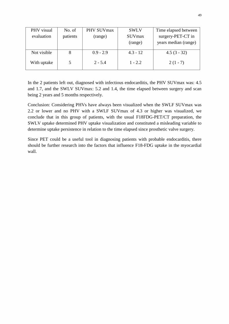
49
PHV visual evaluation
No. of patients
PHV SUVmax (range)
SWLV SUVmax (range)
Time elapsed between surgery-PET-CT in
years median (range)
Not visible 8 0.9 - 2.9 4.3 - 12 4.5 (3 - 32)
With uptake 5 2 - 5.4 1 - 2.2 2 (1 - 7)
In the 2 patients left out, diagnosed with infectious endocarditis, the PHV SUVmax was: 4.5 and 1.7, and the SWLV SUVmax: 5.2 and 1.4, the time elapsed between surgery and scan being 2 years and 5 months respectively.
Conclusion: Considering PHVs have always been visualized when the SWLF SUVmax was 2.2 or lower and no PHV with a SWLF SUVmax of 4.3 or higher was visualized, we conclude that in this group of patients, with the usual F18FDG-PET/CT preparation, the SWLV uptake determined PHV uptake visualization and constituted a misleading variable to determine uptake persistence in relation to the time elapsed since prosthetic valve surgery.
Since PET could be a useful tool in diagnosing patients with probable endocarditis, there should be further research into the factors that influence F18-FDG uptake in the myocardial wall.
50
IAEA-CN-202/148
Utility of Myocardial Perfusion Studies for the Re-stratification of Risk of Ischemic Heart Disease and its Frequent Risk Factors
C. Arroyo Castelán, J. Z. García, R. M. Cuaxospa
Belisario Dominguez Hospital. 4866 ave. Tlahuac corner Zacatlan, San Lorenzo Tezonco, Iztapalapa, Mexico City
Email Address of the Main Author: [email protected]
Introduction: Ischemic heart disease is the second cause of death in Mexico. Risk factors contribute to cardiovascular mortality between 40 and 60%. Risk factors are classified as:
• Not modifiable: those involved do not change when age, gender and heredity. • Modifiable: those who can change the course of ischemic heart disease: dyslipidemia,
hypertension, snuff, diabetes mellitus, oral contraceptives, obesity, sedentary lifestyle and diet.
Single photon tomography synchronized to the electrocardiogram (gated SPECT) myocardial perfusion imaging is a noninvasive imaging technique established in the diagnosis and treatment of coronary disease, provides prognostic information and risk stratification. Objective: Identify the most common risk factors for ischemic heart disease in a population of low socioeconomic status and risk stratification post-test. Method: Mixed Retrospective studies of myocardial perfusion gated SPECT performed in the area of Nuclear Medicine. The indication was risk stratification in coronary artery disease and known diagnose of ischemic heart disease. Results: 413 patients were studied men and women aged 34 to 89 years, with an average age of 58.7 years, 274 men and 139 women, with an incidence higher in the age range 55-61 years in both genders. The most common risk factor was hypertension, dyslipidemia, and smoking in men, hypertension, dyslipidemia and DM in women. 37.7% of patients had two modifiable risk factors (DM and HAS) 47.5%, 3 risk factors dominate the DM, hypertension and smoking and / or dyslipidemia, 6%, 4 risk factors and 2% 5 factors risk.
51
Of the 412: 212 were at high risk for a cardiovascular event (ischemic) and from these 104 were submitted to cardiac catheterization due the presence of moderate to severe ischemia and diminished ejection fraction. Discussion: We note that the higher the number of risk factors is higher your chance of coronary disease. Globalization changed lifestyles from an early age, so therefore the natural history of the disease, begins at younger ages and so their complications. Conclusion: Myocardial perfusion studies have high prognostic value of ischemic heart disease; they help the classification in intermediate-risk patients and / or high pre-test or those who have had myocardial infarction. The objectivity of the data allows us to determine the most suitable treatment, timely and individualized for each patient, reducing costs for the institution and the patient. Its reproducibility facilitates monitoring of patients makes it an ideal technique to assess the prognosis of coronary artery disease in the short and long term.
52
IAEA-CN-202/149
Clinical Experience and Applications of Simultaneous PET/MR Imaging in Cardiology
I. Cho
Department of Nuclear Medicine, Yeungnam University Hospital, Daegu, South Korea
Email Address of the Main Author: [email protected]
PET/MR scanner has been expected to play a key role in research and clinical applications because of the superior tissue contrast and multiparametric functional imaging of MR in conjunction with PET as PET/CT has gained wide acceptance for oncologic imaging in recent years. Simultaneous PET/MR offers a special variety of new possibilities in the field of cardiology. However, the additional value and potential clinical role of these new systems have not been established yet. I will summarize the major advantages, technical requirements and workflow considerations for PET/MR imaging and present clinical experience and potential applications in coronary artery disease and cardiomyopathy for simultaneous PET/MR imaging in the field of cardiology.
53
IAEA-CN-202/150
A Discordance Results between Myocardial Perfusion Imaging with Angiography: A Case Report
A. Elliyantia, A. H. S. Kartamihardjab a Faculty of Medicine Andalas University/Dr. M. Djamil Hospital Padang, West Sumatera, Indonesia b Faculty of Medicine Padjadjaran University/Dr. Hasan Sadikin Bandung, West Java, Indonesia
Email Address of the Main Author: [email protected]
Introduction: Myocardial perfusion imaging is a non-invasive cardiac examination. It identifies areas of relatively reduced myocardial blood flow associated to ischemia or scar by administrated a radiopharmaceutical intravenously for blood flow distribution in myocardium delineation.
Case Presentation: A 50 year-old man was diagnosed a cardiomyopathy and bronchitis disease, refered to Dr.Hasan Sadikin Hospital for myocardial perfusion imaging. He smoked one box of cigarette /day and quit since last year. He had a heart disease family history. Laboratory results showed; Hb 13,5gr/dL, BBS 17/37 mm/hour, blood sugar level 89mg/dl, total cholesterol and triglyceride level were 230mg/dl, 157 mg/dl respectively. Na+ and K+ concentrations were 47 and was 4.4 respectively. ECG showed ST depression with horizontal and descend ST slope at V6,II,III,aVR and aVF. Echocardiography showed dilated CMP, Mod MR, Mild TR, EF 30%. Perfusion imaging was conducted using 99mTc-tetrofosmin stress-rest test protocol. Initial blood pressure was 90/70mmHg and heart rate 121x / min. Patient was complaining exhausted at stage III and stress test was stopped. Heart rate was 138x/min (81% of target HR), blood pressure 100/70 mmHg. Perfusion imaging on stress showed cardiac dilatation and severe blood flow reduction at anterior and inferior walls, septum, apex, antero-lateral and infero-lateral segments which refer to LAD and RCA artery territories. The rest perfusion imaging showed partial reversible perfusion to those areas. Patient asked for more investigation in Singapore and he underwent an angiography test. The test showed 50% stenosis at PLAD. The question came to disagreement between perfusion imaging and angiography results.
Discussion: Interaction between vessels and myocardial related to oxygen consumption and blood vessel velocity. This interaction is different between transmural layers. Effect of contraction on coronary arteries was higher at inner than outer myocardium layer. On the other hand, oxygen consumption is higher at inner than outer myocardium layer. Patients who had no infarct history, on coronary arteries disease, the perfusion will be normal during rest, and the blood vessels velocity on distal of stenosis was managed by auto-regulation, and during exercise the blood vessels velocity reduce significantly because of micro-vascular have been dilated maximally during the rest. Furthermore, in hypo-perfusion vessels (target vessels) have a higher relative cross sectional plaque ratio (RPA) 46%±14% compare to

54
normal/reference vessels (12%±18%) and the coronary flow velocity reserve (CFR) lower than normal p<0,0001, where at target vessel was (2.3±0,5) and at reference vessels was (3,1±0.6).
Conclusion: Angiography result was shown normal at 10-20% of acute or chronic ischemia patients. An angiography gives information of luminal stenosis level and perfusion imaging gives information of luminal stenosis effect on myocardium. This can explain a disagreement between angiography and perfusion imaging. The micro-vascular disturbance be should considered as one condition caused discordance between angiography and perfusion imaging.
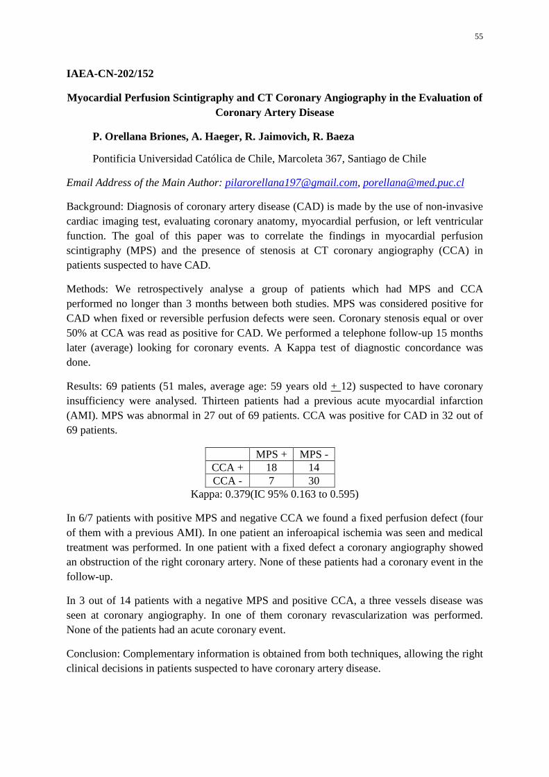
55
IAEA-CN-202/152
Myocardial Perfusion Scintigraphy and CT Coronary Angiography in the Evaluation of Coronary Artery Disease
P. Orellana Briones, A. Haeger, R. Jaimovich, R. Baeza
Pontificia Universidad Católica de Chile, Marcoleta 367, Santiago de Chile
Email Address of the Main Author: [email protected], [email protected]
Background: Diagnosis of coronary artery disease (CAD) is made by the use of non-invasive cardiac imaging test, evaluating coronary anatomy, myocardial perfusion, or left ventricular function. The goal of this paper was to correlate the findings in myocardial perfusion scintigraphy (MPS) and the presence of stenosis at CT coronary angiography (CCA) in patients suspected to have CAD.
Methods: We retrospectively analyse a group of patients which had MPS and CCA performed no longer than 3 months between both studies. MPS was considered positive for CAD when fixed or reversible perfusion defects were seen. Coronary stenosis equal or over 50% at CCA was read as positive for CAD. We performed a telephone follow-up 15 months later (average) looking for coronary events. A Kappa test of diagnostic concordance was done.
Results: 69 patients (51 males, average age: 59 years old + 12) suspected to have coronary insufficiency were analysed. Thirteen patients had a previous acute myocardial infarction (AMI). MPS was abnormal in 27 out of 69 patients. CCA was positive for CAD in 32 out of 69 patients.
MPS + MPS - CCA + 18 14 CCA - 7 30
Kappa: 0.379(IC 95% 0.163 to 0.595)
In 6/7 patients with positive MPS and negative CCA we found a fixed perfusion defect (four of them with a previous AMI). In one patient an inferoapical ischemia was seen and medical treatment was performed. In one patient with a fixed defect a coronary angiography showed an obstruction of the right coronary artery. None of these patients had a coronary event in the follow-up.
In 3 out of 14 patients with a negative MPS and positive CCA, a three vessels disease was seen at coronary angiography. In one of them coronary revascularization was performed. None of the patients had an acute coronary event.
Conclusion: Complementary information is obtained from both techniques, allowing the right clinical decisions in patients suspected to have coronary artery disease.
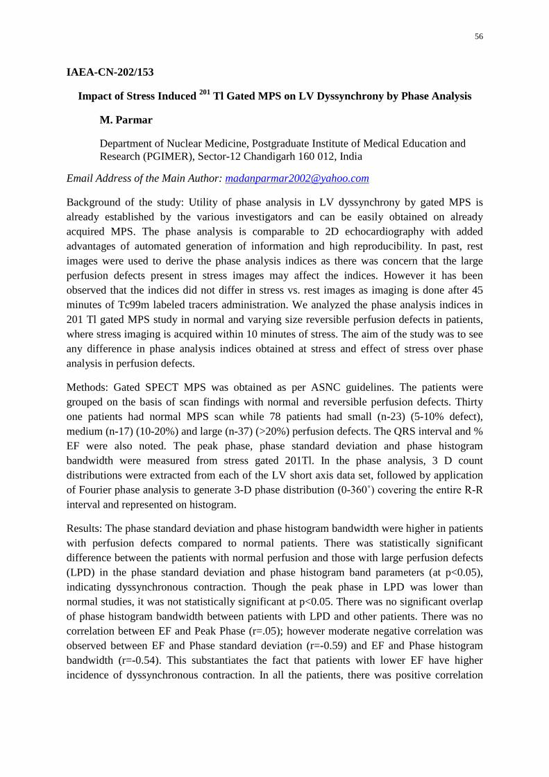
56
IAEA-CN-202/153
Impact of Stress Induced 201 Tl Gated MPS on LV Dyssynchrony by Phase Analysis
M. Parmar
Department of Nuclear Medicine, Postgraduate Institute of Medical Education and Research (PGIMER), Sector-12 Chandigarh 160 012, India
Email Address of the Main Author: [email protected]
Background of the study: Utility of phase analysis in LV dyssynchrony by gated MPS is already established by the various investigators and can be easily obtained on already acquired MPS. The phase analysis is comparable to 2D echocardiography with added advantages of automated generation of information and high reproducibility. In past, rest images were used to derive the phase analysis indices as there was concern that the large perfusion defects present in stress images may affect the indices. However it has been observed that the indices did not differ in stress vs. rest images as imaging is done after 45 minutes of Tc99m labeled tracers administration. We analyzed the phase analysis indices in 201 Tl gated MPS study in normal and varying size reversible perfusion defects in patients, where stress imaging is acquired within 10 minutes of stress. The aim of the study was to see any difference in phase analysis indices obtained at stress and effect of stress over phase analysis in perfusion defects.
Methods: Gated SPECT MPS was obtained as per ASNC guidelines. The patients were grouped on the basis of scan findings with normal and reversible perfusion defects. Thirty one patients had normal MPS scan while 78 patients had small (n-23) (5-10% defect), medium (n-17) (10-20%) and large (n-37) (>20%) perfusion defects. The QRS interval and % EF were also noted. The peak phase, phase standard deviation and phase histogram bandwidth were measured from stress gated 201Tl. In the phase analysis, 3 D count distributions were extracted from each of the LV short axis data set, followed by application of Fourier phase analysis to generate 3-D phase distribution (0-360˚) covering the entire R-R interval and represented on histogram.
Results: The phase standard deviation and phase histogram bandwidth were higher in patients with perfusion defects compared to normal patients. There was statistically significant difference between the patients with normal perfusion and those with large perfusion defects (LPD) in the phase standard deviation and phase histogram band parameters (at p<0.05), indicating dyssynchronous contraction. Though the peak phase in LPD was lower than normal studies, it was not statistically significant at p<0.05. There was no significant overlap of phase histogram bandwidth between patients with LPD and other patients. There was no correlation between EF and Peak Phase (r=.05); however moderate negative correlation was observed between EF and Phase standard deviation (r=-0.59) and EF and Phase histogram bandwidth (r=-0.54). This substantiates the fact that patients with lower EF have higher incidence of dyssynchronous contraction. In all the patients, there was positive correlation
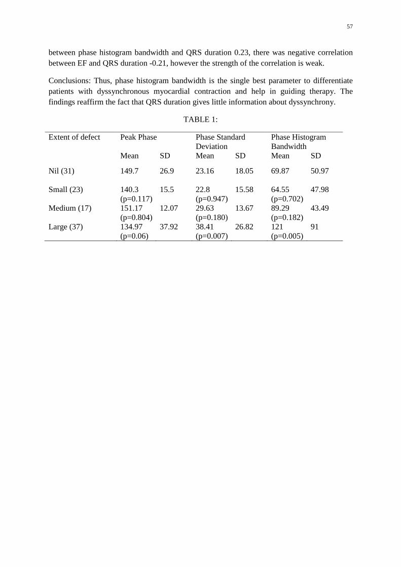
57
between phase histogram bandwidth and QRS duration 0.23, there was negative correlation between EF and QRS duration -0.21, however the strength of the correlation is weak.
Conclusions: Thus, phase histogram bandwidth is the single best parameter to differentiate patients with dyssynchronous myocardial contraction and help in guiding therapy. The findings reaffirm the fact that QRS duration gives little information about dyssynchrony.
TABLE 1:
Extent of defect Peak Phase
Phase Standard Deviation
Phase Histogram Bandwidth
Mean SD Mean SD Mean SD
Nil (31) 149.7 26.9 23.16
18.05 69.87
50.97
Small (23) 140.3 (p=0.117)
15.5 22.8 (p=0.947)
15.58 64.55 (p=0.702)
47.98
Medium (17) 151.17 (p=0.804)
12.07 29.63 (p=0.180)
13.67 89.29 (p=0.182)
43.49
Large (37) 134.97 (p=0.06)
37.92 38.41 (p=0.007)
26.82 121 (p=0.005)
91
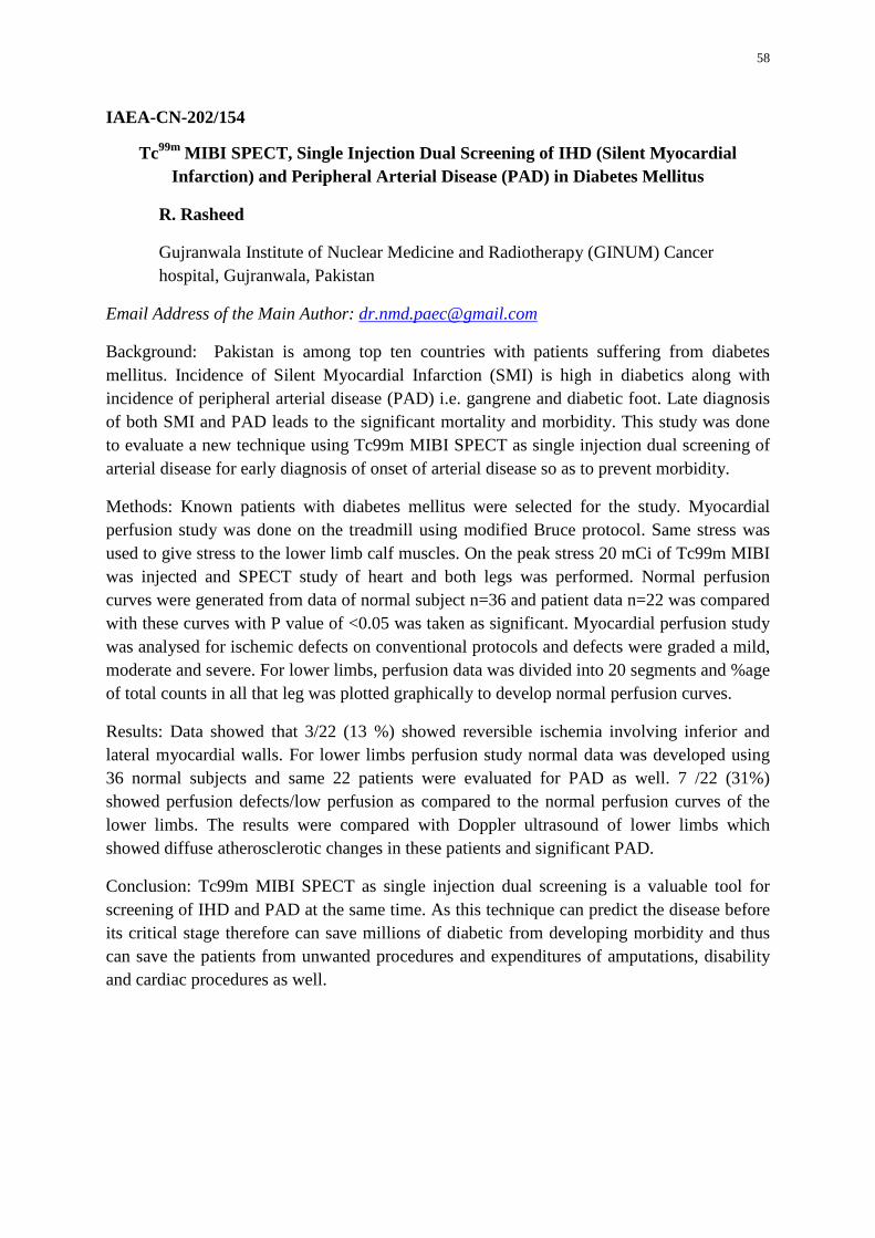
58
IAEA-CN-202/154
Tc99m MIBI SPECT, Single Injection Dual Screening of IHD (Silent Myocardial Infarction) and Peripheral Arterial Disease (PAD) in Diabetes Mellitus
R. Rasheed
Gujranwala Institute of Nuclear Medicine and Radiotherapy (GINUM) Cancer hospital, Gujranwala, Pakistan
Email Address of the Main Author: [email protected]
Background: Pakistan is among top ten countries with patients suffering from diabetes mellitus. Incidence of Silent Myocardial Infarction (SMI) is high in diabetics along with incidence of peripheral arterial disease (PAD) i.e. gangrene and diabetic foot. Late diagnosis of both SMI and PAD leads to the significant mortality and morbidity. This study was done to evaluate a new technique using Tc99m MIBI SPECT as single injection dual screening of arterial disease for early diagnosis of onset of arterial disease so as to prevent morbidity.
Methods: Known patients with diabetes mellitus were selected for the study. Myocardial perfusion study was done on the treadmill using modified Bruce protocol. Same stress was used to give stress to the lower limb calf muscles. On the peak stress 20 mCi of Tc99m MIBI was injected and SPECT study of heart and both legs was performed. Normal perfusion curves were generated from data of normal subject n=36 and patient data n=22 was compared with these curves with P value of <0.05 was taken as significant. Myocardial perfusion study was analysed for ischemic defects on conventional protocols and defects were graded a mild, moderate and severe. For lower limbs, perfusion data was divided into 20 segments and %age of total counts in all that leg was plotted graphically to develop normal perfusion curves.
Results: Data showed that 3/22 (13 %) showed reversible ischemia involving inferior and lateral myocardial walls. For lower limbs perfusion study normal data was developed using 36 normal subjects and same 22 patients were evaluated for PAD as well. 7 /22 (31%) showed perfusion defects/low perfusion as compared to the normal perfusion curves of the lower limbs. The results were compared with Doppler ultrasound of lower limbs which showed diffuse atherosclerotic changes in these patients and significant PAD.
Conclusion: Tc99m MIBI SPECT as single injection dual screening is a valuable tool for screening of IHD and PAD at the same time. As this technique can predict the disease before its critical stage therefore can save millions of diabetic from developing morbidity and thus can save the patients from unwanted procedures and expenditures of amputations, disability and cardiac procedures as well.
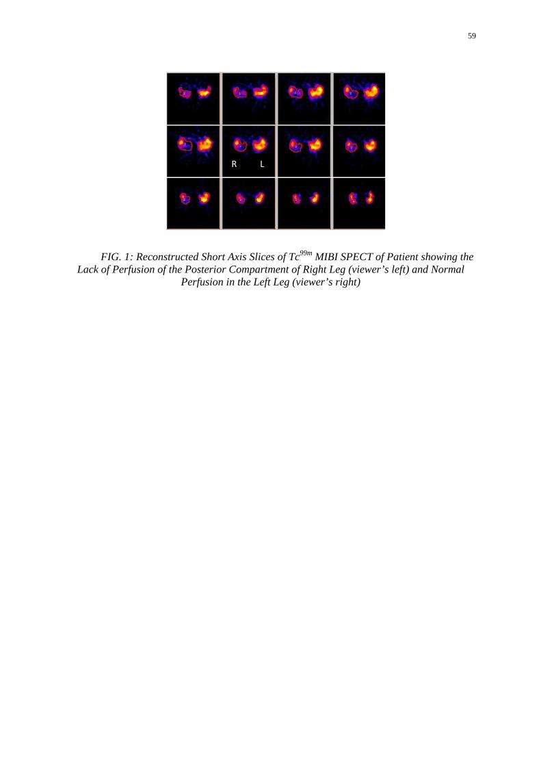
59
FIG. 1: Reconstructed Short Axis Slices of Tc99m MIBI SPECT of Patient showing the Lack of Perfusion of the Posterior Compartment of Right Leg (viewer’s left) and Normal
Perfusion in the Left Leg (viewer’s right)
L R
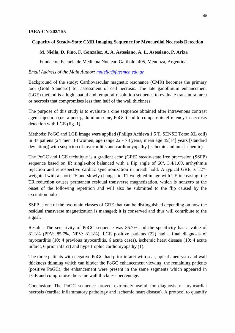
60
IAEA-CN-202/155
Capacity of Steady-State CMR Imaging Sequence for Myocardial Necrosis Detection
M. Niella, D. Fino, F. Gonzalez, A. A. Astesiano, A. L. Astesiano, P. Ariza
Fundación Escuela de Medicina Nuclear, Garibaldi 405, Mendoza, Argentina
Email Address of the Main Author: [email protected]
Background of the study: Cardiovascular magnetic resonance (CMR) becomes the primary tool (Gold Standard) for assessment of cell necrosis. The late gadolinium enhancement (LGE) method is a high spatial and temporal resolution sequence to evaluate transmural area or necrosis that compromises less than half of the wall thickness.
The purpose of this study is to evaluate a cine sequence obtained after intravenous contrast agent injection (i.e. a post-gadolinium cine, PoGC) and to compare its efficiency in necrosis detection with LGE (fig. 1).
Methods: PoGC and LGE image were applied (Philips Achieva 1.5 T, SENSE Torso XL coil) in 37 patiens (24 men, 13 women, age range 22 - 78 years, mean age 45[14] years [standard deviation]) with suspicion of myocarditis and cardiomyopathy (ischemic and non-ischemic).
The PoGC and LGE technique is a gradient echo (GRE) steady-state free precession (SSFP) sequence based on IR single-shot balanced with a flip angle of 60º, 3.4/1.69, arrhythmia rejection and retrospective cardiac synchronization in breath hold. A typical GRE is T2*-weighted with a short TE and slowly changes to T1-weighted image with TE increasing; the TR reduction causes permanent residual transverse magnetization, which is nonzero at the onset of the following repetition and will also be submitted to the flip caused by the excitation pulse.
SSFP is one of the two main classes of GRE that can be distinguished depending on how the residual transverse magnetization is managed; it is conserved and thus will contribute to the signal.
Results: The sensitivity of PoGC sequence was 85.7% and the specificity has a value of 81.3% (PPV: 85.7%, NPV: 81.3%). LGE positive patients (22) had a final diagnosis of myocarditis (10; 4 previous myocarditis, 6 acute cases), ischemic heart disease (10; 4 acute infarct, 6 prior infarct) and hypertrophic cardiomyopathy (1).
The three patients with negative PoGC had prior infarct with scar, apical aneurysm and wall thickness thinning which can hinder the PoGC enhancement viewing, the remaining patients (positive PoGC), the enhancement were present in the same segments which appeared in LGE and compromise the same wall thickness percentage.
Conclusion: The PoGC sequence proved extremely useful for diagnosis of myocardial necrosis (cardiac inflammatory pathology and ischemic heart disease). A protocol to quantify
61
tissue contrast using such sequence makes PoGC a very valuable tool for routine clinical CMR.
FIG 1. 54 year old man with acute myocardial infarction. A. End diastolic long-axis pre-gadolinium cine MRI imaging B. End diastolic long-axis PoGC, show transmural enhancement in inferior and middle-anterior wall. C. Long-axis LGE, show enhancement in same areas as in B. D. Short-axis LGE, show enhancement in same areas as in B.
62
IAEA-CN-202/156
Influence of a Low-Carbohydrate Diet on the Assessment of Myocardial Viability with 18F- Fluorodeoxyglucose PET: Comparison with the Euglycemic Hyperinsulinemic
Clamp
F. Rodrigues Filhoa, J. Soares Jra, M. lzakia, M. C. P. Giorgia, R. M. A. Catapirraa, R. Abea, C. G. M. Vinagreb, G. G. Cerria, J. C. Meneghettia
aNuclear Medicine Department bLipid Metabolism Laboratory, Heart Institute (InCor), University of Sao Paulo Medical School, Sao Paulo - SP, Brazil
Email Address of the Main Author: [email protected]
Background: In patients with myocardial infarction and left ventricular dysfunction, the evidence of myocardial viability has important clinical implications. Positron emission tomography (PET) using 18F-fluoro-desoxi-glucose (18FDG) is considered the gold standard for viability detection. The euglycemic hyperinsulinemic clamping (CLAMP) before 18FDG injection stimulates uptake of both glucose and 18FDG in the myocardium, including areas of hibernating myocardium (MH), and it is one of the most used protocols. However, this imaging protocol has an increased risk for hypoglycemia and is relatively time-consuming. We used a new protocol with a 24 hours low- carbohydrate diet (DIET) before 18FDG injection, aiming to reduce insulin levels and increase free fatty acids (FFA). This protocol stimulates the normal myocardium to use FFA, not glucose, but the area of hibernating myocardium (viable area) may not use oxidative metabolism with FFA, keeping glucose uptake and becoming a hot spot at PET images. The aim of this study was to compare both techniques by segments, regions, vascular territories and patients.
Methods: Thirty patients with previous myocardial infarction and left ventricular dysfunction were studied. All of them underwent in to a SPECT perfusion scan with 99111Tc-sestamibi and two 18FDG PET studies to asses myocardial viability, one with CLAMP (PET-CLAMP) and another using a 24 hours low-carbohydrate diet (PET-DIET). For the analysis, the myocardium was divided into 17 segments, 5 regions and 3 vascular territories. A visual analysis was carried out according to the following score indicating radiotracer uptake: 0 = normal to 4 = absent. Myocardial viability was defined as the presence of normal or mild l y reduced FOG uptake in a n area with reduced perfusion (99mTc- sestamibi uptake). We also performed subgroup analysis in diabetes (OM) and non-diabetic patients (NOM).
Results: While using CLAMP protocol, six (20%) patients had hypoglycemia. None of the patients had hypoglycemia after using DIET. High agreement rates were observed with visual analysis in comparing mismatch segments (kappa=O,78 - substantial concordance), with similar agreement rates in comparing regions (0,80), vascular

63
territories (0,79) and patient (0,79) analysis. In subgroup analysis, OM (8) presented with kappa=0, 70 and NOM (22) with kappa=0,78.
Conclusion: This has led to the suggestion that PET-DIET study proved an excellent agreement in detects mismatch areas assessed by PET-CLAMP, with probably more safety. The same results occurs in the in diabetes and non-diabetic patients subgroups.
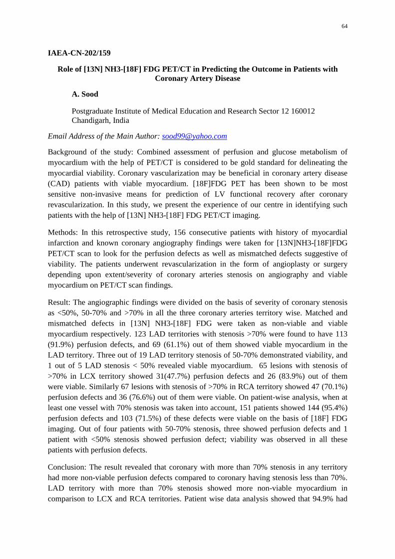
64
IAEA-CN-202/159
Role of [13N] NH3-[18F] FDG PET/CT in Predicting the Outcome in Patients with Coronary Artery Disease
A. Sood
Postgraduate Institute of Medical Education and Research Sector 12 160012 Chandigarh, India
Email Address of the Main Author: [email protected]
Background of the study: Combined assessment of perfusion and glucose metabolism of myocardium with the help of PET/CT is considered to be gold standard for delineating the myocardial viability. Coronary vascularization may be beneficial in coronary artery disease (CAD) patients with viable myocardium. [18F]FDG PET has been shown to be most sensitive non-invasive means for prediction of LV functional recovery after coronary revascularization. In this study, we present the experience of our centre in identifying such patients with the help of [13N] NH3-[18F] FDG PET/CT imaging.
Methods: In this retrospective study, 156 consecutive patients with history of myocardial infarction and known coronary angiography findings were taken for [13N]NH3-[18F]FDG PET/CT scan to look for the perfusion defects as well as mismatched defects suggestive of viability. The patients underwent revascularization in the form of angioplasty or surgery depending upon extent/severity of coronary arteries stenosis on angiography and viable myocardium on PET/CT scan findings.
Result: The angiographic findings were divided on the basis of severity of coronary stenosis as <50%, 50-70% and >70% in all the three coronary arteries territory wise. Matched and mismatched defects in [13N] NH3-[18F] FDG were taken as non-viable and viable myocardium respectively. 123 LAD territories with stenosis >70% were found to have 113 (91.9%) perfusion defects, and 69 (61.1%) out of them showed viable myocardium in the LAD territory. Three out of 19 LAD territory stenosis of 50-70% demonstrated viability, and 1 out of 5 LAD stenosis < 50% revealed viable myocardium. 65 lesions with stenosis of >70% in LCX territory showed 31(47.7%) perfusion defects and 26 (83.9%) out of them were viable. Similarly 67 lesions with stenosis of >70% in RCA territory showed 47 (70.1%) perfusion defects and 36 (76.6%) out of them were viable. On patient-wise analysis, when at least one vessel with 70% stenosis was taken into account, 151 patients showed 144 (95.4%) perfusion defects and 103 (71.5%) of these defects were viable on the basis of [18F] FDG imaging. Out of four patients with 50-70% stenosis, three showed perfusion defects and 1 patient with <50% stenosis showed perfusion defect; viability was observed in all these patients with perfusion defects.
Conclusion: The result revealed that coronary with more than 70% stenosis in any territory had more non-viable perfusion defects compared to coronary having stenosis less than 70%. LAD territory with more than 70% stenosis showed more non-viable myocardium in comparison to LCX and RCA territories. Patient wise data analysis showed that 94.9% had
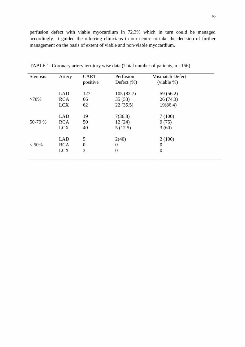
65
perfusion defect with viable myocardium in 72.3% which in turn could be managed accordingly. It guided the referring clinicians in our centre to take the decision of further management on the basis of extent of viable and non-viable myocardium.
TABLE 1: Coronary artery territory wise data (Total number of patients, n =156)
Stenosis Artery CART positive
Perfusion Defect (%)
Mismatch Defect (viable %)
>70%
LAD
127
105 (82.7)
59 (56.2)
RCA 66 35 (53) 26 (74.3) LCX 62 22 (35.5) 19(86.4)
50-70 %
LAD
19
7(36.8)
7 (100)
RCA 50 12 (24) 9 (75) LCX 40 5 (12.5) 3 (60)
< 50%
LAD
5
2(40)
2 (100)
RCA 0 0 0 LCX 3 0 0
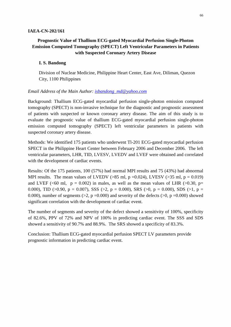
66
IAEA-CN-202/161
Prognostic Value of Thallium ECG-gated Myocardial Perfusion Single-Photon Emission Computed Tomography (SPECT) Left Ventricular Parameters in Patients
with Suspected Coronary Artery Disease
I. S. Bandong
Division of Nuclear Medicine, Philippine Heart Center, East Ave, Diliman, Quezon City, 1100 Philippines
Email Address of the Main Author: [email protected]
Background: Thallium ECG-gated myocardial perfusion single-photon emission computed tomography (SPECT) is non-invasive technique for the diagnostic and prognostic assessment of patients with suspected or known coronary artery disease. The aim of this study is to evaluate the prognostic value of thallium ECG-gated myocardial perfusion single-photon emission computed tomography (SPECT) left ventricular parameters in patients with suspected coronary artery disease.
Methods: We identified 175 patients who underwent Tl-201 ECG-gated myocardial perfusion SPECT in the Philippine Heart Center between February 2006 and December 2006. The left ventricular parameters, LHR, TID, LVESV, LVEDV and LVEF were obtained and correlated with the development of cardiac events.
Results: Of the 175 patients, 100 (57%) had normal MPI results and 75 (43%) had abnormal MPI results. The mean values of LVEDV (>85 ml, p =0.024), LVESV (>35 ml, p = 0.019) and LVEF (<60 ml, p = 0.002) in males, as well as the mean values of LHR (>0.30, p= 0.000), TID (>0.90, p = 0.007), SSS (>2, p = 0.000), SRS (>0, p = 0.000), SDS (>1, p = 0.000), number of segments (>2, p =0.000) and severity of the defects (>0, p =0.000) showed significant correlation with the development of cardiac event.
The number of segments and severity of the defect showed a sensitivity of 100%, specificity of 82.6%, PPV of 72% and NPV of 100% in predicting cardiac event. The SSS and SDS showed a sensitivity of 90.7% and 88.9%. The SRS showed a specificity of 83.3%.
Conclusion: Thallium ECG-gated myocardial perfusion SPECT LV parameters provide prognostic information in predicting cardiac event.
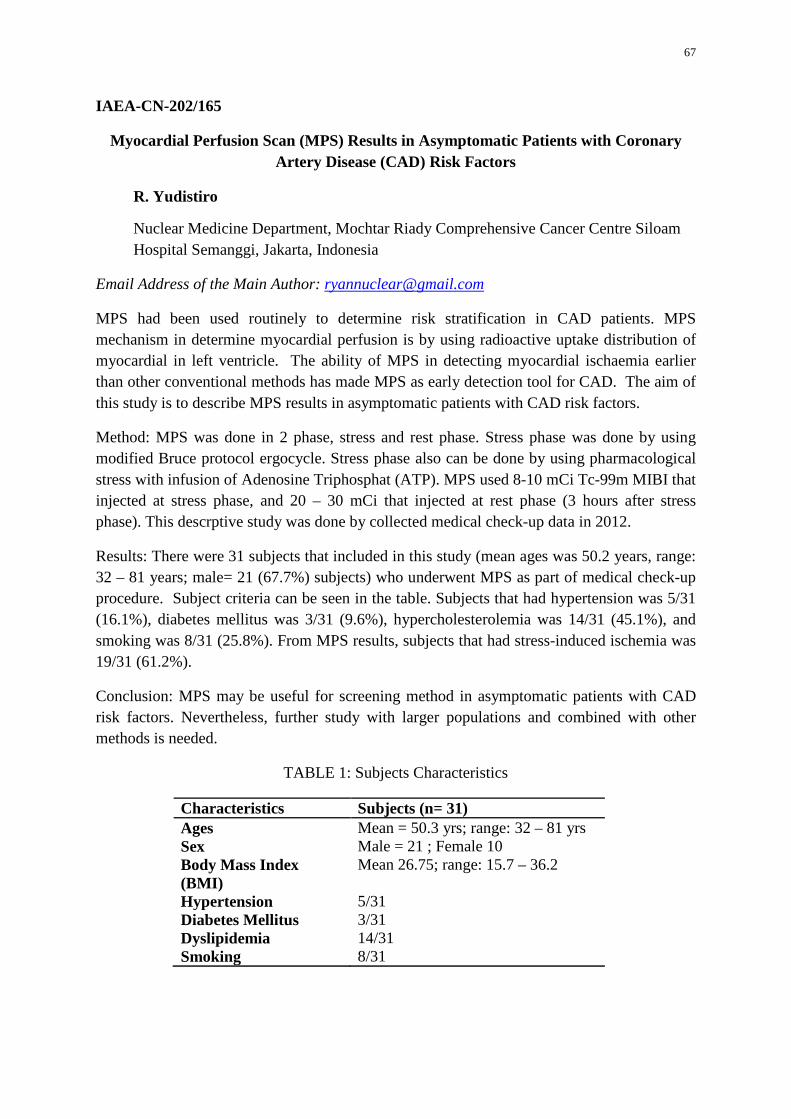
67
IAEA-CN-202/165
Myocardial Perfusion Scan (MPS) Results in Asymptomatic Patients with Coronary Artery Disease (CAD) Risk Factors
R. Yudistiro
Nuclear Medicine Department, Mochtar Riady Comprehensive Cancer Centre Siloam Hospital Semanggi, Jakarta, Indonesia
Email Address of the Main Author: [email protected]
MPS had been used routinely to determine risk stratification in CAD patients. MPS mechanism in determine myocardial perfusion is by using radioactive uptake distribution of myocardial in left ventricle. The ability of MPS in detecting myocardial ischaemia earlier than other conventional methods has made MPS as early detection tool for CAD. The aim of this study is to describe MPS results in asymptomatic patients with CAD risk factors.
Method: MPS was done in 2 phase, stress and rest phase. Stress phase was done by using modified Bruce protocol ergocycle. Stress phase also can be done by using pharmacological stress with infusion of Adenosine Triphosphat (ATP). MPS used 8-10 mCi Tc-99m MIBI that injected at stress phase, and 20 – 30 mCi that injected at rest phase (3 hours after stress phase). This descrptive study was done by collected medical check-up data in 2012.
Results: There were 31 subjects that included in this study (mean ages was 50.2 years, range: 32 – 81 years; male= 21 (67.7%) subjects) who underwent MPS as part of medical check-up procedure. Subject criteria can be seen in the table. Subjects that had hypertension was 5/31 (16.1%), diabetes mellitus was 3/31 (9.6%), hypercholesterolemia was 14/31 (45.1%), and smoking was 8/31 (25.8%). From MPS results, subjects that had stress-induced ischemia was 19/31 (61.2%).
Conclusion: MPS may be useful for screening method in asymptomatic patients with CAD risk factors. Nevertheless, further study with larger populations and combined with other methods is needed.
TABLE 1: Subjects Characteristics
Characteristics Subjects (n= 31) Ages Mean = 50.3 yrs; range: 32 – 81 yrs Sex Male = 21 ; Female 10 Body Mass Index (BMI)
Mean 26.75; range: 15.7 – 36.2
Hypertension 5/31 Diabetes Mellitus 3/31 Dyslipidemia 14/31 Smoking 8/31
68
IAEA-CN-202/166 99mTc-sestamibi Exercise SPECT and Follow-up in Patients Able to Reach 10 Metabolic
Equivalents of Task (METs)
J. S. González
Hospital Clínico Universidad de Chile Santos Dumont 999 1-E, Independencia, Santiago, Chile
Email Address of the Main Author: [email protected]
Good exercise performance during a stress test gives important clinical information. Being able to achieve 10 Metabolic Equivalents of Task (METs) appears to have a good negative predictive value for cardiac events in coronary artery disease (CAD), independent of the cardiac condition.
Aim: to know the prevalence of isotopic ischemia with myocardial SPECT and the outcome in patients who obtained ≥10 METs in their treadmill test.
Methods: We reviewed 2.571 consecutive stress myocardial 99mTc-sestamibi SPECT, 780 of them performed exercise stress test (EST); 152 were able to reach at least 10 METs and ≥85% maximal predicted heart rate (MPHR); 82% males; group age was 56±9 years (range: 26-77 and 5% <40 years). All performed Bruce protocol and were studied with 99mTc-sestamibi SPECT gated in both phases. Record of major or minor cardiac events was performed through phone contact, reviewing clinical files or inquiring in the civil registration office.
Results: SPECT was required due to CAD screening in 75% of the group and to risk stratification in 25% (including myocardial infarction and/or revascularization); 99% was in NYHA functional class I. The electrocardiogram at rest was normal in 41%; with Q wave in 6% and with other conduction abnormalities in 12% (no left bundle branch block included). Main EST parameters were: test duration 9±1 min; MPHR 94±6%; METs 11.5±1.7; 48% suspended for fatigue; 47% reached MPHR and the rest for dyspnea, hypertensive response or angina. EST was positive for ischemia in 16%, negative in 64% and non-conclusive in 20%. SPECT myocardial perfusion was normal in 72%; all had adequate normal size left ventricle function but 10 dilated and 4 with diminished function. There were reversible perfusion defects in 25% of cases (10% with some fixed component) and only 4% with pure fixed defects (the more extended:45% of the LV). Ischemia was moderate or severe in 47% of the reversible cases: mean SDS 8.3; ischemia extension 23%; TPD at stress 20. There was 75% of concordance between reported ischemia with EST and reversibility with SPECT (Cohen kappa: 0.31).
We were able to follow-up 84% of the cases [mean: 30 m ±15; median: 25 m; range: 10-82 m]. There were no cardiac deaths, myocardial infarctions or strokes. There were 8 patients with coronary revascularization procedures, 7 of them performed within 1 year post SPECT;

69
6 out of 8 of them had significant ischemia of diverse extension. Another patient without ischemia was submitted to coronary angiography without intervention.
Conclusion: A quarter of the patients with good exercise capacity submitted to myocardial sestamibi SPECT had reversible perfusion defects. There were no major cardiac events; only 16% of the patients with SPECT ischemia were submitted to revascularization. In good functional capacity patients, this findings support the idea to select with appropriate criteria non-invasive strategies to evaluate CAD, in spite of their relative high prognostic value.
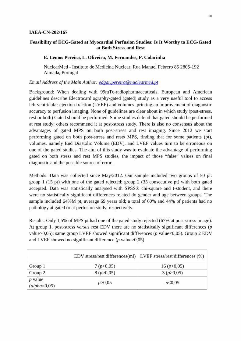
70
IAEA-CN-202/167
Feasibility of ECG-Gated at Myocardial Perfusion Studies: Is It Worthy to ECG-Gated at Both Stress and Rest
E. Lemos Pereira, L. Oliveira, M. Fernandes, P. Colarinha
NuclearMed - Instituto de Medicina Nuclear, Rua Manuel Febrero 85 2805-192 Almada, Portugal
Email Address of the Main Author: [email protected]
Background: When dealing with 99mTc-radiopharmaceuticals, European and American guidelines describe Electrocardiography-gated (gated) study as a very useful tool to access left ventricular ejection fraction (LVEF) and volumes, printing an improvement of diagnostic accuracy to perfusion imaging. None of guidelines are clear about in which study (post-stress, rest or both) Gated should be performed. Some studies defend that gated should be performed at rest study; others recommend it at post-stress study. There is also no consensus about the advantages of gated MPS on both post-stress and rest imaging. Since 2012 we start performing gated on both post-stress and rests MPS, finding that for some patients (pt), volumes, namely End Diastolic Volume (EDV), and LVEF values turn to be erroneous on one of the gated studies. The aim of this study was to evaluate the advantage of performing gated on both stress and rest MPS studies, the impact of those “false” values on final diagnostic and the possible source of error. Methods: Data was collected since May/2012. Our sample included two groups of 50 pt: group 1 (15 pt) with one of the gated rejected; group 2 (35 consecutive pt) with both gated accepted. Data was statistically analysed with SPSS® chi-square and t-student, and there were no statistically significant differences related do gender and age between groups. The sample included 64%M pt, average 69 years old; a total of 60% and 44% of patients had no pathology at gated or at perfusion study, respectively. Results: Only 1,5% of MPS pt had one of the gated study rejected (67% at post-stress image). At group 1, post-stress versus rest EDV there are no statistically significant differences (p value>0,05); same group LVEF showed significant differences (p value<0,05). Group 2 EDV and LVEF showed no significant difference (p value>0,05).
EDV stress/rest differences(ml) LVEF stress/rest differences (%)
Group 1 7 (p>0,05) 16 (p<0,05) Group 2 8 (p>0,05) 3 (p>0,05) p value (alpha=0,05)
p>0,05 p<0,05

71
Conclusion: Statistically analysed data show that there were significant differences between LVEF on the rejected and the accepted gated at group 1; at 33% of studies, the gated study would be differently interpreted if accepted gated wasn’t performed. Despite only 1,5% of pt had one of the gated studies rejected, and so potentially error inductor, we believe it is worthy to perform both stress and rest gated acquisitions, because it allow us to double check gated data, assuring accurate diagnostic. We still are searching for an explanation for rejected gated studies. Further discussion is mandatory and may consider incipient cardiac pathology; technical issues such are different distances at stress and rest between pt and Gama Camera detectors or different pt positioning between stress and rest studies.
72
IAEA-CN-202/168
The Place of Cardiac Computed Tomography Angiography (CCTA) in Cardiac Pathology
M. Mouloudi, T. Bassaid, N. Kriou, N. Boubendir, H. Mahmoudi
Military University Hospital, Ain Beida, Oran, Algeria
Email Address of the Main Author: [email protected]
The coronary CT is not indicated when the patient is presenting typical angina or acute coronary syndrome with explorations confirming a high probability of coronary lesion in this case, coronary angiography is indicated immediately .
However, it is particularity useful for an atypical case, where it is insufficient to indicate in an atypical angina such as coronary angiography with or without risk factors or family history, when the stress test and / or scintigraphy is considered invalid, doubtful or non–contributory. In these cases, the CT scan at first line removes a coronary lesion.
You can then use the high negative predictive value of CCTA (97%) for the case, eliminate a coronary heart disease, and find possible other chest causes. Nearly 35% of coronary angiograms are made to eliminate a coronary origin and are normal, and could be avoided.
For angina patients with recrudescence of symptoms, or for which the stress tests or cardiac scintigraphy were difficult to achieve or with a mistaken interpretation, we can also offer the CCTA as an alternative to traditional angiography, in the cases where revascularization techniques are not always necessary .
Finally, in acute chest pain, and if the clinical setting is ambiguous or not cardiac specific, some others have proposed the emergency scanner before angiography, to the differential diagnosis with other urgent etiologies as aortic dissection or pulmonary embolism, or acute pneumonia, for which the diagnostic sensitivity of the scanner is well proven.
73
IAEA-CN-202/170
Preparation and Preclinical Evaluation of [64Cu]Cu(II)-PTSM: A Tracer for Myocardial Perfusion
J. C. Manrique-Ariasa, V. M. Lara-Camachoa, M. Avila-Garciaa, A. Flores-Morenoa, A. Zarate-Moralesa, P. G. Reyes-Romerob, M. A. Ávila-Rodrigueza a Unidad PET, Facultad de Medicina, Universidad Nacional Autónoma de México, México D.F., C.P. 04510, México b Facultad de Ciencias, Universidad Autónoma del Estado de México, Instituto Literario No. 100, C.P. 50000, Toluca Edo de México, México
Email Address of the Main Author: [email protected]
Background: Copper offers a unique selection of radioisotopes (60Cu, 61Cu,62Cu, 64Cu, and 67Cu) with half-lives ranging from 9.8 min to 61.9 h, suitable for imaging and/or radiotherapy. Due to its decay characteristics, Cu-64 (T1/2=12.7h, β+ [17.4%], β- [39%], E.C. [43.6%]) is an attractive radionuclide with applications in both PET molecular imaging and targeted therapy. This radionuclide has been widely used in the labelling of macromolecules such as peptides, proteins, monoclonal antibodies, and thiosemicarbazone complexes. In this work [64Cu]Cu(II)-pyruvaldehyde-bis(N4-methyl-thiosemicarbazone) ([64Cu]Cu(II)-PTSM), a tracer for myocardial perfusion, was prepared and evaluated.
Methods: 64 Cu was produced in a cyclotron via the 64Ni(p,n)64Cu reaction with 11 MeV protons using natNi electrodeposited on Au backing as target material. Radiochemical separation was performed by ion exchange chromatography, obtaining 64Cu in the chemical form of CuCl2 (0.1M HCl). For radiolabelling, 10 µl of H2-PTSM in DMSO (2 mg/ml), 150 µl of 6M HCl and 20 µl of [64Cu]CuCl2 stock solution (37-50 MBq) were mixed. The mixture was allowed to react at room temperature for 2 min and purified by SPE using a Sep-Pak C18 Light cartridge. [64Cu]Cu(II)-PTSM was eluted with ~500 μl of EtOH. For injection, the ethanolic solution of Cu-PTSM was diluted with 5 ml of isotonic sterile saline and filtered through a hydrophilic 0.22 μm Millex. Radiochemical purity was determined by TLC using SG strips as stationary phase and 80:20 ACN: EtOH (v/v) as mobile phase. TLC-strips were analyzed by autoradiography. Preclinical evaluation in Wistar rats was performed using a Focus 120 microPET.
Results: Radionuclidic purity of 64Cu, 12 h after the end of the bombardment, was >85%, with 61Cu as the main impurity. Labeling and formulation took about 10 min, and radiochemical purity of [64Cu]Cu(II)-PTSM was higher than 98%. MicroPET images 1 h post-injection showed a contrasting heart wall denoting myocardial perfusion, with liver uptake interfering when imaging the inferior heart wall. The mean resting heart/liver ratio (1 h post-injection) was 1.11±0.12.

74
a)
b)
c)
FIG 1. [64Cu]Cu(II)-PTSM axial (a) and coronal (b) images showing uptake in heart and liver, and molecular structure of the compund (c).
Conclusions: The preliminary results obtained in this work warrant the evaluation of other Cu(II) bis(thiosemicarbazone) complexes as myocardial perfusion agents, labelled with positron emitters of Cu with half-lives suitable for its regional distribution from a satellite centre.
75
IAEA-CN-202/171
Evaluation of Left Ventricular Function in Mali using a Radionuclide Method: Application, Methods and Normal Values
S. Sidibé, S. Traoré, R. Diakité
Radiology and Nuclear Medicine Department Point « G » Hospital Bamako, Mali
Email Address of the Main Author: [email protected]
Background: Left ventricular ejection fraction is a good parameter in evaluation of left ventricular function. This parameter can be approach both by radiology including ultrasound method and gamma angiography by using radionuclide.
Method: Sixty-six healthy African volunteers in Mali (27 males and 29 females with mean age 33 years) were studied to demonstrate the feasibility of performing radionulide gamma angiographic studies in Mali.
Results: Normal values for the left ventricular ejection fraction (LVEF) in these Malian subjects were found to be in the range (58±12 %). This value is lower than the LVEF for European people. The reason for this difference is believed to be associated with asymptomatic left ventricular dysfunction in black Malian people. This difference in LVEF values, together with technical details of measuring LVEF in African context and other aspects of procedure, will be discussed.
Key word: Left Ventricular Ejection Fraction, Gamma angioscintigraphy, Black malian people, Asymptomatic left ventricular function.
76
IAEA-CN-202/172
Gated SPECT Functional Assessment in Obese Patients
A. Puente, J. Morales, G. Meléndez
Nuclear Cardiology Department, Centro Médico Nacional “20 de Noviembre”, ISSSTE, Mexico City
Email Address of the Main Author: [email protected]
Background: Several studies have suggested obesity contributes to pathogenesis of abnormal left ventricular cavity remodeling (excentric hypertropy), alteration of dyastolic volumes and ventricular function. Mexico has a high prevalence of overweight and obesity (70%); these changes have not been studied.
Objective: To determine the relation of left ventricular functional parametres in overweight and obese patients evaluated with Gated-Myocardial Perfusion Single-Photon Computed Tomography (g-SPECT)
Methods: Normal g-SPECT of 153 patients without ischemic heart disease were analyzed. Patients with valvular disease, dilated or hypertrophic miocardiopaty were excluded. Variables: age, risk factors; post-stress Left Ventricle Ejection Fraction (LVEF), End-Diastolic Volume (EDV) and End-Systolic Volume (ESV). Body Mass Index (BMI): 18-24.9=Normal, 25-29.9 overweight and ≥ 30 obesity. Statistics: ANOVA, T Student
Results: Average age was 62 ± 10 years, 65% were women; 71% hypertension, 53% dyslipidemia 33% diabetes mellitus. Normal Weight =36, overweight=63 and obesity=54. There were no difference between LVEF in patients with overweight and obesity (p=0.26); the EDV has a light elevation in patients with ≥ 30 BMI (p=0.15)
Conclusions: There were no differences between LVEF in overweight and obese patients. Obese patients have an elevation of EDV without ventricular dysfunction.
77
IAEA-CN-202/174
Rest Thallium-201/Stress Technetium-99m sestamibi Dual-Isotope Myocardial Perfusion Single-Photon Emission Computed Tomography in Detecting of Chronic
Coronary Artery Disease
P. K. Huynh, L. C. Vu, X. Q. Truong, C. X. Nguyen
Cho Ray hospital, 201B Nguyen Chi Thanh Street, District 5, Ho Chi Minh city, Viet Nam
Email Address of the Main Author: [email protected]
Purpose: To investigate diagnostic accuracy of 2-hour protocol of rest thallium-201/stress technetium-99m sestamibi dual-isotope myocardial perfusion SPECT in chronic coronary artery disease.
Patients and Methods: Sixty-seven patients with suspected CAD were enrolled in the prospective study. All patients underwent myocardial perfusion scintigraphy in SPECT with dual isotopes of Tl-201 and dipyridamole stress Tc-99m sestamibi. Rest and stress imaging protocol were performed in 2 hours by dose of 3 mCi Tl-201 and 25 mCi Tc-99m sestamibi. The acquisition parameters includes LEHR collimator, energy peak of 72 and 167 keV for Tl-201 and 140 keV for Tc-99m, 180-degree rotation from RAO to LPO, matrix size 64×64, and 25second/frame/64 frames. The 20-segment model of left ventricle was used in automatic quantitation software. Coronary angiography was used as gold standard. CAD was defined as 50% of lumen stenosis on coronary angiography.
Result: Rest Tl-201/stress tc-99m sestamibi dual-isotope SPECT demonstrated a sensitivity of 94.59% and specificity of 70%, positive predictive value of 79.54% and negative predictive value of 91.3% in detection of coronary artery disease. Sensitivity and specificity for detecting multi-vessel coronary artery disease were 82.75% and 81.57% for the left anterior descending, 77.77% and 91.83% for left circumflex and 94.11% and 82% for right coronary artery.
Conclusion: 2-hour protocol of rest thallium-201/stress technetium-99m sestamibi dual-isotope myocardial perfusion SPECT has high sensitivity, specificity, positive predictive value and negative predictive value in detecting chronic coronary artery disease with greater than 50% stenosis assessed by coronary angiography. Moreover, this imaging protocol gives high imaging quality, time-saving and convenience.
Key words: Myocardial perfusion scintigarphy (MPS), single proton emission computed tomography (SPECT), coronary artery disease (CAD), coronary angiography (CA).
78
IAEA-CN-202/175
Sodium Bicarbonate Augmented 201Tl Rest/Redistribution Myocardial Perfusion SPECT in Detecting Myocardial Viability
K. Khan, A. Jielani, M. Ayub, N. Z. Abbasi, T. Fatima
Institute of Nuclear Medicicne, Oncology and Radiotherapy (INOR), Abbottabad, Pakistan
Email Address of the Main Author: [email protected]
Pharmacological doses sodium bicarbonate induces transient alkalosis, causing intracellular transport of serum potassium.
The aims of this study were (a) whether myocardial thallium-201 uptake can be augmented by pre-treatment with a single bolus of sodium bicarbonate at a pharmacological dose, (b) verify general safety aspects of the sodium bicarbonate intervention and (c) evaluate the clinical implication of augmentation of 201Tl uptake in detecting viable myocardium.
Routine rest/redistribution myocardial scintigraphy was performed twice in 27 adult patients having severe ischemic cardiomyopathy, first without intervention and the second time (within 5 to 10 days) following intravenous administration of sodium bicarbonate (88 mEq in 50 ml) as a slow bolus half an hour prior to the injection of 201Tl. Conventional myocardial thallium study was compared with sodium bicarbonate interventional myocardial scintigraphy with respect to myocardial uptake (count statistics), viability and uptake in normal and abnormal myocardial segments (semi-quantitative scoring), and overall clinical interpretation based on single-photon emission computed tomographic (SPECT) images.
All patients remained asymptomatic after the intervention. A mean increase of 7% and 13% in myocardial count statistics was noted in rest and redistribution acquisitions after the intervention (p<0.05). 74% of patients showed improvement in the viability report (p=0.0001). Summed scores of the patients showed improvement in 93% patients (p<0.0001). 26% of segments showed significant enhancement in the tracer uptake (p<0.0001).
Sodium bicarbonate augmented rest/redistribution thallium scintigraphy may have significant clinical implications for detecting myocardial viability.
79
IAEA-CN-202/177
Gated SPECT Imaging for Evaluation of Coronary Artery Disease - Experience in a Developing Country
S. Afroz
Bangladesh Atomic Energy Commission, E-12/A, Agargaon, Dhaka, Bangladesh
Email Address of the Main Author: [email protected]; [email protected]
Background of the study: The advent of ECG (electrocardiographic) gating has revolutionized the quality of image acquisition in SPECT myocardial perfusion studies. By defining an RR interval of a patient’s ECG into a specific number of time frames, the acquisition software may set a ‘window’ discarding data deviating by a certain percentage from the patient’s average RR interval thereby disregarding arrhythmias and PVCs (preventricular contractions) from the acquired data.
Although this technique is now commonplace in the developed nations, it is fairly novel in the developing world. The aim of this study was to quantify the utility of addition of ECG gating technology to conventional nuclear medicine SPEC T myocardial perfusion studies.
Methods: Forty (40) patients (ages ranging from 27-70 years; 95% male) with ECG evidence of myocardial infarction underwent both non gated and gated myocardial perfusion studies. Following the acquisition of resting images, the patients were exercised to a target heart rate of 85 % of their maximal age and sex predicted heart rates on a treadmill. Three Tc-99m Tetrofosmin SPECT scintigrams were acquired, 1st one at rest, 2nd one after stress without gating and finally the 3rd set was done with ECG gated SPECT imaging.
Results: 84 segments were demonstrated to have normal perfusion in all three scintigrams. However, of the 148 segments where severe hypoperfusion and akinesia were observed in the resting images, 9 were shown to have reversible ischaemia while 139 had fixed perfusion defects using non-gated stress. However, on further evaluation with gated SPECT studies, in addition to the 9 studies where reversible ischaemia was demonstrated, 12 (16%) of the remaining 139 segments with fixed perfusion defects were shown to possess mild hypokinesia. Therefore, 16% of the false negative reports on non-gated SPECT had been corrected using ECG gating technology.
Conclusion: Gated SPECT imaging is a simple technique that may be applied in nuclear medicine laboratories with the simple addition of an ECG monitor and use of relevant software. In this study, the diagnostic value of ECG gated SPECT over non-gated SPECT has been demonstrated.
80
IAEA-CN-202/178
Challenges in Nuclear Cardiology in Senegal
O. Diop, S. Seck-Gassama, E. H. L. Bathily, G. A. Kabre, B. Ndong, R. S. Senghor, A. Kane, M. Mbodj
Nuclear Medicine Department, General Hospital Grand Yoff, Dakar, Senegal
Email Address of the Main Author: [email protected]
Background: Nuclear cardiology has multiple benefits and a particular interest for clinical medicine, of which we should not fail to take advantage. However, many questions arise about the urgency of these examinations in developing countries and the possibility to achieve them, taking into account the multiple challenges encountered to make operational a nuclear medicine department in this context.
Methods: Steps leading to the optimal development of nuclear cardiology go through an identification of challenges and the research of solutions realistic and applicable in our context.
Results: In Senegal, there is one nuclear medicine center to meet the needs of a population of twelve million inhabitants with an increasing number suffering from cardiovascular disorders. In our hospital, eleven cardiolgists performed a mean of thirty tests (echocardiography) every day, in a context of the recent development of cardiac investigations (coronary angiography) and therapeutic applications (coronary stenting) in the country. The nuclear medicine centre is equipped with a dual head Single photon emission computer tomography (SPECT) camera for scitigraphy. Nuclear cardiology represents twenty five percent of the activities of the nuclear medicine department.
The main challenges are A) socio-economic and cultural: ignorance of nuclear cardiology leading to a fear of radioactivity in a population, but also in the medical community and among decision-makers, high cost of nuclear cardiology investigation compared to the low income of people, lack of health insurance system, limited health care budgets ; B) political and structural : planning of the development of human resources for nuclear medicine involving partnerships with the government professional bodies and academic bodies, and C) technical and environmental: safe and appropriate management of the radiopharmaceutical (regular supply, preparation and administration including quality management), maintenance of the equipment. We must also deal with competing medical technologies for diagnosis. Issues related to electricity supply and increased dust levels need to be addressed.
The main advantage of nuclear cardiology is to provide the only widely available and validated method of assessing myocardial perfusion and, hence, is an essential component of modern cardiology. It is sensitive, accurate and cost-effective and gives excellent prognostic information that is not provided by other diagnostic modalities. Nuclear cardiology offers an accurate triage of low and intermediate risk patients, and thereby negating the need for hospitalization and resource utilization for patients who can be managed safely as outpatients.

81
In addition, it is useful to select the right patients for coronary artery bypass graft; it is expected to significantly reduce the financial burden on the public health sector, and foreign currency will be conserved.
Conclusion: The prevalence of cardiovascular disease is increasing in Senegal as well as in other African countries, probably because of more efficient diagnostic tools, but also the recent changes in the mode of life, especially in urban areas. There is a real need to develop operational nuclear cardiology services and overcome socio-economic and cultural, political and structural, technical, challenges to benefit the patients with cardiovascular diseases.
82
IAEA-CN-202/179
Evaluation of Coronary Artery Disease Using Stress ECG and Myocardial Perfusion Imaging in a Developing Country
M. Mbodj, O. Diop, B. Ndong, E. H. L. Bathily, G. A. Kabre, R. S. Senghor, A. Kane, S. Seck-Gassama
Nuclear Medicine Department, General Hospital Grand Yoff (HOGGY), Dakar, Senegal
Email Address of the Main Author: [email protected]
Background: Senegal has a nuclear medicine facility equipped with a dual head single photon emission computed tomography (SPECT) equipment and computerized treadmill system which are used to assess symptomatic and a symptomatic cases of coronary artery disease. In female cases it is often considered that stress test using treadmill can be false positive. This paper presents our experience with 31 cases that underwent evaluation in our nuclear medicine facility.
Methods: Thirty one cases of suspected coronary artery disease (CAD) were undertaken for evaluation with computerized treadmill testing and myocardial perfusion scintigraphy (MPS). The computerized treadmill testing was done using Bruce protocol with twelve lead monitoring. The MPS was performed using a SPECT system after injecting Tc-99m MIBI. The study was done in a two day or one day stress/ rest protocol. The study group included 10 male and 21 females.
Results: Patients were aged 36-77 years with an average of 57 years. The data was processed using company supplied software and images were generated in short axis, vertical long axis and horizontal long axis. Visual impression was coupled with bull’s eye estimation to come to final conclusion about the study. The ST depression was considered as criteria for considering a treadmill test as positive. Of 21 female case, 11 showed normal treadmill test and 8 showed normal stress MPS. Of 10 male case 3 cases showed normal treadmill test and no case showed normal MPS. There was a concordance of results in 6 cases in males and 14 cases in female whereas discordance was noted in 4 males and 7 female cases. The concordance was noted in 60% in males and 66% in female cases.
Conclusion: Myocardial perfusion imaging can be performed effectively in both male and female patients. Our study indicates that there is no statistically difference between concordance and discordance rate between male and female patients when evaluated for presence of coronary artery disease using treadmill testing and MPS. Although MPS is considered to be more sensitive and specific test for assessing coronary artery disease our results indicate that it is equally effective in both sexes.
83
IAEA-CN-202/180
Experience with Performing Myocardial Perfusion Imaging (MPI) in Ischemic Heart Disease in Senegal
S. Seck-Gassama, O. Diop, B. Ndong, E. H. L. Bathily, G.A. Kabre, R. S. Senghor, A. Kane, M. Mbodj
Nuclear Medicine Department, General Hospital Grand Yoff (HOGGY), Dakar, Sénégal
Email Address of the Main Author: [email protected]
Background: Senegal is experiencing the emergence of cardiovascular diseases. It is because of the frequency of vascular risk factors (arterial hypertension, nicotinism, hypercholesterolemia and diabetes mellitus), coronary artery diseases and myocardial infarct constitutes one emerging group of cardiovascular pathologies.
Methods: Myocardial perfusion scintigraphy on a Single-photon emission computed tomography (SPECT) gamma-camera, with one day or two days stress/rest protocols employing 99m technetium-sestamibi or 99m technetium-tetrofosmin was performed in 42 patients in the following circumstances: as a first-line diagnostic tool for the exclusion of coronary artery disease , as part of an investigation strategy in patients with a lower risk of coronary artery disease (based on risk factor calculations) in the prediction of likely future cardiac events, and in patients with established coronary artery disease with persistent symptoms after a myocardial infarct.
Results: Patients were aged 36-77 years with an average of 57 years, and a sex ratio in favor of women (1.62). Hypertension was the predominant cardiovascular risk factor (76.2%), followed by dyslipidemia (56.7%), and diabetes (31%). The chest pain was the main reason of consultation. Electrocardiographic abnormalities were present in 97% of patients. Echocardiography showed impaired left ventricular kinetics (25%). Scintigraphy coupled with a maximal stress test were normal in 33% of patients versus 42% normal with submaximal stress test. The abnormalities observed were reversible ischemia (78%) (predominant in postero- inferior and infero- lateral territories) or non-reversible ischaemia (22%).
Conclusion: Myocardial perfusion imaging seems to be an effective method of evaluating patients with actual or suspected cardiac disease, highly indicated to address the epidemiological transition that we have in our country. Diagnostic and prognostic performances of this investigation are highly dependent on the quality of the test ischemia provocation to which it is coupled. The stress test is undoubtedly the provocation test reference but when the efforts are submaximal they need to be coupled with pharmacological stress in order to obtain the correct interpretation of the test.
84
IAEA-CN-202/183
Nuclear Cardiology Status in Myanmar
K. Myint
Nuclear Medicine Department, Yangon General Hospital, Myanmar
Email Address of the Main Author: [email protected]
Introduction: Non-communicable diseases (NCDs) accounted for 40% of all deaths in Myanmar, of which 21% was accounted for cardiovascular disease. NCDs are emerging as a health problem in both urban and rural communities in Myanmar. Of all NCDs, cardiovascular diseases are known to be the major health problem. Cardiovascular disease, estimated age-standardized mortality rate in 2008 was 355 per 100,000 population. Materials and Methods: Myocardial perfusion scans were indicated for the detection and localization of coronary artery disease and myocardial infarction. Scans were performed with a Siemens Ecam dual head gamma camera. Stress tests were performed with either bicycle exercise test or pharmacologic stress test. Mainly 99mTc-tetrofosmin and occasionally 99mTc-MIBI were used as myocardial perfusion agents. For 1-day, rest-stress protocol, injected doses were 10 and 20 mCi, and for 2-day rest-stress protocol, 10 mCi of radiopharmaceuticals were injected. Conclusion: Nuclear cardiology services with myocardial perfusion scan (MPS) began on August 2003 at the Nuclear Medicine Department, Yangon General Hospital, Myanmar. Number of patients were 80 (2003), 160 (2004), 117 (2005), 100 (2006), 124 (2007), 53 (2008), 63 (2009), 39 (2010), 60 (2011), and 36 (2012). Despite of the fact that metabolic risk factors for coronary artery disease such as mean systolic blood pressure, mean body mass index, mean total cholesterol and behavioural risk factors such as daily tobacco smoking, unhealthy diet, physical inactivity are rising, number of MPS decreased recently due to the irregular availability of 99mTc-generator and cold kits in Myanmar. Measures to improved myocardial perfusion scan and NTG augmented viability assessment services are taken including installation of a SPECT/CT camera in the Yangon General Hospital soon.
85
IAEA-CN-202/184
Prognostic Value of Myocardial Perfusion Test with 99mTc-MIBI in Symptomatic and Asymptomatic Patients with Coronary Risk Factors
G. Fadragas
Hospital Clinico Quirurgico Hermanos Ameijeiras, Hermanos Ameijeiras, Cuba
Email Address of the Main Author: [email protected]
Introduction: With the objective of knowing the prognostic value of myocardial perfusion test with 99mTc-MIBI in symptomatic and asymptomatic patients with coronary risk factors, according to the Framingham Risk Score (FRS) criteria which enable us stratify the patients. If the myocardial perfusion has high sensitivity and specificity, and the FRS has an independent predictive value for clinical coronary artery disease, then, there must be a relationship between them, and their predictive power or prognostic value have to be more when both are combined in the stratification of our patients. From there our interest in studying the level of relationship between the positivity of the myocardial perfusion scans and the probability of coronary artery disease according to the Framingham Risk Score (FRS) criterions.
Methods: A prospective study was realized, the cohorts were longitudinal and observational in which we studied 50 patients between the months of September 2012 and February 2013. We checked their clinical history and established contacts by telephone with the patients at an average follow up of 6 months.
Results: We included 50 patients, 31 females and 19 males, with an average age of 55 years. The majority of had more than three risk factors for coronary artery disease. The Framingham classification of risk showed 28% low risk, 46% moderate risk and 26 % high risk. The average left ventricular ejection fraction (LVEF) was 59.4%. 72% of the total sample was free of cardiovascular events, 28.0% of the asymptomatic patients and 48.3% of the symptomatic patients had events. On the myocardial perfusion test, 52% of the total had at least some defects and of them, 15.4% were asymptomatic. There was no statistical significance when we associated age, sex, risk classification and events free period.
Conclusion: It has demonstrated the relationship between myocardial perfusion scan and other methods of stratification with known prognostic value like the FRS, of which, is an expression that all of them explore from different angles; the complex and dynamic process of coronary arteriosclerosis and the possible clinical impact of them.
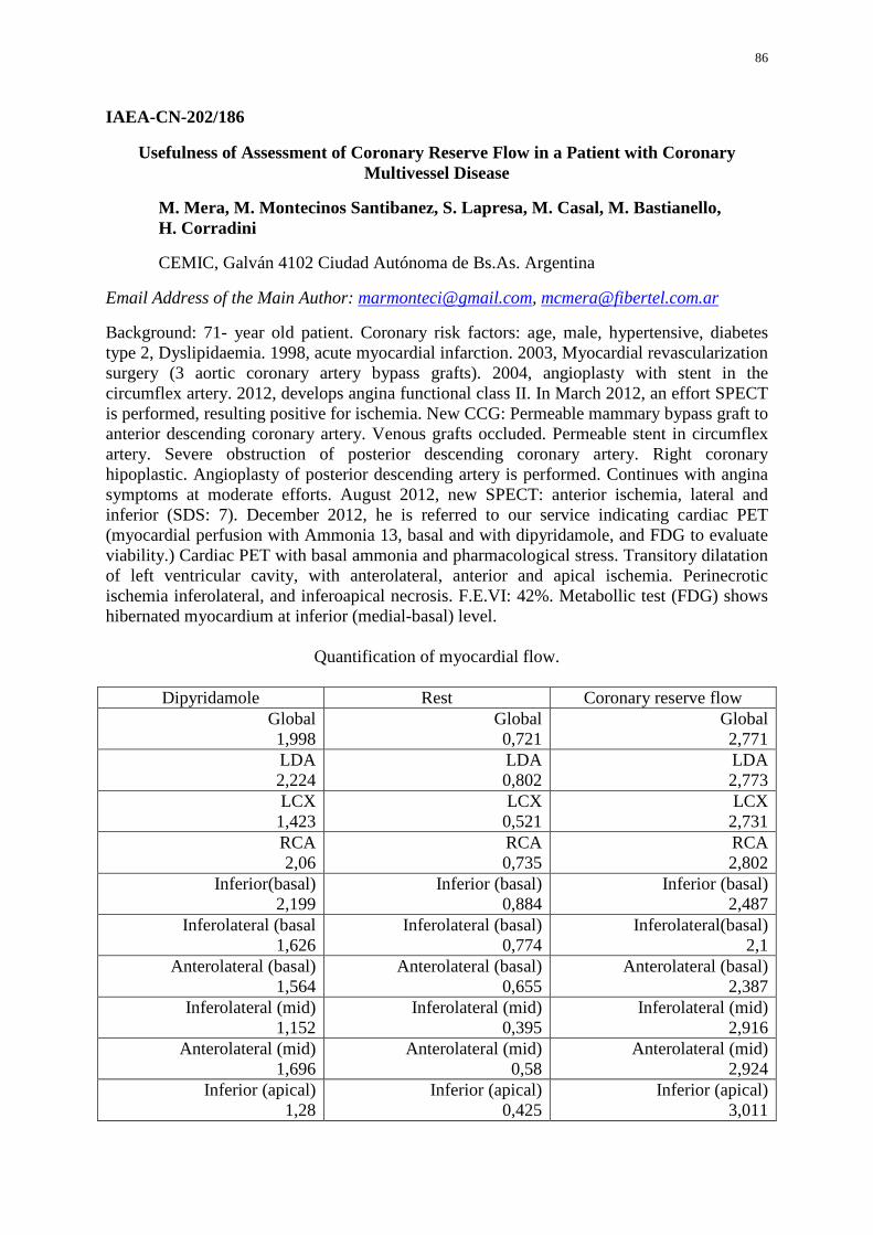
86
IAEA-CN-202/186
Usefulness of Assessment of Coronary Reserve Flow in a Patient with Coronary Multivessel Disease
M. Mera, M. Montecinos Santibanez, S. Lapresa, M. Casal, M. Bastianello, H. Corradini
CEMIC, Galván 4102 Ciudad Autónoma de Bs.As. Argentina
Email Address of the Main Author: [email protected], [email protected]
Background: 71- year old patient. Coronary risk factors: age, male, hypertensive, diabetes type 2, Dyslipidaemia. 1998, acute myocardial infarction. 2003, Myocardial revascularization surgery (3 aortic coronary artery bypass grafts). 2004, angioplasty with stent in the circumflex artery. 2012, develops angina functional class II. In March 2012, an effort SPECT is performed, resulting positive for ischemia. New CCG: Permeable mammary bypass graft to anterior descending coronary artery. Venous grafts occluded. Permeable stent in circumflex artery. Severe obstruction of posterior descending coronary artery. Right coronary hipoplastic. Angioplasty of posterior descending artery is performed. Continues with angina symptoms at moderate efforts. August 2012, new SPECT: anterior ischemia, lateral and inferior (SDS: 7). December 2012, he is referred to our service indicating cardiac PET (myocardial perfusion with Ammonia 13, basal and with dipyridamole, and FDG to evaluate viability.) Cardiac PET with basal ammonia and pharmacological stress. Transitory dilatation of left ventricular cavity, with anterolateral, anterior and apical ischemia. Perinecrotic ischemia inferolateral, and inferoapical necrosis. F.E.VI: 42%. Metabollic test (FDG) shows hibernated myocardium at inferior (medial-basal) level.
Quantification of myocardial flow.
Dipyridamole Rest Coronary reserve flow Global 1,998
Global 0,721
Global 2,771
LDA 2,224
LDA 0,802
LDA 2,773
LCX 1,423
LCX 0,521
LCX 2,731
RCA 2,06
RCA 0,735
RCA 2,802
Inferior(basal) 2,199
Inferior (basal) 0,884
Inferior (basal) 2,487
Inferolateral (basal 1,626
Inferolateral (basal) 0,774
Inferolateral(basal) 2,1
Anterolateral (basal) 1,564
Anterolateral (basal) 0,655
Anterolateral (basal) 2,387
Inferolateral (mid) 1,152
Inferolateral (mid) 0,395
Inferolateral (mid) 2,916
Anterolateral (mid) 1,696
Anterolateral (mid) 0,58
Anterolateral (mid) 2,924
Inferior (apical) 1,28
Inferior (apical) 0,425
Inferior (apical) 3,011

87
Lateral (apical) 1,192
Lateral (apical) 0,31
Lateral (apical) 3,845
Conclusion: The inferolateral-basal, inferobasal and anterobasal segments are the only ones which show response of the coronary reserve flow slightly diminished by pharmacological stimulus. The other tested segments show adequate flow response before vessel-dilating pharmacological stress. It is concluded that is a low ischemic risk test. The perfusion images analyzed qualitatively are discordant with quantitative coronary reserve flow tests in basal conditions and with pharmacological stress.
Patient continues clinically stable with optimization of pharmacological treatment (nitrates, trimetazadine, AAS, statins, beta blockers, and oral hypoglycemics).
88
IAEA-CN-202/187
18F-FDG PET-CT Imaging for the Evaluation of Small- and Medium-size Vessel Vasculitis
S. Voo, M. Kemna, P. van Paassen, J. W. Cohen-Tervaert, M. van Kroonenburgh
Maastricht University Medical Center, P. Debyelaan 25, 6200MD Maastricht, The Netherlands
Email Address of the Main Author: [email protected]
Background of the study: Small- and medium-size blood vessel vasculitis, including Wegener's granulomatosis, is a rather rare clinical entity which leads in most of the cases to life-threatening complications. Early diagnosis of active disease is therefore crucial in preventing a rapid fatal disease progression. Routine diagnostic tools, such as serum inflammatory markers and plasma antineutrophil cytoplasmic antibodies (ANCA), may often produce inconclusive results. 18F-FDG PET-CT is commonly used to identify active inflammatory lesions. This study evaluated the diagnostic value of 18F-FDG PET-CT in patients with suspected relapse of small- and medium-size vessel vasculitis.
Methods: Twenty-three patients with a history of small- and medium-size vessel vasculitis underwent 18F-FDG PET-CT imaging between December 2006 and June 2012 due to suspected relapse of their disease. CRP, as an inflammatory marker, and ANCA levels were determined at the time of scanning.
Results: Despite no significant increase in CRP levels and inconclusive ANCA levels, PET-CT identified foci of active inflammation in 19/23 cases with suspicion of active vasculitis. Common PET-positive sites were nasopharynx (12/19), lungs (11/19), and larynx (10/19). Corresponding tissue biopsies confirmed the diagnosis of active small- and medium-size vessel vasculitis in all these sites. Additional PET-positive sites were found in bone marrow (10/19), aorta (7/19), and thyroid gland (5/19), suggesting a multiorgan involvement in majority of patients (17/19).
Conclusion: PET-CT is able to accurately depict active sites of small- and medium-size vessel vasculitis, including in those in whom other diagnostic markers have been inconclusive. Majority of patients with relapsing disease show a multi-organ involvement on PET-CT.
89
IAEA-CN-202/188
Comparison of Myocardial Perfusion SPECT Acquired with Linear Energy High Resolution Collimators (LEHR) and SMARTZOOM Collimators, Automatic
Quantitative Software Differences
M. Havel, O. Kraft
University Hospital Ostrava, Department of Nuclear Medicine, 17. listopadu 1790, Ostrava 70852, Czech Republic
Email Address of the Main Author: [email protected]
Background: Myocardial perfusion imaging (MPI) is a standard examination performed in our clinic. We have been equipped with new "SMARTZOOM" collimators since 2012. With their installation we upgraded the evaluation software CORRIDOR4DM with appropriate normal databases for assessment and got the CEDARS-SINAI Cardiac Suite of Applications, too. We compared the results acquired with both Linear Energy High Resolution collimators (LEHR) and SMARTZOOM collimators and we also compared outcomes received from both automatic scoring systems.
Methods: For this purpose we utilised random group of 37 patients, (23 male, 14 female, average age 61,5 +- 8,0 years). 27 patients underwent both stress and rest procedure, in 10 patients only the stress study was performed. 22 patients were examined with both LEHR and SMARTZOOM collimators. Acquisition was undertaken on Symbia S double head SPECT camera, LEHR data were processed and reconstructed with filtered back protection, then evaluated with CORRIDOR 4DM software, data acquisited with SPARTZOOM collimators were reconstructed with vendors application and FLASH3D itterative reconstruction and evaluated with CORRIDOR4DM software and CEDARS-SINAI Quantitative Perfusion SPECT and Quantitative Gated SPECT (QPS and QGS) application. We compared parameters of Summed Stress Score (SSS), Sumed Rest Score (SRS), evaluated area of perfusion deficit, ejection fraction, end diastolic and end systolic volume in stress and rest (EFStr, EDVStr, ESVStr, EFRst, EDVRst, ESVRst).
Results: LEHR and SMARTZOOM collimators: SSS was higher for SMARTZOOM collimators in 16 cases (P=0.02), SRS was higher in 11 cases (P= 0.04). Evaluated extension of the perfusion defect did not show significant difference (P=0.16). Significantly higher for LEHR were EFStr (P<0.001) and EFRst (P=0.001), this correlate with significantly lower ESVStr and ESVRst for LEHR (P<0.001).
CORRIDOR4DM and QPS + QGS applications: Comparison of SSS did not show significant difference (P=0.303), but there were differences in SRS (higher values for Corridor4DM, P=0.001). Evaluated extension of the perfusion defect and it’s reversibility portion were significantly higher for Corridor4DM (P=0.001 for both parameters), EFStr and EFRst were higher in Corridor4DM (P<0.001). Differences were found in EDVstr (P<0.001), EDVRst (P<0.001) and ESVStr (P<0.001).

90
Conclusion: Utilising of new imaging systems and various applications could bring confusion; correlation with clinical data in following studies is essential.
91
IAEA-CN-202/189
Combination of Myocardial Perfusion SPECT with Calcium Score Measurement Improved the Detection of Ischemic Cardiomyopathy
L. Henzlováa, M. Kamíneka, I. Metelkováa, M. Budíkováa, P. Korandaa, M. Myslivečeka, H. Crhákováa, E. Buriánkováa, R. Formáneka, E. Sovováb, J. Ostřanskýb, M. Hutyrab
aDepartment of Nuclear Medicine, bDepartment of Internal Medicine - Cardiology, University Hospital, Olomouc, Czech Republic
Email Address of the Main Author: [email protected]
The aim of the study was to assess sensitivity and specificity of myocardial perfusion SPECT in the detection of coronary artery disease (CAD) in patients with a dilated cardiomyopathy, and to investigate the added value of the coronary artery calcium (CAC) as an adjunct to SPECT.
The study group consisted of eighty-one prospectively recruited patients with a dilated left ventricle who were scheduled for elective coronary angiography because of suspected CAD and who underwent gated SPECT imaging (part of them in combination with a CAC score measurement). Patients with known CAD, after myocardial infarction, and coronary revascularization were excluded. The summed difference score (converted to % ischaemic myocardium using 17-segment scoring system), the left ventricular end-diastolic volume (EDV), end-systolic volume (ESV), and ejection fraction (EF) were measured by 4D-MSPECT software. CAC Agatston score >1000 was considered as an extensive CAC. Fifty-four patients had CAD on coronary angiography (≥ 70% stenosis), and 41 of them had reversible perfusion defect (≥ 5% ischaemic myocardium).
Twenty-seven patients had normal arteries or non-significant luminal irregularities, and 21 of them had no significant reversibility (< 5% ischaemia). Patients with angiographically CAD had in comparison with those without CAD significantly higher amount of ischaemic myocardium (14% ± 13% vs. 3% ± 4%, respectively, P <0.05), and not significant lower stress EF, rest EF, and stress EDV/ESV (45%, 48%, and 181/102 mL vs. 48%, 50%, and 183/99 mL, respectively, P = NS). In the detection of significant CAD, sensitivity, specificity, positive predictive value (PPV), and negative predictive value (NPV) of SPECT alone were 80%, 78%, 88%, and 66%, respectively. Combining SPECT with the CAC score (at a cutoff of 1000) markedly improved the sensitivity and NPV to 91% and 81%, respectively, P <0.05), in association with not significant change in specificity (78%) and PPV (89%).
As a conclusion, it can be stated that in patients with a dilated left ventricle, combined SPECT and CAC score can help for identifying high-risk patients with significant CAD despite of normal perfusion SPECT results.
92
IAEA-CN-202/190
Myocardial SPECT CT: Evaluation of Calcium Score during Perfusion SPECT Adds Diagnostic and Prognostic Data
A. Y. Chambi Cotrado
Pró-cardíaco Hospital, UFF, General Polidoro street, 192, Rio de Janeiro, Brazil
Email Address of the Main Author: [email protected]
Background: Myocardial perfusion scintigraphy (MPS) has been widely used to estimate the risk of the presence of coronary artery disease (CAD), as well as in managing this condition. The calcification of coronary arteries is a marker of atherosclerosis and has been assessed by calcium score (CS) is associated with cardiovascular events in medium and long term. Several studies have shown that the combined analysis of an anatomical method, such as the EC, with perfusion scintigraphy making functional assessments provide additional diagnostic and prognostic information. Our objective was to analyze the correlation of the CPM with the presence of coronary calcification on computed tomography (CT) performed for attenuation correction in SPECT-CT (Symbia T2). Methods: An observational, retrospective unicenter. We analyzed the demographic findings, scintigraphy and computed tomography (SPECT-CT) of 62 consecutive patients, from 1 to 15 of 2012, who were referred to our institution to perform the CPM rest and stress associated with CT Low dose for attenuation correction in SPECT CT Symbia T2. Excluded from analysis were 24 patients who had a previous diagnosis of coronary artery disease. The CT was performed without Gated with current average 80 mAS and cut 5mm thick. The analysis was performed on the CS software Osirix ®. Statistical analysis employed the chi-square and Student's T test. Results: 38 patients (12 men) were analyzed without previous CAD, mean age 63 (+ 12 years) and underwent SPECT for the evaluation of CAD. We observed the following distribution of EC in the coronary arteries: (1) zero CE = 17 cases (44.7%), (2) between 1 and EC 17 = 400 cases (44.7%), and (3) greater than EC 4 = 400 cases (10.6%). When we analyze CPM observed in 4 patients (10.6%) had myocardial ischemia. By correlating the presence of coronary calcification observed on CT, scintigraphy with the findings, we found a significant association between the presence of coronary calcium and myocardial ischemia: no patients with zero calcium score showed ischemia, whereas 4 of 21 patients (19 %) with calcium present scintigraphy demonstrated ischemia (p = 0.02). The presence of chest pain that motivated the test request was not correlated with the EC on CT used for attenuation correction (p = 0.06). Conclusion: The analysis of the EC examinations performed on SPECT-CT myocardial perfusion appears to be a useful tool for risk stratification cardiovacular. The presence of

93
coronary calcification correlates with myocardial ischemia, which may correspond to the presence of obstructive coronary lesions, whereas zero EC was associated with normal exams.
94
IAEA-CN-202/191
Evaluation of Cardiac Synchronism by GATED SPECT: Impact of Myocardial Fibrosis
C. C. Wiefels, S. Garcia, J. Cunha de Azevedo, A. Ribeiro Nogueira Oliveira, E. Tinoco Mesquita, L. Evelyn Amorim, L. Rocha Messias, A. Oliveira, T. Cordeiro, T. Camilo, P. Resplandes, S. Marina Ribeiro Miranda, C. Tinoco Mesquita
Nuclear Medicine, Hospital Universitário Antonio Pedro, Universidade Federal Fluminense - Av. Marques do Paraná 303, Centro, Niterói, Rio de Janeiro, Brazil
Email Address of the Main Author: [email protected]
Background: Myocardial perfusion imaging after stress and rest using the SESTAMIBI radiotracer can determine the presence of ischemic areas and fibrosis, evaluate the contraction of the heart muscle and allows the calculation of the ejection fraction of the left ventricle by synchronized technique (gated SPECT). More recently new software development allows the assessment of contractile synchronism using Gated SPECT. The purpose of this study was to evaluate the impact of the presence of fibrosis (fixed perfusion defects) in contractile synchronism in patients with the technique of gated SPECT. Methods: We analyzed sixteen tests of myocardial perfusion imaging with Tc-99m sestamibi in patients (mean age 57 years, 8 men) at the Nuclear Medicine Service of our institution: five patients with normal myocardial perfusion (Group 1 - control), six patients with ischemic areas (Group 2 - reversible defects) and five patients with fibrosis (Group 3 - fixed defects). Relevant data obtained through clinical history were analyzed. We averaged data and subsequent t-test comparing the mean results between groups. The Software used for synchronism analysis was the ECT SyncTool in a Xeleris 3 workstation. Results: The synchronism parameters evaluated showed significant differences in Group 3 (patient with fibrosis) compared to Group 1 (control group 1): (A) SD - standard deviation: Group 1= 16; Group 3= 38 (p = 0.019) in the stress phase and Group 1= 29; Group 3= 52 (p = 0.05) in the resting phase; (B) Bandwidth – Bandwidth in Group 1=48 ; Group 3= 117 (p = 0.047) in the stress phase and Group 1= 77; Group 3= 171 (p = 0.039) in the resting phase. Also significant differences between Group 2 and 3 in the SD parameters and bandwidth in the resting phase were observed. There were no significant differences between the group with ischemia and the control group. Conclusion: These results suggest that myocardial fibrosis is a significant factor in determining contractile dyssynchrony.
95
IAEA-CN-202/192
Predicting Variables of Abnormal Myocardial Perfusion (MP) in Asymptomatic Patients
F. Faccio
Medicina Nuclear San Geronimo, Santiago del Estero 2750 3000 Santa Fe, Argentina
Email Address of the Main Author: [email protected]
Introduction: There is no unified consensus in relation to SPECT for the diagnosis and risk of a coronary artery disease (CAD) in asymptomatic patients (P).
Aims: To determine the existence or non existence of variables presented with higher incidence of abnormal stress/rest (S/R) MP, in asymptomatic P chosen for a diagnosis study.
Materials and Methods: Five hundred and fourteen P were evaluated prospectively. They were derived to the Nuclear Medicine Department to perform a screening S/R MP, because they were considered to be within the range of an intermediate risk for CAD. The following variables were analyzed: Age, Sex, Arterial Hypertension, Diabetes, Dyslipidaemia, Smoking, Stress, Peripheral Artery Disease, CAD family background, Body mass index (BMI), ECG alterations and abnormal Echocardiograms. Exclusion Criteria: 1) Thoracic pain; 2) P with known CAD (ACS; AMI, TCA, By Pass); 3) Dilated Myocardiopathy; 4) Left bundle branch block (LBBB) or its image through a definite pacemaker.
Results: One hundred and forty three abnormal MP (SSS >4) were observed. In the univariate analysis it was detected that the age >70 (p 0.0036), arterial hypertension (p 0.0002), diabetes (p 0.002), stress (p 0.047), peripheral artery disease (p 0.05), BMI > 25 (p 0.0016), ECG alterations (p 0.021), abnormal Echo (p 0.0009) and male (p 0.0000) presented significant differences between normal and abnormal MP. In the multivariate analysis the age > 70 (p 0.0044), male (p 0.0000), arterial hypertension (p 0.01), diabetes (p 0.0057), BMI > 25 (p 0.01), ECG alterations (p 0.02), and abnormal ECHO (p 0.001) remained as predicting variables.
Conclusion: In the asymptomatic P, the presence of arterial hypertension, diabetes, male, age > 70, BMI >25, ECG alterations and abnormal Echo are the variables with a higher power for detecting abnormal MP.
96
IAEA-CN-202/194
Phantom Study of Myocardial Perfusion SPECT-CT
C. J. Trauernicht, T. Kotze, R. Steyn, J. Boniaszczuk
Groote Schuur Hospital, Private Bag, Observatory, 7935, Cape Town, South Africa
Email Address of the Main Author: [email protected]
Background: Prone and supine myocardial perfusion acquisitions each have their own advantages and disadvantages. Supine acquisitions typically produce inferior wall attenuation or may yield images with splanchnic interference. Prone acquisitions typically produce superior imaging of the inferior wall with reduced counts in the anterior wall. The distance of the myocardium to the collimator is larger in a prone acquisition on the Siemens Symbia T6 dual-head SPECT-CT gamma camera.
In 2012 Groote Schuur Hospital purchased a Siemens Symbia T6 SPECT-CT gamma camera, allowing CT attenuation correction of myocardial perfusion studies.
CT attenuation correction for myocardial perfusion studies is controversial; some studies suggest prone and supine acquisitions are superior. In order to decide on the most appropriate imaging protocol while taking patient comfort into consideration, it has become necessary for us to decide whether to image all patients prone and supine, or to use CT attenuation correction only.
Methods: The IAEA kindly donated a cardiac phantom insert for the Jaszczak phantom to the hospital under the RAF 6038 project. A number of defects were simulated with this phantom. CT attenuation corrected images of the phantom were compared with non-attenuation corrected images.
Results: will be presented
Conclusion: Currently both prone and supine image sets are acquired at the hospital during myocardial perfusion studies, as well as a CT scan for attenuation correction. This places a huge strain on the resources (long imaging time), as well as additional dose to the patient from a CT scan. This study will help to determine which protocol to follow in future.
97
IAEA-CN-202/195
Efficacy of Full Fat Milk versus Diluted Lemon Juice to Reduce Interfering Infra-Cardiac Activity of Tc-99m Sestamibi during Myocardial Perfusion Imaging
K. Purbhoo
Department of Nuclear Medicine and Molecular Imaging, Chris Hani Baragwanath Academic Hospital and Charlotte Maxeke Johannesburg Academic Hospital, University of the Witwatersrand, Johannesburg, South Africa
Email Address of the Main Author: [email protected]
The use of Single Photon Emission Computed Tomography (SPECT) myocardial perfusion imaging (MPI), with Technetium – 99m (99mTc) Sestamibi in conjunction with either exercise, pharmacologic stress or both is an established tool for both the diagnosis and prognostication of patients with ischemic heart disease.
For perfusion imaging with SPECT, 99mTc labeled radiopharmaceuticals (Sestamibi or Tetrofosmin) are commonly used. The major metabolic pathway for clearance of Sestamibi is the hepatobiliary system which creates difficulty in both visual and quantitative interpretation of myocardial perfusion particularly of the inferior and infero-septal walls after reconstruction. Diluted lemon juice, an acid-rich drink is an alimentary cholekinetic that facilitates Sestamibi transit through the liver. Whole milk stimulates liver clearance as well as increases peristaltic movement. The aim of the study was to determine which protocol would be the best to reduce interfering infra-cardiac activity and therefore result in an improvement in image quality. We compared the use of full fat milk to diluted lemon juice. All patients referred to our institution for MPI from November 2009 to May 2012 were enrolled in the study. A total of 559 patients were randomized without stratification into two groups. Group 0 (G0) were given diluted lemon juice, 246 patients and full fat milk to group 1 (G1), 313 patients. Raw data of both the stress and rest images were visually and quantitatively assessed by two nuclear medicine physicians for the presence of infra-cardiac activity. The physicians were blinded to the intervention received and the data were reviewed simultaneously. The overall incidence of interfering infra-cardiac activity at stress was 84.1% and 84.5% in G0 and G1, respectively, (p>0.05) and at rest was 91.6%, and 90.1% in G0 and G1, respectively, (p>0.05). The administration of milk, especially for studies done at rest, somewhat resulted in a decrease in the intensity of infra-cardiac activity compared to the lemon juice group. The difference between the two groups however were statistically insignificant.
98
IAEA-CN-202/196
Changes in Myocardial Perfusion in Patients Undergoing Cardiac Resynchronization
C. Martínez, G. Solis, D. López, A. Puente
ISSSTE Centro Médico Nacional “20 de Noviembre” Avenida Coyoacán 540 Delegación Benito Juárez, Distrito Federal, Mexico
Email Address of the Main Author: [email protected]
Cardiac resynchronization therapy has been shown to be effective in improving hemodynamic and functional parameters of the heart, reflecting its benefits in functional class and quality of life, but has not explored the changes in myocardial perfusion produced after this therapy, so we posed the following research question:
What are the changes caused by cardiac resynchronization therapy on myocardial perfusion assessed by SPECT study in patients with cardiac dyssynchrony?
We studied 50 patients undergoing cardiac resynchronization therapy, sent to the Nuclear Cardiology Department, Centro Medico Nacional “20 Noviembre" were performed gated myocardial perfusion SPECT as part of cardiac resynchronization protocol, and control was 3 or 6 months.
We analyzed the general characteristics of patients with chi square, and the variables of perfusion defects and function GATED by McNemar test.
The results demonstrate that the myocardial perfusion study should be part of the study protocol of patient candidates for cardiac resynchronization, because it allows one to determine the etiology of dilated cardiomyopathy. Ischemic patients were those who demonstrated ischemia and viability without revascularized have but forecast that patients are taken to revascularization, were demonstrated functional changes in the gated SPECT variables.
99
IAEA-CN-202/198
Cardiac Adrenergic Innvervation Imaging Assessed with SPECT: Imaging Beyond Planar Cardiac Adrenergic Innvervation Scan
D. Vajauskas, A. E. Tamošiūnas
Vilnius University Hospital, Santariškių klinikos, Radiology and Nuclear medicine center, 2, Santariškių str., Vilnius, Lithuania
Email Address of the Main Author: [email protected]
Background of the study: Global cardiac adrenergic innervation assessed with 123 Iodine labeled metaiodobenzylguanidine (MIBG) SPECT showed promising results for heart failure risk stratification. New data confirms MIBG SPECT value predicting cardiac events for heart failure patients. Although cardiac MIBG SPECT data significantly differs from myocardial perfusion imaging data, especially with lower uptake in apex and inferior wall of left ventricle. This difference could lead to investigators’ misinterpretation. Lack of knowledge in normal cardiac MIBG SPECT imaging and experience evaluating cardiac MIBG SPECT data leads to overestimation of adrenergic denervation area and prediction of poorer prognosis for the patient.
Methods: Cardiac MIBG imaging data were acquired for 91 subjects with NYHA II – IV heart failure and 12 healthy subjects. Images acquired at early and late phase in planar and SPECT mode. Early and late heart to mediastinum (H/M) ratios and washout (WR) ratios were calculated from cardiac MIBG planar images. Early and late summered denervation defect scores, and denervation defect scores difference expressed in absolute units and by percent were calculated from reconstructed cardiac MIBG SPECT data.
Results: Cardiac regional adrenergic innervation heterogeneity was detected despite normal global adrenergic innervation values. As for the patients with impaired cardiac global adrenergic innervation values, the regional cardiac adrenergic innervation assessed with SPECT data could be divided in to four groups: patients with certain cardiac adrenergic denervation defects; patient without certain cardiac denervation defects but with uneven uptake of MIBG in left ventricle - global down regulation of cardiac adrenergic innervation; patients with adrenergic denervation defects localized in areas of normal MIBG uptake reduction in left ventricle - less significant global down regulation of cardiac adrenergic innervation; and patients for whom cardiac SPECT data could not be properly reconstructed due to low count rate from left ventricle - not enough counts to reconstruct SPECT data, or high MIBG uptake in adjacent organs (lungs, liver), when cardiac SPECT data could be reconstructed, but due to large artifacts in inferior wall (liver) or in lateral wall (lung), could not be evaluated properly. Our results suggest that reconstruction of cardiac MIBG SPECT data, when late H/M ratio is below 1,85 in most cases is difficult or impossible.
Conclusion: Our initial results suggest that regional cardiac adrenergic innervation data assessed with SPECT provide additional information next to global cardiac adrenergic innervation and this could lead us to better cardiac events risk assessment for heart failure

100
patients. Although there could be limited cardiac MIBG SPECT value to the patients with greater than moderate reduction of global cardiac adrenergic innervation, assessed from planar cardiac MIBG scans.
The reconstruction and interpretation of cardiac MIBG SPECT data needs good knowledge of normal regional cardiac MIBG uptake in left ventricle, which differs from myocardial perfusion imaging.
101
IAEA-CN-202/199
Abnormalities of Myocardial FDG Uptake during Routine Oncology Positron Emission Tomography Studies
Y. Kmetyuk, O. Moskalets, O. Bondaruk, A. Ashykhmin
Clinical Hospital “Feofania”, Ukrainian Center of Radiosurgery (using PET/CT-technologies), 21 Zabolotnogo str., Kiev, Ukraine
Email Address of the Main Author: [email protected]
Positron emission tomography (PET) with fluorodeoxyglucose (FDG) is a method used widely for the evaluation of cancer and which also has an important role in the evaluation of ischemic heart disease. Recently, increased myocardial FDG uptake has been reported to be related to some types of heart disease, such as endocarditis, hypertrophic or dilated cardiomyopathy and radiation-induced myocarditis. However, FDG PET imaging in patients with non–insulin-dependent diabetes mellitus (NIDDM) is problematic because of poor uptake of FDG. Although non-specific uptake in the myocardium is a common finding as well as in the stomach and bowels in whole-body FDG-PET, patients with cancer or cancer screening incidentally have some heart disease.
The goal of our study was evaluated the FDG pattern in patients with different tumours, health condition and impact of different medications, such as chemotherapy drugs, steroids or beta-blockers.
Methods: Six hundred and fifty patients before and after antitumor treatment with or without comorbidities were enrolled in this study. Patients were referred to our department for whole-body FDG-PET/CT to detect or evaluate malignant tumors. Patients’ age, overweight, blood glucose level, fasting period, insulin injection, steroid treatment and usage of beta-blockers were recorded. Imaging studies were performed on a Biograph 64 PET/CT scanner (Siemens), which produces transaxial, coronal, and sagittal reconstructions of CT, PET, and fusion PET/CT data for interpretation. Results: Increased FDG uptake in the myocardium does not significantly correlate with the blood glucose level, age, or duration of fasting in non-diabetic patients. Only in the uncontrolled diabetic patients did high levels of blood glucose (>7.5 mmol/l) always result in absent or faint myocardial uptake of FDG. However, since glucose metabolism in the fasting state is quite heterogeneous even in the normal myocardium it is difficult to diagnose the presence of ischaemic myocardium by fasting FDGPET imaging alone. Nearly a third of patients demonstrated a significant change in SUVmean values in the myocardium after antracyclin-based chemotherapy Dilated cardiomyopathy shows enlarged left ventricle but more regional homogeneity, and fewer and less severe defects in FDG as compared with ischaemic cardiomyopathy.
Conclusions: A further prospective study with prolonged follow-up of ventricular function is warranted to explore the significance of enhanced FDG uptake as a marker of early

102
identification of antracyclin-induced cardiotoxicity. Moreover the possibility of diagnostics for cancer-independent heart disease will be prolonged.
103
IAEA-CN-202/200
F18-FDG PET/CT in Assessing Myocardial Viability: First Kuwait Experience
A. Al-Shammari, R. Ashkanani, S. Alabsi, S. Alenezi, M. Ghanem, N. Alnefesi
Department of Nuclear Medicine, Mubarak Al-Kabeer Hospital, Kuwait
Email Address of the Main Author: [email protected]
Introduction: Myocardial viability studies are a major contributor to help identify patients who are likely to benefit from coronary revascularization. This will help reduce operative costs and failure rate. Several diagnostic approaches are used to assess myocardial viability. The aim of this article is to summarize the firsthand experience in assessing myocardial viability, using F18-FDG PET/CT imaging, in Kuwait using two different approaches in patient's preparation. Subjects and methods: A comparison between two different protocols of preparing patients pre injection of F18-FDG has been made. 12 patients were prepared (group A) according to first protocol and 11 with the second protocol (group B). The first protocol consists of asking patients to take, a non standardized meal, with low carbohydrate content, followed by his usual treatment of oral hypoglycemic medication or insulin. In the second approach patients are fasting for at least 8 hours before arriving to the department and an oral glucose load and insulin boluses were administered according to the fasting plasma glucose level using a standard protocol. All patients received 10 mCi IV of F18-FDG, and one hour post injection images were acquired. Delayed two hour images were acquired for patients with poor or average quality early images. During a consensus review, two nuclear medicine physicians analyzed the cases commenting on the quality of the image using a 4 point scoring system as (1) excellent, (2) good, (3) average and (4) poor. Results: In all cases of group A, except two cases, patients had the desired glycemic control. The two cases which did not achieve the desired glucose level were rescheduled. All cases achieved the glycemic control in group B and no cases were rescheduled. Out of the two groups, non diabetic patients and patients using insulin were reported to have similar quality of image by the two nuclear physicians. Three of the four patients on oral hypoglycemic drugs of group A were not reported due to poor image quality and delayed imaging did not improve the image quality. All four studies were termed poor quality. In group B patients, four patients on oral hypoglycemic drugs were termed average quality on early imaging, and good quality on delayed. Discussion: While the simplicity of the first approach made it the preferred choice in many institutes, this approach has inherent limitations, particularly related to the patient adherence to the instructions given, the non standardization of the meal, and patient glucose control. In addition, many patients with coronary disease are insulin resistant, and the amount of endogenous insulin released after the meal will not induce maximal stimulation of myocardial glucose/FDG uptake and result in high tracer concentration in the blood pool. This problem was most noted in patients using oral hypoglycemic agents. The mechanism behind this could be that, the amount of endogenous insulin released was not enough in those patients or incompliance of patients to low carbohydrate meal. The theory behind the second approach is that: due to fasting state, the plasma free fatty acid levels are dramatically reduced, and metabolism in insulin-sensitive tissues switches to glucose use. F18-FDG is then injected

104
during the euoglycemic state, resulting in high and rapid myocardial FDG uptake and low tracer concentration in the blood pool, leading to improved image quality. Conclusion: To summarize, this is the first study of myocardial study using F18-FDG to evaluate myocardial viability in Kuwait. In our experience, the glucose loading with insulin sliding scale is more promising in preparing patients for myocardial viability study. This was most evident in patients on oral hypoglycemic drugs.
105
IAEA-CN-202/202
Prone Myocardial Perfusion SPECT Additional Imaging: Review of Results vs. Supine in the Interpretation
I. Berrocal Gamboa, C. Fonseca, M. Torres, J. Salas, U. González
Hospital San Juan de Dios, Cantón Central Frente Parque La Merced, San José, Costa Rica
Email Address of the Main Author: [email protected]
Background: Myocardial perfusion SPECT is a valuable tool with an important role in the diagnosis, prognosis, evaluation of therapy effectiveness and feasibility. However, it is a complex process involving a variety of artifacts that may occur throughout the study involving the patient, the radioisotope and equipment. Additional prone image can be performed to reduce the effects of the artifacts especially those inherent to the patient including: patient motion, diaphragmatic and breast attenuation. Main Goal: To observe and evaluate the change in the interpretation of adding prone imaging when attenuation were suspected in our MPI cardiovascular disease population. Methods: 932 myocardial SPECT reports have been reviewed. These were performed in our institution during 2012 with one day rest-stress protocol. 138 patients had an additional prone imaging, 72 males (52%) mean age 63,15 ± 8,48 y.o. (range: 36-88). They all had at least one cardiovascular risk factor. 63 patients (64%) performed pharmacological stress tests with adenosine infusion intravenously and Bruce protocol exercise stress test in 35. During acquisition we obtained supine imaging for rest and stress phases and additional prone was acquired after stress, if the physician required it, especially when attenuation factors were suspected as a breast or diaphragmatic attenuation, also when was seen extracardiac structures interposition. In addition, 22 patients had defects in anteroseptal territory probably related to LBBB, which were excluded from analysis. Observational and descriptive analysis: Percentages were obtained from different vascular territories affected that disappeared or persisted after additional image. Left ventricular wall motion and thickening were considered for the final report. Results: With supine position SPECT revealed 148 vascular territories with defects suspected of breast and diaphragmatic attenuation, 2 had extracardiac activity interposition and in 2 eventually patient motion were corrected. Prone imaging showed change in the defects in 91 (59.86%) vascular territories and persistence in 61 (40.13%). Due to attenuation was found 97.36% and 2.64% due to the other causes.

106
In addition we observed motility and thickening parameters were consistent with the final interpretation. Conclusion: To add prone imaging is feasible and associated with improved specificity, also reduced the rate of equivocal scans. Centers or hospitals that do not have enough resources for a hybrid equipment or to acquire gadolinium sources can implement this technique and improve the performance of studies and their interpretation. Gold standard or alternative method was necessary in order to confirm the results. Our observations are preliminary and would be interesting to consider a future blind reading among experts to assess interobserver reproducibility.
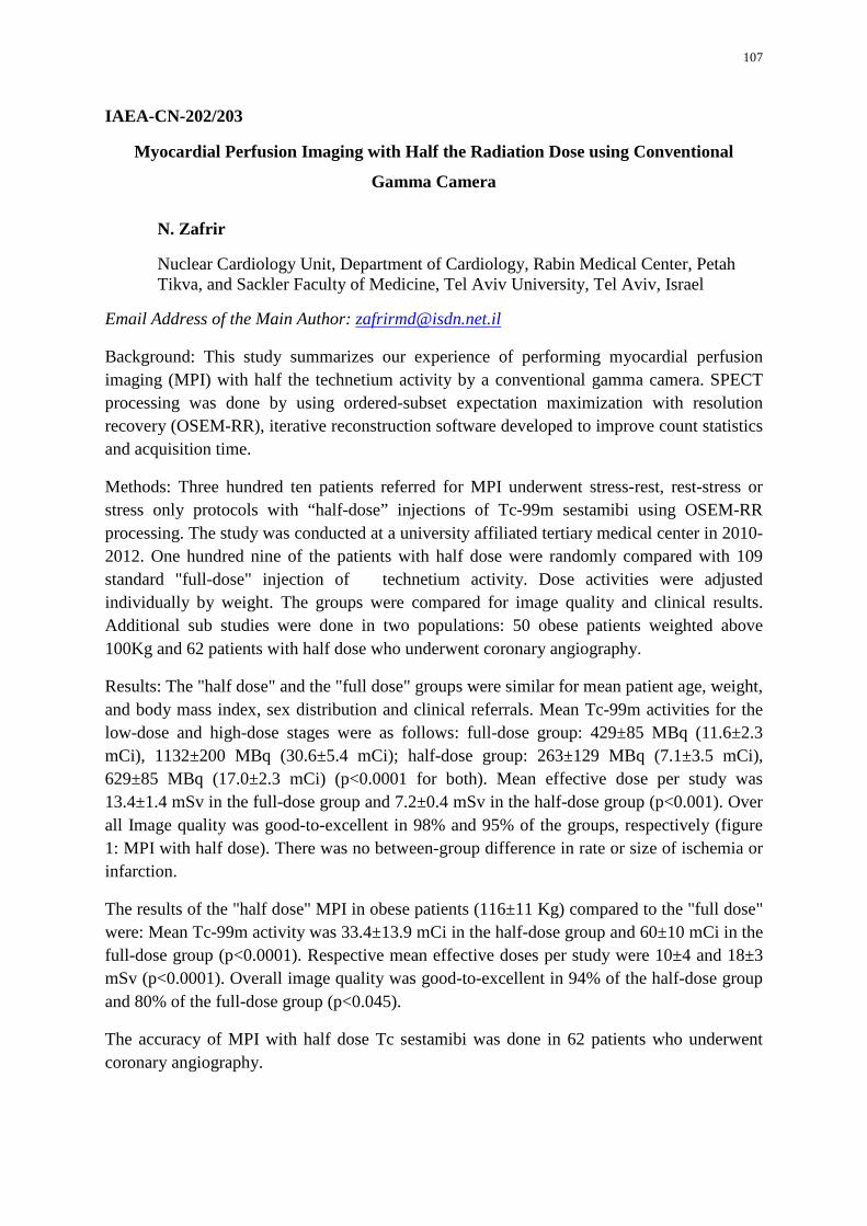
107
IAEA-CN-202/203
Myocardial Perfusion Imaging with Half the Radiation Dose using Conventional
Gamma Camera
N. Zafrir
Nuclear Cardiology Unit, Department of Cardiology, Rabin Medical Center, Petah Tikva, and Sackler Faculty of Medicine, Tel Aviv University, Tel Aviv, Israel
Email Address of the Main Author: [email protected]
Background: This study summarizes our experience of performing myocardial perfusion imaging (MPI) with half the technetium activity by a conventional gamma camera. SPECT processing was done by using ordered-subset expectation maximization with resolution recovery (OSEM-RR), iterative reconstruction software developed to improve count statistics and acquisition time.
Methods: Three hundred ten patients referred for MPI underwent stress-rest, rest-stress or stress only protocols with “half-dose” injections of Tc-99m sestamibi using OSEM-RR processing. The study was conducted at a university affiliated tertiary medical center in 2010-2012. One hundred nine of the patients with half dose were randomly compared with 109 standard "full-dose" injection of technetium activity. Dose activities were adjusted individually by weight. The groups were compared for image quality and clinical results. Additional sub studies were done in two populations: 50 obese patients weighted above 100Kg and 62 patients with half dose who underwent coronary angiography.
Results: The "half dose" and the "full dose" groups were similar for mean patient age, weight, and body mass index, sex distribution and clinical referrals. Mean Tc-99m activities for the low-dose and high-dose stages were as follows: full-dose group: 429±85 MBq (11.6±2.3 mCi), 1132±200 MBq (30.6±5.4 mCi); half-dose group: 263±129 MBq (7.1±3.5 mCi), 629±85 MBq (17.0±2.3 mCi) (p<0.0001 for both). Mean effective dose per study was 13.4±1.4 mSv in the full-dose group and 7.2±0.4 mSv in the half-dose group (p<0.001). Over all Image quality was good-to-excellent in 98% and 95% of the groups, respectively (figure 1: MPI with half dose). There was no between-group difference in rate or size of ischemia or infarction.
The results of the "half dose" MPI in obese patients (116±11 Kg) compared to the "full dose" were: Mean Tc-99m activity was 33.4±13.9 mCi in the half-dose group and 60±10 mCi in the full-dose group (p<0.0001). Respective mean effective doses per study were 10±4 and 18±3 mSv (p<0.0001). Overall image quality was good-to-excellent in 94% of the half-dose group and 80% of the full-dose group (p<0.045).
The accuracy of MPI with half dose Tc sestamibi was done in 62 patients who underwent coronary angiography.
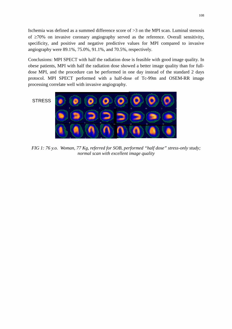
108
Ischemia was defined as a summed difference score of >3 on the MPI scan. Luminal stenosis of ≥70% on invasive coronary angiography served as the reference. Overall sensitivity, specificity, and positive and negative predictive values for MPI compared to invasive angiography were 89.1%, 75.0%, 91.1%, and 70.5%, respectively.
Conclusions: MPI SPECT with half the radiation dose is feasible with good image quality. In obese patients, MPI with half the radiation dose showed a better image quality than for full-dose MPI, and the procedure can be performed in one day instead of the standard 2 days protocol. MPI SPECT performed with a half-dose of Tc-99m and OSEM-RR image processing correlate well with invasive angiography.
FIG 1: 76 y.o. Woman, 77 Kg, referred for SOB, performed “half dose” stress-only study; normal scan with excellent image quality
STRESS
109
IAEA-CN-202/204
Usefulness of the Study of Myocardial Perfusion with Dipyridamole in Clinical Practice
A. A. Chaname Abad
Hospital Nacional Edgardo Rebagliati Martins, Salaverry 490, Jesus Maria, Lima 011 Peru
Email Address of the Main Author: [email protected]
Background of the study: In recent years the development of nuclear medicine has enabled advances in cardiovascular research through myocardial perfusion (MP) and tomographic (SPECT).
Methods: There have been 68 studies of MP-SPECT with dipyridamole in 2012, where it is evaluated, indications, pharmacological effects, side effects, electrocardiographic findings (ECG) and the results of MP.
Having confirmed that there are no contraindications for dipyridamole was administered 0.56 mgrs / Kg / min / via EV in 4 minutes with ECG monitoring and blood pressure. After the application of the drug, was administered MIBI -Tc99m -radiotracer.
According to protocol, an hour later, SPECT images were obtained by pharmacological stimulus, using a General Electric Gamma Camera, model Millennium MG dual-head, 180 ° tomographic advance mode and take, circular orbit, 64x64 matrix with 72 projections. Three hours later, the dose of MIBI -Tc99m was reinjected, corresponding to resting.
The first stage is obtained with multigated system to analyze parietal motility.
Results: 23 patients of the total had MP normal, 45 showed the contrary. Of these 68 patients, 15 had ischemic evidence at ECG during dipyridamole administration which in 100% had defects at MP. The others 53 patients didn't show ECG evidence of ischemia, correlating images from 18 patients with normal PM while the remaining 35 showed no correlation with the images because they had defects at MP.
Comparing the PM with the ECG findings during dipyridamole infusion is remarkable that in 67% of men there was no concordance, compared to 48% of women in which no correlation was found. No correlation has been observed of symptoms after injecting dipyridamole, with findings of MP, being independent of clinical manifestations.
110
Conclusion:
• The Dipyridamole test in our population is a safe test, from the clinical standpoint the side effects observed were mild to moderate and manageable with aminophylline isosorbide SL-EV, etc, being the most common: headache, breathlessness, chest tightness
• This test is particularly useful for studying patients with clinical conditions that limit exercise.
• None of our patients had serious side effects, defining these as those that endanger the patient's life.
• The concordance between MP with ECG records during dipyridamole administration was higher in women than in men, in which there were 15 (51%) who did show ischemic electrocardiographic correlation with MP-SPECT images; while only 13 males (33.33%) had ischemic ECG correlation with PM-SPECT images.
• It is noteworthy that in the majority of patients who showed evidence of ischemia in the test of MP-SPECT showed no electrocardiographic evidence of ischemia, demonstrating the low diagnostic sensitivity of ECG registered during dipyridamole administration to demonstrate this pathology.
• In cases where it has been shown medium and high risk ischemic through the test of MP is advisable to perform coronary angiography to proceed largely and percentage of cases to define myocardial revascularization.
TABLE N°1
RELATIONSHIP BETWEEN FINDINGS PM-SPECT-DPD Y ELECTROCARDIOGRAPHIC DURING DIPYRIDAMOLE ADMINISTRATION
EKG CONCURRING NOT CONCORDANT Total P.M. N° % N° % MEN 13 33% 26 67% 39 Patients WOMEN 15 52% 14 48% 29 Patients
Total 28 41%
40 59%
68 Patients
111
IAEA-CN-202/205
Myocardial Perfusion Imaging in Patients with Diabetes Mellitus
M. R. Mititelu, C. Mazilu
University Emergency Military Hospital “Dr Carol Davila” Bucharest, Romania
Email Address of the Main Author: [email protected]
Aim of this study was to assess the role of myocardial perfusion imaging SPECT in the evaluation and management of patients with diabetes mellitus (DM).
Methods: We perform a retrospective analysis of 128 patients (61f, 67m, mean age 58) referred to our department for stress-rest myocardial perfusion studies. In this group 51 were diabetic patients, 71 non-diabetic. All patients were evaluated after a protocol that included physical examination, ECG in rest and stress, gated SPECT myocardial perfusion imaging in stress and rest and angiographic examination. Acquisition was made after IV injection of 99mTc sestamibi or tetrofosmin, respecting the two days or one day protocol, using a dual head gamma camera Philips Axis. Images were assessed by two specialists in our department. Perfusion defects on stress images were considered fixed defects if remained unchanged on the rest images, and "ischemic" if they were new or worsening after stress imaging. We correlated our findings with coronarographic exam, where lesions were considered “significant” (<50%) and “non-significant” (<50%).
Results: Considering results of MPI – we divided patients in 4 groups: A patients with DM and significant obstruction on angiography – 33 (57.89%); B – pt with DM and non-significant lesions - 24 (42.11%); C – non-diabetics, significant coronary lesions 32 (45.07%); D non-diabetics, non-significant lesions 39 (54.93%). We obtained significant perfusion defects on MPI in 31 pt from group A (93,9%), 3 of group B (12,5%), 29 of group C (90,6%), 3 of group D (7,69%). We detected irreversible defects in 27 pt of group A (81%) and in 21 pt of group C (65%). None of the patients from group B and D had irreversible defects.
Conclusion: Myocardial perfusion SPECT showed a greater percentage of irreversible defects in the group of diabetic patients. Percentage of patients with perfusion defects and non-significant coronary lesions was greater in pts with DM (12.5% vs 7.69%). Concordance with coronarography was better for non-diabetic patients, mainly because diabetic pts usually have microangiopathy, which can't be demonstrated by assessing great vessel permeability. We can therefore consider myocardial perfusion SPECT with 99mTc labeled agents as an important tool for evaluation and management of diabetic patients.
112
IAEA-CN-202/206
Usefulness of SPECT-CT in Myocardial Perfusion Studies
R. Jaimovich, P. Orellana, J. C. Quintana, A. Haeger, D. Vicentini
Unidad de Medicina Nuclear, Hospital Clínico de la Pontificia Universidad Católica de Chile, Marcoleta 367, Santiago, Chile
Email Address of the Main Author: [email protected]
Background: Myocardial Perfusion Imaging (MPI) has a well-established role in coronary artery disease assessment in daily practice. Important clinical decisions including patient management, therapy, risk assessment and screening for coronary angiographic studies are often made according to the results of MPI. Traditional pitfalls of MPI technique include image attenuation artefacts, generally due to diaphragmatic attenuation of the inferior wall on men (more common in our population) and breast attenuation of anterior wall on women. During the last decade a new generation of gammacameras have been introduced in the market, being their characteristic the addition of a computed tomography (CT) scanner in order to perform attenuation correction to the single photon emission computed tomography (SPECT) study and provide anatomical correlation when necessary. The clinical advantages of performing SPECT-CT MPI are not clearly established in our clinical setting. Our aim was to review the impact of the use CT attenuation correction in the evaluation of inferior wall perfusion.
Methods: Retrospective analysis of 74 MPI studies performed on a General Electric SPECT-CT Hawkeye 4 during a one-year period. Three experienced Nuclear Medicine physicians analysed a set of reconstructed images on short axis, long horizontal and vertical axis. Images were displayed with and without applying attenuation correction, as well as end systolic and end diastolic reconstructed images. A consensual report of the inferior wall was obtained: Normal perfusion (Score 0), Equivocal (Score 1), Mild hypoperfusion (Score 2), Moderate hypoperfusion (Score 3), Severe hypoperfusion (Score 4) or Absent perfusion (Score 5). The analysis was performed on both, stress and rest series, in non-corrected and corrected images. On the latter a negative score (-1) was recorded if an over-correction artefact was present. End-systolic and end-diastolic images were evaluated to determine if significative thickening was present in the inferior wall.
Results: 74 MPI were analysed, 39 corresponded to pharmacological stress with dypiridamole.
45 MPI (61%) didn’t show significant differences between corrected and non-corrected images. 27 MPI (36%) had important information added by the attenuation correction images, thus changing the final report. Among these 27 cases, 8 of them showed normal wall thickening, thus orienting the report to attenuation artefact rather than perfusion abnormality. Two MPI (3%) showed overcorrection artefacts. One patient also had a pulmonary mass that was reported as an incidental finding on the CT.
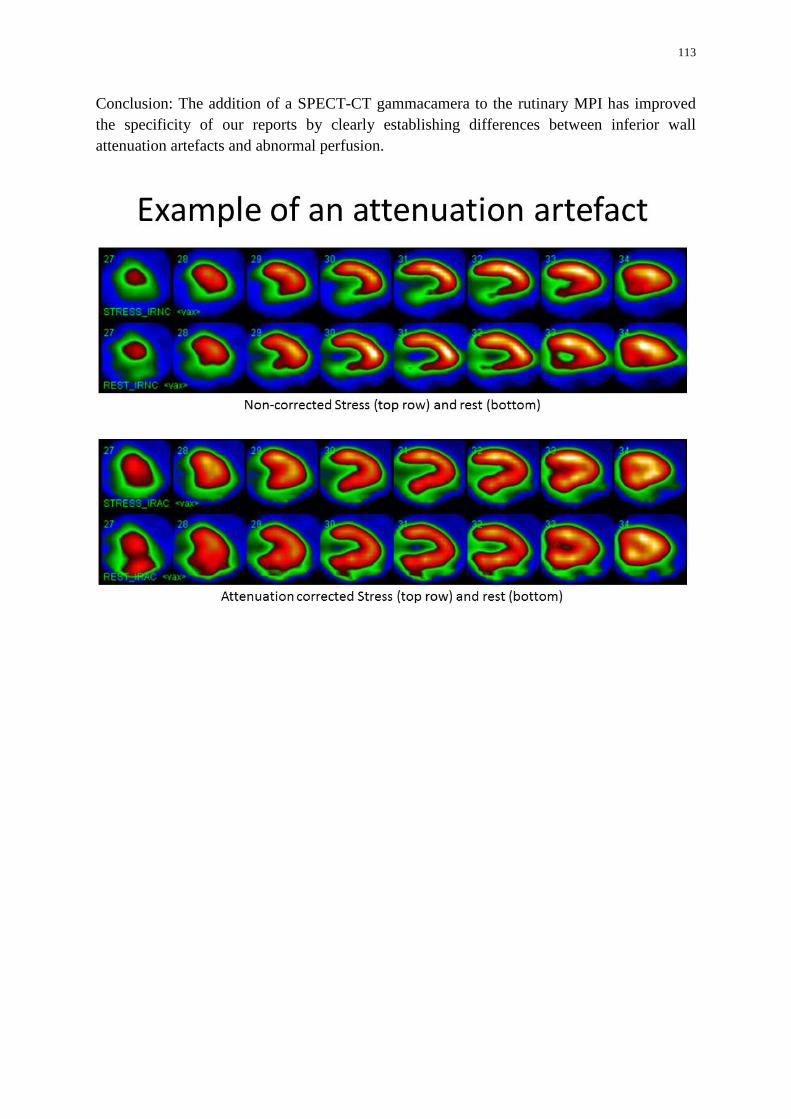
113
Conclusion: The addition of a SPECT-CT gammacamera to the rutinary MPI has improved the specificity of our reports by clearly establishing differences between inferior wall attenuation artefacts and abnormal perfusion.
114
IAEA-CN-202/214
Assessment of Suspected Coronary Arterial Disease using Myocardial Perfusion Scintigraphy (SPECT) Combined with Multi-Detector Computed Tomography in
a Brazilian Population
R.W. Lopes, I. M. F. Pinto, L. E. Mastrocola, F. B. P. Alves
Hospital do Coracao, Rua Desembargador Eliseu Guilherrne, 147, Paraiso, 04004-030, Sao Paulo, Brasil Instituto Dante Pazzanese de Cardiologia, Avenida Dr. Dante Pazzanese, 500, Vila mariana, Sao Paulo, Brasil
Email Address of the Main Author: [email protected]
Background: Coronary artery calcifications are markers of coronary atherosclerosis, but do not correlate well with ischemia or stenosis severity. This study intended to evaluate the combined approach of myocardial perfusion imaging (SPECT) and calcium score (CACS) by multi-detector tomography (MDTC), in a Brazilian population with suspected coronary artery disease (CAD).
Methods: we retrospectively analyzed 413 patients who underwent to SPECT and MDCT in 2009. CACS was automatically defined and Agatston score was semi-automatically calculated. SP ECT and MDCT results were evaluated by semi-objective visual quantification. Associations of tests results with clinical variables were analysed.
Results: We included 177 patients without OAC (71.2% men, mean age 55.6 ± I 0.6 years; range: 33-79 years); normal SPECT was seen in 84.7% and 45.2% had calcium (CA) with normal SPECT. Those with perfusion defects and CA showed more occurrences of: OM; CACS equal or above 75 percentile; and CACS >400. Those with normal perfusion and CA had the highest percentage of dyslipidemia (42.5%).
Conclusions: Patients with normal SPECT may have calcium on MDCT. This combination had association with dyslipidemia. Also, OM, CACS >400 and percentile equal or above 75% were associated with abnormal SPECT and calcium on MOCT.
115
IAEA-CN-202/215
Importance of Adequate Quality Control of Left Ventricular Boundaries with Commercial Software in Gated Myocardial Perfusion SPECT: Analysis of Patients with
Ventricular Dilation and Myocardial Infarction
P. Zhindon Pacurucu
Hospital Clínico Universidad de Chile Santos Dumont 999 1-E , Independencia, Santiago Chile
Email Address of the Main Author: [email protected]
Background: Gated myocardial SPECT allows to know important additional left ventricular (LV) functional parameters such as dilation, decreased ejection fraction (EF) or their changes due to myocardial infarction (MI), ischemia or even stunning. However, automatic methods could conduce to wrong values and incorrect boundary estimations caused by diminished perfusion and or extra cardiac activity that need semiautomatic or manual processing.
Goal: to compare the influence of diverse extension MI and dilation using automatic and manual methods in gated myocardial SPECT.
Methods: We studied 132 patients with gated SPECT using 99mTc-sestamibi referred to evaluate coronary artery disease, 80% males, mean age 63.7 years. The stress was Dipyridamole in 104 and exercise or Dobutamine in 27 cases. We measured end diastolic volume (EDV), end systolic volume (ESV) and LVEF at stress and rest with QGS® program with automatic and manual methods. We included 109 patients with MI, all with Q wave and/or fixed perfusion defects plus any abnormal regional wall motion: 48 non-dilated (EDV <120 ml at rest) and 61 dilated; other 23 patients with dilated LV and no IM, corresponding mostly to ischemic cardiomyopathy were added. We employed paired student t test and Spearman correlation to analyze according to the data normalcy.|
Results: Ten percent of our gated studies did not require modification of fully automatic method in stress and rest processing. MI rest defect extension ranged from 0-55% of LV and total perfusion defect (TPD) from 2 to 55. Correlations between automatic and manual methods for EDV and ESV (stress and rest together) in Group A were r=0.6728 and 0.6461; in Group B r=0.8879 and 0.8938; in Group C r=0.8801 and 0.8590, respectively; all cases p<0.0001. Regarding LVEF, correlation in Group A was r=0.6831, in Group B 0.8961 and in Group C 0.8002; there was more dispersion observed with higher LVEF values. Means ± SD values for volumes and LVEF are displayed in Table 1: globally, a) for volumes, automatic method presented larger values than manual in almost all dilated ventricles, and b) in all cases, automatic method presented larger LVEF. We also analyzed the correlation between both methods for rest EDV according to defect extension also at rest; more dispersion was obtained in non-dilated hearts with smaller lesions (<20% defect extension) compared with a subgroup of larger MI and LV (>35% defect extension) with r=0.773 (mean EDV=108 ml) versus r=0.948 mean EDV=171 ml), respectively.
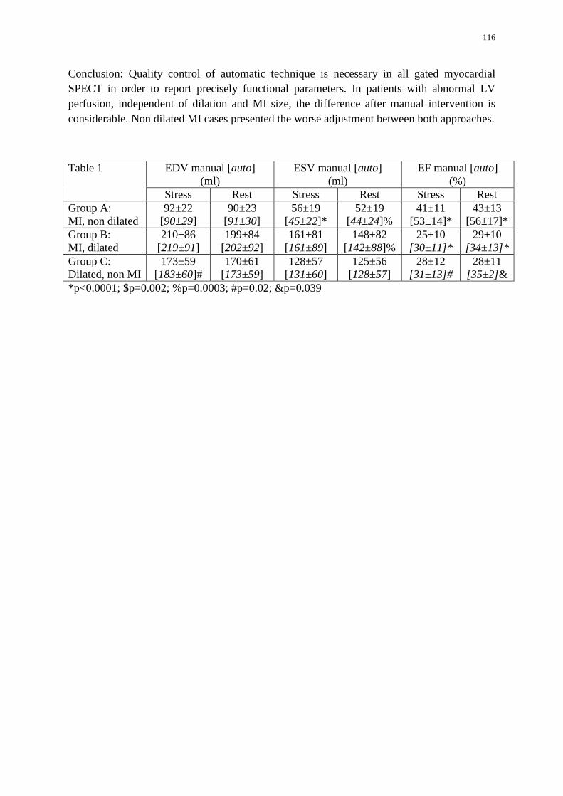
116
Conclusion: Quality control of automatic technique is necessary in all gated myocardial SPECT in order to report precisely functional parameters. In patients with abnormal LV perfusion, independent of dilation and MI size, the difference after manual intervention is considerable. Non dilated MI cases presented the worse adjustment between both approaches.
Table 1 EDV manual [auto] (ml)
ESV manual [auto] (ml)
EF manual [auto] (%)
Stress Rest Stress Rest Stress Rest Group A: MI, non dilated
92±22 [90±29]
90±23 [91±30]
56±19 [45±22]*
52±19 [44±24]%
41±11 [53±14]*
43±13 [56±17]*
Group B: MI, dilated
210±86 [219±91]
199±84 [202±92]
161±81 [161±89]
148±82 [142±88]%
25±10 [30±11]*
29±10 [34±13]*
Group C: Dilated, non MI
173±59 [183±60]#
170±61 [173±59]
128±57 [131±60]
125±56 [128±57]
28±12 [31±13]#
28±11 [35±2]&
*p<0.0001; $p=0.002; %p=0.0003; #p=0.02; &p=0.039
117
IAEA-CN-202/216
Assessment of Peripheral Neuropathy and Microalbuminuria in Coronary Asymptomatic Patients with Diabetes Mellitus Type 2 related with Myocardial Stress
Perfusion Abnormalities
L. T. D. P. Massardo Vega
Hospital Clínico universidad de Chile, Santos Dumont 999 1-E Santiago Chile
Email Address of the Main Author: [email protected]
Background: Diabetes mellitus type 2 (DM2) may affect diverse systems even in early stages and asymptomatic patients. Autonomic peripheral neuropathy (PN) is a serious complication observed usually in delayed stages and suboptimal metabolic control cases. Microalbuminuria (MA) is a good marker of renal damage also related to increased cardiovascular risk. Silent myocardial ischemia may present initially with systolic dysfunction or even fatal infarction.
Objective: To study an association between NP and MA in asymptomatic DM2 patient without known coronary artery disease, with the presence of myocardial perfusion abnormalities using stress test.
Methods:
Population: As a part of An IAEA multicentric project we evaluated 32 DM2 patients with >5 years of disease (average 12 years); they were 61±7 years old; 59% men, body mass index 29±4 kg/m2; coronary asymptomatic, without electrocardiographic Q wave and all with at least another cardiovascular risk factor.
Technique: All patients were studied with 99m Tc-Sestamibi SPECT using Bruce exercise initially ad after 3 years as well as serum profile and HbA1C. Ultrasensitive Reactive C Protein (usRCP), MA and a Michigan Test (MT) for PN detection were also performed at 3 years [Abnormal MT were: >2/10 points or according to physical auto evaluation score].
Analysis: Student t, Pearson r and Cohen k were applied.
Results:
- Main serum and urine mean and SD values at 3 years are shown in Table 1; 43% of the group had HbA1C<7,5%. They were treated medically with intensification of their lipid control.
- Basal SPECT was positive for silent ischemia in 34% initially and in 28% of the group at 3 years (p=ns) with transient perfusion defects (mild to moderate) but only 1 fixed defect corresponding to a myocardial infarction.
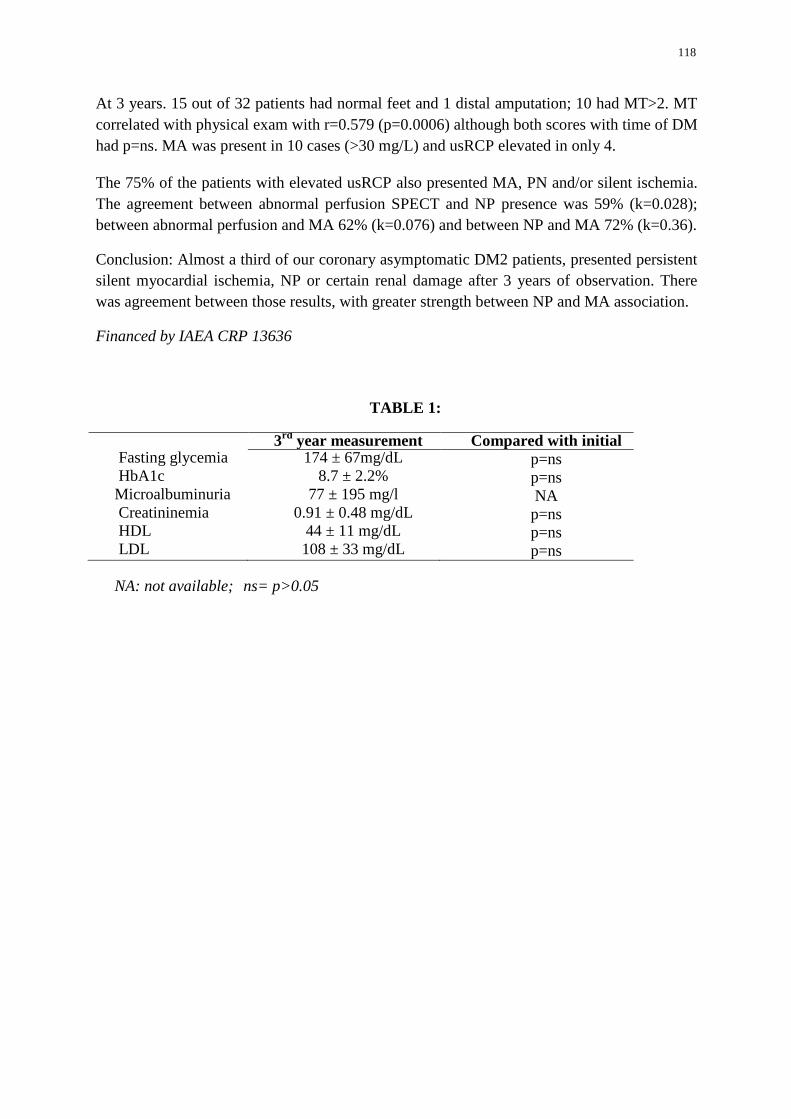
118
At 3 years. 15 out of 32 patients had normal feet and 1 distal amputation; 10 had MT>2. MT correlated with physical exam with r=0.579 (p=0.0006) although both scores with time of DM had p=ns. MA was present in 10 cases (>30 mg/L) and usRCP elevated in only 4.
The 75% of the patients with elevated usRCP also presented MA, PN and/or silent ischemia. The agreement between abnormal perfusion SPECT and NP presence was 59% (k=0.028); between abnormal perfusion and MA 62% (k=0.076) and between NP and MA 72% (k=0.36).
Conclusion: Almost a third of our coronary asymptomatic DM2 patients, presented persistent silent myocardial ischemia, NP or certain renal damage after 3 years of observation. There was agreement between those results, with greater strength between NP and MA association.
Financed by IAEA CRP 13636
TABLE 1:
3rd year measurement Compared with initial Fasting glycemia 174 ± 67mg/dL p=ns HbA1c 8.7 ± 2.2% p=ns Microalbuminuria 77 ± 195 mg/l NA Creatininemia 0.91 ± 0.48 mg/dL p=ns HDL 44 ± 11 mg/dL p=ns LDL 108 ± 33 mg/dL p=ns
NA: not available; ns= p>0.05
119
IAEA-CN-202/217
FDG –PET/CT for Assessment of Abdominal Aortic Infection with and without Graft: Experience in a Single Tertiary Medical Center
H. Shacham Suchman, A. Aharon, V. Kulikov, E. Even-Sapir
Department of Nuclear Medicine, Tel Aviv Sourasky Medical Center, 6 Wiezman Tel Aviv, Israel
Email Address of the Main Author: [email protected]
The results of previous publications in small patent groups suggest a potential use of FDG PET/CT to non-invasively assess the presence and extent of vascular infection. The purpose of the current study was to summarize the diagnostic value of FDG-PET/CT in detecting aortic infection found in a single tertiary medical center (Tel Aviv Sourasky Medical Center, Israel). Methods: 15 consecutive patients, 13 male and two female, age range 20 to 82 years were referred for FDG –PET/CT study from the vascular surgery department for suspected aortic infection, including 8 patients with vascular prosthesis grafts (group A) and 7 with no graft (group B) . Clinical presentation was either fever of unknown origin, severe abdominal pain, bacteremia or laboratory biomarker of infection. Patients were referred for this study as other imaging modalities as CTA and angiography were inconclusive or if had contraindication for IV contrast injection for CT. PET/CT study was performed 90 minutes after the IV injection of 370–666 MBq FDG using the Discovery LS PET/CT system (GE Medical Systems). CT was performed, with no specific breath-holding instructions followed by PET acquisition without changing the patient’s positioning. Studies were read on XELERIS workstation (ELGEMS) equipped with fusion software that enables the display of PET, and fused data. Studies were visually assessed as a consensus reading by two expert nuclear medicine physicians (EES,HL). Final diagnosis was based on histopathology when surgery was performed, microbiologic assays, clinical and imaging follow-up. Results: In group A, 7 of 8 patients had intense increased uptake around the graft raising the possibility of infection. Of these, 6 were true positive (TP), one false positive (FP), one true negative (TN) and no false negative (FN) resulting in a positive predictive value (PPV) of 87.5% and negative predictive value (NPV) of 100%. In group B, only one of the 7 referred patients had increased uptake in an abdominal aortic aneurism, which was proven to be TP. All 6 patients with negative studies had no infection based on further imaging and follow up resulting in PPV of 100% and NPV 100% in this cohort.

120
For all 15 study patients the total sensitivity of FDG-PET/CT for assessment of aortic infection was 100%, specificity 88% PPV88% and NPV 100%. Conclusion: FDG-PET/CT is a valuable noninvasive modality for detection of abdominal aortic infection in patients with or without graft. This modality is being used in our center in patients with clinical or laboratory suspicion for vascular infection, when routine imaging modalities are inconclusive or if contrast enhanced CT cannot be performed.
121
IAEA-CN-202/218
Myocardial Viability: Comparison between Studies Thallium/SPECT and FDG/PET
J. Gómez Garibo, J. Serna, S. Paredes
Hospital General Naval de Alta Especialidad (HOSGENAES), departamento de Medicina Nuclear e Imagen Molecular, México, D.F.
Email Address of the Main Author: [email protected]
Background of the study: The objective of this study is to compare the results of feasibility studies of myocardial perfusion SPECT and PET metabolism and its relationship to the stress test pharmacological and having as Gold Standard arteriography, to know the sensitivity, specificity and diagnostic accuracy of both studies and electrocardiographic correlation.
Methods: We performed a retrospective study in which we compare the results of Tl / SPECT and FDG / PET with your search electrocardiographic correlation for cardiac viability. We selected 16 patients with coronary artery disease (CAD) known have undergone both imaging techniques. Both studies were analyzed by two nuclear physicians with subspecialty in nuclear oncology and radiologists with subspecialty in PET / CT. The final diagnosis was issued based on the results of coronary angiography.
Results: A sensitivity of 70%, a specificity of 77.5%, a positive predictive value of 74%, a negative predictive value of 63.8% and an accuracy of 64.4% for the study Tl / SPECT. For the modality PET / FDG yielded a sensitivity of 90%, a specificity of 100%, a positive predictive value of 100%, a negative predictive value of 85.71% and an accuracy of 93.75%.
Conclusion: It was concluded that the utility of PET metabolic study/FDG for the diagnosis of myocardial viability is superior to SPECT perfusion study/Tl-201, thanks to its high sensitivity, specificity and overall results obtained should be considered as the first imaging study of choice for diagnosis of myocardial viability in the study of patients with known CAD, without dealing with this study that replaces the other as both techniques are complementary in the evaluation of CAD, having the advantage of being non-invasive and have no adverse effects practically taking high sensitivity and specificity in detecting CAD to define its extent and severity data from which they will define their prognosis and proper treatment.
122
IAEA-CN-202/219
Artefacts in SPECT Myocardial Perfusion
M. Zdraveska Kochovskaa, V. Majstorova, D. Pop Gjorcevaa, M. Vavlukisb, L. Peovskab
a Institute of Pathophysiology and Nuclear Medicine “Akad Isak S. Tadzer” Medical faculty Vodnjanska 17, Skopje, Macedonia b University Clinic of Cardiology, Medical Faculty, Skopje, Macedonia
Email Address of the Main Author: [email protected] Background: Myocardial perfusion imaging is an important imaging modality in the management of patients with cardiovascular disease. Performing myocardial perfusion imaging is a complex process and very often a number of variety artefacts may limit the clinical utility of the perfusion myocardial scintigraphy. These factors are related with the nuclear medicine equipment, with patient behaviour during the study performance and the actions of the technologist. The aim of the study was to evaluate common artefacts that appear in certain number of scans performed at the Institute of Pathophysiology and Nuclear Medicine. Methods: The imaging of patients was performed on SIEMENS E-cam Signature, one head SPECT gamma camera connected with ECG. Total number of patient studies that had been analysed was 450 (257 male and 193 female). The detected artefacts mostly arise from patient motion, tissue attenuation, gating and less from the nuclear medicine equipment. Results: Patient motion during acquisition of SPECT is one of the most common causes of artefacts. In 150 patient studies (33%) we have detected this kind of artefact. Patient motion can be detected on rotating cine displays as well as on the static sonograms. In order to produce accurate SPECT images re-imaging of patient might be necessary. Another patient related artefact (tissue attenuation) was detected in 54 patient’s studies (12%). Usually this situation can be overcome and solved by scanning the patients in the prone position. In 23 women’s scans there was found well known breast artefacts which manifested as anterior wall attenuation. Artefacts connected with COR error were not detected in any of the 450 studies. This is the result of routinely performed quality control on the gamma camera. The findings in our cases show that very often artefacts are detected from gating. In 20% of studies there were detected gated artefacts that cannot be used for interpretation. Conclusion: It is important to pay very close attention to technical factors and patient related issues which might arise during SPECT imaging, which can result in imaging artefacts and false positive scan results. It is essential for the technologist and interpreting physician to be aware of this potential source of error and to take appropriate steps to limit them beforehand, correct them when they occur and recognize their impact on the interpretation of the study. Only in that way the percentage of errors can be reduced. This will ensure that MPI has an important and improved role in the management of patients with cardiovascular disease.
123
IAEA-CN-202/220
Value of Gated SPECT Myocardial Perfusion Imaging with and without Attenuation Correction in Diagnosis of Coronary Artery Disease
H. N. Ha, H. M. Le
Nuclear Medicine Department, Tran Hung Dao Hospital, No.1 Tran Hung Dao Street, Hanoi, Vietnam
Email Address of the Main Author: [email protected]
Background: Artefacts hamper the accuracy of myocardial perfusion imaging (MPI). Available Single-Photon Emission Tomography (SPECT) systems may compensate for attenuation frequently observed in MPI images. The aim of this study was to determine value of gated-ECG SPECT MPI with attenuation corrected (AC) to non- attenuation corrected (NAC) SPECT MPI in diagnosis of coronary artery disease.
Methods: 202 consecutive patients suspected coronary artery disease (CAD) underwent Tc99m-sestamibi SPECT MPI and coronary angiography in Department of Nuclear Medicine and Heart Institute, Tran Hung Dao Hospital. The SPECT system with transmission hardware modifications and external gadolinium-153 sources were applied for AC and NAC imaging analysis. The AC and NAC SPECT MPI were visually scored and quantitatively interpreted by consensus reading of two experts, blinded to patient’s coronary angiogram findings with 30 days of SPECT imaging.
Results: In CAD diagnosis, overall sensitivity, specificity, accuracy of gated SPECT MPI with AC were 83.5%, 82.9%, 83.2%, respectively. There was no significant difference between AC and NAC SPECT MPI in diagnosis of CAD in all patient groups. However, for the detection of CAD in female and obese patient groups, receiver operating characteristic (ROC) analyses of visually and quantitatively interpreted images demonstrated significantly improved discrimination (p<0.05). In obese patients, artefacts were frequently observed in left anterior descending coronary artery (LAD) and right coronary artery (RCA) territories. Area under ROC (AUC) of this patient subgroup were 0.65 for AC images compared to 0.6 for NAC images (p<0.01) in detection of LAD stenosis (>70%) whereas AUC were 0.76 for AC images and 0.69 for NAC images in detection of RCA stenosis (p<0.01).
Conclusions: Attenuation corrected SPECT MPI could offer significant benefits to clinical SPECT imaging in female and obese patients suspected CAD in LAD and RCA territories.
124
IAEA-CN-202/221
The Value of Tc-99m MIBI SPECT during Nitrate Administration in Assessment of Viable Myocardium in Patients with Dilated Cardiomyopathy
A. Jakubović Čičkušić, B. Izić, M. Sulejmanović
University Clinical Centre Tuzla, Radiology and Nuclear Medicine Clinic, Bosnia-Herzegovina
Email Address of the Main Author: [email protected]
Background: Tc-99m MIBI myocardial SPECT has shown promise for evaluation of coronary artery disease, but its role in predicting myocardial viability is still under investigation.
The aim of the study was to determine clinical use and efficacy of the 99mTcMIBI with nitrate (ISDN) administration for detection of viable myocardium in patients with dilated cardiomyopathy.
Methods: Thirty-seven patients (27 male and 10 female) with dilated cardioyopathy were studied. All patients were examined under baseline study at rest and after administration ISND. The data were reconstructed in transaxial slices and then reoriented into short, vertical long and horizontal long axis slices. The images were divided into seven different segments for qualitative analysis. The images were interpreted by two independent observers.
Results: The results showed that out of 68 segments with hypoperfusion at resting SPECT, 29 segments (42.67%) had an increase in Tc-99m MIBI uptake during administration ISDN. The degree of improvement in perfusion was related to the age of patients.
Conclusion: The data suggest that use of Tc-99m MIBI SPECT in patients with dilated cardiomyopathy during administration ISDN may be a useful for assessing myocardial viability becouse ISDN augmented 99mTcMIBI protocol in Cardiac SPECT imaging resulting in improved detection of viable but hypoperfused segments.
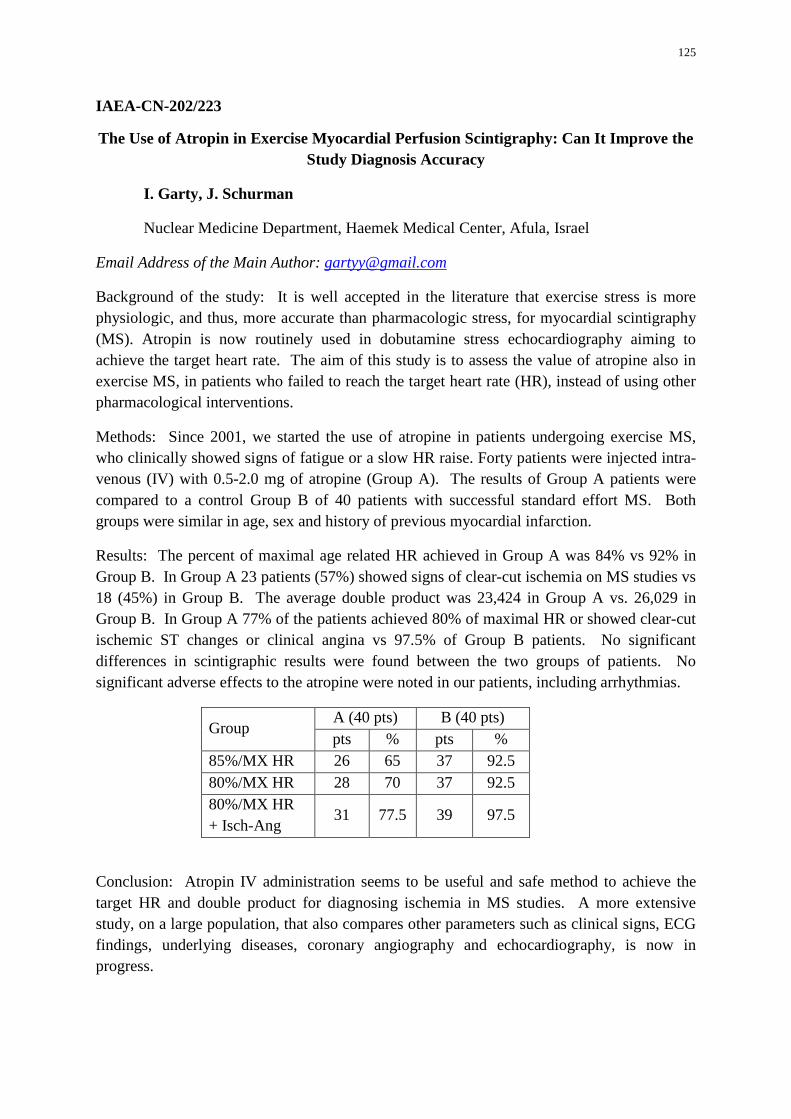
125
IAEA-CN-202/223
The Use of Atropin in Exercise Myocardial Perfusion Scintigraphy: Can It Improve the Study Diagnosis Accuracy
I. Garty, J. Schurman
Nuclear Medicine Department, Haemek Medical Center, Afula, Israel
Email Address of the Main Author: [email protected]
Background of the study: It is well accepted in the literature that exercise stress is more physiologic, and thus, more accurate than pharmacologic stress, for myocardial scintigraphy (MS). Atropin is now routinely used in dobutamine stress echocardiography aiming to achieve the target heart rate. The aim of this study is to assess the value of atropine also in exercise MS, in patients who failed to reach the target heart rate (HR), instead of using other pharmacological interventions.
Methods: Since 2001, we started the use of atropine in patients undergoing exercise MS, who clinically showed signs of fatigue or a slow HR raise. Forty patients were injected intra-venous (IV) with 0.5-2.0 mg of atropine (Group A). The results of Group A patients were compared to a control Group B of 40 patients with successful standard effort MS. Both groups were similar in age, sex and history of previous myocardial infarction.
Results: The percent of maximal age related HR achieved in Group A was 84% vs 92% in Group B. In Group A 23 patients (57%) showed signs of clear-cut ischemia on MS studies vs 18 (45%) in Group B. The average double product was 23,424 in Group A vs. 26,029 in Group B. In Group A 77% of the patients achieved 80% of maximal HR or showed clear-cut ischemic ST changes or clinical angina vs 97.5% of Group B patients. No significant differences in scintigraphic results were found between the two groups of patients. No significant adverse effects to the atropine were noted in our patients, including arrhythmias.
Group A (40 pts) B (40 pts) pts % pts %
85%/MX HR 26 65 37 92.5 80%/MX HR 28 70 37 92.5 80%/MX HR + Isch-Ang
31 77.5 39 97.5
Conclusion: Atropin IV administration seems to be useful and safe method to achieve the target HR and double product for diagnosing ischemia in MS studies. A more extensive study, on a large population, that also compares other parameters such as clinical signs, ECG findings, underlying diseases, coronary angiography and echocardiography, is now in progress.
126
IAEA-CN-202/224
Value of Normal Myocardial Perfusion Images Compared with Non-coronary Stenosis Determined by Coronary Angiographic for One Year Prognosis
L. O. Cabrera Rodriguez, A. Peix, K. Padrón, R. Carrillo, Y. Fernández, E. Mena
Institute of Cardiology, 17 No. 702 Vedado, Havana, Cuba
Email Address of the Main Author: [email protected]
Background: Exercise 99mTc-sestamibi SPECT is widely used for risk stratification, with a good long-term outcome after a normal test, but we have not found any comparative study between nuclear techniques. Purpose: The aim of the study was to compare the prognostic value in one year follow up of two different groups of patients with documented normal coronary arteries or normal perfusion images. Method: We enrolled 178 patients, (mean age 62 ± 15 years, 63% women), 95 (53%) with previous normal myocardial perfusion study (Group 1) and 83 (47%) with documented normal coronary arteries by coronary angiography (Group 2) in the same period of time. Patients were followed up for a mean of 1.4 ±0.6 years . Follow-up endpoints were cardiac mortality, nonfatal myocardial infarction, and coronary revascularization. Results: During the one year follow-up, there was no major adverse cardiac event (MACE) in Group 2, while in Group 1 2% of patients had some kind of cardiac event: one patient needed myocardial revascularization and another suffered a non-fatal myocardial infarction. Typical chest pain was the first criteria for sending patients to coronary angiography (76%). Atypical chest pain was present in 41% and typical chest pain in 53% in patients who were sent to myocardial perfusion study. One year later only 24% patients in Group 1 and 33% in Group 2 suffered typical chest pain. Conclusion: Myocardial perfusion imaging has a good prognostic value at one year follow-up when is compared with coronary angiography for predicting major adverse cardiac events.
127
IAEA-CN-202/225
Is Mechanical Dyssynchrony Related to Myocardial Viability: Simultaneous Evaluation with Gated-Single Photon Emission Computed Tomography
K. Padrón, L. O. Cabrera Rodriguez, A. Peix, J. A. García, R. Carrillo, Y. Fernández, E. Mena
National Cuban Institute of Cardiology and Cardiovascular Surgery, Cuba
Email Address of the Main Author: [email protected]
Introduction: In patients with ischemic dilated cardiomyopathy, the analysis of myocardial viability and mechanical dyssynchrony is very important to determine those patients whose benefit with cardiac resynchronization therapy and whose will be non responders. We had the hypothesis that they both (viability and dyssynchrony) are related.
Purpose: To determine the relation between myocardial viability and mechanical dyssynchrony, they both evaluated simultaneously by gated single photon emission computed tomography myocardial perfusion imaging (gated-SPECT MPI).
Methods: Fifty-four consecutive patients with dilated ischemic myocardiopathy and depressed left ventricular function (less than 40%) were enrolled. They underwent a gated-SPECT MPI following a two-day protocol (rest / rest nitroglycerine (NTG) test) to establish the myocardial viability and gated-SPECT imaging also be used, for the assessment of left ventricular dyssynchrony using a count-based Emory´s method to extract the amplitude and phase from regional left ventricular count changes throughout the cardiac cycle. We compared perfusion imaging, function and synchrony variables, at rest and before NTG in patients viable or non-viable, and we also compared them both. We used Mann-Whitney U and Wilcoxon W test to establish the association between variables; and Fisher´s Exact Test to qualitative variables. The significative value was p<0.05.
Results: The clinical characteristics (age, sex, risk factors, symptoms, anterior myocardial location, coronary anatomy) and functional variables (volume and left ventricular ejection fraction) were similar in both groups (viable and non-viable) in the baseline analysis. The size of necrotic area, the perfusions scores and the elapsed time from the infarct was significantly highest in non-viable group (p<0.01). After the administration of NTG, the viable showed significantly lowest perfusion score, improvement of the ventricular ejection fraction (80% of cases) and reduction of the end systolic volume (76% of cases) (p<0.01); while the non-viable remained invariable.
Viable or non-viable showed mechanical dyssynchrony in the baseline´s phase analysis (100% of cases), but it was highest in non viable group (p<0.05). After NTG, viable (64%) had significative improvement of mechanical dyssynchrony evidenced by reduction of phase standard deviation (SD) and histogram bandwidth (HB) values (p<0.001 versus non-viable), while non-viable experienced a significant increased (79%) in their initial values (p<0.01).

128
Conclusions: There is a relation between myocardial viability and mechanical dyssynchrony evidenced by NTG protocol: The viable improvement mechanical dyssynchrony after NTG, while the non-viable got worse. Gated-SPECT MPI is the only study which can make that simultaneous evaluation.
Clinical implication: These results could be useful to determine which patients with myocardial viability would be responders or not. We are studying that hypothesis now.
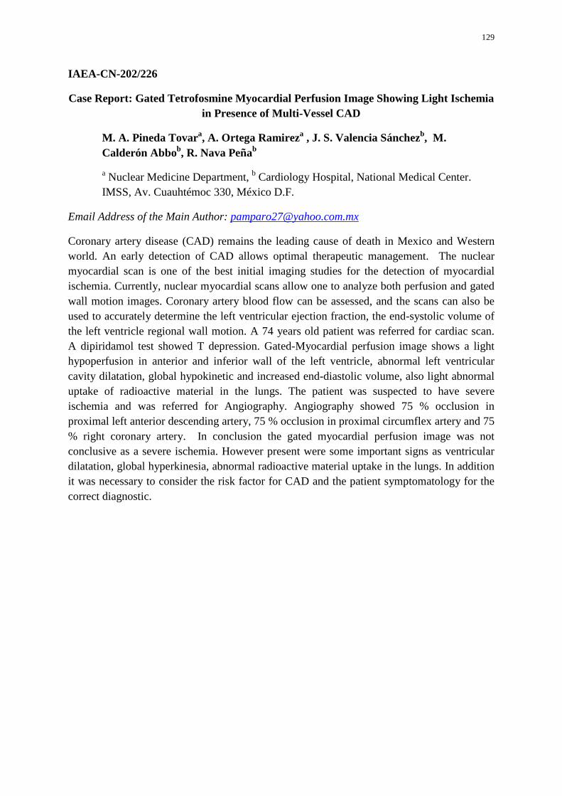
129
IAEA-CN-202/226
Case Report: Gated Tetrofosmine Myocardial Perfusion Image Showing Light Ischemia in Presence of Multi-Vessel CAD
M. A. Pineda Tovara, A. Ortega Ramireza , J. S. Valencia Sánchezb, M. Calderón Abbob, R. Nava Peñab
a Nuclear Medicine Department, b Cardiology Hospital, National Medical Center. IMSS, Av. Cuauhtémoc 330, México D.F.
Email Address of the Main Author: [email protected]
Coronary artery disease (CAD) remains the leading cause of death in Mexico and Western world. An early detection of CAD allows optimal therapeutic management. The nuclear myocardial scan is one of the best initial imaging studies for the detection of myocardial ischemia. Currently, nuclear myocardial scans allow one to analyze both perfusion and gated wall motion images. Coronary artery blood flow can be assessed, and the scans can also be used to accurately determine the left ventricular ejection fraction, the end-systolic volume of the left ventricle regional wall motion. A 74 years old patient was referred for cardiac scan. A dipiridamol test showed T depression. Gated-Myocardial perfusion image shows a light hypoperfusion in anterior and inferior wall of the left ventricle, abnormal left ventricular cavity dilatation, global hypokinetic and increased end-diastolic volume, also light abnormal uptake of radioactive material in the lungs. The patient was suspected to have severe ischemia and was referred for Angiography. Angiography showed 75 % occlusion in proximal left anterior descending artery, 75 % occlusion in proximal circumflex artery and 75 % right coronary artery. In conclusion the gated myocardial perfusion image was not conclusive as a severe ischemia. However present were some important signs as ventricular dilatation, global hyperkinesia, abnormal radioactive material uptake in the lungs. In addition it was necessary to consider the risk factor for CAD and the patient symptomatology for the correct diagnostic.
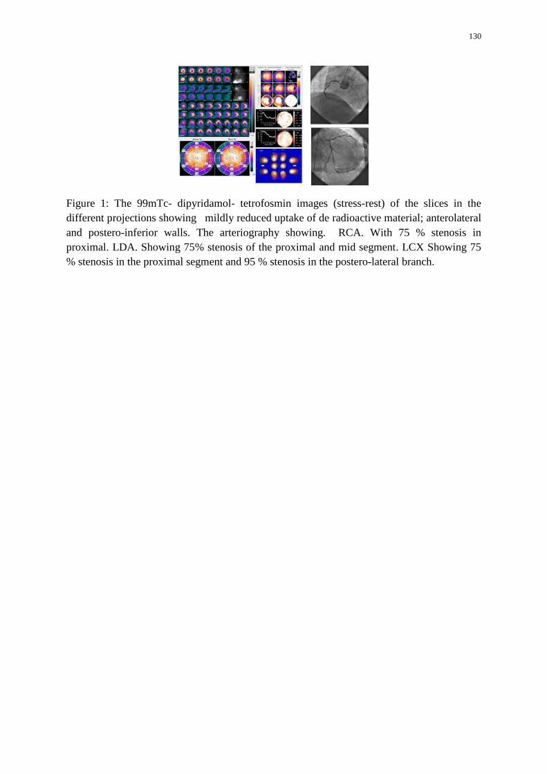
130
Figure 1: The 99mTc- dipyridamol- tetrofosmin images (stress-rest) of the slices in the different projections showing mildly reduced uptake of de radioactive material; anterolateral and postero-inferior walls. The arteriography showing. RCA. With 75 % stenosis in proximal. LDA. Showing 75% stenosis of the proximal and mid segment. LCX Showing 75 % stenosis in the proximal segment and 95 % stenosis in the postero-lateral branch.
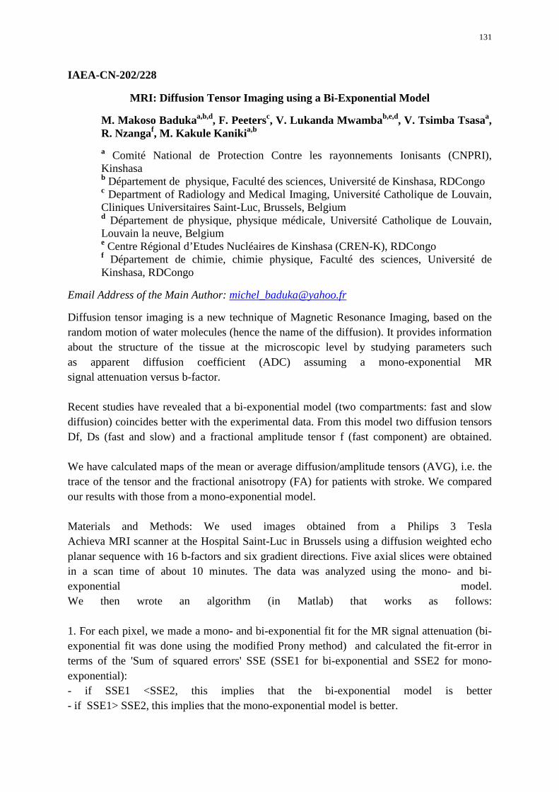
131
IAEA-CN-202/228
MRI: Diffusion Tensor Imaging using a Bi-Exponential Model
M. Makoso Badukaa,b,d, F. Peetersc, V. Lukanda Mwambab,e,d, V. Tsimba Tsasaa, R. Nzangaf, M. Kakule Kanikia,b a Comité National de Protection Contre les rayonnements Ionisants (CNPRI), Kinshasa b Département de physique, Faculté des sciences, Université de Kinshasa, RDCongo c Department of Radiology and Medical Imaging, Université Catholique de Louvain, Cliniques Universitaires Saint-Luc, Brussels, Belgium d Département de physique, physique médicale, Université Catholique de Louvain, Louvain la neuve, Belgium e Centre Régional d’Etudes Nucléaires de Kinshasa (CREN-K), RDCongo f Département de chimie, chimie physique, Faculté des sciences, Université de Kinshasa, RDCongo
Email Address of the Main Author: [email protected]
Diffusion tensor imaging is a new technique of Magnetic Resonance Imaging, based on the random motion of water molecules (hence the name of the diffusion). It provides information about the structure of the tissue at the microscopic level by studying parameters such as apparent diffusion coefficient (ADC) assuming a mono-exponential MR signal attenuation versus b-factor. Recent studies have revealed that a bi-exponential model (two compartments: fast and slow diffusion) coincides better with the experimental data. From this model two diffusion tensors Df, Ds (fast and slow) and a fractional amplitude tensor f (fast component) are obtained. We have calculated maps of the mean or average diffusion/amplitude tensors (AVG), i.e. the trace of the tensor and the fractional anisotropy (FA) for patients with stroke. We compared our results with those from a mono-exponential model. Materials and Methods: We used images obtained from a Philips 3 Tesla Achieva MRI scanner at the Hospital Saint-Luc in Brussels using a diffusion weighted echo planar sequence with 16 b-factors and six gradient directions. Five axial slices were obtained in a scan time of about 10 minutes. The data was analyzed using the mono- and bi-exponential model. We then wrote an algorithm (in Matlab) that works as follows: 1. For each pixel, we made a mono- and bi-exponential fit for the MR signal attenuation (bi-exponential fit was done using the modified Prony method) and calculated the fit-error in terms of the 'Sum of squared errors' SSE (SSE1 for bi-exponential and SSE2 for mono-exponential): - if SSE1 <SSE2, this implies that the bi-exponential model is better - if SSE1> SSE2, this implies that the mono-exponential model is better.
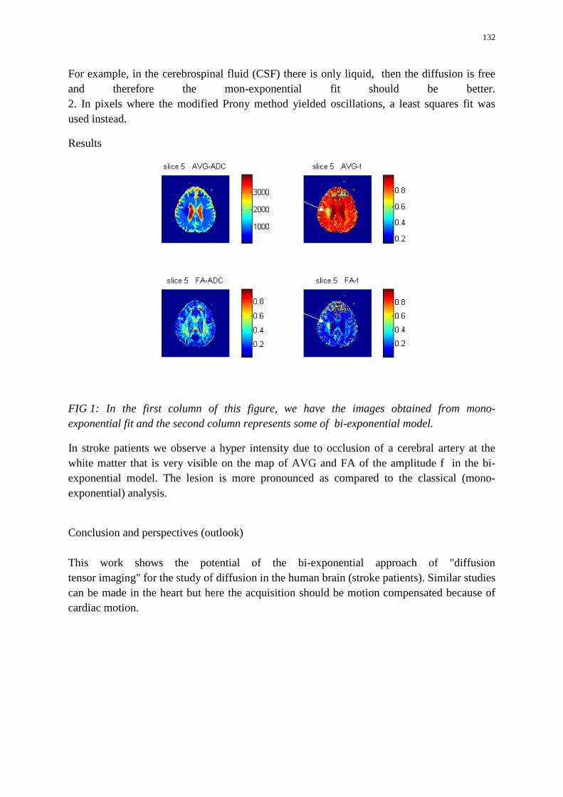
132
For example, in the cerebrospinal fluid (CSF) there is only liquid, then the diffusion is free and therefore the mon-exponential fit should be better. 2. In pixels where the modified Prony method yielded oscillations, a least squares fit was used instead.
Results
FIG 1: In the first column of this figure, we have the images obtained from mono-exponential fit and the second column represents some of bi-exponential model.
In stroke patients we observe a hyper intensity due to occlusion of a cerebral artery at the white matter that is very visible on the map of AVG and FA of the amplitude f in the bi-exponential model. The lesion is more pronounced as compared to the classical (mono-exponential) analysis.
Conclusion and perspectives (outlook) This work shows the potential of the bi-exponential approach of "diffusion tensor imaging" for the study of diffusion in the human brain (stroke patients). Similar studies can be made in the heart but here the acquisition should be motion compensated because of cardiac motion.
133
IAEA-CN-202/229
Coronary Computed Tomography Angiography as a Gatekeeper for Invasive Angiography in Patients with Mild Ischemia by SPECT-MPI in a Developing Country
R. J. Cerci, M. Zapparoli, F. R. Farias, S. S. Zier, C. Cunha, J. J. Cerci, O. Kormann, A. Stier Junior, R. Maruiti, O. Franca Neto, J. V. Vítola. Quanta Diagnóstico e Terapia, Rua Almirante Tamandaré 1000, Curitiba/Paraná, Brazil 80045170
Email Address of the Main Author: [email protected]
Background: To determine the usefulness of a diagnostic strategy based on performing coronary computed tomography angiography (CCTA) as a gatekeeper for invasive angiography (ICA) in patients without known coronary artery disease (CAD) and mild ischemia on SPECT-MPI.
Methods: Data from patients without known CAD, with mild ischemia (SSS ≥ 4 and <8) detected on SPECT-MPI, referred or not to CCTA between 01/01/2012 and 12/31/2012, from the Quanta Diagnostico e Terapia database (Curitiba/ Brazil) were retrospectively analyzed. We modeled two diagnostic scenarios in which patients would be: 1) submitted directly to ICA after the abnormal SPECT; 2) submitted to CCTA, followed by ICA only when obstructive disease was detected. For analysis purposes, the CCTA frequencies of normal, non-obstructive (< 50% stenosis) and obstructive (≥ 50% stenosis) CAD results of the subgroup submitted to CCTA were extrapolated to the whole cohort. Differences between groups were evaluated with chi square test.
Results: During the observation period of 1 year, 7,179 patients were referred for SPECT-MPI. Of these, 1,116 had mild ischemia on SPECT-MPI, and only 719 had no previous history of CAD. CCTA was performed in 103 (14.3%). The clinical and SPECT characteristics of the groups submitted or not to CCTA are presented on the Table. There were 47 (45.6%) normal, 35 (34%) non-obstructive and only 21 (20.4%) obstructive CAD results by CCTA. The only variable predictive of obstructive CAD by CCTA was male gender (35.9% vs. 10% for female gender, P = 0.002), in this cohort where female gender was more prevalent (62.8%). Extrapolating the CCTA results to the whole cohort, the CCTA as a gatekeeper proposed strategy would avoid an unnecessary ICA in 79.6% of patients.
Conclusion: A CAD diagnostic strategy in which CCTA is applied as a gatekeeper to ICA in patients with mild ischemia by SPECT is still underused, but would be very useful, especially in women, lowering the risks of unnecessary invasive procedures and potentially saving significant resources in developing countries.
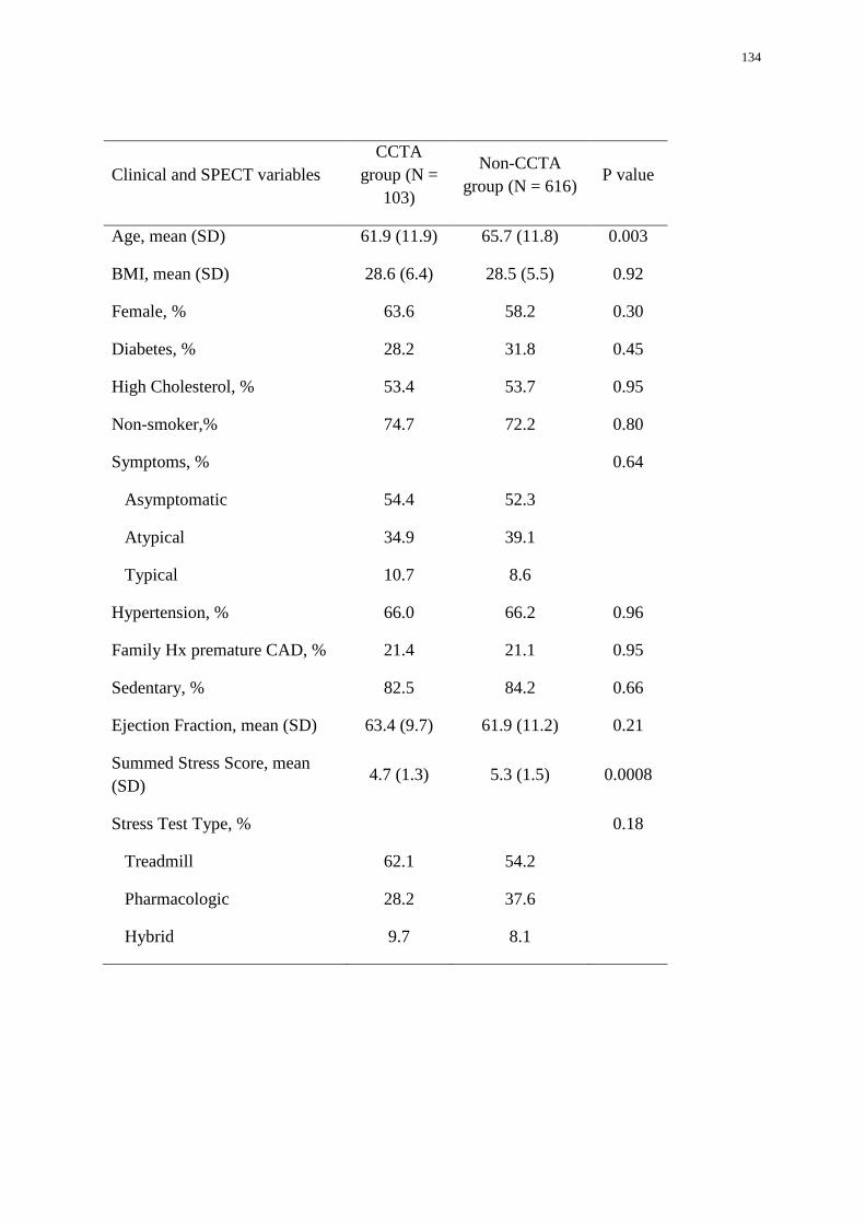
134
Clinical and SPECT variables CCTA
group (N = 103)
Non-CCTA group (N = 616)
P value
Age, mean (SD) 61.9 (11.9) 65.7 (11.8) 0.003
BMI, mean (SD) 28.6 (6.4) 28.5 (5.5) 0.92
Female, % 63.6 58.2 0.30
Diabetes, % 28.2 31.8 0.45
High Cholesterol, % 53.4 53.7 0.95
Non-smoker,% 74.7 72.2 0.80
Symptoms, %
Asymptomatic
Atypical
Typical
54.4
34.9
10.7
52.3
39.1
8.6
0.64
Hypertension, % 66.0 66.2 0.96
Family Hx premature CAD, % 21.4 21.1 0.95
Sedentary, % 82.5 84.2 0.66
Ejection Fraction, mean (SD) 63.4 (9.7) 61.9 (11.2) 0.21
Summed Stress Score, mean (SD)
4.7 (1.3) 5.3 (1.5) 0.0008
Stress Test Type, % 0.18
Treadmill 62.1 54.2
Pharmacologic 28.2 37.6
Hybrid 9.7 8.1
135
IAEA-CN-202/231
False Positive in Myocardial Perfusion SPECT Due to Left Bundle Branch Block Artefact
A. C. Jiménez, A. Alfaro, G. Castro, K. Calvo
Medicina Nuclear, Hospital Calderón Guardia, San José, Costa Rica
Email Address of the Main Author: [email protected]
Single photon emission computed tomography (SPECT) imaging of myocardial perfusion is a sensitive tool for detecting coronary artery disease (CAD). It is important to know and detect image artefacts related to both patient and technical factors.
Left Bundle Branch Block (LBBB) causes reversible septal perfusion defects that could mimic exercise induced septal ischemia. Probably the decrease in septal blood flow is due to asynchronous relaxation of the septum, which is out of phase with diastolic filling of the remainder of the ventricle, but also is postulated that is due to lateral hyperperfusion rather than due to a manifest septal flow decrease. This is most marked in LBBB patients who achieve very high heart rates.
For this reason it is recommended to use pharmacologic stress using vasodilator stress to minimize this artefact. Despite this fact, it is common in our centre that most of the patients with LBBB are referred to do myocardial perfusion SPECT under exercise protocol, which can produce high false positives rates.
We present a case of a 60 year male, Type 2 diabetic and hypertensive, non smoker, with atypical nocturnal chest pain. He was referred to undergo a myocardial perfusion SPECT because of the presence of LBBB.
He completed 6 minutes of Bruce protocol, reaching 9 METS and 100% of the predicted maximum heart rate. The study was acquired with the rest–stress one day sequence with 99m Tc Sestamibi.
On images (Figure), is seen relative reduction of anterior septal perfusion at stress with conserved perfusion at rest. QPS analysis showed SSS: 10, SRS: 4, SDS: 6. TID 0.95.
Due to these findings the patient underwent coronary angiography which showed only a 30% defect in circumflex artery.
To anticipate this reversible septal perfusion artefacts it is important that physicians inspects the ECG and in the presence of LBBB and preferable perform the myocardial perfusion test under vasodilator stress protocols.
The analysis of this clinical case was very important because it helps demonstrate to the referring physicians, how stress test with exercise is not recommended for SPECT imaging of

136
myocardial perfusion, reducing false positive results that may induce and unnecessary coronary angiography.
137
IAEA-CN-202/232
Cardiac and Vascular Effects of Chronic Cocaine Abuse in Young Asymptomatic Subjects
M. Kapitán Oteroa, A. Negrinb, D. Biac, M. Lujambioa, Y. Zócaloc, M. Panb, M. Langhaina, L. Floriod, I. Farroc, J. Torradoc, A. Pascaleb, R. Ferrandoa
a Nuclear Medicine Center, b Toxicology Department, c School of Medicine, CUiiDARTE, Republic University, Montevideo, Uruguay d Cardiovascular Center, Clinics Hospital, University of the Republic, Av. Italia s/n, Montevideo, Uruguay
Email Address of the Main Author: [email protected]
Background: The cardiovascular impact of chronic cocaine use involves different mechanisms. Early effects on ventricular and arterial function in humans have not been enough evaluated yet, particularly using nuclear medicine studies and non-invasive arterial structure and function techniques.
The aim of the study was to characterize the cardiac and vascular effects of chronic cocaine abuse in asymptomatic young individuals.
Methods: Eighteen patients meeting toxicological criteria for cocaine dependence (8 inhaled cocaine hydrochloride, 10 smoked cocaine base paste) with more than 2 years of drug use and withdrawal less than 1 month, with low cardiovascular risk (Framingham Risk Score<5%) were evaluated (Mean [Range] Age 28.7 [20-35] years; 16 men). Occasional use of marijuana, alcohol or BZD was present in most of the patients. Blood samples were obtained (after 9-12 hours overnight fast) for laboratory measurements necessary to quantify global cardiovascular risk. Cardiac performance was evaluated by exercise stress/rest 99mTc-MIBI gated myocardial perfusion SPECT (MPS). End diastolic (EDV) and systolic volume (ESV) and left ventricular ejection fraction (EF) were automatically calculated using Cardiogam software. ESV index (ESVi) and EDV index (EDVi) (ml/m2) were obtained dividing by body surface area. Ischemia was defined as a summed difference score (SDS) ≥3. The correlation between functional MPS parameters and abuse characteristics was analyzed. Common carotid artery intima-media thickness (CIMT; B-Mode echography), aortic pulse wave velocity (PWV, Applanation Tonometry) and endothelial function (Flow-mediated dilation, FMD) were quantified. Using a multiparametric approach, that included CIMT and PWV information, the Arterial Age (AA) was quantified for each subject. Early Vascular Aging (EVA) was evaluated considering the difference between AA and chronological (biological) age. Obtained values were compared to normal values (reference population).
Results: Four patients (22%) had mild ischemia (SDS=3-5). Compared with normal (reference) population, EF was reduced (53±7% vs. 59±8%; p=0.003, t test) while both ESVi (34±7ml/m2 vs. 21±9 ml/m2; p<0.001) and EDVi (74±7 ml/m2 vs. 49±13 ml/m2; p<0.001) were increased in cocaine users. A strong linear relationship was found between EF and ESVi (r=-0,83, p<0,001) but not with EDVi. No relationships were found between functional MPS

138
and arterial parameters with dose or time of cocaine use. Impaired arterial parameters were found: CIMT (62%), PWV (27%) and FMD (35%). When arterial changes were jointly analyzed an AA of 37.1±8.4 years was obtained which resulted in an EVA of 8.7±6.2 (range: 3-24) years.
Conclusions: Cocaine users showed asymptomatic post-stress ventricular dysfunction and subclinical arterial structural and functional detrimental changes associated with an increased cardiovascular risk. The role of variables such as dose, time and frequency of used in the cardiovascular changes associated with cocaine abuse should be investigated in larger series.
139
IAEA-CN-202/234
Identification of Cardiac High Risk Female Patient Profile
A. Oliveiraa,b, P. Crisóstomob, M. Cardonaa, B. Reisc, S. Mirandab, R. Gutterresa, M. Santosc, A. Rochac, E. Mesquitaa,c, C. Mesquitaa,c
a National Commission of Nuclear Energy, 90, General Severiano Street, Zip code: 22290-901, Rio de Janeiro, RJ, Brazil bFederal Fluminense University, 303, Marques do Paraná, Street, Niterói, RJ, Brazil. Zip code: 24030-215 cPró-Cardíaco Hospital, 192, General Polidoro Street, Rio de Janeiro, RJ, Brazil, Zip code: 22280-003
Email Address of the Main Author: [email protected]
Introduction: Myocardial perfusion imaging (MPI) is a procedure widely used to verify and quantify ischemic areas of the heart through images produced at rest and during exercise or pharmacological stress. It has importance for research, monitoring and evaluation of therapeutic response in ischemic heart disease (IHD). For this reason, it is important identify the high-risk group based on the examination results.
Methods: A total of 35 patients with abnormal perfusion in the left anterior descending area were identified and studied. They were divided into 2 groups: The first group with 17 patients that presented abnormalities up to 4 segments, in other words, around 25% of the heart blood circulation is compromised. The second group with 18 patients presented abnormalities in more than 4 segments. The differences between the 2 groups were analyzed considering: age, type of stress test conducted for the MPI, number of segments with ischemia and ischemic burden. Results are presented as mean (standard deviation) form. Data was analyzed using chi-square, Fisher's exact and Mann-Whitney tests. Probability values (p) less than 0.05 were considered statistically significant.
Results: Groups 1 and 2 presented average ages of 68.24 (10.31) and 73.89 (8.94), respectively and p equal to 0.058. There was no statistically significant difference in the number of ischemic segments in group 1 versus group 2: 2.82 (1.02) versus 6.06 (1.21), p less than 0.001. The mean ischemic burden for group 1 and 2, in percentage, were 8.63 (5.18) and 13.00 (5.59) respectively, p equal to 0.033. Comparison between both groups, regarding the type of effort, was significantly different, p equal to 0.004. Group 2 presented majority of patients submitted to pharmacological stress.
Conclusion: Women, who are referred for scintigraphy with pharmacological stress, with high number of ischemic segments and high ischemic burden are the group at highest risk for exams with IHD. Despite the age have not shown a risk factor for IHD the probability value close to the limit of 0.05 suggests that this difference could be modified by increasing the number of patients to be studied (n). This study can help in the selection of subjects for ischemia research and evaluation of results. New approaches increasing n must be made in order to extend these findings.
140
IAEA-CN-202/235
Assessment of Attenuation Correction Effects in Image Quality of Myocardial Perfusion Scintigraphies
A. Oliveiraa,b, P. Crisóstomob, M. Cardonaa, J. Azevedob,c, S. Mirandab, R. Gutterresa, M. Santosc, A. Rochac, E. Mesquitaa,c, C. Mesquitaa,c
a National Commission of Nuclear Energy, 90, General Severiano Street, Zip code: 22290-901, Rio de Janeiro, RJ, Brazil b Federal Fluminense University, 303, Marques do Paraná, Street, Niterói, RJ, Brazil. Zip code: 24030-215 c Pró-Cardíaco Hospital, 192, General Polidoro Street, Rio de Janeiro, RJ, Brazil, Zip code: 22280-003
Email Address of the Main Author: [email protected]
Introduction: The use of Computed Tomography (CT) to generate attenuation coefficients maps, and thus correction of its effects, is the technique most commonly used to improve the accuracy of scintigraphy. This procedure is, generally, known as attenuation correction (AC). Up to 1/5 of abnormal tests interpretation can be modified by the use of this procedure. However this technique can also cause new problems and further studies are necessary to better understanding. This study aims the quantitative analysis of the effects of X-ray attenuation correction using an anthropomorphic phantom.
Methods: Two acquisitions were executed completed simulating a myocardial perfusion scintigraphy (MPS) using anthropomorphic phantom: The first one without the simulated layer of fat female thorax and, the second with the addition of a fat layer. The same parameters were used for acquisition and processing of images for both situations. The values of the counts after myocardial processing for the two tests were compared in order to determine an average factor compensating attenuation. Results are presented as mean (standard deviation) form. t test was used for parametric data and the Wilcoxon and Mann-Whitney for others. We performed cross correlation analysis between the measures. Probability values (p) less than 0.05 were considered statistically significant.
Results: We found a strong correlation (r) between the results with no attenuation correction and without the fat layer (NCNF) and the data without attenuation correction with the fat layer (NCWF), r equal to 0.729 and probability value (p) less than 0.001. There was also a strong correlation between the results of the data with attenuation correction without the fat layer (WCNF) and the data with attenuation correction with the fat layer (WCWF), r equal to 0.662 and p less than 0.001. No correlation was found between the data with and without AC. In all cases, p was greater than 0.05. There were significant differences between NCNF versus NCWF, 6071.11 (1725.24) versus 4892.04 (1325.75), respectively, p less than 0.001. Comparison of data with and without AC showed a significant difference, p less than 0.001. We also observed significant differences in the coefficients of variation with and without AC. Data with AC showed better homogeneity data without AC (33.8% versus 38%, p equal to 0.001).

141
Conclusion: The AC significantly modifies the characteristics of scintigraphic image. These findings may have implications for interpretations of clinical tests and its understanding may elucidate the role of this technique in clinical practice.
142
IAEA-CN-202/236
Breast Attenuation Impact on Anatomic Functional Correlation of Myocardial Perfusion Scintigraphy and Coronary Angiography
A. Oliveiraa,b, N. Moneratc, M. Cardonaa, P. Crisóstomob, J. Azevedob,c, R. Gutterresa, M. Santosc, A. Rochac, E. Mesquitaa,c, C. Mesquitaa,c
a National Commission of Nuclear Energy, 90, General Severiano Street, Zip code: 22290-901, Rio de Janeiro, RJ, Brazil bFederal Fluminense University, 303, Marques do Paraná, Street, Niterói, RJ, Brazil. Zip code: 24030-215 c Pró-Cardíaco Hospital, 192, General Polidoro Street, Rio de Janeiro, RJ, Brazil, Zip code: 22280-003
Email Address of the Main Author: [email protected]
Introduction: Myocardial perfusion scintigraphy (MPS) is one of the most frequently used techniques in the detection and quantification of myocardial ischemia. The gold standard for detection of coronary stenoses is coronary angiography; however the correlation of this technique with noninvasive assessment of ischemia is not perfect. Differences between both techniques are found due to microvascular disease, coronary spasm, and even due to bidimensional X-ray angiography which may underestimate certain complex lesions. One cause of divergence is the presence of artifacts due to attenuation by soft tissue, mainly female breast. This work aims the assessment of concordance degree between anatomical and functional MPS with coronary angiography in female patients presenting perfusion abnormalities in the descending anterior wall.
Methods: A total of 30 patients, who underwent MPS as well as catheterization (CAT) from January 2011 to October 2012, and showed perfusion abnormalities in the anterior wall, were selected to enter the study. Four patients were excluded due to previous revascularization. Data analysis was performed considering: the type of equipment: SPECT/CT Symbia T2 versus SPECT E-CAM Duet; 2) and the criterion choice (per patient or per vessel). Vessel obstruction over 50% was considered as severe stenosis. Results are presented as mean (standard deviation) form. Data was analyzed using t-test, chi-square, Fisher's exact and Mann-Whitney tests. Probability values (p) less than 0.05 were considered statistically significant. Probability values (p) less than 0.05 were considered statistically significant.
Results: Groups 1 and 2 presented average ages of 65.38 (13.02) versus 70.06 (9.21), respectively, p equal to 0.404. The mean weight in kg, was 77.00 (12.69) versus. 75.61 (13.93), p = 0.636, respectively. The average height in meters, was 1.63 (0.08) versus 1.62 (0.08), p equal to 0.802. Concordance between anatomical and functional MPS when compared to CAT showed no significant difference, considering both the criterion as per patient per vessel. In both cases, p was greater than 0.05.
Conclusion: The results suggest that women breast attenuation is still a potential problem for MPS. The use of attenuation correction technique does not solve completely this

143
feature. Further research should be done with different approaches in order to clarify these findings.
144
IAEA-CN-202/237
Myocardial Viability Studies with PET-FDG Associated with Multislice CT Coronary Angiography: Initial Experience in Uruguay
F. Mut, E. Besada, A. Beltrán, A. Panzacchi, G. Lago, J. Gaudiano, M. González, V. Vila, H. Engler
Centro Uruguayo de Imagenología Molecular (CUDIM), Montevideo, Uruguay
Email Address of the Main Author: [email protected]
Background: The application of cardiac metabolic studies associated with noninvasive anatomic evaluation techniques potentially allows the optimization of therapeutic resources in patients with ischemic heart disease. Our purpose was to present the initial experience in the country using cardiac PET / CT.
Methods: Patients with the question of myocardial viability, low LV ejection fraction and possible candidates for cardiac revascularization were enrolled. A 64-slice PET / CT equipment was used. The metabolic study was performed using 12 mCi of 18F-FDG after oral glucose load, followed by serial measurements of plasma glucose levels and IV insulin as needed. CT angiography was performed in retrograde gated mode and atenolol was previously given if the heart rate was ≥ 60 bpm. A perfusion study with 99mTc-MIBI SPECT at rest was obtained on a different day, with nitrate enhancement in some cases. Baseline LVEF was obtained by 2-D echocardiography (echo), gated SPECT with MIBI and gated PET with FDG.
Results: Fourteen patients were studied (13 men), 5 diabetics, with mean age of 57 years. Nine had myocardial infarction, 7 were revascularized and 9 had symptoms of CHF. In 13 cases, a hemodynamic study by catheterization was obtained within 12 months. All had a 18F-FDG PET study and 7 also received CT angiography in the same session; this was not performed in 3 patients with renal failure and in 4 that had very recent angiography. Average LVEF by echo (n = 12) was 21%, by MIBI SPECT (n = 10) 29%, and using PET (n = 10) 28%. In 9 patients with LVEF by echo and PET, the value was 29% and 27% respectively (p=ns). FDG-PET identified myocardial viability in the problem region in 7 patients (50%). Of the 5 patients investigated with MIBI and nitrates, 4 were negative for viability although two of these had significant FDG uptake, whereas one patient had viability with both techniques. With one exception (2 vessel vs. 1 vessel disease), in all cases studied with CT angiography the result was comparable to that of invasive angiography. No adverse effects were observed in the population studied.
Conclusion: Noninvasive functional and anatomic cardiac evaluation with PET / CT as hybrid modality is a safe method that provides clinically relevant information in patients with ischemic heart disease. In this small series, FDG-PET was more sensitive than nitrate-enhanced MIBI for viability, and gated PET measurement of LVEF was comparable to echocardiography and MIBI SPECT.
145
IAEA-CN-202/239
Evaluation of Left Ventricle Ejection Fraction in Oncologic Patients; Comparison of Resting Planar Radionuclide Ventriculography and Resting Gated Myocardial
Perfusion SPECT Techniques
A. N. K. Al-Ibraheem, Z. Alrababa
King Hussein Cancer Center, P.O.Box 1269 Al-Jubeiha Amman 11941 Jordan
Email Address of the Main Author: [email protected]
Background: As number of long-term cancer survivors has increased recently with the introduction of new anti-tumoral drugs, the concern regarding oncologic survivors experiencing chemotherapy-induced cardiotoxicity increases too. Tc99m labelled red blood cells planar radionuclide ventriculgraphy (MUGA) has been regarded for many decades as the standard reference in the assessment of contractile cardiac function. The increasing availability of myocardial perfusion SPECT with provided commercial software to assess ventricular functions has opened a room for this technique in this regard. The aim of the study was to compare the quantitative results obtained from resting MUGA and resting perfusion SPECT in oncologic patients with impaired or border-line left ventricle ejection fraction (LVEF).
Methods: forty two patients (24 women and 18 men, mean age: 52 years, range: 40-72 years) with impaired or border-line LVEF detected by 2D-echocardiography or MUGA before receiving chemotherapy underwent resting perfusion SPECT within one week interval of MUGA study. LVEF was obtained using standard manual techniques on MUGA. An automated software package (AutoQUANT-QGS, Philips JET stream system) was used to measure LVEF on resting perfusion SPECT.
Results: with the planar method, resting LVEF ranged from 33 to 52 and it ranged from 29 to 51 with the SPECT method. Linear correlation was 0.89.
Conclusion: Our results demonstrated a good correlation with respect to resting LFEF between planar radionuclide ventriculography and gated myocardial perfusion SPECT techniques in oncologic patients with potential impaired contractile cardiac function.

146
IAEA-CN-202/240
A Comparative Analysis of Myocardial Perfusion on Gated SPECT Versus Coronary Atherosclerosis and Calcium Score on 64-Slice CT
P. Mohan, U. Kaul, R. Gupta, H. Mahajan
Mahajan Imaging Centre, Fortis Flt. Lt. Rajan Dhall Hospital, Vasant Kunj, New Delhi-110070
Email Address of the Main Author: [email protected]
Background of the study: Non-invasive imaging techniques to exclude coronary artery disease (CAD) with high certainty and to detect its functional consequences at an early stage to guide patient management have been developed. Currently both gated SPECT and multislice CT are available for functional imaging i.e. assessing the haemodynamic consequences of CAD and anatomical imaging i.e. visualizing the coronary artery tree respectively. The aim of the current study was to compare the results of 64-slice CT and gated SPECT on a regional basis (per vessel distribution territory) in patients with known or suspected CAD.
Methods: One hundred and twenty patients underwent both gated SPECT for myocardial perfusion imaging and 64-slice CT for coronary calcium scoring and coronary angiography. The coronary calcium score was determined for each coronary artery. Coronary arteries on multislice CT angiography were classified as having no CAD, insignificant stenosis (<50% luminal narrowing), significant stenosis, or total or subtotal occlusion (>90% luminal narrowing).Gated SPECT findings were classified as normal or abnormal (reversible or fixed defects) and were allocated to the territory of one of the various coronary arteries.
Results: In coronary arteries with a calcium score of 10 or less, the corresponding myocardial perfusion was normal in 94 %. In coronary arteries with extensive calcifications (score > 400), the percentage of vascular territories with normal myocardial perfusion was lower, 52%. Similarly, in most of the normal coronary arteries on 64-slice CT angiography, the corresponding myocardial perfusion was normal on SPECT in >94%. In contrast, the percentage of normal SPECT findings was significantly lower in coronary arteries with obstructive lesions (<57%) or with total or subtotal occlusions (<10%) (P < 0.01). Nonetheless, only 42% of vascular territories with normal perfusion corresponded to normal coronary arteries on multislice CTangiography, whereas insignificant and significant stenosis were present in, respectively, 40% and 18% of corresponding coronary arteries.
Conclusion: Although a relationship exists between the severity of CAD on multislice CT and myocardial perfusion abnormalities on SPECT, analysis on a regional basis showed only moderate agreement between observed atherosclerosis and abnormal perfusion. Accordingly, 64-slice CT and gated SPECT provide complementary rather than competitive information, and further studies should address how these two modalities can be integrated to optimize patient management.
147
IAEA-CN-202/241
Contribution of Myocardial Perfusion Imaging in Cardiovascular Disease in Countries with Limited Resources
I. Tahirou , I. D. J. Moussa, A. Ada, I. Djeomboro, A. Moustapha, H. Harouna, H. Bako, N. Siddo Dodo, M. Saidou, E. Adehossi
Institut des Radio Isotopes, Universite Abdou Moumouni, B.P. 10 727 Niamey, Niger
Email Address of the Main Author: [email protected]
Introduction: Cardiovascular disease is in marked increase in developing countries because of increased cardiovascular risk factors. Myocardial perfusion imaging is a non-invasive and inexpensive diagnostic method. It is practiced in Niger there are 2 years. Method: It is a prospective study over a period of 12 months 37 patients interested. All underwent a stress test and an acquisition. Results: 37 patients were evaluated. Men were the most represented (57%), the average age was 47.24 years. The risk factors found were more hypertension (29%), dyslipidemia (13%), obesity (11%), diabetes (9%) and smoking (5%). The reasons for conducting the review were the atypical chest pain (73%), many risk factors (14%), the balance sheet of post myocardial infarction (11%) and monitoring post-CABG (3%). The stress test was positive in 15 cases or 8.1%. Among them, the myocardial scintigraphy was positive in 10 cases (27%). Abnormalities were found ischemia (80%), necrosis (7%) and the combination of both (13%). Conclusion: Myocardial perfusion imaging is a non-invasive diagnostic reliability giving a cost in patients with cardiovascular factors. It also eliminates the non-coronary origin of chest pain atypical.
148
IAEA-CN-202/244
Imaging-based Cancer Patient Management by Using US and MRI in Oncologic Imaging of Cardiovascular System
E. Slobinaa, N. Maroz-Vadalazhskayab a N.N. Alexandrov National Cancer Center of Belarus, p.o. Lesnoy-2, 223040, Minsk, Belarus b Scientific and Practical Center “Cardiology”, R. Luxemburg str, 220036, Minsk, Belarus
Email Address of the Main Author: [email protected]
Background: Multimodality imaging is playing an increasingly important role in delivering personalized care of cancer patient. Due to lack of PET/CT in country the using of fusion US, MRI or CT gets the special significance especially in multiple tumors cases.
Patient Information: Since November 2010 a 37-year-old female patient underwent: 1) surgical treatment for thyroid cancer, pT2N0M0, I stage; 2) complex treatment for colorectal cancer, pT3N2M1, IV stage; 3) setting of pacemaker for concomitant diagnosis as myocardial dystrophy of complex genesis, transient AV block III stage in syncope. There was severe pain syndrome due to brain metastasis near VII nerve treated by narcotic analgesia.
Imaging Findings: Transthoracic echocardiography in 2D with 3D reconstruction was applied used Philips SONOS 5500 ultrasound machine with 1-5 MHz probes. Ventricular wall is composed of normal myocardium (anterior wall and antero-lateral segments of left ventricle and anterior wall of right ventricle) and tissue with higher density, abnormal structure, postero-lateral spreading and intracavitary growth with extremely decreased right ventricle volume. Echo signs of neotissue capsule or extracardiac growth were not found.
MRI investigation with Gadolinium enhance was executed. Large intramural masses in area of intraventricular septum, posterior wall of both ventricles with abnormal shape and small volume of right ventricle cavity were revealed. These data corresponded the US-investigation and clearly have shown the tumor of myocardium with intramural and intracavitary growth, which could not be operated by open-heart techniques.
Conclusion: Combined US, MRI imaging for cancer patients is useful diagnostic option for heart tumor licking when nuclear medicine diagnostics such as PET/CT and others is impossible due to it lacks.
149
IAEA-CN-202/245
Metabolic Syndrome Indicates Larger Stress Defect Score on GATED SPECT
M. E. Erkan, M. Aşık, A. Yılmaz, M. Z. Yılmaztekin, A. N. Korkmaz, M. Yıldırım, A. S. Doğan
Düzce University, School of Medicine, Department of Nuclear Medicine, Beçi yörükler köyü, Konuralp, Düzce, Turkey
Email Address of the Main Author: [email protected]
Background of the study: Metabolic syndrome (MS), with the main pathogenesis of insulin resistance, is a collection of risk factors associated with an increased risk of cardiovascular disease. The purpose of this study is to investigate the correlation between myocardial perfusion scintigraphic findings and MS.
Methods: We performed GATED SPECT scintigraphy on patients who were referred to our department for the myocardial perfusion scintigraphy with suspected coronary artery disease. They were divided into two groups as MS and non-MS. We confirmed the diagnosis of metabolic syndrome in the presence of at least three of the following parameters: 1) abdominal circumference > 102 cm for men and > 88 cm for women; 2) triglycerides ≥ 150 mg/dL, 3) HDL-C < 40 mg/dL for men and < 50 mg/dL for women, 4) fasting blood glucose ≥ 110 mg/dL, 5) blood pressure (PA) ≥ 130 mmHg or ≥ 85 mmHg. Automated quantitative measurements of summed difference score (SDS) is obtained by subtracting the summed rest score (SRS) from summed stress score (SSS) by using QPS and QGS software. Data were presented as median (25 and 75 of percentiles) and frequency (n,%).
Results: Our study group consisted of 32 MS and 61 non-MS patients. Median age of non-MS and MS were 56 (45-64) and 53 (41-63) (p=0.531). There were 33 (54%) male in non-MS and 12 (37.5%) (p=0.128). Median SDS of non-MS and MS were 2 (0-4) and 3 (1.25-6) (p=0.022). Median ejection fractions were 61 (52-68) and 60 (50-65) (p=0.692).
Conclusion: We found higher SDS in MS. In this group, larger myocardial regions are involved than in non-MS group, therefore, they are likely to have worse cardiac prognosis.
150
IAEA-CN-202/246
Relationship between Insulin Resistance and Myocardial Perfusion SPECT Parameters
M. Aşık, M. E. Erkan, A. Yılmaz, M. Z. Yılmaztekin, A. N. Korkmaz, M. Yıldırım, A. S. Doğan
Düzce University, School of Medicine, Department of Nuclear Medicine, Beçi yörükler köyü, Konuralp, Düzce, Turkey
Email Address of the Main Author: [email protected]
Background of the study: Insulin resistance is shown to be associated with cardiovascular disease. GATED myocardial perfusion SPECT is a simple and easy method, which provides diagnostic and prognostic information on coronary artery disease. The purpose of this study is to investigate the correlation between myocardial perfusion scintigraphic findings and insulin resistance.
Methods: Our study group consisted of 153 non-diabetic patients (80 women and 73 men, mean age 54 ± 12), referred to our department for myocardial perfusion scintigraphy with suspected coronary artery disease between October/2011-April/2012. Prior to exercise or pharmacologic cardiac stress test, blood samples were obtained; fasting insulin and blood glucose levels were measured. Insulin resistance was calculated using HOMA-IR method ([fasting plasma insulin (μU/ml) x fasting plasma glucose (mmol/l)]/ 22.5). Left ventricular quantitative parameters were automatically calculated using quantitative GATED SPECT (QGS) software.
Results: According to the scintigraphic results, 90 (58,8%) of the patients were normal and 48 (31,3%) of the patients had ischemia. Mean insulin values of patients were found to be 9,06±13,95 in normal group and 7,50±5,71 in the ischemic group (p=0,943). Additionally, HOMA-IR was 2,19±3,59 in normal group and 1,71±1,34 in the ischemic group (p=0,368). There was no significant difference between scintigraphic findings and insulin resistance or HOMA-IR results.
Conclusion: We found no significant correlation between insulin resistance (HOMA-IR) and cardiovascular risk parameters obtained from GATED SPECT MPS.
151
IAEA-CN-202/247
Tc-99m Setamibi Detects Carbon Monoxide Poisoning
M. E. Erkan, M. Aşık, A. Sarıtaş, S. Çolakoğlu, E. Kaya, M. Erbaş, M. Yıldırım, A. S. Doğan
Düzce University, School of Medicine, Beçi yörükler köyü, Konuralp, Düzce, Turkey
Email Address of the Main Author: [email protected]
Background of the study: Carbon monoxide (CO) is a well-known toxic gas that binds to hemoglobin with high affinity. This study was planned to assess acute myocardial ischemia mediated by CO poisoning in rats.
Methods: There were four rat groups as Control group, Group I (inhaled 1000 ppm CO), Group II (inhaled 3000 ppm CO) and Group III (inhaled 5000 ppm CO). Immediately after exposure to CO for 30 minutes, planar cardiac images were obtained using Tc-99m sestamibi. Anterior 10-minute, 128x128 matrix images were obtained. Regions of interest around the heart and as background over the lung were obtained. Heart/lung ratios (HLR) were calculated. Spearmen correlation analyses were performed to study the correlation between CO doses and HLR.
Results: A strong negative correlation is detected between HLR and the dose of CO (r=-0.811; p<0.001).
Conclusion: Our results indicate that CO poisoning causes acute myocardial ischemia and severity of the ischemia is increased with higher dose of CO, as shown by Tc-99m setamibi myocardial perfusion study.
152
IAEA-CN-202/248
MRI and Cardiac Ultrasound Utilities in Patients with Acute Myocardial Infarction and No-Reflow Phenomena Early after Invasive Coronary Reperfusion
N. Maroz-Vadalazhskayaa, O.L. Polonetckia, E. L. Slobinab, R. A. Sakovitchc a Scientific and Practical Center “Cardiology”, 110-R.Luxemburg str, 220036, Minsk, Belarus b N.N. Alexandrov National Cancer Center of Belarus, p.o. Lesnoy-2, 223040, Minsk, Belarus c City Clinical Hospital № 2, 25-Fridrikh Engels str, 220030, Minsk, Belarus
Email Address of the Main Author: [email protected]
Background: Endovascular coronary reperfusion in patients with acute myocardial infarction (AMI) is still a first-choice method of treatment, especially early after symptom onset. Nevertheless some patients demonstrated low efficiency of invasive reperfusion therapy with the progression of myocardial dysfunction and worsening of prognosis.
Investigation of intracardiac hemodynamics and myocardial perfusion by using combined myocardial imaging (cardiac ultrasound and MRI) as well as coronary flow imagine (TIMI grade by coronaryangiography (CA) was aimed for estimation of no-reflow coronary phenomena assessment.
Methods: Twenty patients with acute myocardial infarction were undergone CA directly after hospital admission. All patients were treated with anticoagulants, antiplatelet agents, morphine and nitrates since first medical contact. There is no diabetes mellitus or acute inflammative disease. Transthoracic ultrasound in emergency care unit was performed after coronary reperfusion for estimation of chambers size, function and myocardial deformation, as well as cardiac valve competence. MRI with Gadolinium enhancement was used for estimation size and volume of damaged myocardium and myocardial edema at 5-7 days after CA. Blood samples for troponins were collected as it was recommended.
Results: Depressed left ventricle pump function early after endovascular coronary reperfusion (LVEF<45%) was correlated with MRI signs of myocardial edema (Pearson, r=0.43, p=0.01), but not with TIMI flow (p=0.4). MRI volume of impaired but not scared myocardium in the bed of infarct-related coronary artery was significantly large in patients with CA no-reflow phenomena (HR 4.2, p<0.001).
Conclusion: Combined cardiac ultrasound and MRI cardiac imagine can prove a substrate for no-reflow phenomena and drug resistance in patients early after invasive treatment of acute myocardial infarction. MRI data may be used for estimation of acute coronary syndrome prognosis and personalization of treatment.
153
IAEA-CN-202/249
Characterization of Patients Identified as High Risk in Gated SPECT Myocardial Perfusion Imaging
A. Amin, M. Agolti, A. Rodriguez
Medicina Nuclear Clinica Modelo- Paraná-Entre Rios, Argentina
Email Address of the Main Author: [email protected]
Background: Cardiovascular diseases specially ischemics cardiopaty, are the main cause of morbimortality in occidental world. An early diagnosis and identification of the high risk population are fundamental to apply an effective treatment and necessary interventions to reduce the adverse cardiac events. The High Risk criteria are based on perfusion and ventricular function alterations. This determines a heterogeneous group of patients with different types of diseases and stages: Is why we considered analyze our high risk population.
Objectives: 1- to know reference causes. 2- to determine frequency of some risk factors (RF) and some associated comorbilities. 3- to characterize patients taking into consideration the kind of perfusion alteration: necrosis, ischemia, and rest and stress Eyecction Fraction (LVEF), and end diastolic volumes (LVEDV) and the rest-stress differences between them.
Methods: we analyzed every gated SPECT MIBI done in our centre, between 01/01 and 12/31/2011 (759 patients), including 60p. We considered high risk criteria: 1-extense perfusion defect (20% or more of LV), combining necrosis, ischemia or both 2-LVEF equal or less than 35%. We determined the proportion of associated risk factors; severe renal dysfunction-hemodyalisis, atrial fibrillation (AF), LBBB, severe sintomatic vasculopaties; ergometric findings; perfusion and gated study findings, including three aspects: stress and rest LVEF and LVEDV and the difference between them. Then we classified our patients in Ischemic Disease (ID) and Dilated non ischemic Cardiomyopathy (NIDC) and divided the first group in the following subgroups: only ischemia, only necrosis, both and Ischemic Dilated Cardiomyopathy.
Results: We found significant differences in sex, age, LBBB, AF, smoking, hypercholesterolemia incidence, pharmacologic stress; stress findings: chest pain and ST changes; rest and stress LVEF and volumes and delta LVEDV, and severe ventricular dilatation, but not between delta EF and TLVD between ID and NIDC groups. Differences between delta EF and more significant in EF and LVEDV were found when we analyzed the subgroups of ischemic disease.
Conclusion: The incidence of High Risk SPECT-MIBI in our population was 7,9%(60 p), middle age 63,3 years, females 29%. 49% had known coronary disease (32% Myocardial Infarction and 40% bypass or Angioplasty) 12% had LBBB, and AF 9%. 29% were referred for chest pain, 19% for dyspnea, and 17% for heart failure. Hypertension, hipercholesterolemia, smoking, and diabetes were present in 59%, 59%, 39%, 37%, respectively. In the NIDC group 50% were women and more younger (61,3 vs 63,9 y) and

154
they had less RF but they had more AF( 15 vs 7%) and much more LBBB (43 vs. 3 %) than the ID. Rest and stress LVEF (28,8 and 24% vs 37,9 and 33,9 %) was lower and LVEDV (269 and 300 ml vs 214 and 238 ml),was higher in NIDC than ID. The delta LVEDV is lower (12,8 ml vs 21 ml) than the delta LVEF did not change ( 3,3 vs 3,4) in NIDC than ID. In the ID subgroups EF and LVEDV values are significantly different.
155
IAEA-CN-202/250
The Role of CT in Diagnosing Congenital Heart Diseases in Iraq
M. H. G. Baghdadi
Ibn Albitar Center for Cardiac Surgery, Baghdad, Iraq
Email Address of the Main Author: [email protected]
After the last Gulf War, the number of patients with congenital diseases is increasing. A significant part of those diagnosed have congenital heart diseases. It is believed that there is a larger number of patients that remain still undiagnosed.
Traditionally, Echocardiography and invasive angiography were used to evaluate patients with congenital heart diseases. The first diagnosing tool is cheap, available and characterized by its ability to perform haemodynamic assessments, but the window is not always good and results depend on the operator’s hands and experience.
The invasiveness and the risk of high radiation exposure are well-known problems with invasive angiography. Nowadays cardiologists increasingly shift to using multi-slices computed tomography CT in the diagnosis of congenital heart diseases. The volume of such studies increases because CT provides excellent anatomic in formations, in spite of difficulties such as the lack in clinical experience, in the interpretation of this technology and in training opportunities, on top on high cost and limited availability.
Most patients were discovered accidentally as they consulted their physician for atypical symptoms and concluded with an unexpected diagnosis. The common cardiac anomalies in Iraq discovered by CT were that of the aorta, coronary arteries, pulmonary arteries and intracardiac stunts.
Specific anomalies:
- Coaractation of aorta is the most common diagnosis. It is nicely visualized by CT, typically revealing both the aortic abnormality and the presence of collateral vessels. It is also used for patients’ follow-up after intervention. In Iraq, Coaractation of aorta is diagnosed in youth and also in adults.
- CT also efficient in the diagnosis of dysplastic aortic valve and in supra- and sub-aortic stenosis.
- Pulmonary artery stenos and agenesis, pulmonary valve stenosis are readily diagnosed by CT. It is also used in follow-up of patients after surgical repair.
- Aberrant origin of coronary arteries, especially in the right and coronary arteries and left circumflex arteries. Many of those are referred for CT when coronary angiography failed in their proper visualizations. Coronary artery fistula is also nicely visualized by CT.
- Atrial septal defects are diagnosed easily by CT; the communication between the right and the left atria are often clearly visualized.
- Ventricular septal defect is also diagnosed by CT in adults, young patients and children.

156
Conclusion: We feel that CT will play a growing role in the diagnosis and follow-up iof congenital heart disease. It aids in the diagnosis of congenital heart defects in those who are undiagnosed during early childhood, when they present to the emergency room with chest pain. Physicians and surgeons have started to appreciate the imaging power of CT and there is increasingly accumulative experience in using and in interpreting of CT studies.
157
IAEA-CN-202/251
The Acute in Hospital Outcomes of PCI in Ibn-Albetar Hospital
A. S. A. Al-Breesam
Ibn-Albetar Hospital for Cardiac Surgery, Baghdad, Iraq
Email Address of the Main Author: [email protected]
Background of the study: Currently, Percutaneous coronary intervention(PCI) in Iraq has witnessed a great expansion in quantity and quality, large number of vessels and more and more complex lesions are being approached percutaneously in spite of many obstacles facing the performance of PCI including the availability of equipments and facilities and shortage of personnel, still the results are very promising The study was designed to evaluate the acute in-hospital outcomes of PCI in Ibn-Albetar hospital for cardiac surgery.
Methods: It was prospective and observational study. All patients who underwent PCIs in Ibn-AL-Betar hospital for cardiac surgery from the seventeenth of July 2008 to the eighteenth of November of the same year were included in this study. Baseline characteristics were collected from each patient by direct questionnaire and case records various investigations were recorded .The incidence of procedural complications which included angiographic complications and adverse clinical outcome (Death, myocardial infarction (MI), and need for emergency coronary artery bypass grafting( CABG)) during hospitalization was recorded.
Results: There were 213 patients, 184 (86.4%) of them were men and 29 (13.6%) were women. The lesions were 411 critical and 13 intermediate lesions, 415(97.6%) were de novo lesions and 9 (2.4%) were due to in-stent restenosis (ISR) .The majority of procedures were an elective PCI, [203patient of 213 (95.3 %)] while adhoc PCI done in 10 patient of total 213 (4.7 %). The angiographic success rate in non- totally occluded lesions was 99.5% while in totally occluded lesions was 68.2%. The majority of patients had a smooth in–hospital course with One patient had acute stent thrombosis leading to nonfatal Q-wave infarction, and one patient died twenty hours after the procedure, but there was no urgent surgical revascularization procedures during hospital stay. The procedural success rate in non-totally occluded lesions is 98.5% while in totally occluded lesions is 68.2%
Conclusion: PCI is a safe and effective modality in treating coronary artery disease (CAD), with an excellent acute results and negligible major cardiac event during in hospital post procedural period prior to discharge
158
IAEA-CN-202/252
Index of Quantification of Volume Differences in 3D SPECT Stress / Rest Images
S. Synefia
University of Athens, Department of Radiology, Radiation Physics Unit, Aretaieion Hospital, 76 Vas. Sofias str, Athens, Greece
Email Address of the Main Author: [email protected]
The aim of this work consists of modelling the heart left ventricle at stress and rest situation, using the myocardial scintigraphic data and focuses on the possibility of quantification of the differences, obtained in 3D stress/rest images.
15 cardiac patients had completed stress and rest tests by Tc99m tetrofosmin in one-day protocol by a GE-Starcam-4000 gamma-camera; 3D myocardium images were reconstructed by GE Volumetrix software in the GE Xeleris processing system, by FBP reconstruction method, Hanning frequency 0.8 filter and a ramp filter and transferred in a Dicom format. Tomographic image reconstruction in a SPECT camera produces a series of parallel transverse images. The myocardial perfusion was estimated by comparing SPECT slices. The Dicom file, for each patient and each phase is imported to MATLAB for image processing analysis by algorithms that integrate 3D visualization. A series of isocontour surfaces were studied, in order to identify the appropriate threshold value. Based on the threshold value, the myocardium volume was evaluated and reconstructed as 3D image. The difference relating to the rest and stress data of the 3D images was calculated. An Index of Quantification (IQ) was determined to define the quantitative defect size. 3D images irregularities give visual and quantitative evaluation of myocardium perfusion.
The topographic segmentation for analysis of myocardial images gives the opportunity of a better evaluation of small myocardial defects and define the global quantitative defect size as a fraction of the myocardial volume in 3D images that will give confidence in cardiac perfusion efficiency recognition by SPECT. 3D volume visualization increases the reliability of myocardium perfusion diagnosis by SPECT imaging.
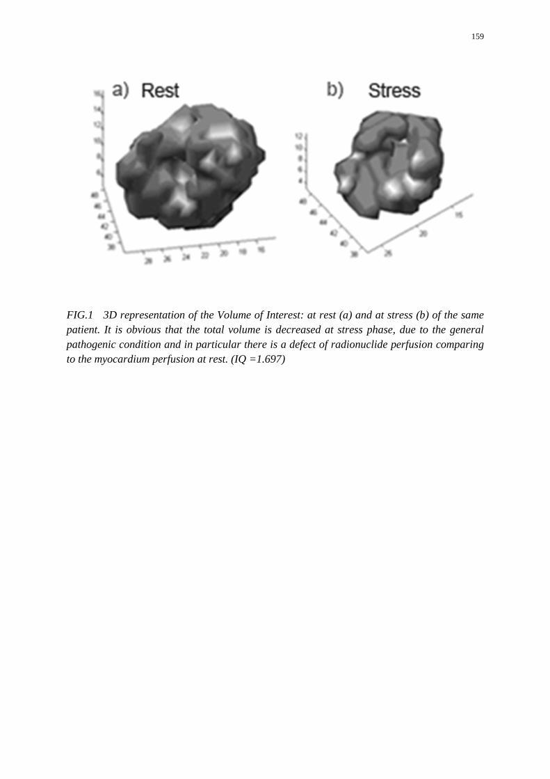
159
FIG.1 3D representation of the Volume of Interest: at rest (a) and at stress (b) of the same patient. It is obvious that the total volume is decreased at stress phase, due to the general pathogenic condition and in particular there is a defect of radionuclide perfusion comparing to the myocardium perfusion at rest. (IQ =1.697)
160
IAEA-CN-202/256
Myocardial Perfusion Abnormality on SPECT MPI and Abdominal Aortic Calcification Detected by DXA
A. Klaipetch, S. Namwongprom, T. Kaewchur, W. Teeyasoontranon, J. Srirattanasak
Faculty of Medicine, Chiang Mai University, 110 Intawaroros road, Chiang Mai, Thailand
Email Address of the Main Author: [email protected]
Background of the study: Atherosclerosis is a systemic disease. Burden of calcium in atherosclerotic plaques was founded to be a risk indicator for cardiovascular disease. Many studies have shown that abdominal aortic calcification (AAC) detected by either lateral lumbar spine radiographs, dual-energy X-ray absorptiometry (DXA) using vertebral fracture assessment (VFA) technique, or computed tomography (CT) scan is an significant predictor for coronary artery disease (CAD) incidence and mortality. DXA has long been a standard diagnostic method to evaluate bone mineral density and to detect osteoporosis. The so called VFA technique is benefit in detecting vertebral fracture and also useful and reliable in detecting AAC with a very low radiation dose to patient. Our study was aimed to find out if AAC detected by VFA has a relationship with myocardial perfusion abnormalities found on MPI.
Methods: Pilot study was performed in 38 patients who came for cardiac gated single photon emission computed tomography myocardial perfusion imaging (SPECT MPI) with certain indications involving CAD at Department of Radiology, Chiang Mai University Hospital. VFA technique of DXA was performed prior to MPI in all patients. The presence and extent of AAC had been analysed and scored, using the Abdominal Aortic Calcification-8 (ACC-8) scoring system. The severity of AAC was classified into 3 levels. Level 1 is absence of AAC (score = 0), level 2 is low AAC (scores 1- 3) and level 3 represents high AAC (scores > 3). The MPI results were categorized into 3 groups regarding the presence and types of the worse major perfusion defect. Normal MPI was in group 1. The reversible perfusion defect represented myocardial ischemia was in group 2 whilst the fixed defect or defect at rest with no or less reversibility was in group 3. Relationship between MPI results and levels of AAC severity was analysed using correlation statistics.
Results: The pilot group of 38 patients included 24 men and 14 women with ages ranging from 38 to 83 years old. Mean age was 64.5 years. SPECT MPI was performed with Thallium-201 in 27 (71%) patients and with Technetium-99m Sestamibi in 11 (29%) patients. Twelfth (31.6%) patients revealed normal MPI. Myocardial ischemia and infarction were found in 15 (39.4%) and 11 (29%) patients, respectively. For the levels of AAC severity, 10 (26.3%) patients showed no AAC while 18 (47.4%) patients had low AAC and 10 (26.3%) patients had high AAC. There is moderate correlation between MPI results and levels of AAC severity (r = 0.467, p < 0.01)

161
Conclusion: Moderate correlation was found between the types of myocardial perfusion abnormality and the levels of AAC severity. VFA technique of DXA to detect AAC might be a screening tool for CAD in patients at risk. Further study with larger number of patients is needed.
162
IAEA-CN-202/257
Correlation of Myocardial Perfusion Imaging and Computed Tomography Coronary Angiography for the Assessment of Coronary Arteries Disease: SQUH Experience
S. S. H. Hussein, H. Al Dhuhli, A. K. Maher Ali
Sultan Qaboos University Hospital, PO Box 38, PC 123, Musact, Oman
Email Address of the Main Author: [email protected]
Background: Myocardial perfusion imaging (MPI) with SPECT is of value for assessing the physiologic significance of coronary lesions. Computed tomography coronary angiography (CTCA) is a new technique to noninvasively detect anatomical coronary stenosis. This study was conducted to correlate the MPI and CTCA for the assessment of Coronary Arteries Disease (CAD).
Methods: From 226 CTCA, studies were performed at Sultan Qaboos University Hospital from May 2011 till May 2012. There were 45 patients who had both MPI and CTCA studies, which were reviewed. The results of the two studies were correlated and the technical problems affected the quality of the studies were reported. Coronary Angiography was not performed in patients if there was discrepancy between the MPI and CTCA studies or agreement on existence of CAD. If both studies were normal Coronary Angiography was performed. MPI was performed in 2 days stress and rest, the patients were stressed with Adenosine or Dobutamine and imaged after injection of Tc-99m Tetrofosmin using Dual Head Gamma Camera the studies were analyzed using 4DM software. CTCA was performed using 64 Multidetector Computed Tomography.
Results: This study included 45 patients, 27 males and 18 females with age range of 34 to 87 age and mean of 58 years. These patients were referred for MPI and CTCA due to suspicious of CAD in 36 patients, 4 had Percutaneous Coronary Intervention for follow up and 5 Dilated Cardiomyopathy to exclude CAD. After exclusion of one study due bowel activity overlying the myocardium the MPI sensitivity was 94%, specificity 96%, Positive Likelihood Ratio 23 and Negative Likelihood Ratio. In MPI there was one false negative result due to 3-vessel disease with balanced ischemia and one false positive study. After exclusion of 7 CTCA studies, 4 due to stents which made it difficult to assess the CAD, 2 due to high pulse and arrhythmias and 1 due to breathing artefact, the CTCA sensitivity was 100%, specificity 89%, Positive Likelihood Ratio 9 and Negative Likelihood Ratio 0. In the CTCA there were 5 patients with stenosis ranged from 60% to 70% which was difficult to determine the significance of the stenosis.
Conclusion: This study showed that CTCA is very sensitive while MPI is very specific in detection of CAD. Combining MPI and CTCA imaging results in improvement in detection of hemodynamically significant CAD. MPI and CTCA might play a potentially important role in the non-invasive diagnosis of coronary artery disease and introduce an objective

163
decision-making tool for assessing the need for interventions in each occluded vessel. CTCA was not useful for the follow up in patients with Percutaneous Coronary Intervention.
164
IAEA-CN-202/259
Conventional Angiography Imaging of Post-traumatic Pseudoaneurysm of the Superficial Femoral Artery
M. Dereli
Cardiovascular Surgery Department, Afyonkocatepe University, Afyonkarahisar Turkey
Email Address of the Main Author: [email protected]
Background: Over the past 50 years the management of vascular trauma has changed from mandatory surgical exploration to selective non-operative treatment, where possible. Accurate, non-invasive, diagnostic imaging techniques are the key to this strategy. The purpose of this review was to define optimal first-line imaging in patients with suspected vascular injury in superficial femoral artery. Methods: A systematic review was performed of literature relating to radiological diagnosis of vascular trauma over the past decade (2000-2010). Studies were included if the main focus was initial diagnosis of blunt or penetrating vascular injury and more than ten patients were included. Results: Of 543 titles identified, 60 articles were incorporated in the systematic review. Most described the use of conventional angiography imaging (CAI). The application of duplex ultrasonography, magnetic resonance imaging / angiography and transoesophageal echocardiography was described, but significant drawbacks were highlighted for each. CAI displayed acceptable sensitivity and specificity for diagnosing vascular trauma in blunt and penetrating vascular injury within the neck and extremity, as well as for blunt aortic injury. Conclusion: Based on the evidence available, CAI should be the first-line investigation for all patients with suspected vascular trauma and no indication for immediate operative intervention.
165
IAEA-CN-202/260
Prospect of Cardiac Imaging Necessity in B. P. Koirala Memorial Cancer Hospital
L. N. Singh, M. Tamang, R. Shaha Chaudhary
B. P. Koirala Memorial Cancer Hospital, Bharatpur Chitwan, Nepal
Email Address of the Main Author: [email protected]
B. P. Koirala Memorial Cancer Hospital is the only national cancer hospital in Nepal. More than 500 patients are attending our hospital from all territories of Nepal and neighbouring country such as India, from its border territory. Among them we have about 100 new cancer cases daily and the rest of the patients are on follow up. Our 80 bed inpatient department holds all sorts of therapeutic facilities of Radiotherapy and Chemotherapy. We encounter a variety of patients suffering from variety of diseases and of mainly late stages, where we go on Chemotherapy and Radiotherapy for a long standing period and develop cardiac toxicity in 20-30% of cases.
We are diagnosing the cardiac toxicity by clinical basis using laboratory tests, ECG and echocardiogram. It is challenging for us to conduct early detection of Cardiac toxicity like carditis and cardiomyopathy with integrated modality. Thus learning and exposure to have cardiac imaging technique with its clinical application is highly needed. We are facing challenges for early diagnosis of therapeutic induced carditis with Nuclear medicine application and modalities.
Cardiotoxicity is a well-known side effect of several cytotoxic drugs, especially of the anthracyclines and can lead to long term morbidity. The mechanism of anthracycline induced cardiotoxicity seems to involve the formation of free radicals leading to oxidative stress. This may cause apoptosis of cardiac cells or immunologic reactions. However, alternative mechanisms may play a role in anthracycline induced cardiotoxicity. Cardiac protection can be achieved by limitation of the cumulative dose. Furthermore, addition of the antioxidant and iron chelator dexrazoxane toanthracycline therapy has shown to be effective in lowering the incidence of anthracycline induced cardiotoxicity. Other cytotoxic drugs such as 5-fluorouracil, cyclophosphamide and the taxoids are associated with cardiotoxicity as well, although little is known about the possible mechanisms. Recently, it appeared that some novel cytotoxic drugs such as trastuzumab and cyclopentenylcytosine also show cardiotoxic side effects.
We are diagnosing the cardiac toxicity by clinical basis using laboratory tests, ECG and echocardiogram. We are having challenges to early detection of Cardiac toxicity like carditis and cardiomyopathy with integrated modality. Thus learning and exposure to have cardiac imaging technique with its clinical application is highly needed.
We are facing challenges for early diagnosis of therapeutic induced carditis with Nuclear medicine application and modalities. Recent advances of Nuclear medicine have enabled us to diagnose many of carditis, cardiomyopathy and other abnormality in more specific and sensitive way. We have ample numbers of Nuclear Physicians, experts and technologists but we are in dire need of Nuclear Medicine modalities, technique and training exposure of new advances to pursue imaging facility to face challenges to combat cardiac ailing patients. Thus, we are in process of establishing Nuclear Medicine department soon with the facility to

166
dispense the early diagnosis of cardiac ill manifestation to meet the challenges of diagnostic dilemma to deliver proper treatment to needed patients.
167
IAEA-CN-202/263
Using Non-Invasive Assays to Improve Detection of Oxidized Low Density Lipoproteins (OxLDL) in Atherosclerosis
B. C. Buia,c, C. C. Giad, M. A. H Nguyena, S. T.Voa,b, J. Shind, K. D. Hoe
a The Center of Molecular Biomedicine, University of Medicine and Pharmacy Ho Chi Minh city, Viet Nam
b The Hospital of University of Medicine and Pharmacy HCM, Viet Nam c Department of Biotechnology, International University, National University of Ho Chi Minh, Viet Nam e Department of Molecular Cell biology, Sungkyunkwan University, Suwon, South Korea
f Deparment of Anatomy, Sungkyunkwan University, Suwon, South Korea
Email Address of the Main Author: [email protected]
Oxidized low-density lipoproteins (OxLDL) such as malondialdehyde-modified LDL (MDA-LDL) are involved in the development atherosclerosis lesions. This study was designed as an approach to examine and characterize technetium-99m (99mTc) labeled MDA-LDL that could serve as standardized protocol to improve detection of OxLDL for the early diagnosis of asymptomatic atherosclerosis. Thirty-two male patients (age from 45 – 62) were divided into control and inflamed groups according to the documented atherosclerosis lesions. For the assessment of myocardial perfusion, 99mTc MDA-LDL were traced the localization in the heart muscle or in the blood pools of the left and right ventricles. Of twenty-four patients were sufficiently showed high to allow external imaging of the lesions 10 to 24 hours after administration of the 99mTc MDA-LDL. The accumulation of 99mTc MDA-LDL were significant in the inflamed group. Histological specimens of the atheroclerosis plaques confirmed the results of inflamed group showed that the specimens of the plaques detected by external imaging contained foam cells and immature fibrocalcific lesions, which strongly immunostained MDA-LDL specific antibodies. Thus, these non-invasive 99mTc MDA-LDL serve as potential biomarkers to identify patients at high risk for atherosclerosis.
168
IAEA-CN-202/264
Patient Peak Skin Dose and Dose Area Product from Interventional Cardiology Procedures
E. M. Antar, I. Duhaini, S. Manaa, H. Al Naemi
Hamad Medical Corporation, PO Box, 3050, Doha, Qatar
Email Address of the Main Author: [email protected]
Introduction: Radiation induced skin effects are deterministic in nature, with a generally accepted threshold dose of 2 Gy. The likelihood and severity of radiation-induced skin injury to the patient as a whole are a function of the highest radiation dose at any point on that patient’s skin: the peak skin dose (PSD). The increase in use of fluoroscopy for complicated interventions, which require long fluoroscopy time coupled with a continued lack of awareness among many interventionists and a lack of dose monitoring or no understanding of dose parameters, has resulted in continued occurrence of skin injuries so, it’s very important to follow up the staff and patients dose evaluation. Objectives: The objective of this study is to obtain information about patient Peak Skin doses (PSD) , Dose Area Product (DAP), Fluoroscopy Time (FT) and Cumulative dose (CD) from PTCA and CA which is the most predominant with respect to high skin doses in addition to other procedures. This study will also help to establish the Reference Dose Level (RDLs) for adult patients undergoing interventional procedures in Qatar which will define recommendations on how to reduce patient dose following the International Commission on Radiation Protection (ICRP). Methods: Four X-ray fluoroscopic machines from HMC Qatar (1 Cath-Lab in Hamad General Hospital & 3 in Heart Hospital) performing interventional Cardiology (IC) procedures were engaged in this study. All machines are manufactured by Siemens with a flat panel detector in a C-arm configuration. Radiation doses for approximately 1700 patients were collected including patients’ age, gender, and fluoroscopy time (FT), and total DAP readings. In order to establish (RDLs), the third quartile of value distribution was calculated. The median values of distribution were also assessed in order to estimate the variation in values. The RDLs were also compared with the RDLs provided by the European Research program (DIMOND) and other references. Result and Discussion: Radiation doses for 1132 patients were collected include patients age, sex, fluoroscopy time (FT), Total DAP reading. The range of Kvp used in these procedures was 50-125 kvp and the fluoroscopy time 0.18 – 52 minutes. For CA procedures, the Dose Area Product values reached 72.14 Gy.cm2 and the Cumulative Dose values reached 752 mGy. On the other hand, the DAP for and CD for PTCA procedures were found to be 143 Gy.cm2 and 2287 mGy respectively. Cumulative Dose (CD) was of the order of 2 Gy, the dose limit for radiation injuries . The relation between the fluoroscopy time and the DAP are also considered. The relation between DAP and PSD was investigated for both diagnostic and therapeutic interventions based on all data. In this study it found that a strong correlation between the Dose Area product and PSD (R2=0.83). Conversely, as shown in, there is a poor correlation between the total fluoroscopy time (FT) and the PSD (R2=0.44). It is noticed that for CA in HMC the DRLS exceeded the Diamond RLs by 20% in DAP and decrease in fluoroscopy time was noticed in HMC for the same procedures.
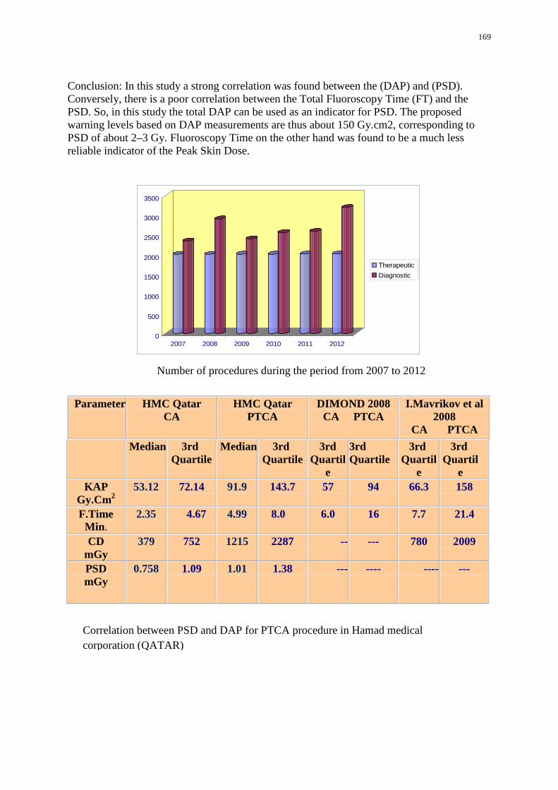
169
Conclusion: In this study a strong correlation was found between the (DAP) and (PSD). Conversely, there is a poor correlation between the Total Fluoroscopy Time (FT) and the PSD. So, in this study the total DAP can be used as an indicator for PSD. The proposed warning levels based on DAP measurements are thus about 150 Gy.cm2, corresponding to PSD of about 2–3 Gy. Fluoroscopy Time on the other hand was found to be a much less reliable indicator of the Peak Skin Dose.
I.Mavrikov et al 2008
CA PTCA
DIMOND 2008 CA PTCA
HMC Qatar PTCA
HMC Qatar CA
Parameter
3rd Quartil
e
3rd Quartil
e
3rd Quartile
3rd Quartil
e
3rd Quartile
Median 3rd Quartile
Median
158 66.3 94 57 143.7 91.9 72.14 53.12 KAP Gy.CmP
2 21.4 7.7 16 6.0 8.0 4.99 4.67 2.35 F.Time
Min. 2009 780 --- -- 2287 1215 752 379 CD
mGy --- ---- ---- --- 1.38 1.01 1.09 0.758 PSD
mGy
0
500
1000
1500
2000
2500
3000
3500
2007 2008 2009 2010 2011 2012
Therapeutic Diagnostic
Number of procedures during the period from 2007 to 2012
Correlation between PSD and DAP for PTCA procedure in Hamad medical corporation (QATAR)
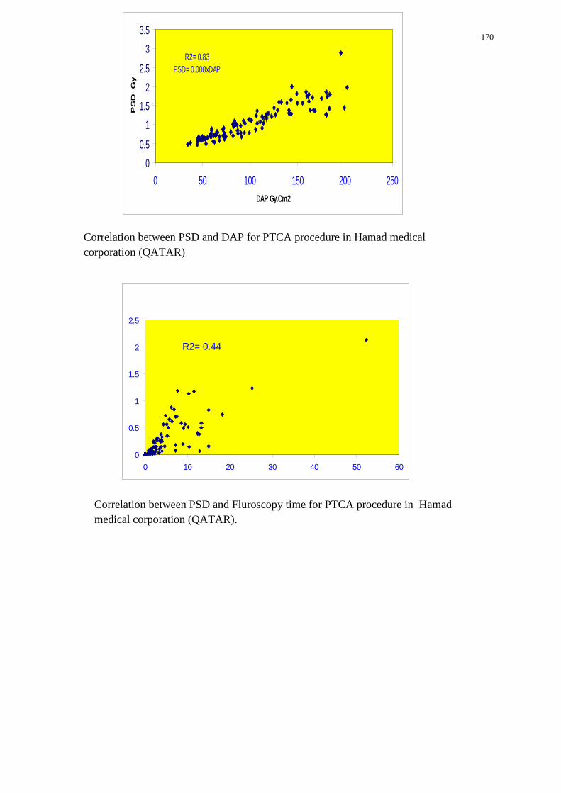
170
00.5
11.5
22.5
33.5
0 50 100 150 200 250DAP Gy.Cm2
PS
D G
y
R
R2= 0.83PSD= 0.008xDAP
R2= 0.44
0
0.5
1
1.5
2
2.5
0 10 20 30 40 50 60
Correlation between PSD and Fluroscopy time for PTCA procedure in Hamad medical corporation (QATAR).
Correlation between PSD and DAP for PTCA procedure in Hamad medical corporation (QATAR)

171

172
USE OF HYBRID IMAGING OR INTEGRATED IMAGING IN CVD MANAGEMENT
173
IAEA-CN-202/132
Comparative Study by Ultrasound and Isotopic Method for the Assessment of Left Ventricular Ejection Fraction in Madagascar
R. A. Rasata
Nuclear Medicine and Biophysics Department, Laboratoire des Radioisotopes, route d’Andraisoro BP 3383, Antananarivo 101, Madagascar
Email Address of the Main Author: [email protected]
The left ventricular ejection fraction (LVEF) reflects the efficiency of the ventricular function.
The main objective of our study is to evaluate the performance of two techniques available in Madagascar, echocardiography and radionuclide angiography, in relation to LVEF.
The study draws on 34 cases of adults who were hospitalised at the Cardiology department at the military hospital in Antananarivo and underwent an echocardiography exam, and who were then recruited for a scintigraphy cardiac at the nuclear medicine department between December 2003 and September 2004.
The results were as follows: A strong correlation exists between isotopic LVEF and echocardiography LVEF (R2 = 0.7522). However, a net overestimate for value of LVEF echocardiography in relation to the value of isotopic LVEF (average LVEF = 42.41%). This overestimate is more marked for isotopic LEVF < 40%. In this range of values (LVEF <40%), the clinical, the radiological and electrical results correlate with all of the isotopic LVEF and do not correlate with certain cases (33%) of LVEF echocardiography.
We conclude that the values of LVEF obtained by the isotopic method are more precise and accurate than the values determined by echocardiography during heart failure.
174
IAEA-CN-202/135
Multimodality Imaging for Assessment of Heart Failure Patients in a PET/CT Center: 13N-ammonia, 18FDG, Coronary CT Angiography and Calcium Score
J. Altamiranoa, G. Estradaa,b a Universidad Nacional Autónoma de México (UNAM). Unidad PET/CT ciclotrón. Edificio de Investigación de la Facultad de Medicina b C.T. Scanner del sur. Rafael Checa No. 3, col San Angel, Distrito Federal, México
Email Address of the Main Author: [email protected]; [email protected]
Background: Heart failure and coronary artery disease are two of the most common illnesses now a day, with high morbidity and mortality. Nuclear imaging can help in the differentiation between patients with ischemic and non ischemic heart failure. In patients with ischemic heart failure, the precise anatomy of the coronary vessels is needed. The CT multislice will provide this information. Nuclear imaging is considered the first choice technique for the evaluation of ischemia and viability, using SPECT and PET/CT. We can also assess left ventricular ejection fraction, which is more reproducible than the echocardiography. PET/CT with 13N-ammonia in conjunction with coronary CT will provide information about ischemia and coronary anatomy. PET/CT with 18FDG is useful in the assessment of myocardial viability. Viable myocardium may have chronically hypoperfused myocardium at rest (known as hibernation) or preserved resting perfusion with reduced flow reserve (known as stunning). The PET with 18FDG had the higher sensitivity, followed by SPECT with thallium-201. Specificity is higher for dobutamine echocardiography. Calcium scoring images are acquired without intravenous contrast to visualize and quantify calcified plaque, a strong indicator of coronary artery disease. When CT calcium score is performed in patients with ischemia a low calcium score result, detect lower event rate than those patients with high calcium. This study describes each of these imaging modalities.
Methods: We performed a retrospective analysis of consecutive patients, who were referred to cardiac studies for clinical indications from January 2011 to February 2013.
Results: Only 4.9% of PET/CT patients that have an appointment in our centre were for cardiac studies. There were a total of 705 studies and 434 patients: 275 for PET/CT with 13N-ammonia, 355 for coronary CT, 54 for morphologic heart for nodal ablation, 17 for calcium score and 4 for viable tissue with 18FDG. Most of the ammonia patients had coronary CT.
Conclusion: Although the field of cardiovascular imaging has experienced marked growth, the percentage of PET/CT patients that had a cardiac PET/CT has not changed. In contrast the number of coronary CT alone has increased. Although the study with higher sensitivity for myocardial viability is the PET/CT with 18FDG, in our centre we only had 4 patients, which indicate that either the referring physician is not aware of the benefits of this study or the cost of performing the study is too high that the referring physician preferred to indicate SPECT with thallium-201. The economic situation in Latin-American countries may contribute to the sub-utilization of the study with higher sensitivity.
175
IAEA-CN-202/139
Radionuclide Ventriculography in the Evaluation of Cardiotoxicity by Chemotherapy
F. Z. T. Huber, W. E. F. M. Alves, M. J. Santos, E. T. Rocha
Hospital de Câncer de Barretos, Rua Antenor Duarte Villela, 1331, Bairro Dr. Paulo Prata, Barretos, Brazil
Email Address of the Main Author: [email protected]
Objectives: The aim of this study was to evaluate the frequency of assessments of left ventricle ejection fraction (LVEF) by radionuclide ventriculography in a sample of patients undergoing chemotherapy for breast cancer in the Cancer Hospital of Barretos (Barretos/SP, Brazil) and analyze when significant changes occurred, associating this information with the chemotherapy regimens and doses achieved. Background: Patients, who are receiving Chemotherapy regimens with anthracycline associated or not with trastuzumab, are at risk of cardiotoxicity and should be monitored for a decrease in LVEF, even when asymptomatic (class I indication according to American College of Cardiology / American Heart Association guidelines). Methods: We selected female patients, who were diagnosed with breast cancer and were under monitoring in the Barretos Cancer Hospital, and who had performed at least two tests of radionuclide ventriculography for assessment of cardiotoxicity with anthracyclines or anthracyclines followed by trastuzumab, having performed necessarily an examination in the period from 03/01/2011 to 15/02/2011. These patients were analyzed mainly according to the chemotherapy regimens and the number, frequency, and average results of the examinations. Results: Thirty patients were studied, the average age was 48.47 years (SD ± 10.08), 70% were treated with anthracyclines followed by trastuzumab, while 30% were treated with anthracyclines without trastuzumab. The overall mean of performed ventriculographies was 4.70 examinations / patient (SD ± 1.34). These tests were conducted on an average frequency of 129.13 days (SD ± 37.99). The overall mean baseline LVEF was 63.63% (± 5.79 SD), and no patient had ≤ 50%. However, the basic tests were performed on the average 82.33 days after initiation of chemotherapy treatment with anthracycline, and 40% of the sample began on average 11.83 days before and 60% on average 145 days after initiation of referred treatment. 43.33% of the sample showed overall change in LVEF (decrease> 10%, LVEF ≤ 50% or both) sometime in the study, 7.7% of these belonged to the group that used anthracyclines not followed of trastuzumab and 92.3% belonged to the group which used anthracyclines followed by trastuzumab. Among the groups, 57.14% of patients who used anthracyclines followed by trastuzumab and 11.11% who used anthracyclines not followed by trastuzumab showed changes in LVEF. In the first group, the change in LVEF happened after an average 7.42 cycles of trastuzumab (± 4.72 SD).

176
Conclusion: It was observed that change in LVEF is higher in the group receiving anthracyclines followed by trastuzumab, and this change when present, appears 7.42 cycles on average after the beginning of therapy with trastuzumab in the sample.
177
IAEA-CN-202/145
Acceptance Testing of a Hybrid Operating Room in the Philippines
A. F. Samilinga, A. Morales, Jr.b aThe Graduate School, University of Santo Tomas, España Manila, Philippines bCenter for Device Regulation, Radiation Health and Research, Food and Drug Administration Philippines, Department of Health, Rizal Avenue, Manila, Philippines
Email Address of the Main Author: [email protected]
Hybrid operating rooms are emerging as the demand for minimally invasive cardiovascular surgery increases. However, these minimally invasive cardiovascular techniques rely more heavily on fluoroscopic imaging for extended periods of time. This will result in greater radiation exposure levels for patients, and therefore newer dose reduction measures will have to be implemented.
This report will discuss the physics test procedures for hybrid operating room x-ray systems that will ascertain whether the x-ray equipment will fulfil the intended purpose or will perform according to specification. These test procedures, also called acceptance tests, must allow the overall evaluation of the equipment’s performance, which will establish the optimal condition under which the equipment will deliver the best output for accurate diagnosis. At the same time, the acceptance test will certify if the x-ray equipment will not subject the patient and the workers to unnecessary radiation exposure that could lead to unwanted radiation-induced stochastic effects.
The widespread use of digital detectors for angiographic imaging equipment has ensured that image distortion is greatly reduced, and smaller focal spots have also improved spatial resolution. However, the increase in patient dose has been noted in previous reports. In order to mitigate this increase, thorough acceptance testing should be carried out in order to determine the highest dose rates delivered by hybrid x-ray imaging systems.
The acceptance tests recommended for hybrid operating rooms include the following: For fluoroscopic mode: Fluoroscopic Image Distortion, Tube Voltage Accuracy, Tube Voltage Reproducibility, Fluoroscopic Output Reproducibility, Fluoroscopic Output Linearity, Resolution Test for Detector with LCD (both Low contrast and High contrast)Test for Fluoroscopic Timer, Maximum Dose Rate, Typical Patient Dose Rates Using a Patient Equivalent Phantom (PEP). For Radiographic/Service Mode, the following tests are recommended: Tube Voltage Accuracy,
Tube Voltage Reproducibility, Timer Accuracy, Radiation Output Reproducibility Radiation Output Linearity, Half Value Layer, Tube Housing Assembly Leakage, Focal Spot Size Check, Grid Alignment, Kilovoltage Tracking and Thickness Tracking.
For the display monitors, viewing tests for gray scale uniformity and contrast using pre-loaded SMPTE and/or AAPM TG-18 patterns are recommended.
178
IAEA-CN-202/162
Automated Diagnosis of Nuclear Cardiac Images Using Advanced Digital Image Processing Techniques
H. K. A. Mahmoud
Engineering Department, Nuclear Research Center, Atomic Energy Authority, Egypt
Email Address of the Main Author: [email protected]
Cardiology images interpretation can be done by cardiologists, radiologists, or physicians who are specialist in nuclear medicine. The interpretation is manually depending heavily on the skills, experience, alertness and consistency of the specialist and requiring most significantly time. Results may suffer from inconsistency and errors, particularly when dealing with large volumes of data. Recently, researchers introduced many systems for automatic interpretation of cardiology images. This paper presents a proposed approach for automatic diagnosis of nuclear cardiac images. In the proposed approach, the features are extracted from each image or from one of its transforms (Discrete Wavelet Transform (DWT), Discrete Cosine Transform (DCT), or Discrete Sine Transform (DST)). The power density spectrum (PDS) or the higher order statistics (Bispectrum and Trispectrum) estimated by different methods are used and compared with the transform domains. These features are used to train the neural networks to create feature database. In testing proposed approach, the features matching process is performed by the neural networks to decide whether these features belong to normal or abnormal image. In the proposed method, 200 cardiac images have been used for training the neural network. And the testing has been done using 100 images. These images have been collected from different patients in Egyptian hospital.
The results show that, the DCT gives the highest diagnosis rate among the transforms domains, the PDS estimated using Multiple Signal Classification (MUSIC) method gives the highest diagnosis rate among the PDS estimation methods or transfer domains. The Trispectrum gives the highest diagnosis rate among all the tested methods.
179
IAEA-CN-202/173
Evaluation of Integrated Medical Imaging for Congenital Aorto-Cardiac Anomalies in Tertiary Referral Centers in Sri Lanka (with Special Emphasis on Paediatric CT and
Radiation Exposure)
A. S. Pallewatte
Consultant Radiologist, The National Hospital of Sri Lanka, Colombo 10, Sri Lanka
Email Address of the Main Author: [email protected]
Background: Evaluation of congenital cardiac and aortic abnormalities is critical to management and challenging to radiologists and clinicians due to complexity of anomalies, neonatal presentation etc. In Sri Lanka, such cases are managed only at the Lady Ridgeway Hospital. The initial imaging tool was echocardiography while 11 % of cases needed further imaging. Although Catheter cardio-angiography is the gold standard, it is invasive with a higher radiation dose and higher complication rates. Therefore in our practice, CT is used as an alternative modality. It has the advantages of lesser invasiveness, ability to visualize arteries and veins, soft tissue anatomy and multi-planar reconstruction. However CT has disadvantages due to radiation dose, patient movement and deficient CT protocols. Our study critically evaluates indications, justification, radiation exposure and usefulness of pediatric CT aortography in relation to other modalities in evaluating congenital aorto-cardiac anomalies.
Method: Data were retrospectively obtained from all Paediatric patients who underwent CT aortography from 01-01-2012 to 31-12-2012 for evaluation of aorto-cardiac anomalies at Lady Ridgeway Childrens’ Hospital. The main equipment used was Siemens Somatom Volume-Zoom 4 slice CT scanner. CT parameters such as Peak kilovoltage (kVP), tube Current (mA), slice thickness and the dose details (CTDIw and Dose Length Product DLP) were recorded from CT console. Patient data such as age, sex, weight, indication for CT, any repeat examinations, echocardiographic data etc were recorded. These were analyzed statistically. Dose data were compared with reference levels. Numbers of patients with the same diagnosis but not undergone CT were also recorded.
Results: Total number patients included were 29. The age varied from 3 days to 8 months. 56 % were male. Weight of patients ranged from 2.1 kg to 10.2 kg. Average kVP and mA were 140 and 70 respectively. Average CTDI & DLP were 7.63 mGy and 163 mGycm respectively. 3D reconstruction was done on all patients. All 29 patients who underwent CT have been initially evaluated by prior echocardiography. (In fact there were 201 patients with the same diagnosis, but underwent only echocardiography but not CT and therefore were not included in this study. 5 patients also underwent catheter angiography). Main indication for CT was pre-surgical demarcation of vascular anomalies. No repeat CT studies were recorded.
Conclusion: Echocardiography being a radiation-safe modality was the commonest used in evaluation of Aorto-cardiac anomalies and it was the only modality used in 89%. All 29 patients who underwent CT scans were initially evaluated by echocardiography. In view of

180
higher radiation dose & anaesthetic risks, CT was reserved only for selected cases where echocardiographic information is inadequate for surgery. It also helps reduce number of catheter angiograms required. CT effectiveness can be further improved by better protocols and provision of a higher slice scanner. Neither nuclear imaging nor MRI was performed in any of our patients due to high cost and limited availability.
181
IAEA-CN-202/176
Potential Benefits of Equilibrium Radionuclide Angiography in HIV-related Cardiac Disorders
Z. Jawa, A. Isa, S. Lawal
Department of Nuclear Medicine, National Hospital Abuja, Nigeria
Email Address of the Main Author: [email protected]
Background: Human Immunodeficiency Virus (HIV) infection is a major health challenge. There is increased number of documented HIV- related cardiac disorders, which include cardiomyopathies, pericardial disease, mitral valve prolapse and coronary artery disease (CAD). Cardiac related HIV disorders are due to lipodystrophy, nutritional deficiencies and antiretroviral cardiotoxicity. The assessment of cardiac function in HIV- related cardiac disease is done mainly using echocardiography, which however, has limitations of geometric assumptions in calculating LVEF and being highly operator dependent. Equilibrium Radionuclide Angiography (ERNA) may provide an alternative, non-invasive, highly reproducible technique for the diagnosis and assessment of cardiac function in HIV-related cardiac disorders.
The objective of this cohort study is to document the usefulness and potential benefits of ERNA in assessing patients with HIV-related cardiac disorders in our clinical setting.
Method: A total of 13 (8 male, 5 female) patients who tested positive for HIV were included in the study. All the patients were on antiretroviral medications and their CD4 counts were <100/mm3. The patients received between 15-30mCi of autologous RBCs labelled using the in vivo technique. A minimum of 16 frames per R-R interval images were acquired using a dual-head MEDISO gamma camera.
Results: All the patients demonstrated low LVEF (25-30%). Nine patients showed dilated cardiac chamber consistent with cardiomyopathy. Three patients showed regional wall motion abnormality suggestive of CAD and 1 patient had surrounding cardiac photopenia consistent with pericardial effusion.
Conclusion: Our data suggest that ERNA seems to be a useful non-invasive technique in the investigation of patients with HIV-related cardiac disease. We are aware of the limitations of our study, which include a highly selective and small number of patients. However, this is the first study which aimed at studying the potential benefit of ERNA in the assessment of HIV-related cardiac diseases. These preliminary results are encouraging and suggest a role of ERNA in the evaluation of patients with HIV-related cardiac disorders. Further studies with a larger patient number are required to validate these data.
182
IAEA-CN-202/201
Correlation of Myocardial Blood Flow and Flow Reserve to Anatomical Lesions using Hybrid PET/CT
H. Bom, H. S. Kim, S. G. Cho
Department of Nuclear Medicine, Chonnam National University Hwasun Hospital, Hwasun, Republic of Korea
Email Address of the Main Author: [email protected]
Background: Noninvasive evaluation of coronary stenosis severity is still a challenge despite vast achievements in new technologies of PET/CT, CT coronary angiography, MRI and echocardiography. Quantitation of myocardial blood flow (MBF) and flow reserve (MFR) using PET confers advantages over other modalities related to functional evaluation of coronary stenosis severity, however, exact localization of culprit lesion is difficult because of difference imaging formats. Authors hypothesized that the correlation of MBF and MFR to anatomical lesions using hybrid cardiac PET/CT technology is feasible and clinically useful. The purpose of this paper is to evaluate the hypothesis as well as review of publications.
Methods: Subjects were 11 patients with known LAD disease studied by both CT coronary angiography and N-13 ammonia PET study. Regional MBF and MFR in LAD segments were measured and gradients over LAD lesions were calculated. Gradients of both MBF and MFR were correlated with severity of coronary stenosis on the interventional coronary angiography and fractional flow reserve in some cases. Conclusion: Recent publications showed added values of MFR in the diagnosis of coronary artery disease (CAD). Although many studies have revealed the value of MBF and MFR of global left ventricle, evaluation of MBF and MFR changes are also possible through coronary arteries using hybrid PET/CT. Therefore measuring regional MBF and MFR and correlation of the information to the anatomical information using hybrid cardiac PET/CT are feasible and useful in the clinical setting.
183
IAEA-CN-202/238
Establishing Nuclear Cardiology Practice with Integrated Imaging in Mauritius and the Difficulties Encountered
A. S. Naojee
Jawaharlal Nehru Hospital, Highway Link Road, Rose Belle, Mauritius
Email Address of the Main Author: [email protected]
Mauritius has a population of 1.2 million inhabitant and non-communicable diseases like diabetes mellitus, hypertension, cancers and coronary artery disease is a major national issue. Cardiac death related to diabetes is 22%, and that for other causes is 33% cardiac death yearly. The number of CAGB (coronary artery bypass graft) perform yearly is about 500 and 90% of which are diabetes. There is only one nuclear medicine center situated at Jawaharlall Nehru Hospital and is equipped one single head SPECT Ecam camera, a dual head SPECT spirit DV Mediso camera and a recent treadmill machine with 12 lead ECG monitor. Among all the studies performed cardiac imaging MPI (myocardial perfusion Imaging) is yet to be established. Some 200 cases have been done since 2001 on an “as and when basis” and the department is striving hard to maintain a good number of cardiac studies. The current practice is to have a cardiologist on site when stressing the patient, a condition which is very difficult as cardiologist are very busy people and the hospital had no cardiac unit attached until recently. Many patients referred for MPI had either physical handicapped or cannot tolerate exercise stress hence MPI could not be performed until the introduction of pharmacological stress recently in November 2012. Earlier stress test was done on cycle ergometer machine which was not practical for our female patients and now it is replaced with new thread mill machine. Another technical difficulty we faced was the quality control with image processing and possibly with quality control of radiopharmaceutical labelling. Lots of our MPI have reversal perfusion defect on stress imaging, a situation which is very embarrassing. Our cardiologists are not making requests for MPI as coronary angiography is a free service offered by the ministry along with other health services. The best situation we found was to keep focusing on nuclear cardiology for several months and try to remedy the situation. Now we are able to perform at least two MPI studies weekly with our new weekly scheduled of technetium generator and working only with the Mediso camera. We yet to have an integrated imaging facility as no SPECT –CT or PET-CT exist here. Our objective is to introduce hybrid imaging through IAEA national projects. We want to further develop nuclear cardiology in Mauritius and have a well-established practice to play a very important role on the management of cardiovascular diseases in Mauritius.
184
IAEA-CN-202/243
Value of Hybrid Imaging-Based Attenuation Correction in the Diagnostic Work-up of
Myocardial Perfusion Scintigraphy
M. F. Bozkurt, E. L. Ergün, Ö. Uğur,
Hacettepe University Faculty of Medicine Department of Nuclear Medicine Ankara, Turkey
Email Address of the Main Author: [email protected]
Aim: Artefacts due to photon absorbtion directly affect the diagnostic specificity of myocardial perfusion scintigraphy (MPS). Attenuation artefacts caused by breast and diaphragm may lead to misdiagnosis as ischemia or infarct and decrease the specificity of the MPS. This study was aimed to assess the value of hybrid SPECT/CT based attenuation correction for the differential diagnosis of artefacts from ischemia and infarction. Patients and Methods: A total of 19 MPS (18 stress, 1 rest) of 18 patients (15 f, mean age: 58±10) was prospectively studied. Stress MPS was done 30 minutes after iv injection of 740 MBq Tc99m-MIBI at peak exercise and rest MPS was performed 60 minutes after Tc99m-MIBI injection with a dual-headed SPECT camera (25 sec/framex32, 180, 64x64). After MPS SPECT, SPECT/CT hybrid imaging (2,5 mAs, 140 kEV) of 15 minutes was done under a SPECT/CT camera with the same MPS protocol. MPS SPECT images reconstructed with filtered-back projection, iterative reconstructed attrenuation corrected and attenuation non-corrected SPECT/CT images were compared to each other both visually and quantitatively. Quantitative comparison was done using 20-segment perfusion scoring map. Also ROIs were drawn over myocardial segments with possible attenuation artefacts and over normally perfused segments and defect/normal ratio (D/R) from all three image sets was calculated for all patients and statistically analysed with paired-t test.
Results: Visual evaluation revealed decreased activity in anterior, anterioseptal, anteriolateral, inferior and inferiolateral walls in correlation with breast (n=10), diaphragma (n=7) and both breast and diaphragma (n=2) attenuation. After SPECT/CT based attenuation correction, 16/19 (84%) of these studies displayed normal findings. Quantitative evaluation revealed statistically significant difference between D/N ratios calculated from filtered-back projected SPECT images and iterative reconstructed attenuation corrected SPECT/CT images (p=0,008). Similar difference between D/N of iterative non-corrected and corrected SPECT/CT images was also found (p=0,002). No significant difference was found between D/N of filtered back projected SPECT images and iterative reconstructed attenuation non-corrected images (p=0,4)

185
Conclusion: Low-dose CT based attenuation correction in hybrid SPECT/CT imaging systems are of great importance for differentiating attenuation artefacts from real perfusion defects at the same imaging session.
186
IAEA-CN-202/253
Perfusion Scintigraphy and Hybrid Imaging in the Diagnosis of Pulmonary Embolism in Pregnancy
N. Ben Rais
Nuclear Medicine Department, Rabat, Morocco
Email Address of the Main Author: [email protected] Background of the study: Pulmonary embolism (PE) is a hot topic. • Pregnancy and the postpartum define a particular population. • Difficulty diagnosis, despite the establishment of clinical and radiological probability score. • Contribution of nuclear medicine is essential for diagnosis. • The anti-coagulation in the PE requires good management especially in pregnant women. Epidemiology: Incidence of VTE (venous thromboembolism) in pregnant women: • 5à12 for 10 000 pregnancies • 7-10 × women the same age Incidence of VTE postpartum: • 15à35 × witnesses of the same age pregnancy + DVT (Deep Vein Thrombosis) 3 × sup + EP in pregnancy DVT left 85% of cases Isolated pelvic TV is more common during pregnancy
New Diagnostic Recommendations: • The validated clinical prediction model: modified Wells score. • The biomarkers: D dimers have no diagnostic value during pregnancy; • BNP, NT-proBNP and troponin are markers of cardiac distension. • Diagnostic imaging is essential: scan V / Q and / or angiography. • The Doppler of MI (Member Inferior) is part of the systematic reviews. Criteria for Well S pretest Probability For Diagnosis and Pulmonary Embolism algorithm for MWS "Modified Well's Score"
Methods: • Period of 3 years (2010 - 2013) • Retrospective study of 17 cases • pregnant women hospitalized for suspected PE
187
Results: • Age is an RF (Risk Factor) independent; after 35 years the risk of DVT is double that of PE is threefold. • The risk of VTE (venous thrombo embolism), progressively increase as we approach the term (Meadle Age: 30 AW) • This risk is higher in post-partum (4 of 17)
RF more specific to pregnancy are found: • The most urgent cesarean (4 of 4) • Parity (≥ to3) multiplies by 2 the risk of VTE (venous thrombo embolism) in our series 9 of 12 multiparous were already at their 3rd pregnancy. • Obesity, RF much discussed, found in 7patientes. • endo utérine infection exposed to a RR × 6 (2 cases) • The search for thrombophilia requires a particular balance. • The hemorrhage and preeclampsia are significant RF. (1 case of PIH)
Other nonspecific RF may exist: • Varicose veins (3 cases) • The Capital (1 case) • The active or passive smoking
Clinical results • Lack of specific clinical signs: Dyspnee in 70%, Chest pain in 52%, palpitation in 35% • Probability clinical score according to a universal Wells score. • The high incidence of intermediate clinical probability complicates the diagnostic and therapeutic PEC: One high clinical case; One Low clinical case and 15 intermediate cases
Paraclinical results: Chest X Ray: normal in 20% of cases (23% in our series). Several signs suggestive but not specific: • Changes electro cardiographic in 87% of cases of PE (normal ECG in 29% of cases in our series) • Normality of ECG n 'removes any diagnosis.
Doppler venous MI is systematic if suspicion of PE. • Made in 6 patients: no DVT was detected. • The diagnosis of DVT solves the problem. • Pelvic DVT is common among pregnant women (diagnostic difficulty).
188
Transthoracic ultrasound: interest in massive PE with hemodynamic instability. • Made in 5 patients: normal except one (dilatation of the right cavities) • If it PE objective signs of right ventricular overload.
Pregnant women angiography is the investigation of choice after the 2nd scan.
Lung scintigraphy is the examination of 1st intention of suspected PE in pregnant women. • A high negative predictive value. • Interpretation based on criteria (modified PIOPED) • Our study 9 cases of systematized perfusion defect suggestive of thromboembolic processes classified as high probability of PE plus 1 case of massive PE, 4 cases interpreted normal??? All patients received anti coagulation is based LMWH (3 cases) or UFH (14 cases). • Relay AVK as soon as possible. • Certainty, LMWH is the treatment of choice in cases of PE. • Good progression after treatment (retrospective confirmation of positive diagnosis) Planar lung scintigraphy requires the injection of low dose of radioactive tracer in pregnancy. A perfusion alone seems sufficient for pregnancy! The CTA is an essential imaging modality for the diagnosis of PE. • A good specificity. (96% according to PIOPED II) • Increased detection rate of PE in segmental. • Reduced acquisition time (faster) • Irradiation of the breast tissue.
New: hybrid imaging • SPECT-CT offers a PPV of 99%, a sensitivity of 99% and a specificity of 98%. • Interest in the differential diagnosis. • The final recommendation of the EANM suggest SPECT-CT as first-line examination for suspected PE.
Conclusion: • The PE is a medical emergency whose clinical diagnosis is often difficult. • Pregnancy and the postpartum period are at high risk of VTE. • Several probability scores to help clinical and radiological diagnosis. • Pregnant women pulmonary scintigraphy examination is pivotal in PSE. • Hybrid imaging rivals scintigraphy. • The treatment of PE requires effective anti-coagulation. • Thromboprophylaxis must go through a risk assessment.
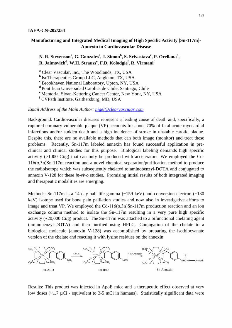
189
IAEA-CN-202/254
Manufacturing and Integrated Medical Imaging of High Specific Activity [Sn-117m]-Annexin in Cardiovascular Disease
N. R. Stevensona, G. Gonzalesa, J. Simonb, S. Srivastavac, P. Orellanad, R. Jaimovichd, W.H. Strausse, F.D. Kolodgief, R. Virmanif a Clear Vascular, Inc., The Woodlands, TX, USA b IsoTherapeutics Group LLC, Angleton, TX, USA c Brookhaven National Laboratory, Upton, NY, USA d Pontificia Universidad Catolica de Chile, Santiago, Chile e Memorial Sloan-Kettering Cancer Center, New York, NY, USA f CVPath Institute, Gaithersburg, MD, USA
Email Address of the Main Author: [email protected]
Background: Cardiovascular diseases represent a leading cause of death and, specifically, a ruptured coronary vulnerable plaque (VP) accounts for about 70% of fatal acute myocardial infarctions and/or sudden death and a high incidence of stroke in unstable carotid plaque. Despite this, there are no available methods that can both image (monitor) and treat these problems. Recently, Sn-117m labeled annexin has found successful application in pre-clinical and clinical studies for this purpose. Biological labeling demands high specific activity (>1000 Ci/g) that can only be produced with accelerators. We employed the Cd-116(α,3n)Sn-117m reaction and a novel chemical separation/purification method to produce the radioisotope which was subsequently chelated to aminobenzyl-DOTA and conjugated to annexin V-128 for these in-vivo studies. Promising initial results of both integrated imaging and therapeutic modalities are emerging. Methods: Sn-117m is a 14 day half-life gamma (~159 keV) and conversion electron (~130 keV) isotope used for bone pain palliation studies and now also in investigative efforts to image and treat VP. We employed the Cd-116(α,3n)Sn-117m production reaction and an ion exchange column method to isolate the Sn-117m resulting in a very pure high specific activity (~20,000 Ci/g) product. The Sn-117m was attached to a bifunctional chelating agent (aminobenzyl-DOTA) and then purified using HPLC. Conjugation of the chelate to a biological molecule (annexin V-128) was accomplished by preparing the isothiocyanate version of the chelate and reacting it with lysine residues on the annexin:
N
N
N
NCO2O2C
O2C CO2NH2
117mSn
N
N
N
NCO2O2C
O2C CO2NCS
117mSn H2N Annexin
N
N
N
NCO2O2C
O2C CO2NHCNH
117mSn
Annexin
CSCl2
Sn-ABD Sn-IBD Sn-Annexin
S
Results: This product was injected in ApoE mice and a therapeutic effect observed at very low doses (~1.7 µCi - equivalent to 3-5 mCi in humans). Statistically significant data were

190
obtained showing that apoptotic bodies and macrophages decreased while smooth muscle cells and collagen increased in the cardiac brachiocephalic arteries and the sinotubular junction where VP was observed to occur. The dose dependent results obtained are indicative of plaque stabilization, inflammatory reduction and a positive therapeutic outcome. Early human studies also indicate that imaging with as low as 3 mCi may be possible. Ultra-sound, gamma (planar and SPECT) and CT in-vivo data were taken along with optical, autoradiography and histology data from excised carotid plaque tissue. These imaging results are still being analysed but are indicative of selective uptake of the Sn-annexin in cardiovascular VP and other inflammation sites.
Conclusions: Very high specific activity Sn-117m has been produced and used to label annexin under cGMP to study vulnerable plaque and unstable plaque in animals and humans. Evidence for imaging and positive therapeutic effect has been observed with very low systemic doses (3 mCi in human; 1.7 µCi in mice).
191
IAEA-CN-202/261
Update of Integrated Medical Imaging in Cardiovascular Diseases
M. A. A. Abu Saiba
Al-Sadr Medical City, Kufa University, Kufa, Najaf, Iraq
Email Address of the Main Author: [email protected]
Although a clinical history and physical examination are essential for identification of cardiac disease, additional diagnostic testing is needed in most patients. An overview of the available diagnostic modalities is presented here. The appropriate diagnostic sequence in various clinical situations is presented in other sections. Transthoracic echocardiography typically is the initial diagnostic test in patients who present with evidence of a structural abnormality, such as a pathologic heart murmur or evidence of heart failure. This test provides detailed information on left and right ventricular size and systolic function, valve anatomy and function, pulmonary pressures, the presence of pericardial disease, and the proximal great vessels. Transesophageal echocardiography is appropriate when transthoracic images are non-diagnostic and as the initial imaging test in some clinical situations, such as detection of left atrial thrombus, evaluation of prosthetic mitral valve dysfunction, evaluation of suspected aortic dissection, and in patients with a moderate to high pre-test probability of endocarditis. Advanced cardiac imaging procedures for structural heart disease include radionuclide angiography, cardiac catheterization, CT scanning, and cardiac magnetic resonance (CMR) imaging. Typically, these tests are considered only after echocardiographic evaluation and in consultation with a cardiologist. Coronary angiography is appropriate when the stress test is consistent with CAD, particularly if there is a large area of myocardium at risk. In patients with a very high pre-test probability of disease, coronary angiography is an appropriate initial diagnostic test. Newer diagnostic approaches for coronary disease include CT imaging, CMR imaging, and positron emission tomography (PET). CT can be used for measuring coronary artery calcium (CAC) and for non-invasive coronary angiographic imaging. CAC scores are measured from non-contrast CT images, whereas CT coronary angiography requires a radiocontrast agent and use of a high-resolution scanner, necessitating a higher radiation dose to the patient. CT or CMR coronary angiography is appropriate for identification of anomalous coronary arteries. CT coronary angiography also is appropriate to evaluate coronary anatomy in a patient with new-onset heart failure and in patients with acute chest pain syndromes when the risk of CAD is intermediate but cardiac enzyme results are negative. CT coronary angiography or CMR pharmacologic stress testing may be considered in symptomatic patients with an intermediate probability of CAD when the ECG is abnormal at baseline, the patient is unable to exercise, or previous test results are equivocal. During CT angiography,

192
additional non-cardiac structures are also imaged, and significant non-cardiac findings (for example, lung nodules and infiltrates, pulmonary embolism, hiatal hernia, and aortic dissection) are present in approximately 20% of patients. PET scanning has several advantages compared with conventional pharmacologic nuclear perfusion imaging using single-photon emission computed tomography (SPECT).Compared with SPECT, a PET study is shorter for the patient (45-90 minutes), results in a lower radiation dose, provides evaluation of both myocardial perfusion and function, and reduces interpretive uncertainty, particularly in obese patients. In addition, PET provides the option of quantification of absolute myocardial blood flow and can be combined with CAC scoring. Guidelines for diagnostic use of PET have not yet been established, but it is a reasonable option when other tests are not diagnostic.

193

194
RADIOPHARMACEUTICAL PRODUCTION USING CYCLOTRONS AND RADIONUCLIDE GENERATORS – INCLUDING GOOD MANUFACTURING
PRACTICE AND QUALITY ASSURANCE ASPECTS – WITH SPECIAL REFERENCE TO IMAGING AGENTS FOR CVDs
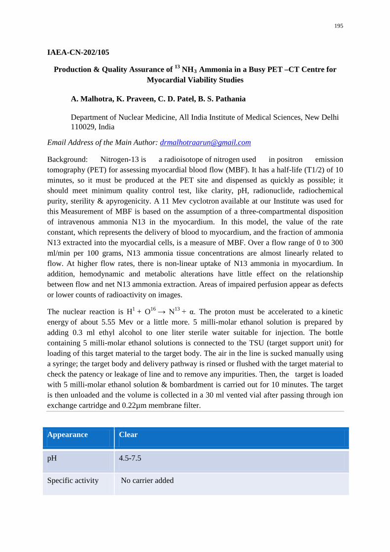
195
IAEA-CN-202/105
Production & Quality Assurance of 13 NH3 Ammonia in a Busy PET –CT Centre for Myocardial Viability Studies
A. Malhotra, K. Praveen, C. D. Patel, B. S. Pathania
Department of Nuclear Medicine, All India Institute of Medical Sciences, New Delhi 110029, India
Email Address of the Main Author: [email protected]
Background: Nitrogen-13 is a radioisotope of nitrogen used in positron emission tomography (PET) for assessing myocardial blood flow (MBF). It has a half-life (T1/2) of 10 minutes, so it must be produced at the PET site and dispensed as quickly as possible; it should meet minimum quality control test, like clarity, pH, radionuclide, radiochemical purity, sterility & apyrogenicity. A 11 Mev cyclotron available at our Institute was used for this Measurement of MBF is based on the assumption of a three-compartmental disposition of intravenous ammonia N13 in the myocardium. In this model, the value of the rate constant, which represents the delivery of blood to myocardium, and the fraction of ammonia N13 extracted into the myocardial cells, is a measure of MBF. Over a flow range of 0 to 300 ml/min per 100 grams, N13 ammonia tissue concentrations are almost linearly related to flow. At higher flow rates, there is non-linear uptake of N13 ammonia in myocardium. In addition, hemodynamic and metabolic alterations have little effect on the relationship between flow and net N13 ammonia extraction. Areas of impaired perfusion appear as defects or lower counts of radioactivity on images.
The nuclear reaction is H1 + O16 → N13 + α. The proton must be accelerated to a kinetic energy of about 5.55 Mev or a little more. 5 milli-molar ethanol solution is prepared by adding 0.3 ml ethyl alcohol to one liter sterile water suitable for injection. The bottle containing 5 milli-molar ethanol solutions is connected to the TSU (target support unit) for loading of this target material to the target body. The air in the line is sucked manually using a syringe; the target body and delivery pathway is rinsed or flushed with the target material to check the patency or leakage of line and to remove any impurities. Then, the target is loaded with 5 milli-molar ethanol solution & bombardment is carried out for 10 minutes. The target is then unloaded and the volume is collected in a 30 ml vented vial after passing through ion exchange cartridge and 0.22µm membrane filter.
Appearance Clear
pH 4.5-7.5
Specific activity No carrier added
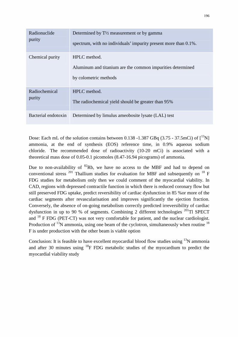
196
Radionuclide purity
Determined by T½ measurement or by gamma
spectrum, with no individuals’ impurity present more than 0.1%.
Chemical purity HPLC method.
Aluminum and titanium are the common impurities determined
by colometric methods
Radiochemical purity
HPLC method.
The radiochemical yield should be greater than 95%
Bacterial endotoxin Determined by limulus ameobosite lysate (LAL) test
Dose: Each mL of the solution contains between 0.138 -1.387 GBq (3.75 - 37.5mCi) of [13N] ammonia, at the end of synthesis (EOS) reference time, in 0.9% aqueous sodium chloride. The recommended dose of radioactivity (10-20 mCi) is associated with a theoretical mass dose of 0.05-0.1 picomoles (8.47-16.94 picograms) of ammonia.
Due to non-availability of 82Rb, we have no access to the MBF and had to depend on conventional stress 201 Thallium studies for evaluation for MBF and subsequently on 18 F FDG studies for metabolism only then we could comment of the myocardial viability. In CAD, regions with depressed contractile function in which there is reduced coronary flow but still preserved FDG uptake, predict reversibility of cardiac dysfunction in 85 %or more of the cardiac segments after revascularisation and improves significantly the ejection fraction. Conversely, the absence of on-going metabolism correctly predicted irreversibility of cardiac dysfunction in up to 90 % of segments. Combining 2 different technologies 201Tl SPECT and 18 F FDG (PET-CT) was not very comfortable for patient, and the nuclear cardiologist. Production of 13N ammonia, using one beam of the cyclotron, simultaneously when routine 18 F is under production with the other beam is viable option
Conclusion: It is feasible to have excellent myocardial blood flow studies using 13N ammonia and after 30 minutes using 18F FDG metabolic studies of the myocardium to predict the myocardial viability study
197
IAEA-CN-202/181
Challenges in Radiopharmacy in Nuclear Cardiology in Senegal
R. S. Senghor, S. Seck-Gassama, M. Mbodj
Biophysic Laboratory and Nuclear Medicine Service / University Cheikh Anta Diop, Dakar, Senegal Nuclear Medicine Department, General Hospital Grand Yoff (HOGGY), Dakar, Senegal
Email Address of the Main Author: [email protected]
Background: At Grand Yoff General Hospital (HOGGY), Nuclear Medicine Service, Radiopharmaceuticals preparation and dispensation are made under the supervision of an engineer biologist with an appropriate training: participation in an IAEA competency based programme with a three month fellowship and involvement to training courses on preparations and quality control procedures. The engineer biologist is supervised by the nuclear medical physician. The aims of this study are to promote best radiopharmacy practices approaches regarding constraints in the work situation to improve quality imaging in nuclear cardiovascular diseases.
Methods: The study presents the project to improve MIBI preparation, referred to quality assurance documents for hot laboratory such as International Atomic Energy Agency, Operational Guidance on Hospital Radiopharmacy (IAEA, 2008).
The sestamibi kit contains a lyophilised and sterile ingredient ready for labelling with Technetium 99m (99mTc) according to the instructions given by the manufacturer using aseptic techniques in a glove box, type Amercare, shielded and depressurized. 99mTc is obtained by elution from 99Mo - 99mTc Ultratechnekow generator supplied by IBD Holland (Mallinckrodt). 99mTc eluates activities are measured with a dose calibrator type Capintec CRC-25R.The percentage of 99Mo in the generator eluates is determined as quality control. The MIBI vial is removed from the fridge, left at room temperature inside the isolator cabinet for 15 minutes, and then put into a lead container while a volume of 99mTc is added aseptically inside it; the reaction vial is agitated to dissolve the lyophilised material and it is placed in the heat water bath. After heating the vial is placed in a lead shield and cooled at room temperature inside the isolator cabinet to be refreshed during 15 minutes. The radiochemical purity is not analyzed prior to administration of 99mTc-sestamibi due to a lack of materials.
Result: 35 cardiac investigations were carried out in the Nuclear Medicine Department of HOGGY during the study period. Without determining the labelling yield of 99mTc-sestamibi, a few poor quality scans are observed. To promote quality radiopharmacy practices, the Nuclear Medicine Service at HOGGY needs to overcome many challenges : low awareness on the importance of good Radiopharmacy practice amongst policy makers, health administrators and nuclear medicine staff, shortage of personnel enough trained in Radiopharmacy practice with emphasis on receipt, storage and safe handling of

198
radiopharmaceuticals, irregular supply of radiopharmaceuticals reagents kits and Tc-99m generators, lack of some hot lab equipment for preparation and quality control : a shielded heating block boiling for a good preparation of MIBI, a Radiochromatography scanner to complete Radiochemical purity tests, Aluminium ion breakthrough to complete Chemical purity tests, lack of routine nuclear medicine personnel monitoring, and contamination surveys for radiation exposure
Conclusion: Our nuclear cardiology scans show most of the time quite good imaging due to the stability of 99mTc-sestamibi and the respect of the procedures, however, this study defines requirements for optimal radiopharmacy practice regarding equipment, facilities and work procedures in the respect of radiation protection rules, and awareness of decision makers.
199
IAEA-CN-202/182
Performance of the Ultratechnekow Generator in Nuclear Cardiology in Senegal
R. S. Senghor, L. Dieng, M. Diarra, S. Seck-Gassama
Nuclear Medicine Department, General Hospital Grand Yoff (HOGGY), Dakar, Senegal
Email Address of the Main Author: [email protected]
Background: In the Nuclear Medicine Department of HOGGY, 99m technetium (99m Tc) is used to compound radiopharmaceuticals in nuclear cardiology. 99mTc is obtained by elution from 99Mo - 99mTc Ultratechnekow generator supplied by IBD Holland (Mallinckrodt). To provide best quality imaging, a protocol was established to determine the performance of the generator by calculating the elution efficiency and determining the 99Mo content of the eluates.
Methods: The study was performed in the Nuclear Medicine Department, General Hospital Grand Yoff (HOGGY). The protocol concerned the performance of the 99Mo - 99mTc Ultratechnekow FM generator and the purity of the eluate. 99Mo - 99mTc Ultratechnekow FM generator is a closed system providing sterile 99mTc eluate on a daily basis. 99Mo - 99mTc Ultratechnekow FM generators are available with a range of 99Mo activities, 6 – 10 GBq, sterile evacuated vials are supplied with the generator offering 5ml elution volume. Daily elution of the 99Mo - 99mTc Ultratechnekow FM generator were carried out using aseptic techniques in a glove box, type Amercare, shielded and depressurized. 99mTc eluates activities were measured with a dose calibrator type Capintec CRC-25R. 99Mo - 99mTc Ultratechnekow FM generator was eluted in evacuates sterile vials with 5ml of saline The performance of the generator is expressed by the elution efficiency which is defined as the fraction of eluted Tc99m activity of the theoretically available radioactivity at the time of elution. The elution efficiency is given as a percentage. The purity of generator eluate was determined by the 99Mo breakthrough.
Results: The study was performed over two weeks, and quality assessments of the 99Mo - 99mTc Ultratechnekow FM generator from IDB Holland showed normal values of 99Mo content in the eluates. The elution efficiency of the generator showed a yield which decreased from 87.94% to 79% on the sixth day to reach 60% on day 14. Normal values expected for this type of generator are close to 1.
Conclusion: Nuclear cardiology is a medical specialty based on the use of radiopharmaceuticals for diagnosis. These radiopharmaceuticals once administrated to patients, provide a functional imaging of the heart and the perfusion of the myocard. In our routine practice 99mTc is the unique radionuclide that we used coupled with MIBI or tetrofosmine.
Our study has shown that the elution efficiency is a quality criterion to evaluate 99Mo - 99mTc Ultratechnekow FM generator and to assure the purity the generator eluate. The

200
results emphasised the importance of planing the cardiac investigations as soon as possible after reception of the generator and the necessity to respect the manufacturing practice procedures.
201
IAEA-CN-202/242
TAEA-PAF Produced Radioisotopes and Radiopharmaceuticals for Cardiovascular Diseases
E. H. Has, A. Tanrikut, A. N. Yuksel, S. Unal, B. Yildiz
Saraykoy Nuclear Research and Training Center, Saray Mahallesi, Atom Caddesi, No:27 Kazan-Ankara, Turkey
Email Address of the Main Author: [email protected]
Cardiovascular diseases (CVDs) are an important group of non-communicable diseases. Millions of people fight back CVDs from all around the world. Thence CVDs are one of the main priorities in the health care systems of many countries. Molecular nuclear medicine practises are used for treatment and diagnosis of many diseases. Medical imaging including molecular nuclear medicine is very important. It offers lots of advantages for diagnostic and therapeutic decision making. Humankind derives benefit from these advantages for diagnosis, treatment, prognosis and follow-up in the management of CVDs. Nuclear medicine techniques such as single photon emission computed tomography (SPECT), positron emission tomography (PET), echocardiography, computed tomography (CT), magnetic resonance imaging (MRI) are used for treatment and diagnosis of CVDs. The use of radioisotopes is an important part of these medical practices. Many radioisotopes such as 201Tl, 99mTc, 15O, 13N, 82Rb are used for treatment and diagnosis of CVDs. 201Tl, 99mTc are used as SPECT agents and 15O, 13N, 82Rb,18F are used as PET agents. Main policy of Turkish Atomic Energy Authority (TAEA) is to provide necessary infrastructure for nuclear technology and get benefit from all products of nuclear technology in Turkey. Proton Accelerator Facility (PAF) is the first facility of its kind and serving an important nuclear technological infrastructure in Turkey. End users of medical sectors, research and development sectors are benefiting from this facility. Along with activities of private sector entities, TAEA is developing a national infrastructure with the capability of domestic production of various radioisotopes and radiopharmaceuticals. Proton accelerator in the facility is CYCLONE-30 (Ion Beam Applications-Belgium) type and its proton energy is 15-30 MeV (variable) and has four target systems. Three of them are for radioisotope production and one of them is for research and develeopment.18F is being produced as PET radioisotope and 123I, 201Tl, 67Ga, 111In are being produced as SPECT radioisotopes. Radiopharmaceutical forms of these radioisotopes are Na123I, 201TlCl, 67Ga[Cit], 111InCl3 and 18FDG. All of these radioisotopes are being produced under good manufacturing practises and quality assurance aspects rules. Radioisotopes planned to be produced in future at TAEA-PAF are 15O, 11C, 13N and 124I. TAEA-PAF produced radioisotopes which are used for CVDs are 201Tl and 18F. In future different radioisotopes such as 15O, 13N, 11C which are also used for CVDs, will be produced at TAEA-PAF.

202
ISSUES OF MEDICAL PHYSICS, INSTRUMENTATION AND IMAGE PROCESSING AND ANALYSIS RELATED TO CVD IMAGING
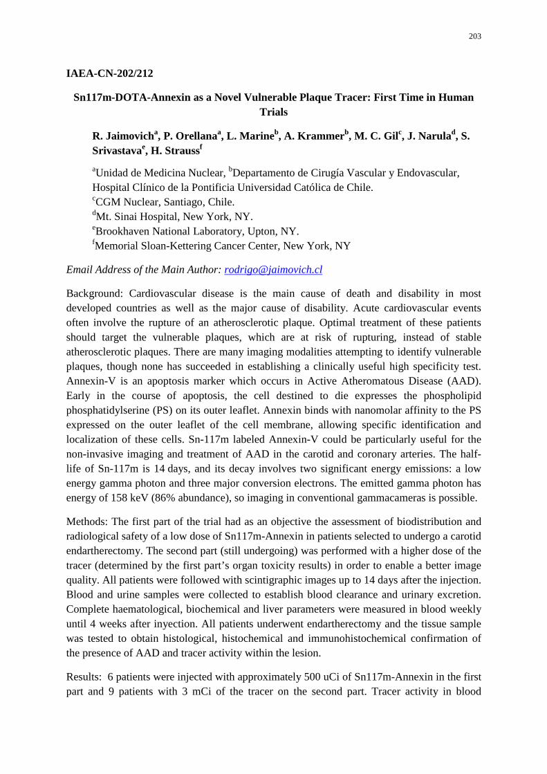
203
IAEA-CN-202/212
Sn117m-DOTA-Annexin as a Novel Vulnerable Plaque Tracer: First Time in Human Trials
R. Jaimovicha, P. Orellanaa, L. Marineb, A. Krammerb, M. C. Gilc, J. Narulad, S. Srivastavae, H. Straussf aUnidad de Medicina Nuclear, bDepartamento de Cirugía Vascular y Endovascular, Hospital Clínico de la Pontificia Universidad Católica de Chile. cCGM Nuclear, Santiago, Chile. dMt. Sinai Hospital, New York, NY. eBrookhaven National Laboratory, Upton, NY. fMemorial Sloan-Kettering Cancer Center, New York, NY
Email Address of the Main Author: [email protected]
Background: Cardiovascular disease is the main cause of death and disability in most developed countries as well as the major cause of disability. Acute cardiovascular events often involve the rupture of an atherosclerotic plaque. Optimal treatment of these patients should target the vulnerable plaques, which are at risk of rupturing, instead of stable atherosclerotic plaques. There are many imaging modalities attempting to identify vulnerable plaques, though none has succeeded in establishing a clinically useful high specificity test. Annexin-V is an apoptosis marker which occurs in Active Atheromatous Disease (AAD). Early in the course of apoptosis, the cell destined to die expresses the phospholipid phosphatidylserine (PS) on its outer leaflet. Annexin binds with nanomolar affinity to the PS expressed on the outer leaflet of the cell membrane, allowing specific identification and localization of these cells. Sn-117m labeled Annexin-V could be particularly useful for the non-invasive imaging and treatment of AAD in the carotid and coronary arteries. The half-life of Sn-117m is 14 days, and its decay involves two significant energy emissions: a low energy gamma photon and three major conversion electrons. The emitted gamma photon has energy of 158 keV (86% abundance), so imaging in conventional gammacameras is possible.
Methods: The first part of the trial had as an objective the assessment of biodistribution and radiological safety of a low dose of Sn117m-Annexin in patients selected to undergo a carotid endartherectomy. The second part (still undergoing) was performed with a higher dose of the tracer (determined by the first part’s organ toxicity results) in order to enable a better image quality. All patients were followed with scintigraphic images up to 14 days after the injection. Blood and urine samples were collected to establish blood clearance and urinary excretion. Complete haematological, biochemical and liver parameters were measured in blood weekly until 4 weeks after inyection. All patients underwent endartherectomy and the tissue sample was tested to obtain histological, histochemical and immunohistochemical confirmation of the presence of AAD and tracer activity within the lesion.
Results: 6 patients were injected with approximately 500 uCi of Sn117m-Annexin in the first part and 9 patients with 3 mCi of the tracer on the second part. Tracer activity in blood
204
cleared after 24 hours. Urine tracer excretion at day 7 was less than 5% of total urinary activity. No significant clinical changes or blood test abnormalities were detected. Imaging of atherosclerotic plaques was not achieved in these patients, although tissue analysis confirmed the presence of the tracer on all inflammatory plaques. One patient had an abdominal aortic aneurism that showed Sn117m-Annexin uptake in the scintigraphy.
Conclusion: A novel inflammatory tracer, specifically targeting vulnerable atherosclerotic plaques, is presented. Biological safety and imaging capability is demonstrated. Clinical utility is still under study.

205

206
QUALITY MANAGEMENT, QUALITY CONTROL, QUALITY ASSURANCE AND AUDITS IN INTEGRATED MEDICAL IMAGING AND NUCLEAR MEDICINE
PRACTICE
207
IAEA-CN-202/102
Patient Dose Assessment in Interventional Cardiology Procedures in Algeria
N. Khelassi-Toutaouia, A. Merada, A. Toutaouia, Z. Brahimia, S. Saadia, B. Mansourib a Département de physique médicale, Centre de Recherche Nucléaire d’Alger, 2 Bd Frantz Fanon BP 399 Alger RP, Algérie b Service d’Imagerie Médicale, Centre Hospitalier Universitaire Lamine Debaghine, Boulevard Saïd Touati, Bab El Oued, Alger, Algérie
Email Address of the Main Author: [email protected], [email protected]
Background of the study: To evaluate patient doses in Interventional Cardiology procedures (IC) in Algeria. The procedures that have been taken into account are Coronary angiography (CA), Percutaneus Transluminal Coronary Angioplasty (PTCA) and Valvuloplasty (Vp).
Methods: The study consists of evaluating the Maximum Skin Dose (MSD) and the Dose Area Product (DAP) in 48 IC procedures, either diagnostic or therapeutic. For this purpose, gafchromic films XR type R were interposed between the x-ray beam and the patient before undergoing the procedure allowing the MSD to be measured. One public hospital (CHU Bab El Oued) and one specialised Cardiology Service (Clinique Maouche) were selected for this study.
Results: The results revealed large variations in MSD (0.05-3.3 Gy) and DAP (20-292 mGy.cm2). The mean MSD was 0.202 Gy in coronary angiography (CA), 1.162 Gy in Percutaneus Transluminal Coronary Angioplasty (PTCA), 1.86 Gy in CA+PTCA and 0.58 Gy in Valvuloplasty (Vp). The correlation of DAP with MSD was significant (r ~ 0.7) whereas its correlation with fluoroscopy time was less significant (r ~ 0.5).
Conclusion: The highest MSD values were found in PTCA, which is a therapeutic procedure. Two PTCAs out of the whole 48 investigated procedures showed MSDs higher than the threshold of 2 Gy corresponding to deterministic effects (respectively 3.0 Gy and 3.3 Gy). The large variations in MSD values stress the need to continuously monitor patient dose in IC procedures with special emphasis in PTCA procedure.
208
IAEA-CN-202/131
Improvement of Nuclear Cardiology in Morocco
N. Ben Rais
Ibn Sina Hospital, Chis University Mohammed V Souissi, Rue Mfadel Cherkaoui Al Irfane, Rabat, Morocco
Email Address of the Main Author: [email protected]
Background of the study: Cardiovascular diseases (CVD) appear among the principal causes of death and disability in the world: each year nearly 17,3 million patients die of the cardiovascular of diseases. The World Health Organization’s projections increase to 23,6 million the number of people who will die of one CVD from here 2030. With the increase in the life expectancy of the Moroccans (men: 73 years and women: 78 years) and the improvement of the health of the population, Morocco is experiencing a transition from its epidemiologic profile and an increase in the cardiovascular diseases, accentuated by sedentariness, the nicotinism and obesity. As the leading cause of mortality in Morocco, the CVD are responsible of more than 100000 deaths per year. One out of two Moroccans dies of a cardiac disease. Morocco is a country at risk of CVD: 33% of the adult population has arterial hypertension, 13% are overweight, 6% are diabetic and 29% have high cholesterol. These CVD risk factors expose many people to heart attacks and myocardial infarctions.
Methods: In this work, we will bring back the incidence of the ischaemic cardiopathy in the diabetic and hypertensive patient; the goal of this work is to underline the frequency of the ischaemic disease in this group of patients and the interest of the cardiac tomoscintigraphy in the detection of the quiet ischaemic disease. It is an exploratory study relating to 150 diabetic and hypertensive patients with the services of internal medicine and cardiology.
Results: All patients have had diabetes and hypertension for at least 30 months. The average age of patients is 56 years old with a male majority of 56%. Nearly 84% of patients with diabetes type 2 and are use oral anti-diabetes medication. Other identified primary risk factors are obesity in 30% and smoking in 45% of cases. The diagnosis was made at the clinic, the ECG, the echocardiogram and stress test power for almost all patients. Coronary artery disease was found in 45% of cases with frequency of asymptomatic in 56% of cases represented by myocardial ischemia in 55% of cases, the IDM in 7% of cases. SPECT cardiac cost limited the number of patients underwent the myocardial tomo scintigraphy. For that only forty-six patients underwent this exam. The SPECT myocardial scintigraphy has demonstrated its effectiveness to identify silent ischemia. Coronary angiography was performed in all patients who require angioplasty Symptomatic forms were observed in 43.3% and represented by stable angina in 60% of cases, unstable angina in 7% of cases and 33% in L IDM cases. Revascularization was required in 23% of cases.

209
Conclusion: These data underscore the frequency of ischemic heart disease in diabetic and hypertensive patients with predominantly asymptomatic forms as well as the value of routine screening especially cardiac SPECT.
210
IAEA-CN-202/158
Image Quality and Radiation Dose using Low Tube Kv in ECG Gated 64 Slice CT Coronary Angiography
D. H. Salamaa,, A. M. Badawib. a Lecturer of Radiodiagnosis, National Center for Research and Radiation Technology, Atomic Energy Authority, Cairo, Egypt b Consultant Cardiology, Chief of Cardiovascular CT, the International Cardioscan Cairo, Egypt.
Email Address of the Main Author: [email protected]
Objectives: The purpose of our study was to compare the image quality and dose reduction of 64 slice ECG gated coronary angiography using 100 kVp instead of 120 kVp.
Material and methods: Eighty patients with a normal BMI ( < 25 kg/m) who had been referred for coronary CTA and did not have a history of coronary revascularization were included. Forty patients (group 1) were examined with low kV protocol (100- KV) and then compared to 40 patients examined with standard KV protocol (120 KV). Image quality was assessed and effective dose was determined based on dose length product.
Results: Imaging with 100 KVp instead of 120 KVp was associated with 45 % reduction in radiation dose. There were no significant differences between the two scanning protocols in the Mean Image Quality or interpretability.
Conclusion: The use of lower tube voltage leads to significant reduction in radiation exposure in CCTA. The use of 100 KVp tube voltage should be considered in dose reduction strategies for coronary CTA for individuals with a normal BMI.
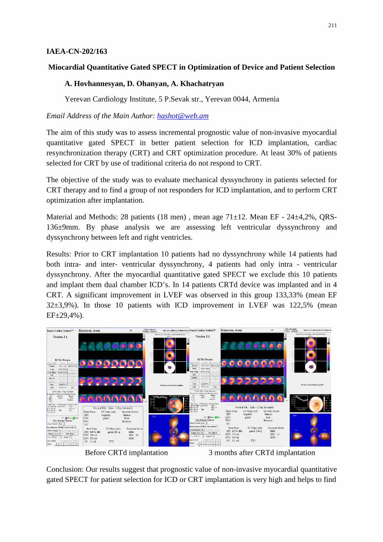
211
IAEA-CN-202/163
Miocardial Quantitative Gated SPECT in Optimization of Device and Patient Selection
A. Hovhannesyan, D. Ohanyan, A. Khachatryan
Yerevan Cardiology Institute, 5 P.Sevak str., Yerevan 0044, Armenia
Email Address of the Main Author: [email protected]
The aim of this study was to assess incremental prognostic value of non-invasive myocardial quantitative gated SPECT in better patient selection for ICD implantation, cardiac resynchronization therapy (CRT) and CRT optimization procedure. At least 30% of patients selected for CRT by use of traditional criteria do not respond to CRT.
The objective of the study was to evaluate mechanical dyssynchrony in patients selected for CRT therapy and to find a group of not responders for ICD implantation, and to perform CRT optimization after implantation.
Material and Methods: 28 patients (18 men) , mean age 71±12. Mean EF - 24±4,2%, QRS- 136±9mm. By phase analysis we are assessing left ventricular dyssynchrony and dyssynchrony between left and right ventricles.
Results: Prior to CRT implantation 10 patients had no dyssynchrony while 14 patients had both intra- and inter- ventricular dyssynchrony, 4 patients had only intra - ventricular dyssynchrony. After the myocardial quantitative gated SPECT we exclude this 10 patients and implant them dual chamber ICD’s. In 14 patients CRTd device was implanted and in 4 CRT. A significant improvement in LVEF was observed in this group 133,33% (mean EF 32±3,9%). In those 10 patients with ICD improvement in LVEF was 122,5% (mean EF±29,4%).
Before CRTd implantation 3 months after CRTd implantation
Conclusion: Our results suggest that prognostic value of non-invasive myocardial quantitative gated SPECT for patient selection for ICD or CRT implantation is very high and helps to find

212
the group who will be real responders. The improvement in LVEF in ICD group could be explained by optimizing AV timing and rhythm stabilization.
213
IAEA-CN-202/169
Pulmonary Embolism: Diagnostic Strategies
S. Moussouni, T. Bassaide, H. Mahmoudi, N. Merzougui, S. Belaribi, R. Koudjeti
Military University Hospital, Aine Beida, Oran, Algeria
Email Address of the Main Author: [email protected]
Pulmonary embolism (PE) is a common life-threatening condition (and a relatively common cardiovascular emergency) that is notoriously difficult to diagnose on purely clinical grounds with a significant mortality reduction when a specific treatment is performed.
Although pulmonary angiography usually remains in literature the usual standard modality for detection of PE, its high rates of morbidity and mortality justify the current need for an accurate and non-invasive diagnosis. The accuracy and main limitations of D-dimer testing, ventilation-perfusion scintigraphy, lower extremity venous sonography and spiral CT are successively reviewed. MR perspectives are also considered.
Suspected high-risk and non-high-risk PE are two distinct situations that must be distinguished because the diagnostic strategies differ. Concurrently with the diagnosis of PE, prognostic assessment is required for risk stratification and therapeutic decision-making.
Diagnostic algorithms based on non-invasive exams have recently been proposed: actually, algorithms including spiral CT and lower extremity sonography allow the lowest mortality and the best cost effective rates.
214
IAEA-CN-202/193
Estimation of Effective Doses for Patients in Interventional Cardiology
A. Skopljak-Beganović, B. Hanić, A. Beganović, M. Gazdić-Šantić, M. Kulić, M. Spužić, A. Drljević, D. Samek
Deparment of Medical Physics and Radiation Safety, Clinical Centre of Sarajevo University, Bolnička 25, Sarajevo 71000, Bosnia-Herzegovina
Email Address of the Main Author: [email protected]
The number of interventional cardiology procedures, as well as other radiological procedures, such as cardiac nuclear medicine, cardiac computed tomography (CT) and electrophysiology is increasing over the past years. They contribute an important share to the medical exposure.
Doses received by patients in interventional cardiology are usually described by kerma-area product (KAP) or cumulative air-kerma at the interventional reference point (KIRP). One of the indicators used to evaluate potential detriment of ionizing radiation is the effective dose. Its value can be obtained using different techniques; most of them rely on Monte Carlo simulations of radiological examination. The procedure consists of multiple series, each having different examination data, such as tube potential, craniocaudal (CC) angle, anterior oblique (AO) angle, field size, total filtration, as well as kerma-area product. The recorded values were used to evaluate effective dose received by the patient per series. The results were summarized, giving the value of total effective dose received by a patient.
The study included 56 patients. Procedures included diagnostic (coronary angiography, ventriculography and aortography) and therapeutic (percutaneous transluminal coronary angioplasty or stent placement). The average KAP per patient was 31.08 Gycm², while estimated average effective dose was 8.2 mSv. Thus, the average conversion factor between KAP and effective dose was estimated to be 0.263 mSv/Gycm². The average value of cumulative air-kerma, which is a reliable indicator of the peak skin-dose, is 647.7 mGy.
Doses received by patients are consistent with data reported in the literature. The estimated conversion factor between KAP and effective dose in dependent on techniques used, therefore the discrepancies between obtained value and literature data is expected.

215
IAEA-CN-202/197
Appropriate Use Criteria for Cardiac Radionuclide Imaging - Compliance and Relationship to Clinical Impact
A. Prata Jeronimo, A. I. Santos, I. Henriksson, H. Pereira
Hospital Garcia de Orta, E.P.E. – Avenida Torrado da Silva, 801-951 Almada – Portugal
Email Address of the Main Author: [email protected]
Aim of the study: To evaluate referrals for myocardial perfusion scintigraphy (MPS) in terms of its compliance with 2009 Appropriate Use Criteria for Cardiac Radionuclide Imaging and assess its relationship to clinical impact.
Methods: We have retrospectively selected 107 patients, mean age 66.5 years (95%CI:64.5;68.6), referred to our institution for MPS between 1st January and 30th May 2011. In this sample population, we have characterized gender, medical speciality of referral (cardiologist versus non cardiologist), presence of previous history of coronary heart disease (CAD) and probability of CAD in patients without previous history. The 2009 Appropriate Use Criteria for Cardiac Radionuclide Imaging were used to classify appropriateness of referrals, score them and define clinical indication for the exam. We have used Mann-Whitney test to evaluate if there were statistically significant differences of appropriateness score between gender and medical speciality of referral. In 53 patients we were able to assess the follow up and evaluated the clinical relevance of the MPS result in patient management, defined as inducing a change in therapy/imaging approach.
Results: Our data consisted on 60 male (56.1%) and 47 female (43.9%) patients, referred mainly by cardiologists (81/107=75.7%), with a majority of patients with previous history of CAD (60/107=56.1%). We had a non-appropriateness rate of 13.1% (14/107). We have not found statistically significant differences of appropriateness scores between males (average 6.9 – CI95%:6.4;7.4) and females (average 7.1 – CI95%:6.6;7.6) – p=0.63; neither between cardiologists (average 6.8 – CI95%: 6.4; 7.3) and non-cardiologists referrals (average 7.5 – CI95%:7.0;8.0) – p=0.41. Considering patients follow up, we found that the result of the MPS had had clinical impact in 37/47 (78.7%) of the appropriate referrals and in 5/6 (83.3%) of the non-appropriate referrals.
Discussion/Conclusion: Our non-appropriateness rate is similar to what has been published by other authors. Opposite to other published studies, we have not found significant differences in appropriateness scores between gender and type of medical speciality of referral, but maybe this is a bias introduced by our exclusive hospital referral. We have had a high clinical impact of the study in appropriate referrals, confirming the utility of appropriate criteria for referral decisions. Nevertheless, in one patient classified as non-appropriate referral, the MPS was positive for ischemia and crucial for the therapy decision, which suggests some caution in refusing exams exclusively based on appropriateness scores.
216
IAEA-CN-202/207
Comparison of Ejection Fraction Left Ventricular Obtained by Method Manual and Automatic in the Nuclear Medicine Institute Sucre-Bolivia
E. Huanca Sardinas, M. R. Vásquez Ibáñez, A. Zambrana Zelada, M. Torrez Cabero
Instituto de Medicina Nuclear, Universidad Mayor Real y Pontificia de San Francisco Xavier de Chuquisaca, Sucre, Bolivia
Email Address of the Main Author: [email protected];[email protected]
One of the functions of professional nuclear medical technologist is the process of studies acquired in Nuclear medicine centres, among which are referred to the cardiac studies of perfusion of myocardial and MUGA (Multi Gatillado) which provide information of left ventricular ejection fraction. This information is very important when it comes to medical decision-making. For comfort or time pressure usually these trials perform them with semi-automatic or automatic programmes. The objective of this work was the compare results of Equilibrium Left Ventricular Ejection Fraction both manually and using automated programs.
A retrospective study, of a total of 326 MUGA in equilibrium studies, took a random sampling of 25 isotopic MUGA corresponding to 13 women between the ages of 36 and 12 men between 17-69 years and 68 years respectively. Studies performed by marking red blood cells in vivo, the acquisition was in projection OAI, looking for a good separation of the Ventricular septal, saturating the image to 6 Megacuentas and a cycle average of 24 images. Each of 25 isotopic studies was processed by three programs: 2 view manual, semi-automatic all view, Semiauto-9funitonals. Seventy-five trials were performed.
The results showed women averaged LVEF 48% with manual program 2 view, regarding LVEF 49% men, appreciating a decrease with semi-automatic programs of LVEF 42% and 43% compared to men LVEF 40% and 41%. The standard deviation of the ejection fraction of the left ventricle in females reflects a value of 3.34, in relation to 4.9 in the male sex. The variation of the ejection fraction obtained both manual and automatic, is significant, so it must educate the operator to carry out prosecutions taking into account that the information provided can help a prompt and effective treatment. Manual processing gives the option of making interest areas of the left ventricle at the end of diastole and end of systole, which can be done taking into account the background. As processing with automatic and semi programs, they have the option of making a single area of interest which applies an automatic stroke adjusting both in end of diastole and systole end not considering the background area.
So it is concluded that the result of the ejection fraction is operator dependent, being the Nuclear Medicine Technologist responsible for presenting this information to the Nuclear physician for the subsequent report of the study of MUGA in balance.
217
IAEA-CN-202/208
Efficiency of In-Vivo Labeling of RBCs for MUGA Scans at Ibadan, Oyo-State, Nigeria
I. Lawal, Y. Onimode, B.O.A. Osifo
University College Hospital, Queen Elizabeth Road, Ibadan, Nigeria
Email Address of the Main Author: [email protected]
Background of the study: Radionuclide ventriculography (RNV) or gated equilibrium cardiac blood pool Imaging (MUGA) is a Nuclear Medicine technique designed to evaluate the function of the ventricles of the heart thus allowing informed diagnostic intervention in heart failure. Common clinical indication for MUGA scan are: known or suspected coronary heart diseases (CAD); known or suspected congestive heart failure (CHF); evaluation of ventricular function in patient undergoing chemotherapy; assessment of ventricular function in patients with Ventricular stenosis and/or insufficiency. A radiopharmaceutical tracer remaining in the blood could provide information on cardiac pumping action with application of appropriate technique.99m Tc-RBC is the agent of choice. In view of the large content of RBC in blood, low susceptibility for damage and easy stable labeling of RBC with Tc-99m. In order to further improve the reliability of labeling and accuracy of the quantitation of the MUGA study, in vivo/in vitro procedures are used. Typical chase is 15mCi for rest and 25-30mCi for stress MUGA.
Methods: There are three general methods of labeling red blood cells with Tc-99m. All are simple, rapid, reliable and inexpensive. However for the purpose of this study, the in-vivo method which is the most practical will be used. It involves the following steps: (1) 1mg of Sn2+ in form of pyrophosphate (PYP) given by I.V. (2)20 minutes wait for Sn2+ to diffuse into RBC (3) I.V. injection of 25mCi Tc-99m pertechnetate (4) 10min wait for radio-labeling to take place. (5) Expected labeling efficiency is 80-85%.
Results: Results are not yet available.
Conclusion: Will be provided at the end of the study.
218
IAEA-CN-202/210
Validation of LVEFs from Gated Cardiac Blood Pool Studies at Ibadan
Y. A. Onimode, B. O. A. Osifo
Department of Nuclear Medicine, University College Hospital, Ibadan, Oyo State, Nigeria
Email Address of the Main Author: [email protected]
Background: Multiple gated cardiac blood pool scintigraphy (MUGA scan) is a simple, physiological method for deriving estimated LVEF which is relatively devoid of side effects. Some clinical indications for its use include monitoring of cardiac function in patients on cardiotoxic anthracycline-based chemotherapy, evaluation of anthracycline-induced cardiac dysfunction, and in the preoperative prediction of cardiac risk. The scan has been acclaimed as the gold standard for determining left ventricular ejection fraction (LVEF). However, each Nuclear Medicine Department must determine normal ranges of LVEF for its service population in order to validate the results derived from these scans.
Methods: From March to August 2013, a prospective study will be carried out involving one hundred and fifty healthy male and female subjects aged 18-55 years recruited from the local hospital and university environments. Subjects will be screened for eligibility by clinical assessment, and with investigations such as chest x-rays, electrocardiographs, and packed cell volume. After having obtained informed written consent, the in vivo method of red cell labelling will be utilized; an initial direct intravenous administration of stannous pyrophosphate, followed thirty minutes later by a second intravenous injection of Tc-99m pertechnetate via another venous site. MUGA scintigraphy will be performed using a Siemens eCam gamma camera fitted with a low-energy all-purpose or ultra-high resolution collimator while subjects are connected to a cardiac gating device. Thereafter, resting LVEFs will be computed from regions of interest generated automatically and manually about the left ventricle in the view of the heart granting the best septal separation of the two ventricles. Values generated from both methods with a maximal disparity of 5 percent between the two will be accepted. Upper and lower limits of LVEF will be derived using the 95 percent limits about the mean value. Subsequently, age- and gender-appropriate LVEF ranges will be generated for use in the department, and will be published for use in the hospital. Similar studies will be performed in Nuclear Medicine facilities in the country once they are established.
Results and Conclusion: These will be produced upon completion of the study.
219
IAEA-CN-202/211
Pattern of Clinician Referral of Patients for Gated Cardiac Blood Pool Studies
Y. A. Onimode, M. D. T. Vangu, B. O. A. Osifo
Department of Nuclear Medicine, University College Hospital, Ibadan, Oyo State, Nigeria
Division of Nuclear Medicine, Charlotte Maxeke Johannesburg Academic Hospital, Johannesburg, South Africa
Email Address of the Main Author: [email protected]
Background: In clinical practice, multi-gated cardiac blood pool scintigraphy (MUGA scan) has been validated in the management of patients being considered for cardiotoxic anthracycline-based chemotherapy, and in the evaluation of those patients who have developed cardiotoxicity from the use of these chemotherapeutic agents. Anthracyclines are indicated in the therapy of patients with haematological malignancies, breast cancer, Kaposi’s sarcoma. An initial baseline scan is recommended prior to commencement of intervention, with follow-up scans performed as indicated in the course of treatment.
Method: One hundred and ninety-nine scans performed between 2007 and 2008 were reviewed retrospectively. The aim of the study was to assess the manner in which clinicians referred patients for MUGA scans and the appropriateness with which they incorporated the scan into patient management. Patient data was retrieved from case notes and analysed.
Results and Conclusion: pending end of study.
220
IAEA-CN-202/222
The Challenge of Diagnosing Coronary Heart Diseases (CHD) in Women: the Special Role of ECG-Gated SPECT Myocardial Scintigraphy (MS)
I. Garty
Nuclear Medicine Department, Haemek Medical Center, Afula, Israel
Email Address of the Main Author: [email protected]
Background of the study: Cardio vascular diseases (CVD) are the number 1 health threat and the main cause of death in American women, according to the official publication of the American Heart Association. In women, heart disease is too often a "silent killer", and often is presented by non-specific symptoms. Furthermore, investigators from the University of Michigan found that women with heart disease tend to minimize their symptoms far more than men. Unfortunately, Coronary Angiography (CA) can look "normal" in female patients who actually have significant CHD. Based on the above mentioned facts and observations many authors consider the early diagnosis of CVD in women as a real challenge. The aim of this study is to try and improve the non-invasive imaging modalities for earlier and more accurate diagnosis of CVD in women, especially in patients with suspected false negative CA.
Methods: During the last 10 years, (2003-2012) 4200 patients (1570 women and 2630 men) were admitted to our department for myocardial scintigraphy. All of them underwent ECG-gated SPECT study. 2813 patients: 480 (17%) women and 2333 (83%) men achieved full stress ergometric study; prior to the injection of the radiopharmaceutical the rest (1387) patients (44% women and 35% men) underwent drug induced stress studies.
Results: Comparison of the results of stress studies to the scintigraphic studies showed that among 480 females with full stress studies, 382 (79.5%) showed positive stress versus 1520 among 2333 males (65%). Among 382 female patients with positive stress study 304 (79.5%) showed positive scintigraphic study. Among 1520 females with positive stress studies 1270 (83.5%) showed positive scintigraphic studies. Among 98 females with negative stress studies 42 (44%) showed positive scintigraphy versus 670 (82.5%) among the 813 males with negative stress study. 1720 (75%) of 2286 patients with positive scintigraphy were sent for further evaluation by CA versus 220 (11.5%) patients with negative scintigraphic studies. The comparison of CA results to ECG stress and scintigraphic results in males and females are now in progress.
Conclusions: 1. Fewer female patients are referred to stress scintigraphic studies compared to male patients. 2. More positive ECG stress studies in women probably due to the high percent of women that arrive too late for imaging evaluation. 3. More false positive scintigraphic results in women. 4. Due to the lower sensitivity of CA in women the combination of ECG exercise study results and myocardial scintigraphy results may considerably improve the diagnostic accuracy. 5. SPECT myocardial scintigraphy was proved to be an efficient non-invasive modality in women with suspected CHD and is much

221
more sensitive than ECG study alone. 6. To women that are unable to achieve full ergo metric stress, drug induced myocardial scintigraphy is recommended. 7. Due to the difficulties in diagnosing CHD in diabetic women, myocardial scintigraphy should be the first choice in evaluating diabetic women with suspected CHD.
222
IAEA-CN-202/227
18F-FDG Cardiac PET: Good or Bad Patient Preparation for Good Image Quality - How to Know It in Advance
S. A. Rogan, A. Balenovic
Nuclear Medicine Departament PET/CT Centre, Vocarska 106, Zagreb, Croatia
Email Address of the Main Author: [email protected]
Background: Fluordeoxyglucose (FDG) enters myocardial cells by the same transport mechanism as glucose. Because of slow dephosphorilation rate of FDG in cells, it becomes trapped in the myocardium, allowing adequate time to image regional uptake by PET. The maximize FDG uptake in myocardium results in superior image quality and reduces occasional irregular FDG uptake. Namely, hormonal level in the blood must favor utilization of glucose over fatty acids. Although, we think to know how to prepare patient before FDG administration, some of images are not good enough for proper image interpretation. Methods: In our center we made 30 FDG viability examinations. We are using two main approaches to stimulate myocardial glucose metabolism with oral or intraveneous (IV) glucose loading. In non-diabetic patient after a fasting period of at least 6 hours (induction of endogenous insulin) oral administration of 25-100 g glucose is sufficient for optimal glucose loading; but IV loading is also used. If needed insulin is administered, too. Thirty minutes later, if blood glucose level is <7,8 mmol/l 5.4 mCi of 18F-FDG is injected and after 45-90 minutes after injection acquisition can be started. Diabetic patients pose a challenge: they have limited ability to produce endogenous insulin but also their cells are less sensitive to insulin stimulation. Simple protocol of fasting-oral glucose loading scheme is not effective for diabetics, so use of insulin along with close monitoring of blood glucose level is needed. This time-consuming procedure could cause delay of FDG administration or delay in image acquisition (increased decay of 18F FDG). Sometimes insulin administration should be repeated several times until blood glucose reach level for FDG administration. Results: in 22 patients out from 30 images quality was sufficient for viability interpretation, but in 8 patients we found low or inadequate glucose uptake after acquisition. Interpretation of myocardial viability in those patients was uncertain, less reliable for cardiologists. Conclusion: Cardiac metabolism imaging is sometimes simple paradigm, but sometimes searches for the best in people, all together patient, technologist, nurse and nuclear medicine physician. We think that we need more experience or some changes in patient preparation for FDG PET myocardial viability study to have good quality of images.
223
IAEA-CN-202/255
Quality Management Audit in Nuclear Medicine Practices (QUANUM) Experience at King Chulalongkorn Memorial Hospital
U. Vutrapongwatanaa, T. Chaiwatanarata, A. Krisanachindaa, S. Sirisalipocha, S. Tepmongkola, K. Kingpetcha, C. Navikhacheevinb, T. Sontrapornpolb, P. Pasawangb, C. Kotevitheeb, S. Taothongb, S. Sorajwongjaruenb, S. Nipasungb, K. Klaimookb a Division of Nuclear Medicine, Department of Radiology, Faculty of Medicine, Chulalogkorn University, Bangkok, Thailand b Division of Nuclear Medicine, Department of Radiology, King Chulalongkorn Memorial Hospital, Bangkok, Thailand
Email Address of the Main Author: [email protected]
The IAEA Regional Workshop on Quality Management Audits in Nuclear Medicine Practices (QUANUM) was held in November 2012, Bangkok Thailand. After that the audit was requested for King Chulalongkorn Memorial Hospital as the first institute in Southeast Asia. The IAEA sent the Expert team for the mission in March 18-22, 2013. We had approximately 2 months to prepare the documents.
Even though the hospital passed the national Hospital Accreditation to be held every 3-5 years, the audit does not go into the detail of every clinical service. In contrast to the QUANUM, an external audit by nuclear medicine specialists in all branches visited our department for the peer review in nuclear medicine service. The preparation began with the set-up of a “quality management team” which met once or twice a month and became more frequent to once or twice a week to report the preparation progress and plan for further action. 17 QUANUM checklists were reviewed, completed and submitted to IAEA one month prior the mission. All necessary documents and information had been collected with difficulty. The reasons were that we had no good system to collect the documents we produced and also some activities are performed so routinely that the systematic records of the results were missing. The main outcome of this QUANUM audit is the harmonization of all staffs which is the key of our work success.
During one week on audit process, five auditors of one nuclear medicine physician, one medical physicist, one nuclear medicine technologist, one radiopharmacist and an IAEA Technical Officer were working hard on peer review the document, the facilities and the clinical service. It was quite stressful for the auditee on the first day as lacking experience on the in-depth quality auditor process, lacking of the knowledge on the quality system. However, on the following days while the discussion was going on the auditors and the auditee had a chance to get acquainted and the real problems were dissolved resulting in the more relax atmosphere. Our major problem is the documentation preparation leading to the quality management. Without documentation the good quality of clinical service cannot be maintained. At the exit meeting on the last day, the auditor team concluded the list of tasks in

224
criticality, major and minor issues to be rectified. The discussion among the QM team was arranged to conclude the time frame to be accomplished.
QUANUM has been something so abstract and not understandable to us at the beginning. After the IAEA QUANUM mission, not only that we have a chance to “see” what our problems are, but we also understand what the true meaning of “quality management” is, and this is the most important thing to maintain real quality in nuclear medicine service. The hard work of the Audit Team are well appreciated and there are several lessons learnt from the most valuable and unforgettable mission.
225
IAEA-CN-202/262
Drafting and Implementing a Procedure Guideline for Investigation of Hibernating Myocardium in a Resource-Constrained Setting
A. Doruyter, A. Ellmann, J. Warwick, J. Holness
Tygerberg Hospital, Bellville, Cape Town, South Africa
Email Address of the Main Author: [email protected]
Background: Determining the extent of hibernating myocardium is prognostic of improvement in left ventricular ejection fraction post-revascularization. The gold standard for identifying hibernating myocardium is 2-deoxy-2-[fluorine-18] fluoro-D-glucose (18F-FDG) positron emission tomography/ computed tomography (PET/CT) combined with myocardial perfusion imaging (MPI)-PET/CT. Until the availability of 18F-FDG PET/CT, our institution performed either nitrate-enhanced rest technetium-99m (99mTc) sestamibi MPI single photon emission tomography (SPECT) to identify viable segments in regions with regional wall movement abnormalities (RWMA) on echocardiography or rest-redistribution thallium-201. With the recent acquisition of a PET/CT camera, cardiology requested a switch to a 18F-FDG-based protocol for this indication. Since MPI PET radiopharmaceuticals were unavailable, we decided to perform 18F-FDG PET/CT combined with 99mTc-sestamibi MPI SPECT/CT for which a standard operating procedure (SOP) was drafted.
Methods: A review of the relevant literature was performed. Guidelines published by the EANM/ESC and ASNC as well as an international departmental guideline on cardiac FDG studies were used as a departure point for the SOP.
Results: It was decided that patients be selected in consultation with the cardiology department. Selection criteria were either previous myocardial infarction or ischaemic heart disease and RWMA; with left ventricular dysfunction. Only patients planned for possible revascularization were investigated. Only non-diabetics were included in the trial period, with diabetics included later. A same day rest 99mTc-sestamibi MPI SPECT/CT, 18F-FDG PET/CT was performed, occasionally replaced by a 2-day protocol for practical reasons. Departmental preparation involved ordering a supply of acipimox, aspirin, rapid-acting insulin (for diabetics) and a standard meal (for non-diabetics). Patients were prepared according to the protocol. On the day of the study the patient followed a protocol (different for diabetic and non-diabetics), supervised by a registrar (resident), with the use of a standardized tick sheet. Patients received acipimox, aspirin, insulin (in diabetics), standardized meal (in the case of non-diabetics), injections of radiopharmaceuticals and scans at predetermined time points. Gated MPI SPECT/CT was obtained followed by a non-gated 18F-FDG PET/CT. To make SPECT and PET studies comparable, various detailed adjustments were made to matrix size and filters. PET images were reconstructed with AC while both AC and non-AC MPI SPECT reconstructions were performed. Interpretation and reporting was as described in the EANM guidelines with good acceptance by the referring cardiologists. Several unresolved issues remain which will be described in the presentation.

226
Conclusion: 18F-FDG PET/CT combined with 99mTc-sestamibi MPI SPECT/CT is a feasible option for the investigation of hibernating myocardium in settings without access to PET MPI agents.

227

228
RADIATION PROTECTION FOR PERSONNEL AND DOSE REDUCTION FOR PATIENTS
229
IAEA-CN-202/137
Radiation Protection for Personnel during Chronic Total Occlusion Recanalization
L. Price, A. Pascoal
King's Health Partners Academic Health Science Centre, Faraday Building , Denmark Hill, London, SE5 9RS, UK
Email Address of the Main Author: [email protected]
Percutaneous recanalization of total coronary occlusions (CTOs) is performed in hospital catheterization laboratories under local anaesthetic. Multiple fluoroscopy projections and long fluoroscopy times are used to direct guide wires and catheters to the CTO which is recanalized using balloon angioplasty alone or with stenting. CTOs are regarded as the most technically challenging and time-consuming subset of percutaneous coronary intervention. Hence the need to identify good and deficient radiation protection practices for all personnel involved.
The study recorded dose measurements for personnel involved with CTO procedures. The aim was investigate how radiation exposure varies between personnel and is influence by their practice in order to optimise radiation protection practices during CTO recanalization.
An active dosimetry system (i2 Raysafe) composed of individual radiation sensors (dosimeters) that transmit dose data to a remote base unit was used to collect and store radiation exposure to the personal involved in a series of CTO procedures. Dosimeters were worn by personal outside the lead apron at chest height and in some cases thyroid height to assess the variation of radiation exposure to unprotected tissue between team members. A reference dosimeter was positioned on the C arm (at 45 degrees below the isocentre) to normalize dose data from personnel dosimeters. Personnel were monitored over a four day CTO training session which consisted of 16 cases where recanalization had previously failed.
The personnel involved with CTOs include a primary cardiologist who stands closest to the detector head followed by a secondary cardiologist, scrub nurse and radiographer. A run nurse and physiologist were present in the laboratory but not for the full procedure. Cardiologists visiting from other centres internationally participated as primary and secondary cardiologists and in an observational capacity. Positions of personnel and radiation practices were recorded during the procedure as well exposures details.
Preliminary analysis of occupational doses in millisieverts and normalized to the reference dosimeter have been compared for different personnel and staff groups intra and inter CTO procedure. The highest occupational dose was obtained by observing cardiologists who received an average normalized dose of 30.1% (all procedures, all observers) in relation to the reference dosimeter. This was mainly attributed to the practice of one visiting cardiologist who tended to stand in front of the ceiling suspended lead screen, by the detector head in order view images displayed on a monitor. The second highest occupational dose was

230
received by run nurses who received an average radiation dose of 10.1% in relation to the reference dosimeter. It was noted that on occasions one run nurse spoke to the patient during an exposure. The average normalized dose for scrub nurses, secondary and primary cardiologists were 1.8%, 2.4% and 2.7% respectively.
In conclusion preliminary analysis indicates that; Radiation exposure of personal involved in CTO procedures showed noticeable variations, unexpected results were found for certain personnel such as an observing cardiologist due to radiation protection awareness and attention is required for nurse exposure to optimise radiation protection practice.
231
IAEA-CN-202/209
Safety and Risk Assessment for Personnel and Patients in In-Vivo Labeling for MUGA Scans at Ibadan, Oyo-State Nigeria
I. Lawal, Y. Onimode, B.O.A. Osifo
University College Hospital, Queen Elizabeth Road, Ibadan, Nigeria
Email Address of the Main Author: [email protected]
Background of the study: Advances in cardiac imaging procedures such as MUGA scans have improved the ability to assess and treat cardiovascular conditions. MUGA (Multigated acquisition scan) is a nuclear medicine procedure designed to evaluate the function of the ventricles of the heart thus allowing informed diagnostic intervention. It is performed by labeling patient’s red blood cells with a radiotracer such as technetium-99m and measuring activity over the anterior chest as the radioactive blood flow through the large vessels and the heart chamber. MUGA scan procedures during the preparation stages through scanning frequently expose both patients and personnel to ionizing radiation. The international recommendation for ionizing radiation requirements for protection includes justification, limitation, and optimization. Radiation exposure may increase risk of carcinogenesis. In the case of the personnel, the danger lie in the exposure to radiation over a long period of time or injudicious exposure to direct radiation as a result of inadequate shielding or disregard of safety factors. Radiopharmaceuticals must be handled according to the relevant radiation protection requirements to protect the personnel, patient and environment. Protecting the patient from unnecessary radiation exposure include not only the cardinal principles of radiation protection but also the outcome of labeling procedures of radiopharmaceutical. Any unsafe exposure beyond the ALARA principle is considered unnecessary excess exposure. These principles are part of the continuum of competent skills to bring about radiation safety for patients and personnel involved. Radiation dose to a patient from MUGA scans should be 7mSV.
Methods: Personnel and patient will be monitored for probable overexposure and protocol for preparation of Radiopharmaceuticals including preparation of individual doses for patients will be monitored
Results: Results are not yet available
Conclusion: Will be provided at the end of the study.
Related Documents