VETERINARY PRACTICE GUIDELINES 2019 AAHA Dental Care Guidelines for Dogs and Cats* Jan Bellows, DVM, DAVDC, DABVP (Canine/Feline), Mary L. Berg, BS, LATG, RVT, VTS (Dentistry), Sonnya Dennis, DVM, DABVP (Canine/Feline), Ralph Harvey, DVM, MS, DACVAA, Heidi B. Lobprise, DVM, DAVDC, Christopher J. Snyder, DVM, DAVDC y , Amy E.S. Stone, DVM, PhD, Andrea G. Van de Wetering, DVM, FAVD ABSTRACT The 2019 AAHA Dental Care Guidelines for Dogs and Cats outline a comprehensive approach to support companion animal practices in improving the oral health and often, the quality of life of their canine and feline patients. The guidelines are an update of the 2013 AAHA Dental Care Guidelines for Dogs and Cats. A photographically illustrated, 12-step protocol describes the essential steps in an oral health assessment, dental cleaning, and periodontal therapy. Recommendations are given for general anesthesia, pain management, facilities, and equipment necessary for safe and effective delivery of care. To promote the wellbeing of dogs and cats through decreasing the adverse effects and pain of periodontal disease, these guidelines emphasize the critical role of client education and effective, preventive oral healthcare. ( J Am Anim Hosp Assoc 2019; 55:---–---. DOI 10.5326/JAAHA-MS-6933) AFFILIATIONS From All Pets Dental, Weston, Florida (J.B.); Beyond the Crown Veterinary Education, Lawrence, Kansas (M.L.B.); Stratham-Newfields Veterinary Hos- pital, Newfields, New Hampshire (S.D.); Department of Small Animal Clin- ical Sciences, College of Veterinary Medicine, University of Tennessee, Knoxville, Tennessee (R.H.); Main Street Veterinary Dental Hospital, Flower Mount, Texas (H.B.L.); Department of Surgical Sciences, School of Vet- erinary Medicine, University of Wisconsin-Madison, Madison, Wisconsin (C.J.S.); Department of Small Animal Clinical Sciences, College of Veter- inary Medicine, University of Florida, Gainesville, Florida (A.E.S.S.); and Advanced Pet Dentistry, LLC, Corvallis, Oregon (A.G.VdW.). CONTRIBUTING REVIEWERS R. Michael Peak, DVM, DAVDC (The Pet Dentist, Tampa, Florida); Jeanne R. Perrone, CVT, VTS (Dentistry) (VT Dental Training, Plant City, Florida); Kevin S. Stepaniuk, DVM, FAVD, DAVDC (Veterinary Dentistry Education and Consulting Services, LLC, Ridgefield, Washington). Correspondence: [email protected] (C.J.S.) * These guidelines were supported by a generous educational grant from Boehringer Ingelheim Animal Health USA Inc., Hill’ s® Pet Nutrition, Inc., and Midmark. They were subjected to a formal peer-review process. These guidelines were prepared by a Task Force of experts convened by the American Animal Hospital Association. This document is intended as a guideline only, not an AAHA standard of care. These guidelines and recom- mendations should not be construed as dictating an exclusive protocol, course of treatment, or procedure. Variations in practice may be warranted based on the needs of the individual patient, resources, and limitations unique to each individual practice setting. Evidence-based support for spe- cific recommendations has been cited whenever possible and appropriate. Other recommendations are based on practical clinical experience and a consensus of expert opinion. Further research is needed to document some of these recommendations. Because each case is different, veterinarians must base their decisions on the best available scientific evidence in conjunction with their own knowledge and experience. Note: When selecting products, veterinarians have a choice among those formulated for humans and those developed and approved by veterinary use. Manufacturers of veterinary-specific products spend resources to have their products reviewed and approved by the FDA for canine or feline use. These products are specifically designed and formulated for dogs and cats and have benefits for their use; they are not human generic products. AAHA suggests that veterinary professionals make every effort to use veterinary FDA-approved products and base their inventory-purchasing decisions on what product is most beneficial to the patient. y C. Snyder was the chair of the Dental Care Guidelines Task Force. NAD (nonanesthetic dentistry); PD (periodontal disease staging); VOHC (Veterinary Oral Health Council); VTS (Dentistry) (Veterinary Technician Specialist[s] in Dentistry) ª 2019 by American Animal Hospital Association JAAHA.ORG 1

Welcome message from author
This document is posted to help you gain knowledge. Please leave a comment to let me know what you think about it! Share it to your friends and learn new things together.
Transcript

VETERINARY PRACTICE GUIDELINES
2019 AAHA Dental Care Guidelines for Dogsand Cats*Jan Bellows, DVM, DAVDC, DABVP (Canine/Feline), Mary L. Berg, BS, LATG, RVT, VTS (Dentistry), Sonnya Dennis,DVM, DABVP (Canine/Feline), Ralph Harvey, DVM, MS, DACVAA, Heidi B. Lobprise, DVM, DAVDC, Christopher J.Snyder, DVM, DAVDCy, Amy E.S. Stone, DVM, PhD, Andrea G. Van de Wetering, DVM, FAVD
ABSTRACT
The 2019 AAHA Dental Care Guidelines for Dogs and Cats outline a comprehensive approach to support companion animal
practices in improving the oral health and often, the quality of life of their canine and feline patients. The guidelines are an update of
the 2013 AAHA Dental Care Guidelines for Dogs and Cats. A photographically illustrated, 12-step protocol describes the essential
steps in an oral health assessment, dental cleaning, and periodontal therapy. Recommendations are given for general anesthesia,
painmanagement, facilities, and equipment necessary for safe and effective delivery of care. To promote the wellbeing of dogs and
cats through decreasing the adverse effects and pain of periodontal disease, these guidelines emphasize the critical role of client
education and effective, preventive oral healthcare. (J Am Anim Hosp Assoc 2019; 55:---–---. DOI 10.5326/JAAHA-MS-6933)
AFFILIATIONS
From All Pets Dental, Weston, Florida (J.B.); Beyond the Crown Veterinary
Education, Lawrence, Kansas (M.L.B.); Stratham-Newfields Veterinary Hos-
pital, Newfields, New Hampshire (S.D.); Department of Small Animal Clin-
ical Sciences, College of Veterinary Medicine, University of Tennessee,
Knoxville, Tennessee (R.H.); Main Street Veterinary Dental Hospital, Flower
Mount, Texas (H.B.L.); Department of Surgical Sciences, School of Vet-
erinary Medicine, University of Wisconsin-Madison, Madison, Wisconsin
(C.J.S.); Department of Small Animal Clinical Sciences, College of Veter-
inary Medicine, University of Florida, Gainesville, Florida (A.E.S.S.); and
Advanced Pet Dentistry, LLC, Corvallis, Oregon (A.G.VdW.).
CONTRIBUTING REVIEWERS
R. Michael Peak, DVM, DAVDC (The Pet Dentist, Tampa, Florida); Jeanne R.
Perrone, CVT, VTS (Dentistry) (VT Dental Training, Plant City, Florida);
Kevin S. Stepaniuk, DVM, FAVD, DAVDC (Veterinary Dentistry Education
and Consulting Services, LLC, Ridgefield, Washington).
Correspondence: [email protected] (C.J.S.)
* These guidelines were supported by a generous educational grant from
Boehringer Ingelheim Animal Health USA Inc., Hill’s® Pet Nutrition, Inc.,
and Midmark. They were subjected to a formal peer-review process.
These guidelines were prepared by a Task Force of experts convened by the
American Animal Hospital Association. This document is intended as a
guideline only, not an AAHA standard of care. These guidelines and recom-
mendations should not be construed as dictating an exclusive protocol,
course of treatment, or procedure. Variations in practice may be warranted
based on the needs of the individual patient, resources, and limitations
unique to each individual practice setting. Evidence-based support for spe-
cific recommendations has been cited whenever possible and appropriate.
Other recommendations are based on practical clinical experience and a
consensus of expert opinion. Further research is needed to document some
of these recommendations. Because each case is different, veterinarians must
base their decisions on the best available scientific evidence in conjunction
with their own knowledge and experience.
Note: When selecting products, veterinarians have a choice among those
formulated for humans and those developed and approved by veterinary
use. Manufacturers of veterinary-specific products spend resources to have
their products reviewed and approved by the FDA for canine or feline use.
These products are specifically designed and formulated for dogs and cats
and have benefits for their use; they are not human generic products. AAHA
suggests that veterinary professionals make every effort to use veterinary
FDA-approved products and base their inventory-purchasing decisions on
what product is most beneficial to the patient.
y C. Snyder was the chair of the Dental Care Guidelines Task Force.
NAD (nonanesthetic dentistry); PD (periodontal disease staging); VOHC
(Veterinary Oral Health Council); VTS (Dentistry) (Veterinary Technician
Specialist[s] in Dentistry)
ª 2019 by American Animal Hospital Association JAAHA.ORG 1
IntroductionThe concept that a pet is suffering from oral pain, infection, and
inflammation that may not be apparent but is affecting their quality
of life is a reality that may not always be fully appreciated by
the veterinary profession and often not understood by the pet-
owning public. Compromised dental health can affect a pet’s over-
all health, longevity, quality of life, and interaction with its owner
without exhibiting obvious clinical signs of disease. The purpose of
this document is to provide guidance to veterinary professionals that
will enable them to recognize dental pathology and deliver appropriate
preventive and therapeutic care to their patients, as well as to provide
essential dental education to their clients.
In consideration of our patients’ welfare, veterinary profes-
sionals must understand that dental patients often experience con-
siderable fear, anxiety, stress, pain, and suffering. In order to achieve
optimal clinical success and client satisfaction, it is essential that the
veterinary team address these concerns with every client, beginning
with the first interaction when scheduling an appointment.
The guidelines are based on evidence-based information
whenever possible, althoughwe recognize that relevant data and well-
designed veterinary dental studies have not always been conducted
for all the topics covered in these guidelines. As a result, expert
opinion and the extensive clinical experience of the Task Force
members have been used in writing the guidelines. The collective goal
of the Task Force was to apply the highest level of evidence-based
information available when preparing the guidelines.
The guidelines are intended primarily for general practitioners
and veterinary team members without advanced dental training. The
Task Force encourages all veterinary professionals to continuously
improve their veterinary dentistry knowledge, skills, and treatment
capabilities and to recognize cases needing referral. It is well known
that many pet owners use the internet as a default resource for pet
healthcare information and home treatment.1 However, because of
the specialized nature of dental procedures, including diagnosis and
treatment, professional veterinary care is necessary for maintaining
pet oral health. Therefore, veterinary dentistry represents an op-
portunity for a primary care practice to demonstrate a high level of
service and professional expertise to its clients and to positively
impact patient comfort and wellbeing.
The guidelines are intended to be a first-line resource in helping
practitioners achieve that essential goal. Readers should consider the
guidelines to be an extension and update of the 2013 AAHA Dental
Care Guidelines for Dogs and Cats (hereafter referred to as the 2013
AAHA Dental Care Guidelines), which continue to be a relevant
source of medically appropriate information on veterinary den-
tistry.2 Although the 2013 AAHA Dental Care Guidelines are an
excellent, basic resource for clinicians, the 2019 guidelines published
here provide important new information. This includes (1) an ex-
panded and updated discussion of commonly performed veterinary
dental procedures, supported by photos that illustrate oral pathol-
ogy and therapeutic techniques; (2) criteria for periodontal disease
staging; (3) the importance for addressing pain and stress in dental
patients; and (4) client communication tips for explaining the im-
portance and rationale behind specific dental and oral procedures.
Client education is a particularly important and often underap-
preciated aspect of veterinary dentistry. Without the pet owner’s
understanding and acceptance of the veterinarian’s oral health rec-
ommendations, the decision to pursue dental cleaning, oral evalu-
ation, and treatment will seem optional. Applying the AAHA Dental
Care Guidelines with an emphasis on client communication will
enhance your practice by providing your clients with services that
address a critical component of canine and feline healthcare.
Dental TerminologyAlthough dental terminology is constantly being defined, current
definitions applicable to veterinary dentistry are shown in Table 1.
Readers will find it helpful to review these definitions before reading
the remainder of the guidelines.
Veterinary dentistry is a discipline within the scope of veterinary
practice that involves the professional consultation, evaluation, di-
agnosis, prevention, and treatment (nonsurgical, surgical, or related
procedures) of conditions, diseases, and disorders of the oral cavity
and maxillofacial area and their adjacent and associated structures.
Veterinary dental diagnoses are made and treatments performed by a
licensed veterinarian, within the scope of his or her education,
training, and experience, in accordance with the ethics of the pro-
fession and applicable law.
The term “dental” has lost favor as an all-purpose descriptive
term because it does not adequately define a particular procedure to
be performed. For example, specific diagnostic and treatment ter-
minology should be used to describe procedures such as a complete
oral health assessment, orthodontics, periodontal surgery, and ad-
vanced oral surgery. Using specific diagnostic and treatment ter-
minology will help staff and clientele understand the importance
and specifics of a scheduled procedure.
Additional information on veterinary dental nomenclature can
be found on the American Veterinary Dental College (AVDC) website
(avdc.org/Nomenclature/Nomen-Intro.html).
Anatomy and PathologyA comprehensive knowledge of oral and dental anatomy and
physiology is imperative for recognizing and treating disease in the
oral cavity and teeth. Veterinarians must understand the location,
2 JAAHA | 55:2 Mar/Apr 2019

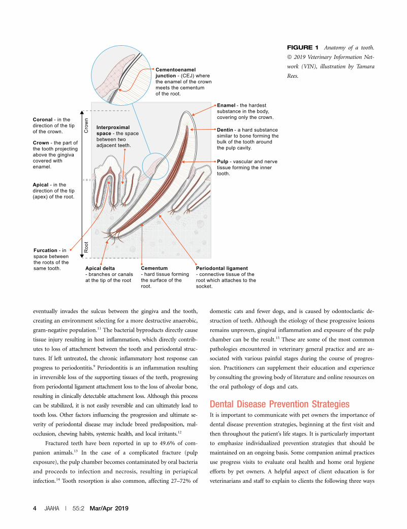
purpose, and function of the structures of the head and oral cavity
shown in Figure 1.3–5 Dogs and cats have two generations of teeth
(diphyodont), with the roots being longer than crowns. Most of the
permanent tooth is composed of dentin, with the central portion of
the tooth being the pulp chamber containing blood vessels, nerves,
lymphatics, connective tissue, and odontoblasts (Figure 1).6 The
tooth supporting structures, or “periodontium,” consist of the
gingiva, periodontal ligament, cementum, and alveolar bone. The
periodontal ligament attaches the tooth in the alveolus by being
affixed between the cementum and the alveolar bone (Figure 1).3,7
There are many pathologic processes that affect the oral cav-
ity of dogs and cats (congenital, infectious, traumatic, neoplastic,
autoimmune, and others). The most common and significant disease
is the inflammation of the tissues of the periodontium, or periodontal
disease. The clinical terms used to describe the active process of
periodontal disease include gingivitis and periodontitis. Gingivitis,
the earliest stage of periodontal disease, is described as inflammation
confined to the gingiva and commonly induced by bacterial plaque.
Gingivitis is reversible and preventable.8,9 Plaque-induced gingivitis
can be reversed by removal of the bacteria above as well as below the
gingival margin and prevented with consistent plaque-removing
home oral hygiene efforts.10 Calculus, or bacterial plaque that has
become calcified by salivary minerals, is mostly an irritant and is
relatively nonpathogenic.8,9
The bacterial population at the tooth surface is initially com-
posed of gram-positive, aerobic bacteria. The bacterial biofilm
TABLE 1
Definitions That Pertain to Dental Care Guidelines
Terminology Definition
Dental chart A written and graphical representation of the mouth, with adequate space to indicate pathology and procedures (see the “2013AAHA Dental Care Guidelines” for included items).
Dental prophylaxis A procedure performed on a healthy mouth that includes oral hygiene care, a complete oral examination, and techniques to preventdisease and to remove plaque and calculus above and beneath the gum line under anesthesia before periodontitis has developed.Note: The words “prophy,” “prophylaxis,” and “dental” are often misused in veterinary medicine. More descriptive terms to usefor the dental procedures that are commonly performed in companion animal dentistry to prevent periodontitis are COPAT,COHAT, and an oral ATP visit.
Dentistry The evaluation, diagnosis, prevention, and/or treatment of abnormalities in the oral cavity, maxillofacial area, and/or associatedstructures. Nonsurgical, surgical, or related procedures may be included.
Endodontics The treatment and therapy of conditions affecting the pulp.
Exodontia (extraction) A surgical procedure performed to remove a tooth.
Gingivitis Inflammation of the gingiva with or without loss of the supporting structure(s) shown with X-rays.
Home oral hygiene Measures taken by pet owners that are intended to control or prevent plaque and calculus accumulation.
Oral surgery The practical manipulation and incising of epithelium of hard and soft tissue for the purpose of improving or restoring oral health andcomfort.
Orthodontics The evaluation and treatment of malpositioned teeth for the purposes of improving occlusion and patient comfort and enhancing thequality of life.
Periodontal disease A disease process beginning with gingivitis and progressing to periodontitis when left untreated.
Periodontitis A destructive process involving the loss of supportive structures of the teeth, including the periodontium (i.e., gingiva, periodontalligament, cementum, and/or alveolar bone).
Periodontal surgery Invasive treatment necessary to re-establish or rehabilitate periodontal attachment structures. This is indicated for patients withpockets .5 mm, stage 2 and 3 furcation exposure, or inaccessible root structures.
Periodontal therapy Treatment of tooth-supporting structures in the presence of existing periodontal disease; includes dental cleaning as defined belowand one or more of the following procedures: gingival curettage for nonsurgical removal of plaque, calculus, and debris in gingivalpockets; root planing periodontal flaps; regenerative surgery; gingivectomy-gingivoplasty; and the local application ofantimicrobials.
Periodontium The supporting structures of teeth, including (1) periodontal ligament, (2) gingiva, (3) cementum, and (4) alveolar and supportingbone.
Pocket A pathologic space between supporting structures and the tooth, extending apically from the normal attachment location of thegingival epithelial attachment.
Professional dental cleaning Scaling (supragingival and subgingival plaque and calculus removal) of teeth with power or hand instrumentation, tooth polishing,and oral examination performed by a trained veterinary healthcare provider under general anesthesia.
Some definitions were derived from previously published descriptions2
COHAT, comprehensive oral health, assessment, and treatment; COPAT, comprehensive oral prevention, assessment, and treatment; oral ATP, oral assessment, treatment,and prevention.
Dental Guidelines
JAAHA.ORG 3
eventually invades the sulcus between the gingiva and the tooth,
creating an environment selecting for a more destructive anaerobic,
gram-negative population.11 The bacterial byproducts directly cause
tissue injury resulting in host inflammation, which directly contrib-
utes to loss of attachment between the tooth and periodontal struc-
tures. If left untreated, the chronic inflammatory host response can
progress to periodontitis.9 Periodontitis is an inflammation resulting
in irreversible loss of the supporting tissues of the teeth, progressing
from periodontal ligament attachment loss to the loss of alveolar bone,
resulting in clinically detectable attachment loss. Although this process
can be stabilized, it is not easily reversible and can ultimately lead to
tooth loss. Other factors influencing the progression and ultimate se-
verity of periodontal disease may include breed predisposition, mal-
occlusion, chewing habits, systemic health, and local irritants.12
Fractured teeth have been reported in up to 49.6% of com-
panion animals.13 In the case of a complicated fracture (pulp
exposure), the pulp chamber becomes contaminated by oral bacteria
and proceeds to infection and necrosis, resulting in periapical
infection.14 Tooth resorption is also common, affecting 27–72% of
domestic cats and fewer dogs, and is caused by odontoclastic de-
struction of teeth. Although the etiology of these progressive lesions
remains unproven, gingival inflammation and exposure of the pulp
chamber can be the result.15 These are some of the most common
pathologies encountered in veterinary general practice and are as-
sociated with various painful stages during the course of progres-
sion. Practitioners can supplement their education and experience
by consulting the growing body of literature and online resources on
the oral pathology of dogs and cats.
Dental Disease Prevention StrategiesIt is important to communicate with pet owners the importance of
dental disease prevention strategies, beginning at the first visit and
then throughout the patient’s life stages. It is particularly important
to emphasize individualized prevention strategies that should be
maintained on an ongoing basis. Some companion animal practices
use progress visits to evaluate oral health and home oral hygiene
efforts by pet owners. A helpful aspect of client education is for
veterinarians and staff to explain to clients the following three ways
FIGURE 1 Anatomy of a tooth.
ª 2019 Veterinary Information Net-
work (VIN), illustration by Tamara
Rees.
4 JAAHA | 55:2 Mar/Apr 2019
preventive oral health products work: (1) mechanical (abrasion), (2)
nonmechanical (chemical), and (3) a combination of mechanical
and chemical modes of action. Some experts prefer oral health
products that have dual action because all the teeth can benefit from
the combination of mechanistic activities.
In most patients, periodontal disease is a preventable condition.
Fractured teeth can often be prevented by appropriate selection of
dental chews and toys and behavior modification for separation
anxiety and cage-biting.
Preventing Periodontal DiseasePrevention of periodontal disease begins at the first visit, either for a
puppy or kitten, as well as for a new adult patient. Recommendations
for young patients include the following:
· A complete oral examination of the deciduous dentition will
assess any missing, unerupted, or slow-to-erupt teeth. The oc-
clusion should also be evaluated at this time, as well as deter-
mination of abnormal jaw length and teeth that are contacting
other teeth or soft tissue. In such cases, early extraction may be
needed.
· As permanent teeth start to erupt, it is critical to address any
retained or persistent deciduous teeth. Immediate extraction of
persistent deciduous teeth can help prevent displacement of the
erupting permanent teeth that can result in a malocclusion, or
that can exacerbate periodontal disease due to crowding.
Retained deciduous teeth without a replacement permanent
tooth can remain stable, although extraction may be necessary
in cases of unstable dentition. Young pets with missing perma-
nent teeth should have intraoral dental radiographs taken to
confirm that the teeth are truly not present, as unerupted teeth
can be problematic.
· Home oral hygiene training can be started for clients owning
pets having erupted, permanent dentition. Juvenile patients
actively exfoliating deciduous teeth may experience discomfort
associated with home dental care efforts, and negative experi-
ences should be avoided.
· The owner of any puppy or kitten who will be smaller than 20–
25 lbs at maturity should be informed that the level of dental
care and prevention for their pet is likely to be more involved
than that of a larger dog. Brachycephalic breeds also tend to have
more dental issues due to the rotation and crowding of teeth.
· A true dental prophylaxis (complete dental cleaning, polishing,
and intraoral dental radiographs in the absence of obvious
lesions) is recommended by 1 yr of age for cats and small- to
medium-breed dogs, and by 2 yr of age for larger-breed dogs.
During the procedure, any hidden conditions such as unerup-
ted or malformed (dysplastic) teeth can be identified and
treated. Ideally, periodontal therapy should then be provided
at an interval to optimally manage periodontal disease in this
preventable stage.
If periodontal disease with attachment loss is already present in
the patient, a complete dental assessment, intraoral radiographs,
cleaning, polishing, and any necessary treatment will help address any
current disease and optimally prevent further disease progression.
Appropriate and effective home oral hygiene (see the “Client
Communication and Education” section and resources at aaha.org/
dentistry) can help maintain oral health in between dental therapy
procedures. In most patients, effective periodontal prevention can
help keep the oral cavity in a relatively pain-free and healthy state,
favorably impacting the systemic health and welfare of the patient.
Clarification of the Impact of Periodontal Health onSystemic HealthThe long-held dogma that specific oral bacteria are directly re-
sponsible for infection in distant organs is oversimplified and difficult
to prove.16,17 There is an association shown between periodontal
disease and systemic health parameters, and in human medicine, the
presence of chronic inflammation associated with periodontitis has
been recognized to likely negatively impact overall systemic
health.18–25 The systemic spread of inflammatory mediators and
cytokines and bacterial endotoxins from periodontal pathogens can
impact the vascular system throughout the body and even cause
histological changes in distant organs.26–28 Management or resolu-
tion of the inflammation associated with periodontitis is likely to
have greater clinical impact that just considering antibacterial ef-
forts.25,29,30 Although evidence demonstrating the direct correlation
between systemic disease and oral and dental infections may be
difficult to prove, the positive impact on patient quality of life is
often clinically demonstrated and widely experienced.
Patient Assessment, Evaluation, andDocumentationHistory and Physical ExaminationA thorough history of patient health should always include an
evaluation and update on systemic maladies as well as an evaluation
and review of oral hygiene efforts performed by the pet owner.
Proactive management of oral health includes documenting any
efforts by the client to provide home dental care. These include tooth
brushing; type of diet fed; access to “chews,” treats, and toys; in-
formation on chewing habits; and updating any current or previous
professional or home dental care. A thorough physical examination
should be performed to evaluate all body systems regardless of
species, breed, age, health status, and temperament. Patients pre-
senting for complaints separate from the oral cavity should be
Dental Guidelines
JAAHA.ORG 5
evaluated for the primary complaint. Appropriate diagnostic tests
and treatments should then be recommended. Patients with un-
derlying health conditions should be appropriately assessed so that
general anesthesia associated with dental or other procedures can be
safely performed.
Conscious Oral EvaluationThe conscious oral evaluation is an important first step to antici-
pating procedural extent and preparing and educating clients re-
garding anticipated findings while under general anesthesia. In many
instances, the examiner will underestimate the presence of disease
during conscious evaluation, only to have the full extent of oral
pathology revealed by periodontal probing and intraoral radiography.
Examination of the conscious patient can be facilitated by use of
individualized pharmacologic and nonpharmacologic protocols
designed to reduce anxiety, stress, and pain. For anxious, conscious
patients, there should be no hesitation to recommend use of anxi-
olytics to facilitate an awake oral examination. For established pa-
tients, anxiety can be effectively relieved by administering trazodone
in dogs and gabapentin in cats, ideally the evening before and at least
2 hr before presentation if deemed safe and appropriate. For new
patients who are difficult to assess, rapid-acting sedatives or anxi-
olytics such as butorphanol, acepromazine, dexmedetomidine, or
alfaxalone are recommended. The use of anxiolytics and sedatives
should not replace the need for procedure-associated analgesic
strategies but will support the analgesic efficacy of analgesic medi-
cations. Additional, nonpharmacologic techniques of compassionate
restraint that can help facilitate conscious patient evaluation include
low-stress handling, use of pheromones, reduction of excess noise,
and the use of highly palatable treats as a distraction. These tech-
niques reduce conflict escalation and ensure the safety of the patient,
the client, and veterinary staff. Familiarization with techniques de-
scribed in the American Association of Feline Practitioners’ Feline-
Friendly Handling Guidelines is recommended.31
All physical exam findings should be recorded in the medical
record (Table 2). Aside from general physical exam findings, visual
attention should be paid to the head and oral cavity, and the visual
evaluation should be performed with appropriate palpation. Specific
signs associated with oral disease include pain on palpation; hali-
tosis; drooling; viscous or discolored saliva; dysphagia; asymmetric
calculus accumulation or gingivitis; resorbing teeth; discolored,
fractured, mobile, or missing teeth; extra teeth; gingival inflamma-
tion and bleeding; loss of gingiva and bone; and abnormal or painful
temporomandibular joint range of motion. Occlusion should be
evaluated to ensure the patient has a functional, comfortable bite.32
The head should be evaluated and palpated including inspec-
tion and retropulsion of the globes, lymph nodes, nose, lips, teeth,
mucous membranes, gingiva, vestibule, dorsal and ventral aspects of
the tongue, tonsils, salivary glands and ducts, and assessment of the
caudal oral cavity and gag reflex if it can be safely elicited. Any and
all abnormalities (including abnormal swellings or masses) should
be recorded in the medical record.
Careful attention to a conscious oral evaluation provides the
practitioner with an opportunity to demonstrate oral pathology and
educate the client about potential treatment options. Full appreci-
ation for the spectrum of treatment options will likely not be known
until additional information can be gathered from the radiographic
interpretation and additional anesthetized oral examination findings
such as pulp exposure, furcation exposure, tooth mobility, or
periodontal pocketing. Pre-emptive discussion of oral findings with
the client provides additional time for the client to consider what
treatment options may be offered once anesthetized oral exam
findings are collected. Periodontal probing for pockets or furcation
exposure or dental probing to evaluate for pulp exposure or tooth
resorption should never be performed on an awake patient. Inad-
vertent or deliberate contact with sensitive or painful areas such as the
exposed pulp risks hurting the pet and exposing the owner or staff to
being bit. Additionally, the pet may become averse to objects being
introduced into its mouth. This tends to undermine the patient’s trust
in human handlers and is counterproductive to coaching the client to
try various home oral hygiene tools or preventive care techniques.
Unconscious Oral EvaluationOnly after the patient has been anesthetized can a complete and
thorough oral evaluation be successfully performed.33 The
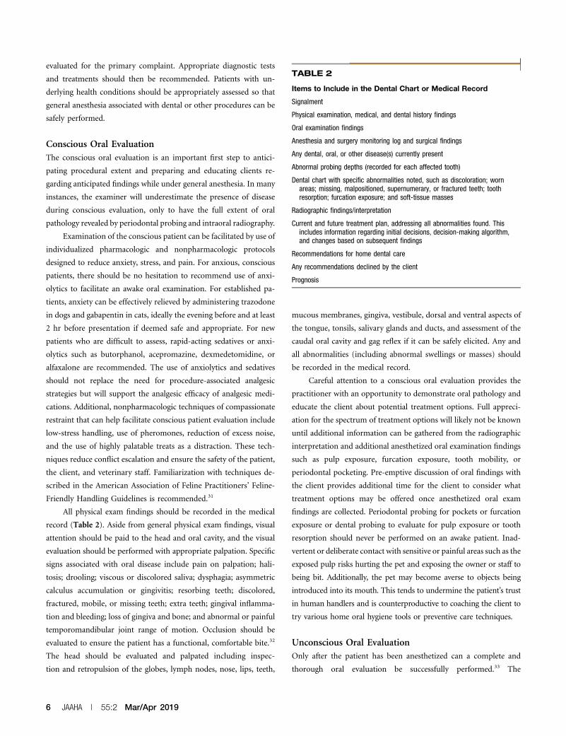
TABLE 2
Items to Include in the Dental Chart or Medical Record
Signalment
Physical examination, medical, and dental history findings
Oral examination findings
Anesthesia and surgery monitoring log and surgical findings
Any dental, oral, or other disease(s) currently present
Abnormal probing depths (recorded for each affected tooth)
Dental chart with specific abnormalities noted, such as discoloration; wornareas; missing, malpositioned, supernumerary, or fractured teeth; toothresorption; furcation exposure; and soft-tissue masses
Radiographic findings/interpretation
Current and future treatment plan, addressing all abnormalities found. Thisincludes information regarding initial decisions, decision-making algorithm,and changes based on subsequent findings
Recommendations for home dental care
Any recommendations declined by the client
Prognosis
6 JAAHA | 55:2 Mar/Apr 2019
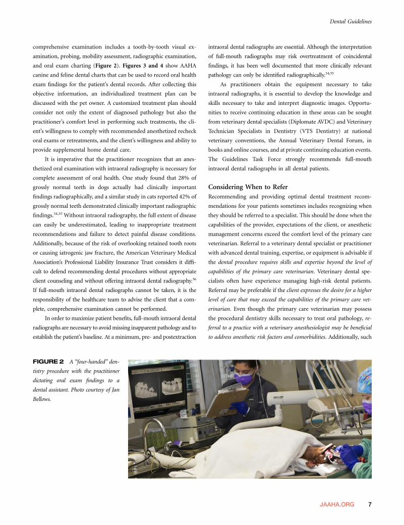
comprehensive examination includes a tooth-by-tooth visual ex-
amination, probing, mobility assessment, radiographic examination,
and oral exam charting (Figure 2). Figures 3 and 4 show AAHA
canine and feline dental charts that can be used to record oral health
exam findings for the patient’s dental records. After collecting this
objective information, an individualized treatment plan can be
discussed with the pet owner. A customized treatment plan should
consider not only the extent of diagnosed pathology but also the
practitioner’s comfort level in performing such treatments, the cli-
ent’s willingness to comply with recommended anesthetized recheck
oral exams or retreatments, and the client’s willingness and ability to
provide supplemental home dental care.
It is imperative that the practitioner recognizes that an anes-
thetized oral examination with intraoral radiography is necessary for
complete assessment of oral health. One study found that 28% of
grossly normal teeth in dogs actually had clinically important
findings radiographically, and a similar study in cats reported 42% of
grossly normal teeth demonstrated clinically important radiographic
findings.34,35 Without intraoral radiography, the full extent of disease
can easily be underestimated, leading to inappropriate treatment
recommendations and failure to detect painful disease conditions.
Additionally, because of the risk of overlooking retained tooth roots
or causing iatrogenic jaw fracture, the American Veterinary Medical
Association’s Professional Liability Insurance Trust considers it diffi-
cult to defend recommending dental procedures without appropriate
client counseling and without offering intraoral dental radiography.36
If full-mouth intraoral dental radiographs cannot be taken, it is the
responsibility of the healthcare team to advise the client that a com-
plete, comprehensive examination cannot be performed.
In order to maximize patient benefits, full-mouth intraoral dental
radiographs are necessary to avoidmissing inapparent pathology and to
establish the patient’s baseline. At a minimum, pre- and postextraction
intraoral dental radiographs are essential. Although the interpretation
of full-mouth radiographs may risk overtreatment of coincidental
findings, it has been well documented that more clinically relevant
pathology can only be identified radiographically.34,35
As practitioners obtain the equipment necessary to take
intraoral radiographs, it is essential to develop the knowledge and
skills necessary to take and interpret diagnostic images. Opportu-
nities to receive continuing education in these areas can be sought
from veterinary dental specialists (Diplomate AVDC) and Veterinary
Technician Specialists in Dentistry (VTS Dentistry) at national
veterinary conventions, the Annual Veterinary Dental Forum, in
books and online courses, and at private continuing education events.
The Guidelines Task Force strongly recommends full-mouth
intraoral dental radiographs in all dental patients.
Considering When to ReferRecommending and providing optimal dental treatment recom-
mendations for your patients sometimes includes recognizing when
they should be referred to a specialist. This should be done when the
capabilities of the provider, expectations of the client, or anesthetic
management concerns exceed the comfort level of the primary care
veterinarian. Referral to a veterinary dental specialist or practitioner
with advanced dental training, expertise, or equipment is advisable if
the dental procedure requires skills and expertise beyond the level of
capabilities of the primary care veterinarian. Veterinary dental spe-
cialists often have experience managing high-risk dental patients.
Referral may be preferable if the client expresses the desire for a higher
level of care that may exceed the capabilities of the primary care vet-
erinarian. Even though the primary care veterinarian may possess
the procedural dentistry skills necessary to treat oral pathology, re-
ferral to a practice with a veterinary anesthesiologist may be beneficial
to address anesthetic risk factors and comorbidities. Additionally, such
FIGURE2 A “four-handed” den-
tistry procedure with the practitioner
dictating oral exam findings to a
dental assistant. Photo courtesy of Jan
Bellows.
Dental Guidelines
JAAHA.ORG 7

referral practices may include access to other individuals with ex-
pertise in managing patients with underlying comorbidities that
jeopardize the safety of the anesthetic event, especially involving
patients with cardiac disease, chronic renal disease, diabetes, or
hyperadrenocorticism.
Dental ProceduresGeneral ConsiderationsNonsurgical dental proceduresmust be performed by a licensed veterinarian,
a credentialed technician, or a trained veterinary assistant under veterinarian
supervision in accordance with applicable state or provincial practice acts.
Oral surgery, including surgical extractions, must be performed only by
trained, licensed veterinarians. State-by-state regulations concerning
what licensed technicians can perform are summarized at avma.org/
Advocacy/StateAndLocal/Pages/sr-dental-procedures.aspx.
Anesthesia allows the practitioner and assistants to carry out
dental procedures in a safe and effective manner, minimizing the risk of
injury. Anesthesia recommendations and techniques are discussed in
the “Anesthesia, Sedation, and Analgesia Considerations” section.
All dental procedures need to use a consistent method to record
pathological findings, recommended treatments, treatment performed,
and treatment declined, as well as future planned treatment and pre-
vention recommendations in the medical record.
Practitioners should be aware that transient bacteremia from the
oral cavity is commonplace and increased during oral procedures,
and therefore, risk for seeding other remote surgical locations is
possible. Combining dental and other surgical procedures should be
performed with caution. The risk of multiple anesthetic events
should be weighed against the risk of complicated healing in the
presence of significant periodontal disease.37
Positioning and safety of the patient is important. The head and
neck should be stabilized when forces are being applied in the mouth.
The use of spring-loaded mouth gags must be avoided as it may
compromise blood flow, which may cause myalgia, neuralgia,
blindness, or trauma to the temporomandibular joint. If a mouth
prop is necessary, do not fully open the mouth or overextend the
temporomandibular joint.38
Whenever possible, practitioners and assistants should dem-
onstrate healthy ergonomic practices to avoid chronic injury. Ac-
tivities and procedures that cause excessive reaching, bending, and
twisting should be limited. For example, instruments and equipment
should be arranged where they can be easily grasped. Supplies should
FIGURE 3 AAHA canine dental record (aaha.org/dental_resources). FIGURE 4 AAHA feline dental record (aaha.org/dental_resources).
8 JAAHA | 55:2 Mar/Apr 2019

be placed as close as possible to the working area and at working
height to decrease stretching and bending. Sufficient space should
be allowed to enable turning the whole body, using a swivel stool.
Essential Steps Before, During, and After the DentalCleaning and Periodontal TherapyThe essential steps for a professional dental cleaning and periodontal
therapy are as follows:
1. Perform an oral evaluation on the conscious patient before
administering anesthesia. Avisual assessment can suggest whether
periodontal disease exists and its extent.
2. Radiograph the entire mouth of the anesthetized patient using
intraoral film or intraoral digital radiographic systems.
3. Scale the teeth supra- and subgingivally using a hand scaler
(supragingivally), curette (subgingivally), or an appropriately
powered ultrasonic scaler followed by a curette inserted subgingi-
vally to remove additional plaque and calculus (Figure 5). Do
not use a rotary scaler, which excessively roughens the tooth
enamel.39,40 Elimination of calculus is essential because it acts as
a retention matrix for plaque and toxins harmful to the tooth’s
support. Curettes are designed to assist in the removal of sub-
gingival plaque and calculus for root planing and curettage
(soft tissue removal in diseased periodontal pockets). Curettes
have a smooth, rounded heel and toe opposite the cutting
surface. The rounded back makes curettes less traumatic to soft
tissues compared with sickle scalers.
Every professional teeth cleaning should include hand scaling
of the accessible root surfaces (Figures 6, 7). Aggressive curet-
tage and scaling causing cementum removal is discouraged.
Cementum covering the roots contains cell-activating proteins
that encourage reattachment. Dentin does not contain these
proteins. Subgingival ultrasonic treatment causes cavitation
and disruption of the subgingival ecosystem and biofilm. The
design and safety of thin, long, ultrasonic periodontal tips de-
crease the need to aggressively root-plane teeth affected by
periodontal disease.
4. Crown polishing is recommended after cleaning and scaling, to
assist in the reduction of microabrasions on the enamel. Polish
the teeth using a low-speed hand piece prophy angle and
polishing cup running at no more than 3000 rpm. Polish
with prophy paste or fine-grit pumice because medium or
course grit can contribute to enamel loss, microabrasions,
and predisposition for plaque accumulation. The use of dis-
posable prophy angles and individually packaged prophy
paste is strongly recommended to avoid cross contamination.
5. Perform oral evaluation using a periodontal probe after dental
scaling and full-mouth radiographs have been obtained. Each
tooth should be probed in at least six places parallel to the
roots. The probing depth should not be greater than 2–3 mm
in a midsized dog and 1 mm in a midsized cat. Oral exam-
ination abnormal findings should be charted.41
6. Perform subgingival irrigation to remove debris and polishing
paste and to inspect the crown and subgingival areas. Use the
air or water syringe to inspect the visible subgingival areas for
remaining calculus requiring removal (Figure 8).
7. After sharing examination findings, a therapy plan, related fees,
and informed consent with the pet owner, perform indicated
periodontal therapy or extractions. Periodontal disease staging
and appropriate treatment are as follows:
· Stage 1 (periodontal disease staging [PD]1, gingivitis): Den-
tal scaling, polishing, irrigation, home dental care.
· Stage 2 (PD2, early periodontal disease with ,25% attach-
ment loss): PD1 care plus locally applied antimicrobials
and/or subgingival scaling if pocketing exists.
· Stage 3 (PD3, established periodontal disease with 25–50%
attachment loss): Periodontal treatment including periodontal
surgery will only be successful if the client is committed to
consistently administering home dental care. Extraction indi-
cated if client and patient will not commit to daily home oral
hygiene. Periodontal therapy: closed- or open-root planing 6
locally applied antimicrobials or advanced periodontal treatment
such as guided tissue regeneration.
· Stage 4 (PD4, advanced periodontal disease with .50% at-
tachment loss): Extraction or periodontal surgery including
osseous resective or additive procedures followed by con-
sistently performed home dental care; prognosis is consid-
ered guarded.
Extraction site packing, which includes bone autografts, allografts,
or synthetic products, may be appropriate in select extraction sites
where the remaining supporting bone is at risk for fracture during
the period of extraction site healing, for example, in a dog’s
mandibular first molar or canine. These products are used to
facilitate bone healing when concern over bone integrity or
strength exists. The use of extraction site packing is contra-
indicated in the presence of osteomyelitis or infection.42–44
Periodontal surgery is performed to remove deep debris,
eliminate pockets, and to extract teeth. When pocketing or
gingival recession exceeds 50% of the root support, extrac-
tion is indicated and should be performed by trained veter-
inarians or referred for treatment by a veterinary dental
specialist when the practitioner does not have the expertise,
equipment, or facilities to perform treatment. It is recom-
mended that extraction sites .1 mm should be sutured with
Dental Guidelines
JAAHA.ORG 9
absorbable suture (4-0 or smaller) to keep blood clots in and food
and debris out.
8. Administer either systemic or local perioperative antibiotics
where indicated. The use of antibiotics in veterinary den-
tistry must be assessed on a case-by-case basis. Therapeutic
antimicrobials should be used appropriately in the surgical
setting. Most dental procedures are considered to be clean-
contaminated procedures, meaning that after extractions,
systemic antibiotics are usually not indicated.45–47
Preoperative antibiotics given several days before surgery
may be administered in cases of PD4 for the purpose of
making tissues more amenable to surgical handling. Intra-
operative antibiotics may be indicated in patients with sys-
temic risk factors, such as subaortic stenosis, systemic
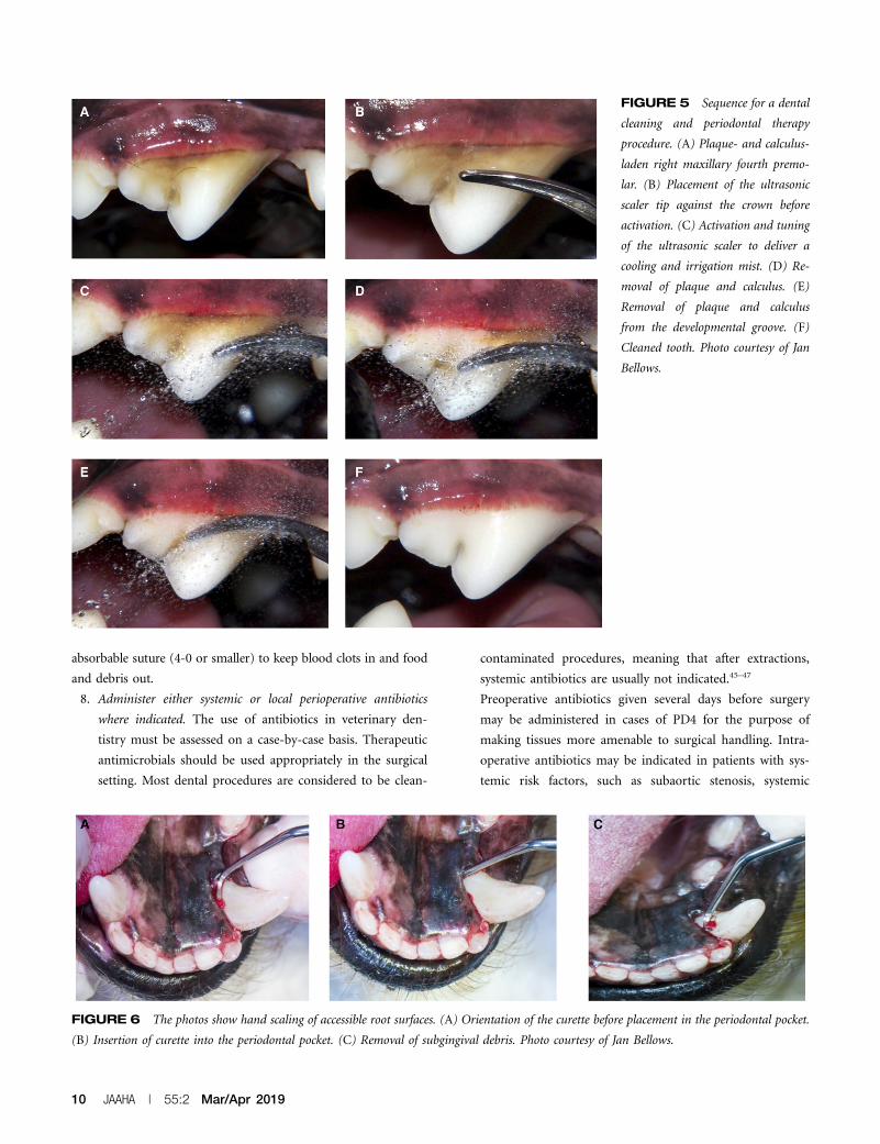
FIGURE 5 Sequence for a dental
cleaning and periodontal therapy
procedure. (A) Plaque- and calculus-
laden right maxillary fourth premo-
lar. (B) Placement of the ultrasonic
scaler tip against the crown before
activation. (C) Activation and tuning
of the ultrasonic scaler to deliver a
cooling and irrigation mist. (D) Re-
moval of plaque and calculus. (E)
Removal of plaque and calculus
from the developmental groove. (F)
Cleaned tooth. Photo courtesy of Jan
Bellows.
FIGURE 6 The photos show hand scaling of accessible root surfaces. (A) Orientation of the curette before placement in the periodontal pocket.
(B) Insertion of curette into the periodontal pocket. (C) Removal of subgingival debris. Photo courtesy of Jan Bellows.
10 JAAHA | 55:2 Mar/Apr 2019

immunosuppression, and orthopedic implants placed in the
last 12–18 mo. Appropriate clinical judgment for each indi-
vidualized patient is necessary. Postoperative antibiotics are
indicated when radiographic evidence of presumed osteomy-
elitis is present. Clindamycin (5.5 mg/kg per os q 12 hr) and
amoxicillin-clavulanic acid (13.75 mg/kg per os q 12 hr) are
both approved for use in cases of dental infections and
should be prescribed for a full 7–14 day course.
The use of locally applied antimicrobials (LAA), also called
perioceutics, may be indicated where a .5 mm cleaned
pocket exists in PD2 or PD3 cases (Figure 9). The purpose
of use is to improve periodontal health and encourage reat-
tachment to a normal level.48 PD4 cases require more inva-
sive periodontal debridement and management; however,
locally applied antimicrobials (LAA) may also be a compo-
nent.
9. Apply antiplaque substances such as barrier sealants. It is impor-
tant for practitioners to understand the appropriate indications
for the use of sealants. The term “sealant” in human dentistry is
a substance applied to teeth to prevent tooth decay. In veteri-
nary medicine, barrier sealants are applied to decrease the ac-
cumulation of plaque (Figure 10). Although the use of barrier
sealants has been shown to decrease accumulation of plaque
subgingivally, it does not totally prevent accumulation of sub-
gingival plaque, the occurrence of periodontal disease, the need
for home oral hygiene, or professional dental therapy.49–51
The use of resin-bonded sealants is designed to treat damaged
tooth structure (e.g., fractured or abraded teeth without pulp ex-
posure) by sealing exposed dentin tubules, thus decreasing sensi-
tivity and risk for bacterial migration leading to pulpitis. A
complete examination and intraoral radiographs are necessary be-
fore using any bonded sealant to identify nonvital teeth and other
pathology. Application of these products requires appropriate
training and radiographic follow-up in 6 mo to reconfirm tooth
vitality. Inappropriate use may result in increased dental pain, risk
FIGURE 8 Compressed air used to visualize the root surface and
subgingival calculus. Photo courtesy of Jan Bellows.
FIGURE 7 The subgingival curette blade is introduced atrau-
matically below the gumline with the face of the instrument nearly
parallel to the root surface. At the bottom of the sulcus, the handle is
adjusted, causing the down (cutting) edge of the instrument to contact
the root surface. Plaque, calculus, and debris is removed on the upward
pull stroke. ª 2019 Veterinary Information Network (VIN), illustra-
tion by Tamara Rees.
Dental Guidelines
JAAHA.ORG 11

for infection, and loss of tooth vitality. The use of resin-bonded
sealants in cases of tooth resorption is contraindicated.52,53
10. Biopsy all abnormal masses visualized grossly or radiograph-
ically and submit samples for histopathologic evaluation by a
pathologist qualified in oral tissues analysis.54
11. Maintain an open airway via intubation until the animal is
swallowing and is in sternal recumbency. Maintain body tem-
perature and continue intravenous fluid support as needed.
Continuously monitor and record vital signs until the pa-
tient is awake. Continue pain management while the pet is
in the hospital and upon discharge.55–57
12. Provide instruction on home oral hygiene. The Veterinary
Oral Health Council (VOHC) Accepted Products web page
(vohc.org/accepted_products.html) lists products that have
been scientifically proven to be effective in retarding accu-
mulation of dental plaque and/or calculus.58,59
Anesthesia, Sedation, and AnalgesiaConsiderationsFear of anesthesia is the most common cause of clients’ decisions to
forego dental procedures for their pets.60 Canine and feline patients in
need of medical or surgical procedures requiring anesthesia can be
managed to maintain a favorable balance between risk and derived
benefit. Medically important and indicated procedures should not be
absolutely discouraged based on chronologic age or most underlying
comorbidities. The most recent AAHA/AAFP Pain Management
Guidelines provide the entire veterinary care team an opportunity to
revisit the pathophysiology of pain and intervention strategies and
associated pharmacology/pharmacokinetics of treatment.
General anesthesia with endotracheal intubation, appropriate
monitoring, and physiologic support is necessary for dental proce-
dures, including dental cleaning and scaling as well as more advanced
dental care. Expert opinion and published data strongly support the
use of general anesthesia for dentistry. So-called “anesthesia-free”
dentistry has not been shown to be safer or comparable to the ca-
pacity to supra- and subgingivally clean teeth in an anesthetized
patient and is therefore unacceptable.2,61
Any dog or cat presenting for anesthesia should be considered on an
individual basis. Anesthesia for older dental patients and those with
comorbidities requires special attention. Each patient will have specific
physiologic alterations or diseases unique to that individual. Thus, the
anesthetic protocol needed for one patient typically will be quite different
from that needed for another. The use of local anesthetics as dental blocks
dramatically decreases the depth of general anesthesia needed, and thereby
helps support blood pressure, decreases ventilatory depression, provides
analgesia, and generally increases safety. Additionally, anxiolytic adminis-
tration prior to veterinary visits has become routine to decrease stress in
some patients. The synergistic effect between anxiolytics and other drugs
necessitates consideration for decreased amount of premedication, induction
agents, and maintenance anesthetics necessary to achieve the desired effect
and should be considered when formulating an anesthetic plan.
As with any patient, a thorough and complete history and
preanesthetic examination should be completed. Any previous anes-
thetic experience with the patient should be noted, and close attention
should be paid to any anesthetic complications or abnormal responses.
Aminimumdatabase including laboratory evaluation and imagingwill
be individually developed. Additional diagnostics will be indicated for
some dental patients based on clinical signs, practical availability, and
client consultation. Any abnormal preanesthetic findings should be
thoroughly evaluated and delaying the anesthesia and surgery should
be considered if necessary to address any potential problem areas
identified. Veterinarians must be in tune with their clients, their pa-
tient’s psychosocial issues, and the existing human–animal bond.
Often, stressed and compromised animals do not thrive at the vet-
erinary practice, away from their families and homes.
Considerations should be made to make the dental stay brief
and less stressful. Outpatient techniques with prompt return of the
patient to familiar settings and routines are highly desirable for all
FIGURE 9 Injection of perioceutic into a 5 mm cleaned, bleeding
periodontal pocket. Photo courtesy of Jan Bellows.
12 JAAHA | 55:2 Mar/Apr 2019

dental patients. A gentle approach, both in pharmacology and in the
application of clinical techniques, is especially important and will
benefit all patients. Support of the human–animal bond is an im-
portant goal, and dedicated emphasis on the reduction of fear, stress,
and pain is always warranted and primarily addressed through
management and behavioral modification. Anesthetic management
represents a powerful combination of additional modalities.
General AnesthesiaFor outpatient dental anesthesia, it is useful to select perioperative
medications that (1) typically provide for a rapid and complete
recovery (propofol or alfaxalone), (2) can be carefully reversed
(diazepam, midazolam, opioids, and dexmedetomidine), (3) can be
totally eliminated by supported ventilation (isoflurane, sevoflurane,
or desflurane), or (4) do not have substantial intrinsic toxicity or
significant adverse effects should drug effects persist (diazepam,
midazolam, or butorphanol). In situations in which delayed or in-
adequate recovery is recognized, physiologic support including judi-
cious fluid therapy, support of body temperature, ventilatory support,
and extended postanesthetic care should be provided. It is worth
noting that there is a strong consensus among veterinary anesthesi-
ologists to reverse dexmedetomidine only when medically necessary,
which allows the beneficial residual sedation to continue after the
completion of procedures in order to facilitate and ease recovery. If
necessary, consider using a low dose of atipamezole in cats.62
Adequate fluid replacement should be given to help prevent a
renal crisis and to help maintain a proper perioperative hemody-
namic state. The rate of IV fluid administration will depend on the
particular patient’s needs, but will generally be in the range of 3–
5 mL/kg/hr.63
Careful planning and additional attention to drug and dosage
selection is important to safely manage high-risk patients. Some
injectable general anesthetic agents need to be used with care in
higher-risk patients (including geriatric animals) because of the
typically altered hemodynamics, pharmacokinetics, and pharmaco-
dynamics; decreased plasma protein binding; and decreased ability
for hepatic metabolism and renal excretion in compromised animals.
Brachycephalic breeds and their associated airway conformations
warrant particularly close attention during the induction and re-
covery periods to avoid hypoxia and prevent dyspnea.
Inhalant general anesthetics are the anesthetics of choice in
many small animal patients, especially for procedures lasting longer
than 10–15 min. The inhalants isoflurane and sevoflurane offer the
advantage for outpatient anesthesia of rapid adjustment of inhaled
and alveolar anesthetic dose and effect. However, inhalational in-
duction of anesthesia (by either mask or chamber) is contra-
indicated in almost all clinical situations.64
Dose-dependent vasodilatation and hypotension preclude the use
of higher doses of inhalant anesthetics in many higher-risk patients.
Dose-sparing anesthesia achieved using lower doses of synergistically
acting injectable systemic agents (e.g., a fentanyl infusion) with local
anesthetic techniques allows for the maintenance of partial IV anes-
thesia (PIVA) with comparatively low doses of inhalants. In other
words, “less is more.” In more extreme cases, injectable agents (total
IV anesthesia [TIVA]) are best used in conjunction with intubation
and oxygen supplementation but without inhalant anesthesia. This
approach can often support markedly improved hemodynamics.
Patients should be preoxygenated for 2–5 min before anesthetic
induction to help prevent hypoxia from developing during induction.
Every anesthetized patient should be intubated to protect and
maintain a patent airway. The safety that often has been associated
with inhalants, as opposed to injectable anesthetics, is partly due to
the customary, if not obligatory, provision of supplemental oxygen as
the carrier gas for the volatile anesthetics. Endotracheal intubation
FIGURE 10 Application of antiplaque sealant. (A) Barrier sealant gel professionally applied to a cat’s gingival sulcus; home plaque prevention
gel is then reapplied weekly by the pet owner. (B) Application of hydrophilic gingival dental sealant professionally applied to a dog’s gingival sulcus;
reapplication is recommended every 6 mo. Photo courtesy of Jan Bellows.
Dental Guidelines
JAAHA.ORG 13

and administration of supplementary oxygen can easily be incorpo-
rated into injectable general anesthetic techniques and substantially
adds to patient safety. If anesthesia is deep enough to allow for
placement of an endotracheal tube, then the patient is no longer able
to protect its airway from either obstruction or aspiration of regur-
gitated or foreign material. Adherence to proper techniques protects
our personnel and practices from waste anesthetic gases.64
SedationIn select cases in which teeth cleaning, polishing, and extractions are
not anticipated, heavy sedation may be appropriate and sufficient to
collect limited baseline information. Examples include a targeted
intraoral radiograph recheck and a more involved preliminary ex-
amination of the oral cavity. When making the decision to use se-
dation versus general anesthesia, there are three considerations: (1)
protecting the patient, (2) protecting personnel, and (3) protecting
equipment. The loss of intrinsic airway protection requires us to place
an endotracheal tube and serves as an operational distinction between
sedation and anesthesia. The use of reversible agents, such as alpha-
agonists, or boluses of induction agents, such as propofol combined
with a quiet and dim environment and care to avoid stimulation, may
provide sufficient chemical restraint to meet these ends.
Sedation-only procedures generate limitations including risking
aspiration of fluids and aerosolized bacteria into the airways and
substandard ability to monitor ventilatory capacity without a proper
endotracheal tube in place. Because of the brief duration of action
and efforts to minimize depth of sedation, challenges arise sur-
rounding the ability to appropriately monitor patient hemodynamics
because time and patient handling (additional stimuli) are necessary
to properly affix monitoring equipment. This results in difficulties
monitoring the adequacy of sedation even with well-trained and
dedicated staff. Because of the absence of reaching a surgical plane of
anesthesia, sedation risks self-inflicted injury from the patient’s re-
flexes when attempting to probe subgingivally during an oral exam
and unnecessary risk for damage to equipment if bitten. Personnel
health must also be considered during sedated procedures because
an absence of a proper endotracheal tube while delivering inhalant
gas risks human exposure to waste gas, ultrasonic scaling with in-
appropriate irrigation results in increased bacterial aerosolization,
and abrupt patient response to stimuli risks bite injury.
Local AnalgesiaAnyone performing oral surgical or periodontal procedures should
be familiar with dental nerve block techniques, including a thorough
knowledge of oral anatomy and analgesic agents and their applica-
tion. Administration of local anesthetics will decrease the amount of
required inhalant anesthetic, will decrease the required amount of
other analgesics, and will ease the transition to administering
postoperative oral pain medications at home. Specific techniques for
local anesthetic dental nerve blocks (indications, doses, and specific
techniques) are described in detail by Niemiec et al., Beckman, and
Gracis, and others.61,65–68 Three approaches for the maxillary nerve
block are well described and offer choices based on anatomy and
personal preference.66 The maxillary tuberosity approach, using ei-
ther an intra- or extraoral (via the buccal pouch) access, allows for a
very short needle insertion just posterior to the caudal molar and
maxillary tuberosity. Both the subzygomatic approach and the
technique of advancing the needle through the infraorbital canal
provide access to the maxillary nerve as alternatives. Care is taken to
avoid damage to the maxillary or infraorbital neurovascular bundle
and inadvertent vascular or intraneural injection. Molars may not be
adequately blocked using the infraorbital nerve block technique alone,
but anesthesia should be reliable from the third or fourth premolar
and the more rostral structures including the canine teeth.67
The mandibular or inferior alveolar block can be performed at
the angle of the mandible. The more successful intraoral approach
technique is recommended.68 More rostral block at the mental fo-
ramen is less effective.60 Rarely, the lingual branch will be anesthetized
with a mandibular nerve block, and a very few patients may bite their
tongue during recovery. Recovery of the patient in sternal recum-
bency with the tongue between the jaws may decrease this risk.
Regardless of the local anesthetic technique or site, always as-
pirate to avoid intravascular injection of local anesthetic. Other uses
of local anesthetics may contribute to the basic nerve block tech-
niques and include “splash blocks,” infiltration anesthesia, intra-
osseus anesthesia, intraseptal injection, periodontal ligament or
intraligamentary injection, and intrapulpal injection.66
Nonanesthetic DentistryNonanesthetic dentistry (NAD), also referred to as anesthesia-
free dentistry, is a procedure in which the teeth are scaled and
polished without the benefit of general anesthesia. NAD is
considered not appropriate because of patient stress, injury, risk
of aspiration, and lack of diagnostic capabilities. Because this
procedure is intended to only clean the visible surface of the
teeth, it provides the pet owner with a false sense of benefit to
their pet’s oral health.69,70
Veterinary dentistry relies on detailed examination by a veter-
inarian with thorough knowledge of oral anatomy, physiology, and
pathology to make an accurate diagnosis. The examination includes
radiographs, requiring the animal to be motionless, as well as the use
of costly equipment in the oral cavity. Periodontal probing (noxious
stimulus) is also required to allow appropriate diagnosis and
treatment recommendations.
14 JAAHA | 55:2 Mar/Apr 2019
Removal of plaque and calculus is the most common treatment
recommended and performed for the treatment of periodontal
disease. It requires that subgingival surfaces be cleaned. This process
is uncomfortable, and at times painful, for the patient. Removal of
supragingival calculus alone is purely cosmetic and ineffective to treat
disease. The processes described above are not possible in a conscious
dog or cat. Without general anesthesia, an accurate diagnosis cannot
be made, patient pain cannot be addressed, the patient’s airway
cannot be protected from aspiration, and disease cannot be ap-
propriately treated.
When NAD is performed, the owner may be under the false
impression that the pet was not stressed by restraint, that pain was
managed, and that oral disease was accurately diagnosed and treated.
Patients who undergo NAD may go for long periods with untreated
disease, leading to more costs to health status (disease progression
and pain) and increased costs to the client. Peer-reviewed data
addressing the safety and efficacy of this controversial procedure are
very limited.71–73
The risks of anesthesia in healthy or minimally compromised
patients are very low when performed by appropriately trained in-
dividuals. A veterinarian concerned about the risk of anesthetizing a
patient may seek the assistance of a diplomate of the American
Veterinary Dental College or a diplomate of the American College of
Veterinary Anesthesia and Analgesia.74 See aaha.org/dentistry for
additional resources for discussing the risks of NAD.
Addressing PainFor both veterinary professionals and pet owners, the ability to
recognize dental pain is limited because dogs and cats often mask
overt signs of oral discomfort. Untreated dental pain may be indi-
rectly demonstrated by halitosis, teeth chattering, weight loss, change
in eating habits, lethargy, and change in behavior with reluctance to
engage in the human–animal bond. A short course of oral pain
medication may provide objective improvement to the patient’s
quality of life, thus bolstering support for the dental procedure.
It is imperative to recognize the importance of pre-emptive,
intraprocedural, and postprocedural dental pain management. The
use of pre-emptive multimodal analgesia with synergistic comple-
mentary classes of analgesics is obligatory and effective in managing
dental procedural pain.
Pre-emptive versus postprocedural nonsteroidal anti-
inflammatory agents may be most effective but would not be se-
lected for patients with hypovolemia, dehydration, chronic renal
disease, azotemia, and other risk factors.
Opioids are often used alone or in combination with tran-
quilizers in the dental patients as preanesthetic medications. Use of
anxiolytics and sedatives does not replace primary analgesics but will
support analgesic efficacy. Various opioid agonists, opioid agonist-
antagonists, and partial agonists have great value.
The Role of Technicians and AssistantsCredentialed veterinary technicians and veterinary assistants have a
prominent role in canine and feline dental care. Highly efficient
veterinary dental practices fully use and empower them in both the
exam room and the dental suite. The Guidelines Task Force strongly
encourages veterinary practices to support the training and education
of their veterinary technicians and assistants to assume a larger and
appropriate role in dental practice. In the exam room, they should
obtain a patient medical and dental history. They should be able to
explain to the client the dental procedures indicated, answer ques-
tions, translate veterinary diagnoses into lay terms, and reassure the
client by demonstrating expertise in dentistry.
In the dental suite, a credentialed veterinary technician should
perform both a conscious and anesthetized initial oral exam and dictate
charting to a veterinary assistant, take diagnostic radiographs, perform
cleaning procedures, and place regional blocks if indicated. Because
extractions are considered oral surgery, they should not be performed by
veterinary technicians. Veterinarians need to provide the appropriate level
of oversight and supervision as required by their state practice acts (www.
avma.org/Advocacy/StateAndLocal/Pages/sr-dental-procedures.aspx).
Veterinary technicians and assistants are the veterinary team’s pa-
tient advocates and client educators. They should spend time with the
pet owner before and at the time of discharge, explaining the procedures
and treatments performed, home oral hygiene, and medications. In
addition, they should interview the client to determine the best home
dental care options for the pet and advise, demonstrate, and instruct the
owners on how to provide quality home oral hygiene for their pet.
Practices should encourage continuing education and training
of veterinary team members. Enabling team members to increase the
level of their training and education brings satisfaction and con-
tributes to the retention of skilled personnel. Delineation of duties
based upon the training and education of the staff also benefits the
practice by fully using the team and ensuring patient safety. Many
skills in dentistry should be only performed by credentialed veter-
inary technicians with the knowledge base to understand how to
perform a skill and understand why a procedure is performed and the
risks associated with each task.
The highest level of training and certification is the Veterinary
Technician Specialist in Dentistry, designated as VTS (Dentistry).
This certification is issued by the Academy of Veterinary Dental
Technicians and awarded to credentialed veterinary technicians who
complete a rigorous 2 yr process of education. VTS (Dentistry)
training includes both didactic and experiential learning culminating
in a credential examination. Although most credentialed veterinary
Dental Guidelines
JAAHA.ORG 15

technicians may not have the interest to pursue VTS (Dentistry)
training, companion animal practices should support and encourage
basic and advanced continuing education in dentistry for all team
members. Trained veterinary assistants are valued members of the
practice team and should act as assistants to the credentialed vet-
erinary technician. Care should be given to assure that veterinary
assistants are only performing tasks appropriate to their skill level and
their state’s practice act.
Facility, Equipment, and Operator SafetyRequirementsFacility RequirementsExcellent dental care for canine and feline patients requires an
efficient, organized, and safe work environment. As a result of
environmental contamination that occurs during many dental
procedures, a dedicated space in a low-traffic area separate from
the sterile surgical suite is necessary. Other requirements include
appropriate ventilation, an anesthetic scavenging system, and
adequate surgical lighting and magnification. This allows ade-
quate visualization for oral treatments and surgery. The proce-
dure table must be impervious and sanitizable and allow for
drainage because dental procedures typically produce a large
amount of water.
Materials, Instruments, and EquipmentAn assortment of correct dental surgical instruments is essential for
adequate dental care. A “one size fits all” approach to dental surgical
equipment is inadequate. Several different sizes of dental luxators,
elevators, periosteal elevators, scalers, curettes, and mechanical scaler
inserts make for a more comprehensive oral surgery suite. Dental
instruments must be in proper working order and properly stored,
with defective instruments repaired or discarded and replaced. Other
dental materials, consumable dental equipment, and products must
not be expired. As with any surgical instruments, all dental instru-
ments must be cleaned and autoclaved between each use and stored
in a sterile manner until the next use. Instruments may be autoclaved
according to procedure, such as examination materials, suture packs,
oral surgery instruments, exodontia instruments, periodontal surgery
instruments, and materials. Additionally, materials used for guided
tissue regeneration must be sterile and perioceutics used according to
manufacturer recommendations. It goes without saying that proper
knowledge of instrument use and storage is essential. Single-use items
must be discarded after each patient use. If barrier sealants and dentin
sealants are used, each must be selected and applied appropriately.
References from the 2013 AAHA Dental Care Guidelines provide
recommendations and information on ordering equipment.8,75–78 A
basic assortment of recommended materials and instruments for
veterinary dentistry is listed in Table 3. See aaha.org/dentistry for
additional resources on instruments.
Operator ProtectionPathogens and debris such as calculus, tooth fragments, plaque, water
spray, and prophy paste are aerosolized during dental procedures. It is
prudent to irrigate the oral cavity with a 0.12% chlorhexidine so-
lution before dental scaling, tooth sectioning, and drilling to decrease
bacterial aerosolization.79 The safety of the operator is ensured
during dental procedures by using radiographic, oral, respiratory,
skin, eye, and ear protective devices as noted in the 2013 AAHA
Dental Care Guidelines.2 Ergonomic considerations for personnel
performing dental procedures include proper seating, fatigue
mats for standing, and proper positioning of both the patient and
materials to minimize immediate and chronic operator injuries.
Instruction on proper instrument handling techniques should be
provided.2 Radiographic safety precautions should be adhered to at all
times while radiographs are obtained. Radiation protection should
include the use of a lead shield or apron, thyroid shield, and a ra-
diation dosimeter. Sources of radiation in the dental suite include
direct exposure to the primary beam, scatter radiation, and leakage
from the tube head. The operator and other staff should not be in the
path of the primary beam. Scatter radiation can be minimized by
standing at least 6 ft from the source, maintaining a 90–1358 angle
from the path of the primary beam, and avoiding touching the tube
head or housing during exposures. Tests for leakage radiation should
be conducted on a regularly scheduled basis by a licensed profes-
sional.8 See aaha.org/dentistry for more resources on radiation safety.
Client Communication and EducationTerminology and MessagingThe most important step in achieving compliance with oral health
recommendations is getting the client to understand the value and
believe in the importance of regular dental care. This awareness
generally results when client realizes that oral pathology is a source of
chronic pain, infection, and poor quality of life for the pet.20,80 The
majority of dental care takes place in the primary care setting. A
fundamental aspect of delivering high-quality veterinary dental care
is for the practice team to use consistent dental care terminology
and messaging with the client. When this is consistently done using
tools such as a written treatment plan, client compliance with your
oral health recommendations will generally follow.
Dental terminology should reflect the importance and breadth of
the dental and oral disease prevention, diagnostics, and therapies. For
example, the consensus viewpoint of the Guidelines Task Force is that
using the simplified term “prophy” is incorrect and misleading because
our dental patients often have calculus and gingivitis before
16 JAAHA | 55:2 Mar/Apr 2019

prophylactic therapy is recommended.81 Neither prophy nor the term
“dental” adequately convey the breadth or complexity of oral health
services offered in primary care or referral practices. The broader ter-
minology “oral health” better conveys the full scope of this aspect of pet
healthcare. Even the correct use of medical terminology without
properly educating the client as to its meaning is insufficient. The
practice team should avoid acronyms, overly simplified terminology,
and medical jargon when discussing oral healthcare with clients. Per-
haps more than most other aspects of veterinary care, proper use of
oral health terminology is directly linked to client understanding, ac-
ceptance, and compliance with your recommendations.
Explaining the Role of AnesthesiaThe client should be told that their pet needs a comprehensive,
anesthetized oral exam and dental radiographs in order to perform a
preventive cleaning or dental-periodontal therapy. It is also vital that
the client understands the distinction between awake and anes-
thetized dental procedures. When the veterinarian explains that an
awake patient must be anesthetized for a proper exam and therapy,
clients understand the central role of anesthesia in oral healthcare.
This often leads to a discussion of the client’s concerns about an-
esthesia, a common reason given by pet owners who decline oral
care. Clients and, sadly, practitioners are often susceptible to un-
supported myths about unreasonable risks of general anesthesia.
It is helpful to manage client expectations on the need for
general anesthesia early in a patient’s life or at the outset of an oral
health visit. Sample dialog might consist of “Your pet will need an
anesthetized oral exam, dental radiographs, and cleaning between 1
and 2 yr of age, even if no abnormalities are seen on the awake
exam.34,35 After that, a periodic anesthetized oral exam, treatment,
cleaning, and prevention should be given throughout her lifetime. We
will develop a prevention plan that will work for you and your pet.”
Discussing Regular Oral HealthcareOral health should be discussed at the first appointment and every
visit thereafter. A 6 mo awake oral health exam is appropriate. The
patient should be evaluated for permanent dentition, retained or
persistent deciduous teeth, unerupted teeth, and crowding. A regular
awake exam can identify oral health problems that can be effectively
treated at an early stage with minimal discomfort to the pet. For
example, extraction of persistent deciduous teeth when the per-
manent tooth begins to erupt can avoid more extensive intervention
later on.82
Although most clients will not have experience with diabetes,
heart disease, or a ruptured cruciate ligament, they are all familiar
with their personal oral health. It is helpful to use the client’s existing
personal hygiene habits for themselves and their children as a bridge
to dental care recommendations for a pet. Client education topics
and recommendations will differ based on the pet’s age and breed
and the client’s prior misconceptions about pet oral healthcare.83 By
discussing dental care at each appointment, the client becomes fa-
miliar with the concept of oral health. Using “road-mapping”
techniques and providing the client with written instructions and
recommendations will facilitate in-depth pet oral health discussions
at later healthcare visits. See aaha.org/dentistry for additional client
education resources.
Nonanesthetic Scaling of TeethClients should be informed that groomers and others should never be
allowed to scale a pet’s teeth. Scaling of teeth must always be accom-
panied by polishing and must only be done by trained veterinary per-
sonnel operating in a clinical setting with an anesthetized animal.
Explain that NAD scaling is a cosmetic procedure, does not improve
oral or systemic health, and can cause pain, fear, bleeding, and infection.
Recommending Home Oral Hygiene and ProductsAdvise clients regularly of the potential damage done by inap-
propriate chew toys and the benefits of regular home dental care. In
offering this guidance, specific recommendations are more likely to
be followed. For example, explain that “Your dog will benefit from
feeding this food and using this toothpaste and toothbrush at least
once a day,” or “The prescription diet is working well to control
tartar (calculus) and plaque on the back teeth, but I would like you
to add these dental wipes for the incisor and canine teeth.” Clients
must be told specifically that brushing only removes plaque, not
calculus. Brushing needs to be done daily to be of benefit. Brushing
teeth with already inflamed gingiva will cause pain and animal
aversion. Even daily brushing does not preclude the need for
anesthetized exams, radiographs, and therapy, the same as in hu-
man dental care.
Discussing the Anesthetized Oral ExamIt is critical to have a written protocol to avoid misunderstanding
regarding an anesthetized oral exam. It is not until the mouth and
oral radiographs have been evaluated under anesthesia that a full
treatment plan and the costs of a dental procedure can be accurately
determined. As soon as the anesthetized oral exam and full-mouth
intraoral radiographs have been reviewed, the findings and treatment
plan can be discussed with the client. The consent form must clearly
state that if the owner cannot be contacted, any findings requiring
additional treatment that are not already specifically on the consent
will not be performed, and a separate appointment and procedure
will be scheduled. Advise the owner beforehand that if more extensive
disease is found, staging procedures may be recommended. Clients
Dental Guidelines
JAAHA.ORG 17
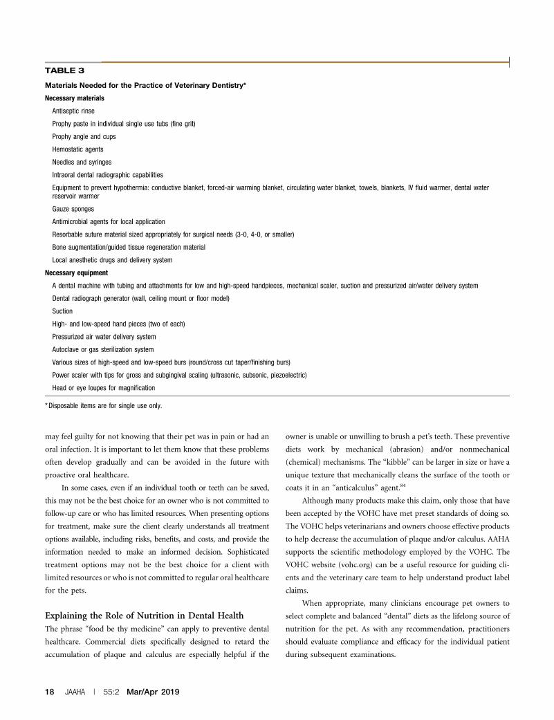
may feel guilty for not knowing that their pet was in pain or had an
oral infection. It is important to let them know that these problems
often develop gradually and can be avoided in the future with
proactive oral healthcare.
In some cases, even if an individual tooth or teeth can be saved,
this may not be the best choice for an owner who is not committed to
follow-up care or who has limited resources. When presenting options
for treatment, make sure the client clearly understands all treatment
options available, including risks, benefits, and costs, and provide the
information needed to make an informed decision. Sophisticated
treatment options may not be the best choice for a client with
limited resources or who is not committed to regular oral healthcare
for the pets.
Explaining the Role of Nutrition in Dental HealthThe phrase “food be thy medicine” can apply to preventive dental
healthcare. Commercial diets specifically designed to retard the
accumulation of plaque and calculus are especially helpful if the
owner is unable or unwilling to brush a pet’s teeth. These preventive
diets work by mechanical (abrasion) and/or nonmechanical
(chemical) mechanisms. The “kibble” can be larger in size or have a
unique texture that mechanically cleans the surface of the tooth or
coats it in an “anticalculus” agent.84
Although many products make this claim, only those that have
been accepted by the VOHC have met preset standards of doing so.
The VOHC helps veterinarians and owners choose effective products
to help decrease the accumulation of plaque and/or calculus. AAHA
supports the scientific methodology employed by the VOHC. The
VOHC website (vohc.org) can be a useful resource for guiding cli-
ents and the veterinary care team to help understand product label
claims.
When appropriate, many clinicians encourage pet owners to
select complete and balanced “dental” diets as the lifelong source of
nutrition for the pet. As with any recommendation, practitioners
should evaluate compliance and efficacy for the individual patient
during subsequent examinations.
TABLE 3
Materials Needed for the Practice of Veterinary Dentistry*
Necessary materials
Antiseptic rinse
Prophy paste in individual single use tubs (fine grit)
Prophy angle and cups
Hemostatic agents
Needles and syringes
Intraoral dental radiographic capabilities
Equipment to prevent hypothermia: conductive blanket, forced-air warming blanket, circulating water blanket, towels, blankets, IV fluid warmer, dental waterreservoir warmer
Gauze sponges
Antimicrobial agents for local application
Resorbable suture material sized appropriately for surgical needs (3-0, 4-0, or smaller)
Bone augmentation/guided tissue regeneration material
Local anesthetic drugs and delivery system
Necessary equipment
A dental machine with tubing and attachments for low and high-speed handpieces, mechanical scaler, suction and pressurized air/water delivery system
Dental radiograph generator (wall, ceiling mount or floor model)
Suction
High- and low-speed hand pieces (two of each)
Pressurized air water delivery system
Autoclave or gas sterilization system
Various sizes of high-speed and low-speed burs (round/cross cut taper/finishing burs)
Power scaler with tips for gross and subgingival scaling (ultrasonic, subsonic, piezoelectric)
Head or eye loupes for magnification
*Disposable items are for single use only.
18 JAAHA | 55:2 Mar/Apr 2019

Offering supplemental treats to pets can be an important part of
the human–animal bond. However, choosing treats that support oral
health is also helpful. There is a broad array of treats that have been
accepted by the VOHC to decrease plaque and calculus accumulation.
The Guidelines Task Force believes that there is not a strong rationale
for offering hard treats (antlers, synthetic, or natural bones) that
could damage the structural integrity of the tooth, ultimately leading
to unnecessary pain and infection for the pet. See aaha.org/dentistry
for additional resources on appropriate treat selection.
Discussion at the Discharge AppointmentWritten and verbal client communication is fundamental to the
maintenance of pet oral health. This dialog should address all
procedures and potential complications, immediate postoperative
home oral care (e.g., no brushing until surgically manipulated gingiva
has healed), medications and their side effects, and any diet changes
that might be necessary (e.g., soft or premoistened food in the
immediate postoperative period). If clients are not properly advised
of normal postanesthesia behavior and postoperative side effects
affecting a pet, they may be reluctant to allow another procedure.
Sharing the patient’s dental chart, photographs, and radiographs
with the client at the discharge appointment is helpful in illustrating the
extent of the pre-existing pathology and effects of treatment. Clients
better appreciate inapparent oral pathology once they see the visual
evidence of its effects and the benefits or therapeutic intervention. See
aaha.org/dentistry for additional client education resources.
Establish an appointment for a follow-up or recheck exami-
nation in 10–14 days, even if the procedure performed is limited to
a prophylactic cleaning. Rechecks will help determine owner
compliance and the need for continued care. At the discharge
appointment, clients are generally more focused on postanesthesia
care than on continued oral hygiene. The recheck is an opportu-
nity to affirm the positive steps that the owner has already taken in
caring for the pet’s oral health and to establish an ongoing treat-
ment plan.
SummaryNot only is oral health a fundamental aspect of overall pet health and
wellbeing, but veterinary dentistry is now considered a standard
component of companion animal medicine. Any full-service, primary
care companion animal practice should have the capability to perform
basic canine and feline dental examinations and procedures, including
those performed under general anesthesia. This capability assumes that
the veterinarian and other clinical staff have the expertise and essential
resources necessary to perform veterinary dentistry. These include
facilities, materials, and equipment, including barrier protection,
specific for veterinary dentistry. It is important that the practice team
routinely apply the nomenclature and terminology specifically asso-
ciated with veterinary dentistry. This is done not only to ensure
precision in performing dental procedures but also to educate clients
about the unique aspects of oral healthcare.
Lifetime oral health assumes that individualized periodontal
disease prevention and treatment plans will be implemented.
Avoiding and managing the inflammation, pain, and potential for
systemic infection associated with periodontal disease are strong
contributors to the pet’s quality of life and longevity.
Evaluation and documentation of dentition and oral pathology
involves oral evaluation of both the conscious and anesthetized
patient. Initial evaluation of the conscious patient can be facilitated
by pharmacologic protocols to reduce the patient’s stress and anxiety.
A comprehensive oral health assessment involving radiography requires
general anesthesia. It is important to recognize that many grossly
normal teeth in dogs and cats have clinically important pathology or
abnormalities that can be detected only by intraoral radiography per-
formed under general anesthesia. Because dental procedures can be
painful, intra- and postoperative pain management, often using mul-
timodal protocols, is an essential component of veterinary dentistry.
The guidelines describe a step-by-step process for the procedures
that are typically performed for canine and feline dental patients. These
include oral examination, radiography, tooth scaling, periodontal disease
staging, plaque and calculus removal and mitigation, general anesthesia,
and instructing pet owners on home oral hygiene. Although some of
these procedures are often performed in a referral setting, they are all
within the capabilities of properly trained and equipped primary care
practices. Additional resources can be found at aaha.org/dentistry.
Oral healthcare is a necessary aspect of lifetime pet wellness.However,
because veterinary dentistry involves general anesthesia, many clients are
hesitant to consider dental procedures for their pet. For this reason, client
education plays a pivotal role in successfully integrating veterinary dentistry
into your practice’s service offerings and incorporating dental care into a
lifetime healthcare plan for canine and feline patients.
The authors gratefully acknowledge the contributions of Mark
Dana of the Kanara Consulting Group, LLC in the preparation
of the guidelines manuscript, Jan Bellows for the photographic
images, and Tamara Rees of VIN for the illustrations.
REFERENCES
1. American Animal Hospital Association-American Veterinary Medical
Association Preventive Healthcare Guidelines Task Force. Development
of new canine and feline preventive healthcare guidelines designed to
improve pet health. J Am Anim Hosp Assoc 2011;47(5):306–11.
2. Holmstrom SE, Bellows J, Juriga S, et al; American Veterinary Dental
College. 2013 AAHA dental care guidelines for dogs and cats. J Am Anim
Hosp Assoc 2013;49(2):75–82.
Dental Guidelines
JAAHA.ORG 19

3. Evans HE, de Lahunta A. Miller’s Anatomy of the Dog. 4th ed. Elsevier
Saunders, St. Louis; 2013:80–113, 197–200, 428–456, 541–545,
708–730.
4. Lewis JR, Reiter AM. Anatomy and physiology. In: Niemiec BA, ed. Small
Animal Dental, Oral and Maxillofacial Disease. Boca Raton (FL): Mason
Publishing; 2010:9–38.
5. American Veterinary Dental College. AVDC nomenclature. Available at:
https://www.avdc.org/Nomenclature/Nomen-Dental_Anatomy.html.
Accessed April 6, 2019.
6. Pashley DH, Liewehr FR. Structure and functions of the dentin-pulp
complex. In: Cohen S, Hargreaves KM, eds. Pathways of the Pulp. 9th
ed. St. Louis: Mosby Elsevier; 2006:460–513.
7. Wolf HF, Rateitschak EM, Rateitschak KH, et al. Structural biology. In:
Rateitshak KH, Wolf HF, eds. Color Atlas of Dental Medicine: Periodon-
tology. 3rd ed. Stuttgart (Germany): Thieme; 2005:7–20.
8. Wiggs RB, Lobprise HB. Periodontology. In: Veterinary Dentistry: Prin-
ciples and Practice. Philadelphia: Lippencott-Raven; 1997:186–231.
9. Carranza FA, Takei HH. Rationale for periodontal treatment. In: New-
man MG, Takei HH, Klokkevold PR, eds. Carranza’s Clinical Periodon-
tology. 10th ed. St. Louis: Saunders; 2006:630–5.
10. Merin RL. Results of periodontal treatment. In: Newman MG, Takei HH,
Klokkevold PR, eds. Carranza’s Clinical Periodontology. 10th ed. St. Louis:
Saunders; 2006:1206–14.
11. Quirynen M, Teughels W, Kinder Haake S, et al. Microbiology of peri-
odontal diseases. In: Newman MG, Takei HH, Klokkevold PR, eds.
Carranza’s Clinical Periodontology.10th ed. St. Louis: Saunders; 2006:134–
69.
12. Novak, MJ. Classification of diseases and conditions affecting the
periodontium. In: Newman MG, Takei HH, Klokkevold PR, eds. Car-
ranza’s Clinical Periodontology. 10th ed. St. Louis: Saunders; 2006:100–9.
13. Soukup JW, Hetzel S, Paul A. Classification and epidemiology of trau-
matic dentoalveolar injuries in dogs and cats: 959 injuries in 660 patient
visits (2004-2012). J Vet Dent 2015:32(1):6–14.
14. Niemiec BA. Fundamentals of endodontics. Vet Clin North Am Small
Anim Pract 2005;35(4):837–68.
15. Reiter AM, Lewis JR, Okuda A. Update on the etiology of tooth re-
sorption in domestic cats. Vet Clin North Am Small Anim Pract 2005;
35(4):913–42.
16. Tou SP, Adin DB, Castleman WL. Mitral valve endocarditis after dental
prophylaxis in a dog. J Vet Intern Med 2005;19(2):268–70.
17. Peddle GD, Drobatz KJ, Harvey CE, et al. Association of periodontal
disease, oral procedures, and other clinical findings with bacterial
endocarditis in dogs. J Am Vet Med Assoc 2009;234(1):100–7.
18. DeBowes, LJ, Mosier D, Logan E, et al. Association of periodontal disease
and histological lesions in multiple organs from 45 dogs. J Vet Dent 1996;
13(2):57–60.
19. Pavlica Z, Petelin M, Juntes P, et al. Periodontal disease burden and
pathological changes in organs of dogs. J Vet Dent 2008;25(2):97–105.
20. Glickman LT, Glickman NW, Moore GE, et al. Evaluation of the risk of
endocarditis and other cardiovascular events on the basis of the severity
of periodontal disease in dogs. J Am Vet Med Assoc 2009;234(4):486–94.
21. Glickman LT, Glickman NW, Moore GE, et al. Association between
chronic azotemic kidney disease and the severity of periodontal disease
in dogs. Prev Vet Med 2011;99(2–4):103–200.
22. Cave NJ, Bridges JP, Thomas DG. Systemic effects of periodontal disease
in cats. Vet Q 2012;32(3–4):131–44.
23. Kouki MI, Papadimitriou SA, Kazakos GM, et al. Periodontal disease as a
potential factor for systemic inflammatory response in the dog. J Vet
Dent 2013;30(1):26–9.
24. Pizzo G, Guiglia R, Lo Russo L, et al. Dentistry and internal medicine:
from the focal infection theory to the periodontal medicine concept. Eur
J Intern Med 2010;21(6):496–502.
25. Nibali L, Tatarakis N, Needleman I, et al. Clinical review: Association
between metabolic syndrome and periodontitis: a systematic review and
meta-analysis. J Clin Endocrinol Metab 2013;98(3):913–20.
26. DeBowes LJ. The effects of dental disease on systemic disease. Vet Clin
North Am Small Anim Pract 1998;28(5):1057–62.
27. Rawlinson JE, Goldstein RE, Reiter AM, et al. Association of periodontal
disease with systemic health indices in dogs and the systemic response to
treatment of periodontal disease. J Am Vet Med Assoc 2011;238(5):601–9.
28. Stepaniuk K. Periodontology. In: Lobprise HB, Dodd JB, eds. Wigg’s
Veterinary Dentistry: Principles and Practice. Hoboken (NJ): Wiley; 2019.
[in production].
29. Chapple IL, Genco R; working group 2 of the joint EFP/AAP workshop.
Diabetes and periodontal diseases: consensus report of the Joint EFP/
AAP Workshop on Periodontitis and Systemic Diseases. J Periodontol
2013; 84(4 Suppl):S106–12
30. Lamster IB, Pagan M. Periodontal disease and the metabolic syndrome.
Int Dent J 2017;67(2):67–77.
31. Rodan I, Sundahl E, Carney H, et al; American Animal Hospital Asso-
ciation. AAFP and ISFM feline-friendly handling guidelines. J Feline Med
Surg 2011;13(5):364–75.
32. American Veterinary Dental College. American veterinary dental college
position statement: orthodontic procedures. Available at: http://avdc.org/
Orthodontic_Procedures.pdf. Accessed April 8, 2018.
33. American Veterinary Medical Association. Veterinary dentistry policy.
Available at: https://www.avma.org/KB/Policies/Pages/AVMA-Position-
on-Veterinary-Dentistry.aspx. Accessed April 8, 2018.
34. Verstraete FJ, Kass PH, Terpak CH. Diagnostic value of full-mouth ra-
diography in dogs. Am J Vet Res 1998;59(6):686–91.
35. Verstraete FJ, Kass PH, Terpak CH. Diagnostic value of full-mouth ra-
diography in cats. Am J Vet Res 1998;59(6):692–5.
36. AVMA PLIT. Professional Liability Newsletter. 2015;34(3). Available at:
https://www.im3vet.com/iM3US/media/Documents/pro-
fessional_liability_Dental_X-ray.pdf.
37. González Navarro B, Jané Salas E, Estrugo Devesa A, et al. Bacteremia
associated with oral surgery: A review. J Evid Based Dent Pract 2017;
17(3):190–204.
38. Stiles J, Weil AB, Packer RA, et al. Post-anesthetic cortical blindness in
cats: twenty cases. Vet J 2012;193(2):367–73.
39. Brine EJ, Marretta SM, Pijanowski GJ, et al. Comparison of the effects of
four different power scalers on enamel tooth surface in the dog. J Vet
Dent 2000;17(1):17–21.
40. White DJ. Dental calculus: Recent insights into occurrence, formation,
prevention, removal and oral health effects on supragingival and sub-
gingival deposits. Eur J Oral Sci 1997;105:508–22.
41. Armitage, G. Manual periodontal probing in supportive periodontal
therapy. Periodontol 2000 1996;12:33–9.
42. Beckman BW. Patient management for periodontal therapy. In: Niemiec
B, ed. Veterinary Periodontology. Ames (IA): Wiley-Blackwell; 2012:305–12.
43. Niemiec BA. Advanced non-surgical therapy. In: Veterinary Periodon-
tology. Ames (IA): Wiley-Blackwell; 2012:154–69.
44. Niemiec BA. The complete dental cleaning. In: Veterinary Periodontology.
Ames (IA): Wiley-Blackwell; 2012:129–53.
45. Cionca N, Giannopoulou C, Ugolotti G, et al. Amoxicillin and metro-
nidazole as an adjunct to full-mouth scaling and root planing of chronic
periodontitis. J Periodontol 2009;80(3):364–71.
20 JAAHA | 55:2 Mar/Apr 2019

46. Crossley DA, Penman S. In: Manual of Small Animal Dentistry. 2nd ed.
Shurdington, (United Kingdom): British Small Animal Veterinary As-
sociation; 1995:105–13.
47. Sarkiala E, Harvey C. Systemic antimicrobials in the treatment of
periodontitis in dogs. Semin Vet Med Surg (Small Anim) 1993;8(3):
197–203.
48. Johnston TP, Mondal P, Pal D, et al. Canine periodontal disease
control using a clindamycin hydrochloride gel. J Vet Dent 2011;28(4):
224–9.
49. Eschner A. Larsen D. OraVet barrier sealant and OraVet plaque prevention
gel prevent dental plaque and calculus, proven by clinical trial. Technical
Bulletin TSB-4-0006-FTB. Merial Limited, Duluth, Georgia, 2004.
50. Bellows J, Carithers DS, Gross SJ. Efficacy of a barrier gel for reducing
the development of plaque, calculus, and gingivitis in cats. J Vet Dent
2012;29(2):89–94.
51. Sitzman C. Evaluation of a hydrophilic gingival dental sealant in beagle
dogs. J Vet Dent 2013;30(3):150–5.
52. Theuns P, Niemiec BA. Bonded sealants for uncomplicated crown
fractures. J Vet Dent 2011;28(2):130–2.
53. Woodward TM. Bonded sealants for fractured teeth. Top Companion
Anim Med 2008;23(2):91–6.
54. Huffman LJ. Oral examination. In: Niemiec BA, ed. Small Animal Dental,
Oral and Maxillofacial Disease: A Colour Handbook. London: Mason
Publishing; 2010:39–61.
55. Tranquilli WJ, Grimm KA, Lamont LA. Analgesic drugs. In: Pain Man-
agement for the Small Animal Practitioner. Jackson (WY): Teton New
Media; 2000:13–30.
56. Quality of Care: Pain Management. Lakewood (CO): American Animal
Hospital Association Standards of Accreditation; 2003.
57. Pascoe PJ. Anesthesia and pain management. In: Verstraete FJ, Lommer
MJ, eds. Oral and Maxillofacial Surgery in Dogs and Cats. St. Louis:
Saunders-Elsevier; 2012:23–42.
58. Theyse LFH, Vrieling HE, Dijkshoorn NA. A comparative study of 4
dental home care regimens in client owned cats. In: Proceedings of the
Hill’s European Symposium on Oral Care; March 19–21 2003; Amster-
dam. pp 60–3.
59. Miller BR, Harvey CE. Compliance with oral hygiene recommendations
following periodontal treatment in client-owned dogs. J Vet Dent 1994;
11(1):18–9.
60. Peak M. Marketing veterinary dentistry: Creating value. In: Florida
Veterinary Medical Association Convention, 2008.
61. Niemiec BA, Gawor J, Nemec A, et al. World Small Animal Dental As-
sociation Global Dental Care Guidelines. Available at: http://www.wsava.
org/WSAVA/media/Documents/Guidelines/Dental-Guidleines-for-en-
dorsement_0.pdf. Accessed June 17, 2018.
62. Martin-Flores M, Sakai D, Honkavaara J, et al. Cardiovascular effects
of low-dose intravenous atipamezole and MK-467 in anesthetized
cats receiving dexmedetomidine. Vet Anaesth Analg 2017;44(5):1262.
e11.
63. Davis H, Jensen T, Johnson A, et al; American Association of Feline
Practitioners; American Animal Hospital Association. 2013 AAHA/AAFP
fluid therapy guidelines for dogs and cats. J Am Anim Hosp Assoc 2013;
49(3):149–59.
64. Bednarski R, Grimm K, Harvey R, et al; American Animal Hospital
Association. AAHA anesthesia guidelines for dogs and cats. J Am Anim
Hosp Assoc 2011;47(6):377–85.
65. Beckman B. Anesthesia and pain management for small animals. Vet Clin
North Am Small Anim Pract 2013;43(3):669–88.
66. Gracis M. The oral cavity. In: Campoy L, Reed M, eds. Small Animal
Regional Anesthesia and Analgesia. Ames (IA): Wiley-Blackwell; 2013:
119–40.
67. Snyder CJ, Snyder LB. Effect of mepivacaine in an infraorbital nerve
block on minimum alveolar concentration of isoflurane in clinically
normal anesthetized dogs undergoing a modified form of dental
dolorimetry. J Am Vet Med Assoc 2013;242(2):199–204.
68. Goudie-DeAngelis E, Snyder C, Raffe M, et al. A pilot study comparing
the accuracy of two approaches to the inferior alveolar nerve block in
canine cadavers. Intern J Appl Res Vet Med 2016;14(1)49–58.
69. American Veterinary Dental College. Dental scaling without anesthesia.
Available at: https://www.avdc.org/dentalscaling.html. Accessed April 16,
2018.
70. American Veterinary Dental College. Anesthesia free dentistry: know the
facts. Available at: http://avdc.org/AFD/. Accessed April 16, 2018.
71. Moser SA. Getting to the root of anesthesia-free veterinary dental care.
DVM360 Magazine 2013;11(1).
72. Urbieta M, Sur S, Hardigan P, et al. Professional outpatient preventive
dentistry (POPD): Can it be done safely and effectively without the use
of general anesthesia? Integrative Vet Care 2013;3.
73. Burns K. Below the surface of anesthesia free dentistry. J Am Vet Med
Assoc 2016;248(3):242–58.
74. Smith LJ. Personal communication, December 10, 2013. Available at:
http://acvaa.org/docs/Anesthesia_Free_Dentistry.pdf. Accessed April 16,
2018.
75. Holmstrom SE, Frost-Fitch P, Eisner ER. Veterinary Dental Techniques
for the Small Animal Practitioner. 3rd ed. Philadelphia: WB Saunders;
2004.
76. Holmstrom SE. Veterinary Dentistry: ATeam Approach. 2nd ed. St. Louis:
Elsevier; 2012.
77. Bellows J. Small Animal Dental Equipment, Materials and Techniques. 1st
ed. Ames (IA): Blackwell; 2004.
78. Mulligan T, Aller MS, Williams CA. Atlas of Canine and Feline Dental
Radiography. Trenton (NJ): Veterinary Learning Systems; 1998.
79. Logothetis DD, Martinez-Welles JM. Reducing bacterial aerosol con-
tamination with a chlorhexidine gluconate pre-rinse. J Am Dent Assoc
1995;126(12):1634–9.
80. Azarpazhooh A, Tenenbaum HC. Separating fact from fiction: use of
high-level evidence from research syntheses to identify diseases and
disorders associated with periodontal disease. J Can Dent Assoc 2012;
78:c25.
81. Lund EM, Armstrong PJ, Kirk CA, et al. Health status and population
characteristics of dogs and cats examined at private veterinary practices
in the United States. J Am Vet Med Assoc 1999;214(9):1336–41.
82. American Veterinary Dental College. Malocclusions and orthodontics in
dogs and cats. Available at: https://www.avdc.org/malandortho.html.
Accessed February 25, 2018.
83. Babbitt SG, Krakowski Volker M, Luskin IR. Incidence of radiographic
cystic lesions associated with unerupted teeth in dogs. J Vet Dent 2016;
33(4):226–33.
84. Jensen L, Logan E, Finney O, et al. Reduction in accumulation of plaque,
stain, and calculus in dogs by dietary means. J Vet Dent 1995;12(4):161–3.
Dental Guidelines
JAAHA.ORG 21
Related Documents