Neuropsychologia 40 (2002) 2084–2099 Vestibulo-ocular and optokinetic impairments in left unilateral neglect Fabrizio Doricchi a,c,∗ , Isabelle Siegler b , Giuseppe Iaria a,c , Alain Berthoz b a Fondazione Santa Lucia IRCCS, Laboratoire Europeen des Neurosciences de l’Action (LENA), Rome, Italy b LPPA College de France, Laboratoire Europeen des Neurosciences de l’Action (LENA) and CNRS, Paris, France c Dipartimento di Psicologia, Università degli Studi di Roma “La Sapienza”, Rome, Italy Received 16 October 2001; received in revised form 22 March 2002; accepted 22 March 2002 Abstract Right brain damaged patients affected by left unilateral neglect (N+) typically fail to explore the contralesional space. For the first time, this study investigates the dynamic and spatial features of the horizontal vestibular–ocular response (VOR), the optokinetic response (OKR) and the VOR–OKR interaction in six N+ and in five right brain damaged patients without neglect (N−). No lateral asymmetry of the gain (i.e. eye velocity to head velocity ratio) of VOR slow phases was found in either group. In the VOR, N+ had higher frequency of slow-rightward/fast-leftward phases and higher contralesional shift of the beating field (i.e. orbital position of fast phases). In the VOR–OKR, there was an increase of gain in both lateral directions and in both groups even though in N−, there was a lower phase shift between eye and head velocity. In contrast to the VOR, in the VOR–OKR, N+ had higher frequency of slow-leftward/fast-rightward phases. The VOR–OKR interaction also introduced an ipsilesional shift of the beating field in both N+ and N−. In the OKR, N+ showed a drop in the velocity, amplitude and frequency of slow-rightward/fast-leftward phases. These findings potentially suggest that each hemisphere modulates VOR with contralaterally directed slow phases and OKR with ipsilaterally directed slow phases. This organisation could facilitate maintenance or fast recovery of combined VOR + OKR after unilateral brain damage. The same findings suggest that by inducing slow-leftward phases, vestibular and optokinetic stimulation improve left side neglect through the activation of different hemispheric pathways. No ipsilesional deviation of the subjective “straight ahead” was found in N+. These results show that chronic unilateral neglect can be dissociated both from deficits of ipsilesionally directed VOR and from ipsilesional deviation of the subjective midsagittal plane of the body. © 2002 Elsevier Science Ltd. All rights reserved. Keywords: Neglect; Vestibular; Optokinetic; Nystagmus; Space 1. Introduction Anatomical, electrophysiological and brain imaging stud- ies have established that discrete regions of the cerebral cortex receive vestibular input. In monkeys, these regions include the parieto insular vestibular cortex (PIVC) to which other areas of the central sulcus (area 3a) and the prefrontal and frontal cortex are connected to form a “vestibular cor- tical system” [25,33]. In humans, cold water irrigation of the left ear determines nystagmus with slow phases in the direction of the stimulated ear and predominant activation of the temporal–parietal junction, the insula, the putamen and the anterior cingulate in the hemisphere contralateral to the side of stimulation [10,11,26]. Galvanic-vestibular stimulation (exciting both semicircular canals and otolith ef- ferents [31]) determines activations in the temporal parietal ∗ Corresponding author. Present address: Centro Ricerche di Neuropsi- cologia, Fondazione Santa Lucia—IRB, Via Ardeatina, 306-00179 Rome, Italy. Tel.: + 39-6-51501509; fax: +39-6-51501366. E-mail address: [email protected] (F. Doricchi). junction, central and intraparietal sulci and in the premotor regions of the frontal lobe [46]. The interaction between the cortical–subcortical and the brainstem mechanisms control- ling the vestibular–ocular response (VOR) not only takes place bottom–up but also top–down. Lesion studies in the monkey [65] showed that unilateral surgical ablations of the convexity of the inferior parietal lobule (area 7a) reduce the gain of the slow phases of the VOR directed ipsilesion- ally. Bilateral efferent connections from each hemisphere to vestibular nuclei were documented both by retrograde [2,25] and anterograde tracing [24]. In the monkey, cortical projec- tions from the parietal and temporal cortex to the vestibular nuclei are predominantly ipsilateral whereas projections from the frontal (area 6) and somatosensory area (3a) are predominantly contralateral ([2]; for review see [27,33]). Clinical reports documented VOR dysfunction in humans following unilateral brain damage [16]. In hemispherec- tomised patients, the gain of horizontal slow phases di- rected ipsilesionally is reduced and the suppression of slow phases directed contralesionally is impaired [22,57]. Patients with temporal or parietal lesions [4,16] can also suffer from 0028-3932/02/$ – see front matter © 2002 Elsevier Science Ltd. All rights reserved. PII:S0028-3932(02)00049-0

Welcome message from author
This document is posted to help you gain knowledge. Please leave a comment to let me know what you think about it! Share it to your friends and learn new things together.
Transcript
Neuropsychologia 40 (2002) 2084–2099
Vestibulo-ocular and optokinetic impairments in left unilateral neglect
Fabrizio Doricchia,c,∗, Isabelle Sieglerb, Giuseppe Iariaa,c, Alain Berthozba Fondazione Santa Lucia IRCCS, Laboratoire Europeen des Neurosciences de l’Action (LENA), Rome, Italy
b LPPA College de France, Laboratoire Europeen des Neurosciences de l’Action (LENA) and CNRS, Paris, Francec Dipartimento di Psicologia, Università degli Studi di Roma “La Sapienza”, Rome, Italy
Received 16 October 2001; received in revised form 22 March 2002; accepted 22 March 2002
Abstract
Right brain damaged patients affected by left unilateral neglect (N+) typically fail to explore the contralesional space. For the firsttime, this study investigates the dynamic and spatial features of the horizontal vestibular–ocular response (VOR), the optokinetic response(OKR) and the VOR–OKR interaction in six N+ and in five right brain damaged patients without neglect (N−). No lateral asymmetry ofthe gain (i.e. eye velocity to head velocity ratio) of VOR slow phases was found in either group. In the VOR, N+ had higher frequency ofslow-rightward/fast-leftward phases and higher contralesional shift of the beating field (i.e. orbital position of fast phases). In the VOR–OKR,there was an increase of gain in both lateral directions and in both groups even though in N−, there was a lower phase shift between eye andhead velocity. In contrast to the VOR, in the VOR–OKR, N+ had higher frequency of slow-leftward/fast-rightward phases. The VOR–OKRinteraction also introduced an ipsilesional shift of the beating field in both N+ and N−. In the OKR, N+ showed a drop in the velocity,amplitude and frequency of slow-rightward/fast-leftward phases. These findings potentially suggest that each hemisphere modulates VORwith contralaterally directed slow phases and OKR with ipsilaterally directed slow phases. This organisation could facilitate maintenanceor fast recovery of combined VOR+ OKR after unilateral brain damage. The same findings suggest that by inducing slow-leftward phases,vestibular and optokinetic stimulation improve left side neglect through the activation of different hemispheric pathways. No ipsilesionaldeviation of the subjective “straight ahead” was found in N+. These results show that chronic unilateral neglect can be dissociated bothfrom deficits of ipsilesionally directed VOR and from ipsilesional deviation of the subjective midsagittal plane of the body.© 2002 Elsevier Science Ltd. All rights reserved.
Keywords:Neglect; Vestibular; Optokinetic; Nystagmus; Space
1. Introduction
Anatomical, electrophysiological and brain imaging stud-ies have established that discrete regions of the cerebralcortex receive vestibular input. In monkeys, these regionsinclude the parieto insular vestibular cortex (PIVC) to whichother areas of the central sulcus (area 3a) and the prefrontaland frontal cortex are connected to form a “vestibular cor-tical system”[25,33]. In humans, cold water irrigation ofthe left ear determines nystagmus with slow phases in thedirection of the stimulated ear and predominant activationof the temporal–parietal junction, the insula, the putamenand the anterior cingulate in the hemisphere contralateralto the side of stimulation[10,11,26]. Galvanic-vestibularstimulation (exciting both semicircular canals and otolith ef-ferents[31]) determines activations in the temporal parietal
∗ Corresponding author. Present address: Centro Ricerche di Neuropsi-cologia, Fondazione Santa Lucia—IRB, Via Ardeatina, 306-00179 Rome,Italy. Tel.: + 39-6-51501509; fax:+39-6-51501366.
E-mail address:[email protected] (F. Doricchi).
junction, central and intraparietal sulci and in the premotorregions of the frontal lobe[46]. The interaction between thecortical–subcortical and the brainstem mechanisms control-ling the vestibular–ocular response (VOR) not only takesplace bottom–up but also top–down. Lesion studies in themonkey [65] showed that unilateral surgical ablations ofthe convexity of the inferior parietal lobule (area 7a) reducethe gain of the slow phases of the VOR directed ipsilesion-ally. Bilateral efferent connections from each hemisphere tovestibular nuclei were documented both by retrograde[2,25]and anterograde tracing[24]. In the monkey, cortical projec-tions from the parietal and temporal cortex to the vestibularnuclei are predominantly ipsilateral whereas projectionsfrom the frontal (area 6) and somatosensory area (3a) arepredominantly contralateral ([2]; for review see[27,33]).
Clinical reports documented VOR dysfunction in humansfollowing unilateral brain damage[16]. In hemispherec-tomised patients, the gain of horizontal slow phases di-rected ipsilesionally is reduced and the suppression of slowphases directed contralesionally is impaired[22,57]. Patientswith temporal or parietal lesions[4,16] can also suffer from
0028-3932/02/$ – see front matter © 2002 Elsevier Science Ltd. All rights reserved.PII: S0028-3932(02)00049-0
F. Doricchi et al. / Neuropsychologia 40 (2002) 2084–2099 2085
this latter deficit. Interestingly, an opposite pattern of VORimpairments was recently documented by Ventre-Domineyet al.[66] in a patient with a right temporal–parietal damageshowing higher frequency and gain of slow phases directedipsilesionally.
In natural settings, the VOR is functionally and synergis-tically coupled with the optokinetic response (OKR). Thisinteraction favours gaze stabilisation on visual targets duringhead–body rotation. The OKR is also subjected to the influ-ence of higher cortical and subcortical structures. In humans,unilateral lesions of the temporal–parietal–occipital junctiontypically reduce the gain and frequency of the slow phasesof the OKR directed toward the damaged side[4,39,59].These clinical observations were confirmed by fMRI studiesshowing larger activations of V5[18,29], of the intraparietalsulcus and the putamen[29] in the hemisphere ipsilateral tothe direction of horizontal optokinetic stimulation.
No evidence is currently available on the VOR of uni-lateral brain damaged patients affected by contralesionalattentional neglect. These patients are characterised by de-fective exploration and representation of the contralesionalspace. This is most frequently caused by a right brain dam-age involving the inferior parietal lobe[62] or the frontallobe and basal ganglia[37]. Failure to remember alreadyexplored locations can contribute to reiterating the inspec-tion of the ipsilesional space[38]. Neglect is doubly dis-sociated from primary sensory or motor impairments andcan be temporarily ameliorated by unilateral proprioceptivestimulation [60,62] or vestibular and optokinetic stimula-tion inducing slow phases toward the neglected hemispace([15,54,56], for review see[60,62]). This stimulation issupposed to orient attention toward the neglected space[28]and re-centre the pathological ipsilesional deviation of anegocentric spatial reference frame based on the multimodalintegration of visual, proprioceptive and vestibular inputs.In the intact organism, this frame of reference is alignedto the sagittal body midline and is used to code the leftand right horizontal hemispace[3,62,63]. Ventre et al.[64]found reduced gain and frequency of ipsilesionally directedhorizontal VOR and spontaneous contralesional nystagmusin monkeys and cats with neglect due to unilateral pari-etal or collicular ablations. According to Ventre et al.[64],these modifications of the VOR should not be consideredas primary symptoms but, rather, as secondary effects ofunilateral disruption of the neural system providing a bal-anced multimodal representation of the space lying on thetwo sides of the body midsagittal plane.
We previously reported[39] that right brain damagedpatients with left unilateral neglect may show a drop in thefrequency, amplitude and speed of slow and fast phasesevoked by optokinetic stimulation moving ipsilesionally.Whether a similar (or complementary) deficit of the VORand the VOR–OKR interaction is associated with neglectstill remains to be investigated. The study of lateral hori-zontal VOR and OKR deficits consequent to unilateral braindamage should provide cues about the way the synergistic
VOR–OKR interaction is functionally distributed withinand between the cerebral hemispheres. Most importantly,defining the lateral VOR and OKR impairments sufferedby neglect patients should clarify whether vestibular oroptokinetic stimulation inducing slow phases toward thecontralesional side improves neglect through re-activationof damaged or spared central VOR and OKR mechanisms.
It is well known that the pauci-synaptic vestibulo-ocularreflex (the “true” VOR) can only be tested by imposingrapid head-on-trunk rotations (i.e. impulse test) accordingto the method defined by Halmagyi et al.[34]. Submit-ting subjects to angular whole-body rotations tests thepauci-synaptic brainstem reflex plus other higher-ordercomponents of the vestibular response including the corti-cal component brought by projections from the cortex tothe vestibular nuclei[7,17]. In the present study, we shallconsider VOR the combination of both lower and higherorder components of the vestibular response.
1.1. Method
1.1.1. PatientsThe patients were six unilateral right brain damaged pa-
tients with signs of left unilateral neglect (N+) and fiveright unilateral brain damaged patients with no signs of ne-glect (N−). None of the patients showed clinical vestibularsymptoms. The presence of neglect was evaluated with thefollowing tests.
(a) Line cancellation[1]: The score is the number of linescancelled (total score range: 0–21; 0–11 on the left, 0–10on the right).
(b) Letter cancellation[19]: The score is the number oftarget-letters cancelled (total score range: 0–104; 0–53on the left, 0–51 on the right).
(c) Wundt-Jastrow area illusion test[48,53]: The score isthe frequency of perception of the optical illusion whenthe two fans are oriented toward the contralesional orthe ipsilesional side of space (score range: 0–20).
(d) Sentence reading test[53]: The score is the number ofsentences read without omissions (score range: 0–6).
(e) Line bisection task: In this task, patients were required tomark the subjective centre of a horizontal line (20 cm; sixtrials) presented on a sheet of A3 paper with its objectivecentre aligned to the midsagittal plane of the patient’strunk. Ipsilesional deviations of the subjective midlinewere coded as positive values (in cm) and contralesionaldeviations as negative ones.
In order to disclose the possible presence of ipsilesionaldeviation of the subjective sagittal head–body midline, allpatients were also administered a “straight ahead” positiontask. In this task, which was carried out in complete dark-ness, the experimenter slowly moved a dimly lighted red ledon a horizontal bar located 60 cm away from the patient. Thebar was parallel to the coronal plane of the patient’s trunkand its centre was aligned to the midsagittal plane of the
2086 F. Doricchi et al. / Neuropsychologia 40 (2002) 2084–2099
patient’s trunk. At the beginning of each trial, the led waspositioned 20◦ to the left or to the right of the bar centre. Insix trials, the led was moved from the left to the right andin six trials from the right to the left. These trials were ran-domly intermixed. The patient verbally guided the displace-ment of the led and adjusted its position until it was judged tobe straight in front of him. In one session, the task was per-formed with the head free to move and in a second sessionwith the midsagittal plane of the head restrained and alignedto the trunk midsagittal plane. Ipsilesional deviations fromthe objective “straight ahead” were coded as positive values(in cm) and contralesional deviations as negative ones.
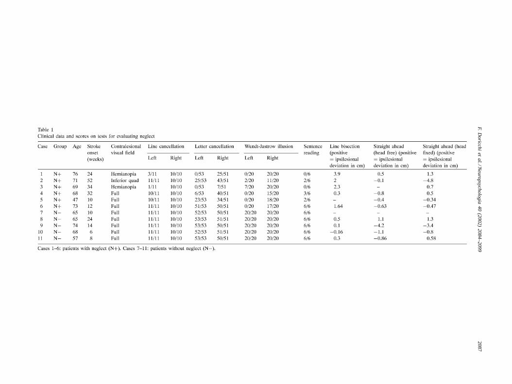
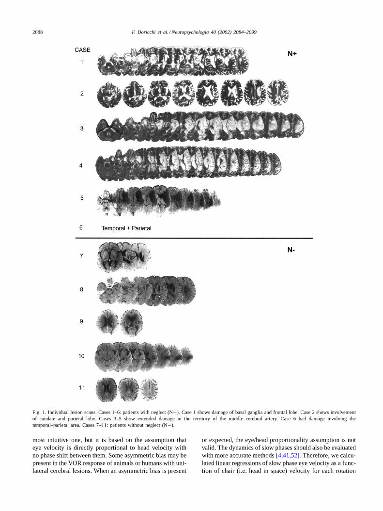
Individual clinical data, results of tests evaluating neglectand of the line bisection and “straight ahead” position tasksare reported inTable 1. Concomitant visual field defectswere evaluated with Goldman perimetric testing. The siteand size of the lesion was evaluated through MRI or CTscans. The individual scans are reported inFig. 1 (for case10, only the report of MRI scans was available).
1.1.2. Evaluation of VOR and OKRThe dc horizontal and vertical EOG were recorded with
electrodes placed at the outer canthi and above and belowthe right eye. EOG calibration was performed with subjectsfixating leds positioned 20◦ to the right and to the left ofa central led aligned to the head–body midsagittal plane. Acalibration was performed before each trial. EOG signalswere amplified, filtered, digitalized (sampling rate 200 Hz)and stored on a PC for off-line processing.
In separate sessions, patients were tested in three differentconditions on a computer controlled rotating chair (model“Rotomac”, Megaris s.a.s.). The software controlling themovements of the chair also allowed the recording and stor-ing of chair position with a resolution of 0.072◦ of arc. Anoptokinetic drum surrounded the chair. The drum was stripedwith vertical black and white stripes (each subtending 6◦ ofthe visual angle). The diameter of the drum was 116 cm andits central vertical axis was centred on the patient’s head.The following testing conditions were organised.
(a) VOR: The chair was sinusoidally rotated (frequency0.05 Hz) in the dark at two different peak velocities: 30and 60◦/s.
(b) VOR+ OKR (visual–vestibular interaction): The chairwas sinusoidally rotated (frequency 0.05 Hz) within theilluminated static optokinetic drum at two different peakvelocities: 30 and 60◦/s.
(c) OKR: The illuminated optokinetic drum was rotatedaround the static chair at two different constant veloci-ties: 15 and 30◦/s.
In the VOR and VOR+ OKR modes of stimulation, thedirection of the first movement (ipsilesional/ contralesional)was balanced among trials. Each trial consisted of two cyclesof rotation, and two trials were performed for each peak ve-locity. The 0.05 Hz frequency was chosen to allow compari-son with the single case study of Ventre-Dominey et al.[66]
(where similar asymmetries of the VOR were found at 0.02,0.05 and 0.1 Hz) and with studies in monkeys with unilat-eral parietal ablations[65] (where comparable asymmetriesof the VOR were found within the range of 0.02–0.07 Hztogether with a peak asymmetry at 0.05 Hz). In order not tostrain patients with a large number of trials, no other fre-quency was used.
The OKR was recorded during drum rotations lasting 60 s(one ipsilesional and one contralesional). In all conditions,the patient was seated in the rotating chair with head, armsand legs restrained by appropriate rests and straps. A securitybelt further stabilised the patient’s body.
At the beginning of each VOR trial, patients fixated a dimblue led fixed to the chair and centred on the head midsagittalplane for 3 s. When the blue led was switched off, a redground-fixed led (aligned to the blue one) was switched onand patients had to fixate it for 3 s. When the red led wasswitched off the rotation started. At the end of the trial, theblue led was switched on again for 3 s and patients wereasked to fixate it again. At the beginning of VOR+ OKRand OKR trials, a led was presented aligned to the headmidsagittal plane. The led was removed by the examinerbefore the chair rotation started and it was presented againat the end of the rotation. This procedure ensured control ofany dc drift of the EOG during trials. Rare trials in whichdrift was present were discarded. At the beginning of eachtrial, patients were simply asked to keep their eyes open.
The local ethical committee approved the experimentalprotocol of the present study.
1.2. Data analysis
For each mode of stimulation, nystagmic eye movementswere analysed thanks to a specially designed Matlab pro-gram. The beginning and end of each slow and relative fastphase were first selected by visual inspection of eye move-ment recordings.
1.2.1. VOR and VOR+ OKRFor each slow and fast phase, a regression polynomial of
degree 3 was fitted to the recorded data of eye movements, aswell as to the corresponding chair (i.e. head in space) move-ment. The number of phases was calculated in each trial. Foreach patient, the mean frequency of fast/slow phases was cal-culated for each mode, direction (ipsilesional/contralesional)and peak velocity (30◦/s or 60◦/s) of rotation.
1.2.2. Slow phasesThree main intrinsic parameters were calculated from re-
gression polynomials: phase amplitude, mean velocity of theeye and mean gain of the response during the phase (i.e. theratio of eye velocity to chair velocity). For each patient, thesevalues were then averaged across trials to obtain a meanvalue for each mode, direction (ipsilesional/contralesional)and peak velocity (30◦/s or 60◦/s) of rotation. This firstmethod of evaluating VOR and VOR+ OKR gain is the
F.D
oricch
ie
ta
l./Ne
uro
psych
olog
ia4
0(2
00
2)
20
84
–2
09
92087
2088 F. Doricchi et al. / Neuropsychologia 40 (2002) 2084–2099
Fig. 1. Individual lesion scans. Cases 1–6: patients with neglect (N+). Case 1 shows damage of basal ganglia and frontal lobe. Case 2 shows involvementof caudate and parietal lobe. Cases 3–5 show extended damage in the territory of the middle cerebral artery. Case 6 had damage involving thetemporal–parietal area. Cases 7–11: patients without neglect (N−).
most intuitive one, but it is based on the assumption thateye velocity is directly proportional to head velocity withno phase shift between them. Some asymmetric bias may bepresent in the VOR response of animals or humans with uni-lateral cerebral lesions. When an asymmetric bias is present
or expected, the eye/head proportionality assumption is notvalid. The dynamics of slow phases should also be evaluatedwith more accurate methods[4,41,52]. Therefore, we calcu-lated linear regressions of slow phase eye velocity as a func-tion of chair (i.e. head in space) velocity for each rotation
F. Doricchi et al. / Neuropsychologia 40 (2002) 2084–2099 2089
direction. The slope of each regression gave the second es-timation of gain. We also analysed the value of the interceptof the regression (corresponding to eye velocity when headvelocity is null). A non-zero intercept of the regression lineindicates the presence of a phase shift in the dynamics of theocular response compared to the dynamics of the head–bodyrotation.
1.2.3. Fast phasesIndividual averaged amplitude and eye position in the or-
bit at the midpoint of each phase (i.e. shift of the beatingfield) were calculated. The shift of the beating field wastaken as positive when it was in the direction of fast phases(i.e. in the direction of chair motion) and negative whenit was in the direction of slow phases (i.e. opposite to thedirection of chair motion).
1.3. OKR
1.3.1. Slow phasesMean amplitude and velocity were calculated for each
experimental condition (i.e. directionx velocity of drumrotation).
1.3.2. Fast phasesMean frequency and amplitude were measured for each
experimental condition.
2. Results
2.1. Clinical data and measures of neglect
N+ and N− did not differ for age (N+ = 67.3 years,N− = 65.8 years;F < 1). Patients from both groups werestudied in the chronic phase of their stroke. The stroke onsetof N− tended to be, but was not, significantly more recentthan that of N+ (N+ = 27.3 weeks, N− = 12.4 weeks;F(1, 9) = 3.8,P = 0.08). N+ performed worse than N− inthe letter cancellation task (F(1, 9) = 12.3, P = 0.006; leftside: N+ = 17.5, N− = 52.6 cancelled items; right side:N+ = 33.1, N− = 50.4 cancelled items), sentence readingtask (F(1, 9) = 14.5, P = 0.004; N+ = 2.1, N− = 6),Wundt-Jastrow illusion task (F(1, 9) = 96,P < 0.001; leftside: N+ = 1.5, N− = 20; right side: N+ = 16.8, N− =20) and in the line bisection test (F(1, 7) = 7.5, P = 0.02;N+ = +2.02 cm, N− = +0.18 cm). Compared with N−,in the line cancellation task, N+ cancelled less items on theleft side of the sheet but not on the right side (group effect:F(1, 9) = 2.7, P = 0.13; left side: N+ = 7.6, N− = 11items, planned comparisonP = 0.04; right side: N+ = 10,N− = 10).
A group (N+, N−) × starting position (left, right)ANOVA showed that in the head free condition N+ hadipsilesional deviation of the “straight ahead” in left toright trials (4.1 cm) and contralesional deviation in right to
left trials (−4.9 cm; group× starting position interaction:F(1, 7) = 10.16;P = 0.01). N− had comparable deviationin both types of trials (left to right:−1.1 cm; right to left:−1.4 cm). In the head restrained condition, no significanteffect or interaction was found (N+: left to right = 1.8 cm,right to left = −2.9 cm; N−: left to right = −0.65 cm, rightto left = −0.67). When the same ANOVAs were repeateddisregarding the starting position, no significant ipsilesionaldeviation of the subjective “straight ahead” as found in N+.(Head free: N+ = −0.28 cm, N− = −1.2 cm, F(1, 7) <
1; head restrained: N+ = −0.51 cm, N− = −0.58 cm;F(1, 8) < 1; see individual data inTable 1).
2.2. VOR and VOR+ OKR
2.2.1. GainIndividual mean gain values were submitted to a group
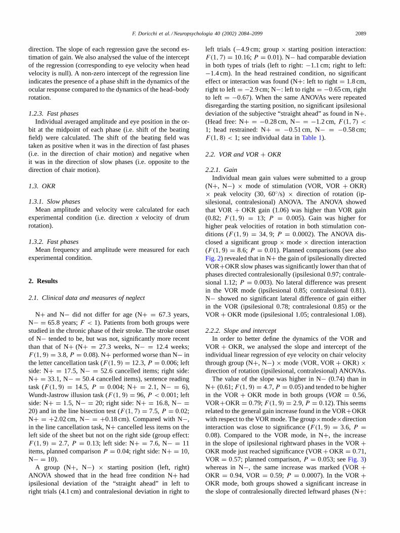
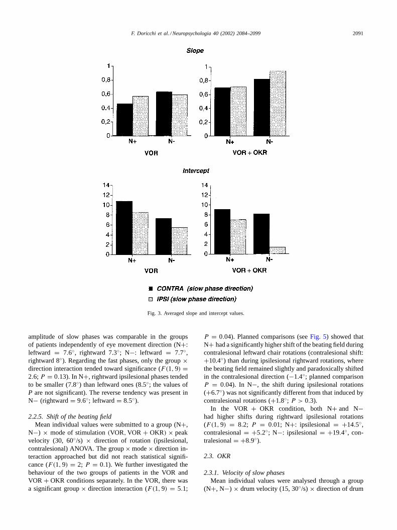
(N+, N−) × mode of stimulation (VOR, VOR+ OKR)× peak velocity (30, 60◦/s) × direction of rotation (ip-silesional, contralesional) ANOVA. The ANOVA showedthat VOR + OKR gain (1.06) was higher than VOR gain(0.82; F(1, 9) = 13; P = 0.005). Gain was higher forhigher peak velocities of rotation in both stimulation con-ditions (F(1, 9) = 34, 9; P = 0.0002). The ANOVA dis-closed a significant group× mode× direction interaction(F(1, 9) = 8.6; P = 0.01). Planned comparisons (see alsoFig. 2) revealed that in N+ the gain of ipsilesionally directedVOR+OKR slow phases was significantly lower than that ofphases directed contralesionally (ipsilesional 0.97; contrale-sional 1.12;P = 0.003). No lateral difference was presentin the VOR mode (ipsilesional 0.85; contralesional 0.81).N− showed no significant lateral difference of gain eitherin the VOR (ipsilesional 0.78; contralesional 0.85) or theVOR + OKR mode (ipsilesional 1.05; contralesional 1.08).
2.2.2. Slope and interceptIn order to better define the dynamics of the VOR and
VOR + OKR, we analysed the slope and intercept of theindividual linear regression of eye velocity on chair velocitythrough group (N+, N−) × mode(VOR, VOR + OKR) ×direction of rotation (ipsilesional, contralesional) ANOVAs.
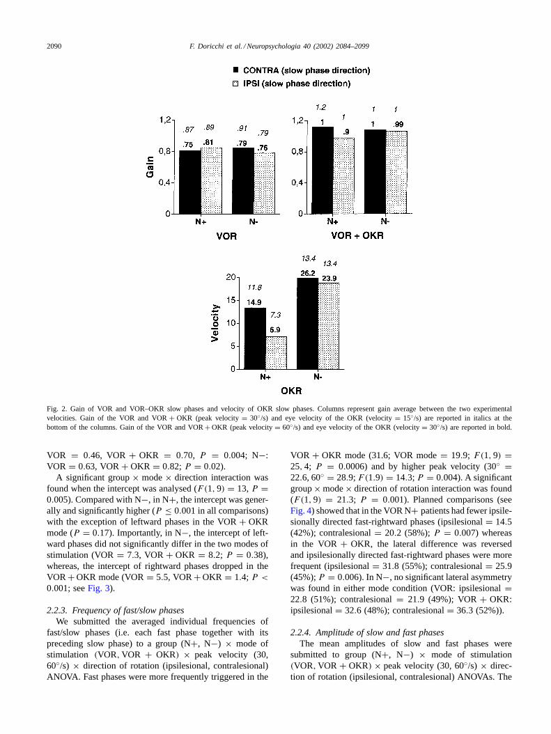
The value of the slope was higher in N− (0.74) than inN+ (0.61;F(1, 9) = 4.7,P = 0.05) and tended to be higherin the VOR+ OKR mode in both groups (VOR = 0.56,VOR+OKR = 0.79;F(1, 9) = 2.9,P = 0.12). This seemsrelated to the general gain increase found in the VOR+OKRwith respect to the VOR mode. The group×mode×directioninteraction was close to significance (F(1, 9) = 3.6, P =0.08). Compared to the VOR mode, in N+, the increasein the slope of ipsilesional rightward phases in the VOR+OKR mode just reached significance (VOR+OKR = 0.71,VOR = 0.57; planned comparison,P = 0.053; seeFig. 3)whereas in N−, the same increase was marked (VOR+OKR = 0.94, VOR = 0.59; P = 0.0007). In the VOR+OKR mode, both groups showed a significant increase inthe slope of contralesionally directed leftward phases (N+:
2090 F. Doricchi et al. / Neuropsychologia 40 (2002) 2084–2099
Fig. 2. Gain of VOR and VOR–OKR slow phases and velocity of OKR slow phases. Columns represent gain average between the two experimentalvelocities. Gain of the VOR and VOR+ OKR (peak velocity= 30◦/s) and eye velocity of the OKR (velocity= 15◦/s) are reported in italics at thebottom of the columns. Gain of the VOR and VOR+ OKR (peak velocity= 60◦/s) and eye velocity of the OKR (velocity= 30◦/s) are reported in bold.
VOR = 0.46, VOR + OKR = 0.70, P = 0.004; N−:VOR = 0.63, VOR+ OKR = 0.82; P = 0.02).
A significant group× mode× direction interaction wasfound when the intercept was analysed (F(1, 9) = 13,P =0.005). Compared with N−, in N+, the intercept was gener-ally and significantly higher (P ≤ 0.001 in all comparisons)with the exception of leftward phases in the VOR+ OKRmode (P = 0.17). Importantly, in N−, the intercept of left-ward phases did not significantly differ in the two modes ofstimulation (VOR= 7.3, VOR+ OKR = 8.2; P = 0.38),whereas, the intercept of rightward phases dropped in theVOR+ OKR mode (VOR= 5.5, VOR+ OKR = 1.4; P <
0.001; seeFig. 3).
2.2.3. Frequency of fast/slow phasesWe submitted the averaged individual frequencies of
fast/slow phases (i.e. each fast phase together with itspreceding slow phase) to a group (N+, N−) × mode ofstimulation (VOR, VOR + OKR) × peak velocity (30,60◦/s) × direction of rotation (ipsilesional, contralesional)ANOVA. Fast phases were more frequently triggered in the
VOR + OKR mode (31.6; VOR mode= 19.9; F(1, 9) =25, 4; P = 0.0006) and by higher peak velocity (30◦ =22.6, 60◦ = 28.9; F(1.9) = 14.3; P = 0.004). A significantgroup× mode× direction of rotation interaction was found(F(1, 9) = 21.3; P = 0.001). Planned comparisons (seeFig. 4) showed that in the VOR N+ patients had fewer ipsile-sionally directed fast-rightward phases (ipsilesional= 14.5(42%); contralesional= 20.2 (58%);P = 0.007) whereasin the VOR+ OKR, the lateral difference was reversedand ipsilesionally directed fast-rightward phases were morefrequent (ipsilesional= 31.8 (55%); contralesional= 25.9(45%);P = 0.006). In N−, no significant lateral asymmetrywas found in either mode condition (VOR: ipsilesional=22.8 (51%); contralesional= 21.9 (49%); VOR+ OKR:ipsilesional= 32.6 (48%); contralesional= 36.3 (52%)).
2.2.4. Amplitude of slow and fast phasesThe mean amplitudes of slow and fast phases were
submitted to group (N+, N−) × mode of stimulation(VOR, VOR + OKR) × peak velocity (30, 60◦/s) × direc-tion of rotation (ipsilesional, contralesional) ANOVAs. The
F. Doricchi et al. / Neuropsychologia 40 (2002) 2084–2099 2091
Fig. 3. Averaged slope and intercept values.
amplitude of slow phases was comparable in the groupsof patients independently of eye movement direction (N+:leftward = 7.6◦, rightward 7.3◦; N−: leftward = 7.7◦,rightward 8◦). Regarding the fast phases, only the group×direction interaction tended toward significance (F(1, 9) =2.6; P = 0.13). In N+, rightward ipsilesional phases tendedto be smaller (7.8◦) than leftward ones (8.5◦; the values ofP are not significant). The reverse tendency was present inN− (rightward= 9.6◦; leftward= 8.5◦).
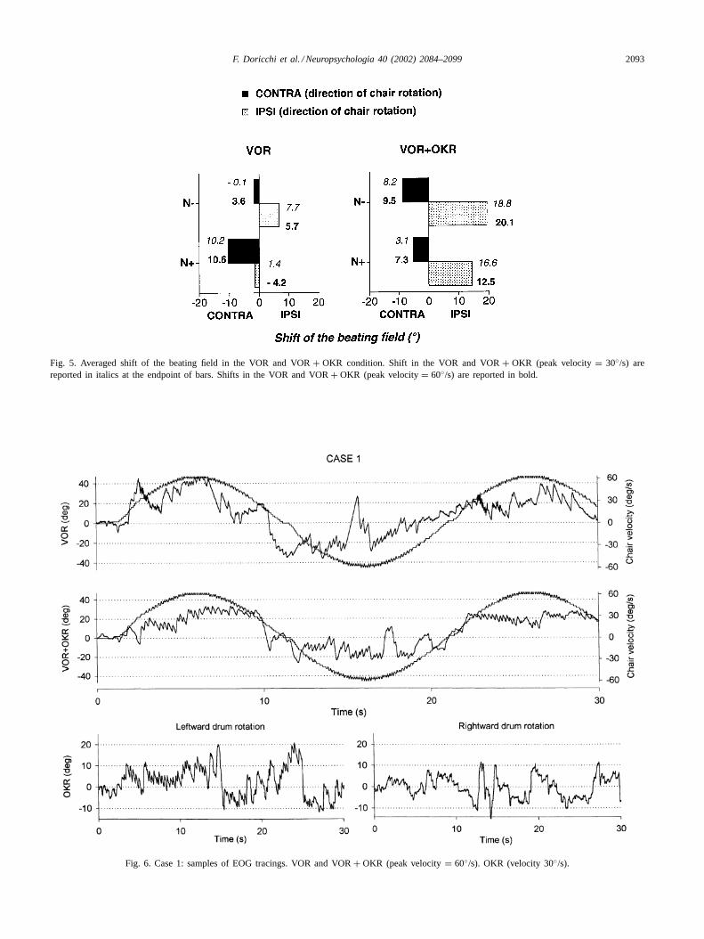
2.2.5. Shift of the beating fieldMean individual values were submitted to a group (N+,
N−) × mode of stimulation(VOR, VOR + OKR) × peakvelocity (30, 60◦/s) × direction of rotation (ipsilesional,contralesional) ANOVA. The group× mode× direction in-teraction approached but did not reach statistical signifi-cance (F(1, 9) = 2; P = 0.1). We further investigated thebehaviour of the two groups of patients in the VOR andVOR + OKR conditions separately. In the VOR, there wasa significant group× direction interaction (F(1, 9) = 5.1;
P = 0.04). Planned comparisons (seeFig. 5) showed thatN+ had a significantly higher shift of the beating field duringcontralesional leftward chair rotations (contralesional shift:+10.4◦) than during ipsilesional rightward rotations, wherethe beating field remained slightly and paradoxically shiftedin the contralesional direction (−1.4◦; planned comparisonP = 0.04). In N−, the shift during ipsilesional rotations(+6.7◦) was not significantly different from that induced bycontralesional rotations (+1.8◦; P > 0.3).
In the VOR + OKR condition, both N+ and N−had higher shifts during rightward ipsilesional rotations(F(1, 9) = 8.2; P = 0.01; N+: ipsilesional = +14.5◦,contralesional= +5.2◦; N−: ipsilesional= +19.4◦, con-tralesional= +8.9◦).
2.3. OKR
2.3.1. Velocity of slow phasesMean individual values were analysed through a group
(N+, N−) × drum velocity (15, 30◦/s) × direction of drum

2092 F. Doricchi et al. / Neuropsychologia 40 (2002) 2084–2099
Fig. 4. Averaged frequencies of slow/fast phases in the VOR, VOR+ OKR and OKR. Frequencies of the VOR and VOR+ OKR (peak velocity= 30◦/s)and the OKR (velocity= 15◦/s) are reported in italics at the bottom of the columns. Frequencies of the VOR and VOR+ OKR (peak velocity= 60◦/s)and the OKR (velocity= 30◦/s) are reported in bold. Corresponding percentage values are reported inside columns, irrespective of velocity of stimulation.
rotation (ipsilesional, contralesional) ANOVA. There was amain effect of group, with N+ having slower phases thanN− (F(1, 9) = 10.7; P = 0.009), drum velocity (F(1, 9) =13.7; P = 0.004) and a main effect of direction of rota-tion indicating that slow-leftward phases were quicker thanrightward ones (F(1, 9) = 21.2; P = 0.001). There wasa significant group× direction interaction (F(1, 9) = 10;P = 0.01). Planned comparisons (seeFig. 3) showed thatin N+, the velocity of contralesional slow-leftward phases(13.3◦/s) was higher than that of rightward ones (7.1◦/s). Nolateral difference was found in N− (ipsilesional= 18.7◦/s;contralesional= 19.8◦/s).
Since in some N+ (cases 3 and 6) rightward ipsilesionalslow phases were completely absent, we repeated the anal-ysis after removal of these patients. The group× directioninteraction was again significant (F(1, 7) = 5.3; P = 0.05)and planned comparisons showed that in N+, the veloc-ity of contralesional slow-leftward phases (14.4◦/s) wasstill higher than that of ipsilesional rightward ones (9.4◦/s;P = 0.004).
2.3.2. Frequency of fast/slow phasesA group (N+, N−) × drum velocity (15, 30◦/s) × direc-
tion of drum rotation (ipsilesional, contralesional) ANOVAperformed on the individual mean frequencies of fast/slowphases showed a significant main effect for group (F(1, 9) =14.1; P = 0.004), direction (F(1, 9) = 14.7; P = 0.003)as well as a close to significant group× direction interac-tion (F(1, 9) = 4.5; P = 0.06). Planned comparisons (seeFig. 4) showed that N+ had more frequent ipsilesional (51.1)than contralesional fast phases (12.5;P = 0.001). In N−,there was no significant lateral difference (ipsilesional=86.4, contralesional= 75.3; P = 0.2).
2.3.3. Amplitude of slow and fast phasesThe mean amplitudes of slow and fast phases were sub-
mitted to group (N+, N−) × drum velocity (15, 30◦/s)× direction of drum rotation (ipsilesional, contralesional)ANOVAs. N+ patients with no ipsilesional slow or con-tralesional fast phases (cases 3 and 6) were excludedfrom the analyses. Regarding the slow phases, there was
F. Doricchi et al. / Neuropsychologia 40 (2002) 2084–2099 2093
Fig. 5. Averaged shift of the beating field in the VOR and VOR+ OKR condition. Shift in the VOR and VOR+ OKR (peak velocity= 30◦/s) arereported in italics at the endpoint of bars. Shifts in the VOR and VOR+ OKR (peak velocity= 60◦/s) are reported in bold.
Fig. 6. Case 1: samples of EOG tracings. VOR and VOR+ OKR (peak velocity= 60◦/s). OKR (velocity 30◦/s).
2094 F. Doricchi et al. / Neuropsychologia 40 (2002) 2084–2099
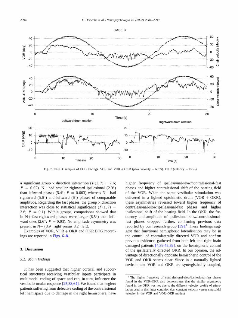
Fig. 7. Case 3: samples of EOG tracings. VOR and VOR+ OKR (peak velocity= 60◦/s). OKR (velocity= 15◦/s).
a significant group× direction interaction (F(1, 7) = 7.6;P = 0.02). N+ had smaller rightward ipsilesional (2.9◦)than leftward phases (5.4◦; P = 0.003) whereas N− hadrightward (5.6◦) and leftward (6◦) phases of comparableamplitude. Regarding the fast phases, the group× directioninteraction was close to statistical significance (F(1, 7) =2.6; P = 0.1). Within groups, comparisons showed thatin N+ fast-rightward phases were larger (6.5◦) than left-ward ones (2.6◦; P = 0.03). No amplitude asymmetry waspresent in N− (8.9◦ right versus 8.2◦ left).
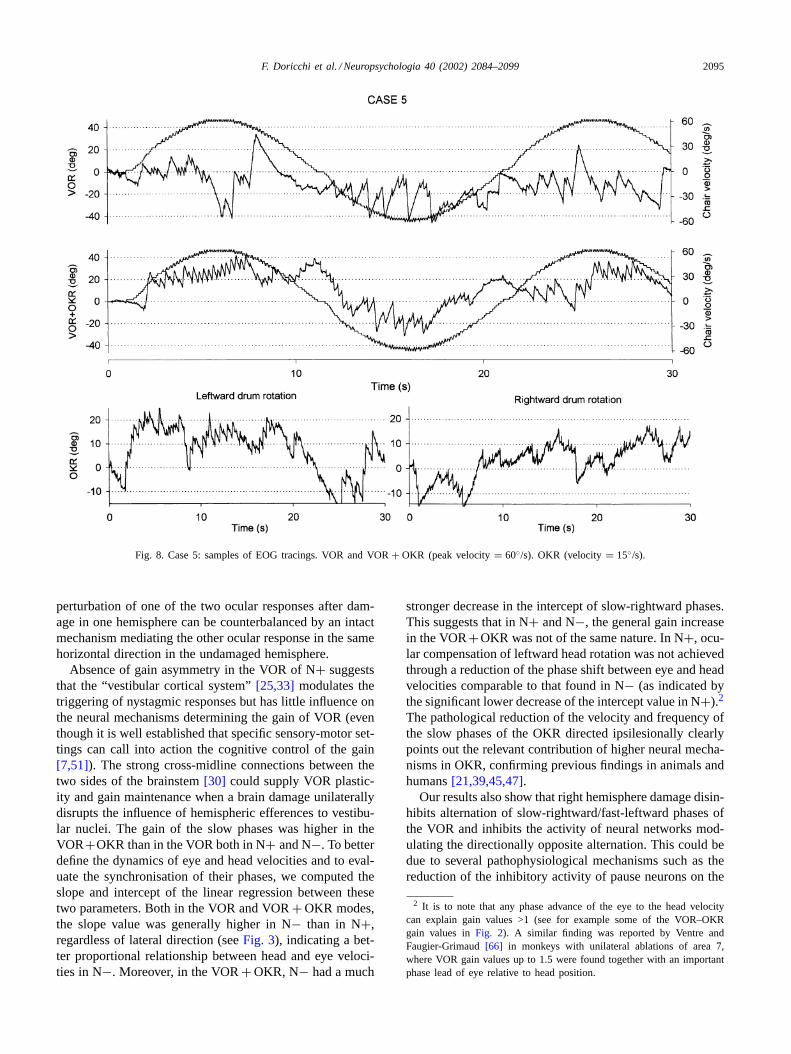
Examples of VOR, VOR+ OKR and OKR EOG record-ings are reported inFigs. 6–8.
3. Discussion
3.1. Main findings
It has been suggested that higher cortical and subcor-tical structures receiving vestibular inputs participate inmultimodal coding of space and can, in turn, influence thevestibulo-ocular response[25,33,64]. We found that neglectpatients suffering from defective coding of the contralesionalleft hemispace due to damage in the right hemisphere, have
higher frequency of ipsilesional-slow/contralesional-fastphases and higher contralesional shift of the beating fieldof the VOR. When the same vestibular stimulation wasdelivered in a lighted optokinetic drum (VOR+ OKR),these asymmetries reversed toward higher frequency ofcontralesional-slow/ipsilesional-fast phases and higheripsilesional shift of the beating field. In the OKR, the fre-quency and amplitude of ipsilesional-slow/contralesional-fast phases dropped further, confirming previous datareported by our research group[39].1 These findings sug-gest that functional hemispheric lateralisation may be inthe control of contralaterally directed VOR and confirmprevious evidence, gathered from both left and right braindamaged patients[4,39,45,59], on the hemispheric controlof the ipsilaterally directed OKR. In our opinion, the ad-vantage of directionally opposite hemispheric control of theVOR and OKR seems clear. Since in a naturally lightedenvironment VOR and OKR are synergistically coupled,
1 The higher frequency of contralesional-slow/ipsilesional-fast phasesfound in the VOR–OKR also demonstrates that the similar asymmetryfound in the OKR was not due to the different velocity profile of stimu-lation used in this latter condition (i.e. constant velocity versus sinusoidalvelocity in the VOR and VOR–OKR modes).
F. Doricchi et al. / Neuropsychologia 40 (2002) 2084–2099 2095
Fig. 8. Case 5: samples of EOG tracings. VOR and VOR+ OKR (peak velocity= 60◦/s). OKR (velocity= 15◦/s).
perturbation of one of the two ocular responses after dam-age in one hemisphere can be counterbalanced by an intactmechanism mediating the other ocular response in the samehorizontal direction in the undamaged hemisphere.
Absence of gain asymmetry in the VOR of N+ suggeststhat the “vestibular cortical system”[25,33] modulates thetriggering of nystagmic responses but has little influence onthe neural mechanisms determining the gain of VOR (eventhough it is well established that specific sensory-motor set-tings can call into action the cognitive control of the gain[7,51]). The strong cross-midline connections between thetwo sides of the brainstem[30] could supply VOR plastic-ity and gain maintenance when a brain damage unilaterallydisrupts the influence of hemispheric efferences to vestibu-lar nuclei. The gain of the slow phases was higher in theVOR+OKR than in the VOR both in N+ and N−. To betterdefine the dynamics of eye and head velocities and to eval-uate the synchronisation of their phases, we computed theslope and intercept of the linear regression between thesetwo parameters. Both in the VOR and VOR+ OKR modes,the slope value was generally higher in N− than in N+,regardless of lateral direction (seeFig. 3), indicating a bet-ter proportional relationship between head and eye veloci-ties in N−. Moreover, in the VOR+ OKR, N− had a much
stronger decrease in the intercept of slow-rightward phases.This suggests that in N+ and N−, the general gain increasein the VOR+OKR was not of the same nature. In N+, ocu-lar compensation of leftward head rotation was not achievedthrough a reduction of the phase shift between eye and headvelocities comparable to that found in N− (as indicated bythe significant lower decrease of the intercept value in N+).2
The pathological reduction of the velocity and frequency ofthe slow phases of the OKR directed ipsilesionally clearlypoints out the relevant contribution of higher neural mecha-nisms in OKR, confirming previous findings in animals andhumans[21,39,45,47].
Our results also show that right hemisphere damage disin-hibits alternation of slow-rightward/fast-leftward phases ofthe VOR and inhibits the activity of neural networks mod-ulating the directionally opposite alternation. This could bedue to several pathophysiological mechanisms such as thereduction of the inhibitory activity of pause neurons on the
2 It is to note that any phase advance of the eye to the head velocitycan explain gain values >1 (see for example some of the VOR–OKRgain values inFig. 2). A similar finding was reported by Ventre andFaugier-Grimaud[66] in monkeys with unilateral ablations of area 7,where VOR gain values up to 1.5 were found together with an importantphase lead of eye relative to head position.
2096 F. Doricchi et al. / Neuropsychologia 40 (2002) 2084–2099
side of the brainstem ipsilateral to the lesion and consequentreduction of threshold activation of the excitatory–inhibitoryburst neurons complex in the contralateral side. Also, theinhibitory interference of brain damage on excitatory pro-jections from right vestibular nuclei to burst driving neuronson the left side of the brainstem could explain reduced trig-gering of fast phases directed rightward.
Finally, neglect patients had higher ipsilesional than con-tralesional shift of the beating field in the lighted VOR–OKRcondition and (paradoxically) higher contralesional shift intotal darkness (i.e. VOR condition). The shift of the beat-ing field was seen as an anticipatory orienting response to-wards sectors of space and stimuli appearing in the directionof the head–body turn[49,50,58]. Following this interpre-tation, our findings can be considered in keeping with dataemphasising the influence of visual input in worsening thepathological ipsilesional deviation of attention suffered byneglect patients[8,36,40].
3.2. Implications for the treatment of unilateral neglect
The directionally opposed VOR and OKR impairmentssuffered by neglect patients suggest that improvement ofneglect by vestibular or optokinetic stimulation inducingcontralesional slow phases is caused by the activation of dif-ferent hemispheric pathways. Cold water irrigation of the leftear produces slow-leftward phases and predominant activa-tion of perisilvian structures in the right hemisphere[10,11].Therefore, temporary amelioration of left unilateral neglectfollowing cold caloric stimulation of the left ear seems dueto prevalent direct activation of the damaged hemisphere.Optokinetic stimulation directed leftward is perturbed by lefthemisphere lesions[5] and in normal subjects causes pre-dominant activation of V5, the intraparietal sulcus and theputamen in the left hemisphere[29]. Thus, amelioration ofleft unilateral neglect after leftward optokinetic stimulationseems due to prevalent indirect re-activation of the damagedright hemisphere by afferences from the undamaged lefthemisphere (this explanation could also apply to the effectsof visual background motion toward the neglected side[44]).
Different and not mutually exclusive physiological mech-anisms may account for neglect improvement after optoki-netic stimulation. Brandt et al.[12] found that leftwardoptokinetic stimulation produces bilateral activation of theparietal–temporal area and the basal ganglia in right braindamaged patients with contralateral pure hemianopia. Theseauthors initially hypothesised that activation in the damagedhemisphere was conveyed by direct retinal-extrastriate con-nections bypassing the damaged striate areas or by transcal-losal afferents from the spared hemisphere. In a subsequentstudy on normal subjects, Bucher et al.[13] demonstratedthat motion stimulation of one hemifield activates middleand middle-superior temporal areas bilaterally while deac-tivating optic radiations contralateral to the side of the stim-ulation. Accordingly, the authors concluded that previousfindings in hemianopics were better explained by transcal-
losal activation. We think that similar transcallosal mech-anisms could also underlie the improvement of unilateralneglect produced by optokinetic stimulation directed con-tralesionally. At the same time, this stimulation could drawattention contralesionally and re-activate, transcallosaly,spared structures in the damaged hemisphere. Conversely,optokinetic stimulation directed ipsilesionally could be in-effective or even worsen neglect (see Vallar et al.[60]), bothby drawing attention ipsilesionally and because disruptionof mechanisms receptive to ipsilesional optokinetic stimula-tion in the damaged hemisphere generally prevents boostingof any residual attentional resource in both hemispheres.
This explanation takes into account different hypotheseson the ameliorative effects of optokinetic stimulation on ne-glect. Gainotti[28] emphasised that contralesionally directedoptokinetic stimulation (or vestibular stimulation producingdirectionally similar oculomotor effects) re-orients covertand overt attention toward the neglected side, contrastingthe pathological ipsilesional bias. Vallar et al.[61] stressedthat contralesional optokinetic stimulation not only has at-tentional effects in neglect patients since it improves theproprioceptive-position sense of both arms (i.e. not only ofthe arm on the side of stimulation direction) whereas stimu-lation directed ipsilesionally worsens position sense bilater-ally. The improvement was interpreted as depending on there-activation of multimodal mechanisms specialised for therepresentation of personal body-space in the damaged righthemisphere[61]. This mechanism seems plausible since inhealthy humans structures of the right hemisphere related toattentional and oculomotor control (posterior parietal cortex,precentral and posterior medial frontal gyrus) can be acti-vated by optokinetic stimulation regardless of its horizontalor vertical direction of[18].
3.3. Implications for the “egocentric reference”hypothesis
Ventre et al. investigated the VOR in cats with unilateralablations of the middle suprasylvian gyrus and superior col-liculus [64] and in monkeys with unilateral ablation of area7a [65]. They found decrease of ipsilesional gain, increaseof contralesional gain and spontaneous nystagmus with slowphases directed contralesionally. Developing the pioneeringobservations made by Hecaen and Massonet[35], Ventreet al. [64] interpreted the predominance of slow phases to-ward the contralesional space as a compensatory response ofthe vestibular system to counteract the ipsilesional displace-ment of visuomotor behaviour produced by the unilaterallesion. According to the “egocentric reference” hypothesis(Ventre et al.[64]), the ipsilesional bias of visuomotor be-haviour is due to interhemispheric unbalance in the activa-tion of multimodal representations of space usually ensuringsymmetry of orienting and the alignment of the subjectivesagittal body midline to the objective one. Our results showthat patients with extensive unilateral brain damage andchronic contralesional neglect have no contralesional bias
F. Doricchi et al. / Neuropsychologia 40 (2002) 2084–2099 2097
in the gain of the VOR, suffer reduced frequency of con-tralesionally directed slow phases and show contralesionalrather than ipsilesional deviation of the beating field. In thesame patients, no ipsilesional deviation of the subjective“straight ahead” was found. This latter finding replicatesprevious data from several independent authors[6,23,55]and, in the present study, was evident at the individual leveland independent from concomitant visual field defects.Taken together, these dissociations do not seem to agreewith the “egocentric reference” hypothesis, which assumesthat the same pathophysiological mechanism underlies con-tralesional neglect, ipsilesional deviation of the subjective“straight ahead” and ipsilesional defects of the VOR. How-ever, the original observations by Ventre et al.[64] weremade within a few days following surgical ablations. It maybe that acute neglect patients show behavioural and vestibu-lar symptoms similar to those described by Ventre et al. inanimals.
Our findings are in keeping with a recent case study byVentre-Dominey et al.[66] documenting decreased gain andreduced frequency of the contralesional VOR in a patientwith a right parietal–temporal damage. The deficits found inthis patient, when compared with the opposite VOR impair-ments found in animals with unilateral parietal or collicularlesions[64], were tentatively attributed to adaptive recoveryduring the subacute phase, to differences in the location ofthe lesion or to inter-species differences in the cortical dis-tribution of vestibular control. Our data and those from thecase study of Ventre-Dominey et al.[66] dovetail both withbrain imaging data indicating relative higher activation ofthe right hemisphere when the slow phases of the VOR aredirected leftward[10,11]and with clinical and experimentalfindings demonstrating hemispheric control of ipsilateralOKR [4,29,39]. This converging evidence does not seemto support the possibility that VOR asymmetries in chronicneglect patients depend on recovery mechanisms causing ashift from ipsilesional to contralesional hemispheric control.The different asymmetries of horizontal VOR impairmentsfound in animals and humans could, therefore, be betterexplained by inter-species differences in the hemisphericdistribution and complexity of mechanisms modulating theVOR. Unicellular recordings in monkeys do not documenta clear-cut hemispheric lateralisation in the control of theVOR and VOR–OKR interaction. Grüsser et al.[32] foundthat in the PIVC area of the monkey, 53% of the neuronsrespond to contralateral rotation in darkness and 37% toipsilateral rotation. Büttner and Buettner[14] found nohemispheric specialisation in area 2v (in the lower sectionof the intraparietal sulcus). Kawano and Sasaki[42] foundthat in area 7 of the monkey, 60% of the neurons respondedto ipsilateral OKR stimulation and the remaining 40% tocontralateral stimulation. Half of the neurons responded tosynergistic VOR–OKR stimulation (i.e. to vestibular or op-tokinetic stimulation inducing slow phases toward the sameside of space) and the other half to anti-synergistic stimu-lation [43]. These data might indicate that in lower species,
the recovery mechanism after unilateral brain damage issubserved by different populations of VOR and bimodalVOR–OKR neurons which, in each hemisphere, preferen-tially respond to ipsilateral or contralateral VOR, OKR orcombined VOR–OKR stimulation.
3.4. Cautions and conclusions
First, caution should be taken in interpreting our find-ings because of the possible effects of inactivation (i.e. di-aschisis) produced by an extensive unilateral hemisphericlesion on brainstem mechanisms regulating the VOR. In thiscase, the alteration of the VOR we tentatively attributedto hemispheric lateralisation could be due to dysfunctionof more peripheral mechanisms. Although this hypothesisshould be taken into consideration, it should be recalled thatVentre-Dominey et al.[66] documented impairments of theVOR similar to those, we found in N+ in a patient witha circumscribed temporal–parietal lesion and no detectablebrainstem damage on MRI examination.
Second, our conclusions about the hemispheric lateralisa-tion of the VOR control are currently based on samples ofright brain damaged patients and should therefore be sub-stantiated by studying patients with lesions of the left hemi-sphere.
Third, previous studies in hemispherectomised patients[22,57]disclosed defects of the horizontal VOR that appeardirectionally opposed to those reported in the present inves-tigation. In many cases, hemispherectomy was performedto bring about relief from seizures with very early onset inthe life of patients. Thus, it cannot be excluded that thesepatients had idiosyncratic patterns of hemispheric vestibularcontrol (and recovery) linked to processes of developmentalfunctional reorganisation due to illness. Nonetheless, the re-port by different authors of a diametrically opposed deficitof the horizontal VOR after unilateral brain damage requiresfurther clarification.
Fourth, another caution concerns the convergence of ourdata with imaging data by Bottini et al.[10,11]. In these stud-ies, which were not specifically aimed at identifying oculo-motor structures related to the control of the VOR, only coldcaloric stimulation of each ear was used. Therefore, brainactivations reflected both sensory-nociceptive and oculomo-tor components. Central brain structures selectively relatedto VOR control should be specifically investigated consid-ering that they should be equally activated both by cold andwarm caloric stimulation inducing VOR toward the samehorizontal direction.
In the present study, N+ had larger lesions than N−. Itis quite possible that this contributed to the greater VORand OKR impairments of N+ and also implies that damageto adjacent, independent, brain structures accounts for theassociation between neglect and VOR/OKR impairments.However, the aim of the present study was to define the pat-tern of VOR and OKR deficits that can be associated withunilateral neglect rather than to demonstrate their higher
2098 F. Doricchi et al. / Neuropsychologia 40 (2002) 2084–2099
frequency or their exclusive link with the syndrome. Thelocalisation and extent of the lesion has a profound impacton the clinical features of the neglect syndrome[9,20].Further studies are therefore needed to specifically assessthe effect of different lesions producing neglect on theVOR and OKR. Conversely, the effectiveness of vestibularand optokinetic stimulation should be assessed in patientswith neglect due to damage of different sectors of theneural network that provide humans with adaptive codingof space.
Acknowledgements
We are greatly indebted to Umberto Cellini, PietroFermani and Dino Moretti for their invaluable technicalskills and help. Prof. Luigi Pizzamiglio, Prof. StefanoPaolucci and two anonymous referees also provided use-ful suggestions. The data from this study were presentedat the joint meeting of the British Neuropsychologi-cal Society and the Societa’ Italiana di Neuropsicologia(London, 25–27 April 2001; abstracts published in Neuro-case 2001; 7:351–56). This research was supported by agrant from the Fondazione Santa Lucia and the LaboratoireEuropeen des Neurosciences de l’Action (LENA) to FabrizioDoricchi.
References
[1] Albert ML. A simple test of visual neglect. Neurology 1973;23:658–64.
[2] Akbarian S, Grusser OJ, Guldin WO. Corticofugal projectionsto the vestibular nuclei in squirrel monkeys: further evidence ofmultiple cortical vestibular fields. Journal of Comparative Neurology1994;332:89–104.
[3] Andersen RA, Snyder LH, Bradley DC, Xing J. Multimodalrepresentations of space in the posterior parietal cortex and its usein planning movements. Annual Review of Neuroscience 1997;20:303–30.
[4] Baloh RW, Yee RD, Honrubia V. Optokinetic nystagmus and parietallobe lesions. Annals of Neurology 1980;7:269–76.
[5] Baloh RW, Jacobson KM, Beykirch K, Honrubia V. Horizontalvestibulo-ocular reflex after acute peripheral lesions. ActaOtolaryngologica 1989;468:323–7.
[6] Bartolomeo P, Chokron S. Egocentric frame of reference: its rolein spatial bias after right hemisphere lesions. Neuropsychologia1999;37:881–94.
[7] Berthoz A. Coopération et substitution entre le système saccadiqueet les “réflexes” d’origine vestibulaire: faut-il réviser la notion de“réflexe”? Revue Neurologique 1989;145:513–26.
[8] Berti A, Maravita A, Frassinetti F, Umilta’ C. Unilateral neglect canbe affected by stimuli in the neglected field. Cortex 1995;31:331–43.
[9] Binder J, Marshall R, Lazar R, Benjamin J, Mohr JP. Distinctsyndromes of hemineglect. Archives of Neurology 1992;49:1187–93.
[10] Bottini G, Sterzi R, Paulesu E, Vallar G, Cappa SF, Erminio F, et al.Identification of the central vestibular projections in man: a positronemission tomography activation study. Experimental Brain Research1994;99:164–9.
[11] Bottini G, Karnath HO, Vallar G, Sterzi R, Frith CD,Frackowiak RSJ, et al. Cerebral representation for egocentric space:
functional–anatomical evidence from caloric vestibular stimulationand neck vibration. Brain 2001;124:1182–96.
[12] Brandt T, Stephan T, Bense S, Yousry TA, Dieterich M. Hemifieldvisual motion stimulation: an example of interhemispheric crosstalk.NeuroReport 2000;11:2803–9.
[13] Bucher SF, Dieterich M, Seelos K, Brandt T. Sensorimotor cerebralactivation during optokinetic nystagmus: a functional MRI study.Neurology 1997;49:1370–7.
[14] Büttner U, Buettner UW. Parietal cortex (2v) neuronal activity in thealert monkey during natural vestibular and optokinetic stimulation.Brain Research 1978;153:392–7.
[15] Cappa S, Sterzi R, Vallar G, Bisiach E. Remission of hemineglectand anosognosia during vestibular stimulation. Neuropsychologia1987;25:775–82.
[16] Carmichael EA, Dix MR, Hallpike CS. Lesions of the cerebralhemispheres and their effects upon optokinetic and caloric nystagmus.Brain 1954;77:345–72.
[17] Collewijn H. The vestibulo-ocular reflex: is it an independentsubsystem? Revue Neurologique 1989;145:502–12.
[18] Dieterich M, Bucher SF, Seelos KC, Brandt T. Horizontal or verticaloptokinetic stimulation activates visual motion-sensitive, ocular motorand vestibular cortex areas with right hemispheric dominance: anfMRI study. Brain 1998;121:1479–95.
[19] Diller L, Ben-Yishay Y, Gerstman LJ, Goodkin R, Gordon W,Weinberg J. Studies in cognition and rehabilitation in hemiplegia.In: Rehabilitation monograph 1974, No. 50. New York: UniversityMedical Center.
[20] Doricchi F, Angelelli P. Misrepresentation of horizontal space inleft unilateral neglect: role of hemianopia. Neurology 1999;52:1845–52.
[21] Dürsteler MR, Würtz RH. Pursuit and optokinetic deficits followingchemical lesions of cortical areas MT and MST. Journal ofNeurophysiology 1988;60:940–65.
[22] Estanol B, Romero R, Saenz de Viteri M, Mateos JH, CorveraJ. Oculomotor and oculovestibular functions in a hemispherectomypatient. Archives of Neurology 1980;37:365–8.
[23] Farne’ A, Ponti F, Ladavas E. In search of biased egocentric referenceframes in neglect. Neuropsychologia 1998;36:611–23.
[24] Faugier-Grimaud S, Ventre J. Anatomic connections of inferiorparietal cortex (area 7) with subcortical structures related tovestibulo-ocular function in monkey. Journal of ComparativeNeurology 1989;280:1–14.
[25] Faugier-Grimaud S, Baleydier C, Magnin M, Jeannerod M. Directbilateral cortical projections to the vestibular complex in macaquemonkey. In: Thier P, Karnath HO, editors. Parietal lobe contributionsto orientation in 3D space. Berlin: Springer, 1997. p. 57–76.
[26] Friberg L, Olsen TS, Roland PE, Paulson OB, Lassen NA. Focalincrease of blood flow in the cerebral cortex of man during vestibularstimulation. Brain 1985;108:603–9.
[27] Fukushima K. Corticovestibular interactions: anatomy, electro-physiology, and functional considerations. Experimental BrainResearch 1997;117:1–16.
[28] Gainotti G. The role of spontaneous eye movements in orientingattention and in unilateral neglect. In: Robertson IH, Marshall JC,editors. Unilateral neglect: clinical and experimental studies. Hove(UK): Lawrence Erlbaum, 1993. p. 107–22.
[29] Galati G, Pappata S, Pantano P, Lenzi GL, Samson Y, PizzamiglioL. Cortical control of optokinetic nystagmus in humans: apositron emission tomography study. Experimental Brain Research1999;126:149–59.
[30] Galiana HL, Outerbridge JS. A bilateral model for central neuralpathways in vestibuloocular reflex. Journal of Neurophysiology;51:210–41.
[31] Goldberg JM, Smith CE, Fernandez C. Relation between dischargeregularity and responses to externally applied galvanic currentsin vestibular nerve afferents of the squirrel monkey. Journal ofNeurophysiology 1984;51:1236–56.
F. Doricchi et al. / Neuropsychologia 40 (2002) 2084–2099 2099
[32] Grüsser OJ, Guldin W, Harris L, Lefebre JC, Pause M. Corticalrepresentation of head-in-space movement and same psychophysicalexperiments on head movement. In: Berthoz A, Vidal PP, Graf W,editors. The head–neck sensory motor system. New York: OxfordUniversity Press, 1992. p. 497–509.
[33] Guldin WO, Grusser OJ. Is there a vestibular cortex? Trends inNeurosciences 1998;21:254–9.
[34] Halmagyi GM, Curthoys IS, Cremer PD, Henderson CJ, Todd MJ,Staples MJ, et al. The human horizontal vestibulo-ocular reflex inresponse to high-acceleration stimulation before and after unilateralvestibular neurectomy. Experimental Brain Research 1990;81:479–90.
[35] Hecaen H, Massonet J. Les troubles visuoconstructif par lesionparieto-occipitale droite: role des perturbations vestibulaires.Encephale 1951;40:122–79.
[36] Hyaltason M, Tegner R. Darkness improves line bisection inunilateral spatial neglect. Cortex 1992;28:353–8.
[37] Husain M, Kennard C. Visual neglect associated with frontal lobeinfarction. Journal of Neurology 1996;243:652–7.
[38] Husain M, Mannan S, Hodgson T, Wojciulik E, Driver J, KennardC. Impaired spatial working memory across saccades contributes toabnormal search in parietal neglect. Brain 2001;124:941–52.
[39] Incoccia C, Doricchi F, Galati G, Pizzamiglio L. Amplitude andspeed change of the optokinetic response in patients with and withoutneglect. NeuroReport 1995;6:2137–40.
[40] Kaplan RF, Verfaellie M, Meadows ME, Caplan L, Pessin MS, DanaDeWitt L. Changing attentional demands in left hemispatial neglect.Archives of Neurology 1991;48:1263–6.
[41] Katsarkas A, Galiana HL, Smith HL. Vestibulo-ocular reflex (VOR)biases in normal subjects and patients with compensated vestibularloss. Acta Otolaryngologica 1995;115:476–83.
[42] Kawano K, Sasaki M. Neurons in the posterior parietal associationcortex of the monkey activated during optokinetic stimulation.Neuroscience Letters 1981;22:239–44.
[43] Kawano K, Sasaki M. Response properties of neurons in posteriorparietal cortex of monkey during visual-vestibular stimulation.Part II. Optokinetic neurons. Journal of Neurophysiology 1984;51:352–60.
[44] Kerkhoff G, Schindler I, Keller I, Marquardt C. Visual backgroundmotion reduces size distortion in spatial neglect. NeuroReport1999;10:319–23.
[45] Kömpf D. The significance of optokinetic nystagmus asymmetry inhemispheric lesions. Neuroophthalmology 1986;6:61–4.
[46] Lobel E, Kleine JF, Bihan DL, Leroy-Willig A, Berthoz A. FunctionalMRI of galvanic vestibular stimulation. Journal of Neurophysiology1998;80:2699–709.
[47] Lynch JC, McLaren JW. Optokinetic nystagmus deficits followingparieto–occipital cortex lesions in monkeys. Experimental BrainResearch 1983;49:125–30.
[48] Massironi M, Antonucci G, Pizzamiglio L, Vitale M, Zoccolotti P.The Wundt-Jastow illusion in the study of spatial hemi-inattention.Neuropsychologia 1988;26:161–6.
[49] Meier RK, Dieringer N. The role of compensatory eye orientationand head movements in the rat for image stabilization and gazeorientation. Experimental Brain Research 1993;96:54–64.
[50] Melvill Jones G. Predominance of anti-compensatory oculomotorresponse during rapid head rotation. Aviation Space andEnvironmental Medicine 1964;35:365–968.
[51] Melvill Jones G, Guitton D, Bethoz A. Changing patterns of eye-headcoordination during 6H of optically reversed vision. ExperimentalBrain Research 1988;69:531–44.
[52] Paige GD. Nonlinearity and asymmetry in the human vestibulo-ocularreflex. Acta Otolaryngologica 1989;108:1–8.
[53] Pizzamiglio L, Antonucci G, Judica A, Montenero P, Razzano C,Zoccolotti P. Cognitive rehabilitation of the hemineglect disorder inchronic patients with unilateral brain damage. Journal of Clinicaland Experimental Neuropsychology 1992;14:901–23.
[54] Pizzamiglio L, Frasca R, Guariglia C, Incoccia C, Antonucci G.Effect of optokinetic stimulation in patients with visual neglect.Cortex 1990;26:535–40.
[55] Pizzamiglio L, Committeri G, Galati G, Patria F. Psychophysicalproperties of line bisection and body midline perception in unilateralneglect. Cortex 2000;36:469–84.
[56] Rubens AB. Caloric stimulation and unilateral visual neglect.Neurology 1985;35:1019–24.
[57] Sharpe JA, Lo AW. Voluntary and visual control of the vestibuloocularreflex after cerebral hemidecortication. Annals of Neurology1981;10:164–72.
[58] Siegler I, Israel I, Berthoz A. Shift of the beating field ofvestibular nystagmus: an orientation strategy? Neuroscience Letters1998;254:93–6.
[59] Smith JL, Cogan DG. Optokinetic nystagmus: a test for parietal lobelesions. American Journal of Ophthalmology 1959;48:187–93.
[60] Vallar G, Guariglia C, Rusconi ML. Modulation of the neglectsyndrome by sensory stimulation. In: Thier P, Karnath HO, editors.Parietal lobe contributions to orientation in 3D space. Berlin:Springer, 1997. p. 556–78.
[61] Vallar G, Guariglia C, Magnotti L, Pizzamiglio L. Optokineticstimulation affects both vertical and horizontal deficits of positionsense in unilateral neglect. Cortex 1995;31:669–83.
[62] Vallar G. Spatial hemineglect in humans. Trends in CognitiveSciences 1998;2:87–96.
[63] Vallar G, Lobel E, Galati G, Berthoz A, Pizzamiglio L, LeBihan D. Afronto-parietal system for computing the egocentric spatial frame ofreference in humans. Experimental Brain Research 1999;124:281–6.
[64] Ventre J, Flandrin JM, Jeannerod M. In search of egocentricreference: a neurophysiological hypothesis. Neuropsychologia1984;22:797–806.
[65] Ventre J, Faugier-Grimaud S. Effects of posterior parietal lesions(area7) on VOR in monkeys. Experimental Brain Research1986;62:654–8.
[66] Ventre-Dominey J, Vighetto A, Denise P. Vestibulo-ocular dys-function induced by cortical damage in man: a case report.Neuropsychologia 1999;37:715–21.
Related Documents