Ultrarapid caspase-3 dependent apoptosis induction by serine/threonine phosphatase inhibitors KE Fladmark 1 , OT Brustugun 1 , R Hovland 1 , R Bøe 1 , BT Gjertsen 1 , B Zhivotovsky 2 and SO Døskeland* ,1 1 Cell Biology Research Group, Department of Anatomy and Cell Biology, University of Bergen, A ˚ rstadveien 19, N-5009 Bergen, Norway 2 Institute of Environmental Medicine, Division of Toxicology, Karolinska Institute, S-171 77 Stockholm, Sweden * Corresponding author: SO Døskeland, University of Bergen, Department of Anatomy and Cell Biology, A ˚ rstadveien 19, N-5009 Bergen, Norway. Tel: +47 55 58 63 76; Fax: +47 55 58 63 60; E-mail: [email protected] Received 8.3.99; revised 20.7.99; accepted 26.8.99 Edited by S Kumar Abstract The protein phosphatase (PP) inhibitors nodularin and microcystin-LR induced apoptosis with unprecedented rapidity, more than 50% of primary hepatocytes showing extensive surface budding and shrinkage of cytoplasm and nucleoplasm within 2 min. The apoptosis was retarded by the general caspase inhibitor Z-VAD.fmk. To circumvent the inefficient uptake of microcystin and nodularin into non- hepatocytes, toxins were microinjected into 293 cells, Swiss 3T3 fibroblasts, promyelocytic IPC-81 cells, and NRK cells. All cells started to undergo budding typical of apoptosis within 0.5 – 3 min after injection. This was accompanied by cytoplasmic and nuclear shrinkage and externalization of phosphatidylserine. Overexpression of Bcl-2 did not delay apoptosis. Apoptosis induction was slower and Z-VAD.fmk independent in caspase-3 deficient MCF-7 cells. MCF-7 cells stably transfected with caspase-3 showed a more rapid and Z- VAD.fmk dependent apoptotic response to nodularin. Rapid apoptosis induction required inhibition of both PP1 and PP2A, and the apoptosis was preceded by increased phosphoryla- tion of several proteins, including myosin light chain. The protein phosphorylation occurred even in the presence of apoptosis-blocking concentrations of Z-VAD.fmk, indicating that it occurred upstream of caspase activation. It is suggested that phosphatase-inhibiting toxins can induce caspase-3 dependent apoptosis in an ultrarapid manner by altering protein phosphorylation. Keywords: apoptosis; protein phosphorylation; caspases; phos- phatase inhibitors; cyanobacterial toxins Abbreviations: PP1, phosphoprotein phosphatase type 1; PP2A, phosphoprotein phosphatase type 2A Introduction Regulated (‘programmed’) cell death often occurs by a morphologically distinct series of events, termed apopto- sis. 1,2 Apoptotic cells are recognized by cell shrinkage, cell surface budding and chromatin hypercondensation. Specific cleavage of DNA, 2 RNA, 3 and ‘flipping’ of phospholipids to the external face of the cell membrane 4,5 are also commonly associated with apoptosis. Caspases are believed to be instrumental in the execution of regulated death. So far 14 members (caspase 1 – 14) of the caspase family have been described. 6,7 Some of these, like procaspase-8 and -2, can undergo autoactivation through multimerization, 8,9 and in turn activate downstream procaspases 10 – 12 and other proapopto- tic proteins. 13 The Bcl-2 protein family contains members (Bcl- 2, Bcl-x L ) that can protect against apoptosis. 16 Actions of Bcl- 2 include the prevention of mitochondrial release of cytochrome c 15,16 as well as of the apoptogenic action of cytoplasmic cytochrome c. 17 The mammary carcinoma cell line MCF-7 lacks both procaspase-3 and the ability to undergo apoptosis in response to injected cycochrome c 18,19 and is a model to test for death which is independent of cytochrome c and caspase-3. Altered protein phosphorylation (‘dysphosphorylation’) is known to be an important modulator of apoptosis induction and possibly apoptosis execution. This can occur either through action on protein kinases or protein phospha- tases. 20 – 22 It is intriguing that serine/threonine protein phosphatases 1 and 2A (PP1 and PP2A) are targeted by an array of chemically distinct microbial toxins. 23 We and others have shown that cell-permeant protein phosphatase inhibitors like okadaic acid 24,25 are general inducers of apoptosis. 26 – 29 We have postulated that the inhibitors act by short-cutting normal pathways of apoptosis induction. 23 A logical consequence of this would be that they could act faster than other apoptosis-inducers, if allowed immediate access to the intracellular compartment. This hypothesis was tested in the present study (1) by microinjecting a number of cell types with the water soluble phosphatase inhibitors 23 nodularin and microcystin, and (2) by exposing primary rat hepatocytes to high concentrations of these toxins in the medium. Hepatocytes are unique among mammalian cells in having an efficient transport system for microcystin and nodularin. 30 The question of whether protein phosphorylation was involved in toxin-induced death was addressed by temporal comparison of the cellular phosphoprotein pattern (studied by two-dimen- sional electrophoresis) with the development of apoptotic indices of cell death. It was also of interest to know whether inhibition of PP1, PP2A or both was required for rapid apoptosis induction, and whether the phosphorylation changes were upstream or downstream of caspase activation. Cell Death and Differentiation (1999) 6, 1099 – 1108 ª 1999 Stockton Press All rights reserved 13509047/99 $15.00 http://www.stockton-press.co.uk/cdd

Welcome message from author
This document is posted to help you gain knowledge. Please leave a comment to let me know what you think about it! Share it to your friends and learn new things together.
Transcript
Ultrarapid caspase-3 dependent apoptosis induction byserine/threonine phosphatase inhibitors
KE Fladmark1, OT Brustugun1, R Hovland1, R Bùe1,
BT Gjertsen1, B Zhivotovsky2 and SO Dùskeland*,1
1 Cell Biology Research Group, Department of Anatomy and Cell Biology,University of Bergen, AÊ rstadveien 19, N-5009 Bergen, Norway
2 Institute of Environmental Medicine, Division of Toxicology, KarolinskaInstitute, S-171 77 Stockholm, Sweden
* Corresponding author: SO Dùskeland, University of Bergen, Department ofAnatomy and Cell Biology, AÊ rstadveien 19, N-5009 Bergen, Norway.Tel: +47 55 58 63 76; Fax: +47 55 58 63 60; E-mail: [email protected]
Received 8.3.99; revised 20.7.99; accepted 26.8.99Edited by S Kumar
AbstractThe protein phosphatase (PP) inhibitors nodularin andmicrocystin-LR induced apoptosis with unprecedentedrapidity, more than 50% of primary hepatocytes showingextensive surface budding and shrinkage of cytoplasm andnucleoplasm within 2 min. The apoptosis was retarded by thegeneral caspase inhibitor Z-VAD.fmk. To circumvent theinefficient uptake of microcystin and nodularin into non-hepatocytes, toxins were microinjected into 293 cells, Swiss3T3 fibroblasts, promyelocytic IPC-81 cells, and NRK cells. Allcells started to undergo budding typical of apoptosis within0.5 ± 3 min after injection. This was accompanied bycytoplasmic and nuclear shrinkage and externalization ofphosphatidylserine. Overexpression of Bcl-2 did not delayapoptosis. Apoptosis induction was slower and Z-VAD.fmkindependent in caspase-3 deficient MCF-7 cells. MCF-7 cellsstably transfected with caspase-3 showed a more rapid and Z-VAD.fmk dependent apoptotic response to nodularin. Rapidapoptosis induction required inhibition of both PP1 and PP2A,and the apoptosis was preceded by increased phosphoryla-tion of several proteins, including myosin light chain. Theprotein phosphorylation occurred even in the presence ofapoptosis-blocking concentrations of Z-VAD.fmk, indicatingthat itoccurredupstreamof caspase activation. It issuggestedthat phosphatase-inhibiting toxins can induce caspase-3dependent apoptosis in an ultrarapid manner by alteringprotein phosphorylation.
Keywords: apoptosis; protein phosphorylation; caspases; phos-phatase inhibitors; cyanobacterial toxins
Abbreviations: PP1, phosphoprotein phosphatase type 1; PP2A,phosphoprotein phosphatase type 2A
Introduction
Regulated (`programmed') cell death often occurs by amorphologically distinct series of events, termed apopto-sis.1,2 Apoptotic cells are recognized by cell shrinkage, cellsurface budding and chromatin hypercondensation. Specificcleavage of DNA,2 RNA,3 and `flipping' of phospholipids to theexternal face of the cell membrane4,5 are also commonlyassociated with apoptosis. Caspases are believed to beinstrumental in the execution of regulated death. So far 14members (caspase 1 ± 14) of the caspase family have beendescribed.6,7 Some of these, like procaspase-8 and -2, canundergo autoactivation through multimerization,8,9 and in turnactivate downstream procaspases10 ± 12 and other proapopto-tic proteins.13 The Bcl-2 protein family contains members (Bcl-2, Bcl-xL) that can protect against apoptosis.16 Actions of Bcl-2 include the prevention of mitochondrial release ofcytochrome c15,16 as well as of the apoptogenic action ofcytoplasmic cytochrome c.17 The mammary carcinoma cellline MCF-7 lacks both procaspase-3 and the ability to undergoapoptosis in response to injected cycochrome c18,19 and is amodel to test for death which is independent of cytochrome cand caspase-3.
Altered protein phosphorylation (`dysphosphorylation') isknown to be an important modulator of apoptosis inductionand possibly apoptosis execution. This can occur eitherthrough action on protein kinases or protein phospha-tases.20 ± 22 It is intriguing that serine/threonine proteinphosphatases 1 and 2A (PP1 and PP2A) are targeted byan array of chemically distinct microbial toxins.23 We andothers have shown that cell-permeant protein phosphataseinhibitors like okadaic acid24,25 are general inducers ofapoptosis.26 ± 29 We have postulated that the inhibitors actby short-cutting normal pathways of apoptosis induction.23
A logical consequence of this would be that they could actfaster than other apoptosis-inducers, if allowed immediateaccess to the intracellular compartment. This hypothesiswas tested in the present study (1) by microinjecting anumber of cell types with the water soluble phosphataseinhibitors23 nodularin and microcystin, and (2) by exposingprimary rat hepatocytes to high concentrations of thesetoxins in the medium. Hepatocytes are unique amongmammalian cells in having an efficient transport system formicrocystin and nodularin.30 The question of whetherprotein phosphorylation was involved in toxin-induceddeath was addressed by temporal comparison of thecellular phosphoprotein pattern (studied by two-dimen-sional electrophoresis) with the development of apoptoticindices of cell death. It was also of interest to know whetherinhibition of PP1, PP2A or both was required for rapidapoptosis induction, and whether the phosphorylationchanges were upstream or downstream of caspaseactivation.
Cell Death and Differentiation (1999) 6, 1099 ± 1108ã 1999 Stockton Press All rights reserved 13509047/99 $15.00
http://www.stockton-press.co.uk/cdd
Results
Intracellularly delivered protein phosphataseinhibitors induce apoptosis ultrarapidly
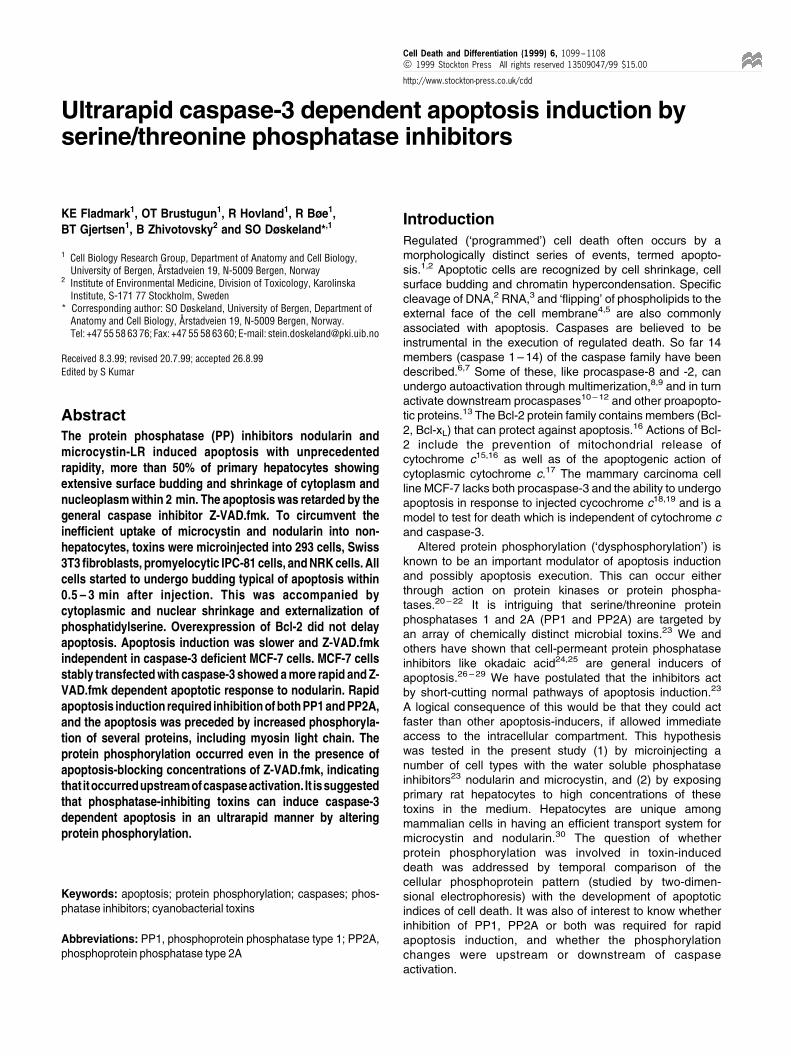
Inhibitors of protein phosphatases 2A and 1 can induceapoptotic cell death in most, if not all, higher eukaryoticcells.27,29 Studies of the rate of apoptosis induction byphosphatase inhibitors have been hampered by therelatively slow cellular uptake of the commonly usedmembrane permeant inhibitors, tautomycin, okadaic acidand calyculin A31 and by the lack of cellular uptake ofthe polar inhibitors microcystin-LR and nodularin in allcells except hepatocytes.30 In introductory experimentscells were microinjected with all the mentioned inhibitorsto test how rapidly cells could develop apoptosisfollowing abrupt inhibition of their phosphatases. Tauto-mycin, okadaic acid and calyculin A all failed to produceapoptosis even when injected at concentrations up to1 mM (data not shown). This could be due to rapid effluxor to preferential distribution to intracellular membranes ofthese hydrophobic compounds. Microinjection of eithermicrocystin-LR (not shown) or nodularin producedextensive budding and rounding of Swiss 3T3 fibro-blasts, human HEK 293 cells, and normal rat kidney(NRK) cells in a remarkably short time (Figure 1).Semilogarithmic plots allowed a quantitative estimate ofapoptosis progression as a function of time (Figures 1A ±C, 2A, 3A). The rectilinear nature of the plots showedthat the entrance of cells into apoptosis obeyed firstorder kinetics. The apparent lag period before the onsetof apoptosis was particularly short in HEK 293 cells(520 s) and fibroblasts (about 30 s). Such rapidapoptosis induction has not been reported previously.
The `flipping' of phosphatidylserine from the inner tothe outer side of the plasma membrane has beenfound to be associated with apoptosis.4,5 To test forthis phenomenon fibroblasts were injected with nodular-in while bathed in a medium containing FITC-annexinV, which binds with high affinity to phosphatidylserine.When FITC-annexin V was added to already apoptoticcells, labeling was observed after 0.5 ± 1 min, suggest-ing that external phosphatidylserine should be obser-vable within less than 1 min. Surface fluorescencecould be seen in injected cells shortly after they hadstarted budding (Figure 1A, inset). When the lag ofstaining is considered, it appeared that phosphatidylser-ine became externalized about simultaneously with cellbudding. The staining was not due to penetration ofFITC-annexin V into the cell with staining ofintracellular phosphatidylserine, since the apoptotic cellsexcluded propidium iodide. Only cells that weredeliberately injected under supraoptimal pressure toproduce necrosis stained positively with both annexinV and propidium iodide (Figure 1A).
Condensation of chromatin was judged by fluorescencemicroscopy of cells stained with bisbenzimide to visualizeDNA. In Swiss 3T3 fibroblasts, human HEK 293 cells,and normal rat kidney (NRK) cells chromatin condensa-tion occurred a few minutes after injection of nodularin(Figure 1A, inset; data not shown).
The ultrarapid apoptosis induction by phosphataseinhibitors depends on caspase-3 and is notprevented by Bcl-2 overexpression
Since only a limited number of cells can be sampled within thefirst few minutes after microinjection, biochemical studiesrequired a cell model where the toxin could act withoutmicroinjection. Ultrarapid induction of apoptosis could beachieved in hepatocytes without microinjection: nearly half ofthe hepatocytes showed apoptotic budding already after 2 minexposure to medium with 16 mM microcystin-LR (Figure 2A) or5 mM nodularin (Figure 3A). The morphological changes
Figure 1 Rate of apoptosis induction after microinjection of nodularin. Swiss3T3 fibroblasts (A), HEK 293 cells (B) or NRK fibroblasts (C) were injectedwith 250 mM nodularin and scored for apoptosis by light microscopy. Thedotted lines indicate the time when 50% of the cells had become apoptotic.Insets in (A): Combined Hoffman modulated contrast and fluorescencemicrographs of cells before injection, 120 s after injection or 150 s afterinjection. Left and lower right insets show fixed cells whose DNA was stainedwith Hoechst 33342. Upper right inset shows unfixed cells whosephosphatidylserine was stained by externally applied FITC-conjugatedAnnexin-V. Both a necrotic (n) and an apoptotic cell (a) are shown. Thenecrotic cell was deliberately produced by injection under high pressure andshows nuclear staining by propidium iodide, which did not penetrate into theapoptotic cell. Insets in (B): Hoffman modulated contrast images of HEK 293cells before and 5 min after nodularin injection. Insets in (C): Phase contrastimages of NRK cells before and 5 min after injection of nodularin. The errorbars in the main panels represent S.E.M. values from at least three separateexperiments. Further details are given in the Materials and Methods section.Bar, 10 mm
Ultrarapid apoptosis inductionKE Fladmark et al
1100
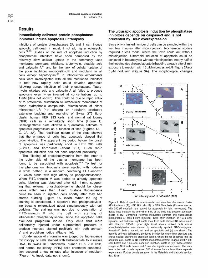
appeared to represent bona fide apoptosis. Both cytoplasmicand nuclear volume decreased by about 20% after 4 minincubation with a high concentration of microcystin (Figure2B). At this stage no buds had been cleaved off the cell, so lossof cell fragments could not account for the cytoplasmicshrinkage. The cell buds defined by light microscopy werefound to contain organelles like mitochondria by electronmicroscopy (Figure 2C). This distinguished them from `toxicblebs', which have been described as a reversible phenom-enon in cells exposed to toxic stress.32 Hypercondensation ofchromatin (Figure 2C, lowest cell) occurred 80 ± 120 min aftersurface budding and shrinkage (Figure 2D).
In order to probe for caspase involvement the oligopep-tide Z-VAD.fmk was tested for ability to block nodularin-
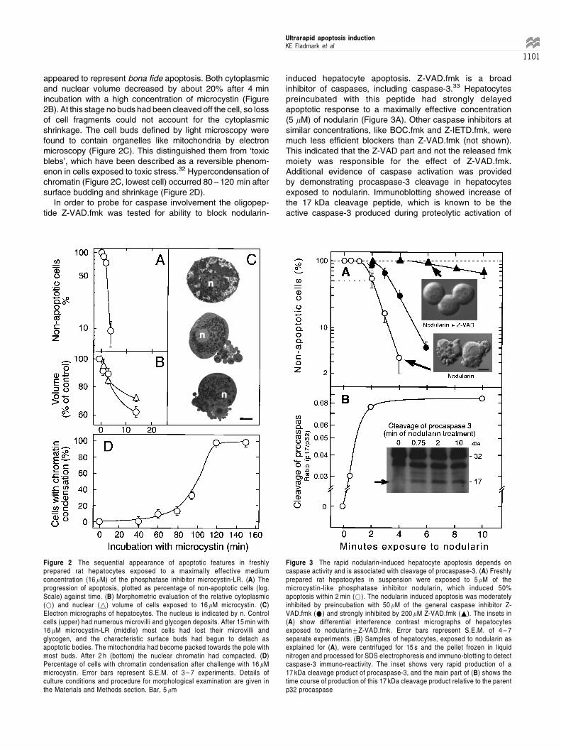
induced hepatocyte apoptosis. Z-VAD.fmk is a broadinhibitor of caspases, including caspase-3.33 Hepatocytespreincubated with this peptide had strongly delayedapoptotic response to a maximally effective concentration(5 mM) of nodularin (Figure 3A). Other caspase inhibitors atsimilar concentrations, like BOC.fmk and Z-IETD.fmk, weremuch less efficient blockers than Z-VAD.fmk (not shown).This indicated that the Z-VAD part and not the released fmkmoiety was responsible for the effect of Z-VAD.fmk.Additional evidence of caspase activation was providedby demonstrating procaspase-3 cleavage in hepatocytesexposed to nodularin. Immunoblotting showed increase ofthe 17 kDa cleavage peptide, which is known to be theactive caspase-3 produced during proteolytic activation of
Figure 2 The sequential appearance of apoptotic features in freshlyprepared rat hepatocytes exposed to a maximally effective mediumconcentration (16 mM) of the phosphatase inhibitor microcystin-LR. (A) Theprogression of apoptosis, plotted as percentage of non-apoptotic cells (log.Scale) against time. (B) Morphometric evaluation of the relative cytoplasmic(*) and nuclear (~) volume of cells exposed to 16 mM microcystin. (C)Electron micrographs of hepatocytes. The nucleus is indicated by n. Controlcells (upper) had numerous microvilli and glycogen deposits. After 15 min with16 mM microcystin-LR (middle) most cells had lost their microvilli andglycogen, and the characteristic surface buds had begun to detach asapoptotic bodies. The mitochondria had become packed towards the pole withmost buds. After 2 h (bottom) the nuclear chromatin had compacted. (D)Percentage of cells with chromatin condensation after challenge with 16 mMmicrocystin. Error bars represent S.E.M. of 3 ± 7 experiments. Details ofculture conditions and procedure for morphological examination are given inthe Materials and Methods section. Bar, 5 mm
Figure 3 The rapid nodularin-induced hepatocyte apoptosis depends oncaspase activity and is associated with cleavage of procaspase-3. (A) Freshlyprepared rat hepatocytes in suspension were exposed to 5 mM of themicrocystin-like phosphatase inhibitor nodularin, which induced 50%apoptosis within 2 min (*). The nodularin induced apoptosis was moderatelyinhibited by preincubation with 50 mM of the general caspase inhibitor Z-VAD.fmk (*) and strongly inhibited by 200 mM Z-VAD.fmk (~). The insets in(A) show differential interference contrast micrographs of hepatocytesexposed to nodularin+Z-VAD.fmk. Error bars represent S.E.M. of 4 ± 7separate experiments. (B) Samples of hepatocytes, exposed to nodularin asexplained for (A), were centrifuged for 15 s and the pellet frozen in liquidnitrogen and processed for SDS electrophoresis and immuno-blotting to detectcaspase-3 immuno-reactivity. The inset shows very rapid production of a17 kDa cleavage product of procaspase-3, and the main part of (B) shows thetime course of production of this 17 kDa cleavage product relative to the parentp32 procaspase
Ultrarapid apoptosis inductionKE Fladmark et al
1101
procaspase-3. The increase was maximal already after2 min of nodularin exposure (Figure 3B). This is anunusually rapid cleavage of procaspase-3, which isconsidered to be a `downstream' caspase, and has beenshown to be activated at the earliest 10 min after Fas-induction.18
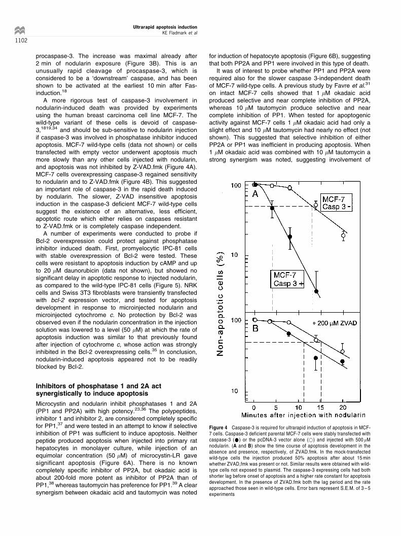
A more rigorous test of caspase-3 involvement innodularin-induced death was provided by experimentsusing the human breast carcinoma cell line MCF-7. Thewild-type variant of these cells is devoid of caspase-3,1819,34 and should be sub-sensitive to nodularin injectionif caspase-3 was involved in phosphatase inhibitor inducedapoptosis. MCF-7 wild-type cells (data not shown) or cellstransfected with empty vector underwent apoptosis muchmore slowly than any other cells injected with nodularin,and apoptosis was not inhibited by Z-VAD.fmk (Figure 4A).MCF-7 cells overexpressing caspase-3 regained sensitivityto nodularin and to Z-VAD.fmk (Figure 4B). This suggestedan important role of caspase-3 in the rapid death inducedby nodularin. The slower, Z-VAD insensitive apoptosisinduction in the caspase-3 deficient MCF-7 wild-type cellssuggest the existence of an alternative, less efficient,apoptotic route which either relies on caspases resistantto Z-VAD.fmk or is completely caspase independent.
A number of experiments were conducted to probe ifBcl-2 overexpression could protect against phosphataseinhibitor induced death. First, promyelocytic IPC-81 cellswith stable overexpression of Bcl-2 were tested. Thesecells were resistant to apoptosis induction by cAMP and upto 20 mM daunorubicin (data not shown), but showed nosignificant delay in apoptotic response to injected nodularin,as compared to the wild-type IPC-81 cells (Figure 5). NRKcells and Swiss 3T3 fibroblasts were transiently transfectedwith bcl-2 expression vector, and tested for apoptosisdevelopment in response to microinjected nodularin andmicroinjected cytochrome c. No protection by Bcl-2 wasobserved even if the nodularin concentration in the injectionsolution was lowered to a level (50 mM) at which the rate ofapoptosis induction was similar to that previously foundafter injection of cytochrome c, whose action was stronglyinhibited in the Bcl-2 overexpressing cells.35 In conclusion,nodularin-induced apoptosis appeared not to be readilyblocked by Bcl-2.
Inhibitors of phosphatase 1 and 2A actsynergistically to induce apoptosis
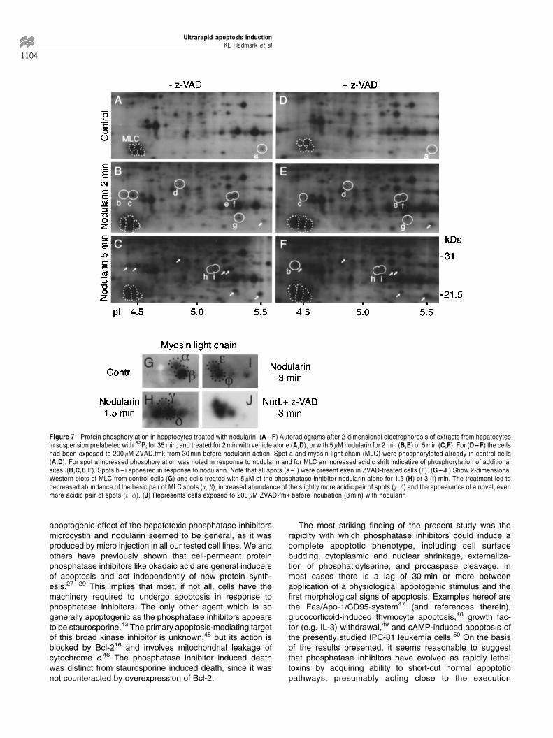
Microcystin and nodularin inhibit phosphatases 1 and 2A(PP1 and PP2A) with high potency.23,36 The polypeptides,inhibitor 1 and inhibitor 2, are considered completely specificfor PP1,37 and were tested in an attempt to know if selectiveinhibition of PP1 was sufficient to induce apoptosis. Neitherpeptide produced apoptosis when injected into primary rathepatocytes in monolayer culture, while injection of anequimolar concentration (50 mM) of microcystin-LR gavesignificant apoptosis (Figure 6A). There is no knowncompletely specific inhibitor of PP2A, but okadaic acid isabout 200-fold more potent as inhibitor of PP2A than ofPP1,38 whereas tautomycin has preference for PP1.39 A clearsynergism between okadaic acid and tautomycin was noted
for induction of hepatocyte apoptosis (Figure 6B), suggestingthat both PP2A and PP1 were involved in this type of death.
It was of interest to probe whether PP1 and PP2A wererequired also for the slower caspase 3-independent deathof MCF-7 wild-type cells. A previous study by Favre et al.31
on intact MCF-7 cells showed that 1 mM okadaic acidproduced selective and near complete inhibition of PP2A,whereas 10 mM tautomycin produce selective and nearcomplete inhibition of PP1. When tested for apoptogenicactivity against MCF-7 cells 1 mM okadaic acid had only aslight effect and 10 mM tautomycin had nearly no effect (notshown). This suggested that selective inhibition of eitherPP2A or PP1 was inefficient in producing apoptosis. When1 mM okadaic acid was combined with 10 mM tautomycin astrong synergism was noted, suggesting involvement of
Figure 4 Caspase-3 is required for ultrarapid induction of apoptosis in MCF-7 cells. Caspase-3 deficient parental MCF-7 cells were stably transfected withcaspase-3 (*) or the pcDNA-3 vector alone (*) and injected with 500 mMnodularin. (A and B) show the time course of apoptosis development in theabsence and presence, respectively, of ZVAD.fmk. In the mock-transfectedwild-type cells the injection produced 50% apoptosis after about 15 minwhether ZVAD,fmk was present or not. Similar results were obtained with wild-type cells not exposed to plasmid. The caspase-3 expressing cells had bothshorter lag before onset of apoptosis and a higher rate constant for apoptosisdevelopment. In the presence of ZVAD.fmk both the lag period and the rateapproached those seen in wild-type cells. Error bars represent S.E.M. of 3 ± 5experiments
Ultrarapid apoptosis inductionKE Fladmark et al
1102
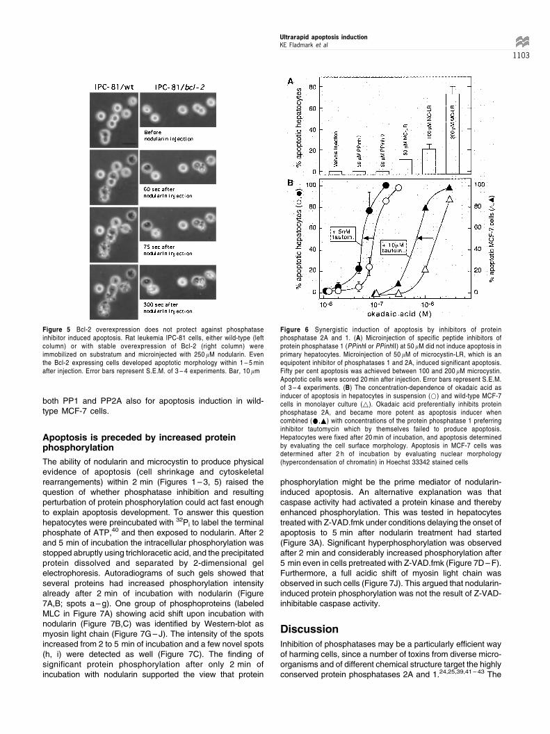
both PP1 and PP2A also for apoptosis induction in wild-type MCF-7 cells.
Apoptosis is preceded by increased proteinphosphorylation
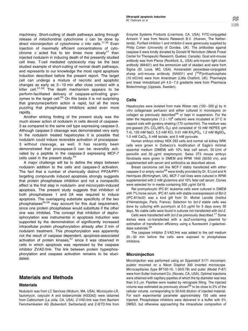
The ability of nodularin and microcystin to produce physicalevidence of apoptosis (cell shrinkage and cytoskeletalrearrangements) within 2 min (Figures 1 ± 3, 5) raised thequestion of whether phosphatase inhibition and resultingperturbation of protein phosphorylation could act fast enoughto explain apoptosis development. To answer this questionhepatocytes were preincubated with 32Pi to label the terminalphosphate of ATP,40 and then exposed to nodularin. After 2and 5 min of incubation the intracellular phosphorylation wasstopped abruptly using trichloracetic acid, and the precipitatedprotein dissolved and separated by 2-dimensional gelelectrophoresis. Autoradiograms of such gels showed thatseveral proteins had increased phosphorylation intensityalready after 2 min of incubation with nodularin (Figure7A,B; spots a ± g). One group of phosphoproteins (labeledMLC in Figure 7A) showing acid shift upon incubation withnodularin (Figure 7B,C) was identified by Western-blot asmyosin light chain (Figure 7G ± J). The intensity of the spotsincreased from 2 to 5 min of incubation and a few novel spots(h, i) were detected as well (Figure 7C). The finding ofsignificant protein phosphorylation after only 2 min ofincubation with nodularin supported the view that protein
phosphorylation might be the prime mediator of nodularin-induced apoptosis. An alternative explanation was thatcaspase activity had activated a protein kinase and therebyenhanced phosphorylation. This was tested in hepatocytestreated with Z-VAD.fmk under conditions delaying the onset ofapoptosis to 5 min after nodularin treatment had started(Figure 3A). Significant hyperphosphorylation was observedafter 2 min and considerably increased phosphorylation after5 min even in cells pretreated with Z-VAD.fmk (Figure 7D ± F).Furthermore, a full acidic shift of myosin light chain wasobserved in such cells (Figure 7J). This argued that nodularin-induced protein phosphorylation was not the result of Z-VAD-inhibitable caspase activity.
Discussion
Inhibition of phosphatases may be a particularly efficient wayof harming cells, since a number of toxins from diverse micro-organisms and of different chemical structure target the highlyconserved protein phosphatases 2A and 1.24,25,39,41 ± 43 The
Figure 5 Bcl-2 overexpression does not protect against phosphataseinhibitor induced apoptosis. Rat leukemia IPC-81 cells, either wild-type (leftcolumn) or with stable overexpression of Bcl-2 (right column) wereimmobilized on substratum and microinjected with 250 mM nodularin. Eventhe Bcl-2 expressing cells developed apoptotic morphology within 1 ± 5 minafter injection. Error bars represent S.E.M. of 3 ± 4 experiments. Bar, 10 mm
Figure 6 Synergistic induction of apoptosis by inhibitors of proteinphosphatase 2A and 1. (A) Microinjection of specific peptide inhibitors ofprotein phosphatase 1 (PPinhI or PPinhII) at 50 mM did not induce apoptosis inprimary hepatocytes. Microinjection of 50 mM of microcystin-LR, which is anequipotent inhibitor of phosphatases 1 and 2A, induced significant apoptosis.Fifty per cent apoptosis was achieved between 100 and 200 mM microcystin.Apoptotic cells were scored 20 min after injection. Error bars represent S.E.M.of 3 ± 4 experiments. (B) The concentration-dependence of okadaic acid asinducer of apoptosis in hepatocytes in suspension (*) and wild-type MCF-7cells in monolayer culture (~). Okadaic acid preferentially inhibits proteinphosphatase 2A, and became more potent as apoptosis inducer whencombined (*,~) with concentrations of the protein phosphatase 1 preferringinhibitor tautomycin which by themselves failed to produce apoptosis.Hepatocytes were fixed after 20 min of incubation, and apoptosis determinedby evaluating the cell surface morphology. Apoptosis in MCF-7 cells wasdetermined after 2 h of incubation by evaluating nuclear morphology(hypercondensation of chromatin) in Hoechst 33342 stained cells
Ultrarapid apoptosis inductionKE Fladmark et al
1103
apoptogenic effect of the hepatotoxic phosphatase inhibitorsmicrocystin and nodularin seemed to be general, as it wasproduced by micro injection in all our tested cell lines. We andothers have previously shown that cell-permeant proteinphosphatase inhibitors like okadaic acid are general inducersof apoptosis and act independently of new protein synth-esis.27 ± 29 This implies that most, if not all, cells have themachinery required to undergo apoptosis in response tophosphatase inhibitors. The only other agent which is sogenerally apoptogenic as the phosphatase inhibitors appearsto be staurosporine.43 The primary apoptosis-mediating targetof this broad kinase inhibitor is unknown,45 but its action isblocked by Bcl-216 and involves mitochondrial leakage ofcytochrome c.46 The phosphatase inhibitor induced deathwas distinct from staurosporine induced death, since it wasnot counteracted by overexpression of Bcl-2.
The most striking finding of the present study was therapidity with which phosphatase inhibitors could induce acomplete apoptotic phenotype, including cell surfacebudding, cytoplasmic and nuclear shrinkage, externaliza-tion of phosphatidylserine, and procaspase cleavage. Inmost cases there is a lag of 30 min or more betweenapplication of a physiological apoptogenic stimulus and thefirst morphological signs of apoptosis. Examples hereof arethe Fas/Apo-1/CD95-system47 (and references therein),glucocorticoid-induced thymocyte apoptosis,48 growth fac-tor (e.g. IL-3) withdrawal,49 and cAMP-induced apoptosis ofthe presently studied IPC-81 leukemia cells.50 On the basisof the results presented, it seems reasonable to suggestthat phosphatase inhibitors have evolved as rapidly lethaltoxins by acquiring ability to short-cut normal apoptoticpathways, presumably acting close to the execution
Figure 7 Protein phosphorylation in hepatocytes treated with nodularin. (A ± F) Autoradiograms after 2-dimensional electrophoresis of extracts from hepatocytesin suspension prelabeled with 32Pi for 35 min, and treated for 2 min with vehicle alone (A,D), or with 5 mM nodularin for 2 min (B,E) or 5 min (C,F). For (D ± F) the cellshad been exposed to 200 mM ZVAD.fmk from 30 min before nodularin action. Spot a and myosin light chain (MLC) were phosphorylated already in control cells(A,D). For spot a increased phosphorylation was noted in response to nodularin and for MLC an increased acidic shift indicative of phosphorylation of additionalsites. (B,C,E,F). Spots b ± i appeared in response to nodularin. Note that all spots (a ± i) were present even in ZVAD-treated cells (F). (G ± J ) Show 2-dimensionalWestern blots of MLC from control cells (G) and cells treated with 5 mM of the phosphatase inhibitor nodularin alone for 1.5 (H) or 3 (I) min. The treatment led todecreased abundance of the basic pair of MLC spots (a, b), increased abundance of the slightly more acidic pair of spots (w, d) and the appearance of a novel, evenmore acidic pair of spots (e, f). (J) Represents cells exposed to 200 mM ZVAD-fmk before incubation (3 min) with nodularin
Ultrarapid apoptosis inductionKE Fladmark et al
1104
machinery. Short-cutting of death pathways acting throughrelease of mitochondrial cytochrome c can be done bydirect microinjection of cytochrome c into cells.17,35 Eveninjection of maximally efficient concentrations of cyto-chrome c acted five to ten times more slowly17 thaninjected nodularin in inducing death of the presently studiedcell lines. T-cell mediated cytotoxicity may be the beststudied example of short-cutting of normal death pathways,and represents to our knowledge the most rapid apoptosisinduction described before the present report. The targetcell can undergo a mixture of necrotic and apoptoticchanges as early as 3 ± 10 min after close contact with akiller cell.51,52 The death mechanism appears to beperforin-facilitated delivery of caspase-activating gran-zymes to the target cell.53 On this basis it is not surprisingthat granzyme/perforin action is rapid, but all the morepuzzling that phosphatase inhibitors acted even morerapidly.
Another striking finding of the present study was themuch slower action of nodularin in cells devoid of caspase-3 as compared to the same cell type expressing caspase-3.Although caspase-3 cleavage was demonstrated very earlyin the nodularin treated hepatocytes it is possible thatnodularin could induce reversible activation of procaspase-3 without cleavage, as well. It has recently beendemonstrated that procaspase-3 can be reversibly acti-vated by a peptide in the caspase-3 expressing MCF-7cells used in the present study.54
A major challenge will be to define the steps betweennodularin addition to the cells and caspase-3 activation.The fact that a number of chemically distinct PP2A/PP1targeting compounds induced apoptosis strongly suggeststhat protein phosphatase inhibition and not a nonspecificeffect is the first step in nodularin- and microcystin-inducedapoptosis. The present study suggests that inhibition ofboth phosphatases 1 and 2A was required to induceapoptosis. The overlapping substrate specificity of the twophosphatases55,56 may account for this dual requirement,since one protein phosphatase would substitute if the otherone was inhibited. The concept that inhibition of depho-sphorylation was instrumental in apoptosis induction wassupported by the demonstration of significantly increasedintracellular protein phosphorylation already after 2 min ofnodularin treatment. This phosphorylation was apparentlynot the result of caspase dependent, apoptosis-associatedactivation of protein kinases,57 since it was observed incells in which apoptosis was repressed by the caspaseinhibitor ZVAD.fmk. The link between protein hyperphos-phorylation and caspase activation remains to be eluci-dated.
Materials and Methods
Materials
Nodularin was from LC Services (Woburn, MA, USA). Microcystin-LR,tautomycin, calyculin A and bisbenzimide (H33342) were obtainedfrom Calbiochem (La Jolla, CA, USA). Z-VAD.fmk was from BachemFeinchemikalien AG (Bubendorf, Switzerland) and Z-IETD.fmk from
Enzyme Systems Products (Livermore, CA, USA). FITC-conjugatedAnnexin V was from Nexins Research B.V. (Hoeven, The Nether-lands). Purified inhibitor-1 and inhibitor-2 were generously supplied byPhilip Cohen (University of Dundee, UK). The antibodies againstcaspase-3 were kindly donated by Donald W Nicholson (Merck FrosstCenter for Therapeutic Research, Quebec, Canada). Goat anti-mouseantibody was from Pierce (Rockford, IL, USA) anti-myosin light chainantibody (M4401) and the ammonium salt of okadaic acid were fromSigma (St. Louis, MO, USA). Horseradish peroxidase-conjugatedsheep anti-mouse antibody (NA931) and [32P]orthophosphate(10 mCi/ml) were from Amersham (Little Chalfont, UK). Pharmalyteand linear immobilized pH 4.0 ± 7.0 gradients were from PharmaciaBiotechnology (Uppsala, Sweden).
Cells
Hepatocytes were isolated from male Wistar rats (120 ± 200 g) by invitro collagenase perfusion and either cultured in monolayers oncollagen as previously described58 or kept in suspension. For thelatter the hepatocytes (1.26106 cells/ml) were incubated at 378C incapped vials with gyratory shaking (175 cycles/min). The medium waspre-gassed (5% CO2/95% O2) and consisted of 10 mM HEPES (pH7.4), 120 mM NaCl, 5.3 mM KCl, 0.01 mM KH2PO4, 1.2 mM MgSO4,1.0 mM CaCl2, 5 mM lactate, and 5 mM pyruvate.
Human embryo kidney HEK 293 cells and normal rat kidney NRKcells were grown in Dulbecco's modification of Eagle's minimalessential medium (DMEM) with 10% fetal calf serum, 50 U/ml ofpenicillin and 50 mg/ml streptomycin. Swiss 3T3 mouse embryofibroblasts were grown in DMEM and RPMI 1640 (50/50 v/v), andsupplemented with serum and antibiotics as described above.
Breast carcinoma cell line MCF-7 stably transfected with eithercaspase-3 or empty vector56 were kindly provided by Dr. S Lord and NHenriques (Birmingham, UK). MCF-7 cell lines were cultured in RPMIsupplemented with 2 mM glutamine and 10% fetal calf serum. Cloneswere selected for in media containing 500 mg/ml G418.
Rat promyelocytic IPC-81 leukemia cells were cultured in DMEMwith 7% horse serum. IPC-81 cells with stable overexpression of Bcl-2(IPC-81/bcl2) was a kind gift from Dr. Michel Lanotte (Institutd'He matologie, Paris, France). Selection for bcl-2 stable cells wasdone by culturing with puromycin at 0.5 mg/ml for 5 days every 30days. No viable cells were found in cultures not transfected with bcl-2.
Cells were transfected with bcl-2 as previously described.17 Somedishes were co-transfected with a lacZ-containing plasmid forestimation of transfection efficiency using a fluorescent b-galactosi-dase substrate.59
The caspase inhibitor Z-VAD.fmk was added to the cell medium20 ± 30 min before the cells were exposed to phosphataseinhibitors.
Microinjection
Microinjection was performed using an Eppendorf 5171 micromani-pulator mounted on a Nikon Diaphot 300 inverted microscope.Microcapillaries (type BF100-10, 1.00/0.78) and puller (Model P-87)were from Sutter Instrument Co. (Novato, CA, USA). Optimal injectionswere obtained with capillary pipettes of which the tip diameter was lessthan 0.3 mm. Pipettes were loaded by retrograde filling. The injectedvolume was estimated as previously shown60 to be close to 2% of thecellular volume, corresponding to 50-fold dilution of injected material.For each experimental parameter approximately 150 cells wereinjected. Phosphatase inhibitors were delivered in a buffer with 5%DMSO, but otherwise approaching the intracellular composition of
Ultrarapid apoptosis inductionKE Fladmark et al
1105
electrolytes.61 Inhibitor-1 was preincubated with 5 mM of the catalyticsubunit of cAMP-dependent protein kinase and Mg2+/ATP prior tomicroinjection to ensure full activation of the inhibitor.62 Additionally,cells injected with inhibitor-1 were sometimes exposed to 200 mM ofcAMP analog (8-chlorothio-cAMP) to ensure continued activation ofendogenous cAMP-dependent protein kinase. Control cells wereinjected with the relevant vehicles. In no case did injection of vehiclealone lead to significant apoptosis.
All injected cell types were cultured in 9.8 cm2 dishes with gridpatterns forming 4-mm2 squares. HEK 293, NRK, Swiss 3T3, andMCF-7 cells (10 000 cells/cm2) were injected 24 ± 48 h after seeding.Hepatocytes (30 000 cells/cm2) were injected after 44 ± 56 h ofculture. IPC-81 and IPC-81-bcl-2 cells to be injected were seeded at30 000 cells/cm2 in dishes precoated with fibronectin (1 mg/cm2), andtreated with 100 nM of the phorbol ester TPA to further improve cellattachment (for details see Vintermyr et al.63)
Scoring of apoptotic morphology
For routine assessment of apoptosis, the cell morphology wasevaluated by inverted phase microscopy using phase- and Hoffmanmodulated-optics. Apoptotic cells were easily discriminated from non-apoptotic (both normal and necrotic) cells by the appearance ofmultiple surface buds. For evaluation of the chromatin condensation,cells fixed in 0.1 M Na-cacodylate buffer (pH 7.4) with 1.5%glutaraldehyde were stained with 1 mg/ml of the DNA-specific dyeHoechst 33342 (bisbenzimide). When surface features were to beillustrated in bisbenzimide-stained cells, they were double-exposedusing both fluorescence and differential interference contrastmicroscopy. The progress of apoptosis in a cell population wasevaluated quantitatively by plotting semilogarithmically the fraction ofcells remaining non-apoptotic against time. In most cases this plotbecame rectilinear after a lag period and its slope gave the rateconstant for the transition from normal to apoptotic phenotype.
For transmission electron microscopy cell aliquots were fixed in10-fold excess of 1.5% glutaraldehyde buffered with 0.1 M Na-cacodylate (pH 7.4) and processed as described previously.27
Estimation of the nuclear and cell volume was determined onsemithin toluidine blue-stained sections using a semiautomaticimage analyzer and a primary magnification of 6306. Phosphati-dylserine was visualized by incubating cells with FITC-conjugatedAnnexin IV (according to64) Since annexin V can bind tophosphatidylserine on the internal side of the plasma membrane ifthe plasma membrane is not intact the cells were counterstainedwith 5 mg/ml propidium iodide to detect membrane leaks. The cellswere scored using a filter set for FITC-staining with a long-pass filterfor the simultaneous detection of propidium iodide. Only propidiumiodine-negative annexin V-staining cells were considered certain tohave externalized phosphatidylserine.
Labeling of cellular phosphoproteins and 2-dimensional gel electrophoresis
Suspension cultures of hepatocytes were preincubated with 32Pi
(1000 mCi/ml) for 35 min before the addition of nodularin. Theincubations were terminated by adding a tenfold excess of ice-cold8% aqueous trichloroacetic acid. Samples were spun (15 0006g for15 min) and washed in 5% aqueous trichloroacetic acid. Cell pelletswere lyzed in 100 ml of a solution containing 9.8 M urea, 100 mM DTE(1,4-dithioerythritol), 1.5% v/v Pharmalyte pH 3.5 ± 10, 0.5% v/vPharmalyte pH 5-6, 4% w/v CHAPS (3-[(3-cholamidopro-pyl)dimethylammonio]-1-propanesulfonate) and 0.2% w/v SDS. The2-dimensional sample separation was as described in,65 using a linear
immobilized pH 4.0 ± 7.0 gradient for the first dimension and 13.75%SDS ± PAGE for the second dimension.
Western blot analysis
For immunodetection (Western blot) of myosin light chain, proteinswere transferred from 2-dimensional gels by electroblotting (70 h at15 V, 48C) onto nitrocellulose membranes. Blots were incubated withanti-myosin light chain antibody followed by horseradish peroxidase-conjugated sheep anti-mouse antibody. The blots were developedusing ECL (Pharmacia, Uppsala, Sweden).
For immunodetection of caspase-3, hepatocytes (106) treated withapoptogen were rapidly centrifuged, and the pellet frozen in liquidnitrogen. The pellet was resuspended in Laemmli buffer supplementedwith a cocktail of protease inhibitors (1 mM PMSF, 1 mM 1,10-phenantroline, 20 mg/ml leupeptin, 5 mg/ml pepstatin and 20 mg/mlcalpain inhibitor I). After boiling for 4 min, the polypeptides wereresolved at 130 V on 12% SDS polyacrylamide gels. The gels weretransblotted onto nitrocellulose membranes (0.2 mm) for 2 h at 100 V.The membranes were blocked overnight in a buffer (50 mM Tris, pH7.5, 500 mM NaCl) containing 1% bovine serum albumin and 5% non-fat milk powder. They were then probed with primary antibodiesagainst p17 in blocking solution without milk, followed by 1 h withsecondary IgG (1 : 10 000 in an identical solution), and then visualizedby ECL according to the manufacturer. The ratio between the cleavageproduct (p17) and procaspase-3 (p32) was determined by computer-assisted densitometric evaluation.
AcknowledgementsWe are indebted to Nina Lied Larsen, Erna FinsaÊs, and Berit Hausvik forexpert technical assistance. The work was funded by The NorwegianCancer Society, the Norwegian Research Council, the Marine Scienceand Technology (MAST III) Program of the European Commission, andthe Novo Nordic Foundation.
References
1. Kerr JF, Wyllie AH and Currie AR (1972) Apoptosis: a basic biological
phenomenon with wide-ranging implications in tissue kinetics. Br. J. Cancer 26:
239 ± 257
2. Wyllie AH, Kerr JF and Currie AR (1980) Cell death: the significance of apoptosis.
Int. Rev. Cytol. 68: 251 ± 306
3. Houge G and Dùskeland SO (1996) Divergence towards a dead end? Cleavage
of the divergent domains of ribosomal RNA in apoptosis. Experientia 52: 963 ±
967
4. Martin SJ, Reutelingsperger CP, McGahon AJ, Rader JA, van Schie RC, LaFace
DM and Green DR (1995) Early redistribution of plasma membrane
phosphatidylserine is a general feature of apoptosis regardless of the initiating
stimulus: inhibition by overexpression of Bcl-2 and Abl. J. Exp. Med. 182: 1545 ±
1556
5. Fadok VA, Voelker DR, Campbell PA, Cohen JJ, Bratton DL and Henson PM
(1992) Exposure of phosphatidylserine on the surface of apoptotic lymphocytes
triggers specific recognition and removal by macrophages. J. Immunol. 148:
2207 ± 22166. Van de Craen M, Van Loo G, Pype S, Van Criekinge W, Van den brande I,
Molemans F, Fiers W, Declercq W and Vandenabeele P (1998) Identification of a
new caspase homologue: caspase-14. Cell Death Differ. 5: 838 ± 846
7. Thornberry NA and Lazebnik Y (1998) Caspases: enemies within. Science 281:
1312 ± 1316
8. Muzio M, Stockwell BR, Stennicke HR, Salvesen GS and Dixit VM (1998) An
induced proximity model for caspase-8 activation. J. Biol. Chem. 273: 2926 ±
2930
Ultrarapid apoptosis inductionKE Fladmark et al
1106
9. Butt JA, Harvey NL, Parasivam G and Kumar S (1998) Dimerization and
autoprocessing of the Nedd2 (caspase-2) precursor requires both the prodomain
and the carboxyl-terminal regions. J. Biol. Chem. 273: 6763 ± 6768
10. Nicholson DW and Thornberry NA (1997) Caspases: killer proteases. Trends
Biochem. Sci. 22: 299 ± 306
11. Zhivotovsky B, Burgess DH, Vanags DM and Orrenius S (1997) Involvement of
cellular proteolytic machinery in apoptosis. Biochem. Biophys. Res. Commun.
230: 481 ± 48812. Villa P, Kaufmann SH and Earnshaw WC (1997) Caspases and caspase
inhibitors. Trends Biochem. Sci. 22: 388 ± 393
13. Luo X, Budihardjo I, Zou H, Slaughter C and Wang X (1998) Bid, a Bcl2 interacting
protein, mediates cytochrome c release from mitochondria in response to
activation of cell surface receptors. Cell 94: 481 ± 490
14. Adams JM and Cory S (1998) The Bcl-2 protein family: arbiters of cell survival.
Science 281: 1322 ± 1326
15. Kluck RM, Bossy-Wetzel E, Green DR and Newmeyer DD (1997) The release of
cytochrome c from mitochondria: A primary site for Bcl-2 regulation of apoptosis.
Science 275: 1132 ± 1136
16. Yang J, Liu X, Bhalla K, Kim CN, Ibrado AM, Cai J, Peng T-I, Jones DP and Wang
X (1997) Prevention of apoptosis by Bcl-2: Release of cytochrome c from
mitochondria blocked. Science 275: 1129 ± 1132
17. Brustugun OT, Fladmark KE, Dùskeland SO, Orrenius S and Zhivotovsky B
(1998) Apoptosis induced by the microinjection of cytochrome c is caspase-
dependent and is inhibited by Bcl-2. Cell Death Differ. 5: 660 ± 668
18. Scaffidi C, Fulda S, Srinivasan A, Friese C, Li F, Tomaselli KJ, Debatin K-M,
Krammer PH and Peter ME (1998) Two CD95 (APO-1/Fas) signalling pathways.EMBO J. 17: 1675 ± 1687
19. Li F, Srinivasan A, Wang Y, Armstrong RC, Tomaselli KJ and Fritz LC (1997) Cell-
specific induction of apoptosis by microinjection of cytochrome c. Bcl-xl has
activity independent of cytochrome c release. J. Biol. Chem. 272: 30299 ± 30305
20. Anderson P (1997) Kinase cascades regulating entry into apoptosis. Microbiol.
Mol. Biol. Rev. 61: 33 ± 46
21. Martins LM, Kottke TJ, Kaufmann SH and Earnshaw WC (1998) Phosphorylated
forms of activated caspases are present in cytosol from HL-60 cells during
etoposide-induced apoptosis. Blood 92: 3042 ± 3049
22. Gjertsen BT and Dùskeland SO (1995) Protein phosphorylation in apoptosis.
Biochim. Biophys. Acta 1269: 187 ± 199
23. Holmes CFB and Boland MP (1993) Inhibitors of protein phosphatases-1 and -
2A; two of the major serine/threonine protein phosphatases involved in cellular
regulation. Curr. Opin. In Struct. Biol. 3: 934 ± 943
24. Honkanen RE, Dukelow M, Zwiller J, Moore RE, Khatra BS and Boynton AL
(1991) Cyanobacterial nodularin is a potent inhibitor of type 1 and type 2A protein
phosphatases. Mol. Pharmacol. 40: 577 ± 583
25. Cohen P, Holmes CF and Tsukitani Y (1990) Okadaic acid: a new probe for thestudy of cellular regulation. Trends Biochem. Sci. 15: 98 ± 102
26. Fladmark KE, Serres MH, Yasumoto T, Larsen NL, Aune T and Dùskeland SO
(1998) Sensitive detection of apoptogenic toxins in suspension cultures of rat
and salmon hepatocytes. Toxicon 36: 1101 ± 1114
27. Bùe R, Gjertsen B, Vintermyr O, Houge G, Lanotte M and Dùskeland S (1991)
The protein phosphatase inhibitor okadaic acid induces morphological changes
typical of apoptosis in mammalian cells. Exp. Cell Res. 195: 237 ± 246
28. Kiguchi K, Glesne D, Chubb CH, Fujiki H and Huberman E (1994) Differential
induction of apoptosis in human breast tumor cells by okadaic acid and related
inhibitors of protein phosphatases 1 and 2A. Cell Growth Differ. 5: 995 ± 1004
29. Yan Y, Shay JW, Wright WE and Mumby MC (1997) Inhibition of protein
phosphatase activity induces p53-dependent apoptosis in the absence of p53
transactivation. J. Biol. Chem. 272: 15220 ± 15226
30. Runnegar MT, Gerdes RG and Falconer IR (1991) The uptake of the
cyanobacterial hepatotoxin microcystin by isolated rat hepatocytes. Toxicon.
29: 43 ± 51
31. Favre B, Turowski P and Hemmings BA (1997) Differential inhibition and
posttranslational modification of protein phosphatase 1 and 2A in MCF7 cells
treated with calyculin-A, okadaic acid, and tautomycin. J. Biol. Chem. 272:13856 ± 13863
32. Majno G and Joris I (1995) Apoptosis, oncosis, and necrosis. An overview of cell
death. Am. J. Pathol. 146: 3 ± 15
33. Zhu H, Fearnhead HO and Cohen G (1995) An ICE-like protease is a common
mediator of apoptosis induced by diverse stimuli in human monocytic THP.1
cells. FEBS Lett. 374: 303 ± 308
34. Srinivasan A, Li F, Wong A, Kodandapani L, Smidt Jr R, Krebs JF, Fritz LC, Wu JC
and Tomaselli KJ (1998) Bcl-xL functions downstream of caspase-8 to inhibit fas-
and tumor necrosis factor receptor 1-induced apoptosis of MCF7 breast
carcinoma cells. J. Biol. Chem. 273: 4523 ± 4529
35. Zhivotovsky B, Orrenius B, Brustugun OT and Dùskeland SO (1998) Injected
cytochrome c induces apoptosis. Nature 391: 449 ± 450
36. Honkanen RE, Zwiller J, Moore RE, Daily RE, Daily SL, Khatra BS, Dukelow M
and Boynton AL (1990) Characterization of microcystin-LR, a potent inhibitor oftype 1 and type 2A protein phosphatases. J. Biol. Chem. 265: 19401 ± 19404
37. Foulkes JG, Strada SJ, Henderson PJ and Cohen P (1983) A kinetic analysis of
the effects of inhibitor-1 and inhibitor-2 on the activity of protein phosphatase-1.
Eur. J. Biochem. 132: 309 ± 313
38. Ishihara H, Martin BL, Brautigan DL, Karaki H, Ozaki H, Kato Y, Fusetani N,
Watabe S, Hashimoto K, Uemura D and Hartshorne DJ (1989) Calyculin A and
okadaic acid: inhibitors of protein phosphatase activity. Biochem. Biophys. Res.
Commun. 159: 871 ± 877
39. MacKintosh C and Klumpp S (1990) Tautomycin from the bacterium
Streptomyces verticillatus. Another potent and specific inhibitor of protein
phosphatases 1 and 2A. FEBS Lett. 277: 137 ± 140
40. Dùskeland AP, Vintermyr OK, Flatmark T, Cotton RGH and Dùskeland SO
(1992) Phenylalanine positively modulates the cAMP-dependent phosphoryla-
tion and negatively modulates the vasopressin-induced and okadaic-acid-
induced phosphorylation of phenylalanine 4 monooxygenase in intact rat
hepatocytes. Eur. J. Biochem. 206: 161 ± 170
41. Lindvall MK, Pihko PM and Koskinen AMP (1997) The binding of Calyculin A to
protein phosphatase-1. A novel spiroketal vector model. J. Biol. Chem. 272:23312 ± 23316
42. Eriksson JE, Toivola D, Meriluoto JA, Karaki H, Han YG and Hartshorne D (1990)
Hepatocyte deformation induced by cyanobacterial toxins reflects inhibition of
protein phosphatases. Biochem. Biophys. Res. Commun. 173: 1347 ± 1353
43. Matsushima R, Yoshizawa S, Watanabe MF, Harada K, Furusawa M,
Carmichael WW and Fujiki H (1990) In vitro and in vivo effects of protein
phosphatase inhibitors, microcystins and nodularin, on mouse skin and
fibroblasts. Biochem. Biophys. Res. Commun. 171: 867 ± 874
44. Bertrand R, Solary E, O'Connor P, Kohn KW and Pommier Y (1994) Induction of a
common pathway of apoptosis by staurosporine. Exp. Cell Res. 211: 314 ± 321
45. Harkin ST, Cohen GM and Gescher A (1998) Modulation of apoptosis in rat
thymocytes by analogs of staurosporine: lack of direct association with inhibition
of protein kinase. Mol. Pharmacol. 54: 663 ± 670
46. Bossy-WetzelE, NewmeyerDD andGreen DR(1998)Mitochondrial cytochrome
c release in apoptosis occurs upstream of DVED-specific caspase activation and
independently of mitochondrial transmembrane depolarization. EMBO J. 17:
37 ± 49
47. Nagata S (1997) Apoptosis by death factor. Cell 88: 355 ± 36548. Nieto MA, Gonza lez A, Gambo n F, DõÂaz-Espada F and Lo pez-Rivas A (1992)
Apoptosis in human thymocytes after treatment with glucocorticoids. Clin. Exp.
Immunol. 88: 341 ± 344
49. Collins MK, Marvel J, Malde P and Lopez Rivas A (1992) Interleukin 3 protects
murine bone marrow cells from apoptosis induced by DNA damaging agents. J.
Exp. Med. 176: 1043 ± 1051
50. Duprez E, Gjertsen BT, Bernard O, Lanotte M and Dùskeland SO (1993)
Antiapoptotic effect of heterozygously expressed mutant RI (Ala 336-Asp)
subunit of cAMP kinase I in a rat leukemia cell line. J. Biol. Chem. 268: 8332 ±
8340
51. Russell JH, Masakowski V, Rucinsky T and Phillips G (1982) Mechanisms of
immune lysis. III. Characterization of the nature and kinetics of the cytotoxic T
lymphocyte-induced nuclear lesion in the target. J. Immunol. 128: 2087 ± 2094
52. Trapani JA, Jans DA, Jans PJ, Smyth MJ, Browne KA and Sutton VR (1998)
Efficient nuclear targeting of granzyme B and the nuclear consequences of
apoptosis induced by granzyme B and perforin are caspase-dependent, but cell
death is caspase-independent. J. Biol. Chem. 273: 27934 ± 27938
53. Atkinson EA, Barry M, Darmon AJ, Shostak I, Turner PC, Moyer RW and
Bleackley RC (1998) Cytotoxic T lymphocyte-assisted suicide. Caspase 3activation is primarily the result of the direct action of granzyme B. J. Biol. Chem.
273: 21261 ± 21266
54. Buckley CD, Pilling D, Henriquez NV, Parsonage G, Threlfall K, Scheel-Toellner
D, Simmons DL, Akbar AN, Lord J and Salmon M (1999) RGD peptides induce
apoptosis by direct caspase-3 activation. Nature 397: 534 ± 539
Ultrarapid apoptosis inductionKE Fladmark et al
1107
55. Wera S and Hemmings BA (1995) Serine/threonine protein phosphatases.
Biochem. J. 311: 17 ± 29
56. Cohen PTW (1997) Novel protein serine/threonine phosphatases: variety is the
spice of life. Trends Biochem. Sci. 22: 245 ± 251
57. Bokoch GM (1998) Caspase-mediated activation of PAK2 during apoptosis:
proteolytic kinase activation as a general mechanism of apoptotic signal
transduction. Cell Death Differ. 5: 637 ± 645
58. Mellgren G, Vintermyr OK and Dùskeland SO (1995) Okadaic acid, cAMP andselected nutrients inhibit hepatocyte proliferation at different stages in G1.
Modulation of the cAMP effect by phosphatase inhibitors and nutrients. J. Cell.
Physiol. 163: 232 ± 240
59. Brustugun OT, Mellgren G, Gjertsen BT, Bjerkvig R and Dùskeland SO (1995)
Sensitive and rapid detection of b-galactosidase expression in intact cells by
microinjection of fluorescent substrate. Exp. Cell Res. 219: 372 ± 378
60. Mellgren G, Vintermyr OK, Bùe R and Dùskeland SO (1993) Hepatocyte DNA
replication is abolished by inhibitors selecting protein phosphatase 2A rather
than phosphatase 1. Exp. Cell Res. 205: 293 ± 301
61. Graessmann M and Graessmann A (1983) Microinjection of tissue culture cells.
Methods Enzymol 101: 482 ± 492
62. Cohen P (1989) The structure and regulation of protein phosphatases. Annu.
Rev. Biochem. 58: 453 ± 508
63. Vintermyr OK, Gjertsen BT, Lanotte M and Dùskeland SO (1993) Microinjected
catalytic subunit of cAMP-dependent protein kinase induces apoptosis in
myeloid leukemic (IPC-81) cells. Exp. Cell Res. 206: 157 ± 161
64. van Engeland M, Ramaekers FCS, Schutte B and Reutelingsperger CPM (1996)A novel assay to measure loss of plasma membrane asymmetry during apoptosis
of adherent cells in culture. Cytometry 24: 131 ± 139
65. Gjertsen BT, Mellgren G, Otten A, Maronde E, Genieser H-G, Jastorff B,
Vintermyr OK, McKnight GS and Dùskeland SO (1995) Novel (Rp)-cAMPS
analogs as tools for inhibition of cAMP-kinase in cell culture. J. Biol. Chem. 270:
20599 ± 20607
Ultrarapid apoptosis inductionKE Fladmark et al
1108
Related Documents