Chapter 31 / Tumor Immune Escape Mechanisms 577 From: Cancer Drug Discovery and Development: Cancer Drug Resistance Edited by: B. Teicher © Humana Press Inc., Totowa, NJ 577 Tumor Immune Escape Mechanisms Yi Ting Koh, BSc, M. Luz García-Hernández, PhD, and W. Martin Kast, PhD CONTENTS INTRODUCTION: IMMUNOSURVEILLANCE OF CANCER TUMOR ANTIGENS IMMUNODETECTION IMMUNOMODULATORY MECHANISMS TUMOR MICROENVIRONMENT ACQUIRING RESISTANCE TO DEATH EFFECTOR MECHANISMS COUNTERATTACK BY THE TUMOR CELLS ANGIOGENIC PROCESSES THAT FACILITATE TUMOR IMMUNE EVASION CONCLUSION ACKNOWLEDGMENTS REFERENCES SUMMARY The immunosurveillance theory postulates that the immune system is able to identify transformed cells and eliminate them. The theory predicts that the incidence of cancer would increase, or the latency period of cancer would decrease, in the absence of a functional immune system. However, the fact that the incidence of only some cancers increases in immunosuppressed patients shows that not all cancers abide by this theory. Most cancers escape immunosurveillance because they are fundamentally “self,” and autoreactive immune cells are usually deleted or anergized so that they do not attack self. The tumors that do face immune pressure are virus-associated cancers and cancers expressing immunogenic tumor antigens. These tumors have, however, evolved mecha- nisms to escape immune eradication. An effective way of escaping immune eradication is to prevent detection. The expression of tumor-associated antigens enhances the immunogenicity of a tumor, and if it is able to reduce the presentation of such markers, then the tumor remains relatively invisible to the immune system and escapes detection. If the tumor does not manage to escape detection, then it can evolve to prevent the activation of the immune response. The immunosuppressive effects of cancer cells are mediated by the secretion of soluble factors, by the expression of inhibitory molecules, 31

Welcome message from author
This document is posted to help you gain knowledge. Please leave a comment to let me know what you think about it! Share it to your friends and learn new things together.
Transcript
Chapter 31 / Tumor Immune Escape Mechanisms 577
From: Cancer Drug Discovery and Development: Cancer Drug ResistanceEdited by: B. Teicher © Humana Press Inc., Totowa, NJ
577
Tumor Immune Escape Mechanisms
Yi Ting Koh, BSc,M. Luz García-Hernández, PhD,and W. Martin Kast, PhD
CONTENTS
INTRODUCTION: IMMUNOSURVEILLANCE OF CANCER
TUMOR ANTIGENS
IMMUNODETECTION
IMMUNOMODULATORY MECHANISMS
TUMOR MICROENVIRONMENT
ACQUIRING RESISTANCE TO DEATH EFFECTOR MECHANISMS
COUNTERATTACK BY THE TUMOR CELLS
ANGIOGENIC PROCESSES THAT FACILITATE TUMOR IMMUNE EVASION
CONCLUSION
ACKNOWLEDGMENTS
REFERENCES
SUMMARY
The immunosurveillance theory postulates that the immune system is able to identifytransformed cells and eliminate them. The theory predicts that the incidence of cancerwould increase, or the latency period of cancer would decrease, in the absence of afunctional immune system. However, the fact that the incidence of only some cancersincreases in immunosuppressed patients shows that not all cancers abide by this theory.Most cancers escape immunosurveillance because they are fundamentally “self,” andautoreactive immune cells are usually deleted or anergized so that they do not attackself. The tumors that do face immune pressure are virus-associated cancers and cancersexpressing immunogenic tumor antigens. These tumors have, however, evolved mecha-nisms to escape immune eradication. An effective way of escaping immune eradicationis to prevent detection. The expression of tumor-associated antigens enhances theimmunogenicity of a tumor, and if it is able to reduce the presentation of such markers,then the tumor remains relatively invisible to the immune system and escapes detection.If the tumor does not manage to escape detection, then it can evolve to prevent theactivation of the immune response. The immunosuppressive effects of cancer cells aremediated by the secretion of soluble factors, by the expression of inhibitory molecules,
31
578 Koh, García-Hernández, and Kast
and by turning the cellular infiltrates into tolerizing cells that can in turn suppress otherpotentially tumor-specific immune cells. Some tumor cells have evolved to becomeresistant to the death effector mechanisms of the immune system. Finally, some tumorshave evolved to turn the immune system against itself by causing the death of theimmune cells through an activation-induced cell death mechanism that normally func-tions to limit the immune response under physiological conditions. These immuneescape mechanisms in combination make the tumor a formidable foe for the immunesystem. Therefore, a well thought out immunotherapy strategy would keep in mind theescape mechanisms the tumor could adopt under immune pressure to direct the mostpropitious strike.
Key Words: Immunosurveillance; immunogenicity; escape mechanisms; tumorantigens; immunodetection; tumor microenvironment.
1. INTRODUCTION: IMMUNOSURVEILLANCE OF CANCER
The immunosurveillance hypothesis states that a physiologic function of the immunecells is to recognize and destroy transformed cells. The concept of immunosurveillancewas first introduced in 1909, when Paul Ehrlich proposed that immunity against cancerwas mediated by “cellular forces” that kept tumors in check (1). The theory was laterappended by Thomas Lewis and Sir MacFarlane Burnet, who proposed that immunologi-cal recognition of transformed cells was a form of homeostatic surveillance that couldallow the body to guard against malignancies (2,3). Implicitly, the hypothesis predictsthat the incidence of cancer would increase or tumor latency periods would be reducedin the absence of immunosurveillance. Epstein-Barr virus (EBV)-related neoplasms areexamples of cancers usually controlled by immunosurveillance that increase in incidencein immunosuppressed individuals. EBV is a lymphotropic herpes virus that affects themajority of individuals (4), and causes little significant disease in a healthy immunocom-petent person. It establishes itself within the nucleus of B-lymphocytes expressing theCD21 molecule during the initial infection, and remains in the body in a state of latencyfor that individual’s lifetime. This latent state is associated with the production of viralproteins like Epstein-Barr nuclear antigen and latent membrane proteins that protect theB-cell from apoptosis and allow intermittent low-grade viral replication (5). The viralreplication is usually held in check by cytotoxic T-cells (CTLs) driven EBV-specificimmunosurveillance in healthy individuals. However, in the immunosuppressed trans-plant recipient, the impaired EBV-specific CTL response leads to an increase in viralreplication and ultimately, to B-cell transformation. EBV-induced posttransplantlymphoproliferative disorder (PTLD) is the most common neoplasm found in pediatricrenal transplant recipients (6). The incidence of PTLD is four times higher amongstpediatric than adult transplant recipients (7), possibly because of the fact that a largerproportion of the children are EBV naive pretransplant and therefore have little immunitytowards the virus. The significant increase in virus-associated cancers in immunocompromisedpatients, and the finding that preemptive antiviral therapy in hematopoietic stem celltransplantation prevented EBV-associated PTLD (8) supports that immunosurveillancereduces the incidence of virus-induced tumors in an immunocompetent host.
Additional evidence supporting the role of immunosurveillance comes from studies onthe incidence of neoplasms amongst transplant patients under long-term immunosup-pression and immunocompromised human immunodeficiency virus (HIV)-infectedpatients. Skin cancer was noted to be increased in patients receiving long-term immuno-
Chapter 31 / Tumor Immune Escape Mechanisms 579
suppression because of a solid-organ transplant (9); also, the risk of malignancy in renaltransplant patients receiving long-term immunosuppression was considerably higher (10).HIV-associated immunosuppression has been linked to a greater increase in cases ofKaposi sarcoma, non-Hodgkin’s lymphoma, and invasive cervical cancer (11,12). Most ofthe malignancies observed in the immunosuppressed and immunocompromised patientswere noted to be associated with viral-infections, such as B-cell lymphomas (EBV),Kaposi sarcoma (human herpes simplex virus 8) and cervical cancers (human papillomavirus). This makes the virus-associated cancers excellent targets for immunotherapy.
1.1. Most Cancers Slip Through the Immunosurveillance NetIt is important to note that the cancers that immunosuppressed patients are at an
increased risk of developing are not the same as those that are most commonly found inthe general populace. This implies that most cancers are not covered under theimmunosurveillance theory, and many cancers develop simply because the immunesystem does not recognize them as foreign in the first place. Cancer cells are basically“altered self-cells” and may not be very immunologically different from normal cells. Infact, most cancer cells escape immunosurveillance, because they simply do not satisfy theprimary condition of the immunosurveillance theory, which requires the distinction oftransformed cells from normal cells. Central and peripheral tolerance mechanisms suchas the clonal deletion by ubiquitous self-antigens and clonal inactivation by tissue-specific antigens presented in the absence of costimulatory signals ensure that the immunesystem does not attack self. Apart from the virus-associated cancers, most cancers are notimmunogenic, because the antigens that they express are self-antigens against which theimmune system has been tolerized. However, this does not mean that immunotherapycannot work on these cancers; it just has to be achieved by breaking immunologicaltolerance to self-antigens and at the cost of autoimmunity. Therefore, such immuno-therapy strategies can only be done in tissues that can be spared because all cells of thesetissues, including the nontransformed cells will be susceptible to immune destruction.
1.2. Immunotherapy—the Need to Pick the Right Target
The success and specificity of immunotherapy strategies is absolutely contingent onthe choice of the target antigen. Focusing the immune response on antigens truly uniqueto the tumor increases the specificity of the response and reduces the chances of devel-oping autoimmunity. In contrast, directing the immune attack at tumor-associated anti-gens detected in both tumor and normal cells could lead to the immune destruction of theself tissues. Naturally, there exists the probability of tumor escape by various means ofdownregulating the expression of the target antigen because of the immune pressureexerted on the tumor. Therefore, a greater number of available antigenic targets wouldallow immunotherapy strategies to cast a wider net to counteract tumor escape mecha-nisms. The academy of cancer immunology has a website that contains links to severaldatabases set up with the purpose of characterizing tumor antigens that elicit immuneresponses in humans (http://www.cancerimmunity.org/statics/databases.htm). The char-acterization and identification of novel tumor antigens is also fundamental to the designof improved therapeutic or prophylactic cancer vaccination schemes. Although mostinterest has been focused on identifying antigens that could be good targets for CD8+
CTLs that kill transformed cells expressing antigenic peptides in the context of majorhistocompatibility complex (MHC) class I molecules, currently however, efforts have
580 Koh, García-Hernández, and Kast
been turned to identify antigens recognized by CD4+ T-helper (Th) cells that enhance andamplify the immune response through costimulation and the local production of cyto-kines. A consensus exists that a combined vaccine based on CD8+ and CD4+ T-cellepitopes would improve the efficacy of therapeutic cancer vaccines substantially.
The cells within any particular tumor may contain their own individual mutations;therefore, the tumor is rather heterogeneous in its susceptibility to any sort of therapy.This accounts for the escape variants that evolve after chemotherapy or immunotherapy.To conduct immunotherapy, in addition to choosing an antigenic target that providesspecificity, the knowledge of how cancers react in response to immune pressure wouldhelp to make the proposed treatment plan more encompassing so that it does not failbecause of tumor escape variants.
2. TUMOR ANTIGENS2.1. Tumor Antigens: How to Identify the Enemy
Tumor antigens are processed and presented to the adaptive immune system as shortpeptide fragments known as epitopes on major MHC class I and MHC class II molecules.The MHC class I molecules are expressed by nearly all nucleated cells of the body, andnormally present peptides that are generated endogenously in the cells. It is imperativethat the cancer cell presents some form of immunogenic antigen in order for the CD8+
CTLs to recognize the tumor cell and destroy it. The CD8+ T-cells are the key effectorsof antitumor immunity mediated by the adaptive immune system, and they recognizeantigenic epitopes presented in the context of MHC class I molecules. CD4+ Th cells alsoplay an important role in antitumor immunity (13), as they enhance and amplify theimmune response through costimulation and the local production of cytokines. Th cellsrecognize antigens presented in the context of MHC class II molecules whose expressionis limited to professional antigen-presenting cells (APCs) such as dendritic cells (DCs).The presentation of antigenic epitopes derived from the tumor cells allows the immunesystem to distinguish between normal and transformed cells and direct the immune attackbased on these antigens. Tumor antigens can be classified into five major groups basedon their expression patterns: mutational antigens, shared tumor-specific antigens, differ-entiation antigens, overexpressed antigens, and viral antigens (14).
2.1.1. MUTATIONAL ANTIGENS
Mutational antigens are derived from ubiquitous proteins that are mutated in tumorcells. Point mutations, chromosomal translocations, deletions, or gene insertions can leadto the generation of unique tumor antigens distinct for each tumor. The mutational anti-gens are highly tumor-specific, and some may also be involved in the transformationprocess. Chronic myelogenous leukemia is characterized by the presence of Bcr-Abl, afusion product resulting from the translocation of the of cellular Abelson tyrosine kinasefrom chromosome 9 to a 5.8-kb breakpoint cluster region on chromosome 22 (15). Thedetection of Bcr-Abl junctional epitopes that bind to both MHC class I human leukocyteantigen (HLA)-A2 (16) and MHC class II DR4 (17,18) demonstrate that mutationalantigens can potentially induce potent immune responses and may be involved in thenatural antitumor response in patients. On the other side of the high specificity of muta-tional antigens is that their potential value as generic cancer vaccines in immunotherapyis limited, as such mutations may not be shared by many patients.
Chapter 31 / Tumor Immune Escape Mechanisms 581
2.1.2. SHARED TUMOR-SPECIFIC ANTIGENS
Shared tumor-specific antigens are antigens whose expression is usually silenced innormal tissues but are activated in tumors of various histological types. Expression ofthese antigens on normal tissues has only been detected on placental trophoblasts andtesticular germ cells that do not express MHC class I molecules. Hence, these antigensare usually not presented to the immune system and can be considered tumor-specific andare also known as cancer–testis antigens. The prototype shared tumor-specific antigensare the melanoma antigen genes, which are normally expressed in testis and placenta andoverexpressed in melanoma, bladder cancer, breast cancer, lung cancer, and prostatecancer (19).
2.1.3. TISSUE-SPECIFIC DIFFERENTIATION ANTIGENS
Differentiation antigens lack the specificity of tumor-specific shared antigens, as theyare differentiation markers that are expressed not just by the malignant cells, but also bythe normal cells of the same origin as the cancer cells. Tyrosinase, for example is expressedby both normal melanocytes and most melanoma cells. Targeting such antigens wouldalso result in the autoimmune destruction of the normal tissue as has been demonstratedby the vitiligo (20) induced after vaccination against tyrosinase in melanoma patients.Immunotherapy strategies based on such antigens should be reserved to tissues that arenot vital for survival, as exemplified by the targeting of the prostate-specific antigen thatcould lead to the destruction of the prostate tissues in prostate cancer.
2.1.4. OVEREXPRESSED ANTIGENS
T-cell activation is dependent on a minimum number of T-cell receptor/peptide/MHCcontacts (21); therefore, the overexpression of many proteins in cancer cells could leadto the generation of an immune response to these self-proteins. The high levels of mutantor wild-type p53 expressed in many cancers make it a potential immunotherapy target,and it has been used against colorectal cancer without inducing autoimmunity (22).However, because these overexpressed proteins are expressed by many normal tissues,it is difficult to assess the safety threshold for each antigen that does not result in wide-spread autoimmunity.
2.1.5. VIRAL ANTIGENS
Viral antigens are foreign and are only found on infected cells, thereby making themideal targets because of their high specificity. Although viruses have evolved their ownset of immune evasion strategies, immunotherapy of virus-associated cancers can bedirected against viral-antigens vital for viral replication or growth. The human papillomavirus (HPV) E6 and E7 proteins interfere with normal cell-cycle regulation (23,24) andare required for the viral life cycle (25,26). Diverse immunotherapy strategies directedagainst HPV E7 and HPV E6 (27) have led to promising results (28).
2.2. Tumor-Associated Antigens Can Induce ToleranceQualitative and quantitative changes have been observed in the glycolipids and gly-
coproteins on the cell surface of tumor cells (29,30). The cell surface location of theseantigens make them good candidates for therapeutic and diagnostic purposes, becausethey are accessible to both the cellular and humoral components of the immune system.The mucins are the most extensively studied group of glycoproteins. Mucins are large
582 Koh, García-Hernández, and Kast
glycoproteins with high-carbohydrate content expressed by a variety of normal andmalignant epithelial cells. The mucins CA-125 and CA-19-9 have been detected in ova-rian carcinomas (31,32), whereas mucin (Muc)-1 has been found in breast carcinomas(33). Under physiological conditions, Muc-1 is expressed on the apical surface of breastductal epithelium and is inaccessible to the immune system. In ductal carcinomas of thebreast however, Muc-1 looses its apical polarization and displays new carbohydrate andpeptide epitopes, thereby becoming an accessible target for the immune cells. Muc-1 canbe easily detected by monoclonal antibodies, and also contains T-cell epitopes and hasbeen used as a target for tumor vaccination schemes (34). However, it has been shownrecently that tumor-derived Muc-1 mucins were responsible for the impaired maturationand function of monocyte-derived DCs. Tumor derived-Muc-1 changed the cytokinerepertoire of the DCs and resulted in their development into interleukin (IL)-10high IL-12low regulatory APCs (35) as a novel mechanism of tumor immune evasion.
3. IMMUNODETECTION3.1. Stealth and Camouflage—Escaping Immunodetection
Two arms of the immune system work complementarily in immunodetection. Theadaptive immune system detects the presence of a transformed cell by scanning foraltered self-cells. The innate immune system detects the presence of a transformed cellby looking out for missing self. Therefore, in order to escape successfully both arms ofthe immune system, cancer cells have evolved a joint strategy of both stealth and cam-ouflage. They have to hide the tumor antigens they express and disguise themselves assomething that the body will not reject. The CTLs of the adaptive immune system rec-ognize antigens bound on MHC class I molecules expressed by nearly all nucleated cellsof the body. If the MHC class I molecule on the tumor cells presents a viral or aberrantpeptide, then the antigen-specific CTLs eliminate the tumor cell. The fetus is an allograftthat survives within the maternal host despite its low expression of allogenic MHCmolecules that would usually result in immune destruction by the natural killer (NK) cellsof the innate immune system. The same immune evasion strategies utilized by the fetus“camouflage” the cancer cells and enable them to escape the NK cells. Together, thisstealth and camouflage strategy described in the following two subheadings enables thecancer cells to evade detection.
3.1.1. EVADING THE CTLS
Tumor cells often have an altered expression pattern of class I molecules, as a consequenceof profound defects in the antigen processing pathway. This promotes poor expressionor loss of class I peptide presentation, which permits tumor cell escape from CTL killing.Different mechanisms that lead to loss of class I molecules have been described so far(36). Production of immunosuppressive molecules that downregulate the expression ofMHC class I on nucleated cells and defects in the antigen processing machinery have beenclearly demonstrated by examining tissue samples from several cancers. Recently, bymicrodissection and reverse transcription-polymerase chain reaction, a problem in thepresentation of class I peptide was detected in transformed colon cells (37,38). Profounddefects in the processing and presentation of peptides were found to be caused by anaccumulation of the HLA class I heavy chain in the cytoplasm of neoplastic cells,biallelic inactivation of the β-2 microglobulin, downregulation of the low-molecular-weight protein (LMP)7, and deregulation of the transporter associated with antigen
Chapter 31 / Tumor Immune Escape Mechanisms 583
processing (TAP) 2. All these defects allow the colon carcinoma cells to become “invis-ible” to the adaptive immune system. In addition, histological samples showed down-regulation of the proteosome multicatalytic complex subunits LMP-2 and LMP-7 inprostate and renal carcinoma, small cell lung carcinoma, and non-small cell lung cancer(39–42). All these examples indicate that class I down-regulation is an important mecha-nism of tumor escape.
3.1.2. TRICKING THE NK CELLS
Despite the reduction in MHC class I expression, tumors are still susceptible to attackfrom immune cells. Tumor cells that lack MHC class I expression are an attractive targetfor the NK cells of the innate immune system. NK cells bind to the polymorphic deter-minants of the MHC class I molecules through killer-cell inhibitory receptors (KIRs)(43). The interaction between KIRs and MHC class I molecules is inhibitory in nature,and on ligation leads to inhibition of NK-cell cytotoxicity, maintaining tolerance towardsself-tissue. The downregulation of MHC class I molecules on the cell surface of tumorcells will therefore normally lead to the NK-mediated killing of the tumor cells. Anotherway the NK cells keep track of MHC class I expression is through the heterodimer CD94-NKG2A, which recognizes nonclassical MHC class I molecules such as HLA-E (44).HLA-E presents the signal peptides from the classical MHC class I molecules (HLA-A,-B, and -C), and downregulation of any haplotype molecule in particular would normallyresult in a reduction of cell surface HLA-E and an increase of the susceptibility of tumorcells to NK-mediated killing.
In order to escape NK-mediated killing, cancer cells have evolved to establish toler-ance using similar mechanisms as those found in fetal–maternal interactions. HLA-G isa nonclassical MHC class I molecule expressed in the placenta and helps to maintaintolerance to the fetus. It is expressed by many cancers like melanoma, renal carcinoma,lung carcinoma, glioblastoma, and ovarian cancer. It is upregulated through the localexpression of environmental factors such as cytokines, stress factors, and chemothera-peutic agents (45,46). HLA-G exerts its immunoinhibitory effects through at least threeKIRs expressed by nearly all cells of the immune system (47,48), and therefore haspowerful immunosuppressive effects (49,50). In renal carcinoma, HLA-G expression ontumor cells blocks the cytolytic activity of lymphocyte activated killer cells and CTLs,promoting the evasion of the immune response (51). Soluble HLA-G has also beendetected in the plasma of patients suffering from malignant melanoma, glioma, breast,and ovarian cancer (52) and can result in local or systemic immunosuppressive effects.However, the signal peptide for HLA-G also serves as a peptide ligand for HLA-E. Theinteraction between the CD94-NKG2 and HLA-E presenting a nonamer from the theHLA-G signal peptide can lead to inhibition or activation of NK-cytotoxicity, dependingon the inhibitory or activating nature of the CD94–NKG2 heterodimer (53–55).
Stress and cellular transformation causes some malignant cells to express MHC classI chain-related (MIC) molecules and UL16-binding protein 1 that are ligands for the NK-activating NKG2D receptor (56). The triggering of NK-activating receptors can result inNK-mediated cytotoxicity of cell types that still express significant level of MHC classI molecules in vitro. NKG2D is also expressed by CTLs and results in their activationwhen triggered. To avoid being killed by NK cells, tumor cells can produce soluble MICs(sMICs) that block the activating NKG2D receptor. sMICs bind to NKG2D, inducing itsendocytosis and degradation, resulting in a reduced expression of NKG2D on tumor-infiltrating and peripheral blood T-cells in cancer patients (57). In colorectal patients,
584 Koh, García-Hernández, and Kast
NKG2D downregulation by sMICs resulted in the decreased expression of another NK-activating receptor, the natural cytotoxicity receptor, and the CXCR1 and CCR7chemokine receptors. This resulted in homing defects and inactivation of the NKG2D NKpopulation (58).
4. IMMUNOMODULATORY MECHANISMS4.1. Immunological Regulatory Processes Exploited by the Tumor CellsCancer cells are basically self-cells that are no longer regulated by normal cellular
processes and proliferate without control. These aberrant cells are predisposed to accu-mulating genetic errors that place them in a better position to adapt to changes in theirenvironment. Like organisms predicted by Darwin’s Theory of Natural Selection to adaptto the environment or suffer extinction, immune pressure selects for tumor variants thatare resistant to immune eradication. Apart from the immune evasion strategies listed,modulation of the immune response to incapacitate the antitumor response is a powerfulevolutionary adaptation of the cancer cells. Most of the immunomodulatory mechanismsfound in tumors are based on normal homeostatic control processes of the immuneresponse set in place to prevent unbridled proliferation of the immune cells, or to maintaintolerance towards self-tissues.
4.1.1. DISRUPTING CELL–CELL INTERACTION
To establish a strong and productive interaction, immune cells are required to reinforcetheir cellular communication through the induction of adhesion molecules on their sur-face. The intercellular adhesion molecule (ICAM)-1 is crucial for the formation of theimmunological synapse. ICAM-1 participates in the cell–cell interaction between the NKcell and the malignant cell. Transformed cells have been shown to disrupt this cellularinteraction by producing the matrix metalloproteinase 9, which results in ICAM-1 shed-ding and resistance to NK cell killing (59).
4.1.2. REQUIREMENT FOR A SECOND SIGNAL—A CHANCE TO TURN OFF THE IMMUNE RESPONSE
Recognition is only the first step in triggering an immune response. The productiveinteraction leading to activation requires a second costimulatory signal. The costimulatorysignal is provided by the ligation of B7.1 (CD80) or B7.2 (CD86) molecules on thesurface of APCs to CD28 on the T-cells or NK cells (60). Although CD28 plays a vitalrole in the induction of T-cell activation, other members of the CD28 family such as CTL-associated antigen (CTLA)-4, programmed death (PD)-1, and inducible costimulator(ICOS) have opposite functions. Engagement of B7 family members with CTLA-4, PD-1,and ICOS leads to inhibition instead of activation of T-cells (60). Accumulating datasuggest that CTLA-4 functions predominantly to regulate activation of naive T-cells inlymphoid organs; ICOS and PD-1 regulate activation and effector phases within andoutside lymphoid organs (61).
PD-1 is a negative regulatory receptor expressed by activated T-cells, B-cells, andmacrophages, which binds to B7-H1 or B7-DC (62,63) expressed on activated DCs, B-cellsand monocytes (64,65). B7.H1 plays an important role in the regulation of the humoraland cellular immune responses, promoting the apoptosis of activated B-cells and T-cellsthat express the ligand PD-1. B7-H1 has been detected in human lung carcinomas,ovary carcinomas, colon carcinomas, and melanomas (66). The expression of B7-H1 ontransfected P815 tumor cells increased the apoptosis of tumor-reactive T-cells and facili-

Chapter 31 / Tumor Immune Escape Mechanisms 585
tated the growth of highly immunogenic B7.1+ tumors in vivo, demonstrating its role intumor-mediated immunosuppression (66).
B7-H4 is a recently discovered B7 family member that causes detrimental effects onT-cell immunity: inhibiting T-cell proliferation, cytokine production, and cell cycle pro-gression. The expression of the putative ligand of B7-H4 is inducible on T-cells, but hasyet to be identified. B7-H4 is not expressed in normal tissues, but is constitutivelyexpressed in 85 and 31%, respectively, of ovarian cancer and lung cancer tissues (67).B7-H4 may have an important role in the immune evasion of these tumors.
4.1.3. CD40—PROVIDING A “HELPING” HAND
Most solid tumors are able to escape immunosurveillance, simply because naive T-cellsnormally circulate between the blood and the secondary lymphoid organs and do notencounter the tumor cells. Tumor-specific protective CTLs can therefore only be inducedif sufficient tumor cells reach the secondary lymphatic organs. Therefore, professionalAPCs that can prime naive T-cells within the lymphoid organs are indispensable in theactivation of natural antitumor response. Immature DCs can pick up antigens derivedfrom apoptotic cells, virus infected cells or neoplastically transformed cells and presentthem on MHC class I molecules in a process known as crosspresentation. Crosspresentationcan either activate or suppress the immune response and has been termed “crosspriming”or “crosstolerance,” respectively. Although crosspriming has been demonstrated to beinefficient and insufficient in inducing protective CTLs (68) on its own, the ability of DCsto present antigens to CD4+ Th cells through MHC class II molecules remains veryimportant, because the presence of Th cells during the priming phase of CTLs contributesignificantly to antitumor immunity. Maturation of DCs is mostly dependent on exposuresignals resulting from inflammation such as exposure to necrotic cells (69) or Toll-likereceptor signaling (70,71). CD4+ T-cells and DCs can provide reciprocal “help” to eachother. Immature DCs can present antigens on MHC class II molecules to the CD4+ T-cellsthat express CD40 ligand (CD40L). The CD40L–CD40 interaction enables the matura-tion of DCs (72). Mature DCs express high levels of costimulatory molecules that providethe costimulation needed for the naive T-cells to proliferate and differentiate. Like theCTLs, the Th cells also require costimulation in order to be fully activated. Absence ofthe second costimulatory signal can lead to a state of anergy or tolerance in the CTL andthe Th cells (73). CD40 ligation of DCs has the capacity to induce high levels of thecytokine IL-12, which polarizes CD4+ T-cells toward a Th1 type, enhances proliferationof CD8+ T-cells, and activates NK cells (74,75).
CD40 is also expressed by B-cells and rescues low-affinity antigen-binding andautoreactive B-cells in germinal centers from Fas–Fas ligand (L)-mediated apoptosis(76). The apoptotic signal is dependent on the activation of the death-inducing signalingcomplex (DISC) that can be inhibited by the Fas-associating protein with death domain-likeinterleukin 1 converting enzyme inhibitory protein (FLIP). CD40 signaling leads to thestabilization of FLIP and to the rescue of Fas-mediated apoptosis (77). CD40 has beendetected on a variety of human cancer cells, from various origins such as bladder, ovarian,colorectal, liver, lung, pancreas, prostate, cervical, and breast (78–80). It has been shown thatCD40 activation on bladder and human gastric carcinoma cells inhibits apoptosis mediatedthrough Fas using a similar mechanism to the one in the B-cell apoptosis rescue (81,82).
In addition, CD40 activation is able to induce an increase in the motility of gastriccarcinoma cells (83), and its expression has been detected in the tumor vasculature of
586 Koh, García-Hernández, and Kast
renal and breast carcinoma as well as in Kaposi’s sarcoma (84), suggesting a potentialrole of CD40 in the angiogenesis and metastasis of cancer. Elevated plasma levels ofsoluble CD40L also correlated with metastatic spread in human lung cancer (83,85). Thehigh serum levels of soluble CD40L have proangiogenic effects (86), as it can induce theincreased transcription of vascular endothelial growth factor (VEGF) by endothelial cellsexpressing CD40 (87).
5. TUMOR MICROENVIRONMENT5.1. The Effects of the Tumor Microenvironmenton the Antitumor Response and Tumor Growth
The pleiotropic effects of cytokines can function to support or suppress the immunesystem. Tumor cells have evolved to produce cytokines that suppress the immune responseand to profit from the proangiogenic effects of some cytokines. Cytokines present in themilieu when naive CD4+ T-cells are activated can skew the balance of development intoTh1 or Th2. Th1 and Th2 cytokines have reciprocal inhibition on the development of thetype of Th response. IL-12 and interferon (IFN)-γ lead to the development of Th1 cellsthat augment cell-mediated immune responses crucial for antitumor immunity. IL-4induces the development of Th2 cells (88,89), which promote humoral responses andinhibit the formation of a Th1 response.
The local production of type 1 cytokines like IFNγ, IL-2, and tumor necrosis factor(TNF)-α favor cell-mediated immunity and is important in the control of tumor growth.Tumor cells have been shown to produce (90) or to induce the production of type 2cytokines through tumor-infiltrating lymphocytes. It has also been suggested that thehypoxic conditions found around tumors may bias the immune response towards a type2 response (91). Type 2 cytokines downregulate the expression of type 1 cytokines,inactivating the cell-mediated antitumor response. Analysis of the cytokine microenvi-ronment from the fresh pleural effusions and tissue samples from several cancers hasrevealed the predominant expression of type 2 cytokines like IL-4 or immunosuppressivecytokines such as transforming growth factor (TGF)-β and IL-10 (92,93). TGF-β and IL-10 suppress the type 1 and proinflammatory responses of the immune system (94,95).
5.1.1. INTERLEUKIN 10IL-10 has potent immunosuppressive effects on APCs and effector T-cells. IL-10
reduces the expression of type 1 cytokines, inhibits antigen-specific T-cell proliferation(96), and inhibits the production of proinflammatory cytokines by macrophages (97) andAPCs (98). DCs matured in vitro in the presence of IL-10 are impaired in their ability toproduce type 1 cytokines, leading to the development of Th2 cells in vivo (99), resultingin the development of a humoral response instead of a cellular response that is morebeneficial for antitumor immunity. IL-10 has also been shown to turn DCs into tolerogenicDCs. Pretreatment of DCs with IL-10 induces an antigen-specific anergy in CTLs (100).In tumors, local production of IL-10 has also been associated with an increase in theexpression of HLA-G, resulting in the induction of tolerance towards the tumor in addi-tion to general immunosuppression (101,102). The exclusion of APCs from the tumormass has also been attributed to the local production of IL-10 (103,104).
5.1.2. TRANSFORMING GROWTH FACTOR-βTGF-β is commonly overexpressed in many cancers and has many immunosuppres-
sive effects, including the inhibition of T-cell proliferation and their development into
Chapter 31 / Tumor Immune Escape Mechanisms 587
CTLs and Th cells (105). TGF-β-overexpressing tumors are particularly aggressive, andhave been correlated with a more malignant phenotype. Apart from its role in tumor-mediated immunosuppression, TGF-β also regulates cellular proliferation, differentia-tion, extracellular matrix production, cell motility, and apoptosis (106,107). Tumor cellshave exploited the pleiotropic effects of TGF-β to its full advantage. Ras is a commonlyactivated oncogene and the cooperation of TGF-β receptor and the Ras oncogene signal-ing pathway has been implicated in the oncogenic and metastatic process in a mammaryepithelial carcinogenesis model (108–111). TGF-β has also been detected in epithelialcompartment and in tumor stroma (112,113), where it may have an important role incontrolling stromal formation within a developing tumor by increasing the synthesis ofmatrix proteins such as collagen, fibronectin, laminin, and tenascin (114). TGF-β is alsoable to induce integrins production important to mediate adhesion and cell migrationthrough the extracellular matrix and induce angiogenesis by inducing PA-1, which inhib-its the conversion of plasminogen into angiogenesis inhibitor; angiostatin (115), therebycontributing to the metastatic ability of tumor cells.
TGF-β also mediates cell cycle arrest and theoretically, should also inhibit tumorgrowth. Binding of TGF-β to the ternary TGF-β receptor complex activates a cascade ofsignal transduction pathways regulated by mothers against DPP homolog (SMAD)2,SMAD3, SMAD4, and mitogen-activated protein kinase (116,117) that negatively regu-late the transcriptional levels of c-Myc and inhibit retinoblastoma protein phosphoryla-tion (108–120), resulting in cell cycle arrest. Tumor escape from TGF-β-mediated cellcycle arrest is accounted for by point mutations, homozygous deletions, gene rearrange-ments, and aberrant transcripts in the RI and RII (121–123) of the TGF-β receptor com-plex. Deletions and mutations of components of the TGF-β receptor signaling pathwaylike SMAD3 and SMAD4 (124) have also been detected. It is yet unknown what themolecular mechanisms are that allow the tumor cells to become insensitive to TGF-β cellcycle arrest effects while remaining sensitive to its induction of migration/invasion.
5.1.3. EFFECTS OF IFN-γ ON ANTITUMOR IMMUNITY
IFN-γ is important for the generation of an effective Th1 response as well as for NKcell-mediated antitumor immunity (125). IFN-γ is a key mediator of antitumor immunity,as it is able to induce the upregulation of many genes containing the IFN responsesequence element. In addition, it has been shown to be essential to tumor rejectionmediated by both CD4+ T-cells and CD8+ T-cells through induction of angiostasis(126,127). IFN-γ exposure can sensitize breast cancer cells to apoptosis by upregulationof caspase 8 (128). Expression of the antigen presentation machinery is also regulated byIFN-γ. IFN-γ upregulates the transcription of transporter associated with antigen process-ing and the proteasome subunits low-molecular-weight protein 2 (129). Tumors maybecome unresponsive to the effects of IFN-γ through defective IFN-γ signaling, allowingthem to gain resistance to IFN-γ-mediated apoptosis (130) and maintain low MHC classI expression levels (131). In hepatocellular carcinoma, there is a correlation between thedegree of metastasis and the poor expression of IFN-γ receptor on tumor cells. In meta-static cases, the decreased expression of IFN-γ receptor on tumor cells causes a consid-erable reduction of MHC class I molecules and Fas on these cells, impairing IFN-γ controlof tumor growth (132).
IFN-γ secretion can also lead to the suppression of the immune response indirectlythrough the upregulation of IFN-γ-inducible genes. Indoleamine 2,3-dioxygenase (IDO)is an IFN-γ-inducible enzyme (133) that catabolizes tryptophan and causes proliferation
588 Koh, García-Hernández, and Kast
arrest of T-lymphocytes because of tryptophan degradation (134,135). IDO-expressingcells create a tryptophan-depleted microenvironment around themselves, as tryptophancrosses the plasma membrane readily through specific transporters to be degraded in thecytosol. Its expression by the placenta is important in the prevention of allorejection ofthe fetus by maternal T-cells (136). IDO is expressed by DCs following ligation of B7.1/B7.2 (137), and may be a mechanism by which DCs regulate T-cell responses (138).Tumor cells can express IDO, and tumor cell lines transfected with IDO in vitro suppressT-cell proliferation (139), and it has been proposed that tumor cells may be able to recruitAPCs and induce tolerogenic IDO-expressing APCs (140). These APCs would then beable to home to draining lymph nodes and tolerize naive T-cells to tumor-derived anti-gens. The discovery of accumulation of IDO-positively staining cells in immunohis-tochemistry studies of lymph nodes from melanoma patients supports this hypothesis(141). IFN-γ production is often taken as a favorable indicator in the antitumor response.In a setting where the tumor cells have evolved to become less sensitive to IFN-γ-inducedapoptosis, the IFN-γ could simply have a negative effect by inducing IDO production andtolerizing the immune system to the tumor.
Another example of the difficulty in accessing the outcome of IFN-γ production onantitumor immunity is illustrated by the interaction between IFN-γ-inducible chemokinesand inducible nitric oxide synthase (iNOS). IFN-inducible CXC chemokines are powerfulinhibitors of angiogenesis (142). Intratumoral production of IFN-inducible chemokineslike CXCL9 and CXL10 is associated with reduced angiogenesis and increased recruit-ment of CD8+ T-cells in renal carcinoma (143). IFN-γ also causes the upregulation ofiNOS that leads to the production of nitric oxide. Nitric oxide is able to upregulate theproduction of angiogenic molecules like IL-8 and VEGF, and downregulate the expres-sion of antiangiogenic chemokines like CXCL9 and CXL10 (144). In hepatocellularcarcinoma, iNOS expression was associated with increased microvascular density, resis-tance to apoptosis mediated by Bcl-2 synthesis, and cell proliferation of malignant cells(145). This illustrates the complexities in trying to predict the outcome of IFN-γ-inducibleproducts on angiogenesis and immune modulation in the tumor microenvironment.
5.1.4. CONSTITUTIVE SIGNAL TRANSDUCER AND ACTIVATOR OF TRANSCRIPTION 3 SIGNALING
Many of the cytokine-activated signaling pathways converge on the signal transducerand activator of transcription (STAT)3 signaling molecule. STAT3 is involved in theregulation of cell differentiation, survival, cytokine, and chemokine production, and isrequired for DC maturation and activation (146,147). The constitutive activation ofSTAT3 has been reported in many cancers (148–150). STAT3 signaling in tumor cellshas been shown to lead to the tumor immune evasion by inhibiting the activation ofproinflammatory cytokines and chemokines, leading to a reduction in the number ofinflammatory infiltrates like macrophages and neutrophils in the tumors (151). STAT3signaling also drives the secretion of factors that lead to the inhibition of DC maturation,thereby preventing the induction of an antitumor T-cell response (151). ConstitutiveSTAT3 activity also confers apoptosis resistance to the tumor cells (152) and upregulatesVEGF expression to stimulate tumor angiogenesis (153). Constitutive STAT3 signalingin tumors results in tumor immune evasion from both the innate and adaptive immunesystem, protects tumors from apoptosis, supports tumor growth through activation ofangiogenesis, and is a clear example of how the tumors can utilize the pleiotropic func-tions of cytokines by the simple dysregulation of a key signaling molecule involved incytokine signaling.
Chapter 31 / Tumor Immune Escape Mechanisms 589
5.2. Cellular Infiltrates in the Tumor: Allies or Enemies?The quantitative and qualitative analysis of the cellular infiltrates in the tumor mi-
croenvironment can lead to a greater understanding of the outcome of the immune modu-latory effects of the tumor. A reduction in the number of tumor-infiltrating DCs inadvanced malignancies can lead to impaired priming and generation of tumor-specificT-cells in the local environment and can be considered as mechanism of immune evasion(154). High numbers of tumor-infiltrating cells may not necessarily be a favorable indi-cator of an effective antitumor response. Some of the tumor-infiltrating cells can betolerogenic cells that can actively downregulate the cellular immune response throughthe production of immunosuppressive molecules. Among these cells, the regulatory T-cellsand NK T-cells are considered key players in the negative regulation of tumor immunitythrough their production of type 2 and immunosuppressive cytokines like IL-4, IL-10,IL-13, and TGF-β (155). Tumor-infiltrating macrophages in the Lewis lung carcinomamodel produce considerable quantities of IDO (156) that suppresses the local T-cellresponse through antigen-specific anergy.
6. ACQUIRING RESISTANCE TO DEATH EFFECTOR MECHANISMS
The immune eradication of tumor cells is mediated by apoptosis that can be inducedby the release of cytotoxic granules or death receptors. Tumors have evolved ways tobecome resistant to the death effector mechanisms, thereby becoming truly imperviousto immune attack. The perforin/granzyme and Fas/FasL pathways are the two maineffector mechanisms by which CTLs and NK cells mediate antitumor immunity (157–158).The downstream effects of both pathways are similar, as they both lead to activation ofthe caspase cascade and mitochondrial-dependent cell death. The caspases and cytochromec released from the mitochondria further synergize by enhancing each others’ activation.
6.1. The Perforin/Granzyme B Pathway
In the granule-mediated pathway, CTLs and NK cells package specialized cytotoxicgranules containing pore-forming perforins and granzymes. Perforins polymerize inresponse to calcium, and are inserted into the target cell membrane to create a channel thatresults in cellular necrosis through disruption of osmotic stability (159). In addition to thecytolytic effect of the perforins, the granzymes in the granules can also induce cellularapoptosis through the activation of caspases. Human CTLs contain five differentgranzymes that have different substrate specificities and modes of action to induce celldeath (160). Granzyme A-induced apoptosis results from single-strand DNA breaks,independent of caspase activation (161). Granzyme B is able to activate both caspase-dependent and caspase-independent pathways of cell death in the target cell (162).Caspase 3 and caspase 8 are direct substrates of granzyme B, and activation of the caspasecascade leads to apoptosis and activation of caspase-activated deoxyribonuclease (CAD)leading to DNA fragmentation (163–165). CAD is normally found in the cytpoplasm inan inactive form bound to its inhibitor ICAD. Caspase 8 cleaves ICAD to release CAD,leading to DNA fragmentation. Granzyme B can also activate the proapoptotic Bcl-2-family member, Bcl-2-interacting domain (Bid) (166,167), through cleavage. Activationof Bid leads to the oligomerization and insertion of proapoptotic Bcl-2-associated Xprotein (Bax) and Bcl-2 antagonist killer 1 into the pore and outer mitochondrial mem-brane (168,169). This eventually results in the release of cytochrome c, mitochondrial
590 Koh, García-Hernández, and Kast
collapse (170), and subsequent release of mitochondrial-derived activator of caspase thatbind to the inhibitors of apoptosis and releases the suppression on caspases for their fullactivation (171,172). The release of cytochrome c can result in the formation of anapoptosome that includes apoptotic protease-activating factor 1 and procaspase 9 (173).The apoptosome is able to activate caspase 9 (174) in the presence of andenosine triph-osphate and activate more caspase 3, augmenting caspase-mediated apoptosis. Overex-pression of a serine protease inhibitor, PI-9/SPI-6, was found in a variety of human andmurine tumors. PI-9/SPI-6 inactivates granzyme B and protects cells against CTL-mediated perforin killing (175).
6.2. Fas-Mediated Apoptosis
The interaction between the death receptor, Fas and its ligand, FasL, leads to thetrimerization of Fas to bring together death domains (DDs) in the cytoplasmic portion ofthe molecules. The DDs then recruit adaptor proteins that form a DISC capable of acti-vating initiator caspases like caspase 8 and caspase 10. Caspase 8 activation leads toactivation of CAD and activation of the mitochondrial-induced death through Bid cleav-age. Mutations in the fas gene, leading to a reduction in Fas expression, have beenreported in many cancers (176–178) as a mechanism of gaining resistance to Fas-mediatedapoptosis. Fas can also be secreted by tumor cells to bind to the FasL on tumor-specificCTLs to protect tumor cells from apoptosis (179).
Decoy receptors containing functional extracellular ligand-binding domains but lack-ing intracellular DD have been found that regulate sensitivity to death-receptor-mediatedapoptosis. DcR3 is a soluble decoy receptor secreted by tumor cells (180,181) andoverexpressed in malignant glioma, pancreatic adenocarcinoma, colon, prostate, lung,and gastrointestinal tumors (182–186). DcR3 binds to FasL and allows tumor cells to gainresistance to Fas/FasL-mediated apoptosis. DcR3 also suppresses the activation anddifferentiation of DCs (187) and macrophages (188) and downregulates T-cell prolifera-tion. The FasL signaling pathway also serves as a local chemoattractant, and the produc-tion of DcR3 results in defective homing by reducing the recruitment of microglialmacrophages, neutrophils, CD4+, and CD8+ T-cells (189,190) as a means of immuneevasion. DcR3 has proangiogenic effects and is able to promote endothelial cell prolif-eration, migration, and the expression of matrix metalloproteinases (191). Altogether, theimmunosuppressive, antiapoptotic, and angiogenic activities of DcR3 can make it animportant player in not just immune evasion but also in tumor growth.
Caspase 8 is the key initiator cell death protease in the death receptors pathway. Itsactivation is dependent on its recruitment to DISC following death receptor engagement.c-FLIP can bind DISC and prevent the activation of caspase 8 (192). c-FLIP is expressedby many cancer cells and represents yet another way by which cancer cells gain resistanceto death-receptor-mediated apoptosis (193).
6.3. Production of Antiapoptotic MoleculesTumor cells can also gain resistance to apoptosis through the production of
antiapoptotic molecules. Members of the Bcl family have either proapoptotic functionsor antiapoptotic functions and control the mitochrondrial-component of apoptosis. Bcl-2and Bcl-XL are commonly overexpressed in cancers and protect cells against apoptosisby preventing cytochrome c release (194). Survivin is involved in the downregulation ofapoptosis in malignant cells. In a prostate cancer cell line PC3, the increased production
Chapter 31 / Tumor Immune Escape Mechanisms 591
of survivin protects cells against apoptosis mediated by TNF-α by preventing the acti-vation of caspase 9 (195). Survivin was also found to cause the upregulation of FasL incolon cancer cells (196).
7. COUNTERATTACK BY THE TUMOR CELLS
Activation-induced cell death is a homeostatic mechanism that controls the magnitudeof the immune response that has been exploited by tumor cells in their counterattackagainst the immune system. Contraction of the immune response after activation iscoordinated Fas-FasL interactions that result in the death of activated cells. FasL expres-sion on tumor cells has been documented in several cancers: hilar cholangiocarcinoma(197), intrahepatic cholangiocarcinoma (198), renal cell carcinoma (199), cervicaladenocarcinoma (200), and melanoma (201). The expression of FasL on malignant cellscan lead to the in situ elimination of tumor-specific T-cells that express Fas on their cellsurface (202). TNF-related apoptosis-inducing ligand is another member of the TNFsuper-family that mediates cell death. TNF-related apoptosis-inducing ligand has beendetected in metastatic gastric carcinoma cells from malignant ascites (203), resulting inthe death of tumor-infiltrating lymphocytes that bear the counter-receptors DR4 and DR5.
Soluble FasL can be released by tumor cells systemically, inducing the death of cir-culating lymphocytes in the periphery. Astrocytomas are known to produce high levelsof soluble FasL (204), which can be cytotoxic to Fas-expressing T-cells. This particularphenomenon has also been detected in colon cancer cells that shed their membraneassociated FasL into the environment (205). Tumors can also combine death-resistancemechanisms with counterattack on the immune system. Renal carcinomas were reportedto decrease the expression of membrane-bound Fas, and secrete soluble FasL (206).
8. ANGIOGENIC PROCESSES THAT FACILITATE TUMORIMMUNE EVASION
Angiogenesis is a vital process in tumor survival. However, some of the angiogenicfactors can indirectly facilitate tumor immune evasion because of their immunosuppres-sive effects. VEGF is a key mediator in both vasculogenesis and angiogenesis (207).VEGF expression is associated with poor prognosis and increased metastatic spreadingin ovarian cancer (208). In addition, VEGF also inhibits T-cell development and contrib-utes to tumor-mediated immune suppression (209). Cyclooxygenase (COX)-2 isoverexpressed in many cancers (210,211), and is implicated in the angiogenic process(212). COX-2 contributes to the production of prostaglandins by catalyzing the oxygen-ation of arachidonic acid to the common precursor of all prostanoids. The various pros-taglandins are synthesized by distinct synthases in different tissues. The local productionof prostaglandin (PG)E2 leads to immunosuppression in the tumor microenvironmentthrough inhibition of T-cell and B-cell proliferation and diminished cytotoxicity of NKcells (213,214). PGE2 is a powerful inhibitor of TNF-α and type 1 cytokine productionand causes the downregulation of the cellular antitumor immune response. Anotherprostaglandin that can negatively affect antitumor immunity is PGD2. PGD2 is the ligandfor the PGD2 receptor expressed on effector memory Th2 cells. An increased COX-2activity and subsequent PGD2 production could promote the trafficking and activationof Th2 cells into tumor, suppressing the production of type 1 cytokines as a form of tumorimmune evasion.
592 Koh, García-Hernández, and Kast
9. CONCLUSION
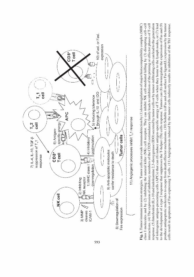
The myriad ways by which cancer cells escape immune eradication could be an indi-cation of the immune pressure it faces. Cancer cells are usually successful in escapingimmunodetection, because many of them are not particularly immunogenic. Cancerimmunotherapy is therefore most successful in situations where the immune system isable to distinguish the transformed cells from surrounding normal cells with which itshares antigens against which the immune system is tolerant. Tumor antigens thereforeserve as the first signals to alert the immune system. Vaccination schemes in cancerimmunotherapy are distinctly different from classical vaccination that is prophylactic.Cancer cells may have already modulated the immune response and therefore nullifiedthe potential therapeutic effects of a vaccine. The accumulation of data on immunogenictumor-derived antigens will increase the arsenal of targets against which efforts can bedirected. It is imperative for researchers and physicians venturing into cancer immuno-therapy to pick their targets carefully, because no immunization scheme can be successfulagainst an enemy that the immune system cannot “see.” The inability of most naiveT-cells to encounter tumor cells early enough in the blood and secondary lymphoidorgans contributes to the lack of immunosurveillance for most types of cancers. Vacci-nation allows for the activation of tumor-specific T-cells and lowers their threshold ofactivation, allowing the activated CTLs to eradicate the tumor cells despite their lowMHC class I expression. This argues for cancer immunotherapy, even for cancers that arenot covered by the immunosurveillance theory, so long as they express antigens that canbe targeted with minimal consequence of autoimmunity. The tumor environment shapedby angiogenic processes, chemokines, cytokines, and cellular infiltrates plays a hugedeterminant in the eradication of the tumor. The presence of T-lymphocytes specific fortumor antigens may not be a good enough indicator for the success of a potential vaccine.Although every tumor is different in itself, understanding the evasion strategies based ontumor type may enable us to support vaccination strategies with other immune modula-tors in order to conduct successful immunotherapies. The immune evasion strategies thattumors are able to adopt and their immunomodulatory effects as a direct consequence ofimmune pressure or as an indirect effect of angiogenesis (Fig. 1), pose as hurdles toexisting natural antitumor activity and therapeutic vaccination schemes. An effectivecancer vaccine needs to create optimal activation conditions, such as adequatecostimulation and a cytokine environment conducive for the Th1 response at the primingphase to prevent antigen-specific anergy or Th2-suppression of the Th1 response. Exist-ing tolerance will have to be broken toward antigens that the immune system is alreadytolerant. Activation of the immune system is the result of the integration of activating andinhibitory signals. Tolerance can be broken by providing “help” in the form of cytokinesand costimulation and by inhibiting tolerogenic stimuli such as immunosuppressivecytokines and inhibitory costimulation. A recent paper outlines strategies to potentiatecancer vaccines by inhibiting the immunosuppressive factors (215). Autoimmune dis-eases are the proof that low levels of autoreactive cells do exist and can turn into potentantigen-specific killers. With the appropriate adjuvants and vaccination strategies, thesecells can be unleashed against the cancer cells to eradicate these altered “self” cells. Thereis great promise for cancer immunotherapy, but there is a need to pick the right targetsand strengthen the immune attack in order to break down the tolerogenic obstacles putup by the tumor cells.
Chapter 31 / Tumor Immune Escape Mechanisms 593
Fig
. 1. T
umor
imm
une
inva
sion
str
ateg
ies.
Tum
or c
ells
can
eva
de im
mun
odet
ecti
on b
y (1
) dow
nreg
ulat
ion
of m
ajor
his
toco
mpa
tibi
lity
com
plex
(MH
C)
clas
s I
mol
ecul
es a
nd b
y (2
) in
hibi
ting
the
nat
ural
kil
ler
(NK
) ce
ll r
ecep
tor.
The
y ca
n in
hibi
t N
K c
ell-
med
iate
d ki
llin
g by
(3)
dis
rupt
ing
cell
–cel
lin
tera
ctio
ns. (
4) T
he e
xpre
ssio
n of
inhi
bito
ry m
embe
rs o
f the
CD
28 c
osti
mul
ator
y fa
mil
y le
ads
to in
hibi
tion
of t
he p
rim
ing
or e
ffec
tor p
hase
s of
T-c
ell
resp
onse
. (5)
Inte
rleu
kin
(IL
)-10
and
indo
leam
ine
2,3-
diox
ygen
ase
(ID
O) s
ecre
ted
by tu
mor
cel
ls o
r by
tum
or-i
nfil
trat
ing
cell
s can
lead
to th
e ge
nera
tion
of to
lero
geni
c an
tige
n-pr
esen
ting
cel
ls (
AP
Cs)
that
can
(6)
indu
ce a
ntig
en-s
peci
fic
aner
gy o
f T
-cel
ls w
hen
they
hom
e to
the
lym
ph n
odes
, or
(7)
lead
to th
e de
velo
pmen
t of
a ty
pe 2
res
pons
e th
at s
uppr
esse
s th
e T
-hel
per
(Th)
cel
ls r
espo
nse.
Tum
ors
can
(8)
dow
nreg
ulat
e th
e ex
pres
sion
of
Fas
and
(9)
prod
uce
anti
apop
toti
c m
olec
ules
to e
scap
e fr
om d
eath
eff
ecto
r mec
hani
sms.
(10)
Sol
uble
(s)F
as a
nd c
ell s
urfa
ce F
as li
gand
(L) e
xpre
ssio
n by
the
tum
orce
lls
resu
lt in
apo
ptos
is o
f F
as-e
xpre
ssin
g T
-cel
ls. (
11)
Ang
ioge
nesi
s in
duce
d by
the
tum
or c
ells
indi
rect
ly r
esul
ts in
inhi
biti
on o
f th
e T
h1 r
espo
nse.
593
594 Koh, García-Hernández, and Kast
ACKNOWLEDGMENTS
M. de la Luz García-Hernández, PhD is supported by the Department of Defensefellowship #PC041078. W. Martin Kast, PhD holds the Walter A. Richter CancerResearch Chair. Yi Ting Koh, BSc is supported by Department of Defense grant#DAMD 17-02-1-0244.
REFERENCES
1. Ehrlich P. Ueber den jetzigen Stand der Karzinomforschung (in German). Ned Tijdschr Geneeskd1909; 5(Pt 1):273–290.
2. Burnet FM. The concept of immunological surveillance. Prog Exp Tumor Res 1970; 13:1–27.3. Thomas L. Discussion. In: Lawrence HS, ed. Cellular and humoral aspects of the hypersensitive states.
New York: Hoeber-Harper, 1959:529–532.4. Henle W, Henle G. Epidemiologic aspects of Epstein-Barr virus (EBV)-associated diseases. Ann N Y
Acad Sci 1980; 354:326–331.5. Miyashita EM, Yang B, Lam KM, Crawford DH, Thorley-Lawson DA. A novel form of Epstein-Barr
virus latency in normal B-cells in vivo. Cell 1995; 80:593–601.6. Pinkerton CR, Hann I, Weston CL, et al. Immunodeficiency-related lymphoproliferative disorders:
prospective data from the United Kingdom Children’s Cancer Study Group Registry. Br J Haematol2002; 118:456–461.
7. Shapiro R, Nalesnik M, McCauley J, et al. Posttransplant lymphoproliferative disorders in adult andpediatric renal transplant patients receiving tacrolimus-based immunosuppression. Transplantation1999; 68:1851–1854.
8. Gruhn B, Meerbach A, Hafer R, Zell R, Wutzler P, Zintl F. Pre-emptive therapy with rituximab forprevention of Epstein-Barr virus-associated lymphoproliferative disease after hematopoietic stem celltransplantation. Bone Marrow Transplant 2003; 31:1023–1025.
9. Randle HW. The historical link between solid-organ transplantation, immunosuppression, and skincancer. Dermatol Surg 2004; 30(Pt 2):595–597.
10. Agraharkar ML, Cinclair RD, Kuo YF, Daller JA, Shahinian VB. Risk of malignancy with long-termimmunosuppression in renal transplant recipients. Kidney Int 2004; 66:383–389.
11. Baillargeon J, Pollock BH, Leach CT, Gao SJ. The association of neoplasms and HIV infection in thecorrectional setting. Int J STD AIDS 2004; 15:348–351.
12. Bellan C, De Falco G, Lazzi S, Leoncini L. Pathologic aspects of AIDS malignancies. Oncogene 2003;22:6639–6645.
13. Velders MP, Markiewicz MA, Eiben GL, Kast WM. CD4+ T cell matters in tumor immunity. Int RevImmunol 2003; 22:113–140.
14. van der Bruggen BP, Zhang Y, Chaux P, et al. Tumor-specific shared antigenic peptides recognizedby human T cells. Immunol Rev 2002; 188:51–64.
15. Rowley JD. Letter: a new consistent chromosomal abnormality in chronic myelogenous leukaemiaidentified by quinacrine fluorescence and Giemsa staining. Nature 1973; 243:290–293.
16. Yotnda P, Firat H, Garcia-Pons F, et al. Cytotoxic T cell response against the chimeric p210 BCR-ABLprotein in patients with chronic myelogenous leukemia. J Clin Invest 1998; 101:2290–2296.
17. Bosch GJ, Joosten AM, Kessler JH, Melief CJ, Leeksma OC. Recognition of BCR-ABL positiveleukemic blasts by human CD4+ T cells elicited by primary in vitro immunization with a BCR-ABLbreakpoint peptide. Blood 1996; 88:3522–3527.
18. Makita M, Azuma T, Hamaguchi H, et al. Leukemia-associated fusion proteins, dek-can and bcr-abl,represent immunogenic HLA-DR-restricted epitopes recognized by fusion peptide-specific CD4+ Tlymphocytes. Leukemia 2002; 16:2400–2407.
19. Scanlan MJ, Gure AO, Jungbluth AA, Old LJ, Chen YT. Cancer/testis antigens: an expanding familyof targets for cancer immunotherapy. Immunol Rev 2002; 188:22–32.
20. Overwijk WW, Lee DS, Surman DR, et al. Vaccination with a recombinant vaccinia virus encodinga “self” antigen induces autoimmune vitiligo and tumor cell destruction in mice: requirement forCD4(+) T lymphocytes. Proc Natl Acad Sci U S A 1999; 96:2982–2987.
21. Viola A, Lanzavecchia A. T cell activation determined by T cell receptor number and tunable thresh-olds. Science 1996; 273:104–106.
Chapter 31 / Tumor Immune Escape Mechanisms 595
22. Menon AG, Kuppen PJ, Van der Burg SH, et al. Safety of intravenous administration of a canarypoxvirus encoding the human wild-type p53 gene in colorectal cancer patients. Cancer Gene Ther 2003;10:509–517.
23. He W, Staples D, Smith C, Fisher C. Direct activation of cyclin-dependent kinase 2 by human papil-lomavirus E7. J Virol 2003; 77:10,566–10,574.
24. Mantovani F, Banks L. The human papillomavirus E6 protein and its contribution to malignant pro-gression. Oncogene 2001; 20:7874–7887.
25. Flores ER, Allen-Hoffmann BL, Lee D, Lambert PF. The human papillomavirus type 16 E7 oncogeneis required for the productive stage of the viral life cycle. J Virol 2000; 74:6622–6631.
26. McMurray HR, Nguyen D, Westbrook TF, McAnce DJ. Biology of human papillomaviruses. Int J ExpPathol 2001; 82:15–33.
27. Eiben GL, da Silva DM, Fausch SC, Le Poole IC, Nishimura MI, Kast WM. Cervical cancer vaccines:recent advances in HPV research. Viral Immunol 2003; 16:111–121.
28. Frazer IH. Prevention of cervical cancer through papillomavirus vaccination. Nat Rev Immunol 2004;4:46–54.
29. Baldus SE, Engelmann K, Hanisch FG. MUC1 and the MUCs: a family of human mucins with impactin cancer biology. Crit Rev Clin Lab Sci 2004; 41:189–231.
30. Carraway KL, Fregien N, Carraway KL, III, Carraway CA. Tumor sialomucin complexes as tumorantigens and modulators of cellular interactions and proliferation. J Cell Sci 1992; 103(Pt 2):299–307.
31. Dietel M, Arps H, Klapdor R, Muller-Hagen S, Sieck M, Hoffmann L. Antigen detection by themonoclonal antibodies CA 19-9 and CA 125 in normal and tumor tissue and patients’ sera. J CancerRes Clin Oncol 1986; 111:257–265.
32. Negishi Y, Furukawa T, Oka T, et al. Clinical use of CA 125 and its combination assay with other tumormarker in patients with ovarian carcinoma. Gynecol Obstet Invest 1987; 23:200–207.
33. Taylor-Papadimitriou J, Burchell JM, et al. MUC1 and the immunobiology of cancer. J MammaryGland Biol Neoplasia 2002; 7:209–221.
34. Finn OJ, Jerome KR, Henderson RA, et al. MUC-1 epithelial tumor mucin-based immunity and cancervaccines. Immunol Rev 1995; 145:61–89.
35. Monti P, Leone BE, Zerbi A, et al. Tumor-derived MUC1 mucins interact with differentiating mono-cytes and induce IL-10highIL-12low regulatory dendritic cell. J Immunol 2004; 172:7341–7349.
36. Garcia-Lora A, Algarra I, Collado A, Garrido F. Tumour immunology, vaccination and escape strat-egies. Eur J Immunogenet 2003; 30:177–183.
37. Cabrera CM, Jimenez P, Cabrera T, Esparza C, Ruiz-Cabello F, Garrido F. Total loss of MHC classI in colorectal tumors can be explained by two molecular pathways: β2-microglobulin inactivation inMSI-positive tumors and LMP7/TAP2 downregulation in MSI-negative tumors. Tissue Antigens2003; 61:211–219.
38. Johnsen AK, Templeton DJ, Sy M, Harding CV. Deficiency of transporter for antigen presentation(TAP) in tumor cells allows evasion of immune surveillance and increases tumorigenesis. J Immunol1999; 163:4224–4231.
39. Sanda MG, Restifo NP, Walsh JC, et al. Molecular characterization of defective antigen processing inhuman prostate cancer. J Natl Cancer Inst 1995; 87:280–285.
40. Korkolopoulou P, Kaklamanis L, Pezzella F, Harris AL, Gatter KC. Loss of antigen-presenting mol-ecules (MHC class I and TAP-1) in lung cancer. Br J Cancer 1996; 73:148–153.
41. Restifo NP, Esquivel F, Kawakami Y, et al. Identification of human cancers deficient in antigenprocessing. J Exp Med 1993; 177:265–272.
42. Seliger B, Hohne A, Jung D, et al. Expression and function of the peptide transporters in escape variantsof human renal cell carcinomas. Exp Hematol 1997; 25:608–614.
43. Boyington JC, Sun PD. A structural perspective on MHC class I recognition by killer cell immunoglo-bulin-like receptors. Mol Immunol 2002; 38:1007–1021.
44. Borrego F, Kabat J, Kim DK, et al. Structure and function of major histocompatibility complex (MHC)class I specific receptors expressed on human natural killer (NK) cells. Mol Immunol 2002; 38:637–660.
45. Rouas-Freiss N, Moreau P, Menier C, Carosella ED. HLA-G in cancer: a way to turn off the immunesystem. Semin Cancer Biol 2003; 13:325–336.
46. Ibrahim EC, Aractingi S, Allory Y, et al. Analysis of HLA antigen expression in benign and malignantmelanocytic lesions reveals that upregulation of HLA-G expression correlates with malignant trans-formation, high inflammatory infiltration and HLA-A1 genotype. Int J Cancer 2004; 108:243–250.
47. Colonna M, Samaridis J, Cella M, et al. Human myelomonocytic cells express an inhibitory receptorfor classical and nonclassical MHC class I molecules. J Immunol 1998; 160:3096–3100.
596 Koh, García-Hernández, and Kast
48. Rajagopalan S, Long EO. A human histocompatibility leukocyte antigen (HLA)-G-specific receptorexpressed on all natural killer cells. J Exp Med 1999; 189:1093–1100.
49. LeMaoult J, Krawice-Radanne I, Dausset J, Carosella ED. HLA-G1-expressing antigen-presentingcells induce immunosuppressive CD4+ T cells. Proc Natl Acad Sci U S A 2004; 101:7064–7069.
50. Rouas-Freiss N, Moreau P, Menier C, Carosella ED. HLA-G in cancer: a way to turn off the immunesystem. Semin Cancer Biol 2003; 13:325–336.
51. Bukur J, Rebmann V, Grosse-Wilde H, et al. Functional role of human leukocyte antigen-G up-regulation in renal cell carcinoma. Cancer Res 2003; 63:4107–4111.
52. Rebmann V, Regel J, Stolke D, Grosse-Wilde H. Secretion of sHLA-G molecules in malignancies.Semin Cancer Biol 2003; 13:371–377.
53. Hofmeister V, Weiss EH. HLA-G modulates immune responses by diverse receptor interactions.Semin Cancer Biol 2003; 13:317–323.
54. Llano M, Lee N, Navarro F, et al. HLA-E-bound peptides influence recognition by inhibitory andtriggering CD94/NKG2 receptors: preferential response to an HLA-G-derived nonamer. Eur J Immunol1998; 28:2854–2863.
55. Soderstrom K, Corliss B, Lanier LL, Phillips JH. CD94/NKG2 is the predominant inhibitory receptorinvolved in recognition of HLA-G by decidual and peripheral blood NK cells. J Immunol 1997;159:1072–1075.
56. Moretta L, Moretta A. Unravelling natural killer cell function: triggering and inhibitory human NKreceptors. EMBO J 2004; 23:255–259.
57. Groh V, Wu J, Yee C, Spies T. Tumour-derived soluble MIC ligands impair expression of NKG2D andT-cell activation. Nature 2002; 419:734–738.
58. Doubrovina ES, Doubrovin MM, Vider E, et al. Evasion from NK cell immunity by MHC class I chain-related molecules expressing colon adenocarcinoma. J Immunol 2003; 171:6891–6899.
59. Fiore E, Fusco C, Romero P, Stamenkovic I. Matrix metalloproteinase 9 (MMP-9/gelatinase B) pro-teolytically cleaves ICAM-1 and participates in tumor cell resistance to natural killer cell-mediatedcytotoxicity. Oncogene 2002; 21:5213–5223.
60. Abken H, Hombach A, Heuser C, Kronfeld K, Seliger B. Tuning tumor-specific T-cell activation: amatter of costimulation? Trends Immunol 2002; 23:240–245.
61. Chen L. Co-inhibitory molecules of the B7-CD28 family in the control of T-cell immunity. Nat RevImmunol 2004; 4:336–347.
62. Blank C, Brown I, Peterson AC, et al. PD-L1/B7H-1 inhibits the effector phase of tumor rejection byT cell receptor (TCR) transgenic CD8+ T cells. Cancer Res 2004; 64:1140–1145.
63. Iwai Y, Ishida M, Tanaka Y, Okazaki T, Honjo T, Minato N. Involvement of PD-L1 on tumor cells inthe escape from host immune system and tumor immunotherapy by PD-L1 blockade. Proc Natl AcadSci U S A 2002; 99:12,293–12,297.
64. Latchman Y, Wood CR, Chernova T, et al. PD-L2 is a second ligand for PD-1 and inhibits T cellactivation. Nat Immunol 2001; 2:261–268.
65. Freeman GJ, Long AJ, Iwai Y, et al. Engagement of the PD-1 immunoinhibitory receptor by a novelB7 family member leads to negative regulation of lymphocyte activation. J Exp Med 2000; 192:1027–1034.
66. Dong H, Strome SE, Salomao DR, et al. Tumor-associated B7-H1 promotes T-cell apoptosis: a poten-tial mechanism of immune evasion. Nat Med 2002; 8:793–800.
67. Choi IH, Zhu G, Sica GL, et al. Genomic organization and expression analysis of B7-H4, an immuneinhibitory molecule of the B7 family. J Immunol 2003; 171:4650–4654.
68. Ochsenbein AF, Sierro S, Odermatt B, et al. Roles of tumour localization, second signals and crosspriming in cytotoxic T-cell induction. Nature 2001; 411:1058–1064.
69. Sauter B, Albert ML, Francisco L, Larsson M, Somersan S, Bhardwaj N. Consequences of cell death:exposure to necrotic tumor cells, but not primary tissue cells or apoptotic cells, induces the maturationof immunostimulatory dendritic cells. J Exp Med 2000; 191:423–434.
70. Kadowaki N, Ho S, Antonenko S, et al. Subsets of human dendritic cell precursors express differenttoll-like receptors and respond to different microbial antigens. J Exp Med 2001; 194:863–869.
71. Schnare M, Barton GM, Holt AC, Takeda K, Akira S, Medzhitov R. Toll-like receptors control acti-vation of adaptive immune responses. Nat Immunol 2001; 2:947–950.
72. Fujii S, Liu K, Smith C, Bonito AJ, Steinman RM. The linkage of innate to adaptive immunity viamaturing dendritic cells in vivo requires CD40 ligation in addition to antigen presentation and CD80/86costimulation. J Exp Med 2004; 199:1607–1618.
73. Appleman LJ, Boussiotis VA. T cell anergy and costimulation. Immunol Rev 2003; 192:161–180.
Chapter 31 / Tumor Immune Escape Mechanisms 597
74. Cella M, Scheidegger D, Palmer-Lehmann K, Lane P, Lanzavecchia A, Alber G. Ligation of CD40 ondendritic cells triggers production of high levels of interleukin-12 and enhances T cell stimulatorycapacity: T-T help via APC activation. J Exp Med 1996; 184:747–752.
75. Lanzavecchia A, Sallusto F. Regulation of T cell immunity by dendritic cells. Cell 2001; 106:263–266.76. Liu YJ, Joshua DE, Williams GT, Smith CA, Gordon J, MacLennan IC. Mechanism of antigen-driven
selection in germinal centres. Nature 1989; 342:929–931.77. Guzman-Rojas L, Sims-Mourtada JC, Rangel R, Martinez-Valdez H. Life and death within germinal
centres: a double-edged sword. Immunology 2002; 107:167–175.78. Ciaravino G, Bhat M, Manbeian CA, Teng NN. Differential expression of CD40 and CD95 in ovarian
carcinoma. Eur J Gynaecol Oncol 2004; 25:27–32.79. Jakobson E, Jonsson G, Bjorck P, Paulie S. Stimulation of CD40 in human bladder carcinoma cells
inhibits anti-Fas/APO-1 (CD95)-induced apoptosis. Int J Cancer 1998; 77:849–853.80. Loro LL, Ohlsson M, Vintermyr OK, Liavaag PG, Jonsson R, Johannessen AC. Maintained CD40 and
loss of polarised CD40 ligand expression in oral squamous cell carcinoma. Anticancer Res 2001;21(1A):113–117.
81. Jakobson E, Jonsson G, Bjorck P, Paulie S. Stimulation of CD40 in human bladder carcinoma cellsinhibits anti-Fas/APO-1 (CD95)-induced apoptosis. Int J Cancer 1998; 77:849–853.
82. Yamaguchi H, Tanaka F, Sadanaga N, Ohta M, Inoue H, Mori M. Stimulation of CD40 inhibits Fas-or chemotherapy-mediated apoptosis and increases cell motility in human gastric carcinoma cells. IntJ Oncol 2003; 23:1697–1702.
83. Roselli M, Mineo TC, Basili S, et al. Soluble CD40 ligand plasma levels in lung cancer. Clin CancerRes 2004; 10:610–614.
84. Pammer J, Plettenberg A, Weninger W, et al. CD40 antigen is expressed by endothelial cells and tumorcells in Kaposi’s sarcoma. Am J Pathol 1996; 148:1387–1396.
85. Sabel MS, Yamada M, Kawaguchi Y, Chen FA, Takita H, Bankert RB. CD40 expression on humanlung cancer correlates with metastatic spread. Cancer Immunol Immunother 2000; 49:101–108.
86. Reinders ME, Sho M, Robertson SW, Geehan CS, Briscoe DM. Proangiogenic function of CD40ligand-CD40 interactions. J Immunol 2003; 171:1534–1541.
87. Flaxenburg JA, Melter M, Lapchak PH, Briscoe DM, Pal S. The CD40-induced signaling pathway inendothelial cells resulting in the overexpression of vascular endothelial growth factor involves Ras andphosphatidylinositol 3-kinase. J Immunol 2004; 172:7503–7509.
88. Dong C, Flavell RA. Cell fate decision: T-helper 1 and 2 subsets in immune responses. Arthritis Res2000; 2:179–188.
89. Reiner SL. Helper T cell differentiation, inside and out. Curr Opin Immunol 2001; 13:351–355.90. Clerici M, Shearer GM, Clerici E. Cytokine dysregulation in invasive cervical carcinoma and other
human neoplasias: time to consider the TH1/TH2 paradigm. J Natl Cancer Inst 1998; 90:261–263.91. Joon YA, Bazar KA, Lee PY. Tumors may modulate host immunity partly through hypoxia-induced
sympathetic bias. Med Hypotheses 2004; 63:352–356.92. Li R, Ruttinger D, Li R, Si LS, Wang YL. Analysis of the immunological microenvironment at the
tumor site in patients with non-small cell lung cancer. Langenbecks Arch Surg 2003; 388:406–412.93. Kim J, Modlin RL, Moy RL, et al. IL-10 production in cutaneous basal and squamous cell carcinomas.
A mechanism for evading the local T cell immune response. J Immunol 1995; 155:2240–2247.94. Kosiewicz MM, Alard P, Liang S, Clark SL. Mechanisms of tolerance induced by transforming growth
factor-β-treated antigen-presenting cells: CD8 regulatory T cells inhibit the effector phase of theimmune response in primed mice through a mechanism involving Fas ligand. Int Immunol 2004;16:697–706.
95. Seo N, Hayakawa S, Takigawa M, Tokura Y. Interleukin-10 expressed at early tumour sites inducessubsequent generation of CD4(+) T-regulatory cells and systemic collapse of antitumour immunity.Immunology 2001; 103:449–457.
96. Garcia-Hernandez ML, Hernandez-Pando R, Gariglio P, Berumen J. Interleukin-10 promotes B16-melanoma growth by inhibition of macrophage functions and induction of tumour and vascular cellproliferation. Immunology 2002; 105:231–243.
97. Fiorentino DF, Zlotnik A, Mosmann TR, Howard M, O’Garra A. IL-10 inhibits cytokine productionby activated macrophages. J Immunol 1991; 147:3815–3822.
98. Mitra RS, Judge TA, Nestle FO, Turka LA, Nickoloff BJ. Psoriatic skin-derived dendritic cell functionis inhibited by exogenous IL-10. Differential modulation of B7-1 (CD80) and B7-2 (CD86) expression.J Immunol 1995; 154:2668–2677.
598 Koh, García-Hernández, and Kast
99. De Smedt T, Van Mechelen M, De Becker G, Urbain J, Leo O, Moser M. Effect of interleukin-10 ondendritic cell maturation and function. Eur J Immunol 1997; 27:1229–1235.
100. Steinbrink K, Jonuleit H, Muller G, Schuler G, Knop J, Enk AH. Interleukin-10-treated human den-dritic cells induce a melanoma-antigen-specific anergy in CD8(+) T cells resulting in a failure to lysetumor cells. Blood 1999; 93:1634–1642.
101. Urosevic M, Dummer R. HLA-G and IL-10 expression in human cancer—different stories with thesame message. Semin Cancer Biol 2003; 13:337–342.
102. Mukherjee P, Ginardi AR, Madsen CS, et al. MUC1-specific CTLs are non-functional within a pan-creatic tumor microenvironment. Glycoconj J 2001; 18:931–942.
103. Garcia-Hernandez ML, Hernandez-Pando R, Gariglio P, Berumen J. Interleukin-10 promotes B16-melanoma growth by inhibition of macrophage functions and induction of tumour and vascular cellproliferation. Immunology 2002; 105:231–243.
104. Huang S, Xie K, Bucana CD, Ullrich SE, Bar-Eli M. Interleukin 10 suppresses tumor growth andmetastasis of human melanoma cells: potential inhibition of angiogenesis. Clin Cancer Res 1996;2:1969–1979.
105. Luethviksson BR, Gunnlaugsdottir B. Transforming growth factor-β as a regulator of site-specificT-cell inflammatory response. Scand J Immunol 2003; 58:129–138.
106. Roberts AB, Wakefield LM. The two faces of transforming growth factor β in carcinogenesis. Proc NatlAcad Sci U S A 2003; 100:8621–8623.
107. Wakefield LM, Roberts AB. TGF-β signaling: positive and negative effects on tumorigenesis. CurrOpin Genet Dev 2002; 12:22–29.
108. Janda E, Lehmann K, Killisch I, et al. Ras and TGFβ cooperatively regulate epithelial cell plasticityand metastasis: dissection of Ras signaling pathways. J Cell Biol 2002; 156:299–313.
109. Oft M, Peli J, Rudaz C, Schwarz H, Beug H, Reichmann E. TGF-β1 and Ha-Ras collaborate in modulat-ing the phenotypic plasticity and invasiveness of epithelial tumor cells. Genes Dev 1996; 10:2462–2477.
110. Oft M, Heider KH, Beug H. TGFβ signaling is necessary for carcinoma cell invasiveness and metas-tasis. Curr Biol 1998; 8:1243–1252.
111. Weijzen S, Velders MP, Kast WM. Modulation of the immune response and tumor growth by activatedRas. Leukemia 1999; 13:502–513.
112. Kai T, Taketazu F, Kawakami M, et al. Distribution of transforming growth factor-β and its receptorsin gastric carcinoma tissue. Jpn J Cancer Res 1996; 87:296–304.
113. Mizoi T, Ohtani H, Miyazono K, Miyazawa M, Matsuno S, Nagura H. Immunoelectron microscopiclocalization of transforming growth factor β 1 and latent transforming growth factor β 1 binding proteinin human gastrointestinal carcinomas: qualitative difference between cancer cells and stromal cells.Cancer Res 1993; 53:183–190.
114. Roberts AB. Molecular and cell biology of TGF-β. Miner Electrolyte Metab 1998; 24(2–3):111–119.115. O’Mahony CA, Albo D, Tuszynski GP, Berger DH. Transforming growth factor-β 1 inhibits generation
of angiostatin by human pancreatic cancer cells. Surgery 1998; 124:388–393.116. Tsukazaki T, Chiang TA, Davison AF, Attisano L, Wrana JL. SARA, a FYVE domain protein that
recruits Smad2 to the TGFβ receptor. Cell 1998; 95:779–791.117. Zhang Y, Feng XH, Derynck R. Smad3 and Smad4 cooperate with c-Jun/c-Fos to mediate TGF-β-
induced transcription. Nature 1998; 394:909–913.118. Ewen ME, Sluss HK, Whitehouse LL, Livingston DM. TGF β inhibition of Cdk4 synthesis is linked
to cell cycle arrest. Cell 1993; 74:1009–1020.119. Pietenpol JA, Munger K, Howley PM, Stein RW, Moses HL. Factor-binding element in the human c-
myc promoter involved in transcriptional regulation by transforming growth factor β 1 and by theretinoblastoma gene product. Proc Natl Acad Sci U S A 1991; 88:10,227–10,231.
120. Sola S, Ma X, Castro RE, Kren BT, Steer CJ, Rodrigues CM. Ursodeoxycholic acid modulates E2F-1 and p53 expression through a caspase-independent mechanism in transforming growth factorβ1-induced apoptosis of rat hepatocytes. J Biol Chem 2003; 278:48,831–48,838.
121. Chen T, Carter D, Garrigue-Antar L, Reiss M. Transforming growth factor β type I receptor kinasemutant associated with metastatic breast cancer. Cancer Res 1998; 58:4805–4810.
122. Goggins M, Shekher M, Turnacioglu K, Yeo CJ, Hruban RH, Kern SE. Genetic alterations of thetransforming growth factor β receptor genes in pancreatic and biliary adenocarcinomas. Cancer Res1998; 58:5329–5332.
123. Kim IY, Ahn HJ, Zelner DJ, et al. Genetic change in transforming growth factor β (TGF-β) receptortype I gene correlates with insensitivity to TGF-β 1 in human prostate cancer cells. Cancer Res 1996;56:44–48.
Chapter 31 / Tumor Immune Escape Mechanisms 599
124. Zhou S, Buckhaults P, Zawel L, et al. Targeted deletion of Smad4 shows it is required for transforminggrowth factor β and activin signaling in colorectal cancer cells. Proc Natl Acad Sci U S A 1998;95:2412–2416.
125. Kelly JM, Takeda K, Darcy PK, Yagita H, Smyth MJ. A role for IFN-γ in primary and secondaryimmunity generated by NK cell-sensitive tumor-expressing CD80 in vivo. J Immunol 2002;168:4472–4479.
126. Qin Z, Schwartzkopff J, Pradera F, et al. A critical requirement of interferon γ-mediated angiostasis fortumor rejection by CD8+ T cells. Cancer Res 2003; 63:4095–4100.
127. Qin Z, Blankenstein T. CD4+ T cell-mediated tumor rejection involves inhibition of angiogenesis thatis dependent on IFN γ receptor expression by nonhematopoietic cells. Immunity 2000; 12:677–686.
128. Ruiz-Ruiz C, Ruiz dA, Rodriguez A, Ortiz-Ferron G, Redondo JM, Lopez-Rivas A. The up-regulationof human caspase-8 by interferon-γ in breast tumor cells requires the induction and action of thetranscription factor interferon regulatory factor-1. J Biol Chem 2004; 279:19,712–19,720.
129. Seliger B, Hammers S, Hohne A, et al. IFN-γ-mediated coordinated transcriptional regulation of thehuman TAP-1 and LMP-2 genes in human renal cell carcinoma. Clin Cancer Res 1997; 3:573–578.
130. Liu K, Abrams SI. Coordinate regulation of IFN consensus sequence-binding protein and caspase-1 inthe sensitization of human colon carcinoma cells to Fas-mediated apoptosis by IFN-γ. J Immunol 2003;170:6329–6337.
131. Dovhey SE, Ghosh NS, Wright KL. Loss of interferon-γ inducibility of TAP1 and LMP2 in a renal cellcarcinoma cell line. Cancer Res 2000; 60:5789–5796.
132. Nagao M, Nakajima Y, Kanehiro H, et al. The impact of interferon γ receptor expression on themechanism of escape from host immune surveillance in hepatocellular carcinoma. Hepatology 2000;32:491–500.
133. Hassanain HH, Chon SY, Gupta SL. Differential regulation of human indoleamine 2,3-dioxygenasegene expression by interferons-γ and -α. Analysis of the regulatory region of the gene and identificationof an interferon-γ-inducible DNA-binding factor. J Biol Chem 1993; 268:5077–5084.
134. Terness P, Bauer TM, Rose L, et al. Inhibition of allogeneic T cell proliferation by indoleamine 2,3-dioxygenase-expressing dendritic cells: mediation of suppression by tryptophan metabolites. J ExpMed 2002; 196:447–457.
135. Fallarino F, Grohmann U, Vacca C, et al. T cell apoptosis by tryptophan catabolism. Cell Death Differ2002; 9:1069–1077.
136. Munn DH, Zhou M, Attwood JT, et al. Prevention of allogeneic fetal rejection by tryptophan catabo-lism. Science 1998; 281:1191–1193.
137. Munn DH, Sharma MD, Mellor AL. Ligation of B7-1/B7-2 by human CD4(+) T cells triggersindoleamine 2,3-dioxygenase activity in dendritic cells. J Immunol 2004; 172:4100–4110.
138. Munn DH, Sharma MD, Lee JR, et al. Potential regulatory function of human dendritic cells expressingindoleamine 2,3-dioxygenase. Science 2002; 297:1867–1870.
139. Mellor AL, Keskin DB, Johnson T, Chandler P, Munn DH. Cells expressing indoleamine 2,3-dioxygenase inhibit T cell responses. J Immunol 2002; 168:3771–3776.
140. Munn DH, Mellor AL. IDO and tolerance to tumors. Trends Mol Med 2004; 10:15–18.141. Lee JR, Dalton RR, Messina JL, et al. Pattern of recruitment of immunoregulatory antigen-presenting
cells in malignant melanoma. Lab Invest 2003; 83:1457–1466.142. Belperio JA, Keane MP, Arenberg DA, et al. CXC chemokines in angiogenesis. J Leukoc Biol 2000;
68:1–8.143. Kondo T, Ito F, Nakazawa H, Horita S, Osaka Y, Toma H. High expression of chemokine gene as a
favorable prognostic factor in renal cell carcinoma. J Urol 2004; 171(Pt 1):2171–2175.144. Hellmuth M, Paulukat J, Ninic R, Pfeilschifter J, Muhl H. Nitric oxide differentially regulates pro- and
anti-angiogenic markers in DLD-1 colon carcinoma cells. FEBS Lett 2004; 563:98–102.145. Peng JP, Zheng S, Xiao ZX, Zhang SZ. Inducible nitric oxide synthase expression is related to angio-
genesis, bcl-2 and cell proliferation in hepatocellular carcinoma. J Zhejiang Univ Sci 2003; 4:221–227.146. Levy DE, Lee CK. What does Stat3 do? J Clin Invest 2002; 109:1143–1148.147. Takeda K, Akira S. STAT family of transcription factors in cytokine-mediated biological responses.
Cytokine Growth Factor Rev 2000; 11:199–207.148. Coffer PJ, Koenderman L, de Groot RP. The role of STATs in myeloid differentiation and leukemia.
Oncogene 2000; 19:2511–2522.149. Mora LB, Buettner R, Seigne J, et al. Constitutive activation of Stat3 in human prostate tumors and cell
lines: direct inhibition of Stat3 signaling induces apoptosis of prostate cancer cells. Cancer Res 2002;62:6659–6666.
600 Koh, García-Hernández, and Kast
150. Song JI, Grandis JR. STAT signaling in head and neck cancer. Oncogene 2000; 19:2489–2495.151. Wang T, Niu G, Kortylewski M, et al. Regulation of the innate and adaptive immune responses by Stat-3
signaling in tumor cells. Nat Med 2004; 10:48–54.152. Catlett-Falcone R, Landowski TH, Oshiro MM, et al. Constitutive activation of Stat3 signaling confers
resistance to apoptosis in human U266 myeloma cells. Immunity 1999; 10:105–115.153. Niu G, Wright KL, Huang M, et al. Constitutive Stat3 activity up-regulates VEGF expression and tumor
angiogenesis. Oncogene 2002; 21:2000–2008.154. Xiang ST, Zhou SW, Guan W, et al. Tumor infiltrating dendritic cells and Mucin1 gene expression in
benign prostatic hyperplasia and prostate cancer (in Chinese). Zhonghua Nan Ke Xue 2003; 9:497–500.155. Terabe M, Berzofsky JA. Immunoregulatory T cells in tumor immunity. Curr Opin Immunol 2004;
16:157–162.156. Friberg M, Jennings R, Alsarraj M, et al. Indoleamine 2,3-dioxygenase contributes to tumor cell
evasion of T cell-mediated rejection. Int J Cancer 2002; 101:151–155.157. Kagi D, Vignaux F, Ledermann B, et al. Fas and perforin pathways as major mechanisms of T cell-
mediated cytotoxicity. Science 1994; 265:528–530.158. Smyth MJ, Thia KY, Street SE, MacGregor D, Godfrey DI, Trapani JA. Perforin-mediated cytotoxicity
is critical for surveillance of spontaneous lymphoma. J Exp Med 2000; 192:755–760.159. Trapani JA, Smyth MJ. Functional significance of the perforin/granzyme cell death pathway. Nat Rev
Immunol 2002; 2:735–747.160. Smyth MJ, Kelly JM, Sutton VR, et al. Unlocking the secrets of cytotoxic granule proteins. J Leukoc
Biol 2001; 70:18–29.161. Beresford PJ, Xia Z, Greenberg AH, Lieberman J. Granzyme A loading induces rapid cytolysis and a
novel form of DNA damage independently of caspase activation. Immunity 1999; 10:585–594.162. Trapani JA, Sutton VR. Granzyme B: pro-apoptotic, antiviral and antitumor functions. Curr Opin
Immunol 2003; 15:533–543.163. Sharif-Askari E, Alam A, Rheaume E, et al. Direct cleavage of the human DNA fragmentation factor-
45 by granzyme B induces caspase-activated DNase release and DNA fragmentation. EMBO J 2001;20:3101–3113.
164. Thomas DA, Du C, Xu M, Wang X, Ley TJ. DFF45/ICAD can be directly processed by granzyme Bduring the induction of apoptosis. Immunity 2000; 12:621–632.
165. Wolf BB, Schuler M, Echeverri F, Green DR. Caspase-3 is the primary activator of apoptotic DNAfragmentation via DNA fragmentation factor-45/inhibitor of caspase-activated DNase inactivation. JBiol Chem 1999; 274:30,651–30,656.
166. Sutton VR, Davis JE, Cancilla M, et al. Initiation of apoptosis by granzyme B requires direct cleavageof bid, but not direct granzyme B-mediated caspase activation. J Exp Med 2000; 192:1403–1414.
167. Heibein JA, Goping IS, Barry M, et al. Granzyme B-mediated cytochrome c release is regulated by theBcl-2 family members bid and Bax. J Exp Med 2000; 192:1391–1402.
168. Eskes R, Desagher S, Antonsson B, Martinou JC. Bid induces the oligomerization and insertion of Baxinto the outer mitochondrial membrane. Mol Cell Biol 2000; 20:929–935.
169. Wei MC, Lindsten T, Mootha VK, et al. tBID, a membrane-targeted death ligand, oligomerizes BAKto release cytochrome c. Genes Dev 2000; 14:2060–2071.
170. Rostovtseva TK, Antonsson B, Suzuki M, Youle RJ, Colombini M, Bezrukov SM. Bid, but not Bax,regulates VDAC channels. J Biol Chem 2004; 279:13,575–13,583.
171. Goping IS, Barry M, Liston P, et al. Granzyme B-induced apoptosis requires both direct caspaseactivation and relief of caspase inhibition. Immunity 2003; 18:355–365.
172. Verhagen AM, Ekert PG, Pakusch M, et al. Identification of DIABLO, a mammalian protein thatpromotes apoptosis by binding to and antagonizing IAP proteins. Cell 2000; 102:43–53.
173. Bleackley RC, Heibein JA. Enzymatic control of apoptosis. Nat Prod Rep 2001; 18:431–440.174. Zou H, Li Y, Liu X, Wang X. An APAF-1.cytochrome c multimeric complex is a functional apoptosome
that activates procaspase-9. J Biol Chem 1999; 274:11,549–11,556.175. Medema JP, de Jong J, Peltenburg LT, et al. Blockade of the granzyme B/perforin pathway through
overexpression of the serine protease inhibitor PI-9/SPI-6 constitutes a mechanism for immune escapeby tumors. Proc Natl Acad Sci U S A 2001; 98:11,515–11,520.
176. Lee SH, Shin MS, Park WS, et al. Alterations of Fas (APO-1/CD95) gene in transitional cell carcinomasof urinary bladder. Cancer Res 1999; 59:3068–3072.
177. Lee SH, Shin MS, Park WS, et al. Alterations of Fas (Apo-1/CD95) gene in non-small cell lung cancer.Oncogene 1999; 18:3754–3760.
Chapter 31 / Tumor Immune Escape Mechanisms 601
178. Shin MS, Park WS, Kim SY, et al. Alterations of Fas (Apo-1/CD95) gene in cutaneous malignantmelanoma. Am J Pathol 1999; 154:1785–1791.
179. Maas S, Warskulat U, Steinhoff C, et al. Decreased Fas expression in advanced-stage bladder canceris not related to p53 status. Urology 2004; 63:392–397.
180. Pitti RM, Marsters SA, Lawrence DA, et al. Genomic amplification of a decoy receptor for Fas ligandin lung and colon cancer. Nature 1998; 396:699–703.
181. Ashkenazi A, Dixit VM. Apoptosis control by death and decoy receptors. Curr Opin Cell Biol 1999;11:255–260.
182. Bai C, Connolly B, Metzker ML, et al. Overexpression of M68/DcR3 in human gastrointestinal tracttumors independent of gene amplification and its location in a four-gene cluster. Proc Natl Acad SciU S A 2000; 97:1230–1235.
183. Pitti RM, Marsters SA, Lawrence DA, et al. Genomic amplification of a decoy receptor for Fas ligandin lung and colon cancer. Nature 1998; 396:699–703.
184. Roth W, Isenmann S, Nakamura M, Platten M, et al. Soluble decoy receptor 3 is expressed by malignantgliomas and suppresses CD95 ligand-induced apoptosis and chemotaxis. Cancer Res 2001; 61:2759–2765.
185. Takahama Y, Yamada Y, Emoto K, et al. The prognostic significance of overexpression of the decoyreceptor for Fas ligand (DcR3) in patients with gastric carcinomas. Gastric Cancer 2002; 5:61–68.
186. Tsuji S, Hosotani R, Yonehara S, et al. Endogenous decoy receptor 3 blocks the growth inhibitionsignals mediated by Fas ligand in human pancreatic adenocarcinoma. Int J Cancer 2003; 106:17–25.
187. Hsu TL, Chang YC, Chen SJ, et al. Modulation of dendritic cell differentiation and maturation by decoyreceptor 3. J Immunol 2002; 168:4846–4853.
188. Chang YC, Hsu TL, Lin HH, et al. Modulation of macrophage differentiation and activation by decoyreceptor 3. J Leukoc Biol 2004; 75:486–494.
189. Roth W, Isenmann S, Nakamura M, et al. Soluble decoy receptor 3 is expressed by malignant gliomasand suppresses CD95 ligand-induced apoptosis and chemotaxis. Cancer Res 2001; 61:2759–2765.
190. Chen YL, Chen SH, Wang JY, Yang BC. Fas ligand on tumor cells mediates inactivation of neutrophils.J Immunol 2003; 171:1183–1191.
191. Yang CR, Hsieh SL, Teng CM, Ho FM, Su WL, Lin WW. Soluble decoy receptor 3 induces angiogen-esis by neutralization of TL1A, a cytokine belonging to tumor necrosis factor superfamily and exhib-iting angiostatic action. Cancer Res 2004; 64:1122–1129.
192. MacFarlane M, Harper N, Snowden RT, et al. Mechanisms of resistance to TRAIL-induced apoptosisin primary B cell chronic lymphocytic leukaemia. Oncogene 2002; 21:6809–6818.
193. Dutton A, O’Neil JD, Milner AE, et al. Expression of the cellular FLICE-inhibitory protein (c-FLIP)protects Hodgkin’s lymphoma cells from autonomous Fas-mediated death. Proc Natl Acad Sci U S A2004; 101:6611–6616.
194. Yi X, Yin XM, Dong Z. Inhibition of Bid-induced apoptosis by Bcl-2. tBid insertion, Bax translocation,and Bax/Bak oligomerization suppressed. J Biol Chem 2003; 278:16,992–16,999.
195. Fornaro M, Plescia J, Chheang S, et al. Fibronectin protects prostate cancer cells from tumor necrosisfactor-α-induced apoptosis via the AKT/survivin pathway. J Biol Chem 2003; 278:50,402–50,411.
196. Asanuma K, Tsuji N, Endoh T, Yagihashi A, Watanabe N. Survivin enhances Fas ligand expressionvia up-regulation of specificity protein 1-mediated gene transcription in colon cancer cells. J Immunol2004; 172:3922–3929.
197. Li ZY, Zou SQ. Fas counterattack in cholangiocarcinoma: a mechanism for immune evasion in humanhilar cholangiocarcinomas. World J Gastroenterol 2001; 7:860–863.
198. Shimonishi T, Isse K, Shibata F, et al. Up-regulation of fas ligand at early stages and down-regulationof Fas at progressed stages of intrahepatic cholangiocarcinoma reflect evasion from immune surveil-lance. Hepatology 2000; 32(Pt 1):761–769.
199. Sejima T, Isoyama T, Miyagawa I. Alteration of apoptotic regulatory molecules expression duringcarcinogenesis and tumor progression of renal cell carcinoma. Int J Urol 2003; 10:476–484.
200. Kase H, Aoki Y, Tanaka K. Fas ligand expression in cervical adenocarcinoma: relevance to lymph nodemetastasis and tumor progression. Gynecol Oncol 2003; 90:70–74.
201. Thomas WD, Zhang XD, Franco AV, Nguyen T, Hersey P. TNF-related apoptosis-inducing ligand-induced apoptosis of melanoma is associated with changes in mitochondrial membrane potential andperinuclear clustering of mitochondria. J Immunol 2000; 165:5612–5620.
202. Bennett MW, O’Connell J, O’Sullivan GC, et al. The Fas counterattack in vivo: apoptotic depletion oftumor-infiltrating lymphocytes associated with Fas ligand expression by human esophageal carci-noma. J Immunol 1998; 160:5669–5675.
602 Koh, García-Hernández, and Kast
203. Koyama S, Koike N, Adachi S. Expression of TNF-related apoptosis-inducing ligand (TRAIL) and itsreceptors in gastric carcinoma and tumor-infiltrating lymphocytes: a possible mechanism of immuneevasion of the tumor. J Cancer Res Clin Oncol 2002; 128:73–79.
204. Frankel B, Longo SL, Canute GW. Soluble Fas-ligand (sFasL) in human astrocytoma cyst fluid iscytotoxic to T-cells: another potential means of immune evasion. J Neurooncol 2000; 48:21–26.
205. Song E, Chen J, Ouyang N, Su F, Wang M, Heemann U. Soluble Fas ligand released by colon adeno-carcinoma cells induces host lymphocyte apoptosis: an active mode of immune evasion in colon cancer.Br J Cancer 2001; 85:1047–1054.
206. Gerharz CD, Ramp U, Dejosez M, et al. Resistance to CD95 (APO-1/Fas)-mediated apoptosis in humanrenal cell carcinomas: an important factor for evasion from negative growth control. Lab Invest 1999;79:1521–1534.
207. Ferrara N. VEGF: an update on biological and therapeutic aspects. Curr Opin Biotechnol 2000; 11:617–624.208. Santin AD, Hermonat PL, Ravaggi A, Cannon MJ, Pecorelli S, Parham GP. Secretion of vascular
endothelial growth factor in ovarian cancer. Eur J Gynaecol Oncol 1999; 20:177–181.209. Ohm JE, Gabrilovich DI, Sempowski GD, et al. VEGF inhibits T-cell development and may contribute
to tumor-induced immune suppression. Blood 2003; 101:4878–4886.210. Wang D, Dubois RN. Cyclooxygenase-2: a potential target in breast cancer. Semin Oncol 2004;
31(Suppl 3):S64–S73.211. Altorki N. COX-2: a target for prevention and treatment of esophageal cancer. J Surg Res 2004;
117:114–120.212. Rao M, Yang W, Seifalian AM, Winslet MC. Role of cyclooxygenase-2 in the angiogenesis of colorectal
cancer. Int J Colorectal Dis 2004; 19:1–11.213. Eisengart CA, Mestre JR, Naama HA, et al. Prostaglandins regulate melanoma-induced cytokine
production in macrophages. Cell Immunol 2000; 204:143–149.214. Plescia OJ, Smith AH, Grinwich K. Subversion of immune system by tumor cells and role of prostag-
landins. Proc Natl Acad Sci U S A 1975; 72:1848–1851.215. Spaner DE. Amplifying cancer vaccine responses by modifying pathogenic gene programs in tumor
cells. J Leukoc Biol 2004; 76:338–351.
Related Documents