Timeline of Events The Cell and The Cell Theory Chapter 4: Structure and Function of Cells

Timeline of Events
Jan 02, 2016
Timeline of Events. The Cell and The Cell Theory Chapter 4: Structure and Function of Cells. Microscopes. The first compound microscope was invented by Zacharias Janssen in Middleburg, Holland, around the year 1595. - A compound microscope has two lenses. Jansen’s Microscope. - PowerPoint PPT Presentation
Welcome message from author
This document is posted to help you gain knowledge. Please leave a comment to let me know what you think about it! Share it to your friends and learn new things together.
Transcript
Timeline of Events
The Cell and The Cell TheoryChapter 4: Structure and
Function of Cells
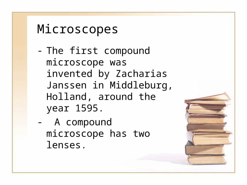
Microscopes
- The first compound microscope was invented by Zacharias Janssen in Middleburg, Holland, around the year 1595.
- A compound microscope has two lenses.
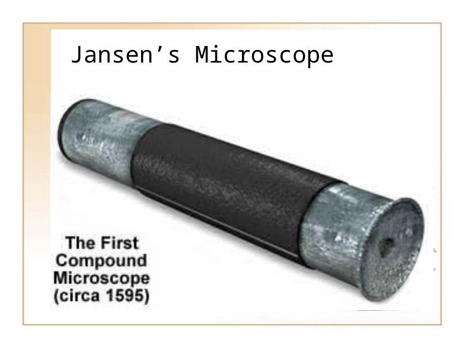
Jansen’s Microscope

Jansen’s Microscope
• The Janssen microscope was capable of magnifying images approximately three times when fully closed and up to ten times when extended to the maximum.
Light Microscopes
• In 1665, Robert Hooke looked at a thin slice of cork from the bark of a cork oak tree.
• He described “a great many little boxes” that reminded him of cubicles or “cells”.
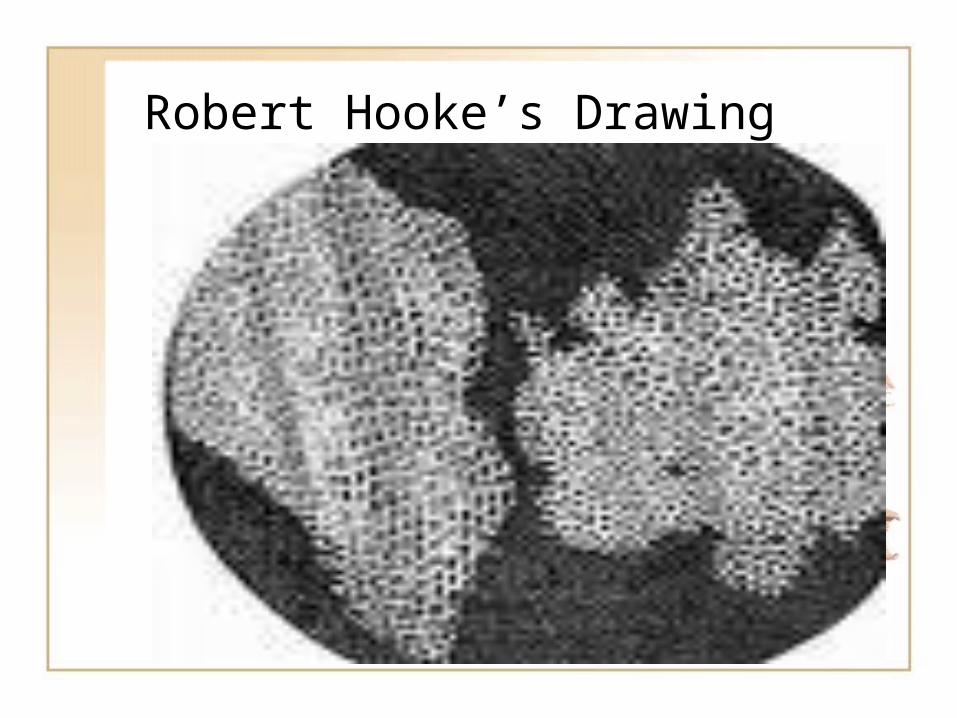
Robert Hooke’s Drawing
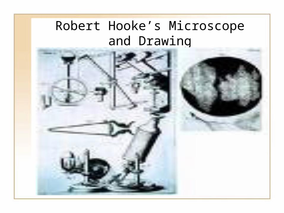
Robert Hooke’s Microscope and Drawing

Cells
• The “little boxes” that Hooke observed were the remains of dead plant cells.
• Hooke coined the word “cells” for the structures he saw under the microscope.
1673 Anton Van Leeuwenhoek• In 1673, Van
Leeuwenhoek was the first person to view living cells.
• He called the organisms “animalcules”.
• We now call them protists.
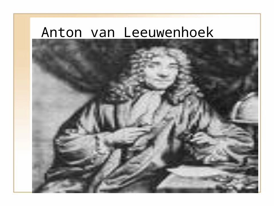
Anton van Leeuwenhoek

1827 Karl von Baer
• In 1827 Karl von Baer discovered the mammalian egg.
• This meant that animals had cells.

Plant and Animal Kingdoms
• In the 1800’s all organisms were placed into only two kingdoms- the Plant Kingdom and the Animal Kingdom.
• 1838: German Botanist Matthias Schleiden concluded that all plants are composed of cells.
• In 1839: German Zoologist Theodor Schwann concluded that all animals are composed of cells.
All organisms are composed of cells.• Schleiden’s and
Schwann’s conclusion led to the first statement of THE CELL THEORY.
• All Living things are composed of cells.

Rudolf Virchow 1855
• In 1855, Rudolf Virchow had evidence that cells came from other cells.
• This was an astonishing statement since in the mid-1800’s, the controversy over spontaneous generation had grown fierce.
• Spontaneous generation states that life can simply “appear”.
The Cell Theory
• All living things are composed of cells. (Schleiden and Schwann)
• Cells are the basic unit of structure and function in an organism.
• Cells come from the reproduction of existing cells. (Virchow)

The first cell structures- organelles• 1857 Kolliker describes
mitochondria in muscle.• Mitochondria are
structures inside of cells where cellular respiration takes place.
• Organelles are cellular structures that perform specific functions.
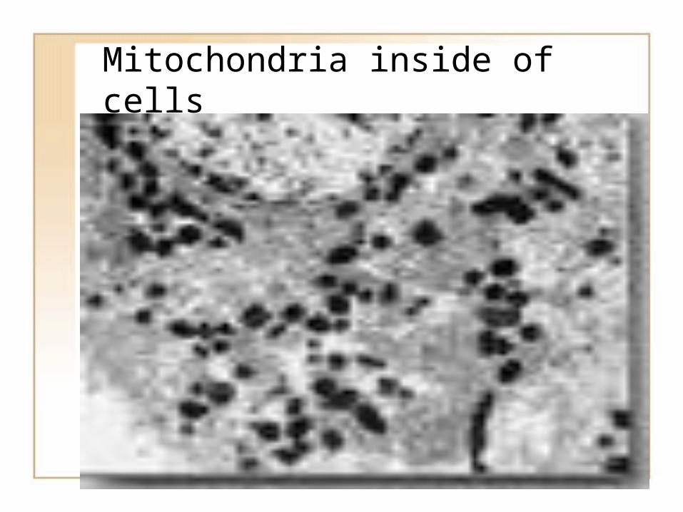
Mitochondria inside of cells

1897 Camillo Golgi
• In 1897, Camillo Golgi discovers the Golgi Apparatus.
• The Golgi Apparatus is an organelle that is called the “packaging plant”.

Electron Microscopes
• Electron microscopes contributed to our knowledge of cells.
• E. Ruska made the first image producing electron microscope in 1933 and used it to take pictures of gold and copper surfaces.
Improving Technology
• Improving technology has allowed scientists to unlock the secrets of the cell.
• There are two major types of electron microscope: the transmission electron microscope (TEM) and the scanning electron microscope (SEM)
1964 Palade
• In 1964 George Palade published his work on the network of membraneous organelles in cells of the guinea pig pancreas.
• This network included the rough endoplasmic reticulum, smooth endoplasmic reticulum, the Golgi Apparatus, and lysosomes.
1996 - Cloning
• 1996 Researchers in Scotland clone a sheep from the adult sheep cell.

2004
• Tissue engineering used to grow new skin and bone for transplant.
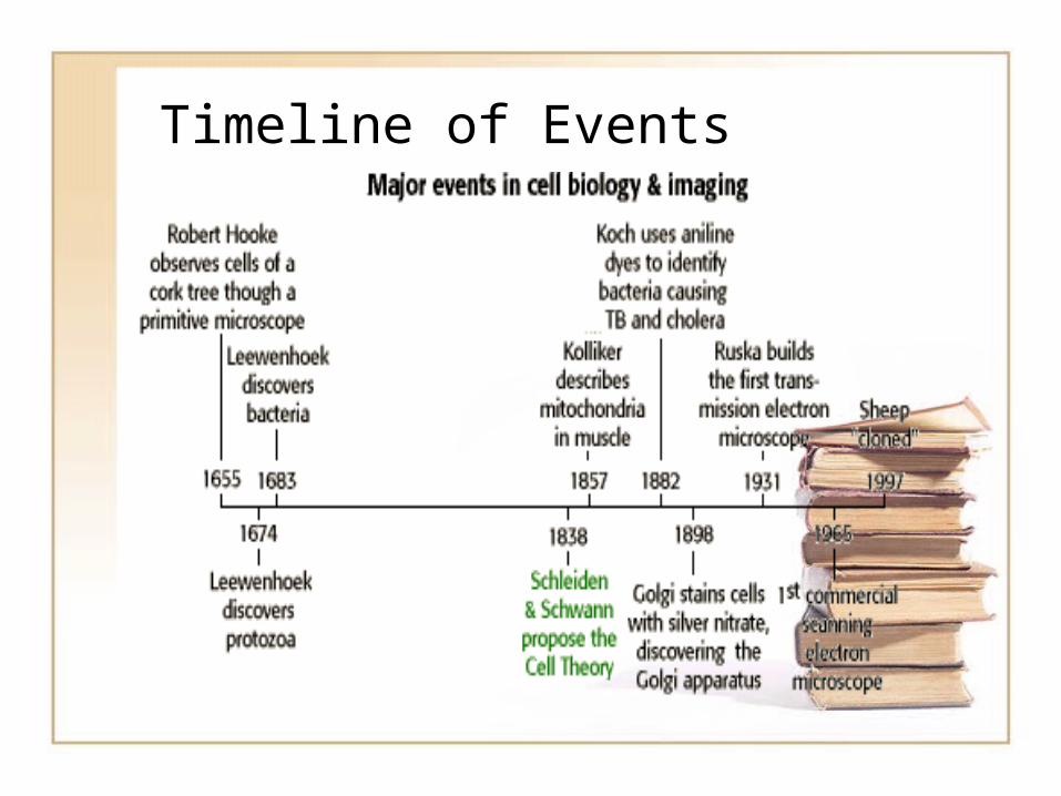
Timeline of Events

Use this Power Point to create a timeline.

Microscopes helped biologists clarify our definition of life.
• All living things:– Consist of organized parts.– Obtain energy from their
surroundings.– Perform chemical reactions.– Change with time– Respond to their
environment– Reproduce

The End
Related Documents