INTRACELLULAR DETECTION OF INFECTIOUS PATHOGENS IN SPERM CELLS This study was funded by and conducted in the diagnostic and research laboratories of Locus Medicus SA, Athens, Greece Speaker: Dr. Angelos Gritzapis, Biologist - Immunologist

This study was funded by and conducted in the diagnostic and research laboratories of Locus Medicus SA, Athens, Greece Speaker: Dr. Angelos Gritzapis,
Dec 27, 2015
Welcome message from author
This document is posted to help you gain knowledge. Please leave a comment to let me know what you think about it! Share it to your friends and learn new things together.
Transcript
INTRACELLULAR DETECTION
OF INFECTIOUS PATHOGENS
IN SPERM CELLSThis study was funded by and conducted in the diagnostic and research laboratories of Locus Medicus
SA, Athens, Greece
Speaker: Dr. Angelos Gritzapis, Biologist - Immunologist
A.D. Gritzapis1 PhD, K. Makarounis2 MD, M. Leventopoulos1 PhD, E. Nossi1, D. Nikolopoulos1, G. Georgoulias3 MD, V. Kapetanios4 MD, V Tsilivakos1 MD, PhD
1: Dept. of Cellular Biology and Immunology, Locus Medicus SA, Athens, Greece2: Urology clinic, Locus Medicus SA, Athens, Greece 3: Dept. of Microbiology, Locus Medicus SA, Athens, Greece 4: Gynecology & Obstetrics clinic, Locus Medicus SA, Athens, Greece
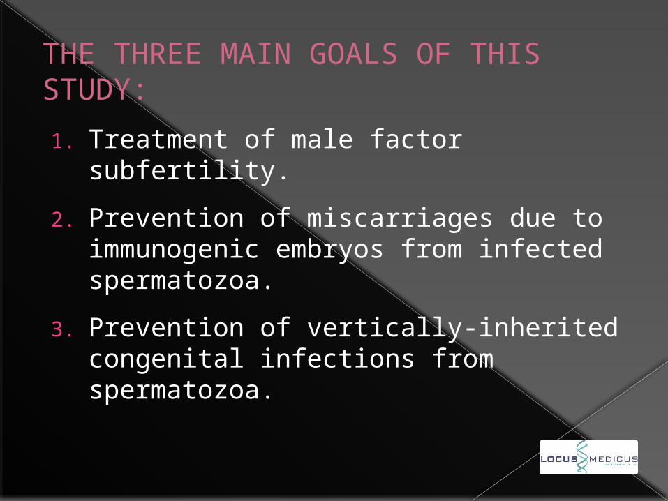
THE THREE MAIN GOALS OF THIS STUDY:
1. Treatment of male factor subfertility.
2. Prevention of miscarriages due to immunogenic embryos from infected spermatozoa.
3. Prevention of vertically-inherited congenital infections from spermatozoa.
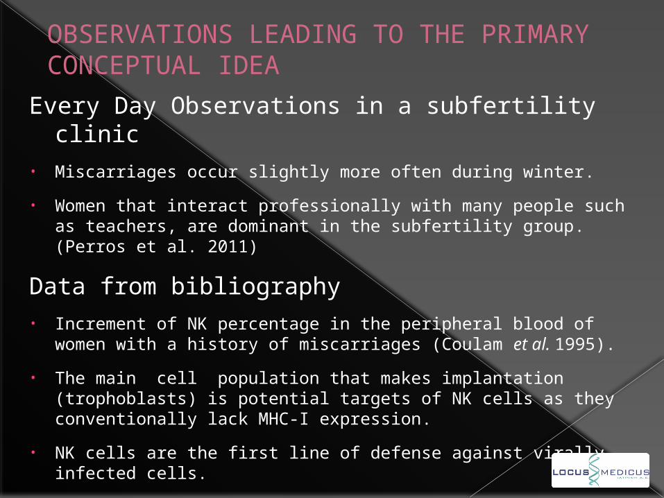
OBSERVATIONS LEADING TO THE PRIMARY CONCEPTUAL IDEA
Every Day Observations in a subfertility clinic• Miscarriages occur slightly more often during winter.
• Women that interact professionally with many people such as teachers, are dominant in the subfertility group.(Perros et al. 2011)
Data from bibliography• Increment of NK percentage in the peripheral blood of women
with a history of miscarriages (Coulam et al. 1995).
• The main cell population that makes implantation (trophoblasts) is potential targets of NK cells as they conventionally lack MHC-I expression.
• NK cells are the first line of defense against virally infected cells.
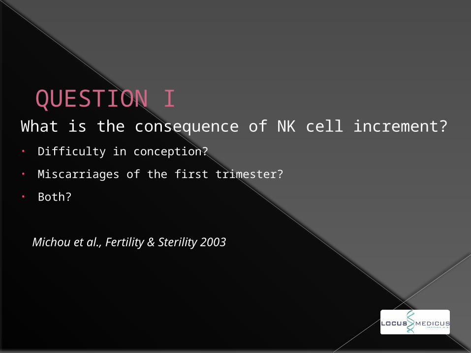
QUESTION IWhat is the consequence of NK cell increment?• Difficulty in conception?
• Miscarriages of the first trimester?
• Both?
Michou et al., Fertility & Sterility 2003
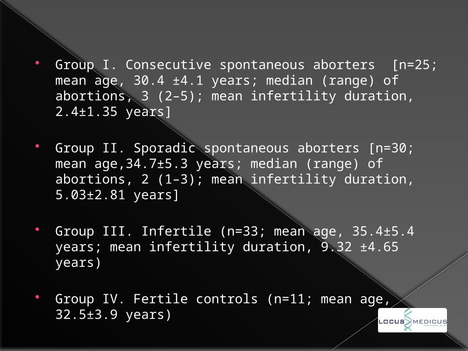
Group I. Consecutive spontaneous aborters [n=25; mean age, 30.4 ±4.1 years; median (range) of abortions, 3 (2–5); mean infertility duration, 2.4±1.35 years]
Group II. Sporadic spontaneous aborters [n=30; mean age,34.7±5.3 years; median (range) of abortions, 2 (1–3); mean infertility duration, 5.03±2.81 years]
Group III. Infertile (n=33; mean age, 35.4±5.4 years; mean infertility duration, 9.32 ±4.65 years)
Group IV. Fertile controls (n=11; mean age, 32.5±3.9 years)
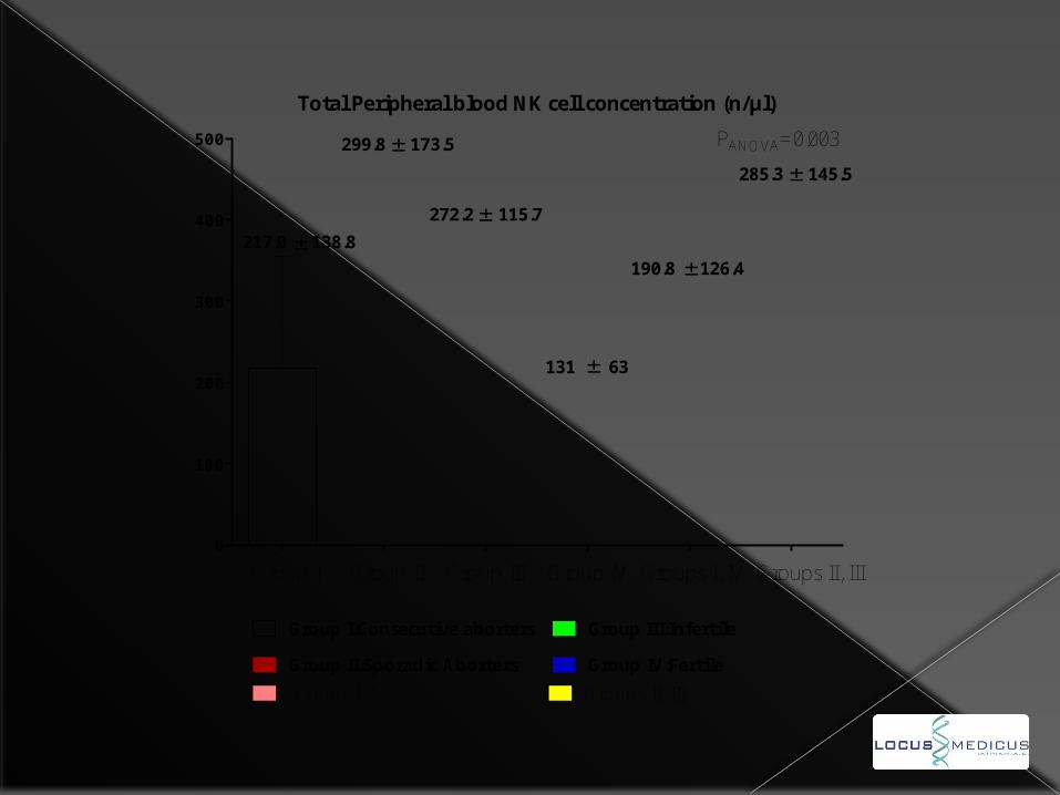
0
100
200
300
400
500
Total Peripheral blood NK cell concentration (n/μl)
Group II:Sporadic Aborters
Group III:Infertile
Group IV:Fertile
Group I:Consecutive aborters
217.0 138.8
299.8 173.5
272.2 115.7
131 63
Group I Group II Group III Group IV
Groups I, IV Groups II, III
190.8 126.4
285.3 145.5
Groups I, IV Groups II, III
PANOVA=0.003
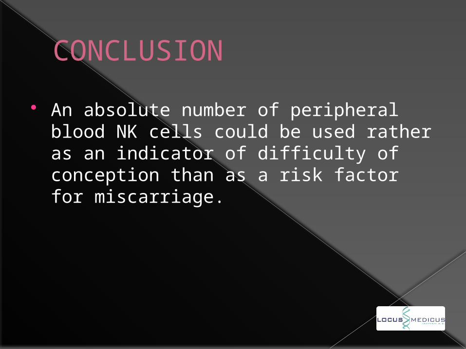
CONCLUSION
An absolute number of peripheral blood NK cells could be used rather as an indicator of difficulty of conception than as a risk factor for miscarriage.
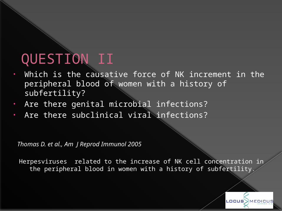
QUESTION II• Which is the causative force of NK increment in the peripheral
blood of women with a history of subfertility?• Are there genital microbial infections?• Are there subclinical viral infections?
Thomas D. et al., Αm J Reprod Immunol 2005
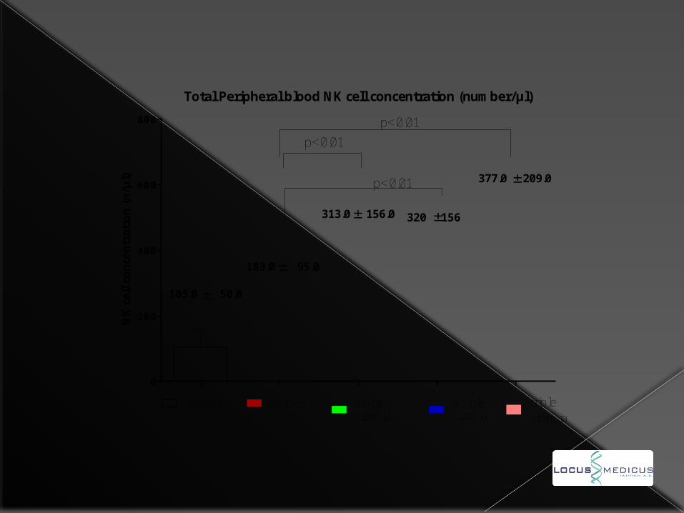
Herpesviruses related to the increase of NK cell concentration in the peripheral blood in women with a history of subfertility.
0
200
400
600
800
Total Peripheral blood NK cell concentration (number/μl)
NK c
ell c
once
ntr
atio
n (n/μ
l)
105.0 50.0
183.0 95.0
313.0 156.0 320 156
tripleviremia
377.0 209.0
doubleviremia
singleviremia
herpes -control
p<0.01
p<0.01
p<0.01
CONCLUSION
Assuming that all women under study remained asymptomatic, these data suggest that subclinical herpesvirus viremia may be an important cause of immunostimulation in women with a history of subfertility by increasing the concentration of NK cells in the peripheral blood.
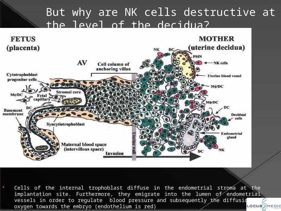
But why are NK cells destructive at the level of the decidua?
Cells of the internal trophoblast diffuse in the endometrial stroma at the implantation site. Furthermore, they emigrate into the lumen of endometrial vessels in order to regulate blood pressure and subsequently the diffusion of oxygen towards the embryo (endothelium is red)
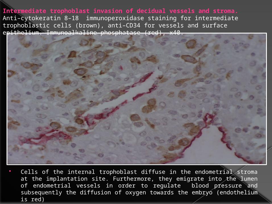
Intermediate trophoblast invasion of decidual vessels and stroma. Anti–cytokeratin 8–18 immunoperoxidase staining for intermediate trophoblastic cells (brown), anti–CD34 for vessels and surface epithelium. Immunoalkaline phosphatase (red), x40.
Cells of the internal trophoblast diffuse in the endometrial stroma at the implantation site. Furthermore, they emigrate into the lumen of endometrial vessels in order to regulate blood pressure and subsequently the diffusion of oxygen towards the embryo (endothelium is red)
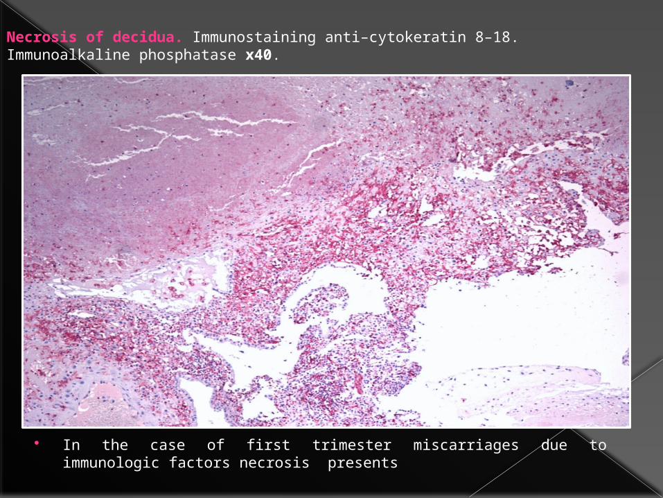
Necrosis of decidua. Immunostaining anti–cytokeratin 8–18. Immunoalkaline phosphatase x40.
In the case of first trimester miscarriages due to immunologic factors necrosis presents
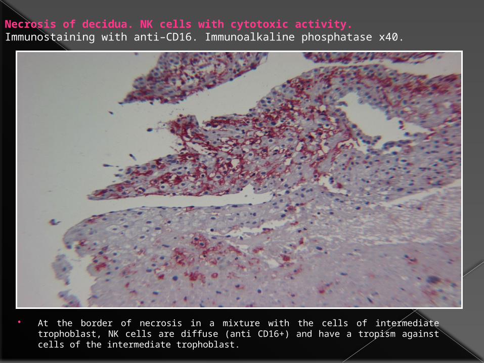
Necrosis of decidua. NK cells with cytotoxic activity. Immunostaining with anti–CD16. Immunoalkaline phosphatase x40.
At the border of necrosis in a mixture with the cells of intermediate trophoblast, NK cells are diffuse (anti CD16+) and have a tropism against cells of the intermediate trophoblast.
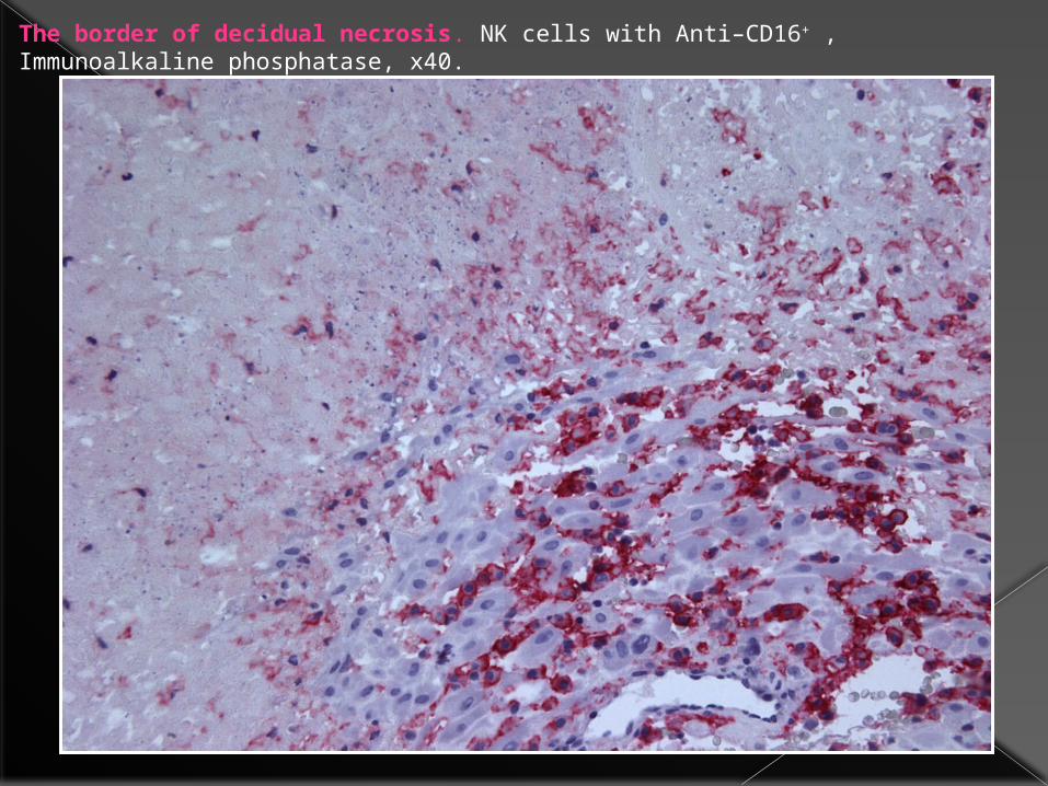
The border of decidual necrosis. NK cells with Anti–CD16+ , Immunoalkaline phosphatase, x40.
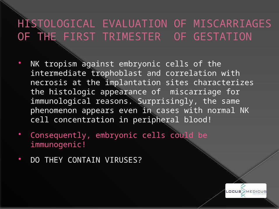
HISTOLOGICAL EVALUATION OF MISCARRIAGES OF THE FIRST TRIMESTER OF GESTATION
NK tropism against embryonic cells of the intermediate trophoblast and correlation with necrosis at the implantation sites characterizes the histologic appearance of miscarriage for immunological reasons. Surprisingly, the same phenomenon appears even in cases with normal NK cell concentration in peripheral blood!
Consequently, embryonic cells could be immunogenic!
DO THEY CONTAIN VIRUSES?
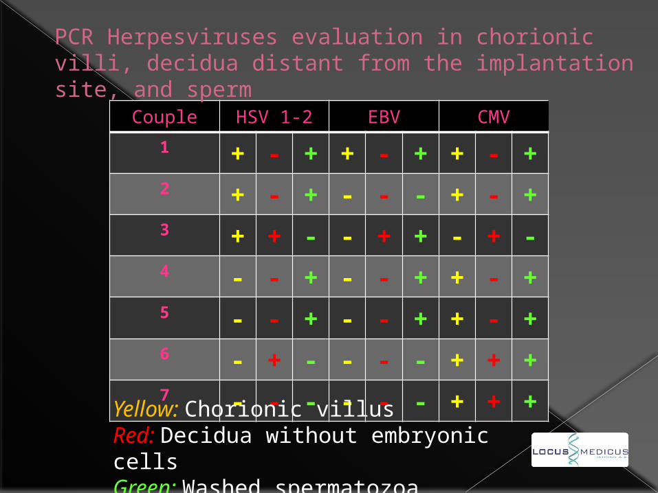
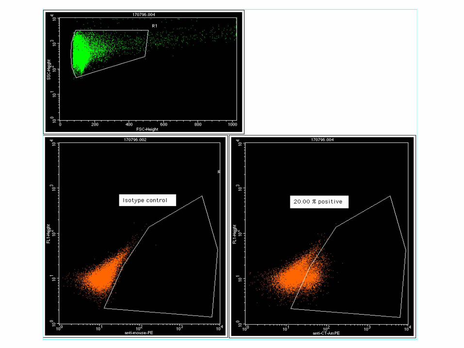
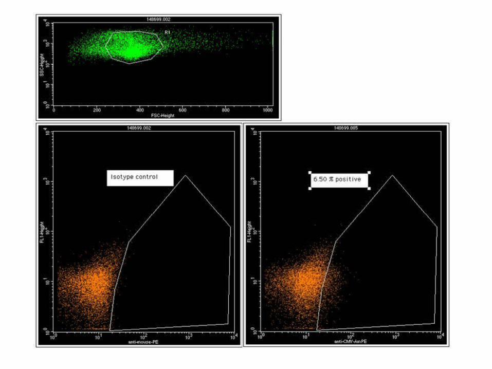
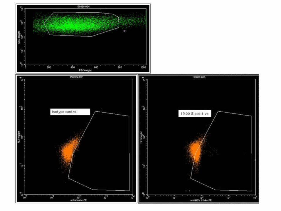
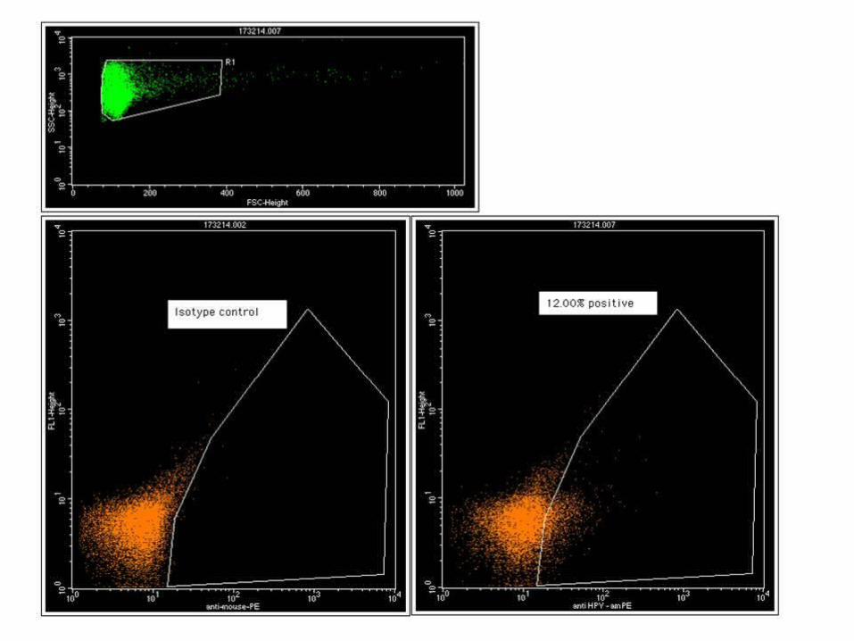
Couple HSV 1-2 EBV CMV1 + - + + - + + - +2 + - + - - - + - +3 + + - - + + - + -4 - - + - - + + - +5 - - + - - + + - +6 - + - - - - + + +7 - - - - - - + + +
Yellow: Chorionic villusRed: Decidua without embryonic cellsGreen: Washed spermatozoa
PCR Herpesviruses evaluation in chorionic villi, decidua distant from the implantation site, and sperm
HSV EBV CMV HHV6 HHV7 HSV EBV CMV HHV6 HHV7
0
20
40
60
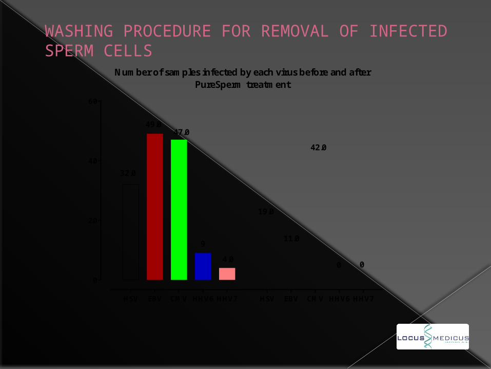
Number of samples infected by each virus before and afterPureSperm treatment
32.0
19.0
49.0
11.0
47.0
42.0
9
04.0
0
WASHING PROCEDURE FOR REMOVAL OF INFECTED SPERM CELLS
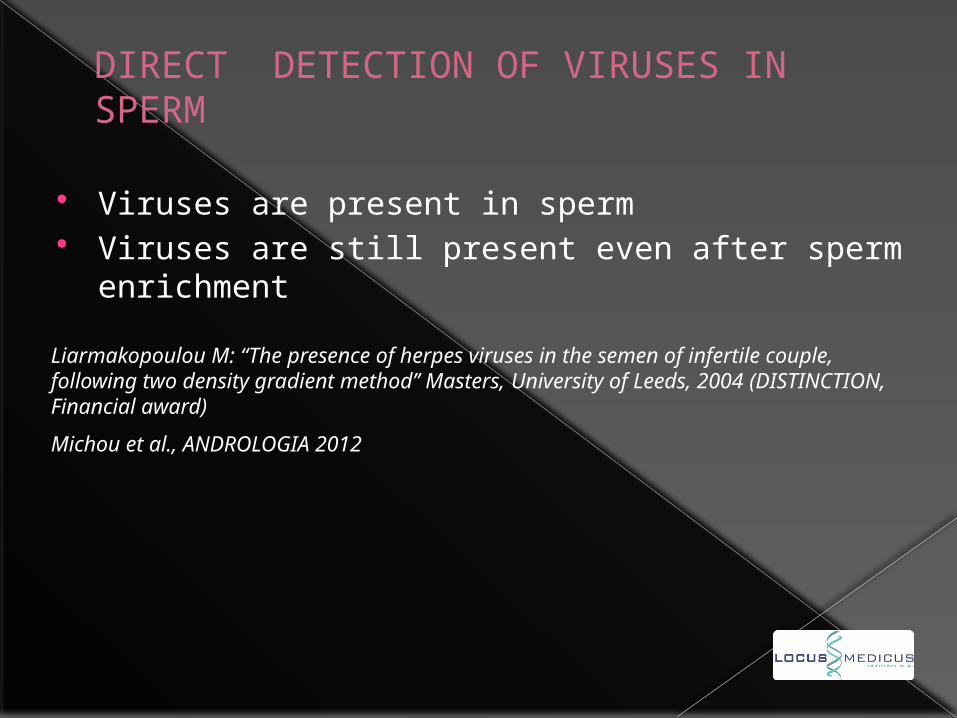
DIRECT DETECTION OF VIRUSES IN SPERM
Viruses are present in sperm Viruses are still present even after sperm
enrichment
Liarmakopoulou M: “The presence of herpes viruses in the semen of infertile couple, following two density gradient method’’ Masters, University of Leeds, 2004 (DISTINCTION, Financial award)
Michou et al., ANDROLOGIA 2012
Regarding the localization of pathogens, the PCR assay can not distinguish between seminal plasma and sperm cells or identify the infected cells.
This is very important as the interior of the spermatozoa but not the membrane, flows into the ovule.
So, a direct and high throughput assay is needed, as for example HBV is already detected in sperm cells by FISH (Huang et al, 2003).
FLOW CYTOMETRY PROTOCOL
Sperm liquefaction
centrifugation
Fixation with 4% PFA.
Membrane permeabilization with saponin and DMSO.
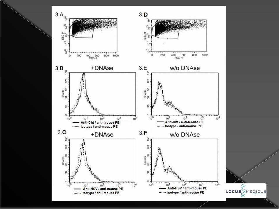
DNase I treatment for 30 min at 37oC as the DNA in sperm cells is very dense.
Incubation with monoclonal antibodies against intracellular pathogens (i.e. Chlamydia trachomatis, clone: Herpes Simplex Virus-HSV, Cytomegalovirus-CMV, Human Papiloma Virus-HPV).
Acquisition in a flow cytometer.
ADNANTAGES OF FLOW CYTOMETRIC METHOD OVER PCR Evaluation of the intracellular presence of
pathogens.
Greater sensitivity as Taq inhibitors should be suppressed by dilutions (Garolla et al. 2013). This can explain the discrepancy between PCR and flow cytometry.
Information on the identity of infected cells.
Lower cost.
Copyright 2012 Malamis & Malamis
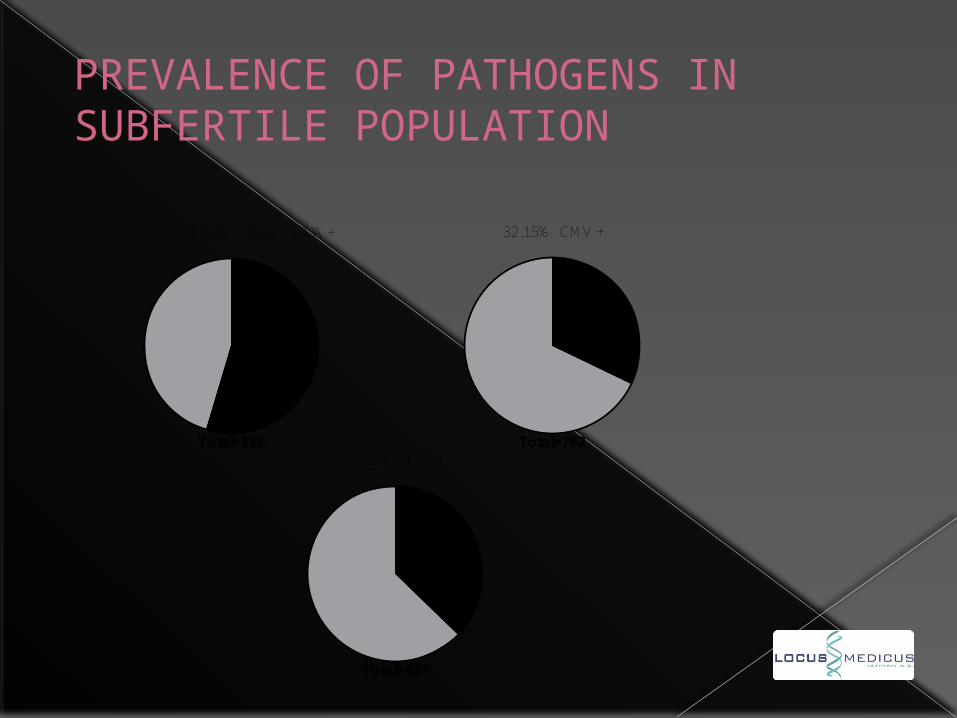
54.63% CHLAMYDIA +
Total=886
32.15% CMV +
Total=76237.22% HSV +
Total=634
PREVALENCE OF PATHOGENS IN SUBFERTILE POPULATION
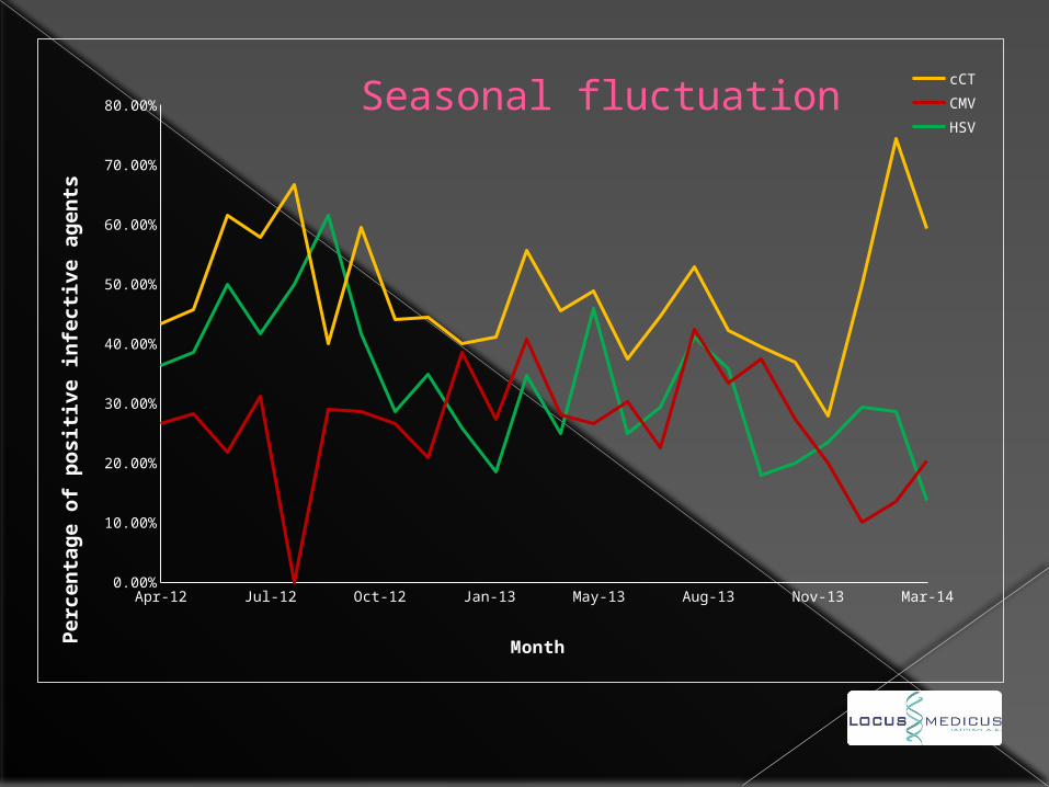
Apr-12 Jul-12 Oct-12 Jan-13 May-13 Aug-13 Nov-13 Mar-140.00%
10.00%
20.00%
30.00%
40.00%
50.00%
60.00%
70.00%
80.00% Seasonal fluctuation cCT
CMV
HSV
Month
Pe
rce
nta
ge
of
po
sit
ive
in
fecti
ve
ag
en
ts
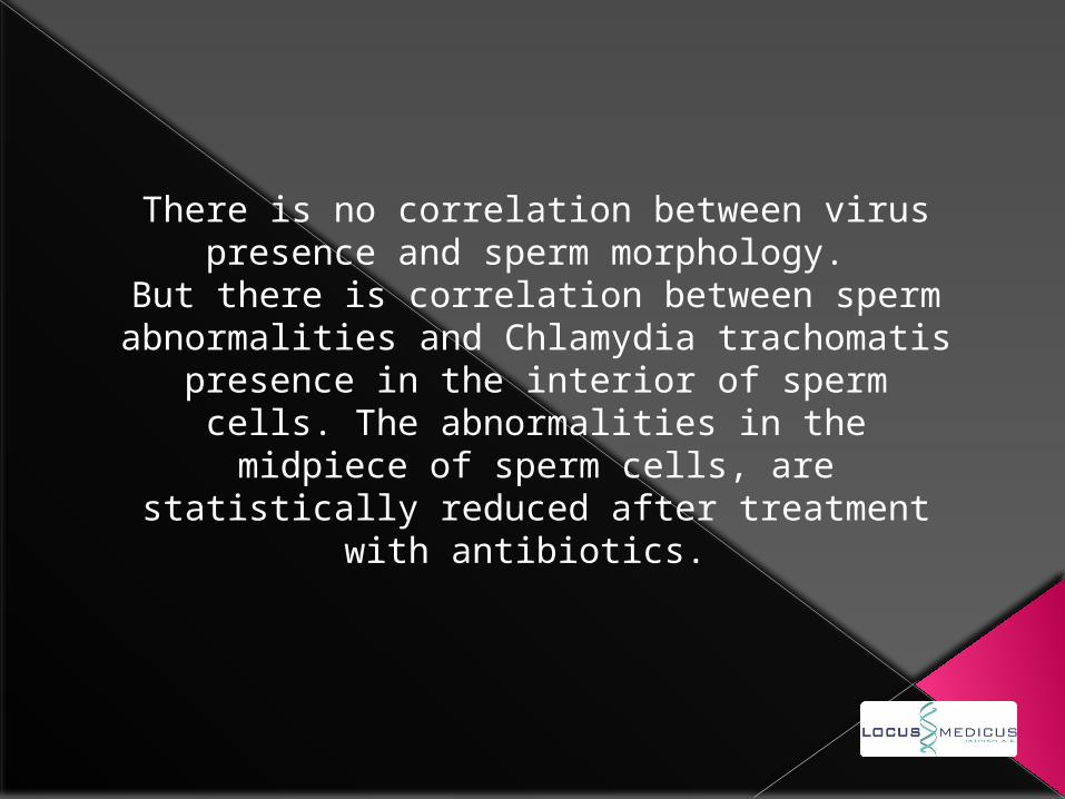
There is no correlation between virus presence and sperm morphology.
But there is correlation between sperm abnormalities and Chlamydia trachomatis presence in the interior of sperm cells. The
abnormalities in the midpiece of sperm cells, are statistically reduced after treatment with
antibiotics.
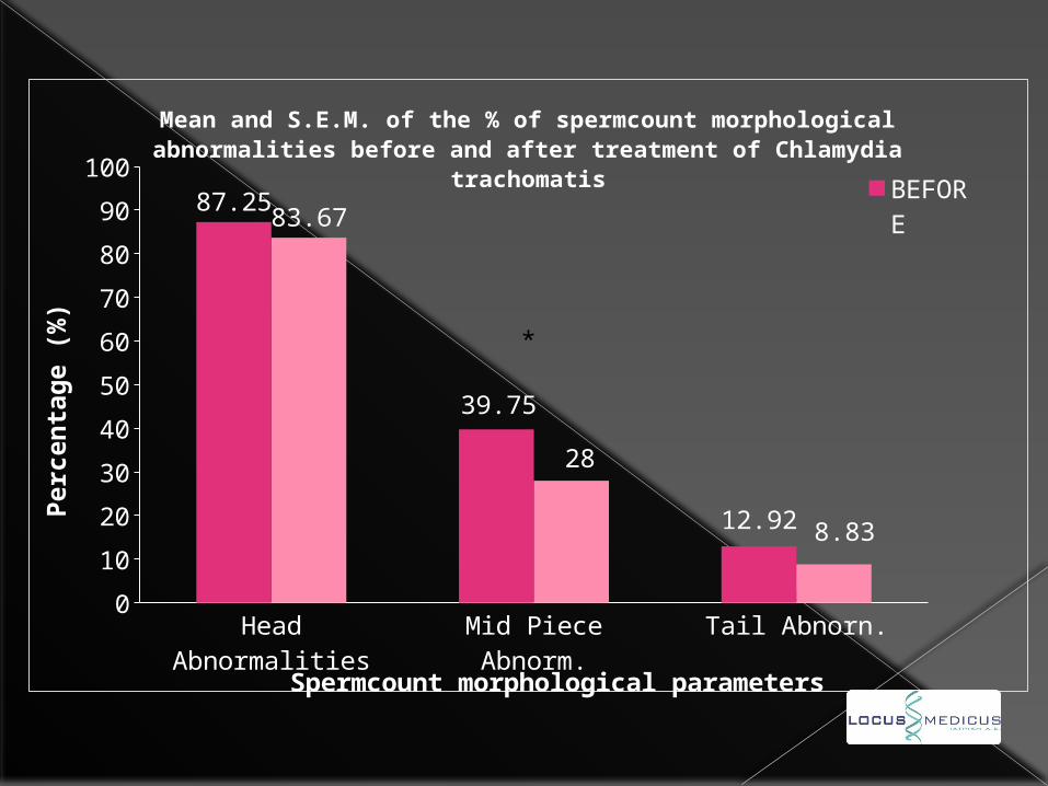
Head Abnormalit
ies
Mid Piece Abnorm.
Tail Abnorn.
0
10
20
30
40
50
60
70
80
90 83.67
28
8.83
Mean and S.E.M. of the % of spermcount morphological abnormalities before and after treatment of Chlamydia
trachomatis BEFOREAFTER
Spermcount morphological parameters
Perc
en
tag
e (
%)
*
0
20
40
60
80
100
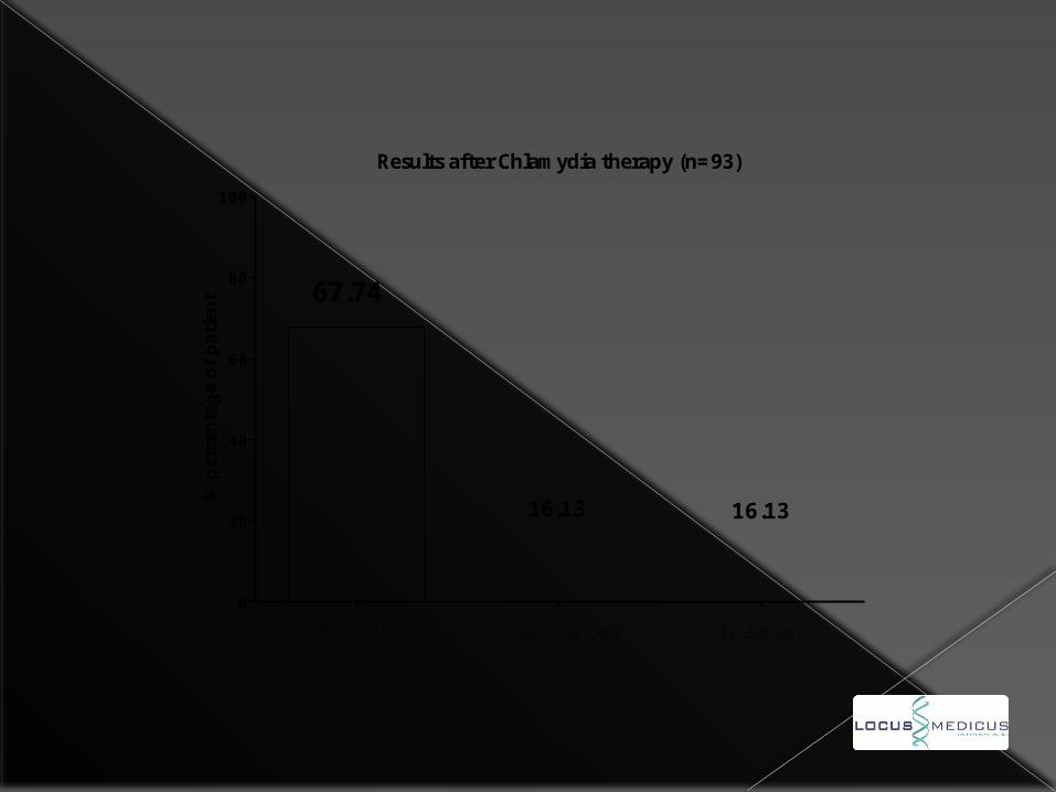
Results after Chlamydia therapy (n=93)
% p
erce
nta
ge
of pat
ient 67.74
16.13
reduction unchanged increment
16.13
cCT0
0.2
0.4
0.6
0.8
1
1.2
1.4
1.6
1.8
2
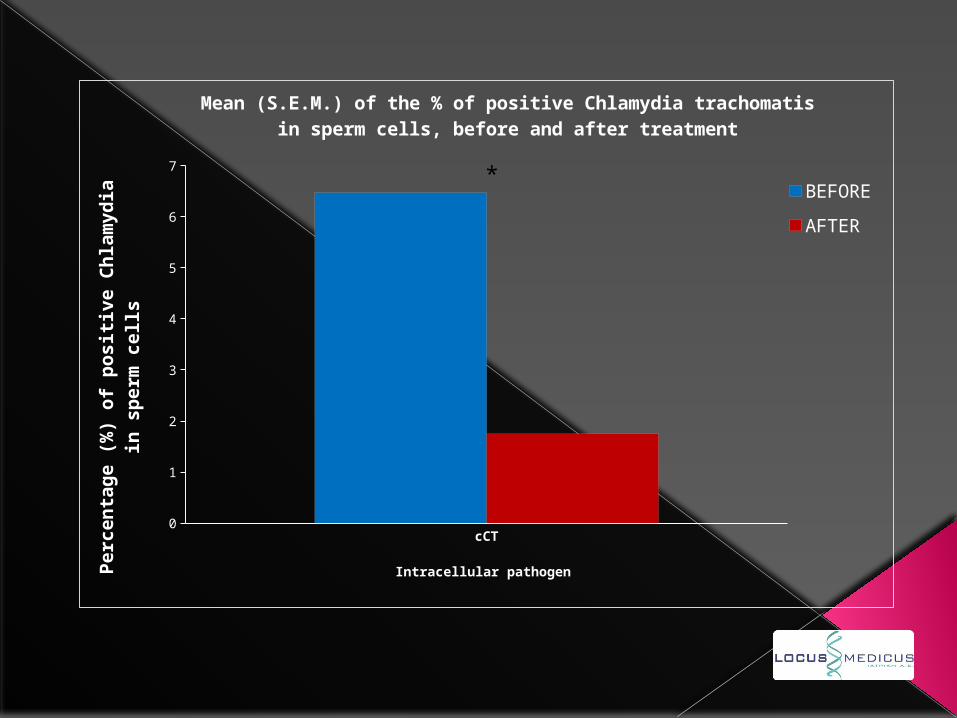
Mean (S.E.M.) of the % of positive Chlamydia trachomatis in sperm cells, before and after treatment
BEFORE
AFTER
Intracellular pathogen
Pe
rce
nta
ge
(%
) o
f p
osit
ive
Ch
lam
yd
ia
in s
pe
rm c
ell
s
*
Series10
0.1
0.2
0.3
0.4
0.5
0.6
0.7
0.8
0.9
1
1.1
1.2
1.3
1.4
1.5
1.6
1.7
1.8
1.9
2
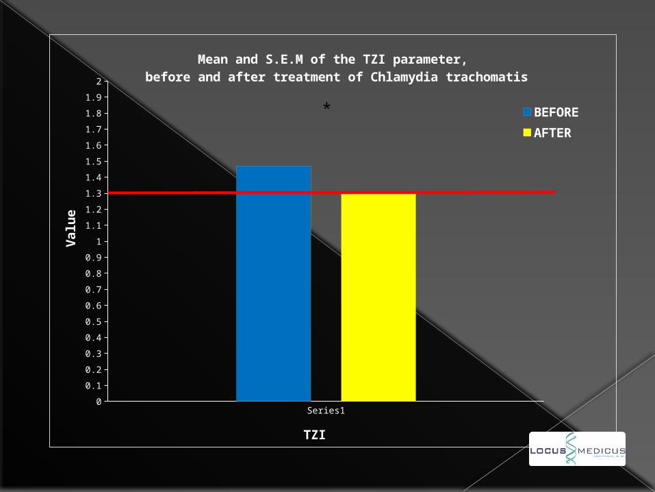
Mean and S.E.M of the ΤΖΙ parameter, before and after treatment of Chlamydia trachomatis
BEFORE
AFTER
TZI
Va
lue
*

POTENTIAL CONSEQUENCES OF THE EXISTENCE OF VIRUS IN THE INTERIOR OF SPERMATOZOA
Incapability of conception due to a) abnormalities of spermatozoa.b) very early rejection of the fetus before the next
menstruation, is misinterpreted as infertility.
Early miscarriage of the fetus with the histologic image described previously, with or without NK increase in the periphery.
Because of the neurotropic properties of Herpesviruses, it is possible that congenital anomalies occur due to them.
In case of fetal survival, there is a strong possibility that T and B cell clones, which normally recognize virus antigens, will be deleted according to the self-tolerance theory during thymic education.
As a result, the newborn organism would be tolerized against intracellular pathogens and therefore more susceptible to them in the future.
SPERM BANKS
The fact that miscarriage is possible due to the intracellular presence of virus, makes their detection by sperm banks necessary.
In addition, the seasonal fluctuation of their presence, makes testing of every sample given by the same donor equally necessary.
Artificial Insemination Techniques (ART) – In vitro Fertilization (IVF)
The high economic and psychological cost of a miscarriage after either ART or IVF, makes intra Sperm Pathogen Immunophenotyping (SPI test) of great importance.

Thank you for your attention.
Related Documents