COMMENTARY The inner workings of stress fibers − from contractile machinery to focal adhesions and back Ariel Livne and Benjamin Geiger* ABSTRACT Ventral stress fibers and focal adhesions are physically coupled structures that play key roles in cellular mechanics and force sensing. The tight functional interdependence between the two is manifested not only by their apparent proximity but also by the fact that ventral stress fibers and focal adhesions are simultaneously diminished upon actomyosin relaxation, and grow when subjected to external stretching. However, whereas the apparent co-regulation of the two structures is well-documented, the underlying mechanisms remains poorly understood. In this Commentary, we discuss some of the fundamental, yet still open questions regarding ventral stress fiber structure, its force-dependent assembly, as well as its capacity to generate force. We also challenge the common approach – i.e. ventral stress fibers are variants of the well-studied striated or smooth muscle machinery – by presenting and critically discussing alternative venues. By highlighting some of the less-explored aspects of the interplay between stress fibers and focal adhesions, we hope that this Commentary will encourage further investigation in this field. KEY WORDS: Cell mechanics, Focal adhesions, Mechanosensitivity, Sarcomeres, Stress fibers Introduction – cellular mechanosensitivity as an emerging paradigm In radical contrast to the common analogy of cells as ‘biochemical machines’ driven primarily by chemical interactions, most cells, throughout our body, constantly generate, transmit, sense and respond to mechanical inputs. We witness such biomechanical processes with every movement we make, every beat of our heart and every breath we take. Force perturbations are created by cells as they push and pull on their immediate surroundings, be it neighboring cells or the extracellular matrix (ECM), as they change their morphology or orientation in response to external mechanical constraints and, of course, as they migrate and divide. The underlying basis of these diverse examples is the ability of essentially all cells, not just muscle cells, to generate and apply force (Discher et al., 2005; Harris et al., 1980; Liang et al., 2010) and, no less important, their capacity to sense ‘informative mechanical cues’ and develop a specific physiological (often biochemical) response. These forces may be divided, superficially, into two types: pushing and pulling. At the molecular level, pushing is typically attributed to the polymerization-based extension of actin bundles or individual fibers. As actin filaments elongate, they can push against any physical obstacle they encounter. Such a pushing mechanism can drive the leading edge of the cell forward during migration (Keren et al., 2008; Mogilner and Oster, 1996; Pollard et al., 2001), and extend thin protrusions to probe the surrounding ECM by using filopodia (Mattila and Lappalainen, 2008) or invade it with invadopodia (Buccione et al., 2004; Revach and Geiger, 2014). It is important to note, however, that cellular pushing does not necessarily take place through cytoskeletal polymerization, as in the cases of bleb (Fackler and Grosse, 2008) and lobopodium formation (Petrie et al., 2014), which are driven by the hydrostatic pressure in the cell. By contrast, pulling forces are generated, primarily, when bipolar bundles of myosin II motor proteins slide along actin fibers (Alberts et al., 2007; Vicente-Manzanares et al., 2009). Notably, myosin II moves along actin filaments in a directed, processive manner; however, when arranged in a bipolar configuration, the myosin motor heads can attach to different actin filaments, sliding one towards the other. This way, cells can apply contractile forces in a number of situations, ranging from classic muscle contraction (striated and smooth) (Gordon et al., 2000) and ECM remodeling (Larsen et al., 2006; Stopak and Harris, 1982) to wound closure (Brugués et al., 2014) and retraction of the trailing edge of migrating cells (Vicente-Manzanares et al., 2007). Nevertheless, contractile forces can also be effectively generated through alternative mechanisms. One such relevant example is the depolymerization of microtubule and actin filaments (see below) (Sun et al., 2010), both of which have been associated with chromosome movement (Kline-Smith and Walczak, 2004) and cytokinesis (Pinto et al., 2012). In this Commentary, we discuss the mechanisms that underlie force generation by stress fibers, the transmission of the apparent contractile (and other) forces to the ECM through their anchorage sites to the matrix, i.e. focal adhesions, and the coordinated response of the two structures to externally applied forces. Stress fibers and focal adhesions are readily formed by many different cell systems in culture, e.g. fibroblasts, epithelial and endothelial cells (Tojkander et al., 2012), and analogous structures are found in vivo (Delon and Brown, 2009; North et al., 1993; Wong et al., 1983). With the growing interest in cellular mechanosensitivity, stress fibers and focal adhesions have drawn increasing attention over the last decades (see, e.g. Chrzanowska-Wodnicka and Burridge, 1996; Geiger et al., 2009; Pelham and Wang, 1997; Wang et al., 2001; Zemel et al., 2010). Here, we refrain from providing a comprehensive literature review of stress fibers and focal adhesions, information that can be readily found in several recent and comprehensive review articles (Burridge and Guilluy, 2015; Burridge and Wittchen, 2013; Geiger et al., 2009; Geiger and Yamada, 2011; Kassianidou and Kumar, 2015; Naumanen et al., 2008; Pellegrin and Mellor, 2007; Smith et al., 2014; Tojkander et al., 2012; Vallenius, 2013). Rather, we highlight and challenge fundamental, yet mostly open, questions regarding the mechanical crosstalk and interdependence between stress fibers and focal adhesions. To that end, we present the main players, and provide a short overview of the different stress fiber types and their specific modes of association with focal adhesions, before exploring the similarities between stress fibers of non-muscle cells and the well-studied Department of Molecular Cell Biology, Weizmann Institute of Science, Rehovot 7610001, Israel. *Author for correspondence ([email protected]) 1293 © 2016. Published by The Company of Biologists Ltd | Journal of Cell Science (2016) 129, 1293-1304 doi:10.1242/jcs.180927 Journal of Cell Science

Welcome message from author
This document is posted to help you gain knowledge. Please leave a comment to let me know what you think about it! Share it to your friends and learn new things together.
Transcript
COMMENTARY
The inner workings of stress fibers − from contractile machinery tofocal adhesions and backAriel Livne and Benjamin Geiger*
ABSTRACTVentral stress fibers and focal adhesions are physically coupledstructures that play key roles in cellular mechanics and force sensing.The tight functional interdependence between the two is manifestednot only by their apparent proximity but also by the fact that ventralstress fibers and focal adhesions are simultaneously diminishedupon actomyosin relaxation, and grow when subjected to externalstretching. However, whereas the apparent co-regulation of the twostructures is well-documented, the underlying mechanisms remainspoorly understood. In this Commentary, we discuss some of thefundamental, yet still open questions regarding ventral stress fiberstructure, its force-dependent assembly, as well as its capacity togenerate force.We also challenge the common approach – i.e. ventralstress fibers are variants of thewell-studied striated or smoothmusclemachinery – by presenting and critically discussing alternativevenues. By highlighting some of the less-explored aspects of theinterplay between stress fibers and focal adhesions, we hope that thisCommentary will encourage further investigation in this field.
KEYWORDS: Cell mechanics, Focal adhesions, Mechanosensitivity,Sarcomeres, Stress fibers
Introduction –cellular mechanosensitivity as an emergingparadigmIn radical contrast to the common analogy of cells as ‘biochemicalmachines’ driven primarily by chemical interactions, most cells,throughout our body, constantly generate, transmit, sense andrespond to mechanical inputs. We witness such biomechanicalprocesses with every movement we make, every beat of our heartand every breath we take. Force perturbations are created by cells asthey push and pull on their immediate surroundings, be itneighboring cells or the extracellular matrix (ECM), as theychange their morphology or orientation in response to externalmechanical constraints and, of course, as they migrate and divide.The underlying basis of these diverse examples is the ability ofessentially all cells, not just muscle cells, to generate and apply force(Discher et al., 2005; Harris et al., 1980; Liang et al., 2010) and, noless important, their capacity to sense ‘informative mechanical cues’and develop a specific physiological (often biochemical) response.These forcesmay be divided, superficially, into two types: pushing
and pulling. At the molecular level, pushing is typically attributed tothe polymerization-based extension of actin bundles or individualfibers.As actin filaments elongate, they can push against any physicalobstacle they encounter. Such a pushing mechanism can drive theleading edge of the cell forward during migration (Keren et al., 2008;Mogilner and Oster, 1996; Pollard et al., 2001), and extend thin
protrusions to probe the surrounding ECM by using filopodia(Mattila and Lappalainen, 2008) or invade it with invadopodia(Buccione et al., 2004; Revach and Geiger, 2014). It is important tonote, however, that cellular pushing does not necessarily take placethrough cytoskeletal polymerization, as in the cases of bleb (Facklerand Grosse, 2008) and lobopodium formation (Petrie et al., 2014),which are driven by the hydrostatic pressure in the cell. By contrast,pulling forces are generated, primarily, when bipolar bundles ofmyosin II motor proteins slide along actin fibers (Alberts et al., 2007;Vicente-Manzanares et al., 2009). Notably, myosin II moves alongactin filaments in a directed, processive manner; however, whenarranged in a bipolar configuration, the myosin motor heads canattach to different actin filaments, sliding one towards the other. Thisway, cells can apply contractile forces in a number of situations,ranging from classic muscle contraction (striated and smooth)(Gordon et al., 2000) and ECM remodeling (Larsen et al., 2006;Stopak andHarris, 1982) towound closure (Brugués et al., 2014) andretraction of the trailing edge of migrating cells (Vicente-Manzanareset al., 2007). Nevertheless, contractile forces can also be effectivelygenerated through alternative mechanisms. One such relevantexample is the depolymerization of microtubule and actin filaments(see below) (Sun et al., 2010), both of which have been associatedwith chromosome movement (Kline-Smith and Walczak, 2004) andcytokinesis (Pinto et al., 2012).
In this Commentary, we discuss the mechanisms that underlie forcegeneration by stress fibers, the transmission of the apparent contractile(and other) forces to the ECM through their anchorage sites to thematrix, i.e. focal adhesions, and the coordinated response of the twostructures to externally applied forces. Stress fibers and focal adhesionsare readily formed by many different cell systems in culture, e.g.fibroblasts, epithelial and endothelial cells (Tojkander et al., 2012),and analogous structures are found in vivo (Delon and Brown, 2009;North et al., 1993; Wong et al., 1983). With the growing interest incellular mechanosensitivity, stress fibers and focal adhesions havedrawn increasing attention over the last decades (see, e.g.Chrzanowska-Wodnicka and Burridge, 1996; Geiger et al., 2009;Pelham andWang, 1997;Wang et al., 2001; Zemel et al., 2010). Here,we refrain from providing a comprehensive literature review of stressfibers and focal adhesions, information that can be readily found inseveral recent and comprehensive review articles (Burridge andGuilluy, 2015; Burridge and Wittchen, 2013; Geiger et al., 2009;Geiger andYamada, 2011;Kassianidou andKumar, 2015; Naumanenet al., 2008; Pellegrin andMellor, 2007; Smith et al., 2014; Tojkanderet al., 2012; Vallenius, 2013). Rather, we highlight and challengefundamental, yet mostly open, questions regarding the mechanicalcrosstalk and interdependence between stress fibers and focaladhesions.
To that end, we present the main players, and provide a shortoverview of the different stress fiber types and their specific modesof association with focal adhesions, before exploring the similaritiesbetween stress fibers of non-muscle cells and the well-studied
Department of Molecular Cell Biology, Weizmann Institute of Science, Rehovot7610001, Israel.
*Author for correspondence ([email protected])
1293
© 2016. Published by The Company of Biologists Ltd | Journal of Cell Science (2016) 129, 1293-1304 doi:10.1242/jcs.180927
Journal
ofCe
llScience
striated myofibrils. On the basis of theoretical and experimentalconsiderations, we postulate that the commonly held comparisonbetween stress fibers and myofibrils is problematic, and should berevised. We also present a set of fundamental open questions thatdeserve to be addressed in future research; these include: (i) whereare the basic stress fiber building blocks (sarcomeres) assembled,and how are they maintained under the constant treadmilling of newactin from the focal adhesions? (ii) How are stress fibers and focaladhesions mechanically coupled and co-regulated, and which of thetwo is the ‘primary driver’? (iii) Are all sarcomeres along the stressfiber identical, both structurally and dynamically? (iv) Stress fibersgenerate a stronger force than theoretically expected from simpleconsiderations (e.g. number of motors, dimensions), whereasmeasurements of their mechanical properties obtained fromdifferent experiments appear to be inconsistent. How can thesefindings be reconciled?Motivated by the above questions, we propose to extend the,
currently, prevailing actomyosin model. We consider the possibilitythat, in addition to the sliding of actin and myosin II filamentsagainst one another, a force-regulated association and dissociationof actin monomers has a key role in the generation of stress-fibertension.
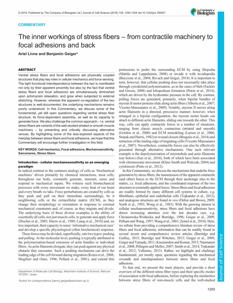
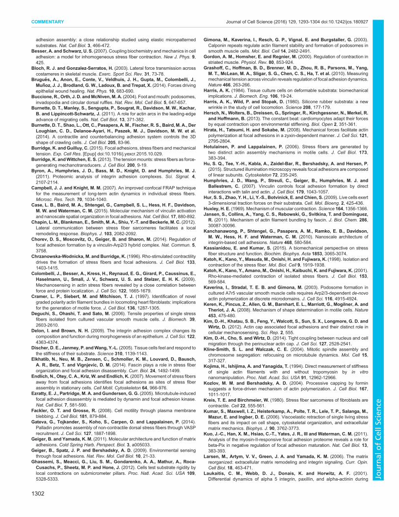
Stress fiber diversity and interdependence with focaladhesionsStress fibers are crosslinked bundles of actin fibers. They are,typically, divided into three types – ventral stress fibers, dorsal stressfibers and transverse arcs – that differ in function, cellular location,structure and composition (Hotulainen and Lappalainen, 2006;Small et al., 1998) (Fig. 1A). Dorsal stress fibers are long, linear andnon-contractile bundles of actin crosslinked by α-actinin, andcomposed of palladin and vasodilator-stimulated phosphoprotein(VASP) (Burnette et al., 2014; Gateva et al., 2014). They appear tobe attached solely to the ventral membrane through focal adhesionsat a single, discrete end, usually near the cell edges. From there, theyextend towards the cell center through formin- and VASP-drivenactin polymerization (Hotulainen and Lappalainen, 2006; Skauet al., 2015; Tojkander et al., 2015), apparently to serve as tracks onwhich transverse arcs can slide centripetally (Hotulainen andLappalainen, 2006; Tee et al., 2015). Transverse arcs, however,are long, curved actin structures along which alternating andrepetitive bands of α-actinin and myosin are arranged. After theirformation at the lamellipodium from Arp2/3- and formin-nucleatedactin fibers (Burnette et al., 2011; Hotulainen and Lappalainen,2006; Tojkander et al., 2011; Zhang et al., 2003), the transverse arcsslide centripetally – not anchored to any focal adhesions – along thedorsal stress fibers in a myosin II-dependent fashion (Tee et al.,2015). They eventually fuse towards the cell center, forming thickand contractile actomyosin bundles. Finally, ventral stress fibersappear to combine the properties of transverse arcs and dorsal stressfibers. These long, linear actin structures typically extend from oneside of the cell to the other, and attach at either end to focal adhesions(Fig. 1B). Towards their center, however, they are made up byalternating bands of α-actinin andmyosin II of non-uniform lengths.Indeed, one of the mechanisms for ventral stress fiber formationinvolves the merger of a transverse arc bundle with two dorsal stressfibers at its sides in a tension-mediated manner (Hotulainen andLappalainen, 2006; Tojkander et al., 2015). Ventral stress fibers alsoinclude two additional subtypes: peripheral stress fibers that runalong the long stable edges of a cell (Prager-Khoutorsky et al., 2011)and actin cap stress fibers, which are draped over the nucleus (Kimet al., 2014). Interestingly, whereas myosin II motors generate
contractile forces in both transverse arcs and ventral stress fibers,their overall effect is quite distinct. In transverse arcs, these forces actwithin the cell to centripetally drive the arcs (Hotulainen andLappalainen, 2006). By contrast, ventral stress fibers transmit themyosin-II-generated forces externally through their anchorage tofocal adhesions, to probe and interact with their microenvironment(Balaban et al., 2001; Grashoff et al., 2010; Sabass et al., 2008).Thus, although dorsal stress fibers and transverse arcs are prominentduring cell spreading, it is the third type of stress fiber, the ventralstress fiber that are most commonly found in mature cells andbelieved to have a key role in cellular mechanosensing (Pellegrin andMellor, 2007). We, therefore, focus in this Commentary on ventralstress fibers (hereafter referring to them as ‘stress fibers’) and discusstheir mechanical coupling through focal adhesions to the ECM.
Located at the ventral membrane of adherent cells, themicrometer-long focal adhesions form a bridge between theterminal segment of stress fibers and the ECM. Their cytoplasmiccomponents attach to stress fibers, whereas their extracellular partsare anchored to the ECM through transmembrane receptors of theintegrin family. Focal adhesions are highly regulated multi-proteincomplexes that comprise over 200 distinct proteins, which areknown collectively as the ‘integrin adhesome’ (Byron et al., 2011;Kuo et al., 2011; Schiller et al., 2011; Winograd-Katz et al., 2014;Zaidel-Bar and Geiger, 2010; Zaidel-Bar et al., 2007; Zamir andGeiger, 2001). Focal adhesion proteins are typically sorted into twofunctional molecular classes: scaffolding proteins (e.g. vinculin,paxillin, talin, zyxin) and signaling proteins (e.g. focal adhesionkinases, specific phosphatases, Rho-family G-protein activators orinhibitors). The former are primarily involved in the formation andmaintenance of a stable structural scaffold, thereby linking the stressfibers to the ECM through integrins. The latter, by contrast, arerecruited to the adhesion sites, where they generate and mediateadhesion-dependent signals that act locally to control thedevelopment and sustainability of focal adhesions and, at thesame time, also globally regulate key cellular processes, such as cellproliferation, differentiation, survival and migration (Wozniak et al.,2004).
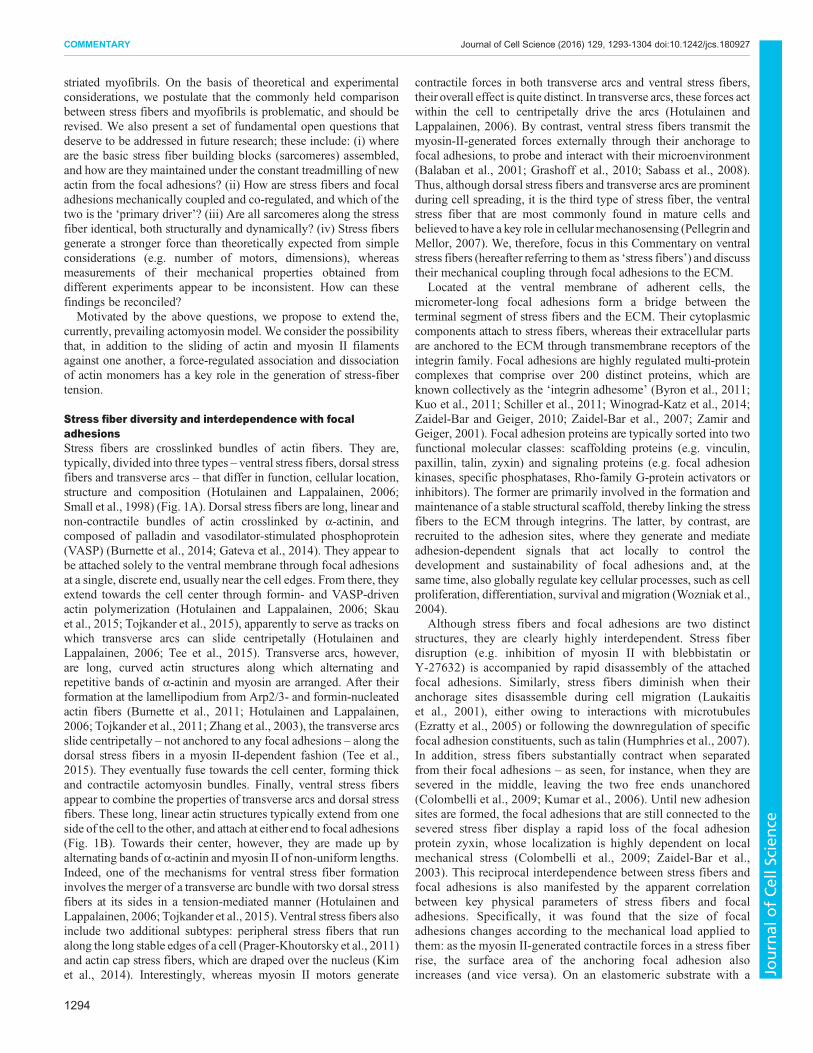
Although stress fibers and focal adhesions are two distinctstructures, they are clearly highly interdependent. Stress fiberdisruption (e.g. inhibition of myosin II with blebbistatin orY-27632) is accompanied by rapid disassembly of the attachedfocal adhesions. Similarly, stress fibers diminish when theiranchorage sites disassemble during cell migration (Laukaitiset al., 2001), either owing to interactions with microtubules(Ezratty et al., 2005) or following the downregulation of specificfocal adhesion constituents, such as talin (Humphries et al., 2007).In addition, stress fibers substantially contract when separatedfrom their focal adhesions – as seen, for instance, when they aresevered in the middle, leaving the two free ends unanchored(Colombelli et al., 2009; Kumar et al., 2006). Until new adhesionsites are formed, the focal adhesions that are still connected to thesevered stress fiber display a rapid loss of the focal adhesionprotein zyxin, whose localization is highly dependent on localmechanical stress (Colombelli et al., 2009; Zaidel-Bar et al.,2003). This reciprocal interdependence between stress fibers andfocal adhesions is also manifested by the apparent correlationbetween key physical parameters of stress fibers and focaladhesions. Specifically, it was found that the size of focaladhesions changes according to the mechanical load applied tothem: as the myosin II-generated contractile forces in a stress fiberrise, the surface area of the anchoring focal adhesion alsoincreases (and vice versa). On an elastomeric substrate with a
1294
COMMENTARY Journal of Cell Science (2016) 129, 1293-1304 doi:10.1242/jcs.180927
Journal
ofCe
llScience
∼10 kPa Young’s modulus, this relationship between force and areawas measured to be ∼5.5 nN/µm2 (Balaban et al., 2001). This isequivalent to a constant shear stress of ∼5.5 kPa applied by focaladhesions on the underlying substrate, some three orders ofmagnitude higher than the maximal shear stress applied by bloodflow on the arterial walls (Samijo et al., 1998).Remarkably, although significant progress has been reached in
recent years in classifying the different stress fiber structures andunderstanding their roles and modes of assembly, little is knownabout the differences between their corresponding adhesion sites.Thus, for example, because focal adhesion composition isdifferentially regulated by mechanical stress (Lavelin et al., 2013;
Yoshigi et al., 2005), the lower tension in dorsal stress fibers relativeto ventral stress fibers (Soiné et al., 2015; Tojkander et al., 2015)might result in substantial variations between their focal adhesions.Namely, we suggest that each type of stress fiber is associated with acharacteristic focal adhesion type. Moreover, the forces transmittedby adhesions associated with the different types of stress fiber mightsignificantly vary not only in magnitude but also in direction.Whereas, ventral stress fibers run parallel to the underlyingsubstrate, thereby predominantly applying tangential forces(Balaban et al., 2001), dorsal stress fibers (Burnette et al., 2014)and actin cap stress fibers (Kim et al., 2014) can anchor to theiradhesion sites at a significant pitch, indicative of considerable
20 μm
BA
10 μm
I
H
30 μm
D
C
F
0.5 μm
G
80 nm
J
E
1 μm
Nucleus
5
1
4
3
2
0.5 μm
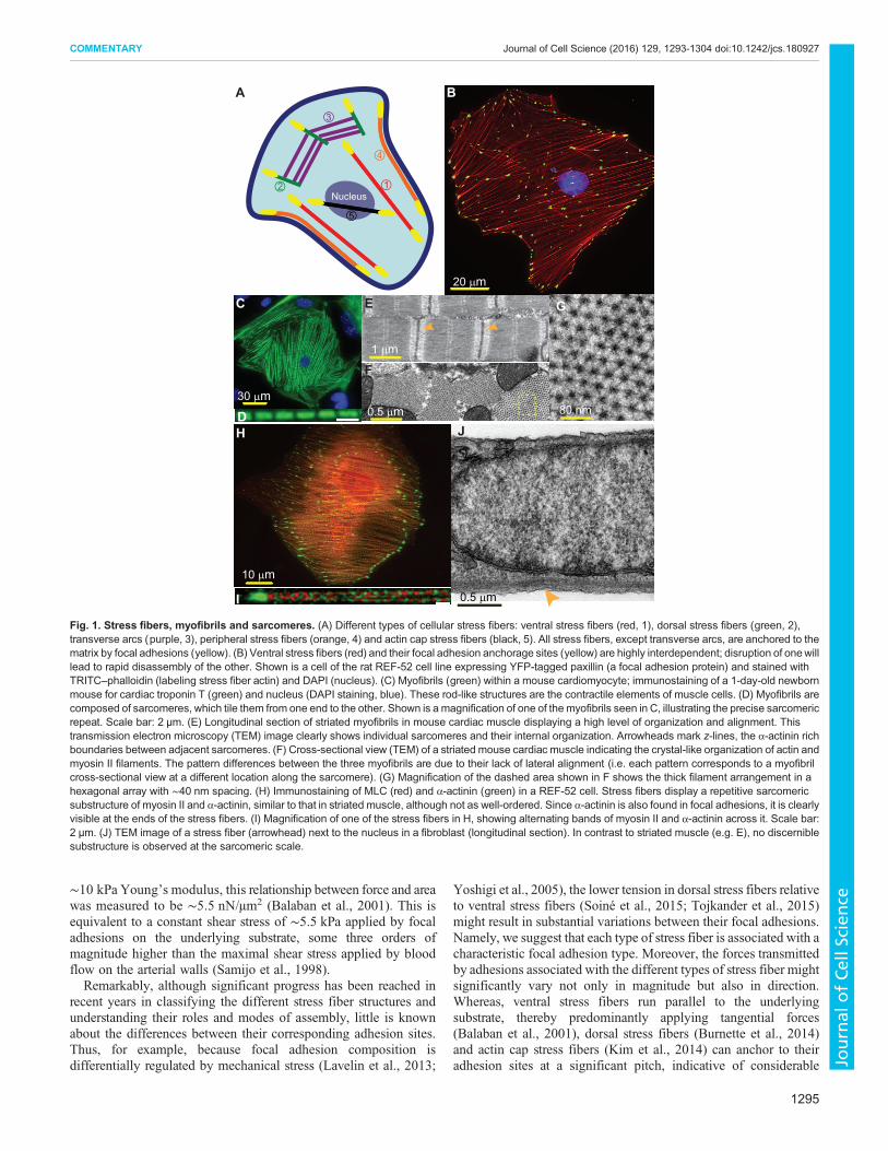
Fig. 1. Stress fibers, myofibrils and sarcomeres. (A) Different types of cellular stress fibers: ventral stress fibers (red, 1), dorsal stress fibers (green, 2),transverse arcs (purple, 3), peripheral stress fibers (orange, 4) and actin cap stress fibers (black, 5). All stress fibers, except transverse arcs, are anchored to thematrix by focal adhesions (yellow). (B) Ventral stress fibers (red) and their focal adhesion anchorage sites (yellow) are highly interdependent; disruption of onewilllead to rapid disassembly of the other. Shown is a cell of the rat REF-52 cell line expressing YFP-tagged paxillin (a focal adhesion protein) and stained withTRITC–phalloidin (labeling stress fiber actin) and DAPI (nucleus). (C) Myofibrils (green) within a mouse cardiomyocyte; immunostaining of a 1-day-old newbornmouse for cardiac troponin T (green) and nucleus (DAPI staining, blue). These rod-like structures are the contractile elements of muscle cells. (D) Myofibrils arecomposed of sarcomeres, which tile them from one end to the other. Shown is a magnification of one of the myofibrils seen in C, illustrating the precise sarcomericrepeat. Scale bar: 2 µm. (E) Longitudinal section of striated myofibrils in mouse cardiac muscle displaying a high level of organization and alignment. Thistransmission electron microscopy (TEM) image clearly shows individual sarcomeres and their internal organization. Arrowheads mark z-lines, the α-actinin richboundaries between adjacent sarcomeres. (F) Cross-sectional view (TEM) of a striated mouse cardiac muscle indicating the crystal-like organization of actin andmyosin II filaments. The pattern differences between the three myofibrils are due to their lack of lateral alignment (i.e. each pattern corresponds to a myofibrilcross-sectional view at a different location along the sarcomere). (G) Magnification of the dashed area shown in F shows the thick filament arrangement in ahexagonal array with ∼40 nm spacing. (H) Immunostaining of MLC (red) and α-actinin (green) in a REF-52 cell. Stress fibers display a repetitive sarcomericsubstructure of myosin II and α-actinin, similar to that in striated muscle, although not as well-ordered. Since α-actinin is also found in focal adhesions, it is clearlyvisible at the ends of the stress fibers. (I) Magnification of one of the stress fibers in H, showing alternating bands of myosin II and α-actinin across it. Scale bar:2 µm. (J) TEM image of a stress fiber (arrowhead) next to the nucleus in a fibroblast (longitudinal section). In contrast to striated muscle (e.g. E), no discerniblesubstructure is observed at the sarcomeric scale.
1295
COMMENTARY Journal of Cell Science (2016) 129, 1293-1304 doi:10.1242/jcs.180927
Journal
ofCe
llScience
out-of-plane contribution (Hur et al., 2009), for which most two-dimensional (2D) traction force microscopy setups are not sensitiveenough. Hence, because current studies are prone to yield a mix ofmeasurements from the potentially different adhesion types, animproved characterization (as in Kim et al., 2012) can influence theresults of focal adhesion analysis. This, in turn, might explain, whyin some cases a linear force–area relationship of focal adhesions isonly partly observed (Oakes et al., 2014; Stricker et al., 2011; Tanet al., 2003), whereas in others it is discerned clearly (Balaban et al.,2001; Trichet et al., 2012).Naturally, stress fibers interact mechanically not only with focal
adhesions but also with additional cellular structures, includingneighboring stress fibers and the actin cortex (see below); similarly,focal adhesions do not exclusively interact with stress fibers. Yet,for the sake of simplicity, we consider a scenario in the nextsections, in which the interface between stress fiber and focaladhesion is the dominant – if not exclusive –mechanical connectionbetween the two systems, and stress-fiber-generated forces aretransmitted to the ECM solely through focal adhesions.
Stress fibers and myofibrils – some similarities, maindifferencesStress fibers and striated muscle myofibrils are mechanisticallydistinct structures but, for the purpose of our discussion, theirapparent similarities are highly appealing. Both are elongated linearstructures that generate actomyosin-based contractile forces. Bothare also organized in alternating bands of myosin II and α-actininacross a long actin cable and, in both systems, there are alternatingsegments in which actin displays an opposite polarity. Given thatmyofibrils are much better characterized than stress fibers (seeBox 1), owing to their fundamental physiological role in muscle it isnot surprising that they often serve as a primary reference system forstress fiber study.Stress fibers display a repetitive segmented substructure that is
reminiscent of the internal order found in striated muscle; bothdisplay ordered actin filaments, crosslinked at one end by α-actininand interconnected at the other end by myosin motors. Just as instriatedmuscle, they are contractile – owing to sliding ofmyosin II onactin fibers – and play a key role in force generation. In view of thisapparent similarity, the contractile units of stress fibers are oftengenerally referred to also as sarcomeres (Kreis and Birchmeier, 1980;Sanger et al., 2006). Nevertheless, beyond these rather superficialsimilarities, fundamental differences exist between stress fiber andmyofibril sarcomeres. First, theirmolecular composition is somewhatdifferent; for example, some of the key components (such as α-actinin, myosin II, actin-binding proteins) have muscle-specific andstress-fiber-specific isoforms. Second, stress fiber contraction isregulated by phosphorylation ofmyosin light chain (MLC), as is seenin smooth muscle, rather than by troponin switching (Huxley, 1969).Third, the stress fiber sarcomere is not as precisely aligned as itscounterpart in muscle (Fig. 1E). Fourth, the sarcomeres along a stressfiber are not uniform, displaying both temporal and spatial variationsin length (Chapin et al., 2012; Peterson et al., 2004). Owing to thesedifferences, key aspects of the organization and contractile functionof stress fiber sarcomeres remain poorly characterized (e.g. thenumber of motors per sarcomere, actomyosin packing, etc.).At the organizational and functional levels, stress fibers are
typically one order of magnitude thinner than myofibrils, and theircontraction is considerably less coordinated. In the same cell, stressfibers may align in different directions, span different lengths(typically, 10–100 µm) and show limited or no sarcomereregistration (Fig. 1H–J). Moreover, along a single stress fiber,
different sarcomere behaviors can be observed. Thus, althoughstress fibers generate contractile forces, they appear far less efficientin doing so than myofibrils. This, naturally, calls into question thesimilarities between the two systems.
One main distinction between stress fibers and myofibrils is thelocation and magnitude of the forces they produce through theircontractile machinery. Stress fiber contractile forces are applied byindividual cells to their immediate environment through focaladhesions. These forces, which may locally rearrange the ECM(Avnur and Geiger, 1981), do not result in significant matrixdeformations – <1 µm over a typical stress fiber length of ∼100 µm(Trichet et al., 2012). Thus, stress fibers generate seeminglyisometric forces, even when they undergo a high level of contraction(Deguchi et al., 2006). In contrast, striated myofibrils can undergosignificant length changes (shortening by up to ∼30%), which isnecessary for skeletal movement and heart contraction. Theresulting non-isometric forces are transmitted over macroscopiclengths; skeletal myofibrils span between bones to form (togetherwith joints) effective lever systems, and cardiac myofibrils ofneighboring cells are mechanically coupled through adherensjunctions along the intercalated discs to efficiently contract theheart. Finally, although myofibrils can also interact locally with thesurrounding ECM (e.g. through costameres or tendons), this isprincipally a secondary effect whose magnitude still remainsunclear (Hersch et al., 2013).
Another clear difference between striated myofibril and stressfiber contractility relates to their activation dynamics. Striated
Box 1. MyofibrilsStriated myofibrils are extended cylindrical structures, spanningthe entire length of muscle cells (∼100 µm in cardiac muscle; a fewmillimeters to tens of centimeters in skeletal muscle). Their fundamentalbuilding block is the sarcomere, which, at rest, is typically ∼2 µm long butis able to shrink by∼30% at the peak of muscle contraction (Lodish et al.,2000). Thus, 102–105 sarcomeres, precisely ordered one after the other,tile a striated myofibril from end to end (see Fig. 1C–E). The sarcomeresthemselves are organized as thick complexes of bipolar myosin IIfilaments (thick filaments) that are flanked by individual actin fibers (thinfilaments) (Alberts et al., 2007). In cross-section, electron microscopyreveals that the thick filaments are arranged in a hexagonal array(∼40 nm spacing), with actin fibers located at the center of each triangleof myosin filaments (Huxley and Faruqi, 1983) (see Fig. 1F,G). Inaddition, the actin filaments of a sarcomere are all oriented with theirpointed (minus) ends pointing inwards, towards the thick filaments at thecenter. At their barbed (plus) end, the filaments of neighboringsarcomeres are crosslinked by α-actinin, thus giving rise to therepetitive sarcomeric structure (Alberts et al., 2007). Finally, althoughmany different proteins are involved in the regulation of myofibril structureand function (Ono, 2010), titin (connectin) and nebulin (Horowits et al.,1986) stand out in their relevance to the mechanical properties of theindividual sarcomere. These giant proteins (∼1 µm long) span from theends of the sarcomere along the thin filaments (nebulin), up to thesarcomere center (titin). Thus, the mechanical behavior of the myofibrildepends not only on its actomyosin machinery, but also on the elasticityof these proteins (Maruyama, 1997).By shortening their sarcomeric units, myofibrils apply contractile forces
to their anchorage sites. This process takes place through a controlledattachment-pulling-detachment cycle of myosin II motors on the actinfibers. In addition, the polarity of the actin fiber in each sarcomereensures that the ratchet-like dynamics of myosin will always lead tosarcomere contraction (i.e. pulling the actin toward the sarcomerecenter). This entire process is regulated by amuscle-specific mechanismthat is based on availability of Ca2+ and ATP (Goody, 2003; Huxley,1969).
1296
COMMENTARY Journal of Cell Science (2016) 129, 1293-1304 doi:10.1242/jcs.180927
Journal
ofCe
llScience

muscles repeatedly shorten and relax, triggered by rapid actionpotentials and Ca2+ release (Alberts et al., 2007). Thus, skeletalmuscles can switch from rest to full contraction within 10–100 ms(Wood, 2012) to drive quick body movement, and cardiac musclesmay beat every 100 ms in small mammals. In contrast, smoothmuscle cells and stress fibers respond much slower. Theircontractility is regulated by phosphorylation of MLC (Katohet al., 2001) and does not depend (solely) on electrical pulses.Consequently, hollow organs (e.g. blood vessels, epithelial tubes orglands) that are enveloped by smoothmuscle, contract and expand attimescales that are up to ∼30× longer than those for striated muscle(Wood, 2012). In addition, stress fiber contractile forces takeminutes to reach peak amplitude (Mbikou et al., 2006; Petersonet al., 2004), with hardly any changes in stress fiber length (Chapinet al., 2012). In effect, it was not until non-muscle cells were shownto be able to induce wrinkles in thin, deformable substrates (Harris,1984) that the contractile nature of stress fibers was, indeed,confirmed. Since then, a number of different methods, ranging fromexplicit measurements (Sugita et al., 2011) to observations of directconsequences – e.g. deformations of the underlying substrate(Balaban et al., 2001; Trichet et al., 2012), or stress fiber retractionfollowing laser cutting (Colombelli et al., 2009; Kumar et al., 2006;Russell et al., 2009) – have established that stress fibers are, in fact,constantly under tension. Therefore, whereas muscles can rapidlyswitch from rest to full power stroke, stress fibers remain at a quasi-constant operational level.The distinct force characteristics of stress fibers, compared to
those of myofibrils, suggest they have a different physiological role.It is conceivable that the relatively low and constant forces generatedby stress fibers are sufficient for cells to monitor and explore themechanical properties of their microenvironment, withoutdamaging the surrounding tissue or the focal adhesions. Inaddition, just as forces are propagated from stress fibers to theECM through focal adhesions, external forces applied to the focaladhesion-stress fiber complex can be sensed by this system, andmodulate its organization and turnover (Livne et al., 2014). This isfurther facilitated by the slow temporal response of a stress fiber;whereas the myofibril actomyosin machinery needs to be fullyassembled at all times to assure a quick response rate, stress fiberscontinuously assemble and disassemble. Thus, the limited co-alignment and registry that lowers the capacity of force generation,might improve the mechanosensing abilities of a cell with regard toprobing and sensing its surroundings from different directions.
In light of the fundamental differences between stress fibers andmyofibrils, we should find a better description of the mechanismunderlying stress fiber mechanics. Moreover, given theinterdependence between stress fibers and focal adhesions, theregulatory role of the adhesion sites also needs to be furtherinvestigated. The questions presented in the following sectionrepresent attempts to highlight and explore the inner workings ofstress fibers.
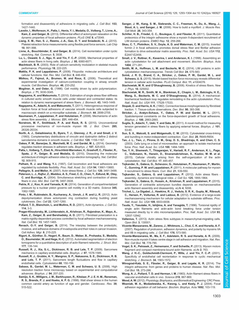
Where are the stress fiber sarcomeric units assembled, andwhat is their fate?We begin our discussion of this aspect with the observation thatactin filaments are continuously polymerized in focal adhesions(Chorev et al., 2014; Skau et al., 2015; Tojkander et al., 2015). Thenewly formed actin filaments are then treadmilled away along theattached stress fibers towards the cell center, at a velocity of 0.02–0.4 µm/min (Endlich et al., 2007; Russell et al., 2011; Tojkanderet al., 2015). As the pointed end of an actin filament always growsaway from the nucleation site, these filaments all possess the sameuniform polarity. Accordingly, the actin crosslinker fascin, whichbinds to unipolar, bundled actin filaments, is only found in focaladhesions and at stress fiber termini (Elkhatib et al., 2014). Incontrast, towards the stress fiber center, actin polarity appears to bebi-directional, as is the case in myofibrils (Cramer et al., 1997)(Fig. 2A,B). Since sarcomeres are found throughout the length ofthe stress fiber, up to the adhesion sites, this suggests two possiblescenarios; either the unipolar actin bundles that emanate from focaladhesions are quickly reorganized (partially or completely) to thealternating polarities observed in the central region (Cramer et al.,1997), or that actin organization along the stress fibers is non-uniform, resulting in variations in elastic and contractile behaviors(Peterson et al., 2004; Vogel et al., 2013).
Actin polymerization at focal adhesions appears to be a force-dependent process (Hirata et al., 2008; Kozlov and Bershadsky,2004). Diminishing stress fiber-generated forces by using inhibitorsof myosin or Rho kinases (Endlich et al., 2007) leads to thecessation of actin treadmilling at the stress fiber end. The fact that,despite the centripetal flow of actin, the length of stress fibersremains constant or even decreases, raises the question of whathappens to all the actin incoming from the focal adhesions. Onepossibility is that the newly created actin pushes against andcompresses the central part of the stress fiber. This, however, doesnot occur in practice, as the central stress fiber sarcomeres tend to
ECM
SFFA
SF
α-actinin
FA
2 μm
B
A C
Fig. 2. Stress fiber organization: from 1D idealization to 2D reality. (A) Schematic of an isolated, single stress fiber (SF, red) running between two focaladhesions (FA, yellow). Stress fibers typically extend a few tens of microns, with a cross-section of less than 0.5 µm, giving rise to their rod-like morphology.(B) Simplified model of the stress fiber substructure illustrating the organization of actin (red), α-actinin (blue) and myosin II (green). The short actin filamentspresent in the stress fiber change from uniform polarity at and near the focal adhesions, to bi-directional polarity inside the stress fiber sarcomeres. Pointed ends ofthe red actin monomers depict their orientation. (C) In mature cells, multiple interconnections are observed between adjacent stress fibers (arrowheads),resulting in the formation of a 2D mechanical network from the 1D structures illustrated in A. This allows stress fibers to respond to external force input in acoordinated manner. Note that finer sub-resolution connections can also exist between neighboring stress fibers [e.g. actin fibers (Xu et al., 2012), microtubules,intermediate filaments] that are not observed in standard fluorescence microscopy.
1297
COMMENTARY Journal of Cell Science (2016) 129, 1293-1304 doi:10.1242/jcs.180927
Journal
ofCe
llScience
expand over time compared to those located closer to the focaladhesions (Elkhatib et al., 2014; Peterson et al., 2004). Anotherpossibility is that, as well-spread cells have multipleinterconnections between stress fibers that together form dynamic2D branched networks (Fig. 2C) (Kumar et al., 2006), the excessactin might be transported away from the main cables into sidebranches (Russell et al., 2011). Moreover, these networks are likelyto be mechanically coupled (Kumar et al., 2006) and can respond toforces in a coordinated manner (Livne et al., 2014). Anotherconsequence of stress fiber branching is that individual stress fibersthat extend between two discrete adhesion sites are rarely observed.Given that stress fiber stability and growth are mechanicallyregulated, it appears that local changes in a single stress fiber (forwhatever reason) are likely to induce long-range effects on the entirestress fiber network.Evidently, actin incorporation into stress fibers occurs not only
at focal adhesions, but also along the entire length of the structure,as was directly observed by fluorescence recovery afterphotobleaching (FRAP) experiments, which demonstrated timesfor actin recovery of ∼10 min (Campbell and Knight, 2007;Hotulainen and Lappalainen, 2006). This recovery rate suggeststhat incorporation of new actin monomers occurs throughout theentire length of stress fibers, and not only at their focal-adhesion-associated ends, as it would take hours for actin polymerizing atfocal adhesions to arrive at the stress fiber center. Thus, althoughstress fibers recruit or release actin throughout their entire length,actin is also continuously polymerized at focal adhesions, and flowscentripetally. However, despite this exchange and new actinpolymerization at adhesion sites, the overall length of stress fibersand the tension along them, remain largely unchanged. How thesedifferent features of the stress-fiber–focal-adhesion system arecoordinated is unclear and deserves further consideration.
What mechanism underlies the coupling and co-regulationbetween stress fibers and focal adhesions through theterminal sarcomere?Physical interactions between focal adhesions and stress fibers,whether related to actin flow or to force transmission from one to theother, take place at the so-called ‘terminal sarcomere’. This is thefinal segment of the stress fiber that still contains myosin II, and
links the sarcomeric chain on the one side and the adhesion site onthe other. In recent years, significant progress has been made in ourunderstanding of the molecular architecture of focal adhesions(Case et al., 2015; Kanchanawong et al., 2010; Patla et al., 2010).Nevertheless, the structural connection between the focal adhesionand the stress fiber terminus remains largely unclear (Gimona et al.,2003; Kaverina et al., 2003).
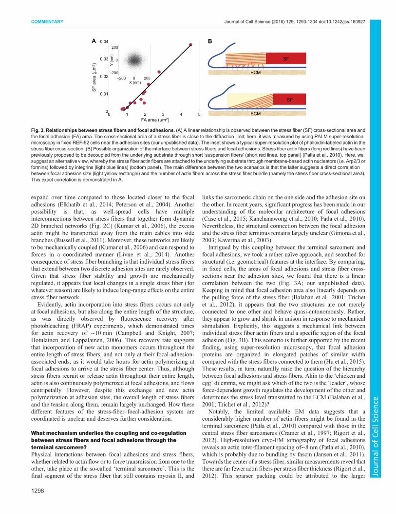
Intrigued by this coupling between the terminal sarcomere andfocal adhesions, we took a rather naïve approach, and searched forstructural (i.e. geometrical) features at the interface. By comparing,in fixed cells, the areas of focal adhesions and stress fiber cross-sections near the adhesion sites, we found that there is a linearcorrelation between the two (Fig. 3A; our unpublished data).Keeping in mind that focal adhesion area also linearly depends onthe pulling force of the stress fiber (Balaban et al., 2001; Trichetet al., 2012), it appears that the two structures are not merelyconnected to one other and behave quasi-autonomously. Rather,they appear to grow and shrink in unison in response to mechanicalstimulation. Explicitly, this suggests a mechanical link betweenindividual stress fiber actin fibers and a specific region of the focaladhesion (Fig. 3B). This scenario is further supported by the recentfinding, using super-resolution microscopy, that focal adhesionproteins are organized in elongated patches of similar widthcompared with the stress fibers connected to them (Hu et al., 2015).These results, in turn, naturally raise the question of the hierarchybetween focal adhesions and stress fibers. Akin to the ‘chicken andegg’ dilemma, we might ask which of the two is the ‘leader’, whoseforce-dependent growth regulates the development of the other anddetermines the stress level transmitted to the ECM (Balaban et al.,2001; Trichet et al., 2012)?
Notably, the limited available EM data suggests that aconsiderably higher number of actin fibers might be found in theterminal sarcomere (Patla et al., 2010) compared with those in thecentral stress fiber sarcomeres (Cramer et al., 1997; Rigort et al.,2012). High-resolution cryo-EM tomography of focal adhesionsreveals an actin inter-filament spacing of∼8 nm (Patla et al., 2010),which is probably due to bundling by fascin (Jansen et al., 2011).Towards the center of a stress fiber, similar measurements reveal thatthere are far fewer actin fibers per stress fiber thickness (Rigort et al.,2012). This sparser packing could be attributed to the larger
SF
ECM
SF
ECM0 1 2 3 4 50
0.01
0.02
0.03
0.04
FA area (μm2)
A B
−200 0 200−200
0
200
Y (n
m)
X (nm)
SF
area
(μm
2 )
Fig. 3. Relationships between stress fibers and focal adhesions. (A) A linear relationship is observed between the stress fiber (SF) cross-sectional area andthe focal adhesion (FA) area. The cross-sectional area of a stress fiber is close to the diffraction limit; here, it was measured by using PALM super-resolutionmicroscopy in fixed REF-52 cells near the adhesion sites (our unpublished data). The inset shows a typical super-resolution plot of phalloidin-labeled actin in thestress fiber cross-section. (B) Possible organization of the interface between stress fibers and focal adhesions. Stress fiber actin fibers (long red lines) have beenpreviously proposed to be decoupled from the underlying substrate through short ‘suspension fibers’ (short red lines, top panel) (Patla et al., 2010). Here, wesuggest an alternative view, whereby the stress fiber actin fibers are attached to the underlying substrate throughmembrane-based actin nucleators (i.e. Arp2/3 orformins) followed by integrins (light blue lines) (bottom panel). The main difference between the two scenarios is that the latter suggests a direct correlationbetween focal adhesion size (light yellow rectangle) and the number of actin fibers across the stress fiber bundle (namely the stress fiber cross-sectional area).This exact correlation is demonstrated in A.
1298
COMMENTARY Journal of Cell Science (2016) 129, 1293-1304 doi:10.1242/jcs.180927
Journal
ofCe
llScience
physical dimensions of either α-actinin (∼35 nm) or myosin II[∼30–50 nm (MacIntosh, 2003)], both of which crosslink thesarcomeric actin. However, as actin is constantly being polymerizedat focal adhesions and treadmilled along the stress fibers,components of the initial terminal sarcomere necessarily formregular sarcomeres over time. This, therefore, raises the question ofwhat happens to the excess of actin fibers in focal adhesions, andwhat other changes in its molecular architecture take place duringthis treadmilling process?
Are all sarcomeres along the stress fiber identical?Variations in sarcomere behavior are observed along the stressfibers. Sarcomeres near the stress fiber center, for example, areslightly longer than their counterparts closer to the termini (Elkhatibet al., 2014; Peterson et al., 2004). However, when stress fibercontractility is stimulated by calyculin A or lipoprotein-a (LPA),this small difference becomes quite pronounced. Thus, a ∼50%expansion of the (originally slightly longer) central sarcomeres isaccompanied by a 30–40% shortening of sarcomeres at theperiphery upon increased phosphorylation of MLC in fibroblasts(Peterson et al., 2004). Furthermore, the concentrations of myosin IIand α-actinin, and their exchange timescales also vary significantlybetween central and peripheral sarcomeres (Peterson et al., 2004).At the single sarcomere level, spontaneous length variations mayalso take place without any apparent external perturbation (Chapinet al., 2012). As the stress fiber is a closed system over such shorttimescales (i.e. its overall length hardly changes), such spontaneousshortening (elongation) events are necessarily accompanied by anelongation (shortening) somewhere else along the structure.Interestingly, when an individual sarcomere suddenly changes itslength, only a small region around it (∼3 sarcomeres on either side)is affected (Chapin et al., 2012).The variations in sarcomere dynamics, however, do not appear to
arise directly from differences in the actin organization along thestress fibers. First, as actin bundles of mixed polarity are assumed tobe more contractile than those of uniform polarity, in which some ofthe myosin heads are attached to the actin fibers in the wrongdirection (Vogel et al., 2013), the bulk of stress fiber contractionshould take place near its center. In reality, the exact oppositebehavior is observed, with the sarcomeres next to focal adhesionsbeing more contractile (Peterson et al., 2004). What, then, is theorigin of the sarcomere variability? While this largely remains anopen question, one interesting direction of research involves thecombination of biochemical signaling processes with stress fibermechanics (Besser and Schwarz, 2007), which showed thatinhomogeneities in biochemical reaction–diffusion fields aroundthe focal adhesions can account for the observed spatial variations insarcomere contractility.Owing to the linear structure of stress fibers, a uniform force
magnitude is propagated across all their sarcomeres. Nevertheless,the observations of a differential contractile behavior suggest thatthe production of this force is not evenly distributed. Localized forcegeneration, whether close to focal adhesions or further away, mightexplain the limited contraction of stress fibers compared tomyofibrils. After all, whereas all the sarcomeres in a myofibrilcontract simultaneously and lead to its dramatic shortening (up to∼30%), the contraction of stress fibers is nearly isometric, withalmost no significant length change. Moreover, in isolated form,stress fibers can shorten to less than 25% of their original length(Katoh et al., 1998). Thus, the limited stress fiber contraction mightserve a physiological purpose. As non-muscle cells need to applyforce to their environment to probe its mechanical properties, it can
be advantageous not to cause any deformations (crucial forcontracting the heart or generating large skeletal movements) thatare too large, which could tear or damage the soft, surroundingECM. In conclusion, the exact location where stress fibercontractility takes place remains unclear.
An enigma of force generation and transduction along stressfibersAn interesting correlation has been observed between the stressfiber-generated contractile forces and the focal adhesion areathrough which they are transmitted to the ECM (Balaban et al.,2001; Trichet et al., 2012). When focal adhesions increase (ordecrease) in size, the traction forces they apply on the underlyingsubstrate increase (or decrease) accordingly. Moreover, althoughfocal adhesion dynamics are strongly related to the chemical andstructural properties of the underlying matrix, different force-areabehaviors are observed when cells are attached to pillar substrates ofsimilar composition but varying effective rigidities (Trichet et al.,2012). Specifically, higher forces per adhesion size were measuredon stiffer substrates, with only little change in chemistry and rigidityof the local surface, with which the focal adhesions directly interact.One interesting explanation for this force-area correlation stemsfrom the molecular composition of focal adhesions. The load-dependent exchange rates of key adhesion components (Lavelinet al., 2013) suggest that under increasing tension, focal adhesionspreferably recruit new proteins and, thus, grow. Inversely, whentension is removed, disassociation rates dominate and the molecularadhesion structure breaks down. However, this explanation, likeother focal adhesion-based and force-oriented approaches, cannotaccount for the pillar measurements, which have shown that theforce-area relationship is dependent on the effective substraterigidity (i.e. pillar geometry) rather than on local surface stiffness(with which the focal adhesions come in to contact). Rigiditysensing, therefore, does not appear to take place solely at the focaladhesion level.
To understand how the stress-fiber–focal-adhesion system sensesmatrix stiffness and responds with different force–area dynamics,the mechanical behavior of the different elements at play need to becharacterized first. Beginning with the relevant structural rigidities,no direct measurements of the elastic properties of focal adhesionsare available to our knowledge. In contrast, stress fibers have beenanalyzed by using a wide range of experimental techniques, leadingto values that differ by more than two orders of magnitude. In vivoestimates for the Young’s modulus of stress fibers range from1–10 kPa as determined by AFM nano-indentation (Lu et al., 2008)to ∼100 kPa, a value that has been obtained by stretching adherentcells through their underlying substrate and measuring the resultingdeformation fields (Nagayama et al., 2011; Nagayama andMatsumoto, 2010). For isolated stress fibers, the obtained values(≥1MPa) differ even more (Deguchi et al., 2006). In addition, stressfiber rigidity can also be inferred indirectly: the Young’s modulus ofa rod-like stress fiber is simply the ratio between the stresses appliedat its ends and the resulting strain. As the internal stress fiber forcesare expected to be identical to those transmitted by focal adhesions,the stresses in both structures should differ according to theirrespective areas (stress is the force transmitted across a unit area).For a wide range of substrate rigidities, focal adhesion tractionstresses are typically 5–20 kPa [(Balaban et al., 2001; Trichet et al.,2012) although the practical limit might even be higher (Ghassemiet al., 2012)] applied over a surface area that is∼100× larger than thecorresponding area of the stress fiber cross-section (Fig. 3A). Thisimplies that internal stresses in stress fibers are ∼100× higher than
1299
COMMENTARY Journal of Cell Science (2016) 129, 1293-1304 doi:10.1242/jcs.180927
Journal
ofCe
llScience
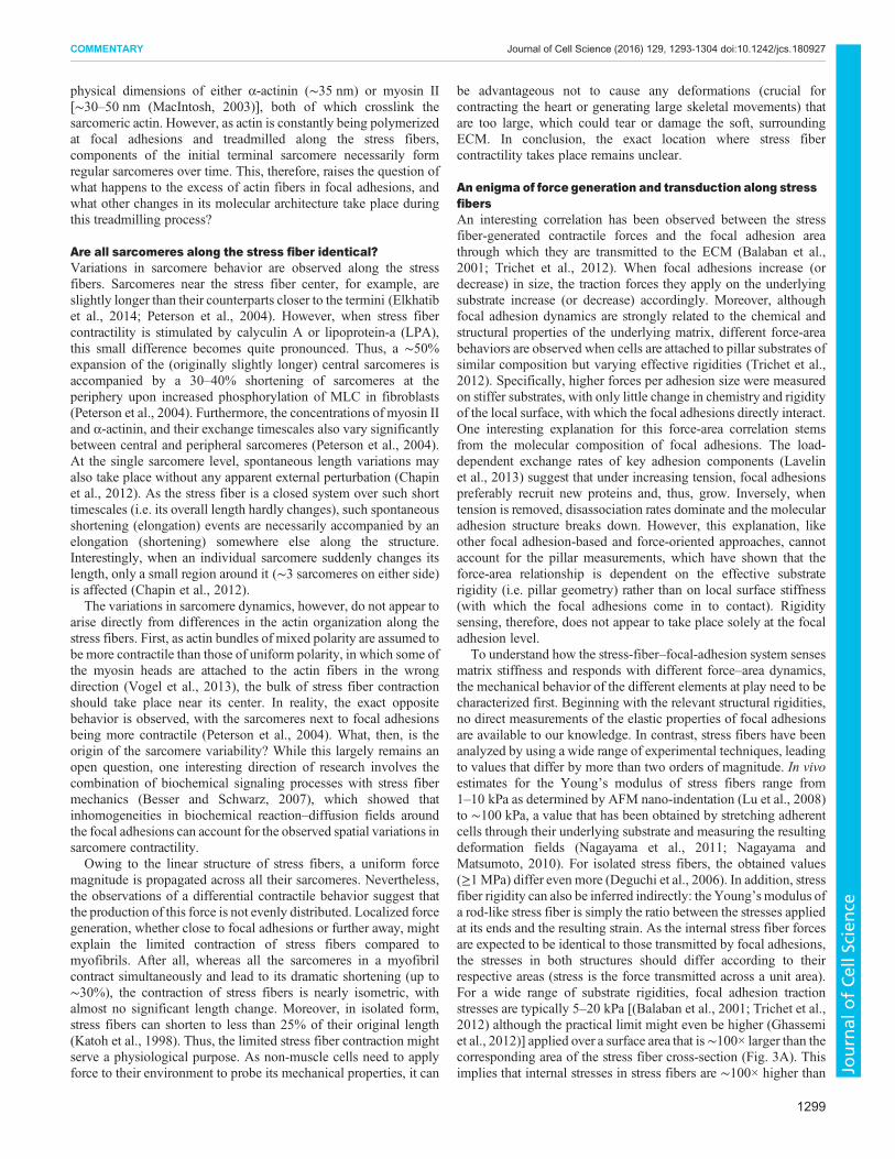
those seen in focal adhesions, i.e. 0.5–2 MPa [in comparison,estimates of the internal stress in striated muscle yield ∼0.3 MPa(Bloch and Gonzalez-Serratos, 2003)]. Moreover, the extensionalstrain in stress fibers (defined as the change in length relative to theoriginal length) is estimated to be in the range of 2–20% (Deguchiet al., 2006). Taken together, these numbers yield a stress fiberYoung’s modulus of at least 3–10 MPa, which is significantlyhigher than that suggested by the experimental results (note that inthis calculation, our unpublished data presented in Fig. 3A wasused). In summary, there is a wide variability, both between thedifferent stress fiber rigidity measurements (Fig. 4A), but alsobetween their indirectly estimated values. However, because allthese numbers are well below the ∼1 GPa rigidity of individual bareactin fibers (Kojima et al., 1994; Tsuda et al., 1996), it stands toreason that the elastic response of stress fibers is dictated rather bysomething else, possibly by the actin crosslinkers themselves.As discussed above, the spacing of actin fibers across the stress
fiber thickness can be estimated from high-resolution EM images(∼35–40 nm spacing, comparable to the lengths of the main stressfiber crosslinkers α-actinin and myosin II). Together with theinternal stress fiber stress, these two numbers enable us to calculatethe contractile force applied on individual actin fibers (the forcebeing the product of the stress and area). Thus, for example, forhighly rigid substrates (e.g. glass), this force is predicted (using ourunpublished data presented in Fig. 3A) to be ∼2 nN (Fig. 4B).Assuming now that this force is generated solely by actomyosincontractility, we ask ourselves how many myosin II power strokesare needed to produce it. As in each cycle of attachment-pulling-detachment an individual motor generates up to ∼2 pN force(Norstrom et al., 2010), then 1000 such cycles would be needed togenerate ∼2 nN. However, because in each cycle, a myosin motoradvances along the actin fiber by one actin monomer, which spans∼5.5 nm (Norstrom et al., 2010), these 1000 cycles would also
amount to a sarcomere shortening of 5.5 µm. As this contraction isfar greater than the sarcomere rest length of 1.5–2 µm (Petersonet al., 2004), it appears that the stress fiber contractile forces cannotbe generated by regular sarcomeres. This puzzling and controversialresult requires further elaboration, and, together with the widevariability of stress fiber rigidity measurements, highlights the needto better clarify our understanding of stress fiber mechanics.
An alternative mechanism of stress fiber force generationbased on regulated global actin turnoverThe considerations discussed above suggest that the one-dimensional picture of a focal adhesion/stress fiber/focal adhesionsystem (Fig. 2A), which is mechanically driven purely by a uniform,actomyosin-based (muscle-like) contraction, might need to bereviewed. It is incompatible with the accumulating experimentaldata for the fine structure of the stress fiber network, the structuraldiversity of the stress fiber sarcomeres, and the isometric nature ofthe actomyosin-based contractile machinery. Filament sliding per semight, therefore, not be the sole generator of force. As indicated,stress fibers are dynamic structures whose building blocks (actin,myosin II, α-actinin and the like) are constantly exchanged with asoluble cytoplasmic pool (Fig. 5A). The exact mechanismsunderlying this exchange processes are still poorly characterized,yet they are most likely to be regulated by a balance between theincorporation of new molecules into the structure, and thedissociation of molecules from it. Along these lines,O’Shaughnessy and colleagues took into account sarcomereremodeling to explain different stress fiber-related measurements(Stachowiak and O’Shaughnessy, 2008; Stachowiak et al., 2014).Essentially, the extent of actin fiber overlap originating from bothends of the stress fiber sarcomere was assumed to have a definedsteady-state value and to be dynamically regulated by actinpolymerization and disassembly rates. It is tempting, however, totake this assumption one step further and consider the possibilitythat the molecular association and dissociation rates of key stressfiber component(s) is affected by the tension along the structure.This is in line with differences in the dissociation rates of specificstress fiber and focal adhesion components observed followingblocking of actomyosin contractility (Fig. 5B) (Lavelin et al., 2013).In addition, a recent study suggests that myosin II-derived forcesinhibit vectorial actin polymerization at focal adhesions (Tojkanderet al., 2015). Thus, it is conceivable that a reduction in stress (forexample, due to actin polymerization at the terminal sarcomeres)increases dissociation events (but without rupturing the structurebecause only a small number of fibers are cut at a time), effectivelyleading to a shortening of the stress fiber. In this manner, theeffective contractile stress of the stress fiber can increase again (Sunet al., 2010) until regaining its optimal tension level [akin to theconstant stress in focal adhesions (Balaban et al., 2001)] (Fig. 5C).These events can take place in discrete regions along the stress fiber,such as its center, which would account for the observedinhomogeneity discussed above. Moreover, stress fiber branchingor interactions with either cortical actin or other cytoskeletalsystems, could locally affect stress fiber mechanics, which, in turn,might be balanced by the proposed force-induced regulation of actinassembly and disassembly.
The above scenario we propose suggests the dynamic regulation ofstress fiber length and tension by a dual mechanism involving, on onehand, actin polymerization and its flow from focal adhesions and, onthe other hand, exchange throughout the entire length of the stressfiber. Accordingly, sarcomere formation and their dissolution dependon the balance between actin dissociation and incorporation (Fig. 5D).
B
A1 10
SF(by nano-
indentation)
Young’smodulus(kPa)
102
SF(by cell
stretching)
103 106
SF(by stretchingisolated SFs)
actin fiber(by nano-
manipulation)
Ff
ECM
Ff
SFFA
Fig. 4. Mechanical propertiesof stress fibers. (A)Stress fiber (SF) rigidity hasbeenanalyzedby usingawide range of experimental techniques (for details seemain text). The resulting Young’s modulus estimates range from ∼1 kPa to>1 MPa, suggesting that an improved understanding of stress fiber structure andmechanics is necessary for the correct interpretation of the existingmeasurements. Nevertheless, the fact that all measurements and estimates arewell below the∼1 GPa rigidity of individual bare actin filaments suggests that theelastic response of stress fibers is not dictated by the constituting actin fibers but,rather, by their crosslinkers. (B) The contractile tension (F) in a stress fiber isgenerated by (and distributed between) the individual comprising actin fibers.These single-filament forces (f ) can be estimated from measurements of F andthe number of filaments per stress fiber cross-section.
1300
COMMENTARY Journal of Cell Science (2016) 129, 1293-1304 doi:10.1242/jcs.180927
Journal
ofCe
llScience
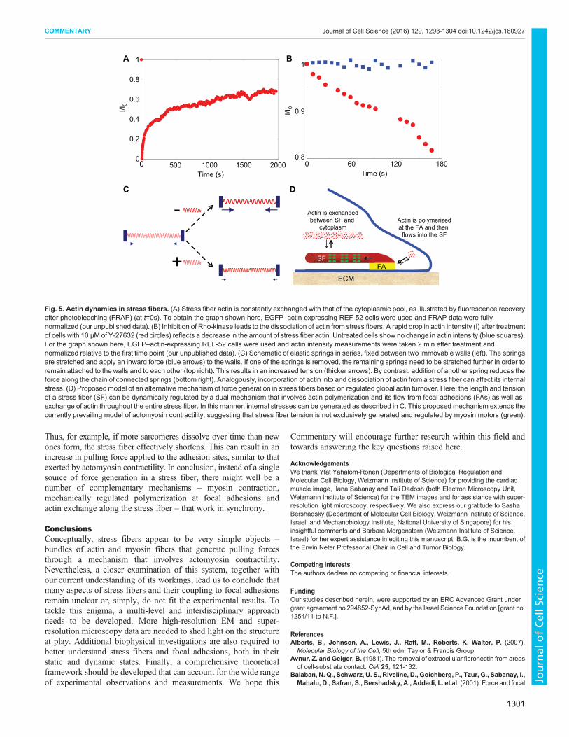
Thus, for example, if more sarcomeres dissolve over time than newones form, the stress fiber effectively shortens. This can result in anincrease in pulling force applied to the adhesion sites, similar to thatexerted by actomyosin contractility. In conclusion, instead of a singlesource of force generation in a stress fiber, there might well be anumber of complementary mechanisms – myosin contraction,mechanically regulated polymerization at focal adhesions andactin exchange along the stress fiber – that work in synchrony.
ConclusionsConceptually, stress fibers appear to be very simple objects –bundles of actin and myosin fibers that generate pulling forcesthrough a mechanism that involves actomyosin contractility.Nevertheless, a closer examination of this system, together withour current understanding of its workings, lead us to conclude thatmany aspects of stress fibers and their coupling to focal adhesionsremain unclear or, simply, do not fit the experimental results. Totackle this enigma, a multi-level and interdisciplinary approachneeds to be developed. More high-resolution EM and super-resolution microscopy data are needed to shed light on the structureat play. Additional biophysical investigations are also required tobetter understand stress fibers and focal adhesions, both in theirstatic and dynamic states. Finally, a comprehensive theoreticalframework should be developed that can account for the wide rangeof experimental observations and measurements. We hope this
Commentary will encourage further research within this field andtowards answering the key questions raised here.
AcknowledgementsWe thank Yfat Yahalom-Ronen (Departments of Biological Regulation andMolecular Cell Biology, Weizmann Institute of Science) for providing the cardiacmuscle image, Ilana Sabanay and Tali Dadosh (both Electron Microscopy Unit,Weizmann Institute of Science) for the TEM images and for assistance with super-resolution light microscopy, respectively. We also express our gratitude to SashaBershadsky (Department of Molecular Cell Biology, Weizmann Institute of Science,Israel; and Mechanobiology Institute, National University of Singapore) for hisinsightful comments and Barbara Morgenstern (Weizmann Institute of Science,Israel) for her expert assistance in editing this manuscript. B.G. is the incumbent ofthe Erwin Neter Professorial Chair in Cell and Tumor Biology.
Competing interestsThe authors declare no competing or financial interests.
FundingOur studies described herein, were supported by an ERC Advanced Grant undergrant agreement no 294852-SynAd, and by the Israel Science Foundation [grant no.1254/11 to N.F.].
ReferencesAlberts, B., Johnson, A., Lewis, J., Raff, M., Roberts, K. Walter, P. (2007).
Molecular Biology of the Cell, 5th edn. Taylor & Francis Group.Avnur, Z. and Geiger, B. (1981). The removal of extracellular fibronectin from areas
of cell-substrate contact. Cell 25, 121-132.Balaban, N. Q., Schwarz, U. S., Riveline, D., Goichberg, P., Tzur, G., Sabanay, I.,
Mahalu, D., Safran, S., Bershadsky, A., Addadi, L. et al. (2001). Force and focal
0 60 1200.8
0.9
1
Time (s)
1
1800 500 1000 1500 20000
0.2
0.4
0.6
0.8
1
Time (s)
C
-
+SF
ECM
Actin is exchangedbetween SF and
cytoplasmActin is polymerizedat the FA and then
flows into the SF
D
A B
I/I0I/I
0
SFSF
FA
Fig. 5. Actin dynamics in stress fibers. (A) Stress fiber actin is constantly exchanged with that of the cytoplasmic pool, as illustrated by fluorescence recoveryafter photobleaching (FRAP) (at t=0s). To obtain the graph shown here, EGFP–actin-expressing REF-52 cells were used and FRAP data were fullynormalized (our unpublished data). (B) Inhibition of Rho-kinase leads to the dissociation of actin from stress fibers. A rapid drop in actin intensity (I) after treatmentof cells with 10 µM of Y-27632 (red circles) reflects a decrease in the amount of stress fiber actin. Untreated cells show no change in actin intensity (blue squares).For the graph shown here, EGFP–actin-expressing REF-52 cells were used and actin intensity measurements were taken 2 min after treatment andnormalized relative to the first time point (our unpublished data). (C) Schematic of elastic springs in series, fixed between two immovable walls (left). The springsare stretched and apply an inward force (blue arrows) to the walls. If one of the springs is removed, the remaining springs need to be stretched further in order toremain attached to the walls and to each other (top right). This results in an increased tension (thicker arrows). By contrast, addition of another spring reduces theforce along the chain of connected springs (bottom right). Analogously, incorporation of actin into and dissociation of actin from a stress fiber can affect its internalstress. (D) Proposedmodel of an alternative mechanism of force generation in stress fibers based on regulated global actin turnover. Here, the length and tensionof a stress fiber (SF) can be dynamically regulated by a dual mechanism that involves actin polymerization and its flow from focal adhesions (FAs) as well asexchange of actin throughout the entire stress fiber. In this manner, internal stresses can be generated as described in C. This proposed mechanism extends thecurrently prevailing model of actomyosin contractility, suggesting that stress fiber tension is not exclusively generated and regulated by myosin motors (green).
1301
COMMENTARY Journal of Cell Science (2016) 129, 1293-1304 doi:10.1242/jcs.180927
Journal
ofCe
llScience
adhesion assembly: a close relationship studied using elastic micropatternedsubstrates. Nat. Cell Biol. 3, 466-472.
Besser, A. and Schwarz, U. S. (2007). Coupling biochemistry andmechanics in celladhesion: a model for inhomogeneous stress fiber contraction. New J. Phys. 9,425.
Bloch, R. J. and Gonzalez-Serratos, H. (2003). Lateral force transmission acrosscostameres in skeletal muscle. Exerc. Sport Sci. Rev. 31, 73-78.
Brugues, A., Anon, E., Conte, V., Veldhuis, J. H., Gupta, M., Colombelli, J.,Mun oz, J. J., Brodland, G. W., Ladoux, B. and Trepat, X. (2014). Forces drivingepithelial wound healing. Nat. Phys. 10, 683-690.
Buccione, R., Orth, J. D. andMcNiven,M. A. (2004). Foot andmouth: podosomes,invadopodia and circular dorsal ruffles. Nat. Rev. Mol. Cell Biol. 5, 647-657.
Burnette, D. T., Manley, S., Sengupta, P., Sougrat, R., Davidson, M. W., Kachar,B. and Lippincott-Schwartz, J. (2011). A role for actin arcs in the leading-edgeadvance of migrating cells. Nat. Cell Biol. 13, 371-382.
Burnette, D. T., Shao, L., Ott, C., Pasapera, A. M., Fischer, R. S., Baird,M. A., DerLoughian, C. D., Delanoe-Ayari, H., Paszek, M. J., Davidson, M. W. et al.(2014). A contractile and counterbalancing adhesion system controls the 3Dshape of crawling cells. J. Cell Biol. 205, 83-96.
Burridge, K. andGuilluy, C. (2015). Focal adhesions, stress fibers and mechanicaltension. Exp. Cell Res. [Epup] doi:10.1016/j.yexcr.2015.10.029.
Burridge, K. andWittchen, E. S. (2013). The tensionmounts: stress fibers as force-generating mechanotransducers. J. Cell Biol. 200, 9-19.
Byron, A., Humphries, J. D., Bass, M. D., Knight, D. and Humphries, M. J.(2011). Proteomic analysis of integrin adhesion complexes. Sci. Signal. 4,2107-2114.
Campbell, J. J. and Knight, M. M. (2007). An improved confocal FRAP techniquefor the measurement of long-term actin dynamics in individual stress fibers.Microsc. Res. Tech. 70, 1034-1040.
Case, L. B., Baird, M. A., Shtengel, G., Campbell, S. L., Hess, H. F., Davidson,M. W. and Waterman, C. M. (2015). Molecular mechanism of vinculin activationand nanoscale spatial organization in focal adhesions.Nat. Cell Biol. 17, 880-892.
Chapin, L. M., Blankman, E., Smith, M. A., Shiu, Y.-T. andBeckerle, M. C. (2012).Lateral communication between stress fiber sarcomeres facilitates a localremodeling response. Biophys. J. 103, 2082-2092.
Chorev, D. S., Moscovitz, O., Geiger, B. and Sharon, M. (2014). Regulation offocal adhesion formation by a vinculin-Arp2/3 hybrid complex. Nat. Commun. 5,3758.
Chrzanowska-Wodnicka, M. and Burridge, K. (1996). Rho-stimulated contractilitydrives the formation of stress fibers and focal adhesions. J. Cell Biol. 133,1403-1415.
Colombelli, J., Besser, A., Kress, H., Reynaud, E. G., Girard, P., Caussinus, E.,Haselmann, U., Small, J. V., Schwarz, U. S. and Stelzer, E. H. K. (2009).Mechanosensing in actin stress fibers revealed by a close correlation betweenforce and protein localization. J. Cell Sci. 122, 1665-1679.
Cramer, L. P., Siebert, M. and Mitchison, T. J. (1997). Identification of novelgraded polarity actin filament bundles in locomoting heart fibroblasts: implicationsfor the generation of motile force. J. Cell Biol. 136, 1287-1305.
Deguchi, S., Ohashi, T. and Sato, M. (2006). Tensile properties of single stressfibers isolated from cultured vascular smooth muscle cells. J. Biomech. 39,2603-2610.
Delon, I. and Brown, N. H. (2009). The integrin adhesion complex changes itscomposition and function during morphogenesis of an epithelium. J. Cell Sci. 122,4363-4374.
Discher, D. E., Janmey, P. andWang, Y.-L. (2005). Tissue cells feel and respond tothe stiffness of their substrate. Science 310, 1139-1143.
Elkhatib, N., Neu, M. B., Zensen, C., Schmoller, K. M., Louvard, D., Bausch,A. R., Betz, T. and Vignjevic, D. M. (2014). Fascin plays a role in stress fiberorganization and focal adhesion disassembly. Curr. Biol. 24, 1492-1499.
Endlich, N., Otey, C. A., Kriz, W. and Endlich, K. (2007). Movement of stress fibersaway from focal adhesions identifies focal adhesions as sites of stress fiberassembly in stationary cells. Cell Motil. Cytoskeleton 64, 966-976.
Ezratty, E. J., Partridge, M. A. and Gundersen, G. G. (2005). Microtubule-inducedfocal adhesion disassembly is mediated by dynamin and focal adhesion kinase.Nat. Cell Biol. 7, 581-590.
Fackler, O. T. and Grosse, R. (2008). Cell motility through plasma membraneblebbing. J. Cell Biol. 181, 879-884.
Gateva, G., Tojkander, S., Koho, S., Carpen, O. and Lappalainen, P. (2014).Palladin promotes assembly of non-contractile dorsal stress fibers through VASPrecruitment. J. Cell Sci. 127, 1887-1898.
Geiger, B. and Yamada, K. M. (2011). Molecular architecture and function of matrixadhesions. Cold Spring Harb. Perspect. Biol. 3, a005033.
Geiger, B., Spatz, J. P. and Bershadsky, A. D. (2009). Environmental sensingthrough focal adhesions. Nat. Rev. Mol. Cell Biol. 10, 21-33.
Ghassemi, S., Meacci, G., Liu, S. M., Gondarenko, A. A., Mathur, A., Roca-Cusachs, P., Sheetz, M. P. and Hone, J. (2012). Cells test substrate rigidity bylocal contractions on submicrometer pillars. Proc. Natl. Acad. Sci. USA 109,5328-5333.
Gimona, M., Kaverina, I., Resch, G. P., Vignal, E. and Burgstaller, G. (2003).Calponin repeats regulate actin filament stability and formation of podosomes insmooth muscle cells. Mol. Biol. Cell 14, 2482-2491.
Gordon, A. M., Homsher, E. and Regnier, M. (2000). Regulation of contraction instriated muscle. Physiol. Rev. 80, 853-924.
Grashoff, C., Hoffman, B. D., Brenner, M. D., Zhou, R. B., Parsons, M., Yang,M. T., McLean, M. A., Sligar, S. G., Chen, C. S., Ha, T. et al. (2010). Measuringmechanical tension across vinculin reveals regulation of focal adhesion dynamics.Nature 466, 263-266.
Harris, A. K. (1984). Tissue culture cells on deformable substrata: biomechanicalimplications. J. Biomech. Eng. 106, 19-24.
Harris, A. K., Wild, P. and Stopak, D. (1980). Silicone rubber substrata: a newwrinkle in the study of cell locomotion. Science 208, 177-179.
Hersch, N., Wolters, B., Dreissen, G., Springer, R., Kirchgessner, N., Merkel, R.and Hoffmann, B. (2013). The constant beat: cardiomyocytes adapt their forcesby equal contraction upon environmental stiffening. Biol. Open 2, 351-361.
Hirata, H., Tatsumi, H. and Sokabe, M. (2008). Mechanical forces facilitate actinpolymerization at focal adhesions in a zyxin-dependent manner. J. Cell Sci. 121,2795-2804.
Hotulainen, P. and Lappalainen, P. (2006). Stress fibers are generated bytwo distinct actin assembly mechanisms in motile cells. J. Cell Biol. 173,383-394.
Hu, S. Q., Tee, Y.-H., Kabla, A., Zaidel-Bar, R., Bershadsky, A. and Hersen, P.(2015). Structured illumination microscopy reveals focal adhesions are composedof linear subunits. Cytoskeleton 72, 235-245.
Humphries, J. D., Wang, P., Streuli, C., Geiger, B., Humphries, M. J. andBallestrem, C. (2007). Vinculin controls focal adhesion formation by directinteractions with talin and actin. J. Cell Biol. 179, 1043-1057.
Hur, S. S., Zhao, Y. H., Li, Y.-S., Botvinick, E. andChien, S. (2009). Live cells exert3-dimensional traction forces on their substrata. Cell. Mol. Bioeng. 2, 425-436.
Huxley, H. E. (1969). Mechanism of muscular contraction. Science 164, 1356-1366.Jansen, S., Collins, A., Yang, C. S., Rebowski, G., Svitkina, T. and Dominguez,
R. (2011). Mechanism of actin filament bundling by fascin. J. Biol. Chem. 286,30087-30096.
Kanchanawong, P., Shtengel, G., Pasapera, A. M., Ramko, E. B., Davidson,M. W., Hess, H. F. and Waterman, C. M. (2010). Nanoscale architecture ofintegrin-based cell adhesions. Nature 468, 580-584.
Kassianidou, E. and Kumar, S. (2015). A biomechanical perspective on stressfiber structure and function. Biochim. Biophys. Acta 1853, 3065-3074.
Katoh, K., Kano, Y., Masuda, M., Onishi, H. and Fujiwara, K. (1998). Isolation andcontraction of the stress fiber. Mol. Biol. Cell 9, 1919-1938.
Katoh, K., Kano, Y., Amano, M., Onishi, H., Kaibuchi, K. and Fujiwara, K. (2001).Rho-kinase-mediated contraction of isolated stress fibers. J. Cell Biol. 153,569-584.
Kaverina, I., Stradal, T. E. B. and Gimona, M. (2003). Podosome formation incultured A7r5 vascular smooth muscle cells requires Arp2/3-dependent de-novoactin polymerization at discrete microdomains. J. Cell Sci. 116, 4915-4924.
Keren, K., Pincus, Z., Allen, G. M., Barnhart, E. L., Marriott, G., Mogilner, A. andTheriot, J. A. (2008). Mechanism of shape determination in motile cells. Nature453, 475-480.
Kim, D.-H., Khatau, S. B., Feng, Y., Walcott, S., Sun, S. X., Longmore, G. D. andWirtz, D. (2012). Actin cap associated focal adhesions and their distinct role incellular mechanosensing. Sci. Rep. 2, 555.
Kim, D.-H., Cho, S. and Wirtz, D. (2014). Tight coupling between nucleus and cellmigration through the perinuclear actin cap. J. Cell Sci. 127, 2528-2541.
Kline-Smith, S. L. and Walczak, C. E. (2004). Mitotic spindle assembly andchromosome segregation: refocusing on microtubule dynamics. Mol. Cell 15,317-327.
Kojima, H., Ishijima, A. and Yanagida, T. (1994). Direct measurement of stiffnessof single actin filaments with and without tropomyosin by in vitronanomanipulation. Proc. Natl. Acad. Sci. USA 91, 12962-12966.
Kozlov, M. M. and Bershadsky, A. D. (2004). Processive capping by forminsuggests a force-driven mechanism of actin polymerization. J. Cell Biol. 167,1011-1017.
Kreis, T. E. and Birchmeier, W. (1980). Stress fiber sarcomeres of fibroblasts arecontractile. Cell 22, 555-561.
Kumar, S., Maxwell, I. Z., Heisterkamp, A., Polte, T. R., Lele, T. P., Salanga, M.,Mazur, E. and Ingber, D. E. (2006). Viscoelastic retraction of single living stressfibers and its impact on cell shape, cytoskeletal organization, and extracellularmatrix mechanics. Biophys. J. 90, 3762-3773.
Kuo, J.-C., Han, X. M., Hsiao, C.-T., Yates, J. R., III and Waterman, C. M. (2011).Analysis of the myosin-II-responsive focal adhesion proteome reveals a role forbeta-Pix in negative regulation of focal adhesion maturation. Nat. Cell Biol. 13,383-393.
Larsen, M., Artym, V. V., Green, J. A. and Yamada, K. M. (2006). The matrixreorganized: extracellular matrix remodeling and integrin signaling. Curr. Opin.Cell Biol. 18, 463-471.
Laukaitis, C. M., Webb, D. J., Donais, K. and Horwitz, A. F. (2001).Differential dynamics of alpha 5 integrin, paxillin, and alpha-actinin during
1302
COMMENTARY Journal of Cell Science (2016) 129, 1293-1304 doi:10.1242/jcs.180927
Journal
ofCe
llScience
formation and disassembly of adhesions in migrating cells. J. Cell Biol. 153,1427-1440.
Lavelin, I.,Wolfenson, H., Patla, I., Henis, Y. I., Medalia, O., Volberg, T., Livne, A.,Kam, Z. and Geiger, B. (2013). Differential effect of actomyosin relaxation on thedynamic properties of focal adhesion proteins. PLoS ONE 8, e73549.
Liang, X. M., Han, S. J., Reems, J.-A., Gao, D. Y. and Sniadecki, N. J. (2010).Platelet retraction force measurements using flexible post force sensors. Lab Chip10, 991-998.
Livne, A., Bouchbinder, E. and Geiger, B. (2014). Cell reorientation under cyclicstretching. Nat. Commun. 5, 3938.
Lu, L., Oswald, S. J., Ngu, H. and Yin, F. C.-P. (2008). Mechanical properties ofactin stress fibers in living cells. Biophys. J. 95, 6060-6071.
MacIntosh, B. R. (2003). Role of calcium sensitivity modulation in skeletal muscleperformance. Physiology 18, 222-225.
Mattila, P. K. and Lappalainen, P. (2008). Filopodia: molecular architecture andcellular functions. Nat. Rev. Mol. Cell Biol. 9, 446-454.
Mbikou, P., Fajmut, A., Brumen, M. and Roux, E. (2006). Theoretical andexperimental investigation of calcium-contraction coupling in airway smoothmuscle. Cell Biochem. Biophys. 46, 233-252.
Mogilner, A. and Oster, G. (1996). Cell motility driven by actin polymerization.Biophys. J. 71, 3030-3045.
Nagayama, K. and Matsumoto, T. (2010). Estimation of single stress fiber stiffnessin cultured aortic smooth muscle cells under relaxed and contracted states: itsrelation to dynamic rearrangement of stress fibers. J. Biomech. 43, 1443-1449.
Nagayama, K., Adachi, A. and Matsumoto, T. (2011). Heterogeneous response oftraction force at focal adhesions of vascular smooth muscle cells subjected tomacroscopic stretch on a micropillar substrate. J. Biomech. 44, 2699-2705.
Naumanen, P., Lappalainen, P. and Hotulainen, P. (2008). Mechanisms of actinstress fibre assembly. J. Microsc. 231, 446-454.
Norstrom, M. F., Smithback, P. A. and Rock, R. S. (2010). Unconventionalprocessive mechanics of non-muscle myosin IIB. J. Biol. Chem. 285,26326-26334.
North, A. J., Galazkiewicz, B., Byers, T. J., Glenney, J. R., Jr and Small, J. V.(1993). Complementary distributions of vinculin and dystrophin define 2 distinctsarcolemma domains in smooth muscle. J. Cell Biol. 120, 1159-1167.
Oakes, P. W., Banerjee, S., Marchetti, M. C. and Gardel, M. L. (2014). Geometryregulates traction stresses in adherent cells. Biophys. J. 107, 825-833.
Patla, I., Volberg, T., Elad, N., Hirschfeld-Warneken, V., Grashoff, C., Fassler, R.,Spatz, J. P., Geiger, B. and Medalia, O. (2010). Dissecting the moleculararchitecture of integrin adhesion sites by cryo-electron tomography.Nat. Cell Biol.12, 909-915.
Pelham, R. J. and Wang, Y.-l. (1997). Cell locomotion and focal adhesions areregulated by substrate flexibility. Proc. Natl. Acad. Sci. USA 94, 13661-13665.
Pellegrin, S. and Mellor, H. (2007). Actin stress fibres. J. Cell Sci. 120, 3491-3499.Peterson, L. J., Rajfur, Z., Maddox, A. S., Freel, C. D., Chen, Y., Edlund, M., Otey,C. and Burridge, K. (2004). Simultaneous stretching and contraction of stressfibers in vivo. Mol. Biol. Cell 15, 3497-3508.
Petrie, R. J., Koo, H. and Yamada, K. M. (2014). Generation of compartmentalizedpressure by a nuclear piston governs cell motility in a 3D matrix. Science 345,1062-1065.
Pinto, I. M., Rubinstein, B., Kucharavy, A., Unruh, J. R. and Li, R. (2012). Actindepolymerization drives actomyosin ring contraction during budding yeastcytokinesis. Dev. Cell 22, 1247-1260.
Pollard, T. D., Blanchoin, L. and Mullins, R. D. (2001). Actin dynamics. J. Cell Sci.114, 3-3.
Prager-Khoutorsky, M., Lichtenstein, A., Krishnan, R., Rajendran, K., Mayo, A.,Kam, Z., Geiger, B. and Bershadsky, A. D. (2011). Fibroblast polarization is amatrix-rigidity-dependent process controlled by focal adhesion mechanosensing.Nat. Cell Biol. 13, 1457-1465.
Revach, O.-Y. and Geiger, B. (2014). The interplay between the proteolytic,invasive, and adhesive domains of invadopodia and their roles in cancer invasion.Cell Adhes. Migr. 8, 215-225.
Rigort, A., Gunther, D., Hegerl, R., Baum, D., Weber, B., Prohaska, S., Medalia,O., Baumeister, W. andHege, H.-C. (2012). Automated segmentation of electrontomograms for a quantitative description of actin filament networks. J. Struct. Biol.177, 135-144.
Russell, R. J., Xia, S.-L., Dickinson, R. B. and Lele, T. P. (2009). Sarcomeremechanics in capillary endothelial cells. Biophys. J. 97, 1578-1585.
Russell, R. J., Grubbs, A. Y., Mangroo, S. P., Nakasone, S. E., Dickinson, R. B.and Lele, T. P. (2011). Sarcomere length fluctuations and flow in capillaryendothelial cells. Cytoskeleton 68, 150-156.
Sabass, B., Gardel, M. L., Waterman, C. M. and Schwarz, U. S. (2008). Highresolution traction force microscopy based on experimental and computationaladvances. Biophys. J. 94, 207-220.
Samijo, S. K., Willigers, J. M., Barkhuysen, R., Kitslaar, P. J. E. H. M., Reneman,R. S., Brands, P. J. and Hoeks, A. P. G. (1998). Wall shear stress in the humancommon carotid artery as function of age and gender. Cardiovasc. Res. 39,515-522.
Sanger, J. W., Kang, S. M., Siebrands, C. C., Freeman, N., Du, A., Wang, J.,Stout, A. L. and Sanger, J. M. (2006). How to build a myofibril. J. Muscle Res.Cell Motil. 26, 343-354.
Schiller, H. B., Friedel, C. C., Boulegue, C. and Fassler, R. (2011). Quantitativeproteomics of the integrin adhesome show a myosin II-dependent recruitment ofLIM domain proteins. EMBO Rep. 12, 259-266.
Skau, C. T., Plotnikov, S. V., Doyle, A. D. and Waterman, C. M. (2015). Invertedformin 2 in focal adhesions promotes dorsal stress fiber and fibrillar adhesionformation to drive extracellular matrix assembly. Proc. Natl. Acad. Sci. USA 112,E2447-E2456.
Small, J. V., Rottner, K., Kaverina, I. and Anderson, K. I. (1998). Assembling anactin cytoskeleton for cell attachment and movement. Biochim. Biophys. Acta1404, 271-281.
Smith, M. A., Hoffman, L. M. and Beckerle, M. C. (2014). LIM proteins in actincytoskeleton mechanoresponse. Trends Cell Biol. 24, 575-583.
Soine, J. R. D., Brand, C. A., Stricker, J., Oakes, P. W., Gardel, M. L. andSchwarz, U. S. (2015). Model-based traction force microscopy reveals differentialtension in cellular actin bundles. PLoS Comput. Biol. 11, e1004076.
Stachowiak, M. R. and O’Shaughnessy, B. (2008). Kinetics of stress fibers. NewJ. Phys. 10, 025002.
Stachowiak, M. R., Smith, M. A., Blankman, E., Chapin, L. M., Balcioglu, H. E.,Wang, S., Beckerle, M. C. and O’Shaughnessy, B. (2014). A mechanical-biochemical feedback loop regulates remodeling in the actin cytoskeleton. Proc.Natl. Acad. Sci. USA 111, 17528-17533.
Stopak, D. andHarris, A. K. (1982). Connective tissuemorphogenesis by fibroblasttraction. 1. Tissue-culture observations. Dev. Biol. 90, 383-398.
Stricker, J., Aratyn-Schaus, Y., Oakes, P. W. and Gardel, M. L. (2011).Spatiotemporal constraints on the force-dependent growth of focal adhesions.Biophys. J. 100, 2883-2893.
Sugita, S., Adachi, T., Ueki, Y. and Sato, M. (2011). A novel method for measuringtension generated in stress fibers by applying external forces. Biophys. J. 101,53-60.
Sun, S. X., Walcott, S. and Wolgemuth, C. W. (2010). Cytoskeletal cross-linkingand bundling in motor-independent contraction. Curr. Biol. 20, R649-R654.
Tan, J. L., Tien, J., Pirone, D. M., Gray, D. S., Bhadriraju, K. and Chen, C. S.(2003). Cells lying on a bed of microneedles: an approach to isolate mechanicalforce. Proc. Natl. Acad. Sci. USA 100, 1484-1489.
Tee, Y. H., Shemesh, T., Thiagarajan, V., Hariadi, R. F., Anderson, K. L., Page,C., Volkmann, N., Hanein, D., Sivaramakrishnan, S., Kozlov, M. M. et al.(2015). Cellular chirality arising from the self-organization of the actincytoskeleton. Nat. Cell Biol. 17, 445-457.
Tojkander, S., Gateva, G., Schevzov, G., Hotulainen, P., Naumanen, P., Martin,C., Gunning, P. W. and Lappalainen, P. (2011). A molecular pathway for myosinII recruitment to stress fibers. Curr. Biol. 21, 539-550.
Tojkander, S., Gateva, G. and Lappalainen, P. (2012). Actin stress fibers -assembly, dynamics and biological roles. J. Cell Sci. 125, 1855-1864.
Tojkander, S., Gateva, G., Husain, A., Krishnan, R. and Lappalainen, P. (2015).Generation of contractile actomyosin bundles depends on mechanosensitiveactin filament assembly and disassembly. eLife 4, 5006.
Trichet, L., Le Digabel, J., Hawkins, R. J., Vedula, S. R. K., Gupta, M., Ribrault,C., Hersen, P., Voituriez, R. and Ladoux, B. (2012). Evidence of a large-scalemechanosensing mechanism for cellular adaptation to substrate stiffness. Proc.Natl. Acad. Sci. USA 109, 6933-6938.
Tsuda, Y., Yasutake, H., Ishijima, A. and Yanagida, T. (1996). Torsional rigidity ofsingle actin filaments and actin-actin bond breaking force under torsionmeasured directly by in vitro micromanipulation. Proc. Natl. Acad. Sci. USA 93,12937-12942.
Vallenius, T. (2013). Actin stress fibre subtypes in mesenchymal-migrating cells.Open Biol. 3, 130001.
Vicente-Manzanares, M., Zareno, J., Whitmore, L., Choi, C. K. and Horwitz, A. F.(2007). Regulation of protrusion, adhesion dynamics, and polarity by myosins IIAand IIB in migrating cells. J. Cell Biol. 176, 573-580.
Vicente-Manzanares, M., Ma, X. F., Adelstein, R. S. and Horwitz, A. R. (2009).Non-muscle myosin II takes centre stage in cell adhesion and migration.Nat. Rev.Mol. Cell Biol. 10, 778-790.
Vogel, S. K., Petrasek, Z., Heinemann, F. and Schwille, P. (2013). Myosin motorsfragment and compact membrane-bound actin filaments. eLife 2, 763.
Wang, J. H.-C., Goldschmidt-Clermont, P., Wille, J. and Yin, F. C.-P. (2001).Specificity of endothelial cell reorientation in response to cyclic mechanicalstretching. J. Biomech. 34, 1563-1572.
Winograd-Katz, S. E., Fassler, R., Geiger, B. and Legate, K. R. (2014). Theintegrin adhesome: from genes and proteins to human disease. Nat. Rev. Mol.Cell Biol. 15, 273-288.
Wong, A. J., Pollard, T. D. and Herman, I. M. (1983). Actin filament stress fibers invascular endothelial cells in vivo. Science 219, 867-869.
Wood,A.W. (2012).Physiology,Biophysics, andBiomedical Engineering. CRCPress.Wozniak, M. A., Modzelewska, K., Kwong, L. and Keely, P. J. (2004). Focal
adhesion regulation of cell behavior. Biochim. Biophys. Acta 1692, 103-119.
1303
COMMENTARY Journal of Cell Science (2016) 129, 1293-1304 doi:10.1242/jcs.180927
Journal
ofCe
llScience
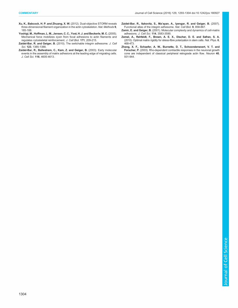
Xu, K., Babcock, H. P. and Zhuang, X. W. (2012). Dual-objective STORM revealsthree-dimensional filament organization in the actin cytoskeleton.Nat. Methods 9,185-188.
Yoshigi, M., Hoffman, L. M., Jensen, C. C., Yost, H. J. andBeckerle, M. C. (2005).Mechanical force mobilizes zyxin from focal adhesions to actin filaments andregulates cytoskeletal reinforcement. J. Cell Biol. 171, 209-215.
Zaidel-Bar, R. and Geiger, B. (2010). The switchable integrin adhesome. J. CellSci. 123, 1385-1388.
Zaidel-Bar, R., Ballestrem, C., Kam, Z. and Geiger, B. (2003). Early molecularevents in the assembly of matrix adhesions at the leading edge of migrating cells.J. Cell Sci. 116, 4605-4613.
Zaidel-Bar, R., Itzkovitz, S., Ma’ayan, A., Iyengar, R. and Geiger, B. (2007).Functional atlas of the integrin adhesome. Nat. Cell Biol. 9, 858-867.
Zamir, E. and Geiger, B. (2001). Molecular complexity and dynamics of cell-matrixadhesions. J. Cell Sci. 114, 3583-3590.
Zemel, A., Rehfeldt, F., Brown, A. E. X., Discher, D. E. and Safran, S. A.(2010). Optimal matrix rigidity for stress-fibre polarization in stem cells. Nat. Phys. 6,468-473.
Zhang, X. F., Schaefer, A. W., Burnette, D. T., Schoonderwoert, V. T. andForscher, P. (2003). Rho-dependent contractile responses in the neuronal growthcone are independent of classical peripheral retrograde actin flow. Neuron 40,931-944.
1304
COMMENTARY Journal of Cell Science (2016) 129, 1293-1304 doi:10.1242/jcs.180927
Journal
ofCe
llScience
Related Documents