/. exp. Biol. 153, 199-224 (1990) 199 Printed in Great Britain © The Company of Biologists Limited 1990 SYNAPTIC CONNECTIONS MADE BY AXONS REGENERATING IN THE CENTRAL NERVOUS SYSTEM OF ADULT MAMMALS BY ALBERT J. AGUAYO, GARTH M. BRAY, MICHAEL RASMINSKY, THOMAS ZWIMPFER, DAVID CARTER AND MANUEL VIDAL-SANZ Center for Research in Neuroscience, The Montreal General Hospital and McGill University, 1650 Cedar Avenue, Montreal, H3G 1A4, Quebec, Canada Summary The restoration of connections in the injured central nervous system (CNS) of adult mammals is hindered by the failure of axons to grow back to their natural fields of innervation. Following transection of the optic nerve of adult rodents, the guided regeneration of retinal ganglion cell (RGC) axons along a transplanted segment of peripheral nerve (PN) has shown that these neurones retain their capacities to form well-differentiated synapses in both normal and abnormal targets. The main aim of this review is to describe the anatomical and functional characteristics of some of these connections and to suggest that their terminal distribution and morphology may be the result of a persistence in these targets of molecular determinants that influence normal connectivity in the intact animal. Introduction It is generally assumed that the successful regeneration of damaged neural connections requires the recapitulation of several critical steps in development, namely the guided growth of axons towards correct targets and the formation of functionally appropriate synapses. The replication of these events in the injured adult CNS must be carried out, however, under conditions that are markedly different from those present in embryogenesis and soon after the birth of the animal. This review of a selected group of recent experiments in adult mammals attempts to highlight some of the similarities and differences observed between events that can lead to the restoration of synaptic connections in the injured nervous system and those that take place during normal development. Development of neuronal connections In the developing organism, the growth and branching of axons and dendrites gives rise to the intricate networks that comprise the adult CNS. The circum- stances that lead the extending processes of neurones to their targets are Key words: synaptogenesis, axotomy, visual system, retina, regeneration, nerve grafts.

Welcome message from author
This document is posted to help you gain knowledge. Please leave a comment to let me know what you think about it! Share it to your friends and learn new things together.
Transcript

/. exp. Biol. 153, 199-224 (1990) 199Printed in Great Britain © The Company of Biologists Limited 1990
SYNAPTIC CONNECTIONS MADE BY AXONSREGENERATING IN THE CENTRAL NERVOUS SYSTEM OF
ADULT MAMMALS
BY ALBERT J. AGUAYO, GARTH M. BRAY, MICHAEL RASMINSKY,THOMAS ZWIMPFER, DAVID CARTER AND MANUEL VIDAL-SANZ
Center for Research in Neuroscience, The Montreal General Hospital and McGillUniversity, 1650 Cedar Avenue, Montreal, H3G 1A4, Quebec, Canada
SummaryThe restoration of connections in the injured central nervous system (CNS) of
adult mammals is hindered by the failure of axons to grow back to their naturalfields of innervation. Following transection of the optic nerve of adult rodents, theguided regeneration of retinal ganglion cell (RGC) axons along a transplantedsegment of peripheral nerve (PN) has shown that these neurones retain theircapacities to form well-differentiated synapses in both normal and abnormaltargets. The main aim of this review is to describe the anatomical and functionalcharacteristics of some of these connections and to suggest that their terminaldistribution and morphology may be the result of a persistence in these targets ofmolecular determinants that influence normal connectivity in the intact animal.
Introduction
It is generally assumed that the successful regeneration of damaged neuralconnections requires the recapitulation of several critical steps in development,namely the guided growth of axons towards correct targets and the formation offunctionally appropriate synapses. The replication of these events in the injuredadult CNS must be carried out, however, under conditions that are markedlydifferent from those present in embryogenesis and soon after the birth of theanimal. This review of a selected group of recent experiments in adult mammalsattempts to highlight some of the similarities and differences observed betweenevents that can lead to the restoration of synaptic connections in the injurednervous system and those that take place during normal development.
Development of neuronal connections
In the developing organism, the growth and branching of axons and dendritesgives rise to the intricate networks that comprise the adult CNS. The circum-
stances that lead the extending processes of neurones to their targets are
Key words: synaptogenesis, axotomy, visual system, retina, regeneration, nerve grafts.

200 A. J. AGUAYO AND OTHERS
unknown, but complex axon-substratum and cell-cell interactions are thought toplay key roles in the formation of neuronal assemblies that subserve specificfunctions (Easter et al. 1985; Edelman, 1987; Rutishauser and Jessel, 1988;Lumsden and Keynes, 1989). Groups of differentiating neurones acquire differentpositions along the developing neuraxis and subserve different roles. While mostcells become interneurones that only synapse with their neighbours, many othersmust extend their axons towards more distant targets. Matrix substrata, cellsurface components and diffusible factors acting on receptive growth cones maystimulate or inhibit the elongation of these axons and influence: (a) their overalldirection and course; (b) the confinement of terminal arborizations to specificclusters, laminae or columns of cells; and (c) the establishment of synaptic contactson appropriate dendrites, axons or somata.
Before and after birth, neural networks are modified further by the naturaldeath of nerve cells, the disappearance of redundant fibres, changes in dendritictrees (Purves et al. 1986), competitive interactions between different afferents andthe shifting and strengthening of terminal connections caused by functionalactivity (Hubel and Wiesel, 1970; Shatz and Sretavan, 1986; Fraser and Perkel,1990). Thus, short-range growth and rearrangements in connectivity, generated bychanges in arborizations and synapses, may continue to be made long after birth.
In the embryo, some of these events may be facilitated by the short distance thatusually separates most neurones from their targets and the time differences in thegenesis, migration and neurite outgrowth normally exhibited by the various classesof nerve cells that populate the nervous system. In addition, it is obvious that manyCNS neurones persistently support a gradual elongation of their axons in harmonywith the growth of the entire organism, an extension that surpasses many timesthat occurring earlier in development, when most of the neural circuitry is firstestablished. While the capacity of many neurones to extend axons and reorganizedendritic and axonal arborizations appears to be retained into adulthood, there is,nevertheless, no evidence that these capacities lead to major changes in thedirection or position of any established long axonal projection in the adultmammalian CNS.
In the mature animal, constraints on fluctuations in connectivity to the narrowrange covered by dendrites and axon terminals could reflect an evolutionary needto maintain the main configuration of developmentally established circuits onceneurogenesis is completed and no new fibre tracts or long association fibres aregenerated. Recent studies of two growth-inhibiting proteins expressed postnatallyby differentiated oligodendrocytes (Schwab, 1990) suggest one possible mechan-ism that may normally curtail fibre tract changes in the white matter whilepermitting the local reorganization of connections in the grey matter. Plasticity inthe grey matter, rich in terminal arborizations, neuronal somata and dendrites,could be influenced locally by repulsive and affinity-determined molecularinteractions (Bonhoeffer and Gierer, 1984; Miiller etal. 1990) that eventually leadto the formation of stable synapses upon appropriate classes of cells. Becaus4terminal synaptogenesis may limit fibre extension (Bernstein and Bernstein,

Regenerative synapto genesis 201
1971), neurone-neurone interactions could also help confine the connectionsmade by axonal arborizations to groups of cells near the sites reached by projectingfibres.
In the hypothetical scheme described, an early stage of directed axonogenesis,largely dependent on interactions with noh-neuronal substrata, makes it possiblefor the axons of certain neurones to reach the more distant regions of the neuraxisthat harbour their specific targets. In mammals, the completion of this stage ofdevelopment appears to be associated with changes in the glial environment thathalt new fibre elongation into the white matter but allow established projections tolengthen during the gradual postnatal growth of the organism. In the neuropile ofcortical and non-cortical regions of the grey matter, the selection of postsynapticneurones and their cellular domains (somata or dendrites) by projecting axons andby interneurones would be regulated locally by surface molecules that induce andsecure synapses but continue to allow narrow fluctuations in local connectivity.The expression of these molecules would be modulated by activity and bycompetitive interactions (Easter etal. 1985; Shatz and Sretavan, 1986; Edelman,1987; Debski et al. 1990). Thus, during postnatal maturation the neuronalcapacities to sustain axonal growth and to form connections would be moderatedby radial interactions with the non-neuronal environment and by influences fromthe target, mediated by terminals and synapses.
Disruption of neuronal circuitries by injurySerious injuries to the brain or spinal cord are commonly followed by persistent
histological alterations and deficits of function. In mammals and other amniotes,the anatomical and functional abnormalities generated by such injuries changewith time, but normal function is seldom restored. Some of the effects of CNSinjuries entail: (a) the immediate death of neuronal and non-neuronal cells at thesite of damage; (b) the interruption of axons and dendrites; (c) the rapid orprotracted loss of axotomized neurones whose somata are some distance from thelesion (Villegas-P6rez et al. 1988, 1989; Sofroniew et al. 1990; and (d) changes inmolecular components of damaged or denervated non-neuronal tissues wheredemyelination and reactive gliosis are well-recognized histological changes(Gasser et al. 1986; Finklestein etal. 1988; Perry and Gordon, 1988; Reier et al.1989; Stoll et al. 1989; Kreutzberg et al. 1990).
Because lost neurones other than those in the olfactory sensory epithelium arenot spontaneously replaced in mammals, recovery from injury in the peripheral orcentral nervous system is largely dependent on: (a) the survival of a criticalnumber of damaged cells; (b) the successful regrowth of interrupted axons; (c) theguidance of these axons to their normal fields of innervation; (d) the branching anddistribution of axon terminals within appropriate sectors; and (e) the establish-ment of functional contacts capable of influencing postsynaptic cells in a
(predictable and beneficial fashion.In the mammalian CNS, the failure of cut axons to regrow is a major obstacle to
202 A. J. AGUAYO AND OTHERS
the restoration of neural connectivity. This lack of axonal re-extension in the brainand spinal cord contrasts with the successful regeneration of cut axons intoperipheral nerve stumps, rich in Schwann cells, extracellular matrix componentsand other non-neuronal elements that express trophic factors (Richardson andEbendal, 1982; Heumann et al. 1987a,b), and critical surface molecules (Carbo-netto et al. 1987; Reichardt et al. 1989). The impressive regrowth that followsperipheral nerve transection, however, often leads to no significant recovery ofuseful function (Mackel et al. 1983), presumably because many regrowing fibresreach inappropriate targets after extending along Bungner bands previouslyoccupied by other axons (Horch and Burgess, 1980). Thus, while axonal extensionis an unavoidable step in the repair process, it does not ensure the restoration ofconnections with functionally and topographically appropriate targets. As indevelopment, pathway guidance to the discrete fields of innervation that containthe normal postsynaptic targets of projection neurones may be critical to the moresubtle local synaptic discriminations made subsequently by axons within suchterritories.
From in vivo studies in adult rodents, it has been proposed that axotomized CNSneurones retain their capacity to initiate and sustain axonal regrowth but thatconditions in the mature CNS inhibit or fail to promote fibre elongation afterinjury (David and Aguayo, 1981). The presence of various inhibitory glialcomponents (Pesheva et al. 1989; Schwab, 1990; Snow et al. 1990), a deficit ofgrowth-promoting molecules (Carbonetto et al. 1987; Sandrock and Matthew,1987) and even the formation of premature synapses with neurones near the site ofdamage (Bernstein and Bernstein, 1971) may all contribute to limit axonalregrowth in the injured CNS.
Certain experimental modifications (Schnell and Schwab, 1990; Schwab, 1990)and substitutions (see below) of the growth-hindering glial environment of theCNS provide a novel opportunity to determine if neuronal connections can berestored by the regrowth of severed axonal projections to selected regions of thebrain or spinal cord of adult mammals.
An experimental in vivo strategy to determine if synaptogenesis can follow axonalregeneration in the central nervous system
The search for restored CNS connectivity as a result of axonal regeneration hasbeen made possible by the demonstration that many different classes of axotom-ized CNS neurones are capable of regrowing lengthy axons when the glialsubstratum present in the adult CNS is replaced by a transplant of non-neuronalcomponents of PN (see Aguayo, 1985). The experimental strategy used in these invivo studies involves by-passing portions of the CNS via a grafted, axonless PNsegment capable of facilitating and guiding the regrowth of the cut central axons toremote CNS targets. Using these techniques, regenerating axons can be led to the^vicinity of their normal fields of innervation or to regions that are not normall™subserved by these projections.
Regenerative synaptogenesis 203
The presence of long regenerated central axons within accessible PN 'bridges'that extend extracranially (Vidal-Sanz et al. 1987; Carter et al. 1989a) orextraspinally (David and Aguayo, 1981) for several centimetres before re-enteringthe CNS facilitates the study of axonal growth and connectivity by anatomical andelectrophysiological techniques, minimizing undue spread of tracers or ambi-guities concerning effective sites of electrical stimulation. For such technicalreasons and because axotomy near the neuronal somata is a pre-requisite forregeneration into the PN grafts (see below), these experiments have concentratedon the responses of long fibre projections axotomized far away from their fields ofinnervation. It is hoped, however, that evidence obtained from these studies mayalso stimulate ideas on the possible capacities for growth and reconnection thatmay reside in the larger population of interneurones that makes up the CNS.
This review focuses on studies of regeneration from one axonal projection, theretinofugal system of adult rodents, where the regrowth of severed retinalganglion cell (RGC) axons along a PN graft linking the eye and the superiorcolliculus (SC) has resulted in the formation of well-differentiated RGC synapsesin the SC (Vidal Sanz et al. 1987; Carter et al. 1989a). In parallel investigations, thesynapses of these regenerated RGC axons were also shown to be capable ofmediating trans-synaptic electrophysiological responses from neurones in the SCafter the retina was stimulated by light (Keirstead et al. 1989)". Finally, PN graftswere used to investigate terminal differentiation and synaptogenesis in exper-iments where regenerating central axons were guided to abnormal targets in theCNS (Zwimpfer et al. 1989, 1990). Descriptions and comments pertinent to thesestudies are given in the paragraphs that follow.
Regenerating retinal ganglion cell axons form synapses in the superior colliculusIn adult hamsters and rats, an autologous PN graft measuring 2-4cm in length
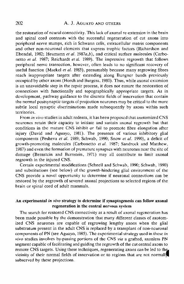
was attached to the ocular stump of optic nerves transected within the orbit (Vidal-Sanz etal. 1987; Bray et al. 1988; Carter et al. 1989a). Approximately 6 weekslater, the distal end of the graft was inserted into the ipsilateral SC (Fig. 1) and thecontralateral optic nerve (ON) was cut intraorbitally to eliminate all normalretinotectal connections.
The PN grafts were attached to optic nerves cut intraorbitally because axotomynear the neuronal perikaryon appears to be a pre-requisite for the growth of RGCsand other CNS axons into peripheral nerve grafts (see Aguayo, 1985). Thispuzzling association between axonal regeneration into these grafts and thedistance that separates the axotomy site from the cell somata may also influenceother cellular responses to axotomy, including the enhanced expression of thegrowth-associated protein GAP-43 (Lozano et al. 1987; Lozano, 1988). Indeed,following ON transection near, but not far from, the eye in adult rats, there is anaugmented GAP-43 immunoreactivity in RGC somata and axons and an increase
)in the axonal transport of the GAP-43 protein (Lozano, 1988). The reason whyaxonal regrowth into PN grafts and enhanced GAP-43 expression both occur

204 A. J. AGUAYO AND OTHERS
under these circumstances remains unclear; a possible explanation is that GAP-43is somehow1 involved in a chain of injury-induced cellular events that modulates thereceptiveness of growth cones to molecules in the non-neuronal environment. Theenhanced expression of these and other molecules in nerve cells injured near thecell body may play a role in the establishment of the neurone-substratuminteractions required to sustain axonal elongation when conditions in the axonalenvironment are propitious. Possible repressor molecules within the long stumpthat remains when the ON is severed far from the retina could also inhibit GAP-43expression in distally axotomized RGCs.
The proximal location of an axonal injury, however, also causes a massiveretrograde degeneration of retinal ganglion cells. In rats, approximately 85% ofthe RGCs are lost 15 days after cutting the ON immediately behind the eye(Villegas-Pe'rez etal. 1988). From these decimated retinas, nearly 20% of thesurviving RGCs regenerate their axons along the entire 3-4 cm length of the PNgrafts (Villegas-Pe'rez et al. 1988). Conversely, after intracranial lesions of the ONthat are 10 mm from the eye, approximately two-thirds of the RGCs survive for 15days after axotomy (Villegas-Pe'rez et al. 1989). This larger population of retinal
Scalp
Fig. 1. Method used to implant peripheral nerve (PN) segments (cross-hatched) asbridges between the retina and the superior colliculus (SC) in adult rats or hamsters.(A) One end of an autologous PN segment was attached to the orbital stump of theoptic nerve (ON) transected immediately behind the eye. The other end of the PNgraft was left under the scalp over the skull. (B) 8-10 weeks later, the unconnected tipof the PN graft was implanted into the SC. After survival times varying from 2 to 18months, orthogradely transported tracers were injected into the vitreous chamber ofthe eye to identify regenerated retinal ganglion cell axons and their terminals.(Reproduced with permission from Vidal-Sanz et al. 1987.)
Regenerative synaptogenesis 205
neurones, however, gives rise to no significant regrowth of axons into PN graftsplaced intracranially, away from the RGC somata (Richardson et al. 1982). Thus,under these experimental conditions, the neuronal propensity to regenerate cutaxons bears a curious relationship to lesions that cause more serious damage tothese cells. It is assumed that the retrograde degeneration of these neuronessubstantially diminishes the pool of viable cells that can give rise to axonalregeneration.
In the animals with PN grafts joining the eye and the dorsal mesencephalon,fluorescent tracers, horseradish peroxidase (HRP) or tritiated amino acids wereinjected intravitreally to label the regrowing axons, as well as their arborizationsand terminals. Retinal axons, isolated from each other by their Schwann cell andbasal lamina ensheathment, were shown to have extended without branching fordistances that were nearly twice as long as those of the normal retinotectalprojections found in the intact adult animal (Vidal-Sanz et al. 1987; Carter et al.1989a).
In the hamsters, light and electron microscope studies of 758 labelled terminalsfrom RGC axons that extended into the SC between 6 and 8 weeks after insertionof the PN graft into the SC were compared with 698 retinotectal terminals fromintact hamsters. It was documented that: (i) the regenerating RGC axonspenetrated the SC for distances of up to 500 /zm and arborized in the superficiallayers of the SC that normally receive most retinal projections; (ii) the ultrastruc-tural features of the newly formed RGC terminals, including their content ofround vesicles and pale mitochondria, resembled normal presynaptic structures;and (iii) the characteristics of their postsynaptic contacts, virtually all of whichwere axo-dendritic and asymmetrical (Gray type I), were also strikingly similar tothose of normal retinocollicular connections (Carter et al. 1989a).
These findings in hamsters, together with similar investigations in the rat(Fig. 2) (Vidal-Sanz et al. 1987; Bray et al. 1988), demonstrate that regeneratingaxons do not necessarily stop growing at the interface of the PN grafts and theCNS, but can penetrate the damaged CNS tissue and form arborizations andsynapses within the nearby grey matter. This extension of the regenerating RGCaxons into the SC was slight in comparison to the lengths they had covered alongthe PN grafts. However, the penetration of the RGC axons into the SC for up to500,1011 from the tip of the graft approximated the lengths of the arborizationsmade by the longest retinal afferents within the normal SC of these rodents (Sachsand Schneider, 1984). Because the SC is a small, dome-shaped structure whoseretino-recipient neurones are normally near the pial surface, this short extensioninto the grey matter should have permitted the RGC axons to reach cells in mostregions of the SC that normally receive retinal inputs. Thus, the small numbers ofRGC axons emerging from the graft, rather than the limited extension of theincoming fibres, may account for the poor re-innervation observed in the SC ofthese animals.
It is of interest that the RGC axons that regenerated into the SC arborizedpreferentially within the superficial layers of the superior colliculus. Since the PN
206 A. J. AGUAYO AND OTHERS
grafts ended in or near the stratum opticum (SO), incoming axons could havegrown dorsally into the stratum griseum superficiale (SGS) or ventrally into thestratum griseum intermediale. However, the vast majority of the identified axonterminals observed were in the SO and SGS, the main retino-recipient layers of thenormal SC. In normal rodents, most retinal inputs to the SC come from thecontralateral eye and terminate mainly in the SGS and the SO, while the smalleripsilateral projection ends principally in the SO (Chalupa and Rhoades, 1979;Frost et al. 1979; Jen et al. 1984). When, in these experiments, the regrowing RGCaxons were all directed via a PN graft to the ipsilateral SC, the synapses made bythese axons were not concentrated in the SO but extended into the SGS, which isnormally a target of the contralateral retinal inputs. Such a broadened deploymentof ipsilateral RGC axons can also be induced experimentally in immature hamsters(Schneider, 1973; Mooney et al. 1985). Similarly, following ON section, thegoldfish retinotectal system, which normally projects contralaterally, can regrowand innervate the main retino-recipient layers of the ipsilateral tecrum if thistectum is deprived of its normal retinal input (Meyer, 1978).
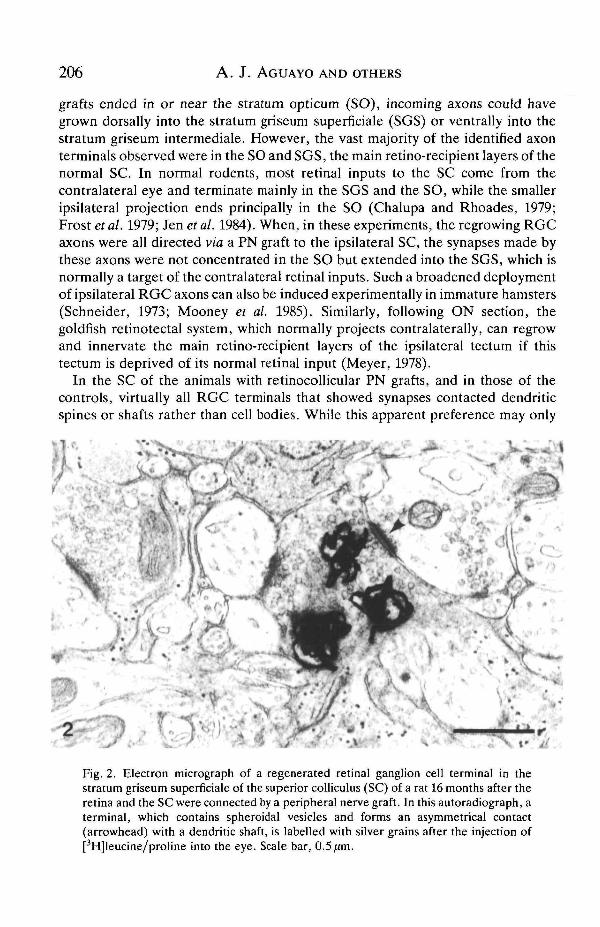
In the SC of the animals with retinocollicular PN grafts, and in those of thecontrols, virtually all RGC terminals that showed synapses contacted dendriticspines or shafts rather than cell bodies. While this apparent preference may only
*; .
Fig. 2. Electron micrograph of a regenerated retinal ganglion cell terminal in thestratum griseum superficiale of the superior colliculus (SC) of a rat 16 months after theretina and the SC were connected by a peripheral nerve graft. In this autoradiograph, aterminal, which contains spheroidal vesicles and forms an asymmetrical contact(arrowhead) with a dendritic shaft, is labelled with silver grains after the injection of[3H]leucine/proline into the eye. Scale bar, 0.5 /im.

Regenerative synapto genesis 207
reflect the high density of dendrites in the grey matter of the SC, the observationthat approximately 60% of the synapses made by normal and regeneratingRGC-SC terminals are on spines rather than on the shafts of dendrites (Carteret al. 1989a) argues against a purely random selection of synaptic targets (Fig. 3).A predilection for precise positions in the dendritic tree is also observed afterneural transplantation (Sotelo and Alvarado-Mallart, 1987; Sotelo, 1989) andwhen sprouting is induced by a partial deafferentation of inputs (Lewis andCotman, 1982). This affinity of the axonal terminals for certain neuronal domainssuggests a discrete, patch-like expression of molecules in postsynaptic neuronesthat selectively stabilizes presynaptic contacts. Such determinants of the regionaldistribution of synaptic contacts may not only be expressed during developmentand regeneration but may also play a role in the maintenance of connectivityduring normal synaptic turnover and the rearrangement of synaptic connectionsthat follows sprouting (Raisman and Field, 1973; Cotman and Nieto-Sampedro,1984). As occurs during development and after sprouting, the availability of suchsites on the surface of the targeted neurone may be a determinant of thearrangement of the contacts made by regenerating axons and target cells. It maybe useful to consider the possibility that mechanisms akin to those involved inspecifying the location of synapses at the neuromuscular junction also play a role indetermining the selective distribution of synapses between neurones (Poo andYoung, 1990; Magill-Solc and McMahan, 1990). Because regenerated RGCsynaptic contacts have been found in the SC for periods of up to 18 months in ratsand 9 months in hamsters, it seems likely that these retinotectal connections arepermanently restored (Bray et al. 1988; Carter et al. 19896).
Postsynaptic structures
Regenerated ry-m c- • ControlE222 Spines• I ShaftsI | Undefined
Fig. 3. Postsynaptic domains of neurones in the superior colliculus contacted by 901regenerated and 471 control retinal ganglion cell terminals. In addition to theillustrated distributions on dendritic shafts and spines, which were similar for theregenerated and control axon terminals, one of the regenerated terminals formed asynapse on the cell body of a neurone.

208 A. J. AGUAYO AND OTHERS
Anatomical characteristics of the synapses made by regenerated retinal ganglioncell axons that regrow into the superior colliculus
Morphometric ultrastructural studies of the synaptic specializations generatedin the SC by the RGC terminals and estimates of the ratio of synaptic contacts tothe perimeter of each terminal indicated that both were similar to those of normalretinotectal connections. The preservation of the ratio between the number ofRGC synaptic contacts made within the neuropile of the SC and the perimeter oftheir terminals may be an indication that the overall size of such terminals isinfluenced by the number of contacts. It is less likely, however, that such ratiosmight denote that the amount of membrane formed by a terminal dictates thenumber of synaptic contacts that is generated.
Some of the regenerated terminals were larger and had longer synaptic junctionsthan controls. Although the size of axon terminals appears to be smaller duringdevelopment than in the mature CNS (Kalil etal. 1986), large terminals have beenreported in other adult vertebrates where synaptogenesis is known to occur as aresult of regeneration or sprouting. In the goldfish optic tectum, for example,regenerating retinotectal terminals are initially large and form longer synapticcontacts (Radel and Yoon, 1985). Furthermore, while the regeneration of axonterminals and synapses has not been studied previously in the adult mammalianCNS, larger terminals and longer, more frequent, synaptic contacts per terminalhave been documented in studies of the collateral sprouting of intact neurones inpartially denervated areas of the CNS (Raisman, 1969; Matthews et al. 1976; Chenand Hillman, 1987; Murray et al. 1987; Steward et al. 1988) and in experimentswhere afferent fibre populations are decreased (Sotelo, 1975; Hillman and Chen,1985).
In the goldfish, in which a full complement of RGC axons may eventually re-innervate the optic tectum following ON crush (Murray et al. 1982), the size of theregenerated retinotectal terminals is reported to return to normal approximately 1year after denervation (Murray and Edwards, 1982; Radel and Yoon, 1985; Hayesand Meyer, 1988). An overall tendency towards smaller terminals has also beenobserved in the SC approximately 9 months after the insertion of the caudal end ofthe PN grafts into the SC of hamsters (D. Carter, G. M. Bray and A. J. Aguayo,unpublished observations). Morphometric studies of RGC terminals in rats, 16-18months after PN grafting, have shown no difference between the size of theregenerated terminals and age-matched controls (M. Vidal-Sanz, A. J. Aguayoand G. M. Bray, unpublished observations). These overall findings suggest that,although the developmental maturation of neuronal connections involves anenlargement of the terminals, regenerated terminals may follow a different patterntypified by a gradual reduction in their size.
While there are obvious differences between the normal and the regeneratedretinotectal projections we have studied, particularly with regard to the number ofRGC axons that innervate the SC, the evidence described here suggests that the.patterns of terminal arborization as well as the morphology and preferentialdistribution of synapses to superficial layers of the SC made by individual
Regenerative synaptogenesis 209
regenerated RGC axons resemble those of retinofugal axons in the SC of rodentsduring: (a) the development of the visual system (Sachs et al. 1986); (b) the re-innervation that follows optic tract lesions in the immediate postnatal period .(So etal. 1981); and (c) the transplantation of foetal retinas to the midbrain of immaturehosts (Hankin and Lund, 1987).
Competitive interactions between different RGC axons, believed to play animportant role in the formation of neural connections (Easter et al. 1985), areunlikely to have influenced the distribution of arborizations and synapses in theseanimals because so few RGC axons reached the SC. It is not known, however, ifthe preferences of RGC axons for certain SC laminae and neuronal domains aredictated by the extensive tectal denervation caused by the severing of the opticnerves and the unavoidable damage to the occipital lobes and the SC that isinflicted by the insertion of the PN grafts into the dorsal midbrain of these animals.Studies of the synaptic distribution of RGC axons guided to an abnormal target,the cerebellum, suggest the possibility that conditions other than those that are adirect result of denervation may play a role in determining synaptic distribution(see below).
A gradual loss of RGCs follows axotomy in the optic nerve and also theregrowth of RGC axons into blind-ended PN grafts (Villegas-P6rez et al. 1988,1989). This protracted cell loss may result from a persistent disconnection of theseneurones with targets that are their main source of trophic support. Because, inrats and hamsters in which the PN grafts were used to bridge the eye and thetectum, no apparent fall in the number of RGC arborizations in the SC has beenobserved during studies that span most of the life of these animals (D. Carter, G.M. Bray and A. J. Aguayo, unpublished observations), it is likely that the restoredRGC connections in the SC may ensure RGC survival.
The studies of axonal regeneration into the SC of adult rodents reviewed herehave not encompassed several other important aspects of RGC regeneration.These include determining more precisely the number and size of the arborizationsformed, the classes of SC neurones that are re-innervated, and whether there is aretinotopic order to the deployment of the RGC axons that reach the SC. Whileother vertebrates such as amphibians and fish appear to be capable of restoring lostretinotectal connections with a great deal of accuracy, it remains to be shown thataxonal regeneration in adult mammals can replicate the pathfinding patterns andthe terminal connectivity that they achieve during normal development. In ourexperiments, the massive loss of RGCs that follows proximal axotomy in the opticnerve has imposed serious limitations on the search for answers to some of thesequestions.
Electrophysiological studies of afferent and efferent connectivity of axotomizedretinal ganglion cells whose axons regenerate along peripheral nerve grafts
Axonal injury not only disrupts connections with target cells innervated by thedamaged axons but may also lead to loss or modification of function of the

210 A. J. AGUAYO AND OTHERS
axotomized neurones themselves. In addition to the dramatic retrograde cell lossthat can follow axotomy, there may be changes in afferent connectivity, patterns ofdendritic arborization or membrane properties of damaged neurones (Purves andLichtman, 1985; Purves, 1975; Mendell etal. 1976; Thanos and Aguayo, 1988). Forthis reason, as a prelude to physiological studies of synaptic connectivity in theretinocollicular grafts, the physiological properties of RGCs were investigatedafter regeneration of their axons into PN gTafts that were not connected to a CNStarget (Keirstead etal. 1985). This was accomplished by teasing axons from PNgrafts inserted into the retina and recording the responses in these axons to visualstimulation of the retina.
The responses to visual stimulation of some of the RGCs with regeneratedaxons were indistinguishable from those characteristic of RGCs in the intactanimal (Fig. 4). These cells responded to light with on, off or on-off responses,and had receptive fields of sizes similar to those of normal RGCs, in some casesaccompanied by a suppressive surround organization like that seen in some RGCsin intact animals. This responsiveness of RGCs with regenerated axons indicates
150
75
II 200
100
0
200
100
0
100
50
0
250
125
0
2s
Fig. 4. Responses to light recorded from the regenerated axons of retinal ganglioncells. (A-E) Peristimulus time histograms of unitary responses to visual stimulipresented during the 2 s period indicated by the bars. In some histograms artefactsrelated to shutter opening and closing are seen in bins at the onset and offset ofillumination. (A,B) On responses with transient (A) and sustained (B) responses toillumination. (C,D) Off responses with depression of firing during illumination andtransient (C) and more protracted (D) increases in firing at the cessation ofillumination. (E) An on-off response with brief increases in firing at both the onset andoffset of illumination. (F) Simultaneous recordings from two regenerated retinalganglion cell axons teased from a single fascicle. During the time indicated by the blackbar, a circle of light 2° in diameter was presented on a tangent screen within the rat'svisual field. The low-amplitude unit discharged in response to the onset of light and itsdischarge was sustained throughout the 2 s of illumination (on response); the large-amplitude unit discharged in response to cessation of illumination (off response). Thecentres of the receptive fields for these two units were identical. (Reproduced withpermission from Keirstead et al. 1985.)

Regenerative synaptogenesis 211
that at least some of the RGCs that regenerate axons into PN grafts are able toretain, or regain, enough of their normal inputs to ensure function.
The number of visually responsive units appeared to decline with time, fewerunits being found in grafts examined 25-28 weeks after insertion in the retina thanin those examined 9-11 weeks after graft insertion. Almost no visually responsiveunits could be found in animals with grafts implanted for 44-48 weeks, despite thefact that the grafts still contained viable axons responsive to electrical stimulation(Keirstead et al. 1988). It is unclear whether this reflects simply the continuingattrition of the RGC population following axotomy (Villegas-Pe'rez et al. 1988) orwhether some surviving RGCs with regenerated axons lose their ability to respondto light, perhaps because of changes in afferent connections or membraneproperties. A similar loss with time of functioning cells with regenerated axons wasfound in a study of the function of brainstem neurones that had regenerated axonsinto PN grafts (Gauthier and Rasminsky, 1988). For both the RGCs and thebrainstem neurones, the regenerating axons were blind-ended and it is possiblethat long-term viability and/or function of the regenerating cells was compromisedbecause of the absence of axonal contact with an appropriate target. As notedabove, retinocollicular synapses, once established following regeneration of RGCaxons through PN grafts, appear to be stable for many months, implying thesurvival of afferent RGCs. Nonetheless, it will be important to establish explicitlyif RGC survival and function are enhanced by axonal contact with appropriate orinappropriate targets.
To assess the function of synapses formed by regenerating RGC axons,hamsters with PN grafts directed from the eye to the ipsilateral SC were studiedelectrophysiologically 15-18 weeks after graft insertion into the SC (Keirsteadet al. 1989) (Fig. 5). Within the superficial 450 pan of the SC, in the vicinity of graftinsertion, units were found that responded with either excitation or inhibition toflashes of light directed towards the retina. The proof that at least some of theseunits were postsynaptic to the afferent regenerated RGC axons established thatthe regenerated synapses observed in the morphological studies were indeed-functional and capable of mediating trans-synaptic responses to light. Light-responsive units tended to be found in clusters within a few hundred micrometresof one another, consistent with the limited morphological observations so farmade of the patterns of arborization of regenerating RGC axons.
The responses to light of most of the units definitively identified as postsynapticwere much less vigorous than that characteristic of retinal ganglion cells withregenerated axons or of visually responsive neurones in the superficial layers of theSC in the intact animal. However, these responses may not have been representa-tive of all reinnervated SC neurones. The electrophysiological criterion used toidentify visually responsive units as postsynaptic would have failed to distinguishbetween responses in securely innervated SC neurones and responses in theJerminals of regenerated RGC axons. This criterion was thus strongly biasedpowards identification of SC neurones that were insecurely rather than securelycontacted by regenerated RGC axons and there may, consequently, have been a

212 A. J. AGUAYO AND OTHERS
large class of briskly responsive SC neurones that could not be identified as such. Itis thus not possible to make generalizations concerning the response properties ofreinnervated SC neurones.
Little information is available concerning the extent or pattern of innervation ofeither intact or reinnervated SC neurones necessary to ensure either normal orattenuated responsiveness to light. Individual synaptic contacts of regeneratedRGC axons on SC neurones appear remarkably normal at an ultrastructural level,but comparisons cannot be made between reinnervated and intact neuronesregarding the extent of reinnervation of individual SC neurones by their afferents.Although it is known that neurones in the retino-recipient laminae of the rodentSC each receive several thousand synaptic contacts (Albers et al. 1990), it is notknown how many of these derive from direct retinal input, how many contacts percell reflect input from individual RGC afferents and how many retinocollicularsynapses need to be activated against the background of many other visual andnon-visual inputs in order to displace the membrane potential of the cell to
A Flash Electrical stimulation <n
20 ms
Fig. 5. Recordings from the superior colliculus (SC) of a hamster with a peripheralnerve (PN) graft directed from the eye to the SC. (A) A single unit 250 fan below thesurface of the SC responds to light flashes to the eye (arrow) with a single spike on fourof ten successive trials. (B) The same unit responds erratically with inconstant latencyto (traces 1) single electrical stimuli (arrow) delivered to the PN graft but responds witha more constant latency, often with multiple spikes, to (traces 2) paired electricalstimuli (arrows) of the same intensity. This pattern of response, reflecting postsynapticsummation of subthreshold EPSPs, is inconsistent with that anticipated from an RGCaxon and thus identifies this unit as an SC neurone. This criterion of exclusion wouldfail to distinguish responses in securely excited SC neurones from responses in RGCaxon terminals. (Reproduced with permission from Keirstead et al. 1989.)
Regenerative synaptogenesis 213
threshold. Other areas giving rise to major inputs to the superficial layer of the SC,such as the visual cortex, the lateral geniculate nucleus and the pretectal nuclei,are themselves deprived of their visual inputs as a result of transection of the opticnerves and are no longer able to exert their influence on SC neurones when a PNgraft replaces the optic nerve. Formation and function of synaptic connectionsbetween regenerated RGC axons and SC neurones thus occurs in a contextsubstantially altered from that in the intact animal. It will not be surprising if theresponse properties of the reinnervated neurones prove to differ substantiallyfrom those of neurones in the intact animal.
It remains an open question to what extent synaptic activation of SC neuronesby regenerated retinocollicular synapses can be translated into behaviouralfunction. Experiments in which foetal retinas are grafted onto neonatal midbrainindicate that the synaptic connections formed between grafted RGCs andmidbrain neurones can mediate pupillary responses (Klassen and Lund, 1989) andPavlovian conditioned responses (Coffey et al. 1989) in response to visualstimulation in the absence of any obvious pattern of spatial organization of tectalreinnervation by the transplanted retina (Galli et al. 1989). However, suchresponses are unlikely to demand the precise retinotopy that characterizes thenormal pattern of innervation of the tectum by the retina. It remains to bedetermined if any retinotopic order can be re-established in the SC of adultrodents following regeneration of RGC axons through PN grafts joining the eyeand the tectum.
Regenerating retinal ganglion cell axons also make synapses when guided to thecerebellum
The restoration of useful function by the regrowth of severed axons may dependnot only on the re-establishment of a critical number of appropriate connectionsbut also on the avoidance or elimination of synapses with abnormal targets(Sperry, 1943; Schneider et al. 1985). The guiding of regenerating central axons toanatomically inappropriate regions of the CNS via PN grafts provides anopportunity to investigate in adult mammals the freedoms and constraints thatgovern the establishment of synaptic interactions. For this purpose, the regrowthof retinal axons was directed to the cerebellar vermis (Zwimpfer et al. 1989), aCNS target that does not normally receive any direct inputs from the eye eitherduring development (Frost, 1984) or in the mature animal. In these experiments,two important components of the regenerative response, axonal elongation andpathway finding, were deliberately rearranged to force axons into a region of theCNS that is both foreign to and distant from the natural RGC targets. The initialobjective was to determine if RGC axons would grow and arborize in thecerebellum. It was anticipated that such uncharacteristic growth would permit new
investigations on the conditions that influence presynaptic differentiation andPostsynaptic preferences within the territory reached by an anomolous growth ofaxons.

214 A. J. AGUAYO AND OTHERS
In these experiments (Zwimpfer et al. 1989), one end of an autologous PNsegment was grafted to the ocular.stump of the ON as described above. The otherend of the graft was extended caudally beyond the tectum and inserted super-ficially into lobules VI and VII on the right side of the cerebellar vermis. To assessthe effects of denervation of the cerebellar cortex on the formation of RGCsynapses, mossy and climbing fibres were interrupted by transecting the rightmiddle and inferior cerebellar peduncles (group A hamsters); this procedure wasomitted in other similarly grafted hamsters (group B). 1-9 months after theinsertion of the PN graft into the cerebellum, RGC terminals were labelled by anintravitreal injection of HRP similar to that used in the studies of the retino-collicular connections described above.
Labelled RGC axons regrew along these grafts and penetrated the cerebellumfor distances of up to 650 fjm. The penetration and overall branching character-istics of the RGC-cerebellar axons were not noticeably different from thoseobserved in the SC when RGC axons reach the tectum via a PN graft (Carter et al.1989a). A striking feature of the RGC axons that arborized within the cerebellumwas that they avoided the white matter and extended preferentially into thegranule cell layer (GCL). Although RGC axons occasionally grew to the level ofPurkinje cell somata, they generally avoided the molecular layer (ML).
By electron microscopy, it was possible to determine that the RGC arboriz-ations made synapses with cerebellar neurones (Fig. 6). In hamsters from bothgroups A and B, 172 labelled retinocerebellar terminals were found in the GCL,while only 12 were observed in the ML. Most RGC terminals made synapses ongranule cell dendrites; only one synapsed on a granule cell soma. The few synapsesin the ML were axodendritic, probably contacting Purkinje cells. The retino-cerebellar connections had the ultrastructural characteristics of well-differen-tiated, asymmetrical (Gray type I) axodendritic synapses. These retinocerebellarconnections appeared to be persistent as they were present up to 9 months afterinsertion of the PN graft. We do not know if the RGC-cerebellar synapses made inthe adult animal can mediate function.
A remarkable feature of many of the RGC terminals that synapsed in thecerebellum was their large size. Their average area was at least three times greaterthan that of either normal or regenerated RGC terminals in the SC and itapproximated that of normal mossy fibre terminals (Palay and Chan-Palay, 1974).The mean size of the retinal terminals in the cerebellum did not changesignificantly during the period of study (1-9 months after the caudal end of thegraft was inserted into the cerebellum). While most normal mossy fibres containeddark mitochondria, those within retinocerebellar terminals were pale, resemblingthe mitochondria of normal and regenerated retinotectal projections (Carter et al.1989a).
Fewer retinocerebellar arborizations were found in group B animals with intactcerebellar pedunculi, but the extensive denervation of the cerebellar cortex causedby the severing of climbing and mossy fibre inputs (group A hamsters) did n «appear to influence either the predominant distribution of RGC axons and
Regenerative synaptogenesis 215
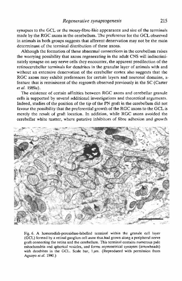
synapses to the GCL or the mossy-fibre-hke appearance and size of the terminalsmade by the RGC axons in the cerebellum. The preference for the GCL observedin animals in both groups suggests that afferent denervation may not be the maindeterminant of the terminal distribution of these axons.
Although the formation of these abnormal connections in the cerebellum raisesthe worrying possibility that axons regenerating in the adult CNS will indiscrimi-nately synapse on any nerve cells they encounter, the apparent predilection of theretinocerebellar terminals for dendrites in the granular layer of animals with andwithout an extensive denervation of the cerebellar cortex also suggests that theRGC axons may exhibit preferences for certain layers and neuronal domains, afeature that is reminiscent of the regrowth observed previously in the SC (Carteretal. 1989a).
The existence of certain affinities between RGC axons and cerebellar granulecells is supported by several additional investigations and theoretical arguments.Indeed, studies of the position of the tip of the PN graft in the cerebellum did notfavour the possibility that the preferential growth of the RGC axons to the GCL ismerely the result of gTaft location. In addition, while RGC axons avoided thecerebellar white matter, where putative inhibitors of fibre adhesion and growth
Fig. 6. A horseradish-peroxidase-labelled terminal within the granule cell layer(GCL) formed by a retinal ganglion cell axon thaUiad grown along a peripheral nervegraft connecting the retina and the cerebellum. This terminal contains numerous palemitochondria and spherical vesicles, and forms asymmetrical synapses (arrowheads)with dendrites in the GCL. Scale bar, ljim. (Reproduced with permission fromAguayo et al. 1990.)
216 A. J. AGUAYO AND OTHERS
(Caroni and Schwab, 1988; Schwab, 1990) may be expressed, they did not favourthe molecular layer, which is totally lacking in myelin. Sprouting of climbingfibres, known to occur in the ML of the mammalian cerebellum after a partiallesion of the inferior olive (Rossi et al. 1989), could, if sprouting were to occurexclusively in this layer, favourably compete for the synaptic sites of Purkinje celldendrites in the molecular layer and thus divert the incoming RGC fibres. Thispossibility also seems an unlikely explanation for the preference of RGC terminalsfor the GCL. The demonstration that climbing fibres can be induced to sproutwithin the ML of the adult rat (Rossi et al. 1989) suggests that the preference ofRGC axons for the GCL is not a result of the ML acting as a mechanical barrier tofibre ingrowth.
A more attractive explanation for these synaptic preferences is that the retino-recipient layers of the superior colliculus and the granule cell layer of thecerebellum express common molecules that play a role in their recognition byRGC growth cones. While no evidence is yet available to explain this predilection,candidates for this role could include neurotransmitters, receptors and trophicfactors. The postulated role of transmitters in recognition and synaptogenesis inthe adult mammalian CNS is not well understood (Lipton and Kater, 1989) butcertain excitatory amino acids or related peptides may be transmitters in bothcerebellar mossy fibres (Somogyi et al. 1986; Olson et al. 1987) and in RGCs(Anderson et al. 1987). Certain receptors, such as the iV-methyl-D-aspartate(NMDA) receptor, may also be involved in neuronal recognition, as suggested bystudies of the amphibian retinotectal system and other neuronal projections indifferent animal species (Debski et al. 1990). While NMD A receptors are plentifulin the granule cell layer, where they are expressed primarily on dendrites ofgranule cells (Olson et al. 1987), they are less numerous within the ML (Monaghanand Cotman, 1985) and do not appear to be present on mature Purkinje cells(Olson et al. 1987). In contrast, NMD A receptors are abundant in the SC andmany other areas of the adult mammalian CNS (Greenamyre et al. 1985).Pertinent examples of the role of trophic factors in establishing and sustaininganomalous connections are the effects of nerve growth factor (NGF) application,or its denervation-related local release, on the anomalous invasion of the CNS byperipheral sympathetic fibres (Levi-Montalcini, 1976; see Crutcher, 1987). Whilethe role of NGF in the retina of mammals is unclear, brain-derived neurotrophicfactor (BDNF) has been shown both to support the survival and growth of RGCsand to be present in the SC (Johnson et al. 1986). Although the presence of BDNFitself has not been reported in the cerebellum of rodents, progress in thesequencing of the BDNF gene (Leibrock et al. 1989) has led to the demonstrationthat, in the cerebellar cortex, mRNAs for BDNF are preferentially distributed inthe GCL (Hofer et al. 1990).
The peculiar morphology of the RGC-cerebellar terminals is an additional issueof interest. Because they are large and synapse predominantly with dendrites inthe granular layer, many of these RGC terminals resemble normal mossy fibreendings in the cerebellar cortex. One possible explanation of their size is that

Regenerative synaptogenesis 217
inappropriate contacts in any abnormal target may generate unusually largeterminals, well beyond the increases in size observed among the regenerated RGCfibres in the SC. This possibility is being explored by determining the character-istics of abnormal synaptic contacts made by RGCs in other inappropriate CNSregions of the brain to which axons are guided via PN grafts (Zwimpfer et al.1990). An alternative explanation is that pre- and postsynaptic interactionsreshape the regenerated terminals to fit the standard morphology of normalregional inputs. The latter hypothesis is consistent with the conclusions drawn byother investigators who have studied the ultrastructural characteristics of theabnormal connections established by axons after the ablation of their normaltargets at critical periods of neural development (Kalil and Schneider, 1975; Kaliland Behan, 1987; Campbell and Frost, 1988). In such experimental circumstances,the size and shape of the terminals formed resembled those of the normalterminals found within these targets.
The postulated influence of interactions between pre- and postsynaptic constitu-ents on the geometry of axon terminals is further highlighted by the knowndisparities that exist between terminals arising from the same neurone butreaching different cells or separate domains of the same neurone. Such adiversification of synaptic morphology was noted by Ram6n y Cajal (1909) in thecochlear nucleus and subsequently described for connections in this (Morest, 1968)and various other neuronal systems. Other examples of such normal diversityinclude the synapses made by normal retinal efferents sharing connections indifferent brain targets (Lund, 1969; Campbell et al. 1984)
General comments
Developmental associated changes in the cellular and molecular components ofthe CNS are thought to curtail the regrowth of axons in the injured CNS ofmammals. In the retina of adult rodents, the replacement of the optic nerve by aperipheral nerve graft enhances, at least temporarily (Villegas-P6rez et al. 1988),the survival of axotomized retinal ganglion cells, facilitates and guides theelongation of these central axons to distant targets (Vidal-Sanz et al. 1987) andpermits the formation of new synapses in the regions of the CNS to which they areguided (Vidal-Sanz et al. 1987; Bray et al. 1988; Carter et al. 1989a; Zwimpfer et al.1989).
Experiments described in this review indicate that, under conditions that permitaxonal regrowth, injured CNS neurones display a remarkable capacity to initiateand sustain the renewed extension of their cut fibres. The guiding of theseregenerated axons to regions of the CNS that are either the natural (the superiorcolliculus) or unnatural (the cerebellar cortex) targets for retinal projectionsresults in the formation of terminal arborizations and synapses.
In the SC, the distribution of the regenerated RGC arborizations and synapsesfcloes not appear to be totally haphazard. Indeed, their terminations are restrictedto the normal retino-recipient layers of the SC and synapses are made on the
218 A. J. AGUAYO AND OTHERS
neuronal domains selected by normal retinotectal projections. Moreover, withtime, regenerated synapses attain normal morphological characteristics and canmediate the trans-synaptic activation of neurones in the SC when the retina isstimulated by light.
As described previously for peripheral axons and for immature central projec-tions that are forced to terminate in unusual targets (see Purves and Lichtman,1985), the RGCs of these adult animals also appear competent to make synapsesupon anatomically inappropriate neurones. In the cerebellum, peculiarities in thedistribution and differentiation of the RGC terminals suggest that there may alsobe preferences in the synaptic selections made by the regenerating RGC fibres thatpenetrate the cortex.
Our overall findings, as well as those of other investigators (see other chapters inthis volume), suggest that connectivity in the CNS may be established andmaintained by graded affinities, functional activities and molecular interdepen-dences that are possibly shared by discrete populations of neurones in differentregions of the neuraxis. During development, spatially restricted and time-dependent events that involve the expression of specific substratum components,the fasciculation of related axons and the presence of gradients of trophicmolecules (Rutishauser and Jessel, 1988) may guide axons to their normal fields ofinnervation and thus narrow the final selection of their postsynaptic partners to thecells encompassed by terminal arborizations.
The resistance of the mature CNS to axonal extension has forced us to create anexperimental artifice to bypass these tissues in order to recreate interactionsbetween the sources and targets of certain central axonal projections. It does nowappear that, upon their arrival at such targets, the regenerated axons are subjectedto conditions that determine the formation of terminal arborizations and thedifferentiation of synapses, an indication of the likely persistence in the neuropileof the injured CNS of adult mammals of the molecular determinants of thesephenomena.
The demonstration that functional synapses can be made in the injured adultmammalian CNS as the end result of an extensive and circuitous axonal regrowthvia the PN grafts raises the possibility that connections may also be restoredspontaneously in the CNS by the short-range regrowth of axons interrupted neartheir targets or through the re-establishment of connections between nerve cells,such as interneurones, whose somata and cellular appendages are close to eachother. Thus, interneurones, and the terminals of longer axons, may expressgrowth capacities and synaptic predilections within the narrow radius defined bytheir immediate glial and neuronal environment. While these capacities for axonalgrowth and synaptogenesis may be retained in the adult CNS, it is difficult toconceive how the time- and substratum-dependent developmental conditions thatguide a multitude of axonal projections to their normal fields of innervation, whileavoiding inappropriate targets, can be fully replicated in the injured CNS to permitthe restoration of immensely complex circuits. The remarkable precision ^order with which many injured neurones in the brain and spinal cord of amphibian
Regenerative synaptogenesis 219
and fish re-establish damaged axonal projections provides an encouraging argu-ment for the continuation of studies of regeneration in the central nervous systemof mammals. It will be important to determine if suppression of the effects ofputative molecules that inhibit axonal growth in the CNS of adult mammals(Schwab, 1990) leads not only to an extensive re-elongation of cut axons but also toa re-expression of the substratum conditions responsible for their guidance.
The technical assistance of M. David, J. Laganiere, S. Shinn, J. Trecarten andW. Wilcox is gratefully acknowledged. DC, TZ and MV-S were supported byMedical Research Council fellowships. The Medical Research Council of Canada,the Multiple Sclerosis Society of Canada, the Spinal Cord Research Foundation,the Daniel Heumann Fund for Spinal Cord Research and the M. Wiesberg VisionRegeneration Fund provided financial support. The laboratories in which many ofthe studies reviewed here were carried out are part of the Canadian Network forthe Study of Neural Regeneration and Functional Recovery.
ReferencesAGUAYO, A. J. (1985). Axonal regeneration from injured neurons in the adult mammalian
central nervous system. In Synoptic Plasticity (ed. C. W. Cotman), pp. 457-484. New York:Guilford Press.
AGUAYO, A. J., CARTER, D. A., ZWIMPFER, T., VIDAL-SANZ, M. AND BRAY, G. M. (1990).Axonal regeneration and synapse formation in the injured CNS of adult mammals. In BrainRepair (ed. A. Bjorklund, A. J. Aguayo and D. Ottoson), pp. 251-272. London: MacmillanPress.
ALBERS, F. J., MEEK, J. AND HOFMANS, T. G. M. (1990). Synaptic morphometry and synapse-to-neuron ratios in the superior colliculus of albino rats. J. comp. Neurol. 291, 220-230.
ANDERSON, K. J., BORJA, M. A., COTMAN, C. W., MOFFETT, J. R., NAMBOODIRI, M. A. A. ANDNEALE, J. H. (1987). /V-Acetylaspartylglutamate identified in the rat retinal ganglion cells andtheir projections in the brain. Brain Res. 411, 172-177.
BERNSTEIN, J. J. AND BERNSTEIN, M. E. (1971). Axonal regeneration and formation of synapsesproximal to the site of lesion following hemisection of the rat spinal cord. Expl Neurol. 30,336-351.
BONHOEFFER, F. AND GIERER, A. (1984). How do retinal axons find their targets on the tectum?Trends Neurosci. 7, 378-381.
BRAY, G. M., VIDAL-SANZ, M. AND AGUAYO, A. J. (1988). Regenerated retino-collicularsynapses eighteen months after substitution of the optic nerve by a peripheral nerve graft inadult rats. Soc. Neurosci. Abstr. 14, 654.
CAMPBELL, G. AND FROST, D. O. (1988). Synaptic organization of anomalous retinal projectionsto the somatosensory and auditory thalamus: Target-controlled morphogenesis of axonterminals and synaptic glomeruli. /. comp. Neurol. 272, 383-408.
CAMPBELL, G., SO, K.-F. AND LIEBERMAN, A. R. (1984). Normal postnatal development ofretinogeniculate axons and terminals and identification of inappropriately-located transientsynapses: Electron microscope studies of horseradish peroxidase-labelled retinal axons in thehamster. Neuroscience 13, 743-759.
CARBONETTO, S., EVANS, D. AND COCHARD, P. (1987). Nerve fiber growth in culture on tissuesubstrata from central and peripheral nervous systems. J. Neurosci. 7, 610-620.
CARONI, P. AND SCHWAB, M. E. (1988). Two membrane protein fractions from rat central myelinwith inhibitory properties for neurite growth and fibroblast spreading. /. Cell Biol. 106,1281-1288.
CARTER, D., BRAY, G. M. AND AGUAYO, A. J. (1989a). Regenerated retinal ganglion cell axonscan form well-differentiated synapses in the superior colliculus of adult hamsters. J. Neurosci.9, 4042-4050.
220 A. J. AGUAYO AND OTHERS
CARTER, D., BRAY, G. M. AND AGUAYO, A. J. (1989ft). Extension and persistence of regeneratedretinal ganglion cell axons in the superior colliculus of adult hamsters. Soc. Neurosci. Abstr.15,872.
CHALUPA, L. M. AND RHOADES, R. W. (1979). An autoradiographic study of the retinotectalprojection in the golden hamster. J. comp. Neurol. 186, 561-570.
CHEN, S. AND HILLMAN, D. E. (1987). Enlargement of synapses in the striatum following partialdeafferentation. Soc. Neurosci. Abstr. 13, 1663.
COFFEY, P. J., LUND, R. D. AND RAWLINS, J. N. P. (1989). Retinal transplant-mediated learningin a conditioned suppression task in rats. Proc. natn. Acad. Sci. U.S.A. 86, 7248-7249.
COTMAN, C. W. AND NIETO-SAMPEDRO, M. (1984). Cell biology of synaptic plasticity. Science 225,1287-1294.
CRUTCHER, K. A. (1987). Sympathetic sprouting in the central nervous system: a model forstudies of axonal growth in the mature mammalian brain. Brain Res. Rev. 12, 203-233.
DAVID, S. AND AGUAYO, A. J. (1981). Axonal elongation into PNS 'bridges' after CNS injury inadult rats. Science 214, 931-933.
DEBSKI, E. A., CLINE, H. T. AND CONSTANTINE-PATON, M. (1990). Activity-dependent tuningand the NMD A receptor. J. Neurobiol. 21,18-32.
EASTER, S. S., PURVES, D., RAKIC, P. AND SPITZER, N. C. (1985). The changing view of neuralspecificity. Science 230, 507-511.
EDELMAN, G. M. (1987). Neural Darwinism. The Theory of Neuronal Group Selection. NewYork: Basic Books Inc.
FlNKLESTEIN, S. P . , APOSTOLIDES, P. J. , CADAY, C. G. , PROSSER, J. , PHILIPS, M. F . ANDKLAGSBRUN, M. (1988). Increased basic fibroblast growth factor (bFGF) immunoreactivity atthe site of local brain wounds. Brain Res. 460, 253-259.
FRASER, S. E. AND PERKEL, D. H. (1990). Competitive and positional cues in the patterning ofnerve connections. /. Neurobiol. 21, 51-72.
FROST, D. O. (1984). Axonal growth and target selection during development: retinalprojections to the ventrobasal complex in neonatal Syrian hamsters. /. comp. Neurol. 230,576-592.
FROST, D. O., SO, K. F. AND SCHNEIDER, G. E. (1979). Postnatal development of retinalprojections in Syrian hamsters: A study using autoradiographic and anterograde degenerationtechniques. Neuroscience 4, 1649-1677.
GALLI, L., RAO, K. AND LUND, R. D. (1989). Transplanted rat retinae do not project in atopographic fashion on the host tectum. Expl Brain Res. 74, 427-430.
GASSER, U. E., WESKAMP, G., OTTEN, U. AND DRAVID, A. R. (1986). Time course of theelevation of nerve growth factor (NGF) content in the hippocampus and septum followinglesions of the septohippocampal pathway in rats. Brain Res. 376, 351-356.
GAUTHIER, P. AND RASMINSKY, M. (1988). Activity of medullary respiratory neuronsregenerating axons into peripheral nerve grafts in the adult rat. Brain Res. 438, 225-236.
GREENAMYRE, J. T., OLSON, J. M. M., PENNEY, J. B. AND YOUNG, A. B. (1985).Autoradiographic characterization of iV-methyl-D-aspartate-, quisqualate- and kainate-sensitive glutamate binding sites. J. Pharmac. exp. Ther. 233, 254-263.
HANKIN, M. H. AND LUND, R. D. (1987). Role of the target in directing the outgrowth of retinalaxons: Transplants reveal surface-related and surface-independent cues. J. comp. Neurol.263, 455̂ 1456.
HAYES, W. P. AND MEYER, R. L. (1988). Normal and regenerating optic fibers in goldfish tectum:HRP-EM evidence for rapid synaptogenesis and optic fiber-fiber affinity. /. comp. Neurol.274, 516-538.
HEUMANN, R., KORSCHING, S., BANDTLOW, C. AND THOENEN, H. (1987a). Changes of nervegrowth factor synthesis in non-neuronal cells in response to sciatic nerve transection. J. CellBiol. 104, 1623-1631.
HEUMANN, R., LINDHOLM, D., BANDTLOW, C , MEYER, M., RADEKE, M. J., MISKO, T. P.,SHOOTER, E. AND THOENEN, H. (19876). Differential regulation of nerve growth factor (NGF)and NGF-receptor mRNA in the rat sciatic nerve during development, degeneration andregeneration; role played by macrophages. Proc. natn. Acad. Sci. U.S.A. 84, 8735-8739. |
HILLMAN, D. E. AND CHEN, S. (1985). Plasticity in the size of presynaptic and postsynaptic
Regenerative synaptogenesis 221
membrane specializations. In Synaptic Plasticity (ed. C. W. Cotman), pp. 39-76. New York:Guilford Press.
HOFER, M., PAGLIUSI, S. R., HOHN, A., LEIBROCK, J. AND BARDE, Y.-A. (1990). Regionaldistribution of brain-derived neurotrophic factor mRNA in the adult mouse brain. EMBO J.9, 2459-2464.
HORCH, K. W. AND BURGESS, P. R. (1980). Functional specificity and somatotopic organizationduring peripheral nerve regeneration. In Nerve Repair and Regeneration (ed. D. L. Jewett andH. R. McCarroll), pp. 105-109. St Louis: C. V. Mosby Co.
HUBEL, D. H. AND WIESEL, T. N. (1970). The period of susceptibility to the physiological effectsof unilateral eye closure in kittens. /. Physiol., Lond. 206, 419-436.
JEN, L. S., SO, K.-F. AND WOO, H. (1984). An anterograde HRP study of the retinocollicularpathways in normal hamsters and hamsters with one eye enucleated at birth. Brain Res. 294,169-173.
JOHNSON, J. E., BARDE, Y.-A., SCHWAB, M. AND THOENEN, H. (1986). Brain-derivedneurotrophic factor supports the survival of cultured retinal ganglion cells. J. Neurosci. 6,3031-3038.
KAMI., R. E. AND BEHAN, M. (1987). Synaptic reorganization in the dorsal geniculate nucleusfollowing damage to visual cortex in newborn or adult hamster. /. comp. Neurol. 257,216-236.
KALIL, R. E., DUBIN, M. W., SCOTT, G. AND STARK, L. A. (1986). Elimination of actionpotentials blocks the structural development of retinogeniculate synapses. Nature 323,156-158.
KALIL, R. E. AND SCHNEIDER, G. E. (1975). Abnormal synaptic connections of the optic tract inthe thalamus after midbrain lesions in newborn hamsters. Brain Res. 100, 690-698.
KEIRSTEAD, S. A., RASMINSKY, M., FUKUDA, Y., CARTER, D. A., AGUAYO, A. J. AND VIDAL-SANZ, M. (1989). Electrophysiologic responses in hamster superior colliculus evoked byregenerating retinal axons. Science 246, 255-258.
KEIRSTEAD, S. A., RASMINSKY, M., VIDAL-SANZ, M. AND AGUAYO, A. J. (1988). Functionalstudies of central nervous system neurons regenerating axons into peripheral nerve grafts. InThe Current Status of Peripheral Nerve Regeneration (ed. T. Gordon), pp. 143-155. NewYork: Alan Liss.
KEIRSTEAD, S. A., VIDAL-SANZ, M., RASMINSKY, M., AGUAYO, A. J., LEVESQUE, M. AND SO,K.-F. (1985). Responses to light of retinal neurons regenerating axons into peripheral nervegrafts in the rat. Brain Res. 359, 402-406.
KLASSEN, H. AND LUND, R. D. (1989). Retinal graft-mediated pupillary responses in rats;restoration of a reflex function in the mature mammalian brain. /. Neurosci. 10, 578-587.
KREUTZBERG, G. W., GRAEBER, M. B., RAIVICH, G. AND STREIT, W. (1990). Neuron-glialrelationship during regeneration of motoneurons. In Regulation of Gene Expression in theNervous System, vol. 59, Neurology and Neurobiology (ed. A. M. Stella, J. de Vellis and J. R.Perez-Polo), pp. 333-341. New York: Wiley-Liss Inc.
LEIBROCK, J., LOTTSPEICH, F., HOHN, A., HOFER, M., HENGERER, B., MASIAKOWSKI, P.,THOENEN, H. AND BARDE, Y.-A-. (1989). Molecular cloning and expression of brain-derivedneurotrophic factor. Nature 341, 149-152.
LEVI-MONTALCINI, R. (1976). The nerve growth factor: Its role in growth, differentiation andfunction of the sympathetic adrenergic neuron. Prog. Brain Res. 45, 235-258.
LEWIS, E. R. AND COTMAN, C. W. (1982). Mechanisms of septal lamination in the developinghippocampus revealed by outgrowth of fibers from septal implants, in. Competitiveinteractions. Brain Res. 233, 29-44.
LIPTON, S. A. AND KATER, S. B. (1989). Neurotransmitter regulation of neuronal outgrowth,plasticity and survival. Trends Neurosci. 12, 265-270.
LOZANO, A. M. (1988). The expression of the growth associated protein GAP-43 in the injuredand regenerating retinal ganglion cells of the adult rat. PhD thesis, McGill University,Montreal, Canada.
J^OZANO, A. M., DOSTER, S. K., AGUAYO, A. J. AND WILLARD, M. B. (1987). Immunoreactivityto GAP-43 in axotomized and regenerating retinal ganglion cells of adult rats. Soc. Neurosci.Abstr. 13, 1389.
222 A. J. AGUAYO AND OTHERS
LUMSDEN, A. AND KEYNES, R. (1989). Segmental patterns of neuronal development in the chickhindbrain. Nature 337, 424-428.
LUND, R. D. (1969). Synaptic patterns of the superficial layers of the superior colliculus of therat. /. comp. Neurol. 135, 197-208.
MACKEL, R., KUNESCH, E., WALDHOR, F. AND STRUPPLER, P. (1983). Reinnervation ofmechanoreceptors in the human glabrous skin following peripheral nerve repair. Brain Res.268, 49-65.
MAGILL-SOLC, C. AND MCMAHAN, U. J. (1990). Synthesis and trasnsport of agrin-like moleculesin motor neurons. /. exp. Biol. 153, 1-10.
MATTHEWS, D. A., COTMAN, C. AND LYNCH, G. (1976). An electron microscopic study of lesion-induced synaptogenesis in the dentate gyrus of the adult rat. II. Reappearance ofmorphologically normal synaptic contacts. Brain Res. 115, 23-41.
MENDELL, L. M., MUNSON, J. B. AND SCOTT, J. G. (1976). Alterations of synapses onaxotomized motoneurones. /. Physiol, Lond. 255, 67-79.
MEYER, R. L. (1978). Deflection of selected optic fibers into a denervated tectum in goldfish.Brain Res. 155, 213-227.
MONAGHAN, D. T. AND COTMAN, C. W. (1985). Distribution of N-methyl-D-aspartate-sensitiveL-[3H]glutamate-binding sites in rat brain. /. Neurosci. 5, 2909-2919.
MOONEY, R. D., KLEIN, B. G., SZCZEPANIK, A. M. AND RHOADES, R. W. (1985). Extensiverecrossing of retinotectal axons after neonatal unilateral superior collicular lesions in hamster.Devi Brain Res. 19, 297-313.
MOREST, D. K. (1968). The collateral system of the medial nucleus of the trapezoid body of thecat, its neuronal architecture and relation to the olivo-cochlear bundle. Brain Res. 9, 288-311.
MULLER, B., STAHL, B. AND BONHOEFFER, F. (1990). In vitro experiments on axonal guidanceand growth-cone collapse. /. exp. Biol. 153, 29-46.
MURRAY, M. AND EDWARDS, M. A. (1982). A quantitative study of the reinnervation of thegoldfish optic tectum following optic nerve crush. J. comp. Neurol. 209, 363-373.
MURRAY, M., SHARMA, S. AND EDWARDS, M. A. (1982). Target regulation of synaptic number inthe compressed retinotectal projection of goldfish. J. comp. Neurol. 209, 374-385.
MURRAY, M., WU, L. F. AND GOLDBERGER, M. E. (1987). Spared root deafferentation of catspinal cord: Anatomical recovery. In Effects of Injury on Trigeminal and SpinalSomatosensory Systems (ed. L. M. Pubols and B. J. Sessle), pp. 261-271. New York: AlanLiss.
OLSON, J. M. M., GREENAMYRE, J. T., PENNEY, J. B. AND YOUNG, A. B. (1987).Autoradiographic localization of cerebellar excitatory amino acid binding sites in the mouse.Neurosaence 22, 913-923.
PALAY, S. L. AND CHAN-PALAY, V. (1974). Cerebellar Cortex. New York: Springer-Verlag.PERRY, V. H. AND GORDON, S. (1988). Macrophages and microglia in the nervous system. Trends
Neurosci. 11, 173-277.PESHEVA, P., SPIESS, E. AND SCHACHNER, M. (1989). Jl-160 amd Jl-180 are oligodendrocyte-
secreted nonpermissive substrates for cell adhesion. J. Cell Biol. 109, 1765-1778.Poo, M. AND YOUNG, S. H. (1990). Diffusional and electrokinetic redistribution at the synapse:
a physicochemical basis of synaptic competition. J. Neurobiol. 21, 157-168.PURVES, D. (1975). Functional and structural changes of mammalian sympathetic neurones
following interruption of their axons. J. Physiol., Lond. 252, 429-463.PURVES, D., HADLEY, R. D. AND VOYVODIC, J. T. (1986). Dynamic changes in the dendritic
geometry of individual neurons visualized over periods of up to three months in the superiorcervical ganglion of living mice. /. Neurosci. 6, 1051-1060.
PURVES, D. AND LICHTMAN, J. W. (1985). Principles of Neural Development. New York: SinauerAssociates Inc.
RADEL, J. D. AND YOON, M. G. (1985). Time-course of ultrastructural changes in regeneratedoptic fiber terminals of goldfish. Brain Res. 342, 168-171.
RAISMAN, G. (1969). Neuronal plasticity in the septal nuclei of the adult rat. Brain Res. 14,25-48.
RAISMAN, G. AND FIELD, P. M. (1973). A quantitative investigation of the development oMcollateral reinnervation after partial deafferentation of the septal nuclei. Brain Res. 50,™241-264.

Regenerative synaptogenesis 223
RAM6N Y CAJAL, S. (1909). Histology du Systeme Nerveux de I'Homme et des Vertebres. Vol. I,pp. 754-838.
REICHARDT, L. S., BIXBY, J. L., HALL, D. E., IGNATIUS, M. J., NEUGEBAUER, K. M. ANDTOMASELLI, K. J. (1989). Integrins and cell adhesion molecules: Neuronal receptors thatregulate axon growth on extracellular matrix and cell surfaces. Devi Neurosci. 11, 332-347.
REIER, P. J., ENG, L. F. AND JAKEMAN, L. (1989). Reactive astrocyte and axonal outgrowth inthe injured CNS. In Neural Regeneration and Transplantation, Frontiers of ClinicalNeuroscience, vol. 6 (ed. F. J. Seil), pp. 183-209. New York: Alan Liss.
RICHARDSON, P. M. AND EBENDAL, T. (1982). Nerve growth activities in the rat peripheral nerve.Brain Res. 246, 57-64.
RICHARDSON, P. M., ISSA, V. M. K. AND SCHEMIE, S. (1982). Regeneration and retrogradedegeneration of axons in the rat optic nerve. J. Neurocytol. 11, 949-966.
Rossi, F., WIKLUND, L., VAN DER WANT, J. J. L. AND STRATA, P. (1989). Climbing fibre plasticityin the cerebellum of the adult rat. Eur. J. Neurosci. 1, 534-547.
RUTISHAUSER, U. AND JESSELL, T. M. (1988). Cell adhesion molecules in vertebrate neuraldevelopment. Physiol. Rev. 68, 819-857.
SACHS, G. M., JACOBSON, M. AND CAVINESS, V. S., JR. (1986). Postnatal changes in arborizationpatterns of murine retinocollicular axons. J. comp. Neurol. 246, 395-408.
SACHS, G. M. AND SCHNEIDER, G. E. (1984). The morphology of optic tract axons arborizing inthe superior colliculus of the hamster. J. comp. Neurol. 230, 155-167.
SANDROCK, A. W. AND MATTHEW, W. D. (1987). Identification of a peripheral nerve neuritegrowth-promoting activity by development and use of an in vitro bioassay. Proc. natn. Acad.Sci. U.S.A. 84, 6934-6938.
SCHNEIDER, G. E. (1973). Early lesions of superior colliculus: Factors affecting the formation ofabnormal retinal projections. Brain Behav. Evol. 8, 73-109.
SCHNEIDER, G. E., JHAVERI, S., EDWARDS, M. A. AND SO, K-F. (1985). Regeneration, re-routing, and redistribution of axons after early lesions: Changes with age, and functionalimpact. In Recent Advances in Restorative Neurology, vol. 1, Upper Motor Neuron Functionsand Dysfunction (ed. J. Eccles and M. Dimitrijevic), pp. 291-310. New York: Karger.
SCHNELL, L. AND SCHWAB, M. E. (1990). Axonal regeneration in the rat spinal cord produced byantibody against myelin-associated neurite growth inhibitors. Nature 343, 269-272.
SCHWAB, M. E. (1990). Myelin-associated inhibitors of neurite growth. Expl Neurol. 109, 2-5.SHATZ, C. J. AND SRETAVAN, D. W. (1986). Interactions between retinal ganglion cells during the
development of the mammalian visual system. A. Rev. Neurosci. 9,171-207.SNOW, D. M., LEMMON, V., CARRINO, D. A., CAPLAN, A. I. AND SILVER, J. (1990). Sulfated
proteoglycans and astroglial barriers inhibit neurite outgrowth in vitro. Expl Neurol. 109,111-130.
So, K. F., SCHNEIDER, G. E. AND AYRES, S. (1981). Lesions of the brachium of the superiorcolliculus in neonate hamsters: Correlation of anatomy with behaviour. Expl Neurol. 72,379-400.
SOFRONIEW, M. V., GALLETLY, N. P., ISACSON, O. ANDSVENDSEN, C. N. (1990). Survival of adultbasal forebrain cholinergic neurons after loss of target neurons. Science 247', 338-342.
SOMOGYI, P., HALASY, K., SOMOGYI, J., STORM-MATHISEN, J. AND OTTERSEN, O. P. (1986).Quantification of immunogold labelling reveals enrichment of glutamate in mossy and parallelfibre terminals in cat cerebellum. Neuroscience 19, 1045-1050.
SOTELO, C. (1975). Synaptic remodeling in mutants and experimental animals. In Aspects ofNeural Plasticity (ed. F. Vital-Durand and M. Jeannerod), pp. 167-190. Paris: INSERM.
SOTELO, C. (1989). Purkinje cell replacement by intracerebellar neuronal implants in an animalmodel of heredodegenerative ataxia: An overview. In Neuronal Grafting and Alzheimer'sDisease (ed. F. Gage, A. Privat and Y. Christen), pp. 34-42. New York: Springer Verlag.
SOTELO, C. AND ALVARADO-MALLART, R. M. (1987). Reconstruction of the defective cerebellarcircuitry in adult Purkinje cell degeneration mutant mice by Purkinje cell replacement throughtransplantation of solid embryonic implants. Neuroscience 20, 1-22.
»PERRY, R. W. (1943). Effect of 180 degrees rotation of the retinal field on visuomotorcoordination. J. exp. Zool. 92, 263-279.
STEWARD, O., VINSANT, S. L. AND DAVIS, L. (1988). The process of reinnervation in the dentate
224 A. J. AGUAYO AND OTHERS
gyms of adult rats: An ultrastructural study of changes in presynaptic terminals as a result ofsprouting. J. comp. Neurol. 267, 203-210.
STOLL, G., TRAPP, B. D. AND GRIFFIN, J. W. (1989). Macrophage function during Walleriandegeneration of rat optic nerve: Clearance of degenerating myelin and la expression.J. Neurosci. 9, 2327-2335.
THANOS, S. AND AGUAYO, A. J. (1988). Changes in dendrites of adult rat ganglion cellsregenerating axons into peripheral grafts. In Post-lesion Neural Plasticity (ed. H. Flohr), pp.129-138. Heidelberg: Springer-Verlag.
VIDAL-SANZ, M., BRAY, G. M., VILLEGAS-PF.REZ, M. P., THANOS, S. AND AGUAYO, A. J. (1987).Axonal regeneration and synapse formation in the superior colliculus by retinal ganglion cellsin the adult rat. J. Neurosci. 7, 2894-2907.
VILLEGAS-P£REZ, M. P., VIDAL-SANZ, M., BRAY, G. M. AND AGUAYO, A. J. (1988). Influences ofperipheral nerve grafts on the survival and regrowth of axotomized retinal ganglion cells inadult rats. J. Neurosci. 8, 265-280.
VILLEGAS-PEREZ, M. P., VIDAL-SANZ, M., BRAY, G. M. AND AGUAYO, A. J. (1989). The distanceof axotomy from the neuronal cell body influences the rate of retrograde degeneration but notlong-term survival of retinal ganglion cells (RGCs). Soc. Neurosci. Abstr. 15, 457.
ZWIMPFER, T., AGUAYO, A. J., BRAY, G. M. AND LAWRENCE, D. G. (1989). Synapse formationby regenerating • retinal ganglion cell axons directed into an inappropriate target (thecerebellar cortex) in adult hamsters. Soc. Neurosci. Abstr. 15, 458.
ZWIMPFER, T. J., INOUE, H. I., AGUAYO, A. J. AND BRAY, G. M. (1990). Regenerating retinalganglion cell axons can form synapses with neurons in four different non-retinal targets in theadult hamster. Soc. Neurosci. Abstr. 16 (in press).
Related Documents