Abstract This arcle describes the treatment of a skeletal Class III paent with a severe anterior cross-bite, open-bite, crowding and a tongue thrust habit. Two treatment opons were proposed: -convenonal orthodoncs to camouflage the skeletal anomaly and tradional orthodoncs/orthognathic surgery approach. The surgical orthodonc approach was selected, but treatment was started 3 years 8 months from the treatment plan presentaon appointment. Surgery was scheduled when pre-surgical orthodoncs was finished; a Le Fort I osteotomy was performed for maxilla advancement and a bilateral sagital split osteotomy (BSSO) for mandibular setback. Post-surgical orthodoncs was applied for finishing and detailing the occlusion. Total treatment me was 24 months. Good facial balance and occlusal relaonships were achieved. Keywords: Surgical orthodonc treatment; Orthognathic surgery; Pre-surgical orthodoncs; Post-surgical orthodoncs; Tongue trust habit; Skeletal class III; Open- bite; Anterior cross-bite. Introducon The primary concern of many paents who seek orthodonc treatment is aesthecs and not for correcon of funconal disability although there is a severe one. Facial and dental aesthecs affect how people are perceived by society and how they see themselves [1]. Low self-esteem and quality of life is related to dentofacial deformity [2]. Social embarrassment, eang problems, discomfort and bullying are some of the issues paents experience as a result of dentofacial deformity [3,4]. In adolescents and young adults can be a tough me to endure if dentofacial deformity is present and it can lead to depression interfering significantly in vitality, social aspects of the individual and mental health [5]. Adolescents and young adults with dentofacial deformity want to start treatment as soon as possible, so teasing related to their appearance can stop. Case Report A female paent came to the University orthodonc department with chief complaint of a severe anterior open- bite and cross-bite, the paent was 13 years and 10 months old with a healthy medical history. A tongue thrust habit was detected, pretreatment records showed a Class III skeletal relaon, proclined upper and retroclined lower incisors (Figure 1 and Table 1). Dental findings showed an Angle Class III malocclusion, anterior cross-bite of -3 mm, -4 mm open-bite, severe upper anterior and moderate lower crowding (Figures 2 and 3). Hand wrist radiograph showed a fiſth stage of skeletal maturaon, the panoramic radiograph showed diminished root length of the upper incisors, lack of roots parallelism and presence of all third molars (Figure 4). Treatment objecves The treatment objecves were to • Achieve beer facial balance • Eliminate the tongue thrust habit • Correct anterior cross-bite • Correct open-bite • Obtain a funconal dental relaonship • Maintain the root length of the teeth (Especially upper incisors) Treatment alternaves The first treatment opon was a conservave camouflage of the skeletal anomaly non-surgical approach: upper and lower first bicuspids extracons to relieve crowding and stripping of the lower incisors or a central lower incisor extracon could eliminate the anterior cross-bite. Case Report iMedPub Journals http://www.imedpub.com/ Vol.2 No.1:7 2017 © Copyright iMedPub | This article is available from: http://www.imedpub.com/dental-and-craniofacial-research/ 1 DOI: 10.21767/2576-392X.100007 Surgical Orthodonc Treatment for a Skeletal Class III Young Paent with Tongue Thrust Habit Montesinos Armando F 1 , Benitez César 1 and Ramirez Lugo Oscar 2 1 Department of Orthodoncs, Post-Graduate Studies and Research Division, Odontology Faculty, Naonal Autonomous University of Mexico, Mexico 2 Department of Maxillofacial Surgery, Post-Graduate Studies and Research Division, Odontology Faculty, Naonal Autonomous University of Mexico, Mexico Corresponding author: Armando Montesinos F, Department of Orthodoncs Post-Graduate Studies and Research Division, Odontology Faculty, Naonal Autonomous University of Mexico, Circuito Instutos S/N Ciudad Universitaria, Colonia Copilco Universidad, Delegación Coyoacán, México Distrito Federal, C.P. 04510, Mexico, Tel: 525555542429; E-mail: [email protected] Received Date: Sept 15, 2016; Accepted Date: May 19, 2017; Published Date: May 28, 2017 Citation: Armando MF, Cesar B, Oscar RL. Surgical Orthodontic Treatment for a Skeletal Class III Young Patient with Tongue Thrust Habit. J Den Craniofac Res. Vol.2 No.1:7. Journal of Dental and Craniofacial Research ISSN 2576-392X

Welcome message from author
This document is posted to help you gain knowledge. Please leave a comment to let me know what you think about it! Share it to your friends and learn new things together.
Transcript
Abstract
This article describes the treatment of a skeletal Class IIIpatient with a severe anterior cross-bite, open-bite,crowding and a tongue thrust habit. Two treatmentoptions were proposed: -conventional orthodontics tocamouflage the skeletal anomaly and traditionalorthodontics/orthognathic surgery approach. The surgicalorthodontic approach was selected, but treatment wasstarted 3 years 8 months from the treatment planpresentation appointment. Surgery was scheduled whenpre-surgical orthodontics was finished; a Le Fort Iosteotomy was performed for maxilla advancement and abilateral sagital split osteotomy (BSSO) for mandibularsetback. Post-surgical orthodontics was applied forfinishing and detailing the occlusion. Total treatment timewas 24 months. Good facial balance and occlusalrelationships were achieved.
Keywords: Surgical orthodontic treatment; Orthognathicsurgery; Pre-surgical orthodontics; Post-surgicalorthodontics; Tongue trust habit; Skeletal class III; Open-bite; Anterior cross-bite.
IntroductionThe primary concern of many patients who seek
orthodontic treatment is aesthetics and not for correction offunctional disability although there is a severe one. Facial anddental aesthetics affect how people are perceived by societyand how they see themselves [1]. Low self-esteem and qualityof life is related to dentofacial deformity [2]. Socialembarrassment, eating problems, discomfort and bullying aresome of the issues patients experience as a result ofdentofacial deformity [3,4]. In adolescents and young adultscan be a tough time to endure if dentofacial deformity ispresent and it can lead to depression interfering significantly in
vitality, social aspects of the individual and mental health [5].Adolescents and young adults with dentofacial deformity wantto start treatment as soon as possible, so teasing related totheir appearance can stop.
Case ReportA female patient came to the University orthodontic
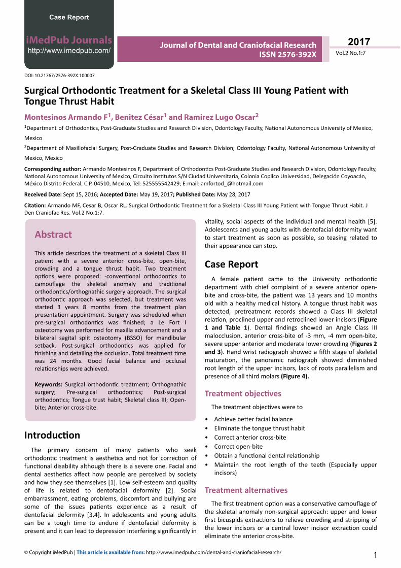
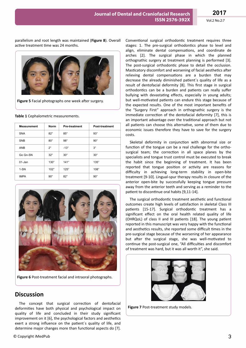
department with chief complaint of a severe anterior open-bite and cross-bite, the patient was 13 years and 10 monthsold with a healthy medical history. A tongue thrust habit wasdetected, pretreatment records showed a Class III skeletalrelation, proclined upper and retroclined lower incisors (Figure1 and Table 1). Dental findings showed an Angle Class IIImalocclusion, anterior cross-bite of -3 mm, -4 mm open-bite,severe upper anterior and moderate lower crowding (Figures 2and 3). Hand wrist radiograph showed a fifth stage of skeletalmaturation, the panoramic radiograph showed diminishedroot length of the upper incisors, lack of roots parallelism andpresence of all third molars (Figure 4).
Treatment objectivesThe treatment objectives were to
• Achieve better facial balance• Eliminate the tongue thrust habit• Correct anterior cross-bite• Correct open-bite• Obtain a functional dental relationship• Maintain the root length of the teeth (Especially upper
incisors)
Treatment alternativesThe first treatment option was a conservative camouflage of
the skeletal anomaly non-surgical approach: upper and lowerfirst bicuspids extractions to relieve crowding and stripping ofthe lower incisors or a central lower incisor extraction couldeliminate the anterior cross-bite.
Case Report
iMedPub Journalshttp://www.imedpub.com/ Vol.2 No.1:7
2017
© Copyright iMedPub | This article is available from: http://www.imedpub.com/dental-and-craniofacial-research/ 1
DOI: 10.21767/2576-392X.100007
Surgical Orthodontic Treatment for a Skeletal Class III Young Patient with Tongue Thrust HabitMontesinos Armando F1, Benitez César1 and Ramirez Lugo Oscar2
1Department of Orthodontics, Post-Graduate Studies and Research Division, Odontology Faculty, National Autonomous University of Mexico,
Mexico2Department of Maxillofacial Surgery, Post-Graduate Studies and Research Division, Odontology Faculty, National Autonomous University of
Mexico, Mexico
Corresponding author: Armando Montesinos F, Department of Orthodontics Post-Graduate Studies and Research Division, Odontology Faculty, National Autonomous University of Mexico, Circuito Institutos S/N Ciudad Universitaria, Colonia Copilco Universidad, Delegación Coyoacán, México Distrito Federal, C.P. 04510, Mexico, Tel: 525555542429; E-mail: [email protected]
Received Date: Sept 15, 2016; Accepted Date: May 19, 2017; Published Date: May 28, 2017
Citation: Armando MF, Cesar B, Oscar RL. Surgical Orthodontic Treatment for a Skeletal Class III Young Patient with Tongue Thrust Habit. J Den Craniofac Res. Vol.2 No.1:7.
Journal of Dental and Craniofacial ResearchISSN 2576-392X
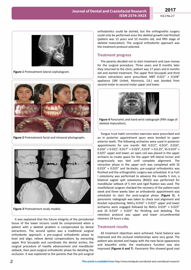
Figure 1 Pretreatment lateral cephalogram.
Figure 2 Pretreatment facial and intraoral photographs.
Figure 3 Pretreatment study models.
It was explained that the future integrity of the periodontaltissue of the lower incisors could be compromised when apatient with a skeletal problem is compensated by dentalextractions. The second option was a traditional surgicalorthodontic approach: a pre-surgical orthodontic phase tolevel and align, relieve dental compensations by extractingupper first bicuspids and coordinate the dental arches; thesurgical procedure of maxilla advancement and mandibularsetback; then post-surgical orthodontic phase for detailing theocclusion. It was explained to the parents that the pre-surgical
orthodontics could be started, but the orthognathic surgerycould only be performed once the skeletal growth had finished(patient was 13 years and 10 months old, and fifth stage ofskeletal maturation). The surgical orthodontic approach wasthe treatment protocol selected.
Treatment progressThe parents decided not to start treatment and save money
for the surgical procedure. Three years and 8 months laterthey returned to the clinic; patient was 17 years and 6 monthsold and started treatment. The upper first bicuspids and thirdmolars extractions were prescribed. MBT 0.022” × 0.028”appliance (3M Unitek, Monrovia, CA.) was bonded fromsecond molar to second molar upper and lower.
Figure 4 Panoramic and hand wrist radiograph (fifth stage ofskeletal maturation).
Tongue trust habit correction exercises were prescribed andan in posterior appointment spurs were bonded to upperanterior teeth. The following archwires were used in posteriorappointments for one month: Niti 0.012”, 0.014”, 0.016”,0.016” × 0.022”, 0.017” × 0.025”, 0.019” × 0.0.25”, SS 0.019” ×0.025” upper and lower; an open coil was placed in the upperarchwire to create space for the upper left lateral incisor andprogressively was tied until complete alignment. Theretraction phase in the upper arch was completed with SS0.019” × 0.025” and tie-backs; pre-surgical orthodontics wasfinished and the orthognathic surgery was scheduled. A Le FortI osteotomy was performed to advance the maxilla 5 mm, abilateral sagital split osteotomy (BSSO) was performed formandibular setback of 5 mm and rigid fixation was used. Themaxillofacial surgeon checked the recovery of the patient eachweek and three weeks later an orthodontic appointment wasscheduled to start the post-surgical phase (Figure 5). Apanoramic radiograph was taken to check root alignment andbracket repositioning, NitiCu 0.016” × 0.022” upper and lowerarchwires were engaged followed by NitiCu 0.019” × 0.025”and SS 0.019” × 0.025” for finishing and detailing. Theretention protocol was upper and lower circumferentialretainers 24 hours a day.
Treatment resultsAll treatment objectives were achieved, Facial balance was
improved and the occlusal relationships were very good. Thepatient was excited and happy with the new facial appearanceand beautiful smile; the masticatory function was alsoimproved (Figures 6 and 7). Panoramic film showed good root
Vol.2 No.2:7
2017
2 This article is available from: http://www.imedpub.com/dental-and-craniofacial-research/
Journal of Dental and Craniofacial ResearchISSN 2576-392X
parallelism and root length was maintained (Figure 8). Overallactive treatment time was 24 months.
Figure 5 Facial photographs one week after surgery.
Table 1 Cephalometric measurements.
Measurement Norm Pre-treatment Post-treatment
SNA 82° 85° 93°
SNB 80° 98° 90°
ANB 2° -13° 3°
Go Gn-SN 32° 30° 30°
01-Jan 130° 141° 135°
1-SN 102° 125° 108°
IMPA 90° 82° 90°
Figure 6 Post-treatment facial and intraoral photographs.
DiscussionThe concept that surgical correction of dentofacial
deformities have both physical and psychological impact onquality of life and concluded in their study significantimprovement on it [6], the psychological factors and aestheticsexert a strong influence on the patient´s quality of life, anddetermine major changes more than functional aspects do [7].
Conventional surgical orthodontic treatment requires threestages: 1. The pre-surgical orthodontics phase to level andalign, eliminate dental compensations, and coordinate dearches [2]. The surgical phase in which the plannedorthognathic surgery at treatment planning is performed [3].The post-surgical orthodontic phase to detail the occlusion.Masticatory discomfort and worsening of facial aesthetics afterrelieving dental compensations are a burden that maydecrease the already diminished patient´s quality of life as aresult of dentofacial deformity [8]. This first stage in surgicalorthodontics can be a burden and patients can really sufferbullying with devastating effects, especially in young adults;but well-motivated patients can endure this stage because ofthe expected results. One of the most important benefits ofthe “Surgery First” approach in orthognathic surgery is theimmediate correction of the dentofacial deformity [7], this isan important advantage over the traditional approach but notall patients can choose this alternative, some of them due toeconomic issues therefore they have to save for the surgerycosts.
Skeletal deformity in conjunction with abnormal size orfunction of the tongue can be a real challenge for the ortho-surgical team; the correction in all space planes by thespecialists and tongue trust control must be executed to breakthe habit since the beginning of treatment. It has beenreported that tongue position or activity are reasons fordifficulty in achieving long-term stability in open-bitetreatment [9-10]. Lingual-spur therapy results in closure of theanterior open-bite by successfully keeping tongue pressureaway from the anterior teeth and serving as a reminder to thepatient to discontinue oral habits [9,11-14].
The surgical orthodontic treatment aesthetic and functionaloutcomes create high levels of satisfaction in skeletal Class IIIpatients [15-17]. Surgical orthodontic treatment has asignificant effect on the oral health related quality of life(OHRQoL) of class II and III patients [18]. The young patientreported in this manuscript was very happy with the functionaland aesthetics results, she reported some difficult times in thepre-surgical stage because of the worsening of her appearancebut after the surgical stage, she was well-motivated tocontinue the post-surgical one, “All difficulties and discomfortof treatment was hard, but it was all worth it”, she said.
Figure 7 Post-treatment study models.
Vol.2 No.2:7
2017
© Copyright iMedPub 3
Journal of Dental and Craniofacial ResearchISSN 2576-392X
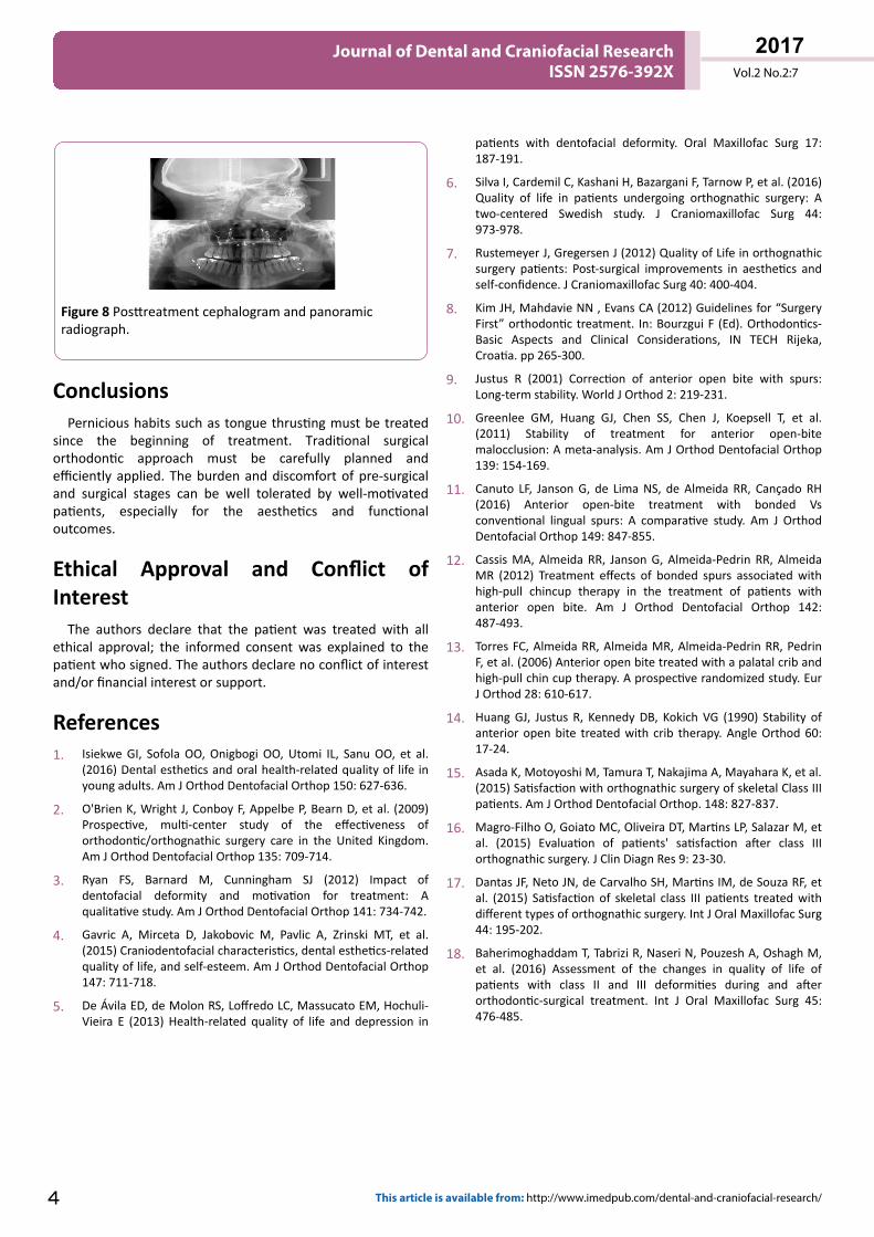
Figure 8 Posttreatment cephalogram and panoramicradiograph.
ConclusionsPernicious habits such as tongue thrusting must be treated
since the beginning of treatment. Traditional surgicalorthodontic approach must be carefully planned andefficiently applied. The burden and discomfort of pre-surgicaland surgical stages can be well tolerated by well-motivatedpatients, especially for the aesthetics and functionaloutcomes.
Ethical Approval and Conflict ofInterest
The authors declare that the patient was treated with allethical approval; the informed consent was explained to thepatient who signed. The authors declare no conflict of interestand/or financial interest or support.
References1. Isiekwe GI, Sofola OO, Onigbogi OO, Utomi IL, Sanu OO, et al.
(2016) Dental esthetics and oral health-related quality of life inyoung adults. Am J Orthod Dentofacial Orthop 150: 627-636.
2. O'Brien K, Wright J, Conboy F, Appelbe P, Bearn D, et al. (2009)Prospective, multi-center study of the effectiveness oforthodontic/orthognathic surgery care in the United Kingdom.Am J Orthod Dentofacial Orthop 135: 709-714.
3. Ryan FS, Barnard M, Cunningham SJ (2012) Impact ofdentofacial deformity and motivation for treatment: Aqualitative study. Am J Orthod Dentofacial Orthop 141: 734-742.
4. Gavric A, Mirceta D, Jakobovic M, Pavlic A, Zrinski MT, et al.(2015) Craniodentofacial characteristics, dental esthetics-relatedquality of life, and self-esteem. Am J Orthod Dentofacial Orthop147: 711-718.
5. De Ávila ED, de Molon RS, Loffredo LC, Massucato EM, Hochuli-Vieira E (2013) Health-related quality of life and depression in
patients with dentofacial deformity. Oral Maxillofac Surg 17:187-191.
6. Silva I, Cardemil C, Kashani H, Bazargani F, Tarnow P, et al. (2016)Quality of life in patients undergoing orthognathic surgery: Atwo-centered Swedish study. J Craniomaxillofac Surg 44:973-978.
7. Rustemeyer J, Gregersen J (2012) Quality of Life in orthognathicsurgery patients: Post-surgical improvements in aesthetics andself-confidence. J Craniomaxillofac Surg 40: 400-404.
8. Kim JH, Mahdavie NN , Evans CA (2012) Guidelines for “SurgeryFirst” orthodontic treatment. In: Bourzgui F (Ed). Orthodontics-Basic Aspects and Clinical Considerations, IN TECH Rijeka,Croatia. pp 265-300.
9. Justus R (2001) Correction of anterior open bite with spurs:Long-term stability. World J Orthod 2: 219-231.
10. Greenlee GM, Huang GJ, Chen SS, Chen J, Koepsell T, et al.(2011) Stability of treatment for anterior open-bitemalocclusion: A meta-analysis. Am J Orthod Dentofacial Orthop139: 154-169.
11. Canuto LF, Janson G, de Lima NS, de Almeida RR, Cançado RH(2016) Anterior open-bite treatment with bonded Vsconventional lingual spurs: A comparative study. Am J OrthodDentofacial Orthop 149: 847-855.
12. Cassis MA, Almeida RR, Janson G, Almeida-Pedrin RR, AlmeidaMR (2012) Treatment effects of bonded spurs associated withhigh-pull chincup therapy in the treatment of patients withanterior open bite. Am J Orthod Dentofacial Orthop 142:487-493.
13. Torres FC, Almeida RR, Almeida MR, Almeida-Pedrin RR, PedrinF, et al. (2006) Anterior open bite treated with a palatal crib andhigh-pull chin cup therapy. A prospective randomized study. EurJ Orthod 28: 610-617.
14. Huang GJ, Justus R, Kennedy DB, Kokich VG (1990) Stability ofanterior open bite treated with crib therapy. Angle Orthod 60:17-24.
15. Asada K, Motoyoshi M, Tamura T, Nakajima A, Mayahara K, et al.(2015) Satisfaction with orthognathic surgery of skeletal Class IIIpatients. Am J Orthod Dentofacial Orthop. 148: 827-837.
16. Magro-Filho O, Goiato MC, Oliveira DT, Martins LP, Salazar M, etal. (2015) Evaluation of patients' satisfaction after class IIIorthognathic surgery. J Clin Diagn Res 9: 23-30.
17. Dantas JF, Neto JN, de Carvalho SH, Martins IM, de Souza RF, etal. (2015) Satisfaction of skeletal class III patients treated withdifferent types of orthognathic surgery. Int J Oral Maxillofac Surg44: 195-202.
18. Baherimoghaddam T, Tabrizi R, Naseri N, Pouzesh A, Oshagh M,et al. (2016) Assessment of the changes in quality of life ofpatients with class II and III deformities during and afterorthodontic-surgical treatment. Int J Oral Maxillofac Surg 45:476-485.
Vol.2 No.2:7
2017
4 This article is available from: http://www.imedpub.com/dental-and-craniofacial-research/
Journal of Dental and Craniofacial ResearchISSN 2576-392X
Related Documents