Suppression of cancer relapse and metastasis by inhibiting cancer stemness Youzhi Li, Harry A. Rogoff, Sarah Keates, Yuan Gao, Sylaja Murikipudi, Keith Mikule, David Leggett, Wei Li, Arthur B. Pardee 1 , and Chiang J. Li 1 Boston Biomedical, Inc., Cambridge, MA 02139 Contributed by Arthur B. Pardee, December 23, 2014 (sent for review December 9, 2014; reviewed by Mikhail V. Blagosklonny and Howard Y. Chang) Partial or even complete cancer regression can be achieved in some patients with current cancer treatments. However, such initial responses are almost always followed by relapse, with the recurrent cancer being resistant to further treatments. The discov- ery of therapeutic approaches that counteract relapse is, there- fore, essential for advancing cancer medicine. Cancer cells are extremely heterogeneous, even in each individual patient, in terms of their malignant potential, drug sensitivity, and their potential to metastasize and cause relapse. Indeed, hypermalig- nant cancer cells, termed cancer stem cells or stemness-high cancer cells, that are highly tumorigenic and metastatic have been iso- lated from cancer patients with a variety of tumor types. More- over, such stemness-high cancer cells are resistant to conventional chemotherapy and radiation. Here we show that BBI608, a small molecule identified by its ability to inhibit gene transcription driven by Stat3 and cancer stemness properties, can inhibit stem- ness gene expression and block spherogenesis of or kill stemness- high cancer cells isolated from a variety of cancer types. Moreover, cancer relapse and metastasis were effectively blocked by BBI608 in mice. These data demonstrate targeting cancer stemness as a novel approach to develop the next generation of cancer thera- peutics to suppress cancer relapse and metastasis. cancer stemness | relapse | BBI608 C urrent cancer treatments ultimately fail owing to metastasis and relapse. Although chemotherapy can induce partial or even complete cancer regression in some patients, such initial responses are invariably followed by relapse, with the recurrent cancer being highly resistant to further chemotherapy, resulting in very limited survival benefits (1– 3). Modern therapeutics designed to specifically target activating mutations are quite ef- fective in inducing cancer regression in patients driven by such activating mutations; however, these treatments are also in- variably followed by relapse with tumors that no longer respond to the targeted agent (2, 3). The discovery of novel therapeutic approaches that counteract cancer relapse is, therefore, urgently required for advancing cancer treatment. The idea that cancer is composed of a group of near-homog- enous, ectopically growing cells has been replaced with a more complex model in which cancer cells are extremely heteroge- neous, even in each individual patient, in terms of their malig- nant potential to metastasize and cause relapse. The increased understanding of the genomic and proteomic complexity of tu- mor heterogeneity further highlights the extreme heterogeneity of cancer cells (4). Subpopulations of cancer cells with extremely high tumori- genic potential, termed cancer stem cells or stem-like cancer cells, have been isolated from cancer patients with a variety of tumor types (5–13) and found to have high stemness properties (5–15). Stemness, initially defined by the expression of stem cells genes, is a property shared by embryonic stem cells and adult stem cells (16). In addition to distinct gene expression profiles, stemness can be measured by a cell’s ability to form spheres when cultured in stem cell media (17). Although it is still un- certain whether cancer stem cells isolated from cancer patients truly qualify as bona fide stem cells and how frequent these cells are, it has been demonstrated that these stemness-high malignant cells are extremely tumorigenic and are resistant to conventional chemotherapies and radiation. Moreover, chemotherapy and ra- diation have been found to induce stemness genes in cancer cells, converting stemness-low cancer cells to stemness-high cancer cells (18, 19). Such highly tumorigenic and drug-resistant stemness-high cancer stem cells are, therefore, likely to be “left over” after chemotherapy or radiotherapy and ultimately responsible for re- lapse (13, 14, 16, 20, 21). However, these stemness-high cancer stem cells are difficult to target owing to activation of prosurvival and antiapoptotic pathways, overexpression of drug efflux pumps, and increased DNA repair capacity (13, 14, 16, 20, 21). Thera- peutic approaches based on the cancer stem cell hypothesis have centered on identifying specific cancer stem cell surface markers and the design of agents to selectively kill these marker-bearing cancer stem cells (22–24). We hypothesized that cancer stemness inhibition can effectively suppress metastasis and relapse. To selectively target cancer stemness, it is critical to identify molecular targets that are re- quired for cancer stemness, but not (or less so) by normal tissue stem cells. The feasibility of this approach has been demon- strated by gene expression profiling, whereby cancer stemness has been shown to more closely resemble embryonic stem cells than adult stem cells (25). Through gene-silencing approaches, we have identified signal transducer and activator of transcrip- tion 3 (Stat3) as critically important for maintaining cancer stem- ness, yet largely dispensable for hematopoietic stem cells. Here we show that BBI608, a small molecule identified by its ability to Significance Current cancer treatments ultimately fail owing to metastasis and relapse. The discovery of therapeutic approaches that counteract relapse and metastasis is, therefore, extremely im- portant for advancing cancer medicine. Hypermalignant cancer cells, termed cancer stem cells or stemness-high cancer cells, have been isolated from patients with a variety of tumor types and found to be highly malignant, tumorigenic, and resistant to chemotherapies. Our data that BBI608, a cancer stemness inhibitor in clinical development, effectively blocks cancer re- lapse and metastasis in xenografted human cancers, suggest targeting cancer stemness as a novel approach to develop the next generation of cancer therapeutics to suppress cancer re- lapse and metastasis. Author contributions: C.J.L. conceived the study; Y.L., H.A.R., S.K., Y.G., S.M., K.M., W.L., A.B.P., and C.J.L. designed research; Y.L., H.A.R., S.K., Y.G., S.M., and K.M. performed research; Y.L., H.A.R., S.K., Y.G., S.M., K.M., D.L., W.L., A.B.P., and C.J.L. analyzed data; and H.A.R. and C.J.L. wrote the paper. Reviewers: M.V.B., Roswell Park Cancer Institute; and H.Y.C., Stanford University. Conflict of interest statement: All authors are employees or advisors of Boston Biomedical, Inc., Sumitomo Dainippon Global Oncology, and declare no equity ownership. 1 To whom correspondence may be addressed. Email: [email protected] or [email protected]. This article contains supporting information online at www.pnas.org/lookup/suppl/doi:10. 1073/pnas.1424171112/-/DCSupplemental. www.pnas.org/cgi/doi/10.1073/pnas.1424171112 PNAS | February 10, 2015 | vol. 112 | no. 6 | 1839–1844 MEDICAL SCIENCES

Welcome message from author
This document is posted to help you gain knowledge. Please leave a comment to let me know what you think about it! Share it to your friends and learn new things together.
Transcript

Suppression of cancer relapse and metastasis byinhibiting cancer stemnessYouzhi Li, Harry A. Rogoff, Sarah Keates, Yuan Gao, Sylaja Murikipudi, Keith Mikule, David Leggett, Wei Li,Arthur B. Pardee1, and Chiang J. Li1
Boston Biomedical, Inc., Cambridge, MA 02139
Contributed by Arthur B. Pardee, December 23, 2014 (sent for review December 9, 2014; reviewed by Mikhail V. Blagosklonny and Howard Y. Chang)
Partial or even complete cancer regression can be achieved insome patients with current cancer treatments. However, suchinitial responses are almost always followed by relapse, with therecurrent cancer being resistant to further treatments. The discov-ery of therapeutic approaches that counteract relapse is, there-fore, essential for advancing cancer medicine. Cancer cells areextremely heterogeneous, even in each individual patient, interms of their malignant potential, drug sensitivity, and theirpotential to metastasize and cause relapse. Indeed, hypermalig-nant cancer cells, termed cancer stem cells or stemness-high cancercells, that are highly tumorigenic and metastatic have been iso-lated from cancer patients with a variety of tumor types. More-over, such stemness-high cancer cells are resistant to conventionalchemotherapy and radiation. Here we show that BBI608, a smallmolecule identified by its ability to inhibit gene transcriptiondriven by Stat3 and cancer stemness properties, can inhibit stem-ness gene expression and block spherogenesis of or kill stemness-high cancer cells isolated from a variety of cancer types. Moreover,cancer relapse and metastasis were effectively blocked by BBI608in mice. These data demonstrate targeting cancer stemness asa novel approach to develop the next generation of cancer thera-peutics to suppress cancer relapse and metastasis.
cancer stemness | relapse | BBI608
Current cancer treatments ultimately fail owing to metastasisand relapse. Although chemotherapy can induce partial or
even complete cancer regression in some patients, such initialresponses are invariably followed by relapse, with the recurrentcancer being highly resistant to further chemotherapy, resultingin very limited survival benefits (1–3). Modern therapeuticsdesigned to specifically target activating mutations are quite ef-fective in inducing cancer regression in patients driven by suchactivating mutations; however, these treatments are also in-variably followed by relapse with tumors that no longer respondto the targeted agent (2, 3). The discovery of novel therapeuticapproaches that counteract cancer relapse is, therefore, urgentlyrequired for advancing cancer treatment.The idea that cancer is composed of a group of near-homog-
enous, ectopically growing cells has been replaced with a morecomplex model in which cancer cells are extremely heteroge-neous, even in each individual patient, in terms of their malig-nant potential to metastasize and cause relapse. The increasedunderstanding of the genomic and proteomic complexity of tu-mor heterogeneity further highlights the extreme heterogeneityof cancer cells (4).Subpopulations of cancer cells with extremely high tumori-
genic potential, termed cancer stem cells or stem-like cancercells, have been isolated from cancer patients with a variety oftumor types (5–13) and found to have high stemness properties(5–15). Stemness, initially defined by the expression of stem cellsgenes, is a property shared by embryonic stem cells and adultstem cells (16). In addition to distinct gene expression profiles,stemness can be measured by a cell’s ability to form sphereswhen cultured in stem cell media (17). Although it is still un-certain whether cancer stem cells isolated from cancer patients
truly qualify as bona fide stem cells and how frequent these cellsare, it has been demonstrated that these stemness-high malignantcells are extremely tumorigenic and are resistant to conventionalchemotherapies and radiation. Moreover, chemotherapy and ra-diation have been found to induce stemness genes in cancer cells,converting stemness-low cancer cells to stemness-high cancer cells(18, 19). Such highly tumorigenic and drug-resistant stemness-highcancer stem cells are, therefore, likely to be “left over” afterchemotherapy or radiotherapy and ultimately responsible for re-lapse (13, 14, 16, 20, 21). However, these stemness-high cancerstem cells are difficult to target owing to activation of prosurvivaland antiapoptotic pathways, overexpression of drug efflux pumps,and increased DNA repair capacity (13, 14, 16, 20, 21). Thera-peutic approaches based on the cancer stem cell hypothesis havecentered on identifying specific cancer stem cell surface markersand the design of agents to selectively kill these marker-bearingcancer stem cells (22–24).We hypothesized that cancer stemness inhibition can effectively
suppress metastasis and relapse. To selectively target cancerstemness, it is critical to identify molecular targets that are re-quired for cancer stemness, but not (or less so) by normal tissuestem cells. The feasibility of this approach has been demon-strated by gene expression profiling, whereby cancer stemnesshas been shown to more closely resemble embryonic stem cellsthan adult stem cells (25). Through gene-silencing approaches,we have identified signal transducer and activator of transcrip-tion 3 (Stat3) as critically important for maintaining cancer stem-ness, yet largely dispensable for hematopoietic stem cells. Here weshow that BBI608, a small molecule identified by its ability to
Significance
Current cancer treatments ultimately fail owing to metastasisand relapse. The discovery of therapeutic approaches thatcounteract relapse and metastasis is, therefore, extremely im-portant for advancing cancer medicine. Hypermalignant cancercells, termed cancer stem cells or stemness-high cancer cells,have been isolated from patients with a variety of tumor typesand found to be highly malignant, tumorigenic, and resistantto chemotherapies. Our data that BBI608, a cancer stemnessinhibitor in clinical development, effectively blocks cancer re-lapse and metastasis in xenografted human cancers, suggesttargeting cancer stemness as a novel approach to develop thenext generation of cancer therapeutics to suppress cancer re-lapse and metastasis.
Author contributions: C.J.L. conceived the study; Y.L., H.A.R., S.K., Y.G., S.M., K.M., W.L.,A.B.P., and C.J.L. designed research; Y.L., H.A.R., S.K., Y.G., S.M., and K.M. performedresearch; Y.L., H.A.R., S.K., Y.G., S.M., K.M., D.L., W.L., A.B.P., and C.J.L. analyzed data;and H.A.R. and C.J.L. wrote the paper.
Reviewers: M.V.B., Roswell Park Cancer Institute; and H.Y.C., Stanford University.
Conflict of interest statement: All authors are employees or advisors of Boston Biomedical,Inc., Sumitomo Dainippon Global Oncology, and declare no equity ownership.1To whom correspondence may be addressed. Email: [email protected] [email protected].
This article contains supporting information online at www.pnas.org/lookup/suppl/doi:10.1073/pnas.1424171112/-/DCSupplemental.
www.pnas.org/cgi/doi/10.1073/pnas.1424171112 PNAS | February 10, 2015 | vol. 112 | no. 6 | 1839–1844
MED
ICALSC
IENCE
S

inhibit gene transcription driven by Stat3 and cancer stemnessproperties, can block spherogenesis of and kill stemness-highcancer cells isolated from a variety of cancer types and inhibitstemness gene expression. Moreover, cancer relapse and metas-tasis were effectively blocked by BBI608 in xenografted humancancers in mice. These data demonstrate targeting cancerstemness as a novel approach to develop the next generation ofcancer therapeutics to suppress cancer relapse and metastasis.
ResultsInhibition of Cancer Relapse and Metastasis by BBI608.Relapse afterchemotherapy treatment is a common phenomenon (26). Wehypothesized that stemness-high cancer cells are responsible forcancer relapse and that stemness inhibition can suppress cancerrelapse. To effectively block cancer relapse it is, therefore, es-sential to target cancer stemness. Through a gene silencing-basedapproach, we identified Stat3 as a key driver for cancer stemness.We next set out to discover a druggable inhibitor of Stat3 totarget cancer stemness, using in silico screening and computa-tional biology to search for Stat3 binders, phenotype-driventesting to determine stemness inhibition, and Stat3-drivengene transcription to test for Stat3 inhibition. Through such anactivity-oriented, quality-driven iterative research process, weidentified BBI608.To evaluate the phenomenon of tumor relapse, we used
a pancreatic cancer xenograft model and the chemotherapyagent gemcitabine. Treatment with gemcitabine inhibited PaCa-2xenograft tumor growth, with tumor growth inhibition of 47.5%on day 41 (Fig. 1A). However, after cessation of treatment on day41, the tumors in the gemcitabine-treated animals soon relapsedand even outgrew the tumors in the vehicle control group (Fig.1A). Treatment with BBI608 also significantly inhibited PaCa-2xenograft tumor growth on day 41 (Fig. 1B). However, unlike thegemcitabine-treated animals, no tumor regrowth was observed inthe animals administered BBI608 during the 22-d posttreatmentobservation period (Fig. 1B). Furthermore, no signs of toxicity asevidenced by body weight measurement were observed (Fig. S1 Aand B). These data suggest that BBI608, unlike chemotherapy,can inhibit the cells within the tumor mass that are responsiblefor tumor relapse.As another model for cancer relapse, we used the intrasplenic-
nude mouse model system (ISMS) model to evaluate cancerrelapse in a metastatic setting. This model involves the injectionof colon cancer cells (HT29) into the spleen capsule of nudemice, and these colon cancer cells can form liver metastasesspontaneously in a few weeks. To test the potential therapeuticrole of BBI608 against metastasis, we used the ISMS model.BBI608 was found to effectively block spleen and liver metas-tases in the ISMS model (Fig. 1C). These data demonstrate thatBBI608 is effective at blocking metastasis in vivo.
Depletion of Stemness-High Cancer Cells by BBI608. We hypothe-sized that the antirelapse activity of BBI608 may be attributed toits effect on stemness-high cancer cells. To test this hypothesis,
A
B
C
Fig. 1. Inhibition of cancer relapse by BBI608. (A) Immunosuppressed micewith established s.c. Paca-2 human pancreatic cancer were given gemcita-bine (120 mg/kg) q3d, or vehicle control i.p. The animals received a totalof 14 doses and were maintained 22 d for posttreatment observation.Tumor size was evaluated periodically during treatment and posttreat-ment observation. Each point represents the mean ± SEM of seventumors. (B) Immunosuppressed mice with established s.c. PaCa-2 humanpancreatic cancer were given BBI608 (20 mg/kg) daily or vehicle controli.p. and monitored as in A. (C ) Immunosuppressed mice bearing intra-splenically inoculated HT29 human colon cancer cells were given dailyBBI608 (20 mg/kg) or vehicle control i.p. Primary tumors at spleen andspontaneous liver metastases were examined macroscopically. A repre-sentative photograph is shown.
A B
Fig. 2. Depletion of stemness-high cancer cells by BBI608 in vivo. Mice wereadministered either vehicle, (A) gemcitabine (120 mg/kg [PaCa2]), (B) car-boplatin (30 mg/kg [FaDu]), or 20 mg/kg of BBI608 i.p. After killing, tumorswere collected after 7 or 14 d of treatment, for Paca-2 and FaDu cells, re-spectively. Single-cell suspensions were obtained after animal killing andsterile removal of tumors. Live cells were then counted and used to measuretheir ability to form spheres when cultured in cancer stem cell media.
1840 | www.pnas.org/cgi/doi/10.1073/pnas.1424171112 Li et al.

we used two human xenograft tumor models. In the pancreatictumor (PaCa2) model animals were treated with vehicle, gem-citabine, or BBI608. Tumors were collected after 7 d of treat-ment, single-cell suspensions obtained from the tumors, andfrequency of stemness-high cancer cells was determined by theirself-renewal capacity as measured by their ability to grow asspheres when cultured under serum-free, attachment-free stemcell culture conditions. Compared with control-treated animals,BBI608 treatment decreased the stemness-high subpopulation byfivefold (Fig. 2A). By contrast, gemcitabine treatment causeda threefold increase in stemness-high cell population. Similardata were also observed in a head and neck tumor (FaDu) modeltreated with vehicle, carboplatin, or BBI608 (Fig. 2B). Thesedata demonstrate that BBI608 is effective at targeting thestemness-high cancer cells in vivo, whereas standard chemo-therapy caused an enrichment of the stemness-high cancer cellsubpopulation.
BBI608 Can Block Survival and Self-Renewal of Stemness-High CancerCells. To determine the effect of BBI608 on stemness-high cancercells, we examined self-renewal of stemness-high cancer cellsisolated or enriched by stem-cell culture selection (Fig. 3A), sidepopulation-based enrichment (Fig. 3B), or cancer stem cell surfacemarkers (Fig. 3C). As shown in Fig. 3A, stemness-high coloncancer cells grown under stem cell culture conditions formspheres, and treatment with BBI608 blocked spherogenesis,suggesting that BBI608 inhibits self-renewal of stemness-highcancer cellsWe determined whether the stemness-high population was
resistant to chemotherapeutics and targeted kinase inhibitors.Stemness-high cancer cells were isolated by sorting for exclusionof Hoechst dye, followed by culture in the serum-free stem cellmedia. Sorting based on Hoechst dye exclusion provides two cellpopulations: a stemness-high side population and a stemness-lownonside population. Both the stemness-high side population andnonside population were killed by BBI608, with the side pop-ulation being more sensitive to BBI608 (Fig. 3B). By contrast,whereas treatment of the nonside population with doxorubicinresulted in inhibition of viability, treatment of the side pop-ulation with doxorubicin had little effect on viability (Fig. 3B).Stemness-high cancer cells were also isolated by sorting for highCD44 expression followed by culture in the serum-free stem cell
media. Treatment of these cells with BBI608 resulted in inhibitionof spherogenesis (Fig. 3C). By contrast, treatment with imatinib,sunitinib, erlotinib, or doxorubicin had little effect on thespherogenesis (Fig. 3C).We next determined whether BBI608 affects normal stem
cells. To address this issue we obtained CD34+ hematopoieticstem cells, treated them with BBI608, and then assessed theirability to form both erythroid and myeloid colonies. No in-hibition of colony formation of CD34+ hematopoietic stem cellswas observed by BBI608 treatment at up to 30 μM (the highestconcentration tested) (Fig. 3D).We next compared the potency of BBI608 and clinically used
targeted therapeutics against stemness-high cancer cells andheterogeneous cancer cells under the same conditions. As shownin Table 1, stemness-high cancer cells displayed between three-fold and 10-fold resistance to all targeted therapeutics tested,whereas the IC50 for BBI608 was lower in the stemness-highcancer cell population than in the bulk cancer cells. We nextdetermined the IC50 of the inhibitory activity of BBI608 instemness-high cancer cells isolated from a variety of humancancer cell lines, including head and neck, lung, brain, colon,liver, ovarian, pancreatic, and kidney cancer cell lines. As shownin Table 2, BBI608 was highly effective at targeting stemness-high cancer cells. These data suggest that stemness-high cancercells are resistant to conventional chemotherapeutic and targetedagents tested but are sensitive to cancer stemness inhibitor BBI608.
A B C
D
Fig. 3. Inhibition of self-renewal of stemness-high cancer cells by BBI608. (A) Spheres derived from DLD1 and HCT116 cell lines were dissociated to single cellsand allowed to form spheres in suspension with cancer stem cell medium for 48 h before treatment with BBI608 (2 μM). After 24 h, the drugs were removedand the cells were cultured in fresh cancer stem cell medium for another 24 h. Representative images are shown. (B) SW480 colon cancer cells were isolated byFACS based on Hoechst dye exclusion and were cultured for 72 h in cancer stem cell conditions before the addition of the indicated concentrations oftherapeutic agents. Sphere growth was scored by counting the number of spheres possessing >50 cells. SP, side population; NSP, nonside population. (C)CD44high FaDu cells were isolated by FACS and were cultured for 72 h in cancer stem cell conditions before the addition of the indicated therapeutic agents(400 nM BBI608, 2 μM imatinib, 10 μM sunitinib, 10 μM erlotinib, 100 nM doxorubicin). Sphere growth was scored by counting the number of spherespossessing >50 cells. Representative images are shown. (D) CD34+ bone marrow mononuclear cells were treated with either DMSO or BBI608 for 6 h at 37 °C.Cells were then washed and plated in complete Methocult H4434 medium. Colonies of both erythroid and myeloid lineages containing >50 cells per colonywere counted. Each treatment was performed in triplicate.
Table 1. Comparison of BBI608 with indicated compounds inregular cancer cells and stemness-high cancer cells
Compound
IC50 (uM)
Bulk cells Cancer stem cells
BBI608 0.395 0.142Sunitinib 2.907 9.011Gefitinib 1.950 22.283Regorafenib 4.705 15.821Erlotinib 1.807 12.172
FaDu cells cultured under normal growth conditions or cancer stem cellgrowth conditions. IC50s were performed in triplicate.
Li et al. PNAS | February 10, 2015 | vol. 112 | no. 6 | 1841
MED
ICALSC
IENCE
S
BBI608 Down-Regulates Stemness Gene Expression. Multiple path-ways regulating the self-renewal of stem cells have been identi-fied (16). Stat3, the target of BBI608, regulates many of thegenes implicated in cancer stem cell self-renewal, includingc-Myc and β-catenin (27–32). We investigated inhibition of thesesignaling pathways after treatment with BBI608. We found thatBBI608 treatment resulted in a dose-dependent decrease inNanog, Axl, Sox-2, Klf4, survivin, c-Myc, Bmi-1, and β-cateninprotein levels (Fig. 4A). Levels of some proteins, such as c-Mycand Axl, were decreased as early as 3 h after treatment, whereasmost were lower at 6 h, with the majority of proteins still reducedafter 24 h (Fig. 4A). To expand on this observation, we analyzed
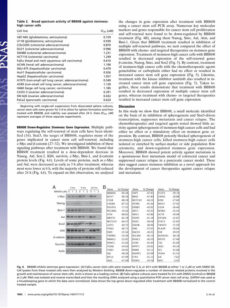
the changes in gene expression after treatment with BBI608using a cancer stem cell PCR array. Numerous key molecularmarkers and genes responsible for cancer stem cell proliferationand self-renewal were found to be down-regulated by BBI608treatment (Fig. 4B), among them Nanog, Smo, Axl, Atm, andBmi-1. Given that BBI608 treatment resulted in inhibition ofmultiple self-renewal pathways, we next compared the effect ofBBI608 with chemo- and targeted therapeutics on stemness geneexpression. Treatment of stemness-high cancer cells with BBI608resulted in decreased expression of the self-renewal genesβ-catenin, Nanog, Smo, and Sox2 (Fig. 5). By contrast, treatmentof stemness-high cancer cells with the chemotherapeutic agentsgemcitabine or carboplatin either had no effect or resulted inincreased cancer stem cell gene expression (Fig. 5). Likewise,treatment with the kinase inhibitor sunitinib also resulted in in-creased cancer stem cell gene expression (Fig. 5). Taken to-gether, these results demonstrate that treatment with BBI608resulted in decreased expression of multiple cancer stem cellgenes, whereas treatment with chemo- or targeted therapeuticsresulted in increased cancer stem cell gene expression.
DiscussionIn this study we show that BBI608, a small molecule identifiedon the basis of its inhibition of spherogenesis and Stat3-driventranscription, suppresses metastasis and cancer relapse. Thechemotherapeutics and targeted agents tested showed little ac-tivity against spherogenesis of stemness-high cancer cells and hadeither no effect or a stimulatory effect on stemness gene ex-pression. By contrast, BBI608 potently blocked spherogenesis ofstemness-high cancer cells, killed stemness-high cancer cellsisolated or enriched by surface-marker or side population flowcytometry, and down-regulated stemness gene expression.Moreover, BBI608 showed potent activity against metastasis ina spontaneous liver metastasis model of colorectal cancer andsuppressed cancer relapse in a pancreatic cancer model. Thesedata suggest cancer stemness inhibition as a novel approach forthe development of cancer therapeutics against cancer relapseand metastasis.
Table 2. Broad spectrum activity of BBI608 against stemness-high cancer cells
Cell line IC50 (uM)
U87-MG (glioblastoma; astrocytoma) 0.729U118 (glioblastoma; astrocytoma) 0.930COLO205 (colorectal adenocarcinoma) 0.870DLD1 (colorectal adenocarcinoma) 0.996SW480 (colorectal adenocarcinoma) 1.231HCT116 (colorectal carcinoma) 1.249FaDu (head and neck squamous cell carcinoma) 0.616ACHN (renal cell adenocarcinoma) 1.190SNU-475 (hepatocellular carcinoma) 0.479Huh7 (hepatocellular carcinoma) 0.926HepG2 (hepatocellular carcinoma) 1.057H1975 (non-small cell lung cancer; adenocarcinoma) 0.549A549 (non-small cell lung cancer; adenocarcinoma) 1.130H460 (large cell lung cancer; carcinoma) 1.185CAOV-3 (ovarian adenocarcinoma) 0.291SW-626 (ovarian adenocarcinoma) 0.432PaCa2 (pancreatic carcinoma) 0.624
Beginning with single-cell suspensions from dissociated sphere cultures,cancer stem cells were grown for 3 d to allow for sphere formation and thentreated with BBI608, and viability was assessed after 24 h. Data (IC50, uM)represent averages of three separate experiments.
A B
Fig. 4. BBI608 inhibits stemness gene expression. (A) FaDu cancer stem cells were treated for 3, 6, or 24 h with BBI608 at either 1 or 2 μM or with DMSO (0).Cell lysates from these treated cells were then analyzed by Western blotting. BBI608 down-regulates a number of stemness related proteins involved in thegrowth and maintenance of cancer stem cells. Actin is shown as a loading control. (B) FaDu sphere cultures were treated for 6 h with DMSO (control) or BBI608at 2 μM. RNA was isolated and reverse transcribed and then the resulting cDNA analyzed using a quantitative PCR cancer stem cell array. GAPDH was used asa housekeeping gene to which the data were normalized. Data shows the top genes down-regulated after treatment with BBI608 normalized to the controltreated sample.
1842 | www.pnas.org/cgi/doi/10.1073/pnas.1424171112 Li et al.
Highly tumorigenic cancer cells have been enriched and isolatedfrom heterogeneous cancer cell populations in patients of varioustumor types (5–15). Such hypermalignant cells, termed cancerstem cells, are characterized by extremely high tumorgenic po-tential, spherogenesis in stem cell media, and expression ofstemness genes (5–15). More recently, it has been demonstratedthat nonstem cancer cells can acquire stemness phenotypes undercertain conditions (33, 34), including clinically relevant che-motherapy or radiotherapy (18, 19). To compare BBI608 withchemotherapeutics agents, we used gemcitabine and carboplatin,which are two commonly used agents for the treatment of a va-riety of human cancers. We reproduced in vivo the situation thatis often found in patients, in which standard therapies target thebulk of the tumor but fail to eradicate the cancer stem cellpopulation, allowing the tumor to relapse (35). We found thattreatment with BBI608 could prevent cancer relapse and metas-tasis in vivo. Moreover, BBI608 significantly reduced the expres-sion of various stemness genes, including Nanog, Sox2, and Oct4(POU5F1). These genes encode key stemness transcription factorsthat are important for the maintenance of pluripotency (36, 37).These data demonstrate the feasibility of inhibiting cancer stem-ness by modulating stemness gene expression.Through a gene silencing-based approach we identified Stat3
as a critically important factor for maintaining cancer stemness(US patent 8,877,803), which has also been shown by other pub-lications (38, 39). We have observed that Stat3 is constitutively
activated in cancer stem cells independent of upstream signalingregulators (US patent 8,877,803). Therefore, cancer stem cells orstemness-high cancer cells are highly sensitive to direct Stat3 in-hibition and are not sensitive to inhibition of upstream kinases,including Janus kinases. In this study we demonstrate that BBI608,a potent small-molecule inhibitor of Stat3 (US patent 8,877,803),has a broad spectrum of activity against stemness-high cancer cells,further supporting a significant role for Stat3 in cancer stemness.Targeting Stat3 is expected to trigger a cascade of down-regulationof stemness genes, via direct or indirect mechanisms (32). Ourdata show that BBI608 completely spares hematopoietic stemcells, a finding that is consistent with an observation made in Stat3conditional knockout mice in which Stat3 was found to be dis-pensable for hematopoietic stem cells owing to compensation byother Stat family members (40). Thus, it seems that cancer stemcells hijacked the essential role of Stat3 in embryonic stem cellsand become highly sensitive to Stat3 inhibition. In keeping withthese in vitro findings, BBI608 is well tolerated and associatedwith no signs of adverse effects on hematopoietic or other normaladult stem cells in preclinical toxicology studies, as well as inclinical trials (41).Cancer relapse and metastasis are the ultimate reasons of the
failure of current cancer therapies (42–44). The initial responsesto chemotherapy and targeted agents are almost always followedby recurrence of resistant cancer (45). The hypothesis that can-cer stemness is at least partially responsible for cancer relapseand metastasis opens a novel avenue of research. Identifying andtargeting the molecular mechanisms that regulate cancer stem-ness should help design the next generation of cancer thera-peutics to block metastasis and suppress relapse.
Materials and MethodsCell Culture and Reagents. Colo205, Dld1, Hct116, RKO, SW480, AGS, MKN-28,MKN-45, 786-0, ACHN, Huh7, Panc1, Du145, HeLa, PC3, and A431 cells wereobtained from ATCC. All cells were maintained in DMEM supplemented with10% (vol/vol) FBS (Gemini) and 1% penicillin/streptomycin (Life Technolo-gies). Cancer stem cells were maintained on noncell adhesive plates inDMEM nutrient mixture F-12 (DMEM/F-12) containing B-27 supplement (LifeTechnologies), 0.6% BSA, 20 ng/mL EGF, 10 ng/mL FGF (R&D Systems), and1% penicillin/streptomycin. Hoechst dye exclusion cell sorting of the sidepopulation was performed as previously described (46). CD44high/CD24low
were isolated by FACS and sorted cells maintained as described above. HumanCD34+ bone marrow cells (AllCells) were cultured in MethoCult media to formcolonies as described by the manufacturer (Stemcell Technologies). BBI608(2-Acetyl-4H,9H-naphtho[2,3-b]furan-4,9-dione) was synthesized at BostonBiomedical. Sunitinib, regorafenib, gefitinib, erlotinib, carboplatin, gemci-tabine, and doxorubicin were purchased from Selleckchem.
Western Blot Analysis. Cells were washed twice with ice-cold PBS and lysed inlysis buffer [50 mM Hepes (pH 7.5), 1% Nonidet P-40, 150 mM NaCl, 1 mMEDTA, and 1× protease inhibitor mixture (EMD Millipore)]. Soluble protein(20 μg) was separated by SDS/PAGE and transferred to nitrocellulose mem-branes. Primary antibodies against c-Myc, Klf4, Nanog, Survivin, Sox2, Bmi-1,Axl, and β-catenin (Cell Signaling) were used in this study. The antigen–antibody complexes were visualized by enhanced chemiluminescence (BioRad).
IC50 Analysis. For cancer stem cells, spheres were dissociated and plated undercancer stem cell culture conditions on coated 96-well plates. After 72 h ofculture, wells were dosed with the indicated compounds. Seventy-two hours(Table 1) or 24 h (Table 2) after dosing, CellTiter-Glo 2.0 was added to eachwell, and the luminescence was measured as described by the manufacturer(Promega). IC50 values were calculated by fitting a four parameter dose–response curve to normalized data using GraphPad Prism software. For bulkcells, cells were plated at 5,000 cells per well on 96-well plates. Twenty-fourhours after plating, cells were treated with the indicated compounds. Via-bility was determined at 72 h as described above.
Quantitative PCR. FaDu cancer stem cells were seeded in attachment-free six-well plates with cancer stem cell media. Media was changed on day 3. On day4 of culture, desired concentrations of BBI608, chemotherapy or targetedtherapeutic, or DMSO were added to wells in quadruplicate. At the
Fig. 5. Comparison between BBI608 and current cancer therapeutics. FaDucancer stem cells were treated for 24 h with DMSO (control), BBI608 (2 μM),sunitinib (20 μM), gemcitabine (2 μM), or carboplatin (32 μM). RNA wasisolated, reverse transcribed, and then analyzed by quantitative PCR forstem cell genes: β-catenin, Nanog, SMO, and SOX2. GAPDH was used asa housekeeping gene to which the data were normalized.
Li et al. PNAS | February 10, 2015 | vol. 112 | no. 6 | 1843
MED
ICALSC
IENCE
S
appropriate time point cells were harvested and RNA extracted using theSimplyRNA kit according to directions on the Promega Maxwell system.Reverse transcription was performed on 1 μg of RNA from each sample usingthe GoScript reverse transcription kit (Promega). Real-time PCR was carriedout using RT2 qPCR Primer Assays and RT2 SYBR Green qPCR Mastermix(SABiosciences). Replicate wells were prepared for each sample. Expressionof genes of interest was normalized to GAPDH.
CD34+ Bone Marrow Cells. Frozen CD34+ bone marrow mononuclear cells(AllCells LLC) were thawed into RPMI 1640 medium (Gibco BRL) containing10% (vol/vol) FBS and DNase I (Stem Cell Technologies). After two washes withmedium, cells were incubated in medium containing DMSO or BBI608 for 6 hat 37 °C. Cells were then washed twice in medium, counted to determine vi-ability, and plated in replicate plates at 4,000 cells per 35-mm dish in completeMethocult H4434 medium (Stem Cell Technologies). Cultures were maintainedat 37 °C for 14 d to allow colony formation. Colonies of both erythroid andmyeloid lineages containing more than 50 cells per colony were counted.
Mouse Models. For xenograft studies, PaCa2 pancreatic cancer cells wereinoculated s.c. into female athymic nude mice (6 × 10⁶ cells per mouse) andallowed to form palpable tumors. Once the tumors reached ∼100 mm3, theanimals were treated i.p. with BBI608 at 20 mg/kg, gemcitabine (120 mg/kg)every three days (q3d), or vehicle control daily (5 consecutive days, followedby a 2-d dosing holiday). The animals received a total of 14 doses of BBI608or vehicle control. Tumors were measured throughout treatment and theposttreatment observation period. To determine whether BBI608 depletescancer stem cells in vivo, female athymic nude mice were administered witheither vehicle, gemcitabine (120 mg/kg), carboplatin (30 mg/kg), or 20 mg/kg
of BBI608 i.p. Single-cell suspensions were obtained after animal killing af-ter 14 d of treatment, and sterile removal of tumors. Live cells were thencounted and used to measure their ability to form spheres when cultured incancer stem cell media. Fresh media was added every 3 d, and sphere for-mation was determined after 10–14 d in culture. Spheres with >50 cells werescored. For the ISMS model, 2 × 10⁶ HT29 cells in 0.1 mL PBS were injectedunder the spleen capsule of the nude mice. The spleen was replaced in theperitoneal cavity, and the incision was closed. Mice were killed when mori-bund or 5 wk after treatment. The spleen, liver, and lungs were removedand examined, and the number of tumor lesions was recorded. Mice weredivided into two groups, a control group given vehicle (n = 4) and the othergroup receiving 20 mg/kg BBI608 (n = 4). Drug was administered via i.p.injection for 5 d/wk and for 4 wk from the fourth day after intrasplenicinjection. The primary tumors at the spleen and spontaneous liver metas-tases were examined macroscopically and confirmed histologically.
Statistical Analysis. All experiments were performed in triplicate. The data forthe cell proliferation assays assay are expressed as the mean ± SD. The SDs forall of the measured biological parameters are displayed in the appropriatefigures. A Student t test was used for single-variable comparisons, anda P value of <0.05 was considered statistically significant.
Animal Permits. The protocol was approved by Institutional Animal Care andUse Committee of Boston Biomedical.
ACKNOWLEDGMENTS. We thank our colleagues at Boston Biomedical for theiradvice, Katherine Geromini for excellent technical help, and Dr. Andrew Keatesfor critical reading of the manuscript.
1. Shekhar MP (2011) Drug resistance: Challenges to effective therapy. Curr Cancer DrugTargets 11(5):613–623.
2. Nakata A, Gotoh N (2012) Recent understanding of the molecular mechanisms for theefficacy and resistance of EGF receptor-specific tyrosine kinase inhibitors in non-smallcell lung cancer. Expert Opin Ther Targets 16(8):771–781.
3. Bucheit AD, Davies MA (2014) Emerging insights into resistance to BRAF inhibitors inmelanoma. Biochem Pharmacol 87(3):381–389.
4. Meacham CE, Morrison SJ (2013) Tumour heterogeneity and cancer cell plasticity.Nature 501(7467):328–337.
5. Al-Hajj M, Wicha MS, Benito-Hernandez A, Morrison SJ, Clarke MF (2003) Prospectiveidentification of tumorigenic breast cancer cells. Proc Natl Acad Sci USA 100(7):3983–3988.
6. Dalerba P, et al. (2007) Phenotypic characterization of human colorectal cancer stemcells. Proc Natl Acad Sci USA 104(24):10158–10163.
7. Eramo A, et al. (2008) Identification and expansion of the tumorigenic lung cancerstem cell population. Cell Death Differ 15(3):504–514.
8. Kim MP, et al. (2011) ALDH activity selectively defines an enhanced tumor-initiatingcell population relative to CD133 expression in human pancreatic adenocarcinoma.PLoS ONE 6(6):e20636.
9. Li C, et al. (2007) Identification of pancreatic cancer stem cells. Cancer Res 67(3):1030–1037.
10. O’Brien CA, Pollett A, Gallinger S, Dick JE (2007) A human colon cancer cell capable ofinitiating tumour growth in immunodeficient mice. Nature 445(7123):106–110.
11. Prince ME, et al. (2007) Identification of a subpopulation of cells with cancer stem cellproperties in head and neck squamous cell carcinoma. Proc Natl Acad Sci USA 104(3):973–978.
12. Singh SK, et al. (2004) Identification of human brain tumour initiating cells. Nature432(7015):396–401.
13. Lapidot T, et al. (1994) A cell initiating human acute myeloid leukaemia after trans-plantation into SCID mice. Nature 367(6464):645–648.
14. Blagosklonny MV (2007) Cancer stem cell and cancer stemloids: From biology totherapy. Cancer Biol Ther 6(11):1684–1690.
15. Visvader JE, Lindeman GJ (2008) Cancer stem cells in solid tumours: Accumulatingevidence and unresolved questions. Nat Rev Cancer 8(10):755–768.
16. Reya T, Morrison SJ, Clarke MF, Weissman IL (2001) Stem cells, cancer, and cancer stemcells. Nature 414(6859):105–111.
17. Chen SF, et al. (2012) Nonadhesive culture system as a model of rapid sphere for-mation with cancer stem cell properties. PLoS ONE 7(2):e31864.
18. Ghisolfi L, Keates AC, Hu X, Lee DK, Li CJ (2012) Ionizing radiation induces stemness incancer cells. PLoS ONE 7(8):e43628.
19. Hu X, et al. (2012) Induction of cancer cell stemness by chemotherapy. Cell Cycle11(14):2691–2698.
20. Milas L, Hittelman WN (2009) Cancer stem cells and tumor response to therapy:Current problems and future prospects. Semin Radiat Oncol 19(2):96–105.
21. Vermeulen L, de Sousa e Melo F, Richel DJ, Medema JP (2012) The developing cancerstem-cell model: Clinical challenges and opportunities. Lancet Oncol 13(2):e83–e89.
22. Jin L, Hope KJ, Zhai Q, Smadja-Joffe F, Dick JE (2006) Targeting of CD44 eradicateshuman acute myeloid leukemic stem cells. Nat Med 12(10):1167–1174.
23. Jin L, et al. (2009) Monoclonal antibody-mediated targeting of CD123, IL-3 receptoralpha chain, eliminates human acute myeloid leukemic stem cells. Cell Stem Cell 5(1):31–42.
24. Kikushige Y, et al. (2010) TIM-3 is a promising target to selectively kill acute myeloid
leukemia stem cells. Cell Stem Cell 7(6):708–717.25. Wong DJ, et al. (2008) Module map of stem cell genes guides creation of epithelial
cancer stem cells. Cell Stem Cell 2(4):333–344.26. Colak S, Medema JP (2014) Cancer stem cells—important players in tumor therapy
resistance. FEBS J 281(21):4779–4791.27. Garner JM, et al. (2013) Constitutive activation of signal transducer and activator of
transcription 3 (STAT3) and nuclear factor κB signaling in glioblastoma cancer stem
cells regulates the Notch pathway. J Biol Chem 288(36):26167–26176.28. Lee TK, et al. (2011) CD24(+) liver tumor-initiating cells drive self-renewal and tumor
initiation through STAT3-mediated NANOG regulation. Cell Stem Cell 9(1):50–63.29. Yu H, Lee H, Herrmann A, Buettner R, Jove R (2014) Revisiting STAT3 signalling in
cancer: New and unexpected biological functions. Nat Rev Cancer 14(11):736–746.30. Bromberg J, Darnell JE, Jr (2000) The role of STATs in transcriptional control and their
impact on cellular function. Oncogene 19(21):2468–2473.31. Bromberg JF, et al. (1999) Stat3 as an oncogene. Cell 98(3):295–303.32. Darnell JE, Jr (2002) Transcription factors as targets for cancer therapy. Nat Rev Cancer
2(10):740–749.33. Gupta PB, et al. (2011) Stochastic state transitions give rise to phenotypic equilibrium
in populations of cancer cells. Cell 146(4):633–644.34. Vermeulen L, et al. (2010) Wnt activity defines colon cancer stem cells and is regulated
by the microenvironment. Nat Cell Biol 12(5):468–476.35. Dean M, Fojo T, Bates S (2005) Tumour stem cells and drug resistance. Nat Rev Cancer
5(4):275–284.36. Rizzino A (2013) Concise review: The Sox2-Oct4 connection: Critical players in a much
larger interdependent network integrated at multiple levels. Stem Cells 31(6):
1033–1039.37. Saunders A, Faiola F, Wang J (2013) Concise review: Pursuing self-renewal and plu-
ripotency with the stem cell factor Nanog. Stem Cells 31(7):1227–1236.38. Gujral TS, et al. (2014) A noncanonical frizzled2 pathway regulates epithelial-
mesenchymal transition and metastasis. Cell 159(4):844–856.39. Marotta LL, et al. (2011) The JAK2/STAT3 signaling pathway is required for growth of
CD44⁺CD24⁻ stem cell-like breast cancer cells in human tumors. J Clin Invest 121(7):
2723–2735.40. Kato Y, et al. (2005) Selective activation of STAT5 unveils its role in stem cell self-
renewal in normal and leukemic hematopoiesis. J Exp Med 202(1):169–179.41. Langleben A, et al. (2013) A dose-escalation phase I study of a first-in-class cancer
stemness inhibitor in patients with advanced malignancies. J Clin Oncol 31(15 Suppl):
2542.42. Dave B, Chang J (2009) Treatment resistance in stem cells and breast cancer.
J Mammary Gland Biol Neoplasia 14(1):79–82.43. Li S, et al. (2014) Model of tumor dormancy/recurrence after short-term chemother-
apy. PLoS ONE 9(5):e98021.44. Li X, et al. (2008) Intrinsic resistance of tumorigenic breast cancer cells to chemo-
therapy. J Natl Cancer Inst 100(9):672–679.45. Holohan C, Van Schaeybroeck S, Longley DB, Johnston PG (2013) Cancer drug re-
sistance: An evolving paradigm. Nat Rev Cancer 13(10):714–726.46. Wulf GG, et al. (2001) A leukemic stem cell with intrinsic drug efflux capacity in acute
myeloid leukemia. Blood 98(4):1166–1173.
1844 | www.pnas.org/cgi/doi/10.1073/pnas.1424171112 Li et al.
Related Documents