Presented by: Dr. Joydeep Mallik GANGA HOSPITAL COIMBATORE

Welcome message from author
This document is posted to help you gain knowledge. Please leave a comment to let me know what you think about it! Share it to your friends and learn new things together.
Transcript

Presented by: Dr. Joydeep Mallik
GANGA HOSPITAL
COIMBATORE
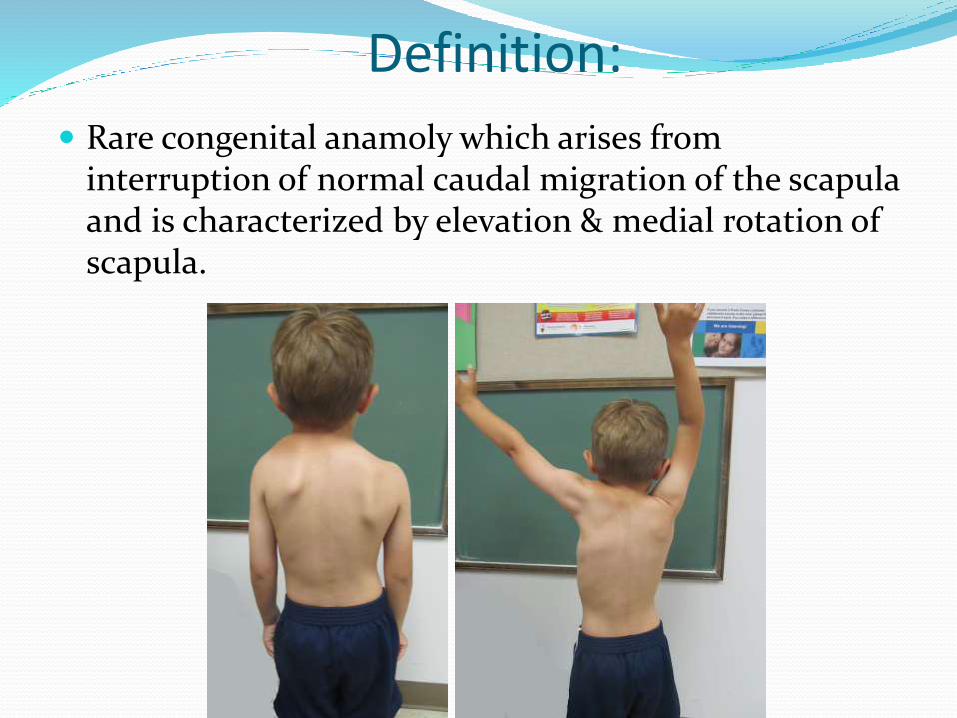
Definition:
Rare congenital anamoly which arises from interruption of normal caudal migration of the scapula and is characterized by elevation & medial rotation of scapula.
Historical significance:
First described by Eulenberg (1863) who described 3 patients.
Willet and Walsham reported 2 cases with anatomic descriptions of this clinical entity (1883)
It is named after Otto Gerhard Karl Sprengel (1852-1915), a German surgeon who described four cases in 1891.
Frequency:most common congenital malformation of the shoulder girdle.
Age – Mostly noticed at birth
Gender : Equal distribution in both sexes
Side – Left side more common than right, bilateral only in 10%
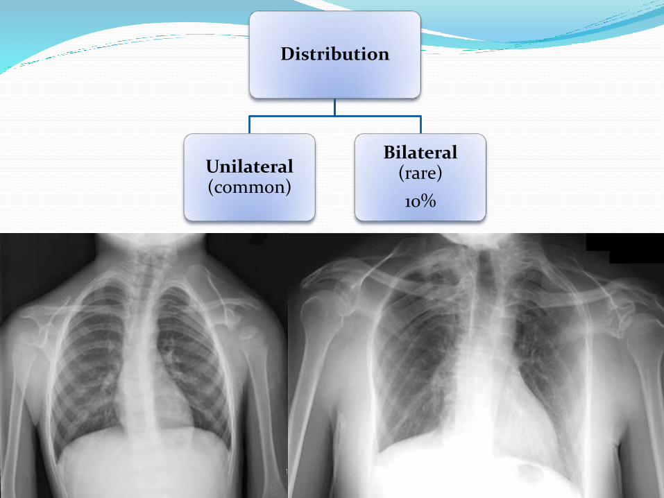
Distribution
Unilateral(common)
Bilateral(rare)
10%

Distribution
Unilateral(common)
Bilateral(rare)
10%
Genetics: The condition is sporadic.
Rarely, autosomal dominant pattern of inheritance.
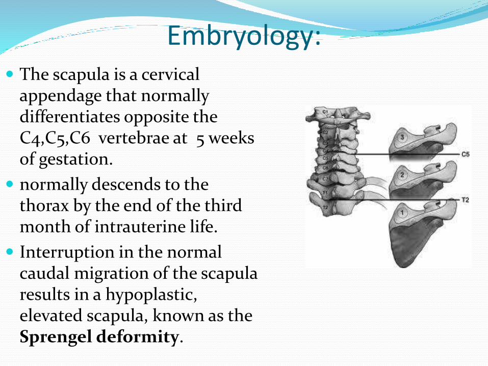
Embryology: The scapula is a cervical
appendage that normally differentiates opposite the C4,C5,C6 vertebrae at 5 weeks of gestation.
normally descends to the thorax by the end of the third month of intrauterine life.
Interruption in the normal caudal migration of the scapula results in a hypoplastic, elevated scapula, known as the Sprengel deformity.
Pathophysiology: Occurs between the 9th and 12th week of gestation.
An arrest in the development of bone, cartilage, and muscle also occurs.
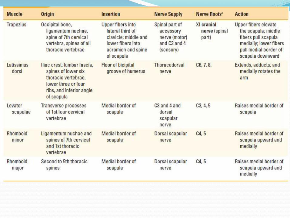
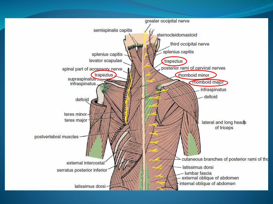
The trapezius, rhomboid, or levator scapulae muscle may be absent or hypoplastic. The serratusanterior muscle may be weak, leading to winging of the scapula. Other muscles, such as the pectoralismajor, latissimus dorsi, or the sternocleidomastoid may be hypoplastic and similarly involved.
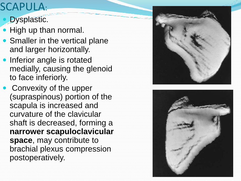
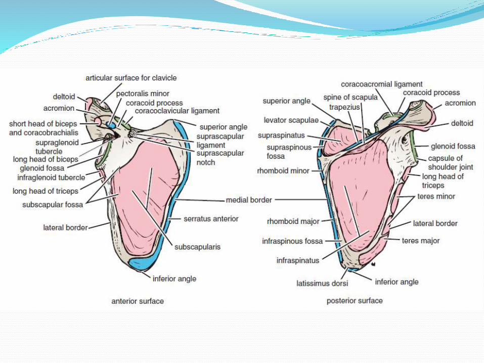
SCAPULA:
Dysplastic.
High up than normal.
Smaller in the vertical plane and larger horizontally.
Inferior angle is rotated medially, causing the glenoidto face inferiorly.
Convexity of the upper (supraspinous) portion of the scapula is increased and curvature of the clavicularshaft is decreased, forming a narrower scapuloclavicularspace, may contribute to brachial plexus compression postoperatively.
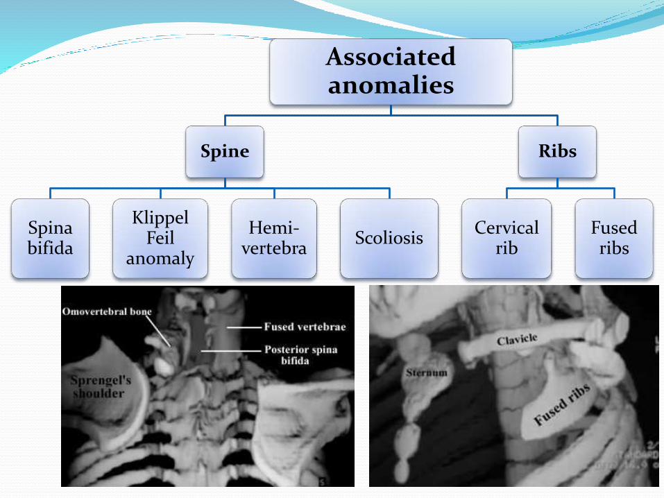
Associated anomalies
Spine
Spinabifida
KlippelFeil
anomaly
Hemi-vertebra
Scoliosis
Ribs
Cervical rib
Fused ribs
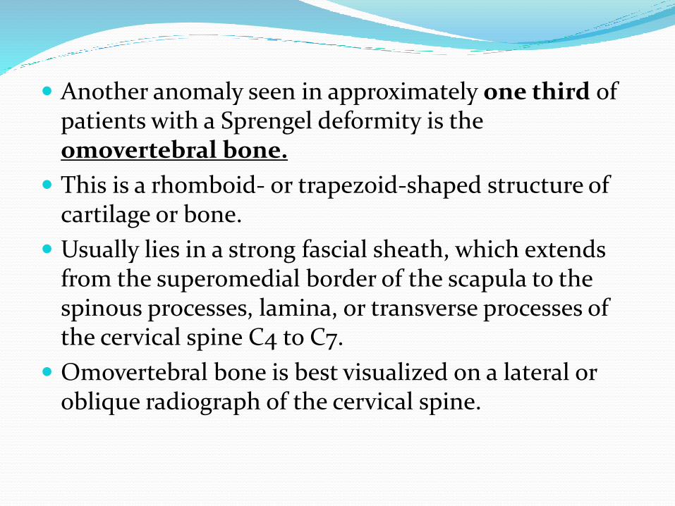
Another anomaly seen in approximately one third of patients with a Sprengel deformity is the omovertebral bone.
This is a rhomboid- or trapezoid-shaped structure of cartilage or bone.
Usually lies in a strong fascial sheath, which extends from the superomedial border of the scapula to the spinous processes, lamina, or transverse processes of the cervical spine C4 to C7.
Omovertebral bone is best visualized on a lateral or oblique radiograph of the cervical spine.
Klippel-Feil syndrome and Sprengel’s deformity
Congenital fusion of at least 2 cervical vertebrae with/without additional spinal/extraspinalmanifestations
Associated Sprengel’s deformity: 7%-42%
Most common congenitally fused segment in Sprengel’s deformity: C6-C7;extensive fusion patterns common
Thorough neurological examination to be done preoperatively to avoid complications during surgery and anesthesia
Short neckLow hair lineRestriction of neck movement

CLINICAL FEATURES Cosmetic
High position of the scapula
Scoliosis
Torticollis
Caput obstiosum (asymmetric distortion of the skull)
Facial asymmetry
Functional
Restricted motions of scapula and shoulder joint
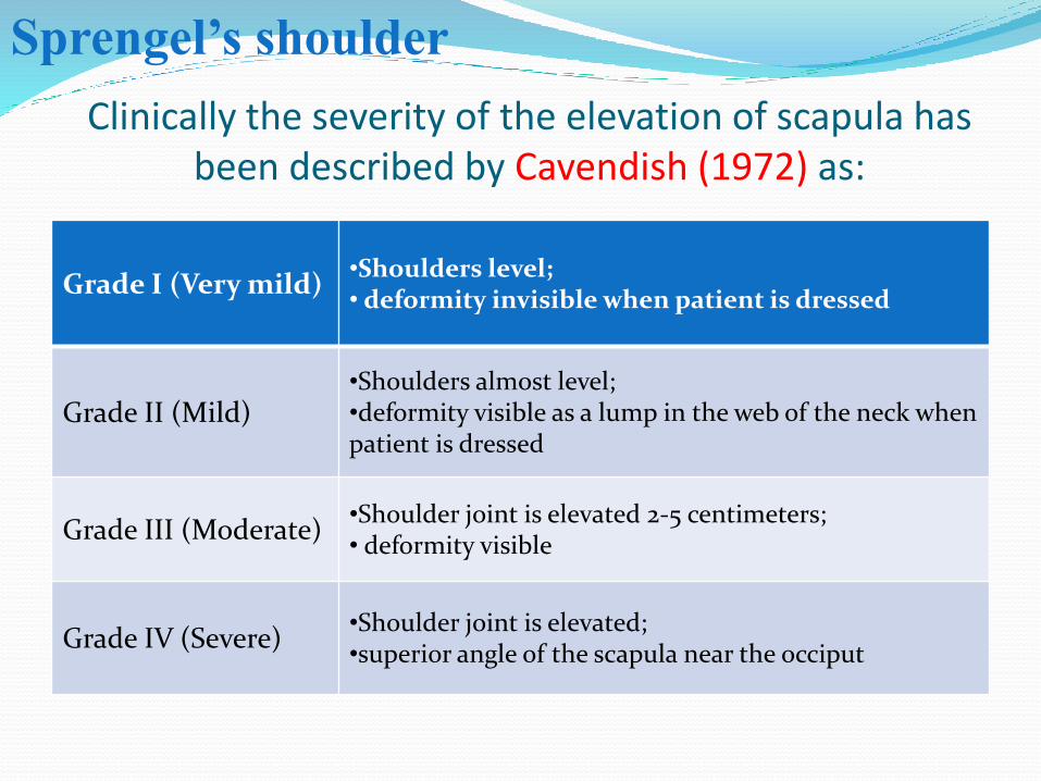
Clinically the severity of the elevation of scapula has been described by Cavendish (1972) as:
Grade I (Very mild)•Shoulders level;• deformity invisible when patient is dressed
Grade II (Mild)•Shoulders almost level; •deformity visible as a lump in the web of the neck when patient is dressed
Grade III (Moderate)•Shoulder joint is elevated 2-5 centimeters;• deformity visible
Grade IV (Severe)•Shoulder joint is elevated; •superior angle of the scapula near the occiput
Sprengel’s shoulder
DIAGNOSIS
The x-ray appearances are characteristics,showing the unduly high situation of the scapula.
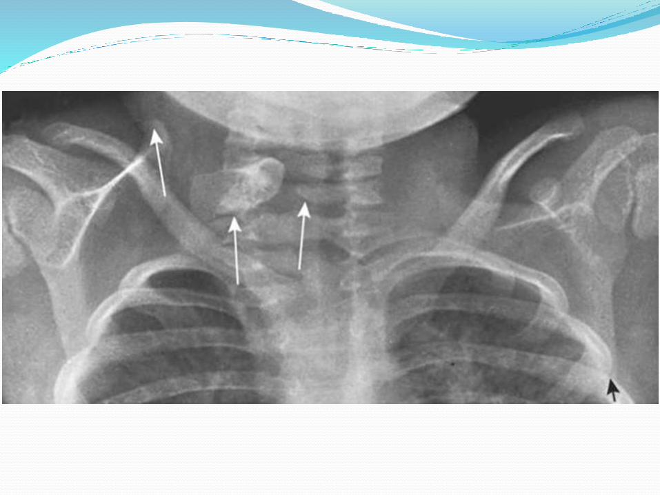
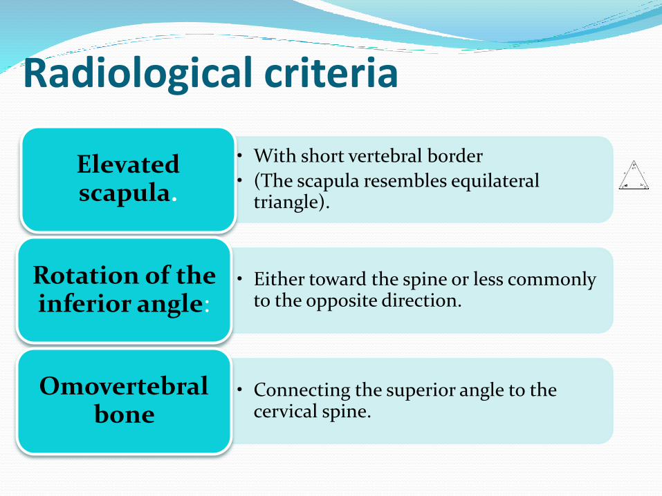
Radiological criteria
• With short vertebral border
• (The scapula resembles equilateral triangle).
Elevated scapula.
• Either toward the spine or less commonly to the opposite direction.
Rotation of the inferior angle:
• Connecting the superior angle to the cervical spine.
Omovertebralbone
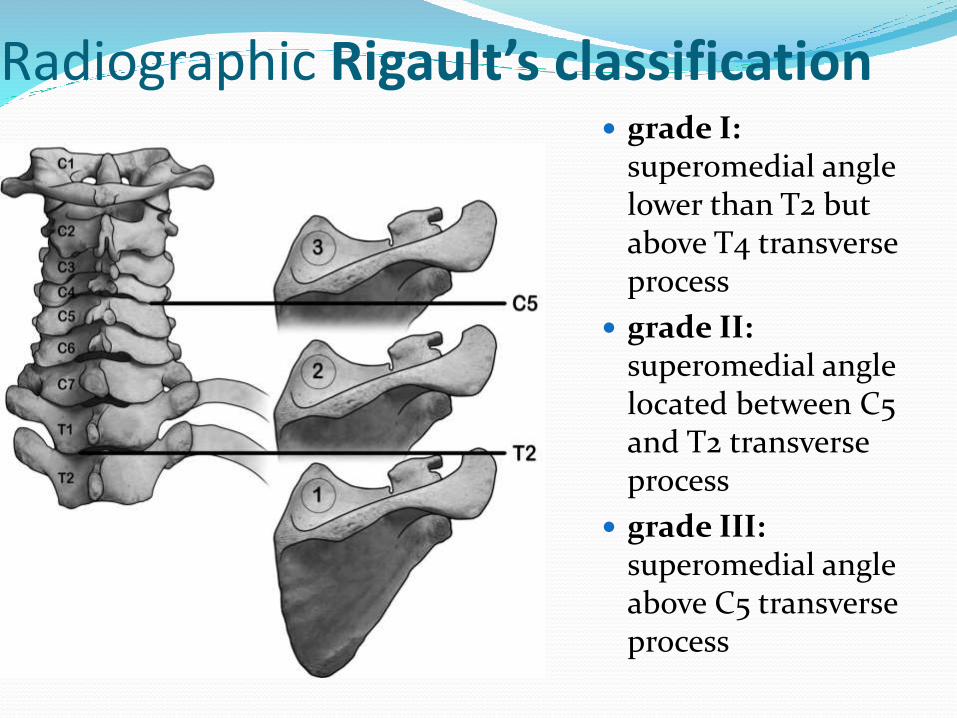
Radiographic Rigault’s classification grade I:
superomedial angle lower than T2 but above T4 transverse process
grade II:superomedial angle located between C5 and T2 transverse process
grade III:superomedial angle above C5 transverse process
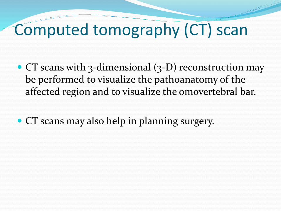
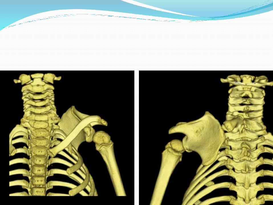
Computed tomography (CT) scan
CT scans with 3-dimensional (3-D) reconstruction may be performed to visualize the pathoanatomy of the affected region and to visualize the omovertebral bar.
CT scans may also help in planning surgery.
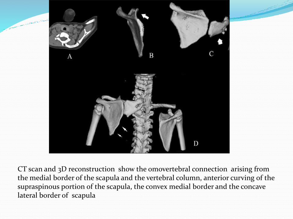
CT scan and 3D reconstruction show the omovertebral connection arising from the medial border of the scapula and the vertebral column, anterior curving of the supraspinous portion of the scapula, the convex medial border and the concave lateral border of scapula
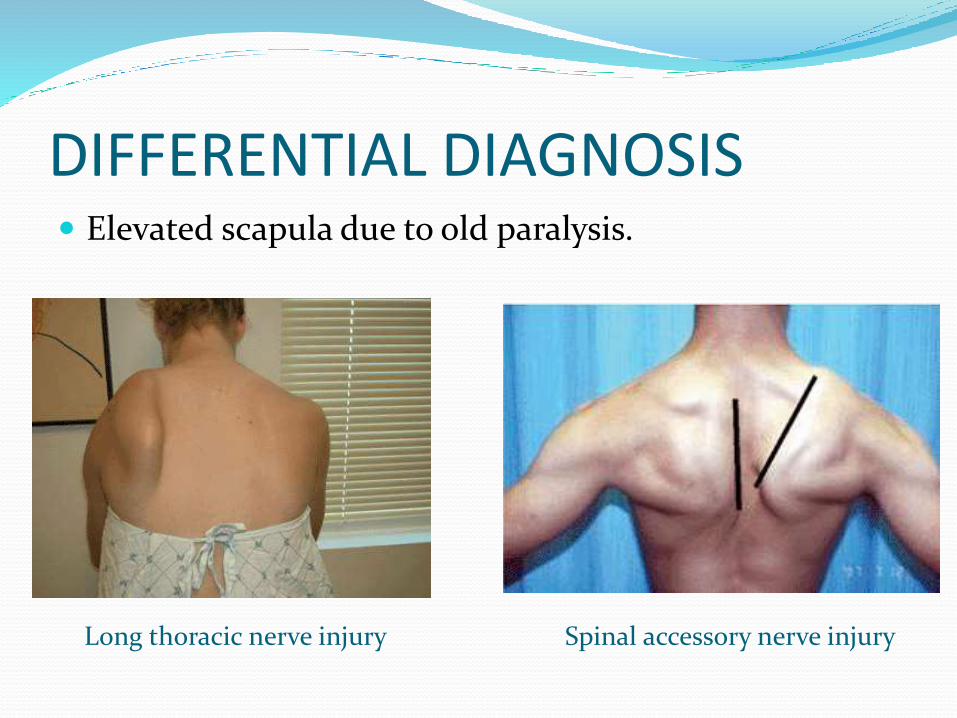
DIFFERENTIAL DIAGNOSIS Elevated scapula due to old paralysis.
Spinal accessory nerve injuryLong thoracic nerve injury
PROGNOSIS
Even if operation is undertaken, the prognosis is not very favorable.
Literatures indicate that while the mobility of the shoulder may be improved, asymmetry almost always persists.
SURGICAL TREATMENT Factors to be assessed
severity of the deformity,
functional impairment,
Age
associated comorbid conditions.
Surgery is best advisable for a patient between 3 and 8 years of age
with moderate or severe cosmetic/ functional deformity.
The presence of associated congenital anomalies may be contraindications to operation.
Surgical intervention before the age of 2 years is extensive and is technically more difficult. Best results are obtained if surgery is performed below the age of 5 years
SURGICAL TREATMENT
The surgical procedures involve a combination of
• (a) scapular lowering with either the shift of the origin or the insertion of the scapular muscles on the spine/ scapula,
• (b) resection of the superomedial border and
• (c) omovertebral bar resection
A clavicular morselization is sometimes recommended as a concomitant deformity of this bone may reduce the correction obtained.
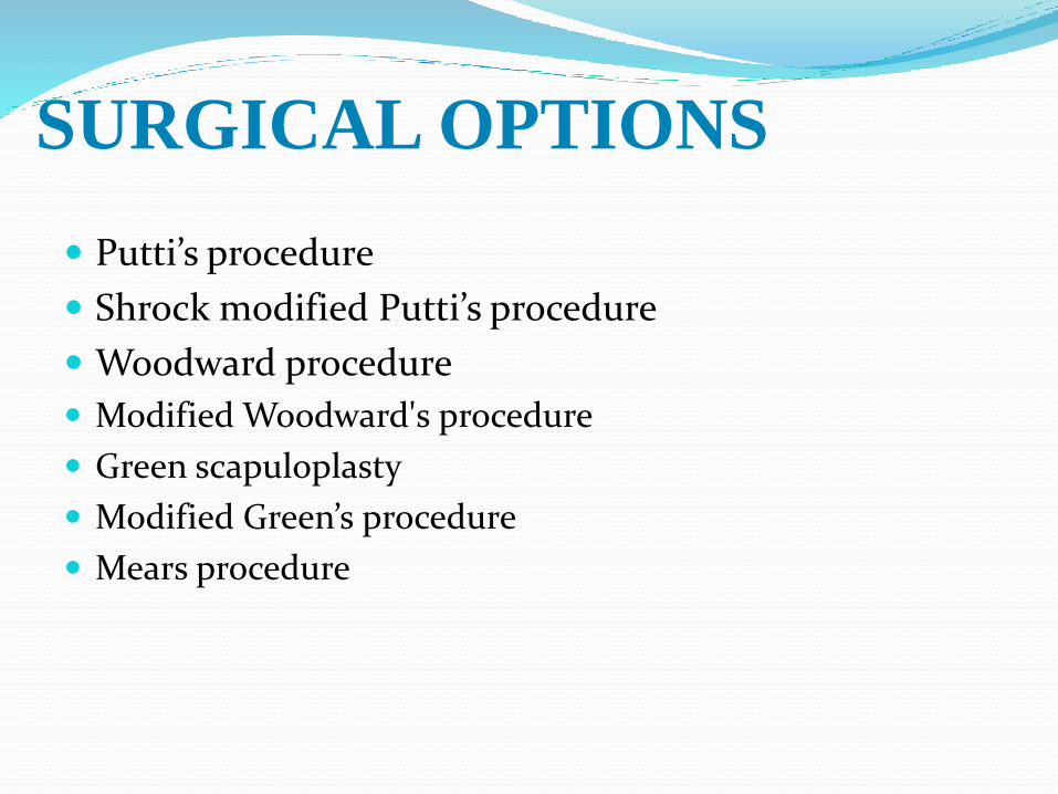
SURGICAL OPTIONS
Putti’s procedure
Shrock modified Putti’s procedure
Woodward procedure
Modified Woodward's procedure
Green scapuloplasty
Modified Green’s procedure
Mears procedure
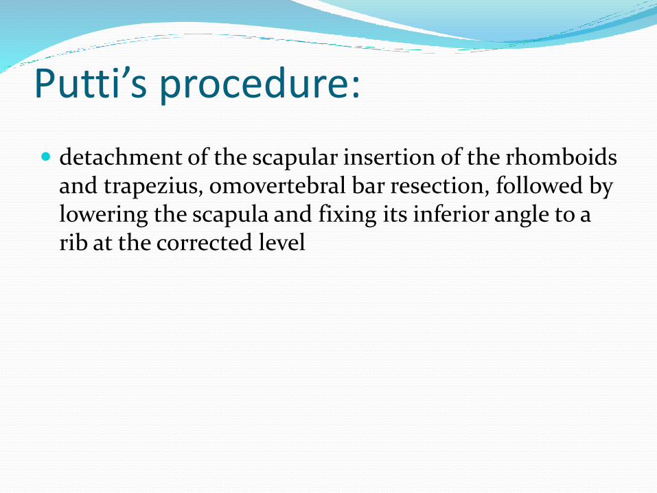
Putti’s procedure:
detachment of the scapular insertion of the rhomboids and trapezius, omovertebral bar resection, followed by lowering the scapula and fixing its inferior angle to a rib at the corrected level
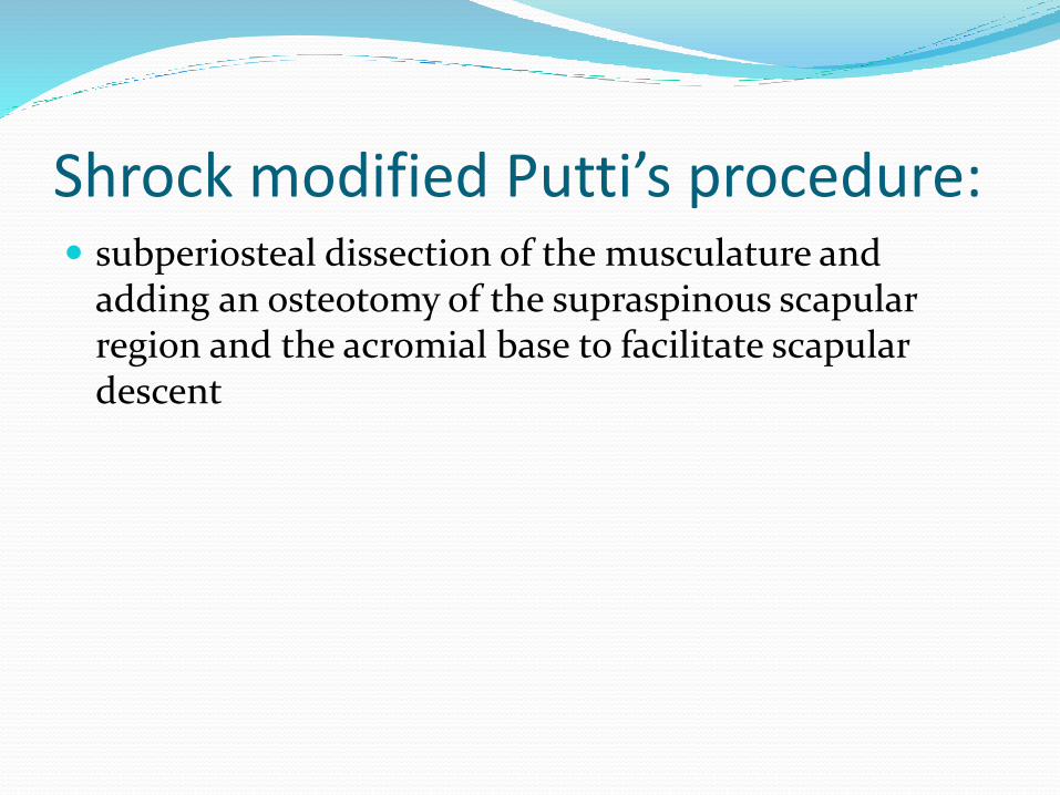
Shrock modified Putti’s procedure: subperiosteal dissection of the musculature and
adding an osteotomy of the supraspinous scapular region and the acromial base to facilitate scapular descent
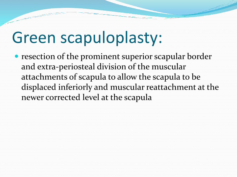
Green scapuloplasty: resection of the prominent superior scapular border
and extra-periosteal division of the muscular attachments of scapula to allow the scapula to be displaced inferiorly and muscular reattachment at the newer corrected level at the scapula
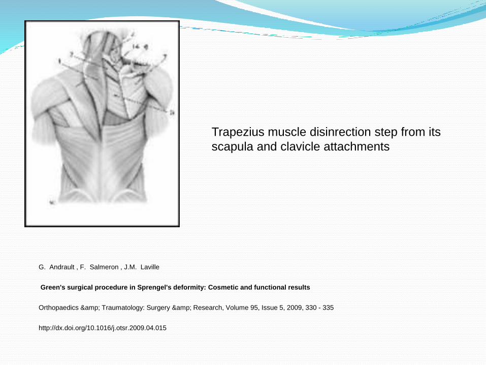
Trapezius muscle disinrection step from its
scapula and clavicle attachments
G. Andrault , F. Salmeron , J.M. Laville
Green's surgical procedure in Sprengel's deformity: Cosmetic and functional results
Orthopaedics & Traumatology: Surgery & Research, Volume 95, Issue 5, 2009, 330 - 335
http://dx.doi.org/10.1016/j.otsr.2009.04.015
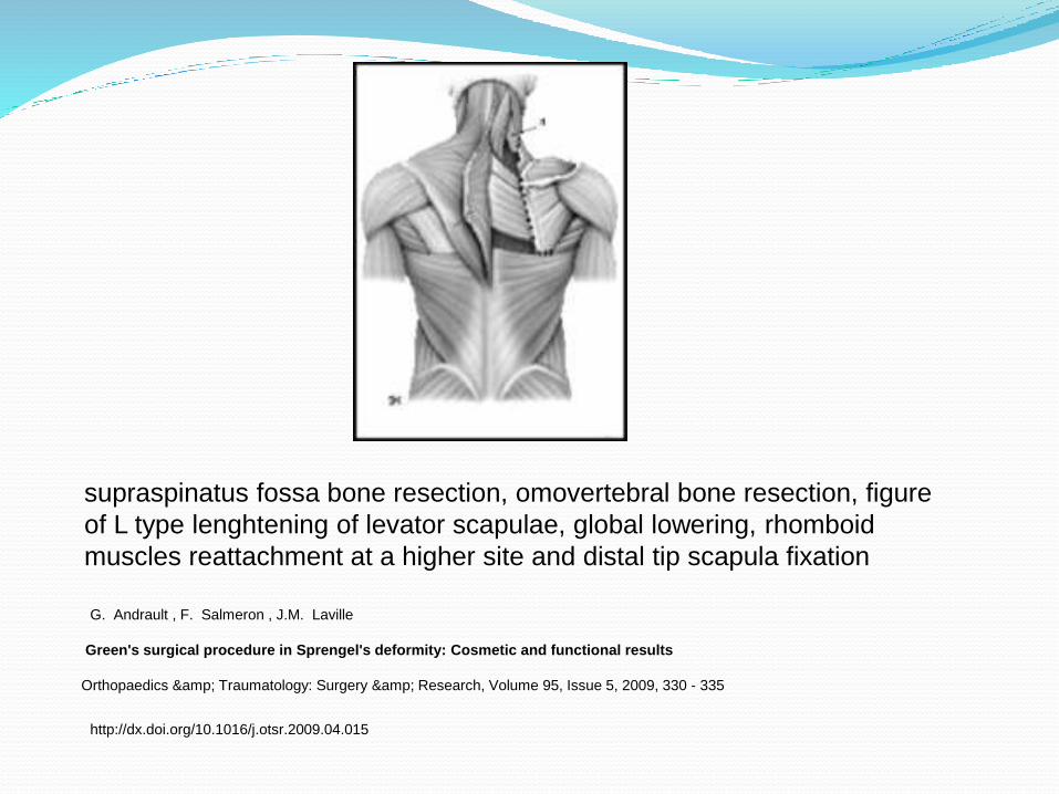
supraspinatus fossa bone resection, omovertebral bone resection, figure
of L type lenghtening of levator scapulae, global lowering, rhomboid
muscles reattachment at a higher site and distal tip scapula fixation
G. Andrault , F. Salmeron , J.M. Laville
Green's surgical procedure in Sprengel's deformity: Cosmetic and functional results
Orthopaedics & Traumatology: Surgery & Research, Volume 95, Issue 5, 2009, 330 - 335
http://dx.doi.org/10.1016/j.otsr.2009.04.015
Modified Green scapuloplasty: Andrault et al. suggested modifications to Green’s
procedure
(a) dis-insertion of supraspinatus,
(b) clavicular osteotomy and
(c) a limited release of the serratus anterior to facilitate the descent of the scapula.
incidence of brachial plexus palsy could be reduced by clavicular osteotomy, and that scapular winging could be prevented by doing only a limited release of serratus anterior from the medial scapular border
Woodward procedure:
Transfer of the origin of the trapezius muscle to a more inferior position on the spinous processes.
This was maintained by placing the scapula in a pocket of the trapezius muscle.
Modified Woodward's procedure:
for achieving better abduction and correction of the glenoid tilt
the scapula was anchored to the lower dorsal vertebrae by a stout absorbable suture placed through the superomedial scapula, so as to externally rotate it and cause lateral displacement of the inferior angle, thereby achieving correction of glenoid vara
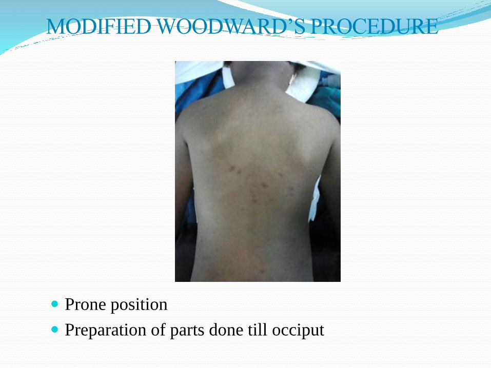
Prone position
Preparation of parts done till occiput
MODIFIED WOODWARD’S PROCEDURE
MODIFIED WOODWARD’S PROCEDURE
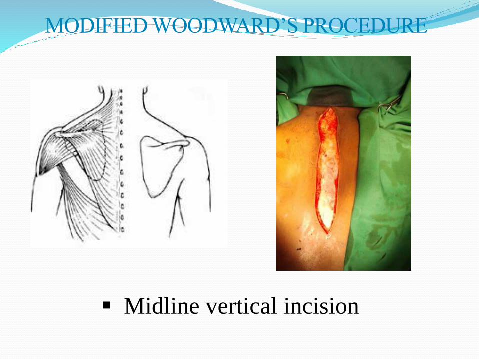
Midline vertical incision
MODIFIED WOODWARD’S PROCEDURE
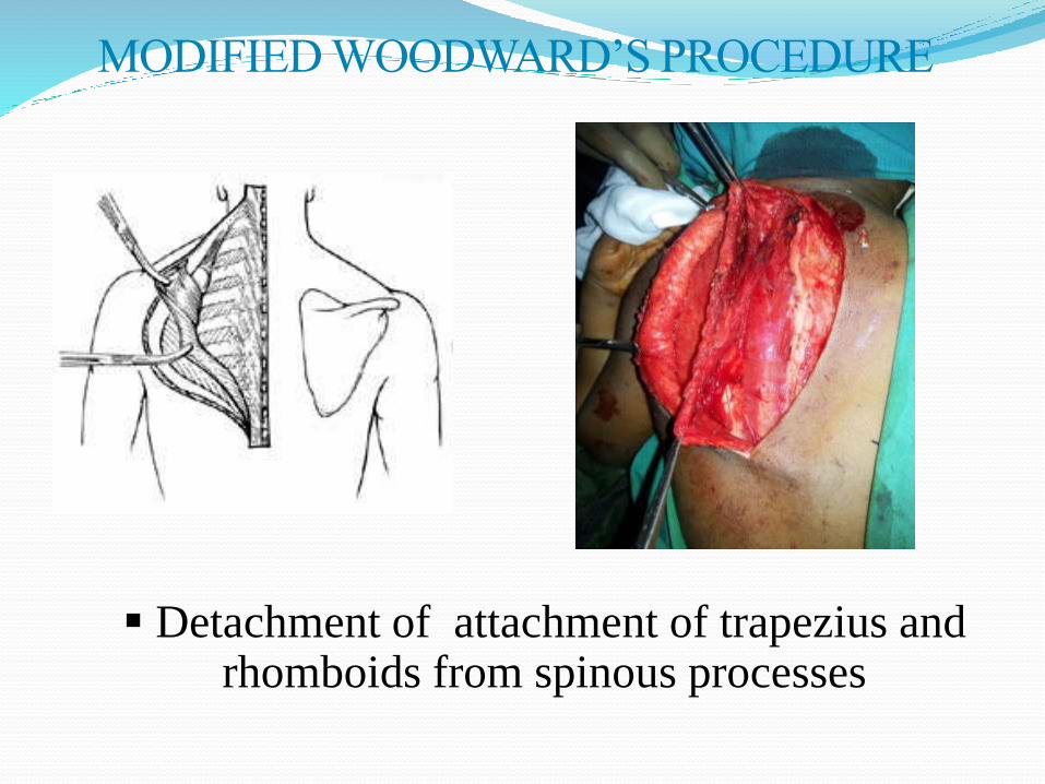
Detachment of attachment of trapezius and rhomboids from spinous processes
MODIFIED WOODWARD’S PROCEDURE
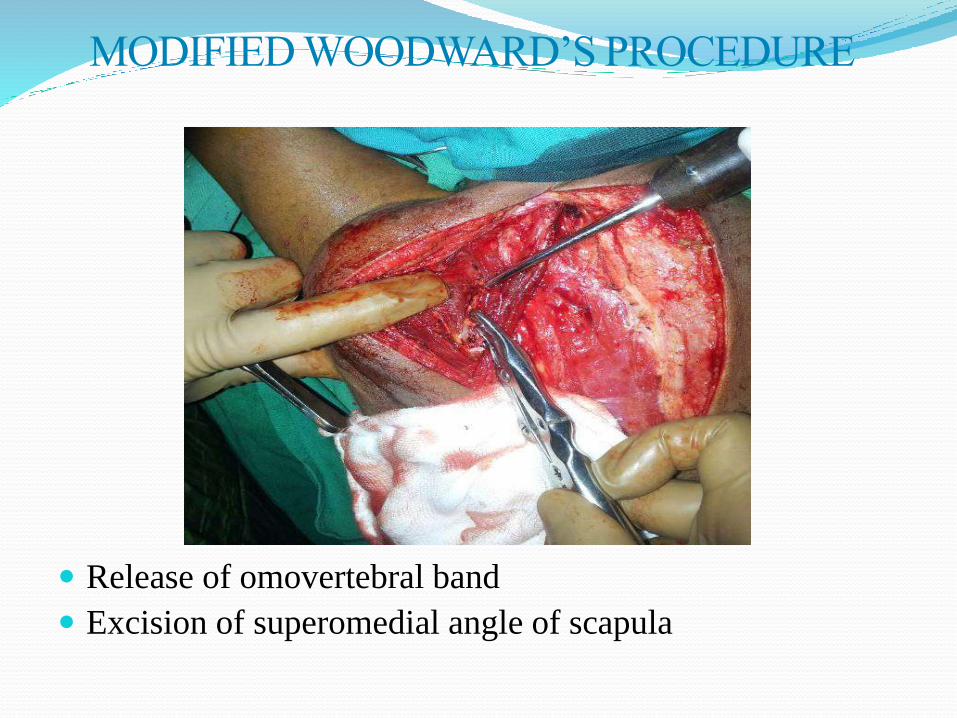
Release of omovertebral band
Excision of superomedial angle of scapula
MODIFIED WOODWARD’S PROCEDURE
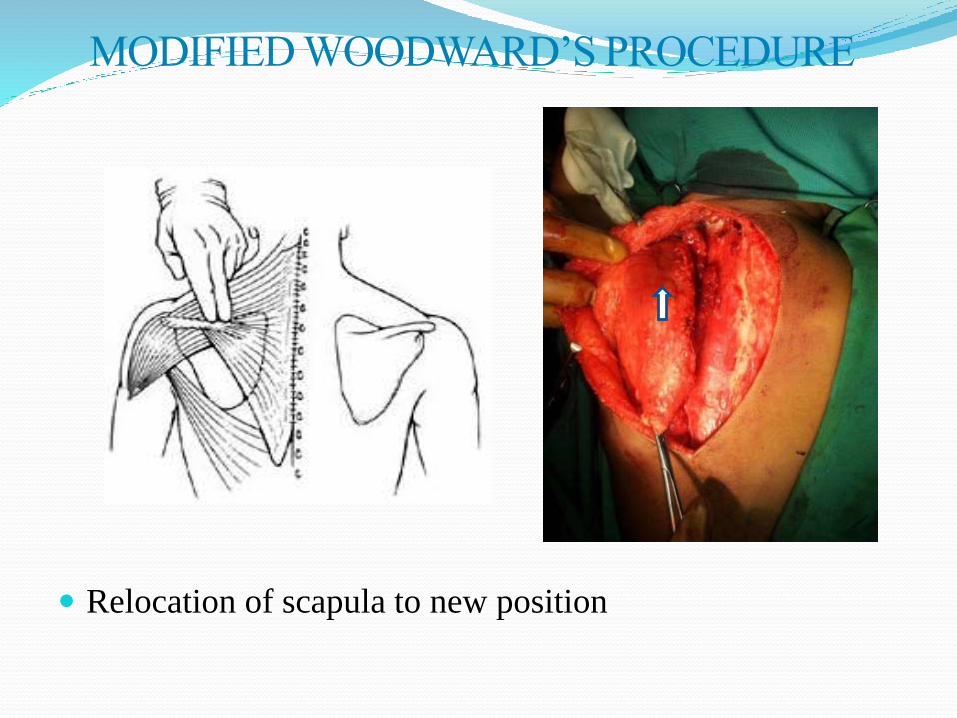
Relocation of scapula to new position
MODIFIED WOODWARD’S PROCEDURE
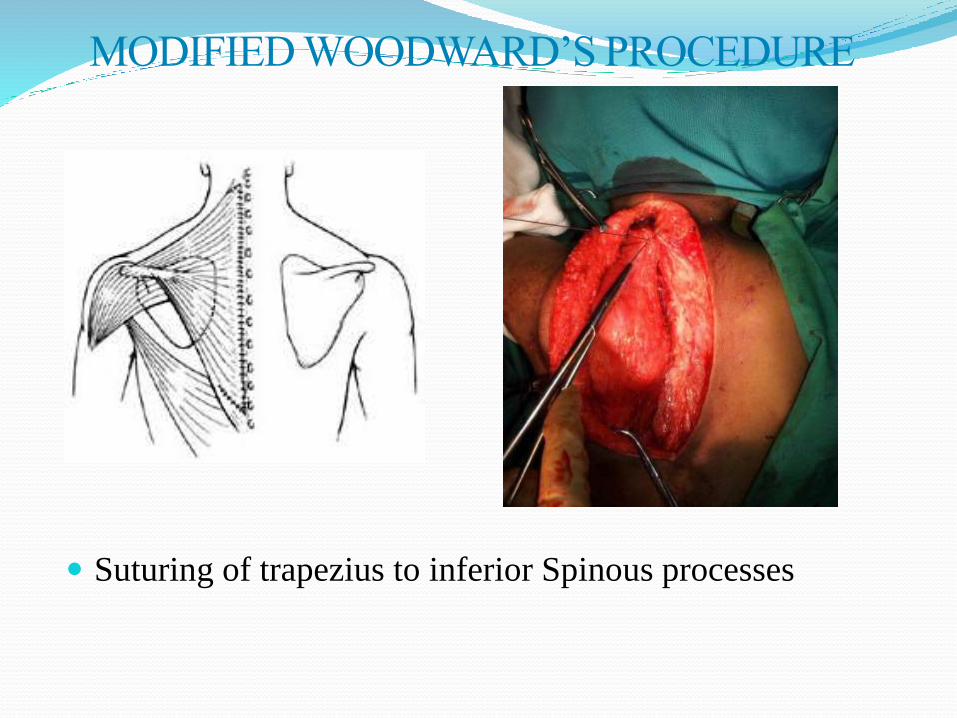
Suturing of trapezius to inferior Spinous processes
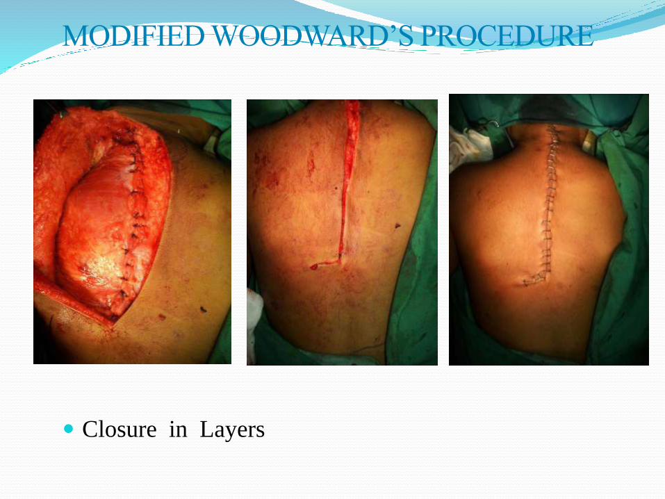
Closure in Layers
MODIFIED WOODWARD’S PROCEDURE

Mears procedure In a report by Mears, the author described a novel
approach-- (a) subperiosteal elevation of the scapular musculature,
(b) extraperiosteal resection of the omovertebral bone,
(c) supraspinatous fossa osteotomy,
(d) release of long head of triceps and a portion of the origin of teres minor from the scapula and
(e) resection of the superolateral border of the scapula to gain abduction
He reported a significant improvement in function following this procedure.
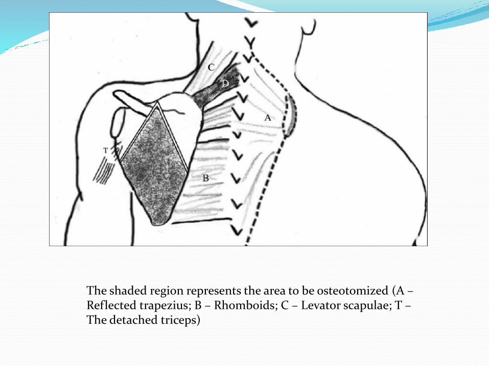
The shaded region represents the area to be osteotomized (A –Reflected trapezius; B – Rhomboids; C – Levator scapulae; T –The detached triceps)
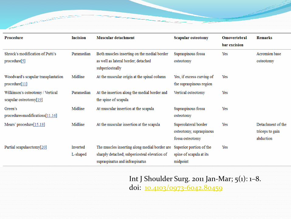
Int J Shoulder Surg. 2011 Jan-Mar; 5(1): 1–8. doi: 10.4103/0973-6042.80459

Postoperative complications Winging of the scapula that may result from
incomplete reattachment of the serratus anterior muscle
Brachial plexus injury To avoid brachial plexus palsy, several authors recommended
morcellization of the clavicle on the ipsilateral side as a first step in the operative treatment of Sprengel deformity.
Keloid formation.
PHYSIOTHERAPY AFTER SURGERY Gradual relaxed passive mobilization of the shoulder and
scapula.
Suitable pain relieving modality like TENS, IFT andhydrocollator packs may be used to induce relaxation.
Special attention is given to achieve early mobility of thescapula and the shoulder abduction and elevation.
Overall mobilization and strengthening of the shoulder girdlemuscles.
Emphasize maximum possible correction of the posture ofshoulder and maintain it.
5/6/201650

THANK YOU
Related Documents