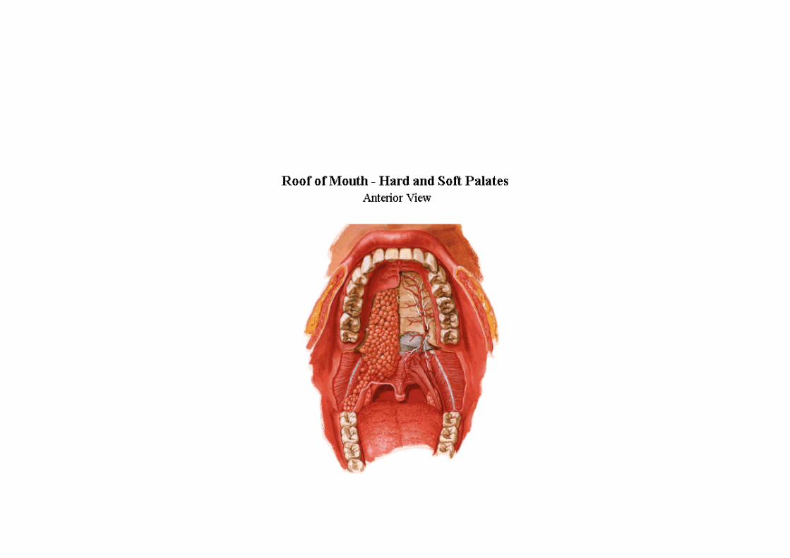
Palate • Lies within alveolar arches, b/w roof of mouth and floor of nasal cavity • Arched antero-posteriorly and side to side • Consist of two parts a) Hard Palate – Anterior 2/3 rd b) Soft Palate – Posterior 1/3 rd

Welcome message from author
This document is posted to help you gain knowledge. Please leave a comment to let me know what you think about it! Share it to your friends and learn new things together.
Transcript

Palate
• Lies within alveolar arches, b/w roof of mouth and floor of nasal cavity
• Arched antero-posteriorly and side to side
• Consist of two partsa) Hard Palate – Anterior 2/3rd
b) Soft Palate – Posterior 1/3rd
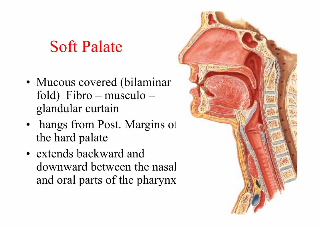
Soft Palate
• Mucous covered (bilaminar fold) Fibro – musculo –glandular curtain
• hangs from Post. Margins of the hard palate
• extends backward and downward between the nasal and oral parts of the pharynx
Soft Palate
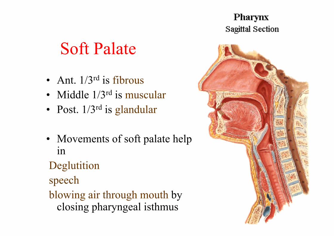
• Ant. 1/3rd is fibrous• Middle 1/3rd is muscular• Post. 1/3rd is glandular
• Movements of soft palate help in
Deglutitionspeechblowing air through mouth by
closing pharyngeal isthmus
Soft palate
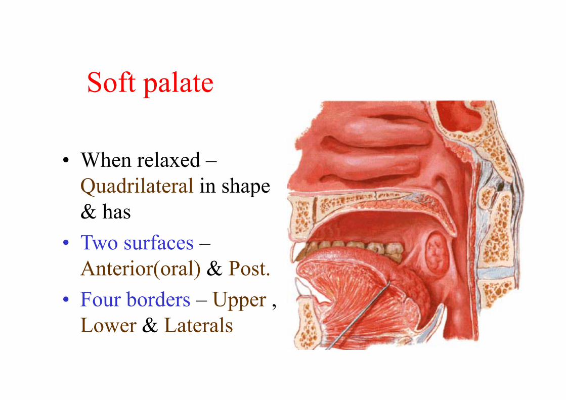
• When relaxed –Quadrilateral in shape & has
• Two surfaces –Anterior(oral) & Post.
• Four borders – Upper , Lower & Laterals
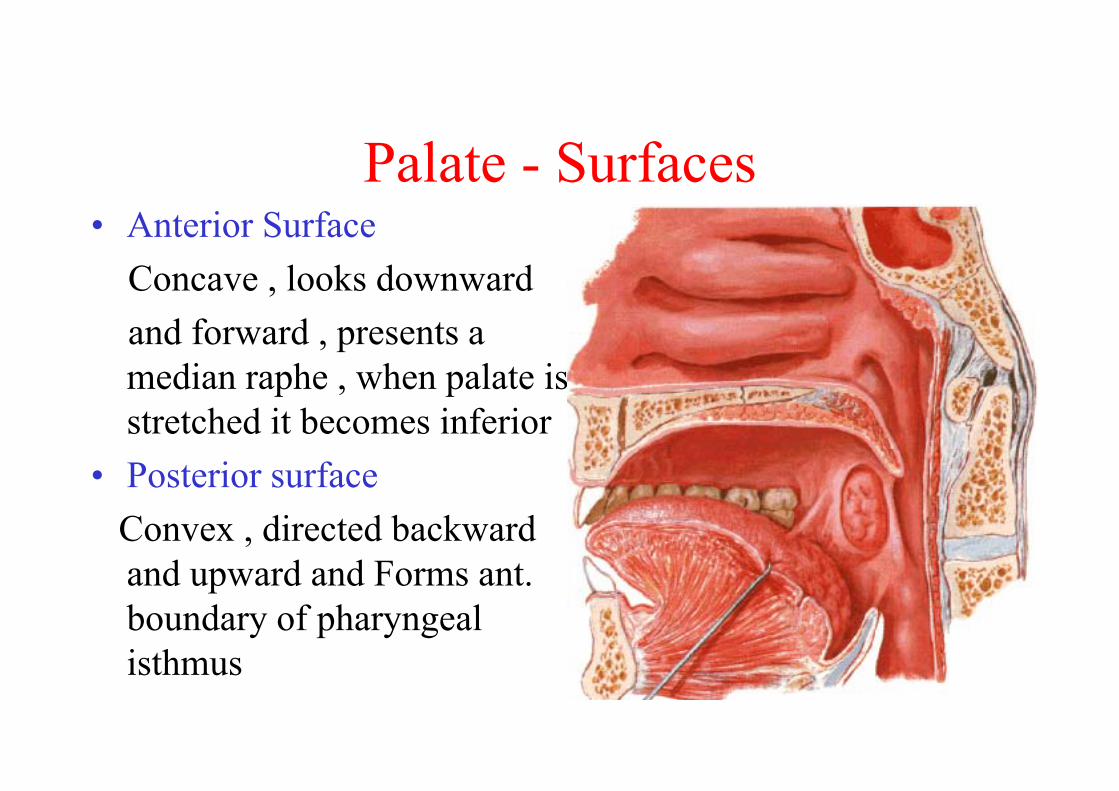
Palate - Surfaces• Anterior Surface
Concave , looks downward and forward , presents a median raphe , when palate is stretched it becomes inferior
• Posterior surfaceConvex , directed backward and upward and Forms ant. boundary of pharyngeal isthmus

Palate - borders
• Upper borderAttached to post. Margins of the hard palate
• Lateral bordersContinuous with the wall of pharynx
• Lower borderFree & presents a conical projection in midline
(Uvula)
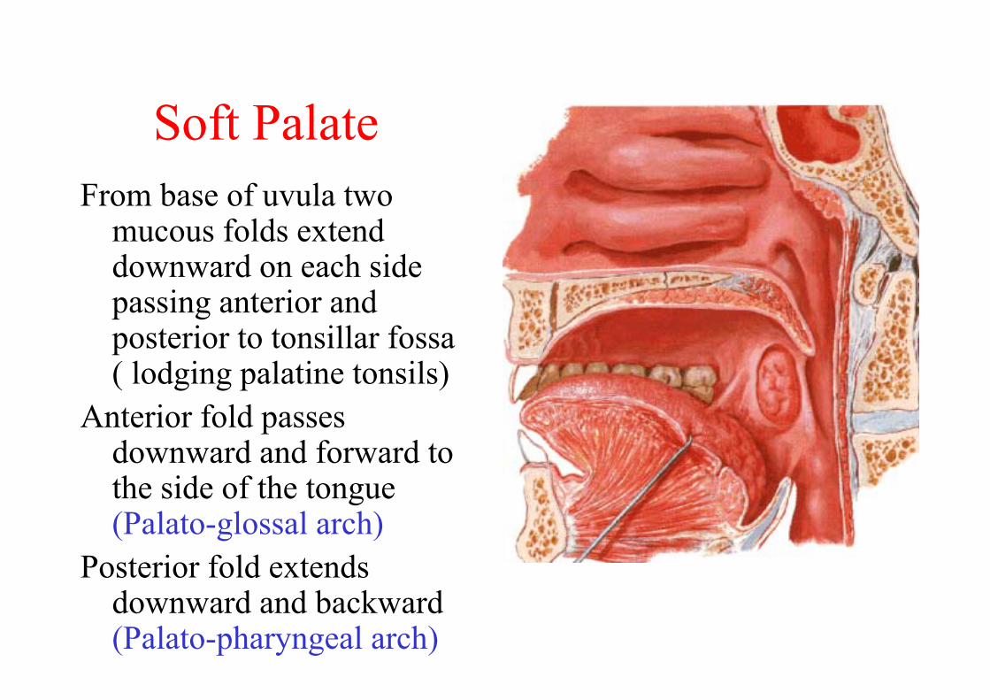
Soft PalateFrom base of uvula two
mucous folds extend downward on each side passing anterior and posterior to tonsillar fossa ( lodging palatine tonsils)
Anterior fold passes downward and forward to the side of the tongue (Palato-glossal arch)
Posterior fold extends downward and backward (Palato-pharyngeal arch)
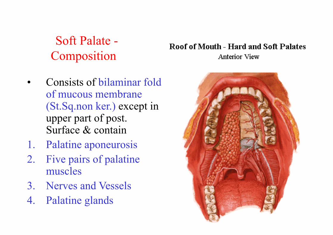
Soft Palate -Composition
• Consists of bilaminar fold of mucous membrane (St.Sq.non ker.) except in upper part of post. Surface & contain
1. Palatine aponeurosis2. Five pairs of palatine
muscles3. Nerves and Vessels4. Palatine glands
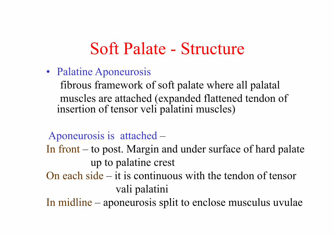
Soft Palate - Structure• Palatine Aponeurosis
fibrous framework of soft palate where all palatal muscles are attached (expanded flattened tendon of
insertion of tensor veli palatini muscles)
Aponeurosis is attached –In front – to post. Margin and under surface of hard palate
up to palatine crestOn each side – it is continuous with the tendon of tensor
vali palatiniIn midline – aponeurosis split to enclose musculus uvulae
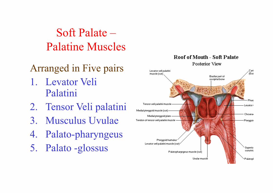
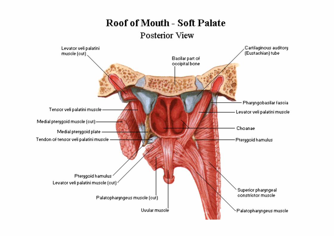
Soft Palate –Palatine Muscles
Arranged in Five pairs1. Levator Veli
Palatini2. Tensor Veli palatini3. Musculus Uvulae4. Palato-pharyngeus5. Palato -glossus
Palatine muscles
Levator Veli palatiniArise fromo Under surface of apex of petrous temporalo From carotid sheatho Medial cartilaginous part of Auditory tubeInsertionUpper surface of Aponeurosis passing in b/w ant &
post. fasciculi of palatopharyngeus
Soft Palate -Muscles
Tensor Veli Palatini – Triangular muscleOrigino Scaphoid fossa of medial pterygoid plateo Lateral fibrous lamina of Auditory tubeo Spine of sphenoidInsertionPalatine Aponeurosis
Soft palate - Muscles
Musculus UvulaeOrigino Post nasal spine of hard palate passes
backward and downward within tubular sheath of aponeurosis
InsertionIn submucous tissue of base of uvula

Nerve Supply• All muscles of soft palate are supplied by
cranial part of Accessory nerve through pharyngeal plexus except Tensor veli palatini which is supplied by the trunk of the mandibular nerve
Vessels and nervesArteries• Greater palatine branch of maxillary artery• Ascending palatine branch of facial artery• Palatine branch of ascending pharyngeal arteryVeinsDrain in pharyngeal venous plexus via paratonsillar
veinsL. NodesDrain in retropharyngeal and upper group of deep
cervical LN
Applied Anatomy
• Diphtheria – paralysis of palatal muscles causing nasal voice , Flattening of arches and regurgitation of food through nose when swallowing
• Cleft Palate
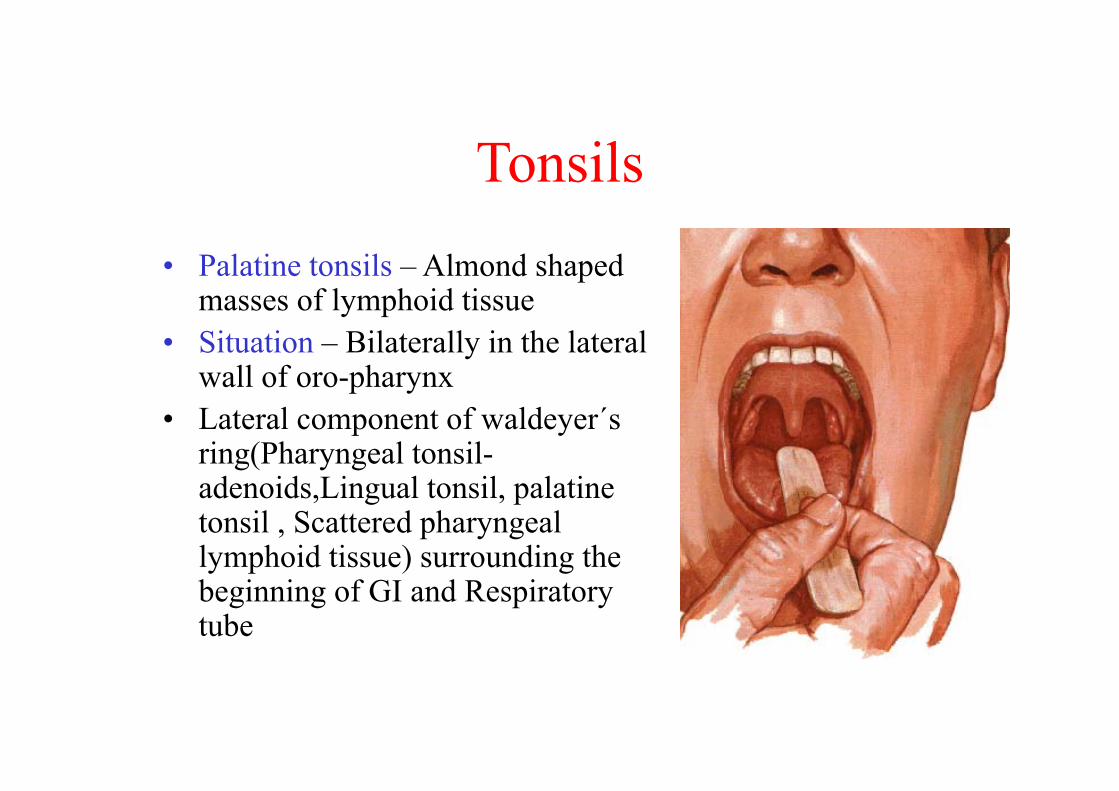
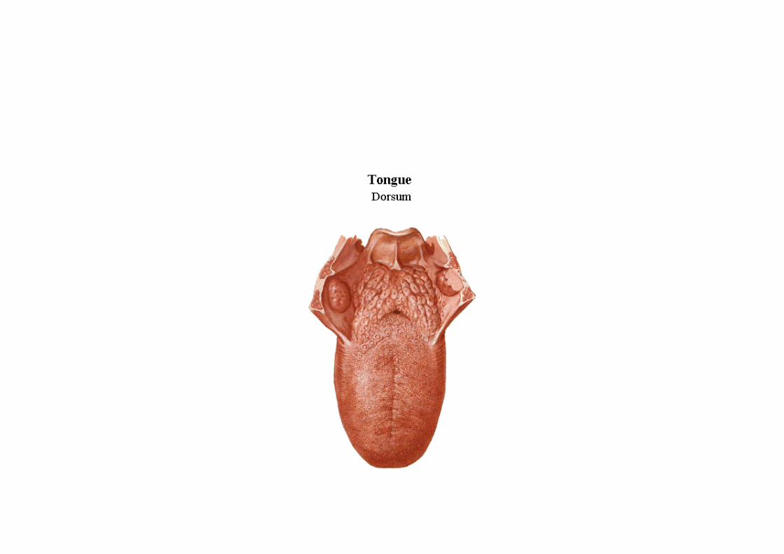
Tonsils• Palatine tonsils – Almond shaped
masses of lymphoid tissue• Situation – Bilaterally in the lateral
wall of oro-pharynx• Lateral component of waldeyer΄s
ring(Pharyngeal tonsil-adenoids,Lingual tonsil, palatine tonsil , Scattered pharyngeal lymphoid tissue) surrounding the beginning of GI and Respiratory tube
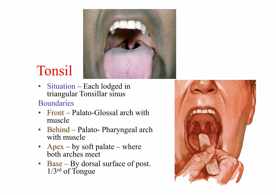
Tonsil• Situation – Each lodged in
triangular Tonsillar sinusBoundaries• Front – Palato-Glossal arch with
muscle• Behind – Palato- Pharyngeal arch
with muscle• Apex – by soft palate – where
both arches meet• Base – By dorsal surface of post.
1/3rd of Tongue
Tonsil
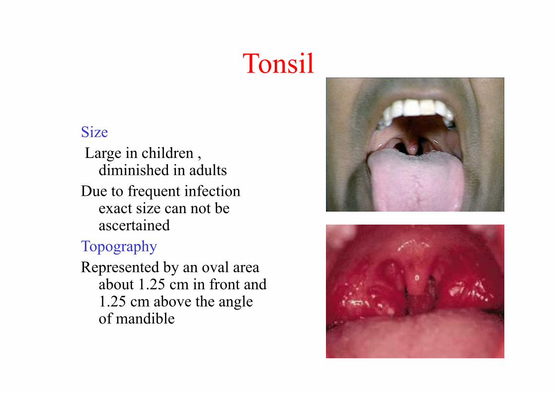
SizeLarge in children ,
diminished in adultsDue to frequent infection
exact size can not be ascertained
TopographyRepresented by an oval area
about 1.25 cm in front and 1.25 cm above the angle of mandible
Tonsil
• Lateral wall of the Tonsillar bed formed from within outward by
1. Pharyngo- basilar fascia2. Few fibres – palatopharyngeus muscle in upper
and post. Part3. Sup. Constrictor muscle of pharynx in 2/3rd of
posterosuperior part4. Styloglossus muscle accompanied by
glossopharyngeal nerve in antero.inferior 1/3rd
Tonsil
• Each tonsil has• Two surfaces –
Medial and lateral• Two borders –
anterior and posterior• Two ends –
upper and lower
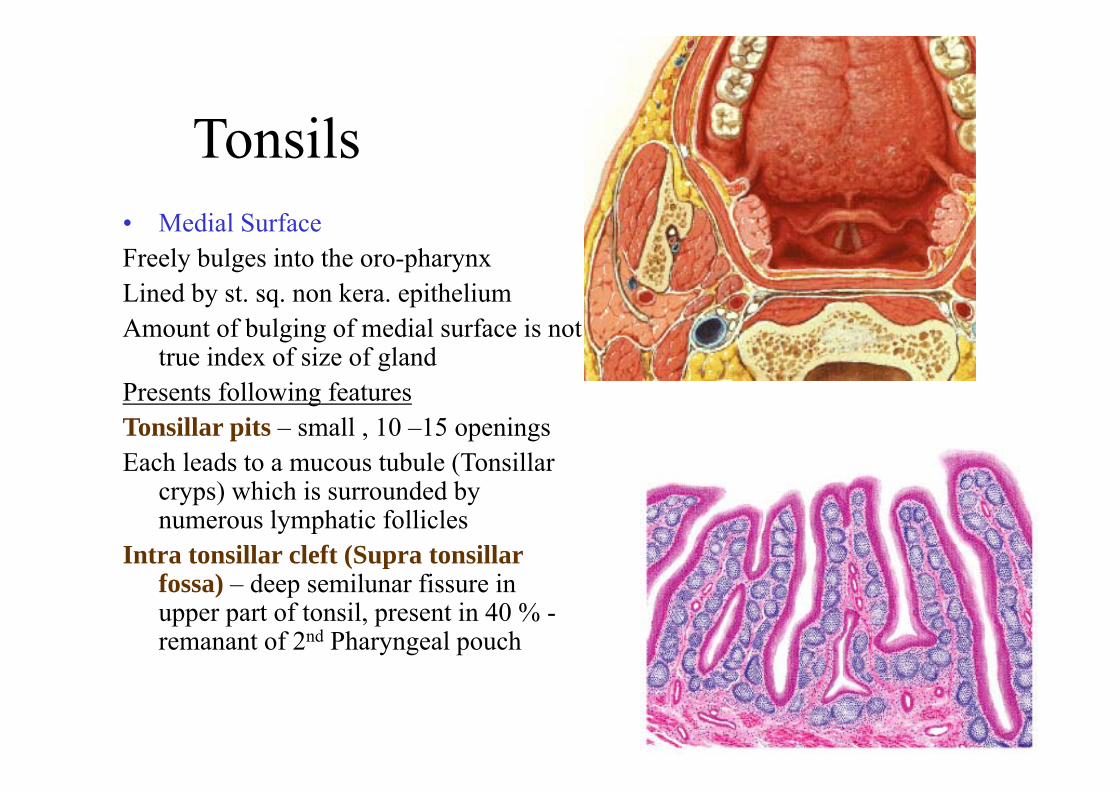
Tonsils• Medial SurfaceFreely bulges into the oro-pharynxLined by st. sq. non kera. epitheliumAmount of bulging of medial surface is not
true index of size of glandPresents following featuresTonsillar pits – small , 10 –15 openingsEach leads to a mucous tubule (Tonsillar
cryps) which is surrounded by numerous lymphatic follicles
Intra tonsillar cleft (Supra tonsillar fossa) – deep semilunar fissure in upper part of tonsil, present in 40 % -remanant of 2nd Pharyngeal pouch
Tonsil – Medial surface
• Embryonic folds1. Plica tringularis – extend backward as
triangular fold from lower part of palatoglossal arch – replaced by lymphoid tissue after birth
2. Plica semilunaris – arches backward from upper part of palatoglossal arch , also replaced by lymphoid tissue after birth
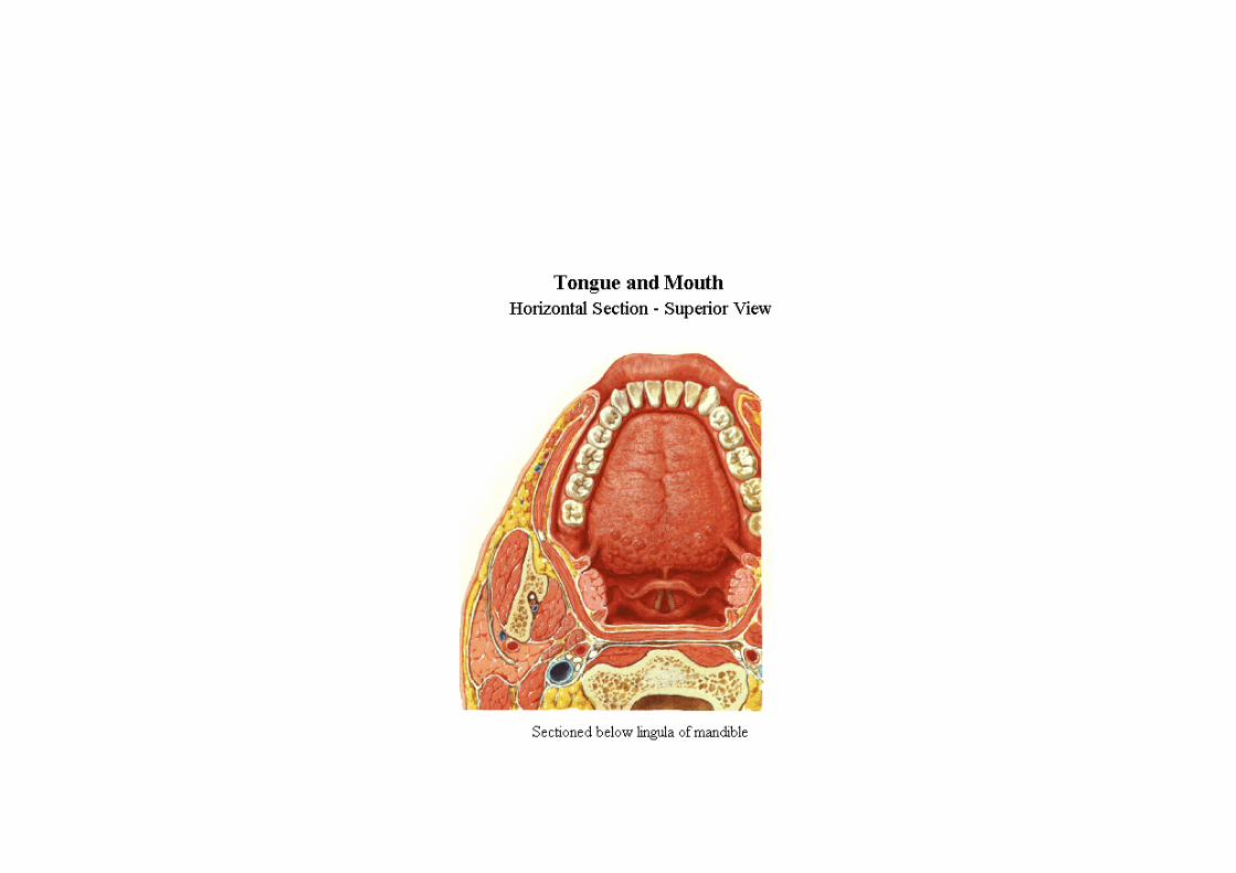
Tonsil – lateral surface(deep)
• Extends above , below and in front beyond the limits of tonsillar sinus
• Surface is covered by a fibrous capsule which is attached below to the side of the tongue
Tonsils – lateral surface
• Relations (from within outward)1. Loose areolar tissue containing
paratonsillar veins2. Pharyngo-basilar fascia3. Superior constrictor muscles of pharynx4. Bucco-pharyngeal fascia containing
pharyngeal plexus of nerves and vessels
5. Arteries Facial artery with its – ascending palatine and
tonsillar branches Ascending pharyngeal artery Internal carotid artery – lies about 2.5 cm behind
and lateral to tonsilar sinus and is separated by fibrofatty tissue
6. Styloglossus , stylopharyngeus and glossopharyngeal nerve
7. Post. Belly of diagastric and stylohyoid muscles8. Medial pterygoid muscle and ramus of mandible
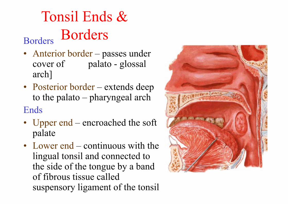
Tonsil Ends & BordersBorders
• Anterior border – passes under cover of palato - glossal arch]
• Posterior border – extends deep to the palato – pharyngeal arch
Ends• Upper end – encroached the soft
palate• Lower end – continuous with the
lingual tonsil and connected to the side of the tongue by a band of fibrous tissue called suspensory ligament of the tonsil
Tonsil
Factors keeping tonsils in positionSuspensory ligament of tonsil – connecting
it with tongueAttachments of palatopharyngeus and
palatogossus muscles to the fibrous capsules of the tonsilsPerivascular stalks which keeps the tonsils
in position
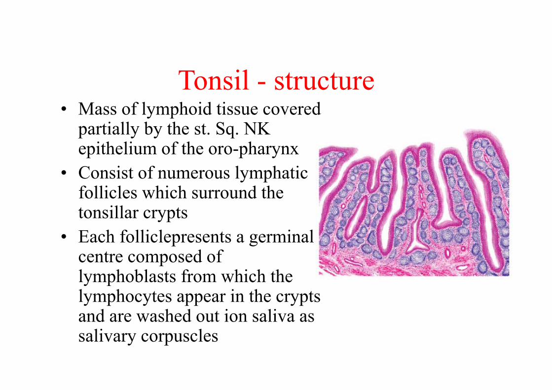
Tonsil - structure• Mass of lymphoid tissue covered
partially by the st. Sq. NK epithelium of the oro-pharynx
• Consist of numerous lymphatic follicles which surround the tonsillar crypts
• Each folliclepresents a germinal centre composed of lymphoblasts from which the lymphocytes appear in the crypts and are washed out ion saliva as salivary corpuscles
Tonsil - vessels• Supplied by four set of arteries1. Anterior tonsillar – from dorsal lingual branch of lingual
artery2. Post. Tonsillar – from ascending palatine br. of facial and
ascending pharyngeal arteries3. Superior tonsillar – from greater palatine artery4. Inferior tonsillar – from facial artery ( Principal artery) –
reaches antero inferior part of tonsil after piercing the superior constrictor muscle and the fibrous capsule
Ligature of these arteries are important particularly inferior tonsillar is an imp step in the surgical removal of the tonsil
Tonsil
• Veins drain into the pharyngeal venous plexus via paratonsillar veins
Lymphatic drainage –Into jugulo - diagastric lymph nodesOne LN situated below and behind the angle of
mandible , in a triangular interval b/w post. Belly of diagastric and the junction of common facial and internal jugular veins( Principal LN of tonsil) primarily enlarged in infections of tonsil
Tonsil - Nerves
• Supplied by glossopharyngeal nerve and greater and lesser palatine branchesfrom the pterygo-palatine ganglion – these convey both general and taste sensatins
Tonsil – Applied Anatomy
• Tonsillitis – infection of tonsils• Referred pain may extend to middle Ear – because
both supplied by Glossopharyngeal nerve• Tonsillectomy – surgical removal of tonsilsComplete removal assessed by noticing the fibrous
capsule – whether intact or notDuring removal damage to paratonsillar veins may
cause exessive venous haemorrhage Post tonsillectomy loss of taste sensation could be
due to involvement of Glossopharyngeal nerve
Related Documents