(CANCER RESEARCH 52, 1347-1351. March I, 1992] Site-dependent Differences in Sensitivity of LOX Human Melanoma Tumors in Nude Rats to Dacarbazine and Mito/olomide, but not to Doxorubicin and Cisplatin1 Inge Kj0nniksen, Knut Breist01, and 0ystein Fodstad2 Department of Tumor Biology, Institute for Cancer Research, The Norwegian Radium Hospital, Oslo, Norway ABSTRACT Three model systems involving LOX human malignant melanoma cells in nude rats were used to compare the chemosensitivity of tumors growing in different tissues. Groups of 4-18 rats with either s.c. xenografts, lung tumor colonies, or bone métastaseswere treated with cisplatin, doxorub- icin, dacarbazine, or mitozolomide. The antitumor effect in the s.c. model was expressed as specific growth delay, and in the experimental metas tasis studies as relative increase in life span (RILS), calculated on the basis of observed disease-free survival. Cisplatin had a moderate but significant effect on the progression of LOX tumor growth in all three systems. Doxorubicin was clearly more efficacious, but for both drugs tumor-free survivors were rare or absent. Importantly, for each of the compounds the levels of response were roughly the same in all three models, with specific growth delay and RILS values in the range of 0.2- 0.3 for cisplatin and 0.5-0.9 for doxorubicin. In contrast, a significant site-dependent difference in sensitivity of the LOX tumors was observed for two alkylating agents. Thus, dacarbazine, which temporarily caused complete regression of s.c. xenografts (specific growth delay = 21.0), showed a moderate activity in the lung tumor model (RILS = 1.0) but had only a limited effect (RILS = 0.4) on bone métastases. Mitozolomide gave a curative effect in 6 of 10 animals with s.c. and in 4 of 4 animals with lung tumors, whereas in the bone metastasis model it was only slightly superior to doxorubicin (RILS = 1.1). In preliminary attempts to elucidate the underlying mechanisms, no site-dependent differences in drug distribution and in two cellular detoxifying systems were detected. The data demonstrate the usefulness of the LOX models for studying the clinically relevant problem of site-dependent tumor response to chemotherapy. INTRODUCTION In clinical chemotherapy of cancer it is a well-known expe rience that métastasesin different tissues may differ signifi cantly in their response to treatment. In a study involving different histologica! types of cancer, Slack and Bross (1) found that skin and lymph node métastaseswere more sensitive to chemotherapy than métastasesin bone and lung. Similarly, Yap et al, (2) demonstrated that in patients with advanced sarcoma, bone and liver métastasesdid not respond as well as secondary intrathoracic and soft tissue tumors. Moreover, it has been shown in osteosarcoma patients that although the combination of high-dose chemotherapy and surgery can prevent or delay development of lung métastases(3-5), secondary tumors in bone marrow may later develop in a significant percentage of the patients. Together, these data suggest that métastasesin bone and bone marrow may be less sensitive to anticancer drugs than tumor manifestations in other tissues. Received 10/4/91; accepted 12/16/91. The costs of publication of this article were defrayed in part by the payment of page charges. This article must therefore be hereby marked advertisement in accordance with 18 U.S.C. Section 1734 solely to indicate this fact. 1Supported by The Norwegian Cancer Society and the Developmental Ther apeutics Program, Division of Cancer Treatment, National Cancer Institute, Bethesda, MD. 2To whom requests for reprints should be addressed, at Department of Tumor Biology, Institute for Cancer Research, The Norwegian Radium Hospital, 0310 Oslo 3, Norway. In an experimental model with Yoshida sarcoma in rats, subcutaneous and lung tumors responded better than tumors in kidney and brain (6), whereas Smith et al. (7) found with an anaplastic cancer in mice that lung métastaseswere markedly more sensitive to cyclophosphamide than the primary tumors. Staroselsky et al. (8) recently showed that secondary deposits of a murine fibrosarcoma differed significantly in sensitivity to doxorubicin. Tumors growing in the cutis and the spleen re sponded well to the drug, whereas lung métastasesdid not. Furthermore, in a study involving a human tumor in immuno- suppressed mice, small lymph node métastaseswere more sen sitive to melphalan than the primary i.m. xenograft (9). The mechanisms involved in tissue-dependent variation in tumor sensitivity are poorly understood, but several potential factors have been suggested (10). These include differences in drug concentrations, tumor cell kinetics, and microenvironmental factors that may affect the chemosensitivity of the tumor cells. To date, none of the experimental model systems used to address these questions have involved bone métastases.We have previously established models in nude rats for studying the growth of LOX human melanoma cells in the bone marrow as well as in the su bent is and lungs (11, 12). The response profile of the LOX tumor shows a wide range in sensitivity to different cytostatic agents (13, 14), and the nude rat models therefore seemed suitable for comparing the effect of individual drugs on the same human tumor residing in different tissues. In the present study we found that bone marrow métastases were much less sensitive than lung and s.c. tumors to treatment with two drugs that have alkylation as their main mechanism of action, dacarbazine and mitozolomide. Attempts have been made to elucidate the mechanisms underlying this site-depend ent difference in response. MATERIALS AND METHODS Animals. Disease-free congenitally athymic nude rats (Han:rnu/rnu Rowett) were bred in our own nude rodent facility and maintained as previously reported (11). Four-week-old rats of both sexes were used. The animal experiments were performed according to institutional and national guidelines for such studies. Tumor Cells. The amelanotic malignant melanoma cell line LOX (IS) was grown in monolayer cultures in RPMI 1640 supplemented with 10% fetal calf serum at 37°C,and cells from near-confluent monolayer cultures were washed twice with phosphate-buffered saline and resuspended in RPMI 1640 before use in the in vivo experiments. The cells were routinely checked for Mycoplasma infections by staining with Hoechst 33258. Subcutaneous Tumor Transplantation. LOX tumor tissue cubes (2 x 2x2 mm) were prepared from s.c. xenografts and inoculated into the flanks of rats. The growth of the resulting tumors was followed by measuring two perpendicular diameters of the tumors with calipers three times weekly. Tumor volumes were calculated according to the formula 0.5 x length x width2, and growth curves were constructed (16). Intravenous Injection of Tumor Cells. Nude rats were anesthetized with halothane (Trofield Surgicals A.C., Zug, Switzerland) and N,( ) and given lateral tail vein injections of 1 x 10'' LOX cells in 0.2 ml of 1347 Research. on February 3, 2018. © 1992 American Association for Cancer cancerres.aacrjournals.org Downloaded from

Welcome message from author
This document is posted to help you gain knowledge. Please leave a comment to let me know what you think about it! Share it to your friends and learn new things together.
Transcript
(CANCER RESEARCH 52, 1347-1351. March I, 1992]
Site-dependent Differences in Sensitivity of LOX Human Melanoma Tumors in
Nude Rats to Dacarbazine and Mito/olomide, but not to Doxorubicin andCisplatin1
Inge Kj0nniksen, Knut Breist01, and 0ystein Fodstad2
Department of Tumor Biology, Institute for Cancer Research, The Norwegian Radium Hospital, Oslo, Norway
ABSTRACT
Three model systems involving LOX human malignant melanoma cellsin nude rats were used to compare the chemosensitivity of tumors growingin different tissues. Groups of 4-18 rats with either s.c. xenografts, lungtumor colonies, or bone métastaseswere treated with cisplatin, doxorub-icin, dacarbazine, or mitozolomide. The antitumor effect in the s.c. modelwas expressed as specific growth delay, and in the experimental metastasis studies as relative increase in life span (RILS), calculated on thebasis of observed disease-free survival. Cisplatin had a moderate butsignificant effect on the progression of LOX tumor growth in all threesystems. Doxorubicin was clearly more efficacious, but for both drugstumor-free survivors were rare or absent. Importantly, for each of thecompounds the levels of response were roughly the same in all threemodels, with specific growth delay and RILS values in the range of 0.2-0.3 for cisplatin and 0.5-0.9 for doxorubicin. In contrast, a significantsite-dependent difference in sensitivity of the LOX tumors was observedfor two alkylating agents. Thus, dacarbazine, which temporarily causedcomplete regression of s.c. xenografts (specific growth delay = 21.0),showed a moderate activity in the lung tumor model (RILS = 1.0) buthad only a limited effect (RILS = 0.4) on bone métastases.Mitozolomidegave a curative effect in 6 of 10 animals with s.c. and in 4 of 4 animalswith lung tumors, whereas in the bone metastasis model it was onlyslightly superior to doxorubicin (RILS = 1.1). In preliminary attemptsto elucidate the underlying mechanisms, no site-dependent differences indrug distribution and in two cellular detoxifying systems were detected.The data demonstrate the usefulness of the LOX models for studying theclinically relevant problem of site-dependent tumor response tochemotherapy.
INTRODUCTION
In clinical chemotherapy of cancer it is a well-known experience that métastasesin different tissues may differ significantly in their response to treatment. In a study involvingdifferent histologica! types of cancer, Slack and Bross (1) foundthat skin and lymph node métastaseswere more sensitive tochemotherapy than métastasesin bone and lung. Similarly, Yapet al, (2) demonstrated that in patients with advanced sarcoma,bone and liver métastasesdid not respond as well as secondaryintrathoracic and soft tissue tumors. Moreover, it has beenshown in osteosarcoma patients that although the combinationof high-dose chemotherapy and surgery can prevent or delaydevelopment of lung métastases(3-5), secondary tumors inbone marrow may later develop in a significant percentage ofthe patients. Together, these data suggest that métastasesinbone and bone marrow may be less sensitive to anticancer drugsthan tumor manifestations in other tissues.
Received 10/4/91; accepted 12/16/91.The costs of publication of this article were defrayed in part by the payment
of page charges. This article must therefore be hereby marked advertisement inaccordance with 18 U.S.C. Section 1734 solely to indicate this fact.
1Supported by The Norwegian Cancer Society and the Developmental Ther
apeutics Program, Division of Cancer Treatment, National Cancer Institute,Bethesda, MD.
2To whom requests for reprints should be addressed, at Department of Tumor
Biology, Institute for Cancer Research, The Norwegian Radium Hospital, 0310Oslo 3, Norway.
In an experimental model with Yoshida sarcoma in rats,subcutaneous and lung tumors responded better than tumors inkidney and brain (6), whereas Smith et al. (7) found with ananaplastic cancer in mice that lung métastaseswere markedlymore sensitive to cyclophosphamide than the primary tumors.Staroselsky et al. (8) recently showed that secondary depositsof a murine fibrosarcoma differed significantly in sensitivity todoxorubicin. Tumors growing in the cutis and the spleen responded well to the drug, whereas lung métastasesdid not.Furthermore, in a study involving a human tumor in immuno-suppressed mice, small lymph node métastaseswere more sensitive to melphalan than the primary i.m. xenograft (9). Themechanisms involved in tissue-dependent variation in tumorsensitivity are poorly understood, but several potential factorshave been suggested (10). These include differences in drugconcentrations, tumor cell kinetics, and microenvironmentalfactors that may affect the chemosensitivity of the tumor cells.
To date, none of the experimental model systems used toaddress these questions have involved bone métastases.Wehave previously established models in nude rats for studyingthe growth of LOX human melanoma cells in the bone marrowas well as in the subent is and lungs (11, 12). The responseprofile of the LOX tumor shows a wide range in sensitivity todifferent cytostatic agents (13, 14), and the nude rat modelstherefore seemed suitable for comparing the effect of individualdrugs on the same human tumor residing in different tissues.In the present study we found that bone marrow métastaseswere much less sensitive than lung and s.c. tumors to treatmentwith two drugs that have alkylation as their main mechanismof action, dacarbazine and mitozolomide. Attempts have beenmade to elucidate the mechanisms underlying this site-dependent difference in response.
MATERIALS AND METHODS
Animals. Disease-free congenitally athymic nude rats (Han:rnu/rnuRowett) were bred in our own nude rodent facility and maintained aspreviously reported (11). Four-week-old rats of both sexes were used.The animal experiments were performed according to institutional andnational guidelines for such studies.
Tumor Cells. The amelanotic malignant melanoma cell line LOX(IS) was grown in monolayer cultures in RPMI 1640 supplementedwith 10% fetal calf serum at 37°C,and cells from near-confluent
monolayer cultures were washed twice with phosphate-buffered salineand resuspended in RPMI 1640 before use in the in vivo experiments.The cells were routinely checked for Mycoplasma infections by stainingwith Hoechst 33258.
Subcutaneous Tumor Transplantation. LOX tumor tissue cubes (2 x2x2 mm) were prepared from s.c. xenografts and inoculated into theflanks of rats. The growth of the resulting tumors was followed bymeasuring two perpendicular diameters of the tumors with calipersthree times weekly. Tumor volumes were calculated according to theformula 0.5 x length x width2, and growth curves were constructed (16).
Intravenous Injection of Tumor Cells. Nude rats were anesthetizedwith halothane (Trofield Surgicals A.C., Zug, Switzerland) and N,( )and given lateral tail vein injections of 1 x 10'' LOX cells in 0.2 ml of
1347
Research. on February 3, 2018. © 1992 American Association for Cancercancerres.aacrjournals.org Downloaded from
SITE-DEPENDENT DIFFERENCES IN TUMOR CHEMOSENSITIVITV
RPMI 1640. The animals were checked daily for up to 3 months.Animals with clinical signs of metastatic lung disease were sacrificedby a lethal dose of halothane/N2O, and the time from the day of tumorcell injection was recorded.
Intracardial Injections. The rats were anesthetized by s.c. inoculationof 0.1 mg/kg fentanyl, 5 mg/kg Onanismi, and 2.5 mg/kg midazolam,and the LV3 injections were performed as described (12). Briefly, amicroinfusion set, containing a 27-gauge needle connected to a 10-crnplastic tube, was used. The needle was inserted into the second intercostal space, and when pulsating oxygenated blood appeared in theplastic tube, a single cell suspension of tumor cells (2 x 10s) was
injected. Animals with early signs of paralysis were killed by halo thane/V<>.and an autopsy was performed.
Chemotherapy Experiments. Groups of five to six 4-week-old ratscarrying s.c. LOX xenografts were treated when the tumors had reacheda size of approximately 4 mm in diameter (after about 1 week) or wereleft as controls. The treatment was given as i.v. injections of IS mg/kgof the investigational drug (17-19) mitozolomide (May and Baker,Ltd., Dagenham, England), 4 mg/kg cisplatin (Bristol-Myers Company,Syracuse, NY), or 4 mg/kg doxorubicin (Farmitalia, Milano, Italy) oras an i.p. injection of 250 mg/kg dacarbazine (medac GmbH, Hamburg,Germany). The treatment was repeated 7 days later. Growth curveswere constructed for each treated and untreated group (7-9 tumors ineach) of rats on the basis of mean tumor volumes, and the antitumoreffects were calculated and expressed as SGD (20), defined as
SGD =
where to is the mean tumor volume doubling time from the start oftreatment.
Rats injected with LOX cells i.v. or in the left cardiac ventricle weretreated with the same drugs and doses on Days 7 and 14 after LOXcell injection. The times until the i.v.-injected animals became dy-spnoeic and until signs of paralysis appeared in the LV-injected ratswere recorded, and the MLS of animals in each group was calculated.The therapeutic effects were expressed as RILS, calculated accordingto the formula
RILSMLS,,e>,,d- MLSCOI
MLSconlr0i
Roentgenographic Studies. The radiological examinations were performed on the hind legs of nude rats that became paraplegic after LVinjection of LOX cells. The animals were sacrificed by an overdose ofhalothane, and the legs were removed and X-rayed using a Senographe500T mammograph (Thompson-CGR Medical Corporation, Columbia, MD) with a molybdenum anode and filter, a O.I mm focus, and a25-kW exposure at 10 mAs. The Ortho M (Kodak) film was exposedin a Kodak MinR cassette with a Kodak MinR screen.
RESULTS
Effect of Chemotherapy
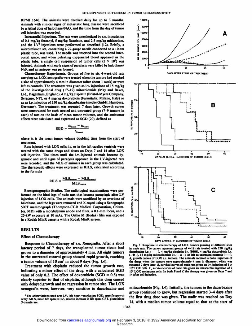
Response to Chemotherapy of s.c. Xenografts. After a shortlatency period of 7 days, the transplanted tumor tissue hadgrown to a diameter of approximately 4 mm. All eight tumorsin the untreated control group showed rapid growth, reachinga tumor volume of 10 cm3 in about 9 days (Fig. \A).
Treatment with cisplatin reduced the tumor growth rate,indicating a minor effect of the drug, with a calculated SGDvalue of only 0.2. The effect of doxorubicin (SGD = 0.5) wasclearly superior to that of cisplatin, although this drug causedonly delayed growth and no regression in tumor size. The LOXxenografts were, however, very sensitive to dacarbazine and
'The abbreviations used are: LV, left heart ventricular; SGD, specific growthdelay; MLS, mean life span; RILS. relative increase in life span; GST, glutathioneS-transferase.
10000-
2 2000-
DAYS AFTER START OF TREATMENT
S•¿�5¿IT, 50-ceo
L 1-.--,
BLk.
20 30 40 50DAYS AFTER I.V. INJECTION OF TUMOR CELLS
a
£ 50-
25-
"T—
10
—¿�I—4D 6020 30 40 50
DAYS AFTER L V INJECTION OF TUMOR CELLS
Fig. 1. Response to chemotherapy of LOX tumors growing at different sitesin nude rats. The curves represent groups of 4-18 rats treated with 250 mg/kgdacarbazine i.p. ( ), 4 mg/kg cisplatin i.v. (•••),4 mg/kg doxorubicin i.v.(- •¿�-), 15 mg/kg mitozolomide i.v. (- A -), or left as untreated controls ( )., I. growth curves of LOX s.c. tumors. The animals received a bolus injection ofthe drugs when the tumors were approximately 4 mm in diameter, which wasrepeated 7 days later. B, survival curves of nude rats given an i.v. injection of 1 x10" LOX cells. C, survival curves of nude rats given an intracardial injection of 210s LOX melanoma cells. In both B and C the therapy was given on Days 7 and14 after cell injection.
mitozolomide (Fig. \A). Initially, the tumors in the dacarbazinegroup continued to grow, but regression started 3-4 days after
the first drug dose was given. The nadir was reached on Day14, with a median tumor volume equal to that at the start of
1348
Research. on February 3, 2018. © 1992 American Association for Cancercancerres.aacrjournals.org Downloaded from
SITE-DEPENDENT DIFFERENCES IN TUMOR CHEMOSENSITIVITY
treatment. Thereafter, regrowth took place, but the growth ratewas somewhat slower than that of the control group. The effectof mitozolomide did not become apparent until Day 7, afterwhich the tumors regressed rapidly with a complete responseof nine of ten tumors at Day 21 (Fig. IA). In six of the ninecases no regrowth was observed throughout an observationperiod of 50 days, whereas three tumors started to regrow at23-29 days after the start of therapy. Treatment with dacarba-zine resulted in a SGD value of 21, whereas the pronouncedeffect of mitozolomide did not permit a meaningful SGDcalculation.
Effect of Chemotherapy on the Survival of Nude Rats withLOX Lung Colonies. In two independent experiments the effectof the four chemotherapeutic agents was tested in animalsinjected i.v. with 1 x 10*LOX cells. In agreement with previous
results (11), the untreated control animals had a mean survivaltime of 20.1 ±3.3 (SD) days (Fig. \B), and upon necropsymore than 200 tumor colonies could be counted on the surfaceof the lungs. Treatment with cisplatin had a marginal effect onthe LOX lung tumors, resulting in MLS ±SD values for thesacrificed rats of 27.4 ±1.8 days, whereas one of the six animalssurvived. In this model, the levels of response to dacarbazine(MLS 39.3 ±8.2) and doxorubicin (MLS 38.5 ±10.2) weresimilar, with one survivor of the seven rats treated in each group(Fig. IB). As was the case in the s.c. experiments, mitozolomidewas also highly effective against the lung tumors. All fourtreated animals were alive at the end of the 90-day observationperiod, in agreement with the results of several previous experiments (11). In conclusion, the sensitivity of the experimentalLOX lung métastasesshowed a drug response profile similarto that of the s.c. tumors, except that dacarbazine was clearlyless effective. In the Mann-Whitney U test all drugs showedstatistically significant effects. When the life span of all surviving animals was set at 90 days, the effect of mitozolomide wassignificantly better than that of doxorubicin and dacarbazine (P= 0.022). Necropsy of the sacrificed rats in the treatment groupsshowed fewer (not counted) but larger lung colonies than werefound in untreated animals.
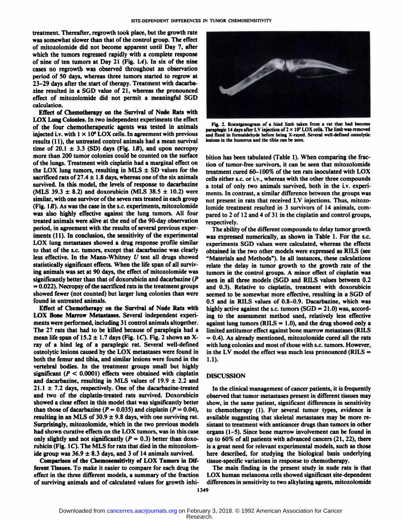
Effect of Chemotherapy on the Survival of Nude Rats withLOX Bone Marrow Métastases.Several independent experiments were performed, including 31 control animals altogether.The 27 rats that had to be killed because of paraplegia had amean life span of 15.2 ±1.7 days (Fig. 1C). Fig. 2 shows an X-ray of a hind leg of a paraplegic rat. Several well-definedosteolytic lesions caused by the LOX métastaseswere found inboth the femur and tibia, and similar lesions were found in thevertebral bodies. In the treatment groups small but highlysignificant (P < 0.0001) effects were obtained with cisplatinand dacarbazine, resulting in MLS values of 19.9 ±2.2 and21.1 ±7.2 days, respectively. One of the dacarbazine-treatedand two of the cisplatin-treated rats survived. Doxorubicinshowed a clear effect in this model that was significantly betterthan those of dacarbazine (P = 0.035) and cisplatin (P = 0.04),resulting in an MLS of 30.9 ±9.8 days, with one surviving rat.Surprisingly, mitozolomide, which in the two previous modelshad shown curative effects on the LOX tumors, was in this caseonly slightly and not significantly (P = 0.3) better than doxorubicin (Fig. 1C). The MLS for rats that died in the mitozolomide group was 36.9 ±8.3 days, and 3 of 14 animals survived.
Comparison of the Chemosensitivity of LOX Tumors in Different Tissues. To make it easier to compare for each drug theeffect in the three different models, a summary of the fractionof surviving animals and of calculated values for growth inhi-
Fig. 2. Roentgenogram of a hind limb taken from a rat that had becomeparaplegic 14 days after LV injection of 2 x 10' LOX cells. The limb was removedand fixed in formaldehyde before being X-rayed. Several well-defined osteolyticlesions in the humérusand the tibia can be seen.
bition has been tabulated (Table 1). When comparing the fraction of tumor-free survivors, it can be seen that mitozolomidetreatment cured 60-100% of the ten rats inoculated with LOXcells either s.c. or i.v., whereas with the other three compoundsa total of only two animals survived, both in the i.v. experiments. In contrast, a similar difference between the groups wasnot present in rats that received LV injections. Thus, mitozolomide treatment resulted in 3 survivors of 14 animals, compared to 2 of 12 and 4 of 31 in the cisplatin and control groups,respectively.
The ability of the different compounds to delay tumor growthwas expressed numerically, as shown in Table 1. For the s.c.experiments SGD values were calculated, whereas the effectsobtained in the two other models were expressed as RILS (see"Materials and Methods"). In all instances, these calculations
relate the delay in tumor growth to the growth rate of thetumors in the control groups. A minor effect of cisplatin wasseen in all three models (SGD and RILS values between 0.2and 0.3). Relative to cisplatin, treatment with doxorubicinseemed to be somewhat more effective, resulting in a SGD of0.5 and in RILS values of 0.8-0.9. Dacarbazine, which washighly active against the s.c. tumors (SGD = 21.0) was, according to the assessment method used, relatively less effectiveagainst lung tumors (RILS = 1.0), and the drug showed only alimited antitumor effect against bone marrow métastases(RILS= 0.4). As already mentioned, mitozolomide cured all the ratswith lung colonies and most of those with s.c. tumors. However,in the LV model the effect was much less pronounced (RILS =1.1).
DISCUSSION
In the clinical management of cancer patients, it is frequentlyobserved that tumor métastasespresent in different tissues mayshow, in the same patient, significant differences in sensitivityto chemotherapy (1). For several tumor types, evidence isavailable suggesting that skeletal métastasesmay be more resistant to treatment with anticancer drugs than tumors in otherorgans (1-5). Since bone marrow involvement can be found inup to 60% of all patients with advanced cancers (21, 22), thereis a great need for relevant experimental models, such as thosehere described, for studying the biological basis underlyingtissue-specific variations in response to chemotherapy.
The main finding in the present study in nude rats is thatLOX human melanoma cells showed significant site-dependentdifferences in sensitivity to two alkylating agents, mitozolomide
1349
Research. on February 3, 2018. © 1992 American Association for Cancercancerres.aacrjournals.org Downloaded from
SITE-DEPENDENT DIFFERENCES IN TUMOR CHEMOSENSITIVITY
Table 1 Effect of chemotherapy on LOX human melanoma tumors growing in different tissues of nude ratsGroups of nude rats with s.c. xenografts, microscopic lung colonies, or micrometastases in bone marrow were treated i.v. with cisplatin (4 mg/kg), doxorubicin (4
mg/kg), mitozolomide (15 mg/kg), or i.p. with dacarbazine (250 mg/kg) on the days given in "Materials and Methods." The size of s.c. tumors was measured three
times weekly for volume calculations. Animals that received LOX cells i.v. or LV were observed daily, and the time to appearance of symptoms of respiratory distressor paraplegia was observed.
Fraction of tumor-free survivors in ratsinoculatedAnimal
groupControl
CisplatinDoxorubicinDacarbazineMitozolomides.c.0/8
0/80/90/7
6/10i.v.0/12
1/61/71/74/4LV4/31
2/121/81/183/14S.C.
(SGD)0.2
0.521.0
Cure"Tumor
growthdelayi.v.
(RILS)0.2
0.81.0
CureLV
(RILS)0.3
0.90.41.1
1Complete response in nine often tumors. In three of these regrowth occurred, starting 23-29 days after the start of therapy.
and dacarbazine, drugs that are chemically related (19) andhave shown activity in malignant melanoma (13). The site-dependent difference in response was particularly striking inthe case of mitozolomide, which had a curative effect on s.c.and lung tumors, whereas bone métastaseswere much lessresponsive. The effect of dacarbazine was pronounced on s.c.tumors, moderate against lung tumor colonies, and smallagainst skeletal métastases.In contrast, for each of the twoother drugs, doxorubicin and cisplatin, similar levels of response were obtained in all three models. Mitozolomide, whichwas significantly more effective than the other drugs againsts.c. and lung tumors, showed only the same moderate activityas doxorubicin in the LV experiments.
Several different explanations for the tissue-dependent differences in chemosensitivity may be considered. The most obvious possibility would be that the degree of tumor responsereflects the drug concentration obtained in the different organs.It would, therefore, be of interest to measure tumor concentrations at the start of therapy, i.e., in small (4 mm) s.c. tumors,and in the métastaseson Day 7 after cell injection. Unfortunately, at this point only micrometastases are present in bonemarrow and lungs. Later, when the rats become paraplegic,LOX skeletal métastasesare still not easily available for drugdistribution studies, since the tumor cells are highly intermingled with normal bone marrow cells in the spine and long bones(12). However, since the level of drug concentration present inmicrometastases presumably is associated with the concentrations achieved in the surrounding normal tissue, we measuredby high-performance liquid chromatography the doxorubicinand mitozolomide concentrations in normal bone marrow andin the lungs of non-tumor-bearing nude rats. No major differences in drug concentration in these tissues were found, neitherin the levels of doxorubicin and its metabolites during the first16 h nor in the mitozolomide concentration in the initial 4-hperiod after drug administration (not shown). The test periodrepresents for both drugs about twice their half-life in plasma.Although care must be taken in the interpretation of these data,it does not seem very likely that the differences in chemosensitivity are caused by pharmacokinetic factors.
It is generally agreed that small tumors respond better tochemotherapy than large ones (7, 9). These differences may berelated to better blood supply to the tumor cells, resulting inhigher rates of cell proliferation and better penetration of thedrugs into the smaller tumors. If such factors operate in oursystem, the response of the LOX micrometastases known to bepresent in the bone marrow and in the lungs at the start oftreatment would be expected to be better than that of the s.c.tumors. This assumption is further supported by the findingthat LOX micrometastases in the bone are particularly wellvascularized, as seen on tissue sections 7 days after cell injection
(not shown). Moreover, in the calculation of the relative increase in life span and specific growth delay shown in Table 1,we have attempted to calibrate for differences in proliferationrates of the tumors growing at the different sites. Therefore, itseems reasonable to conclude that such factors cannot explainthe observed differences in chemosensitivity in our models.
Since tumor cells capable of forming métastasesmay represent a subpopulation of the cells present in the original tumor(23), it is a priori not inconceivable that LOX cells capable offorming bone metastasis may be less responsive to chemotherapy than the LOX cells present in s.c. and lung tumors. Thishypothesis does not seem very likely, considering the differencebetween the four compounds in site-dependent variation insensitivity. We will, however, examine the possibility by testingthe chemosensitivity of tumors established by s.c. réimplanta-
tion of LOX bone marrow métastasesisolated from rats treatedwith mitozolomide or dacarbazine. One might also speculatewhether the alkylation effect of mitozolomide and dacarbazinecould in some way be absorbed to a higher extent by the normalbone marrow cells than by the normal cells in the other tissues.If so, a similar relative resistance of the bone marrow métastases, as seen here with mitozolomide and dacarbazine, wouldbe expected to be present also for other alkylating agents, apossibility that is currently under investigation. A more likelyexplanation may be that interactions between the LOX cellsand the microenvironment in the bone marrow might influencetumor sensitivity at the cellular level.
Several mechanisms have been implicated in tumor cell resistance to chemotherapy, mechanisms which may differ between groups of drugs as well as between specific agents withineach group (24). One mechanism claimed to be involved inprotecting tumor cells from xenobiotics is the isozymes belonging to the GST family. In particular, the anionic isozymeglutathione 5-transferase (GST-n-) has been shown to be ele
vated in a variety of human tumors, including melanomas (25).No difference was detected, however, in the expression of GST-
7Tin s.c., lung, and bone marrow LOX tumors, as assessed byimmunohistochemical examination of frozen tissue sections(not shown). Moreover, antibodies against the nulrl (mult¡drugresistance) gene product stained LOX cells in tumors of allthree sites to a similar, low extent (not shown). Cross-resistanceto alkylating agents is, however, rare in cells of the nulrlphenotype (26).
Another mechanism that possibly could confer resistance toalkylating drugs in the LOX cells is increased cellular expression of {y-methylguanine-DNA methyltransferase. Cells lacking expression of this gene (MER~ phenotype), which is in
volved in DNA repair, are usually quite sensitive to DNA cross-linking agents (27, 28). Since LOX cells in tissue culture have
1350
Research. on February 3, 2018. © 1992 American Association for Cancercancerres.aacrjournals.org Downloaded from
SITE-DEPENDENT DIFFERENCES IN TUMOR CHEMOSENSITIV1TY
been found to be MER ,4 it seemed unlikely that this mecha
nism could be involved in our case. Nevertheless, preliminaryresults in our laboratory indicate that the gene for this transfer-ase is intact in the LOX cells, and we are now investigatingwhether a relationship might be found between tumor cellexpression of the gene in the various tissues and their sensitivityto mito/olomide and dacarbazine. Cisplatin can also causeDNA cross-links, but the responses to different classes of cross-linking agents have been shown to be largely independent ofeach other (29). The possibility that tissue-specific variation intumor expression of the C^-methylguanine-DNA methyltrans-ferase may be present in patients could have significant implications for clinical chemotherapy.
ACKNOWLEDGMENTS
The authors wish to thank Sissel G. Freim and Carin van der Leliefor excellent technical assistance and Frances Jaques for typing themanuscript. The statistical analyses were kindly performed by AreHelseth.
REFERENCES
1. Slack, N. H., and Bross, D. J. The influence of site of metastasis on tumourgrowth and response to chemotherapy. Br. J. Cancer, 32:78-86, 1975.
2. Yap, B-S., Baker, L. H., Sinkovics, J. G., Rivkin, S. E., Bottomley, R.,Thigpen, T., Burgess, M. A., Benjamin, R. S., and Bodey, G. P. Cyclophos-phamide, vincristine, Adriamycin, and DTIC (CYVADIC) combinationchemotherapy for the treatment of advanced sarcomas. Cancer Treat. Rep.,64:93-98, 1980.
3. Giuliano, A. E., Feig, S., and Eilber, F. R. Changing metastatic patterns ofosteosarcoma. Cancer (Phila.), 54: 2160-2164, 1984.
4. Goldstein, H., McNeill, B. J., Zufall, E., Jaffe, N., and Trêves,S. Changingindications for bone scintigraphy in patients with osteosarcoma. Radiology,135: 177-180, 1980.
5. Goorin, A. M., Shuster, J. J., Baker, A., Horowitz, M. E., Meyer, W. H.,and Link, M. P. Changing pattern of pulmonary métastaseswith adjuvantchemotherapy in patients with osteosarcoma: results from the multiinstitu-tional osteosarcoma study. J. Clin. Oncol., 9:600-605, 1991.
6. Holst, E., Sievers, U., and Schmahl. D. Experimentelle Untersuchungen zurChemosensibilität von Impftumoren bei unterschiedlicher Transplantation-slokalisation. Z. Krebsforsch., 76: 325-329, 1971.
7. Smith, K. A., Begg, A. C, and Denekamp, J. Differences in chemosensitivitybetween subcutaneous and pulmonary tumours. Eur. J. Cancer Clin. Oncol.,21: 249-256, 1985.
8. Staroselsky, A. N., Fan, D., O'Brian, C. A., Bucana, C. D., Gupta, K. P.,and Fidler, I. J. Site-dependent differences in response of the UV-2237murine fibrosarcoma to systemic therapy with Adriamycin. Cancer Res., 50:7775-7780, 1990.
9. Selby, P. J., Thomas, J. M., and Peckham, M. J. A comparison of thechemosensitivity of a primary tumour and its métastasesusing a humantumour xenograft. Eur. J. Cancer, 75.-1425-1429, 1979.
10. Phillips, R. M., Bibby, M. C., and Double, J. A. A critical appraisal of thepredictive value of in vitro chemosensitivity assays. J. Nati. Cancer Inst., 82:1457-1468, 1990.
4 R. H. Shoemaker, personal communication.
11. Kjenniksen, I., Storeng, R., Pihl, A., McLemore, T. L., and Fodstad, 0. Ahuman tumor lung metastasis model in athymic nude rats. Cancer Res., 49:5148-5152,1989.
12. Kjunniksen. I., Nesland, J. M., Pihl, A., and Fodstad, 0. Nude rat model forstudying metastasis of human tumor cells to bone and bone marrow. J. Nati.Cancer Inst., «2.-408-412, 1990.
13. Fodstad, 0., Aamdal, S., Pihl, A., and Boyd, M. R. Activity of mitozolomide(NSC 353451), a new imidazotetrazine, against xenografts from humanmelanomas, sarcomas, and lung and colon carcinomas. Cancer Res., 45:1778-1786, 1985.
14. Aamdal, S., Fodstad, 0., Kaalhus, ().. and Pihl, A. Chemosensitivity profilesof human cancers assessed by the 6-day SRC assay on serially xenograftedtumors. Int. J. Cancer, 37: 579-587, 1986.
15. Fodstad, 0., Aamdal, S., McMenamin, M., Nesland, J. M., and Pihl, A. Anew experimental metastasis model in athymic nude mice, the human malignant melanoma LOX. Int. J. Cancer, 41: 442-449, 1988.
16. Fodstad, 0., Aass, N., and Pihl, A. Assessment of tumour growth and ofresponse to chemotherapy of human melanomas in athymic, nude mice. Br.J. Cancer, 46(Supp\. 4): 146-149, 1980.
17. Gibson, N. W., Erickson, L. C., and Hickman, J. A. Effects of the antitumoragent 8-carbamoyl-3-(2-chloro-ethyl)imidazo[5,1 -</)-1,2,3,5-tetrazin-4(3H)-one on the DNA of mouse L1210 cells. Cancer Res., 44:1767-1771, 1984.
18. Gibson, N. W., Hickman, J. A., and Erickson, L. C. DNA cross-linking andcytotoxicity in normal and transformed human cells treated in vitro with 8-carbamoyl-3-(2-chloro-ethyl)imidazo[5,1 -</]-1,2,3,5-tetrazin-4(3//)-one.Cancer Res., 44: 1772-1775, 1984.
19. Stevens, M. F. G., Hickman, J. A., Langdon, S. P., Chubb, D., Vickers, L.,Stone, R., Baig, G., Goddard, C., Gibson, C., Gibson, N. W., Slack, J. A.,Newton, C., Lunt, E., Fizantes, C., and Lavelle, F. Antitumor activity andpharmacokinetics in mice of 8-carbamoyl-3-methyl-imidazo[5,1-rf)-1,2,3,5-tetrazin-4(3//)-one (CCRG 81045; M & B 39831), a novel drug with potentialas an alternative to dacarbazine. Cancer Res., 47: 5846-5852, 1987.
20. Nowak, K., Peckham, M. J., and Steel, G. G. Variation in response ofxenografts of coloréela!carcinomas to chemotherapy. Br. J. Cancer, 37:576-584, 1978.
21. Drew, M., and Dickson, R. B. Osseous complications of malignancy. In: J.J. Lokich (ed.), Clinical Cancer Medicine: Treatment Tactics, p. 97-124.Boston: G. K. Hall, 1980.
22. Enneking, W. F. Metastatic carcinoma. In: Musculoskeletal Tumor Surgery,Vol. 2, pp. 1541-1562. New York: Churchill Livingstone, 1983.
23. Fidler, I. J., and Kripke, M. L. Metastasis results from preexisting variantcells within a malignant tumor. Science (Washington DC), 197: 893-895,1977.
24. Chabner, B. A., and Myers, C. E. Clinical pharmacology of cancer chemotherapy. In: V. T. DeVita, Jr., S. Hellman, and S. A. Rosenberg (eds.).Cancer. Principles & Practice of Oncology, pp. 349-395. Philadelphia: J. B.Lippincott Co., 1989.
25. Shea, T. C., Kelley, S. L., and Henner, W. D. Identification of an anionicform of glutathione transferase present in many human tumors and humantumor cell lines. Cancer Res., 48: 527-533, 1988.
26. Cantwell, B. M. J., Sozzino, J. M., Corns, P., and Harris, A. L. Themultidrug resistant phenotype in clinical practice; evaluation of cross resistance to ifosfamide and mesna after VP 16-213, doxorubicin and vincristine(VPAV) for small cell lung cancer. Eur. J. Clin. Oncol., 24:123-129, 1988.
27. Scudiere, D. A., Meyer, S. A., Clatterbuck, B. E., Mattem, M. R., andZiolkowski, C. H. Sensitivity of human cell strains having different abilitiesto repair O'-methylguanine in DNA to inactivation by alkylating agentsincluding chloroethylnitosoureas. Cancer Res., 44:2467-2474, 1984.
28. Fornace, A. J., Papathanasiou, M. A., Hollander, M. C., and Yarosh, D. B.Expression of the O'-methylguanine-DNA methyltransferase gene MGMTin MER+ and MER~ human tumor cells. Cancer Res., 50:7908-7911,1990.
29. Sariban, E., Kohn, K. W., Zlotogorski, C., Laurent, G., D'Incaici, M., Day,R., HI, Smith, B. H., Kornblith, P. L., and Erickson, L. C. DNA cross-linking responses of human malignant glioma cell strains to chloroethylni-trosoureas, cisplatin, and diaziquone. Cancer Res., 47: 3988-3994, 1987.
1351
Research. on February 3, 2018. © 1992 American Association for Cancercancerres.aacrjournals.org Downloaded from
1992;52:1347-1351. Cancer Res Inge Kjønniksen, Knut Breistøl and Øystein Fodstad Mitozolomide, but not to Doxorubicin and CisplatinMelanoma Tumors in Nude Rats to Dacarbazine and Site-dependent Differences in Sensitivity of LOX Human
Updated version
http://cancerres.aacrjournals.org/content/52/5/1347
Access the most recent version of this article at:
E-mail alerts related to this article or journal.Sign up to receive free email-alerts
Subscriptions
Reprints and
To order reprints of this article or to subscribe to the journal, contact the AACR Publications
Permissions
Rightslink site. Click on "Request Permissions" which will take you to the Copyright Clearance Center's (CCC)
.http://cancerres.aacrjournals.org/content/52/5/1347To request permission to re-use all or part of this article, use this link
Research. on February 3, 2018. © 1992 American Association for Cancercancerres.aacrjournals.org Downloaded from
Related Documents