G. Di Giovanni, V. Di Matteo & E. Esposito (Eds.) Progress in Brain Research, Vol. 172 ISSN 0079-6123 Copyright r 2008 Elsevier B.V. All rights reserved CHAPTER 28 Serotonin/dopamine interaction in memory formation Ignacio Gonza´lez-Burgos 1,2, and Alfredo Feria-Velasco 2 1 Laboratorio de Psicobiologı´a, Divisio´n de Neurociencias, Centro de Investigacio´n Biome´dica de Occidente, Instituto Mexicano del Seguro Social, Guadalajara, Jal., Me´xico 2 Laboratorio de Neurobiologı´a Celular, Centro Universitario de Ciencias Biolo´gicas y Agropecuarias, Universidad de Guadalajara. Guadalajara, Jal., Me´xico Abstract: Both serotonin (5-HT) and dopamine (DA) neurotransmitters play a key role in modulating synaptic transmission in the central nervous system. Such 5-HT- and DA-mediated modulatory activity has been shown to influence a wide variety of cerebral functions, both of an instrumental and cognitive nature. Some brain regions strongly involved in cognition such as the prefrontal cortex, hippocampal formation and corpus striatum, are densely innervated by serotonergic and dopaminergic afferents proceeding from the raphe complex and the mesocorticolimbic or nigrostriatal systems, respectively. Learning and memory are strongly modulated by 5-HT and DA neurotransmitter activity, and in some cases they interact interdependently to sustain the psychobiological organization of these cognitive processes. Learning and memory, at least in part, depend on short- or long-lasting synaptic modifications, mainly occurring at dendritic spines. Indeed, the modulatory influence of 5-HT and DA at the synaptic level may affect the codification of mnemonic information on such spines. In fact, several experimental models of neurotransmitter activity have identified a close association between a 5-HT–DA imbalance and cytoarchitectonic changes underlying learning and memory impairment. Keywords: adaptive; behavior; serotonin; dopamine; learning; memory; dendritic spines Introduction Our understanding of the neurobiological phenom- ena underlying the organization of behaviour has required experimental studies capable of discrimi- nating the participation of the variables involved in these processes. As a result, it is clear that the neurotransmitter activity of different chemical substances is capable of producing a firing pattern in nerve cells. This implies that when confronted with diverse environmental situations, the informa- tion that the brain processes through its balanced biochemical activity is incorporated into reference schemes that reflect the harmonious relationship that individuals maintain with their physical and even psychological environment. On the other hand, the imbalances that might occur under atypical conditions could well lead to discordant individual–environment interactions with respect to the needs of the former, which would provoke behavioural disruptions that will impair the indivi- dual’s capacity to adapt to the situation (Fig. 1). Corresponding author. Tel.: +52 333 6683000–31950; Fax: +52 333 6181756; E-mail: [email protected] DOI: 10.1016/S0079-6123(08)00928-X 603

Welcome message from author
This document is posted to help you gain knowledge. Please leave a comment to let me know what you think about it! Share it to your friends and learn new things together.
Transcript
G. Di Giovanni, V. Di Matteo & E. Esposito (Eds.)
Progress in Brain Research, Vol. 172
ISSN 0079-6123
Copyright r 2008 Elsevier B.V. All rights reserved
CHAPTER 28
Serotonin/dopamine interaction in memoryformation
Ignacio Gonzalez-Burgos1,2,� and Alfredo Feria-Velasco2
1Laboratorio de Psicobiologıa, Division de Neurociencias, Centro de Investigacion Biomedica de Occidente,Instituto Mexicano del Seguro Social, Guadalajara, Jal., Mexico
2Laboratorio de Neurobiologıa Celular, Centro Universitario de Ciencias Biologicas y Agropecuarias,Universidad de Guadalajara. Guadalajara, Jal., Mexico
Abstract: Both serotonin (5-HT) and dopamine (DA) neurotransmitters play a key role in modulatingsynaptic transmission in the central nervous system. Such 5-HT- and DA-mediated modulatory activity hasbeen shown to influence a wide variety of cerebral functions, both of an instrumental and cognitive nature.Some brain regions strongly involved in cognition such as the prefrontal cortex, hippocampal formationand corpus striatum, are densely innervated by serotonergic and dopaminergic afferents proceeding fromthe raphe complex and the mesocorticolimbic or nigrostriatal systems, respectively. Learning and memoryare strongly modulated by 5-HT and DA neurotransmitter activity, and in some cases they interactinterdependently to sustain the psychobiological organization of these cognitive processes. Learning andmemory, at least in part, depend on short- or long-lasting synaptic modifications, mainly occurring atdendritic spines. Indeed, the modulatory influence of 5-HT and DA at the synaptic level may affect thecodification of mnemonic information on such spines. In fact, several experimental models ofneurotransmitter activity have identified a close association between a 5-HT–DA imbalance andcytoarchitectonic changes underlying learning and memory impairment.
Keywords: adaptive; behavior; serotonin; dopamine; learning; memory; dendritic spines
Introduction
Our understanding of the neurobiological phenom-ena underlying the organization of behaviour hasrequired experimental studies capable of discrimi-nating the participation of the variables involved inthese processes. As a result, it is clear that theneurotransmitter activity of different chemicalsubstances is capable of producing a firing pattern
�Corresponding author. Tel.: +52 333 6683000–31950;
Fax: +52 333 6181756; E-mail: [email protected]
DOI: 10.1016/S0079-6123(08)00928-X 603
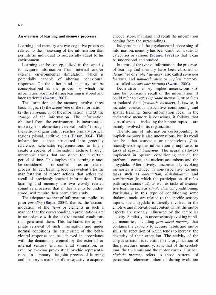
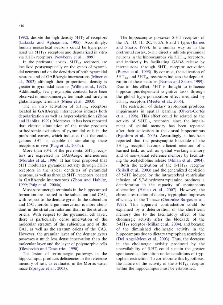
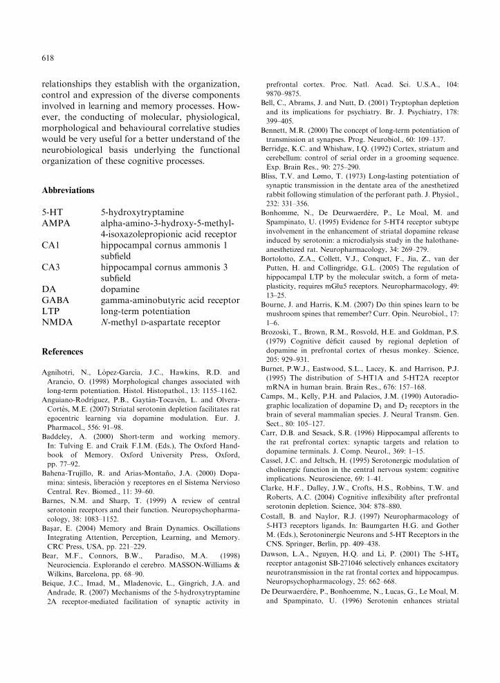
in nerve cells. This implies that when confrontedwith diverse environmental situations, the informa-tion that the brain processes through its balancedbiochemical activity is incorporated into referenceschemes that reflect the harmonious relationshipthat individuals maintain with their physical andeven psychological environment. On the otherhand, the imbalances that might occur underatypical conditions could well lead to discordantindividual–environment interactions with respect tothe needs of the former, which would provokebehavioural disruptions that will impair the indivi-dual’s capacity to adapt to the situation (Fig. 1).
CNS
INDIVIDUAL
ENVIRONMENT
ENVIRONMENTAL DEMANDS
BEHAVIOR
ENVIRONMENTAL DEMANDS
BEHAVIOR
Fig. 1. Schematic representation of the interactive relationships between an individual and his environment. This bidirectional
interaction (upper panel) is characterized by the sensorial (straight lines) and psychological (curve lines) afferent stimulation (solid
arrows) to which an individual is permanently exposed; such afferent information is analysed in the brain (CNS) leading in turn to
structuring behavioural responses (dotted arrows) putatively correspondent to the environmental demands. If such behavioural
responses are in agreement with these, then harmonic adaptation is facilitated in healthy subjects (lower panel; left); on the contrary,
aberrant behaviour could be closely related with psychopathological disorders (lower panel; right).
604
Over and above the observational criteria, theconcept of ‘behaviour’ could be conceptualized asthe result of an individual’s interaction with hisenvironment. From an operational point of view,the information the subject ‘extracts’ from such aninteraction is incorporated into abstract represen-tations in the brain. Such representations arecontrasted both with prior reference schemes, suchas genetically determined response patterns thatfinally permit reflex, invariable, conditionedactions to be taken or by taking decisions to
resolve the problems that such an interactionposes. In this context, the brain activity translatesinto motor actions, which carries an implicit orexplicit message that acquires a higher characterduring the process of verbal communication inhuman beings.
Motor activity (or its absence) results from theprocessing of the information obtained during thisinteraction between the individual and his envi-ronment, and is interpreted as ‘behaviour’. Thus,behaviour would be the series of messages emerging
605
from the nervous system in response, not only tocertain stimuli but also to the information acquiredfrom the environment. This information is decoded,ordered and recoded by virtue of the activation ofneuronal systems that produce an action, be it areflex, or a fixed pattern conditioned by priorexperience, or by reference schemes derived fromspecific situations that require a decision to be takenin response to a specific problem.
Within this framework, brain activity acquires apreponderant character in the interpretation ofbehaviour, for which the experimental study of theunderlying psychoneural processes and the neuro-biological phenomena that they sustain, are criticalto understand their expression, both under normaland pathological circumstances.
Whatever the psychobiological resources withwhich an individual confronts the needs constantlypresented by their interaction with their environ-ment, the fundamental objective of their activity isto achieve a satisfactory adaptation to this. Toachieve this process of adaptation satisfactorily,intrinsic instrumental and cognitive capacitiesparticipate in a relevant fashion, among whichlearning and memory stand out.
The normal psychoneural expression of learningand memory capacity is regulated by the balancedactivity of the diverse brain neurotransmittersystems. The metabolic imbalance of such systems —be it genetic or environmental in origin — couldproduce behavioural disorders that in the worstcase, would impede a harmonious relationship inaccordance with the demands of the surroundings.Thus, the individuals that develop disorders in thecognitive sphere related with alterations in theneurochemical balance of some neurotransmittersystems are incapable of harmoniously interactingwith their social and psychological surroundings.
Two of the neurotransmitters closely relatedwith cognitive function are 5-hydroxytryptamine(5-HT; serotonin) and dopamine (DA). As well astheir neurotransmitter activity, these two biogenicamines possess neuromodulatory activity. Theycan thus exert a direct or indirect influence on theexcitability of neurons that they stimulate.
The neurons that liberate 5-HT and DA arelocated in specific nervous centres. The axonterminals of serotonergic and dopaminergic
neurons are localized in different brain regions inwhich they exert their neurotransmitter and/orneuromodulatory effects. The postsynaptic bio-electric effect after their release depends on thetransduction system of these chemical signals, madeup of the receptor molecules for the correspondingneurotransmitter and where appropriate, by thosethat modulate the neuronal response to otherchemical stimuli through their secondary activity.Thus, the psychoneural effect that they exert willdepend on the brain region in question. Both 5-HTand DA are released in brain regions such as thecerebral cortex, the hippocampus and the corpusstriatum. These three brain regions are involved inthe organization of diverse cognitive processes,most noticeably, learning and memory.
Serotonergic and dopaminergic nerve terminalsmay have an excitatory, inhibitory or modulatoryeffect depending on the chemical receptor locatedin the postsynapse. Furthermore, the neurotrans-mission mediated by 5-HT could be affected tosome extent by DA and vice versa. The terminalsthat liberate 5-HT do so both in the synaptic cleftof specific contacts or as free terminals, whilst DAacts exclusively onto receptors located in thepostsynaptic membrane.
Learning and memory processes are partiallyregulated by the neurotransmitter and neuromodu-latory activity of 5-HT and DA. Such regulation isintrinsically related to the expression of morpho-physiological phenomena of synaptic plasticity thatoccur at dendritic spines of the neurons that possessthem. Thus, the alteration of serotonergic activitycan affect the excitability of certain brain regions byvirtue of modifications in the cytoarchitectonicpattern of principal neurons present therein. More-over, dopaminergic terminals may be located in theneck of the dendritic spines on principal projectionneurons, both in the prefrontal cerebral cortex aswell as in the striatum, while glutamatergicterminals are situated in the head of these samespines. Therefore, a modulatory effect of DA on theexcitatory information afferent to these neurons hasbeen proposed. It then follows that the modifica-tions in the neuronal cytoarchitectonic patterninduced by changes in the 5-HT and DA neuro-transmitter activity can affect the organization ofthe information related to learning and memory.
606
An overview of learning and memory processes
Learning and memory are two cognitive processesrelated to the processing of the information thatpermits an individual to successfully adapt to hisenvironment.
Learning can be conceptualized as the capacityto acquire information from internal and/orexternal environmental stimulation, which ispotentially capable of altering behaviouralresponses. On the other hand, memory can beconceptualized as the process by which theinformation acquired during learning is stored andlater retrieved (Sweatt, 2003).
The ‘formation’ of the memory involves threebasic stages: (1) the acquisition of the information;(2) the consolidation of the information; and (3) thestorage of the information. The informationobtained from the environment is incorporatedinto a type of elementary cerebral ‘buffer’ throughthe sensory organs until it reaches primary corticalregions (visual, auditive, etc.) (Bas-ar, 2004). Thisinformation is then structured into generallyreferenced schematic representations to finallycreate a species of information archive throughmnemonic traces that are stable for a certainperiod of time. This implies that learning cannotbe considered — or studied — as an isolatedprocess. In fact, learning becomes evident after themanifestation of motor actions that reflect therecall of previously learned information. Thus,learning and memory are two closely relatedcognitive processes that if they are to be under-stood, will require their correlative study.
The adequate storage of information implies itsprior encoding (Bas-ar, 2004), that is, the ‘accom-modation’ of the items or elements in such amanner that the corresponding representations arein accordance with the environmental conditionsthat generated them. This facilitates the appro-priate retrieval of such information and undernormal conditions the structuring of the beha-vioural response can be achieved in accordancewith the demands presented by the external orinternal sensory environmental stimulation, oreven by evoking pre-existing psychic representa-tions. In summary, the joint process of learningand memory is made up of the capacity to acquire,
encode, store, maintain and recall the informationcoming from the surroundings.
Independent of the psychoneural processing ofinformation, memory has been classified in variouscategories or systems (Squire, 1992) so that it canbe understood and studied.
In terms of the type of information, the processesof learning and memory have been classified asdeclarative or explicit memory, also called conscious
learning, and non-declarative or implicit memory,also called unconscious learning (Sweatt, 2003).
Declarative memory implies unconscious sto-rage but conscious recall of the information. Itcould refer to events (episodic memory), or to factsor isolated data (semantic memory). Likewise, itincludes conscious associative conditioning andspatial learning. Since information recall in thedeclarative memory is conscious, it follows thatcortical areas — including the hippocampus — aremainly involved in its organization.
The storage of information corresponding toimplicit memory is also unconscious, but its recallcan be either conscious or unconscious. Con-sciously evoking this information is implicated intasks of operant behaviour. The neural pathwaysimplicated in operant conditioning include theprefrontal cortex, the nucleus accumbens and theamygdala. Alternatively, unconsciously evokingmemories is included in non-associative learningtasks such as habituation, dishabituation andsensitization (in which the participation of reflexpathways stands out), as well as tasks of associa-tive learning such as simple classical conditioning.Particularly in this type of conditioning somethalamic nuclei are related to the specific sensoryinputs; the amygdala is directly involved in theemotive and motivational content whilst the motoraspects are strongly influenced by the cerebellaractivity. Similarly, in unconsciously evoking impli-cit memories, including procedural memory thatcontains the capacity to acquire habits and motorskills the repetition of which tends to increase thedexterity of their execution. The activity of thecorpus striatum is relevant to the organization ofthis procedural memory, as is that of the cerebel-lum, the thalamus and the motor cortex. Further,phyletic memory refers to those patterns ofperceptual references inherited during evolution
607
and that are evoked by specific stimuli or by the‘need to act’. Perceptual memory is another formof implicit memory that is evoked unconsciouslyand refers to the neocortical representation ofevents, objects, people, animals, facts, names andconcepts, ranging from elementary sensations tothe formation of abstract concepts.
It has been suggested that only the declarative orexplicit memory can have a temporal dimension(Squire, 1992). In this context, and in accordancewith the time over which the information ismaintained until it is later recalled, this memoryhas been classified as long-term and short-term
memory (Baddeley, 2000). Comparatively, long-term memory is more stable and less labile and theinformation can be maintained for hours, days oryears. As a specialization of cognitive short-termmemory, the working memory (or active short-term
memory) refers to the actualization in the short-term of items of information that are specificallynecessary during the execution of a certain task.The psychoneural dynamic of this type of short-term memory is sustained by the conformation ofdynamic networks between prefrontocortical neu-rons denominated ‘memory fields’; these arealternating, temporally reverberant and remainactive and stable until the behavioural action isexecuted (Williams and Goldman-Rakic, 1995).Both evoking memories and executing motoractions leading to the execution of tasks relatedto the working memory (also called operant)requires the fully conscious processing of theinformation (Sweatt, 2003).
It is therefore evident that the participation ofthe cerebral cortex, hippocampus, striatum, amyg-dala and cerebellum are particularly important inthe functional organization of diverse componentsof learning and memory.
5-HT in learning and memory
5-HT is synthesized from tryptophan obtainedthrough the diet. The experimental manipulationof the availability of this essential amino acid hasbeen used as an experimental paradigm to studythe effect that 5-HT exerts over different psycho-neural processes such as learning and memory.
It has been shown that the generalized depletionof 5-HT in the brain produces an impairment ofshort-term memory but not of long-term memory(Hritcu et al., 2007). However, there is alsoevidence that the availability of tryptophan in thebrain affects both types of memory. It has alsobeen reported that the restriction of tryptophanproduces impairment in the formation of the long-term memory and its consolidation (Schmitt et al.,2000), which would agree with later findings that5-HT affects this type of memory by affecting theinformation encoding phase rather than recall (vander Veen et al., 2006). Alternatively, the activity of5-HT is related to the acquisition, retention andrecuperation of the information associated withshort-term memory. This effect is more pro-nounced in long-term memory, suggesting thatboth types of memory are processed independently(Shirahata et al., 2006).
In repetitive trial assays of short-term memory,greater behavioural efficiency was achieved underconditions of chronic tryptophan depletion(Gonzalez-Burgos et al., 1998), which was con-firmed in a similar behavioural paradigm afterproducing a chemical lesion of the serotonergicraphe-prefrontal pathway (Perez-Vega et al.,2000). These findings could be related to theeffects that 5-HT exerts on behavioural flexibility.Indeed, it has been reported that the depletion ofprefrontal 5-HT produces enduring behaviours,without affecting the capacity to retain or dis-criminate those previously learned (Clarke et al.,2004). In accordance with this, tryptophan restric-tion improves the short-term active memory(working memory) (Riedel et al., 2003), which isdependent on prefrontal cortical activity (Fuster,1997). This would be supported by the activationof 5-HT2A receptors located in prefrontal pyrami-dal neurons that are known to facilitate the spatialworking memory performance (Williams et al.,2002). Similarly, a higher density of 5-HT2A
receptors has been observed after prefrontal5-HT depletion (sent to publication), as well asan enhancement of multiunitary activity of pre-frontal neurons (sent to publication). Thesewould be strongly associated with a more efficientshort-term memory performance along withcytoarchitectural changes in prefrontal pyramidal
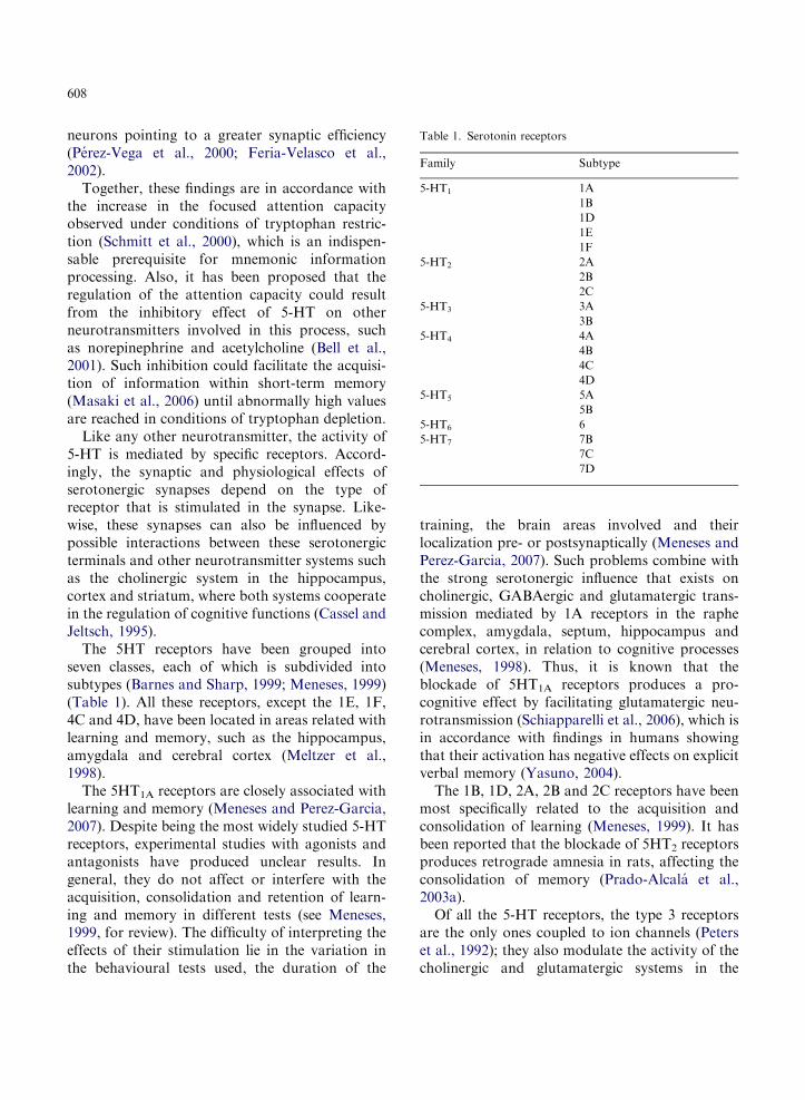
Table 1. Serotonin receptors
Family Subtype
5-HT1 1A
1B
1D
1E
1F
5-HT2 2A
2B
2C
5-HT3 3A
3B
5-HT4 4A
4B
4C
4D
5-HT5 5A
5B
5-HT6 6
5-HT7 7B
7C
7D
608
neurons pointing to a greater synaptic efficiency(Perez-Vega et al., 2000; Feria-Velasco et al.,2002).
Together, these findings are in accordance withthe increase in the focused attention capacityobserved under conditions of tryptophan restric-tion (Schmitt et al., 2000), which is an indispen-sable prerequisite for mnemonic informationprocessing. Also, it has been proposed that theregulation of the attention capacity could resultfrom the inhibitory effect of 5-HT on otherneurotransmitters involved in this process, suchas norepinephrine and acetylcholine (Bell et al.,2001). Such inhibition could facilitate the acquisi-tion of information within short-term memory(Masaki et al., 2006) until abnormally high valuesare reached in conditions of tryptophan depletion.
Like any other neurotransmitter, the activity of5-HT is mediated by specific receptors. Accord-ingly, the synaptic and physiological effects ofserotonergic synapses depend on the type ofreceptor that is stimulated in the synapse. Like-wise, these synapses can also be influenced bypossible interactions between these serotonergicterminals and other neurotransmitter systems suchas the cholinergic system in the hippocampus,cortex and striatum, where both systems cooperatein the regulation of cognitive functions (Cassel andJeltsch, 1995).
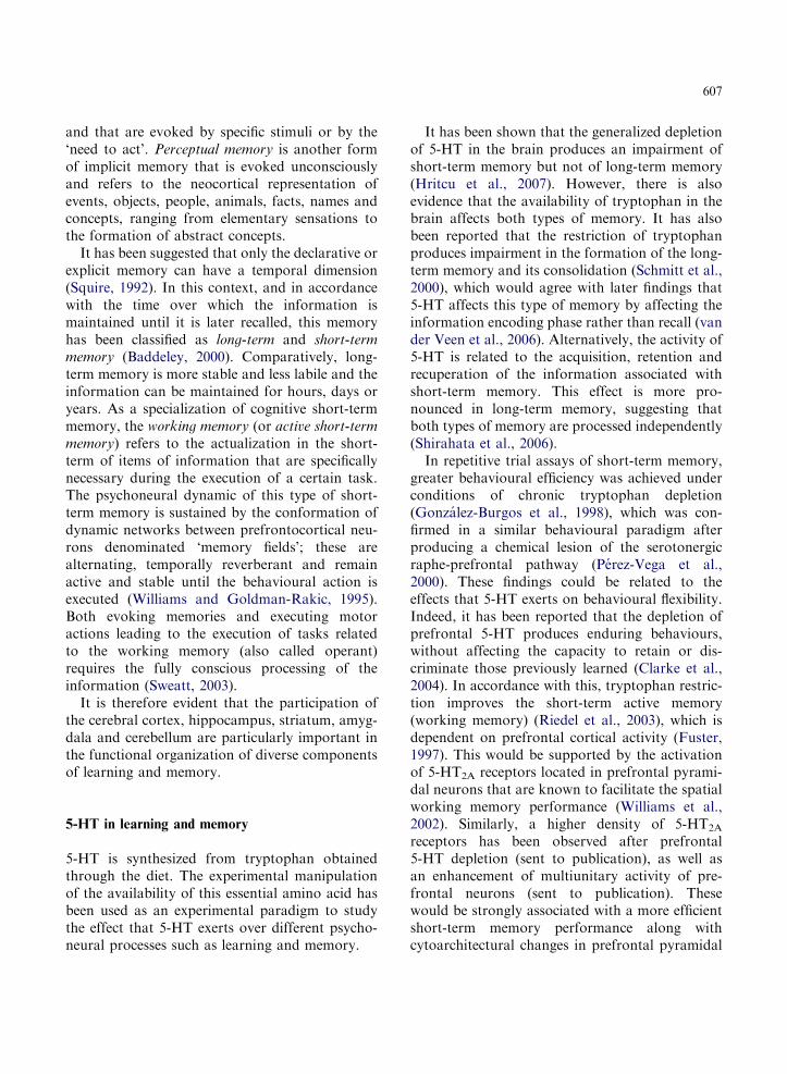
The 5HT receptors have been grouped intoseven classes, each of which is subdivided intosubtypes (Barnes and Sharp, 1999; Meneses, 1999)(Table 1). All these receptors, except the 1E, 1F,4C and 4D, have been located in areas related withlearning and memory, such as the hippocampus,amygdala and cerebral cortex (Meltzer et al.,1998).
The 5HT1A receptors are closely associated withlearning and memory (Meneses and Perez-Garcia,2007). Despite being the most widely studied 5-HTreceptors, experimental studies with agonists andantagonists have produced unclear results. Ingeneral, they do not affect or interfere with theacquisition, consolidation and retention of learn-ing and memory in different tests (see Meneses,1999, for review). The difficulty of interpreting theeffects of their stimulation lie in the variation inthe behavioural tests used, the duration of the
training, the brain areas involved and theirlocalization pre- or postsynaptically (Meneses andPerez-Garcia, 2007). Such problems combine withthe strong serotonergic influence that exists oncholinergic, GABAergic and glutamatergic trans-mission mediated by 1A receptors in the raphecomplex, amygdala, septum, hippocampus andcerebral cortex, in relation to cognitive processes(Meneses, 1998). Thus, it is known that theblockade of 5HT1A receptors produces a pro-cognitive effect by facilitating glutamatergic neu-rotransmission (Schiapparelli et al., 2006), which isin accordance with findings in humans showingthat their activation has negative effects on explicitverbal memory (Yasuno, 2004).
The 1B, 1D, 2A, 2B and 2C receptors have beenmost specifically related to the acquisition andconsolidation of learning (Meneses, 1999). It hasbeen reported that the blockade of 5HT2 receptorsproduces retrograde amnesia in rats, affecting theconsolidation of memory (Prado-Alcala et al.,2003a).
Of all the 5-HT receptors, the type 3 receptorsare the only ones coupled to ion channels (Peterset al., 1992); they also modulate the activity of thecholinergic and glutamatergic systems in the
609
amygdala, hippocampus and entorhinal cortex(Meneses, 1998). These receptors are located inthe soma, axon and/or nerve terminals ofGABAergic interneurons (Zifa and Fillion, 1992),and it has been reported that they participate inthe synaptic organization of information related tolearning and memory (Staubli and Xu, 1995).Indeed, the application of antagonists to thesereceptors in the amygdala provokes an improve-ment in learning (Costall and Naylor, 1997).
The 5-HT4 receptors are located in the habe-nula, hippocampus and the amygdala, and theymediate the slow excitatory and the long-lastingresponse in the hippocampus (Eglen et al., 1995).
The 5HT6 receptor is particularly abundant inthe olfactory bulb, striatum, nucleus accumbens,cerebral cortex and some sub-fields of the hippo-campus (Gerard et al., 1996). The blockade of the5HT6 receptors with antagonists in the ratprefrontal cortex and hippocampus increasesexcitatory neurotransmission, which suggests theirlocalization in interneurons or extrinsic GABAer-gic terminals (Dawson et al., 2001).
The antagonism of the 1A, 2A, 2B, 4 or 6receptors improves only long-term memory, whileantagonism of 1B receptors improves both long-and short-term memory. This suggests that bothtypes of memory appear to function in parallelusing the same signalling cascades, whilst on otheroccasions they are functionally associated in series(Meneses, 2007). It has been shown that serialactivity of short- to long-term memory is favouredby the activity of 5HT1B receptors, whilst the 1A,2A, 2B/2C, 4 and 6 receptors are involved in theiractivity in parallel (Meneses, 2007).
Both the dorsal and medial raphe projectserotonergic afferents to the dorsolateral prefron-tal cortex in the monkey, although the dorsalraphe does so more densely and the afferents comeparticularly from the rostral region. Hence, it wassuggested that the innervation of the dorsal raphecould be most closely related with the coordinationof the excitability of functionally related corticalareas, whilst the innervation of the medial rapheprobably exerts a global influence over corticalactivity (Wilson and Molliver, 1991).
Indeed, 28% of the serotonergic terminals afferentto the prefrontal cortex establish synaptic contacts
with spines (predominantly) and with dendriticshafts, while the remainder are made up of freeterminals (Smiley and Goldman-Rakic, 1996).
Serotonergic innervation to the prefrontal cor-tex occurs in all cortical layers, although it isparticularly dense in layers 1 and 4. In layers 1, 3and 5, only 23% of terminals form synapses, whichare excitatory on the dendritic shafts of interneu-rons, and of these 23%, 8% establish synapseswith dendritic shafts on pyramidal neurons(Smiley and Goldman-Rakic, 1996).
In the prefrontal cortex, 1A and 2A receptorsare expressed in high density (Pazos and Palacios,1985; Pazos et al., 1985) the 5-HT receptors beingthe most abundant receptors in this brain region(Pazos et al., 1985; Pompeiano et al., 1994). In thisneocortical region, the 1A receptors are inhibitorywhereas the 2A receptors are excitatory (Pazoset al., 1985; Pompeiano et al., 1994).
The 5HT1A receptors are present in 60% of theprefrontal pyramidal neurons and in 25% of theGABAergic interneurons (Santana et al., 2004). Ithas been shown that the 1A 5-HT receptors areinvolved in the process of learning acquisition andthat antagonist blockade of these receptors revertssome cognitive deficiencies induced pharmacologi-cally in monkeys (Harder and Ridley, 2000) andrats (Misane and Ogren, 2003).
In the pyramidal cells, the 5-HT1A receptors arepreferentially located in the soma and in the basaldendrites (Riad et al., 2000), whereas 5HT2A
receptors establish synaptic contacts with apicaldendrites (Xu and Pandey, 2000). Although theseneurons are simultaneously excitatory and inhibi-tory, the inhibitory response predominates, per-haps due to the localization of the type 1Areceptors in the axon hillock (Puig et al., 2004b).Furthermore, 30% of the raphe innervation tothe cortex comes from GABAergic terminals(Jankowski and Sesack, 2004), which suggests thatnot only the 5HT1A receptors, but also theGABA(A) receptors might be responsible for theinhibitory responses induced in pyramidal cellsafter electrical stimulation of the raphe nucleus(Puig et al., 2004a).
From in vitro and in vivo studies in rats, thepredominant effect of 5HT in the prefrontal cortexis known to be inhibitory (Jacobs and Azmitia,
610
1992), despite the high density 5HT2 of receptors(Lakoski and Aghajanian, 1985). Accordingly,human neocortical neurons could be hyperpola-rized via 5HT1A receptors and depolarized in vitrovia 5HT2 receptors (Newberry et al., 1999).
In the prefrontal cortex, 5HT2A receptors arelocalized postsynaptically on the spines of pyrami-dal neurons and on the dendrites of both pyramidalneurons and of GABAergic interneurons (Miner etal., 2003) although their proportional density isgreater in pyramidal neurons (Willins et al., 1997).Additionally, few presynaptic contacts have beenobserved in monoaminergic terminals and rarely inglutamatergic terminals (Miner et al., 2003).
The in vitro activation of 5HT2A receptorslocated in GABAergic interneurons induces bothdepolarization as well as hyperpolarization (Zhouand Hablitz, 1999). Moreover, it has been reportedthat electric stimulation of the raphe producesorthodromic excitation of pyramidal cells in theprefrontal cortex, which indicates that the endo-genous 5HT is capable of stimulating thesereceptors in vivo (Puig et al., 2004a).
More than 90% of the prefrontal 5HT3 recep-tors are expressed in GABAergic interneurons(Morales et al., 1996). It has been proposed that5HT modulates pyramidal activity through 5HT2
receptors in the apical dendrites of pyramidalneurons, as well as through 5HT3 receptors locatedin GABAergic interneurons (Zhou and Hablitz,1999; Puig et al., 2004a).
Most serotonergic terminals in the hippocampalformation are located in the subiculum and CA1,with respect to the dentate gyrus. In the subiculumand CA1, serotonergic innervation is more abun-dant in the striatum radiatum than in the stratumoriens. With respect to the pyramidal cell layer,there is particularly dense innervation of themolecular stratum of the subiculum and of theCA1, as well as the stratum oriens of the CA3.However, the granular layer of the dentate gyruspossesses a much less dense innervation than themolecular layer and the layer of polymorphic cells(Oleskevich and Descarries, 1990).
The lesion of serotonergic pathways in thehippocampus produces deficiencies in the referencememory of rats, as evaluated in the Morris watermaze (Sprague et al., 2003).
The hippocampus possesses 5-HT receptors ofthe 1A, 1D, 1E, 2C, 3, 5A, 6 and 7 types (Barnesand Sharp, 1999). In a similar way as in theprefrontal cortex, 5-HT directly inhibits pyramidalneurons in the hippocampus via 5HT1A receptors,and indirectly by facilitating GABA release byinterneurons through 5HT3 receptor activation(Burnet et al., 1995). By contrast, the activation of5HT2A and 5HT2C receptors induces the depolari-zation of these neurons (Barnes and Sharp, 1999).Due to this effect, 5HT is thought to influencehippocampus-dependent cognitive tasks throughthe global hyperpolarization effect mediated by5HT1A receptors (Meeter et al., 2006).
The restriction of dietary tryptophan producesimpairments in spatial learning (Olvera-Corteset al., 1998). This effect could be related to theactivity of 5-HT1A receptors, since the impair-ment of spatial memory has been reportedafter their activation in the dorsal hippocampus(Egashira et al., 2006). Accordingly, it has beenreported that the postsynaptic blockade of the5HT1A receptor favours efficient retention of alearned task, as well as spatial working memoryand of non-spatial reference memory by facilitat-ing the acetylcholine release (Millan et al., 2004).
Both the activation of the 5-HT1A receptor(Seibell et al., 2003) and the generalized depletionof 5-HT induced by the intracerebral ventricularinfusion of 5,7-dihydroxytryptamine produces adeterioration in the capacity of spontaneousalternation (Hritcu et al., 2007). However, thechronic restriction of dietary tryptophan improvesefficiency in the T-maze (Gonzalez-Burgos et al.,1995). This apparent contradiction could beexplained by a deterioration of the short-termmemory due to the facilitatory effect of thecholinergic activity after the blockade of the5-HT1A receptor (Millan et al., 2004), and becauseof the diminished cholinergic activity in thehippocampus due to dietary tryptophan restriction(Del Angel-Meza et al., 2003). Thus, the decreasein the cholinergic activity produced by theunavailability of 5-HT could sustain the greaterspontaneous alternation under conditions of tryp-tophan restriction. To corroborate this hypothesis,the nature of the activity of the 5-HT1A receptorwithin the hippocampus must be established.
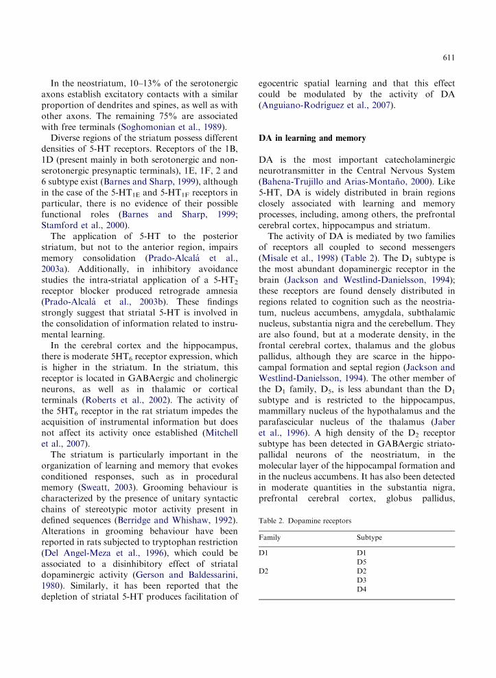
Table 2. Dopamine receptors
Family Subtype
D1 D1
D5
D2 D2
D3
D4
611
In the neostriatum, 10–13% of the serotonergicaxons establish excitatory contacts with a similarproportion of dendrites and spines, as well as withother axons. The remaining 75% are associatedwith free terminals (Soghomonian et al., 1989).
Diverse regions of the striatum possess differentdensities of 5-HT receptors. Receptors of the 1B,1D (present mainly in both serotonergic and non-serotonergic presynaptic terminals), 1E, 1F, 2 and6 subtype exist (Barnes and Sharp, 1999), althoughin the case of the 5-HT1E and 5-HT1F receptors inparticular, there is no evidence of their possiblefunctional roles (Barnes and Sharp, 1999;Stamford et al., 2000).
The application of 5-HT to the posteriorstriatum, but not to the anterior region, impairsmemory consolidation (Prado-Alcala et al.,2003a). Additionally, in inhibitory avoidancestudies the intra-striatal application of a 5-HT2
receptor blocker produced retrograde amnesia(Prado-Alcala et al., 2003b). These findingsstrongly suggest that striatal 5-HT is involved inthe consolidation of information related to instru-mental learning.
In the cerebral cortex and the hippocampus,there is moderate 5HT6 receptor expression, whichis higher in the striatum. In the striatum, thisreceptor is located in GABAergic and cholinergicneurons, as well as in thalamic or corticalterminals (Roberts et al., 2002). The activity ofthe 5HT6 receptor in the rat striatum impedes theacquisition of instrumental information but doesnot affect its activity once established (Mitchellet al., 2007).
The striatum is particularly important in theorganization of learning and memory that evokesconditioned responses, such as in proceduralmemory (Sweatt, 2003). Grooming behaviour ischaracterized by the presence of unitary syntacticchains of stereotypic motor activity present indefined sequences (Berridge and Whishaw, 1992).Alterations in grooming behaviour have beenreported in rats subjected to tryptophan restriction(Del Angel-Meza et al., 1996), which could beassociated to a disinhibitory effect of striataldopaminergic activity (Gerson and Baldessarini,1980). Similarly, it has been reported that thedepletion of striatal 5-HT produces facilitation of
egocentric spatial learning and that this effectcould be modulated by the activity of DA(Anguiano-Rodrıguez et al., 2007).
DA in learning and memory
DA is the most important catecholaminergicneurotransmitter in the Central Nervous System(Bahena-Trujillo and Arias-Montano, 2000). Like5-HT, DA is widely distributed in brain regionsclosely associated with learning and memoryprocesses, including, among others, the prefrontalcerebral cortex, hippocampus and striatum.
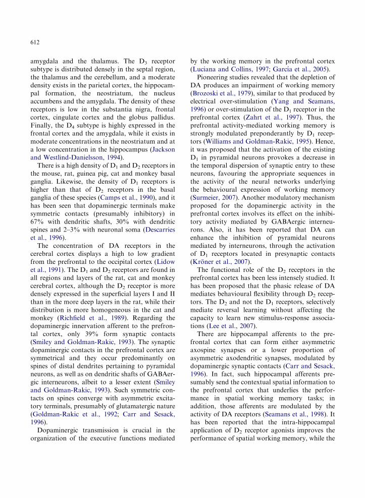
The activity of DA is mediated by two familiesof receptors all coupled to second messengers(Misale et al., 1998) (Table 2). The D1 subtype isthe most abundant dopaminergic receptor in thebrain (Jackson and Westlind-Danielsson, 1994);these receptors are found densely distributed inregions related to cognition such as the neostria-tum, nucleus accumbens, amygdala, subthalamicnucleus, substantia nigra and the cerebellum. Theyare also found, but at a moderate density, in thefrontal cerebral cortex, thalamus and the globuspallidus, although they are scarce in the hippo-campal formation and septal region (Jackson andWestlind-Danielsson, 1994). The other member ofthe D1 family, D5, is less abundant than the D1
subtype and is restricted to the hippocampus,mammillary nucleus of the hypothalamus and theparafascicular nucleus of the thalamus (Jaberet al., 1996). A high density of the D2 receptorsubtype has been detected in GABAergic striato-pallidal neurons of the neostriatum, in themolecular layer of the hippocampal formation andin the nucleus accumbens. It has also been detectedin moderate quantities in the substantia nigra,prefrontal cerebral cortex, globus pallidus,
612
amygdala and the thalamus. The D3 receptorsubtype is distributed densely in the septal region,the thalamus and the cerebellum, and a moderatedensity exists in the parietal cortex, the hippocam-pal formation, the neostriatum, the nucleusaccumbens and the amygdala. The density of thesereceptors is low in the substantia nigra, frontalcortex, cingulate cortex and the globus pallidus.Finally, the D4 subtype is highly expressed in thefrontal cortex and the amygdala, while it exists inmoderate concentrations in the neostriatum and ata low concentration in the hippocampus (Jacksonand Westlind-Danielsson, 1994).
There is a high density of D1 and D2 receptors inthe mouse, rat, guinea pig, cat and monkey basalganglia. Likewise, the density of D1 receptors ishigher than that of D2 receptors in the basalganglia of these species (Camps et al., 1990), and ithas been seen that dopaminergic terminals makesymmetric contacts (presumably inhibitory) in67% with dendritic shafts, 30% with dendriticspines and 2–3% with neuronal soma (Descarrieset al., 1996).
The concentration of DA receptors in thecerebral cortex displays a high to low gradientfrom the prefrontal to the occipital cortex (Lidowet al., 1991). The D1 and D2 receptors are found inall regions and layers of the rat, cat and monkeycerebral cortex, although the D2 receptor is moredensely expressed in the superficial layers I and IIthan in the more deep layers in the rat, while theirdistribution is more homogeneous in the cat andmonkey (Richfield et al., 1989). Regarding thedopaminergic innervation afferent to the prefron-tal cortex, only 39% form synaptic contacts(Smiley and Goldman-Rakic, 1993). The synapticdopaminergic contacts in the prefrontal cortex aresymmetrical and they occur predominantly onspines of distal dendrites pertaining to pyramidalneurons, as well as on dendritic shafts of GABAer-gic interneurons, albeit to a lesser extent (Smileyand Goldman-Rakic, 1993). Such symmetric con-tacts on spines converge with asymmetric excita-tory terminals, presumably of glutamatergic nature(Goldman-Rakic et al., 1992; Carr and Sesack,1996).
Dopaminergic transmission is crucial in theorganization of the executive functions mediated
by the working memory in the prefrontal cortex(Luciana and Collins, 1997; Garcıa et al., 2005).
Pioneering studies revealed that the depletion ofDA produces an impairment of working memory(Brozoski et al., 1979), similar to that produced byelectrical over-stimulation (Yang and Seamans,1996) or over-stimulation of the D1 receptor in theprefrontal cortex (Zahrt et al., 1997). Thus, theprefrontal activity-mediated working memory isstrongly modulated preponderantly by D1 recep-tors (Williams and Goldman-Rakic, 1995). Hence,it was proposed that the activation of the existingD1 in pyramidal neurons provokes a decrease inthe temporal dispersion of synaptic entry to theseneurons, favouring the appropriate sequences inthe activity of the neural networks underlyingthe behavioural expression of working memory(Surmeier, 2007). Another modulatory mechanismproposed for the dopaminergic activity in theprefrontal cortex involves its effect on the inhibi-tory activity mediated by GABAergic interneu-rons. Also, it has been reported that DA canenhance the inhibition of pyramidal neuronsmediated by interneurons, through the activationof D1 receptors located in presynaptic contacts(Kroner et al., 2007).
The functional role of the D2 receptors in theprefrontal cortex has been less intensely studied. Ithas been proposed that the phasic release of DAmediates behavioural flexibility through D2 recep-tors. The D2 and not the D1 receptors, selectivelymediate reversal learning without affecting thecapacity to learn new stimulus-response associa-tions (Lee et al., 2007).
There are hippocampal afferents to the pre-frontal cortex that can form either asymmetricaxospine synapses or a lower proportion ofasymmetric axodendritic synapses, modulated bydopaminergic synaptic contacts (Carr and Sesack,1996). In fact, such hippocampal afferents pre-sumably send the contextual spatial information tothe prefrontal cortex that underlies the perfor-mance in spatial working memory tasks; inaddition, those afferents are modulated by theactivity of DA receptors (Seamans et al., 1998). Ithas been reported that the intra-hippocampalapplication of D2 receptor agonists improves theperformance of spatial working memory, while the
613
antagonist blockade of these receptors impedes itsefficient performance (Wilkerson and Levin, 1999).
The intra-cortical application of D1 receptorantagonists produces an impaired performanceof spatial working memory, both in monkeys(Sawaguchi and Goldman-Rakic, 1991) and rats(Seamans et al., 1995), whilst no effects wereobserved after the pharmacological manipulationof the D2 receptor. In particular, retrospectivememory is processed in the hippocampus and suchinformation is sent to the prefrontal cortex,allowing it to change a response based on newcontent to modify the behaviour prospectively.The activity of the prefrontal D1 receptorsmodulates the incorporation of retrospectiveinformation, while the activation of D2 receptorsparticipates in the structuring of patterns of futureactions (Goto and Grace, 2008).
Through the activation of the D1 receptorslocated in pyramidal neurons, the hippocampaldopaminergic system mediates the acquisition ofnovel information, which can be transformed intolong-term memory if it is biologically significant(Lisman and Grace, 2005). Further, it has beendemonstrated that the activation of the D1
receptors are activated during the formation of apersistent memory trace in the hippocampus(O’Carroll et al., 2006), which would be inaccordance with the facilitation of the inductionof long-term potentiation (LTP) mediated bythe stimulation of D1 receptors (Lemon andManahan-Vaughan, 2006), and in particular ofthe D1 subtype and not the D5 subtype (Granadoet al., 2008).
The role of DA in the striatum is related withthe flexibility of the changes in response patternscharacteristic of implicit learning and memoryprocesses (O’Neill and Brown, 2007) and of thosein which compensation plays an important role.DA release increases in the prefrontal cortex,nucleus accumbens and dorsal striatum whenreward is contingent with the learning of a rulethat guides a task and during behavioural switch-ing, or when some uncertainty exists (Stefani andMoghaddam, 2006). Likewise, it has been reportedthat some neural circuits between the prefrontalcortex, the hippocampal formation and diverseregions of the striatum — those underlying
searching behaviour based on memory (Phillips,2003) — are modulated by an increase of DArelease in such regions, and by the chemicaldecoding of this by D1 receptors (Garcıa et al.,2005).
The nucleus accumbens is part of the ventralstriatum. In the dendritic arborization of themedium spiny projection neurons of the accum-bens shell region, the dopaminergic fibres comingfrom the ventral tegmental area are activated non-specifically when a novel environmental stimulusappears. In turn, the DA released modulates theentry of biologically significant information fromthe prefrontal cortex (attention), the hippocampus(spatial context) and the amygdala (motivation),through D1 receptors whose function is to main-tain a low level of neuronal activity. When theneuron depolarizes after the sustained excitationmediated by cortical, hippocampal and amygdalaafferents, the excitatory D2 receptor contributes tosustaining that depolarizing effect, stabilizing thehigh state of activity and facilitating the generationof the action potential underlying the transmissionof information towards related motor areas,among which the cerebral cortex, globus pallidus,thalamus and the ‘core’ of nucleus accumbensitself can be found (Fernandez-Espejo, 2000).Thus, the acquisition or the extinction of aversiveor appetitive conditioning will be modulated bydopaminergic activity differentially mediated bythe D1 and D2 receptors in the nucleus accumbens.
Dendritic spines in learning and memory
The spines are cytoplasmic elongations disposedperpendicularly to the longitudinal axis of the cellmembrane of neurons’ dendrites. They measurebetween 0.1 and 2 mm in length (Harris andStevens, 1989) and two main anatomical segmentsare defined in them: the neck and the head (Harriset al., 1989).
The spines mediate excitatory synaptic transmis-sion postsynaptically and they generally congre-gate in the dendritic zones farthest away from thesoma, whilst the inhibitory synapses are predomi-nantly located in the dendrites close to the somaand generally on dendritic shafts (Edwards, 1995).
614
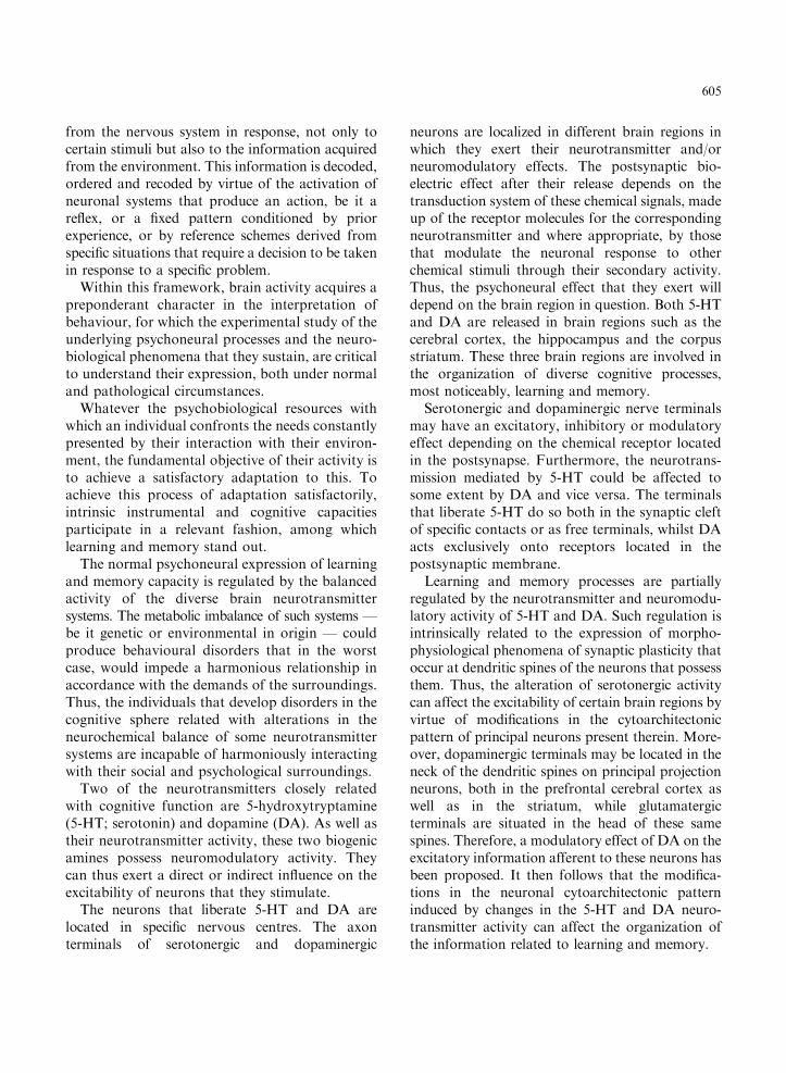
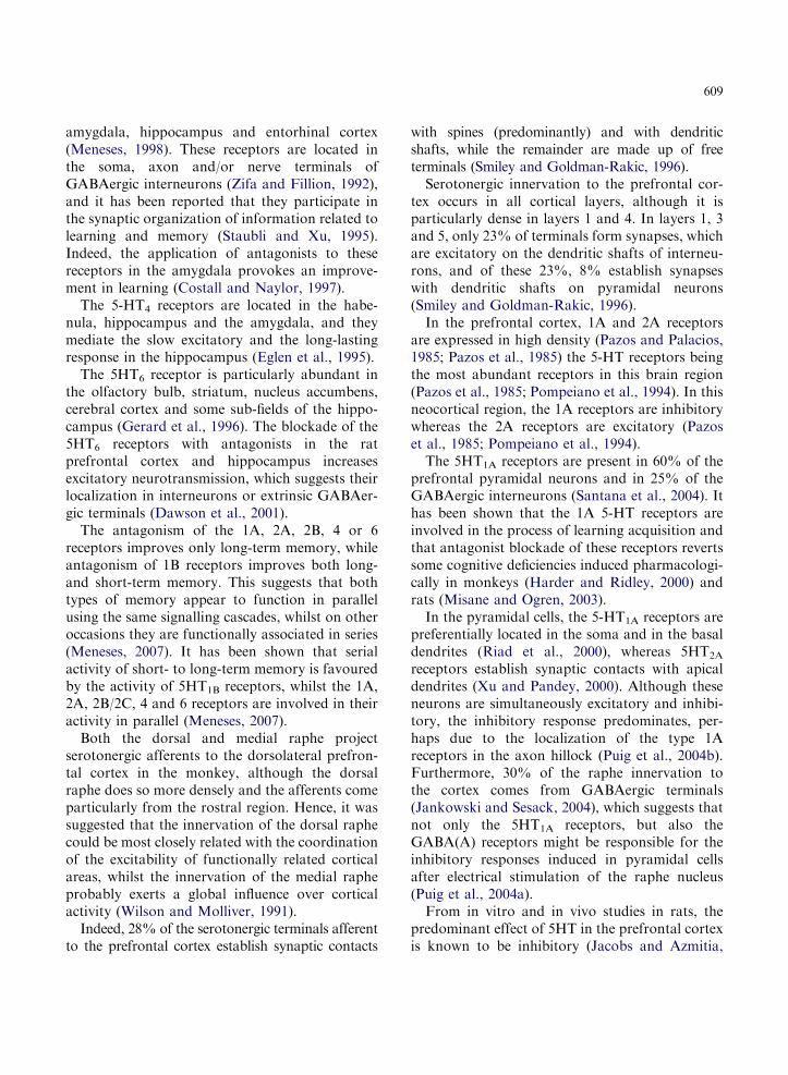
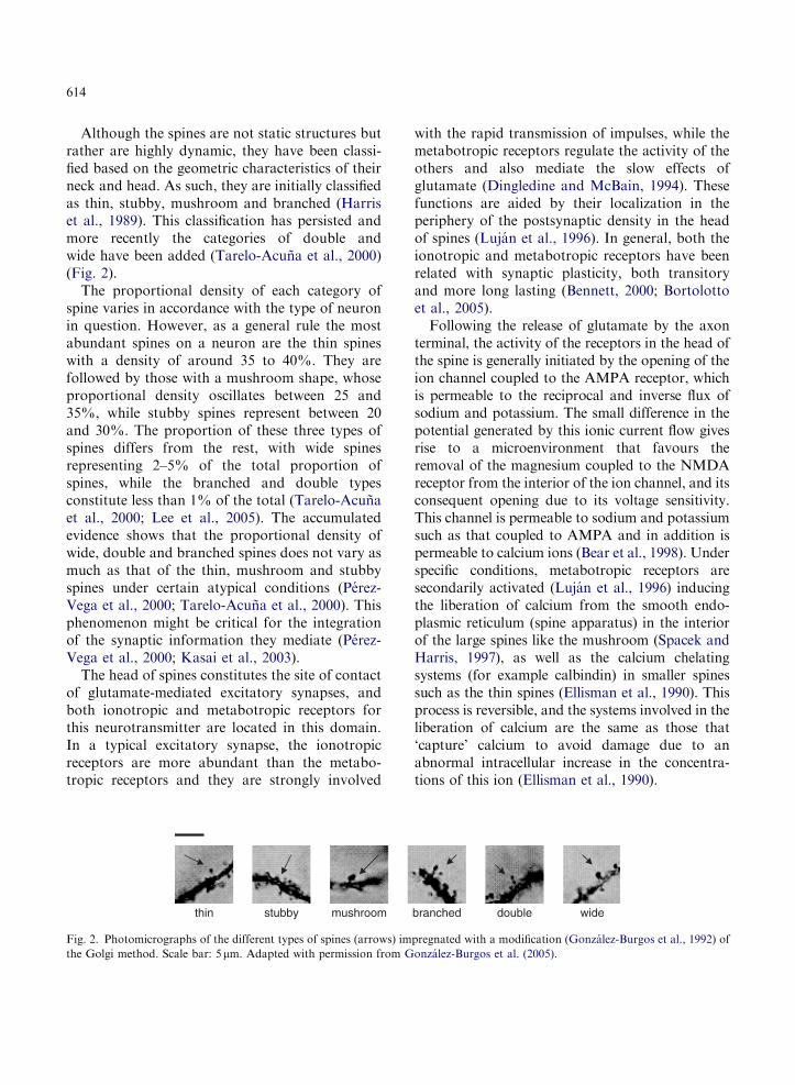
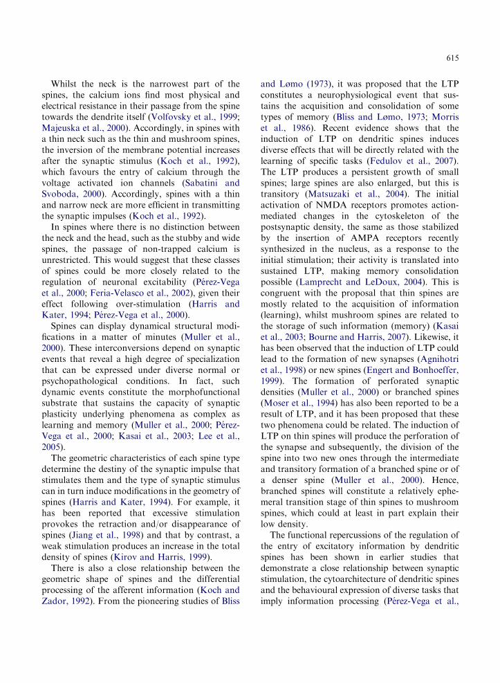
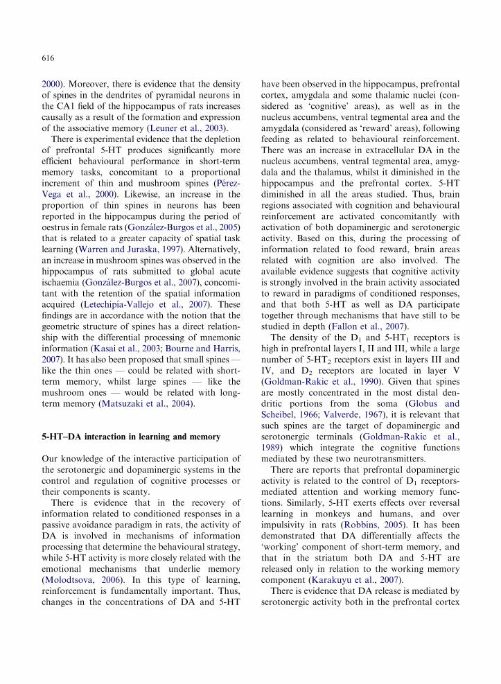
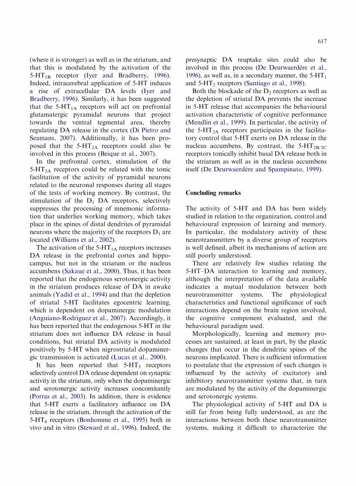
Although the spines are not static structures butrather are highly dynamic, they have been classi-fied based on the geometric characteristics of theirneck and head. As such, they are initially classifiedas thin, stubby, mushroom and branched (Harriset al., 1989). This classification has persisted andmore recently the categories of double andwide have been added (Tarelo-Acuna et al., 2000)(Fig. 2).
The proportional density of each category ofspine varies in accordance with the type of neuronin question. However, as a general rule the mostabundant spines on a neuron are the thin spineswith a density of around 35 to 40%. They arefollowed by those with a mushroom shape, whoseproportional density oscillates between 25 and35%, while stubby spines represent between 20and 30%. The proportion of these three types ofspines differs from the rest, with wide spinesrepresenting 2–5% of the total proportion ofspines, while the branched and double typesconstitute less than 1% of the total (Tarelo-Acunaet al., 2000; Lee et al., 2005). The accumulatedevidence shows that the proportional density ofwide, double and branched spines does not vary asmuch as that of the thin, mushroom and stubbyspines under certain atypical conditions (Perez-Vega et al., 2000; Tarelo-Acuna et al., 2000). Thisphenomenon might be critical for the integrationof the synaptic information they mediate (Perez-Vega et al., 2000; Kasai et al., 2003).
The head of spines constitutes the site of contactof glutamate-mediated excitatory synapses, andboth ionotropic and metabotropic receptors forthis neurotransmitter are located in this domain.In a typical excitatory synapse, the ionotropicreceptors are more abundant than the metabo-tropic receptors and they are strongly involved
thin stubby mushroom
Fig. 2. Photomicrographs of the different types of spines (arrows) im
the Golgi method. Scale bar: 5mm. Adapted with permission from G
with the rapid transmission of impulses, while themetabotropic receptors regulate the activity of theothers and also mediate the slow effects ofglutamate (Dingledine and McBain, 1994). Thesefunctions are aided by their localization in theperiphery of the postsynaptic density in the headof spines (Lujan et al., 1996). In general, both theionotropic and metabotropic receptors have beenrelated with synaptic plasticity, both transitoryand more long lasting (Bennett, 2000; Bortolottoet al., 2005).
Following the release of glutamate by the axonterminal, the activity of the receptors in the head ofthe spine is generally initiated by the opening of theion channel coupled to the AMPA receptor, whichis permeable to the reciprocal and inverse flux ofsodium and potassium. The small difference in thepotential generated by this ionic current flow givesrise to a microenvironment that favours theremoval of the magnesium coupled to the NMDAreceptor from the interior of the ion channel, and itsconsequent opening due to its voltage sensitivity.This channel is permeable to sodium and potassiumsuch as that coupled to AMPA and in addition ispermeable to calcium ions (Bear et al., 1998). Underspecific conditions, metabotropic receptors aresecondarily activated (Lujan et al., 1996) inducingthe liberation of calcium from the smooth endo-plasmic reticulum (spine apparatus) in the interiorof the large spines like the mushroom (Spacek andHarris, 1997), as well as the calcium chelatingsystems (for example calbindin) in smaller spinessuch as the thin spines (Ellisman et al., 1990). Thisprocess is reversible, and the systems involved in theliberation of calcium are the same as those that‘capture’ calcium to avoid damage due to anabnormal intracellular increase in the concentra-tions of this ion (Ellisman et al., 1990).
branched double wide
pregnated with a modification (Gonzalez-Burgos et al., 1992) of
onzalez-Burgos et al. (2005).
615
Whilst the neck is the narrowest part of thespines, the calcium ions find most physical andelectrical resistance in their passage from the spinetowards the dendrite itself (Volfovsky et al., 1999;Majeuska et al., 2000). Accordingly, in spines witha thin neck such as the thin and mushroom spines,the inversion of the membrane potential increasesafter the synaptic stimulus (Koch et al., 1992),which favours the entry of calcium through thevoltage activated ion channels (Sabatini andSvoboda, 2000). Accordingly, spines with a thinand narrow neck are more efficient in transmittingthe synaptic impulses (Koch et al., 1992).
In spines where there is no distinction betweenthe neck and the head, such as the stubby and widespines, the passage of non-trapped calcium isunrestricted. This would suggest that these classesof spines could be more closely related to theregulation of neuronal excitability (Perez-Vegaet al., 2000; Feria-Velasco et al., 2002), given theireffect following over-stimulation (Harris andKater, 1994; Perez-Vega et al., 2000).
Spines can display dynamical structural modi-fications in a matter of minutes (Muller et al.,2000). These interconversions depend on synapticevents that reveal a high degree of specializationthat can be expressed under diverse normal orpsychopathological conditions. In fact, suchdynamic events constitute the morphofunctionalsubstrate that sustains the capacity of synapticplasticity underlying phenomena as complex aslearning and memory (Muller et al., 2000; Perez-Vega et al., 2000; Kasai et al., 2003; Lee et al.,2005).
The geometric characteristics of each spine typedetermine the destiny of the synaptic impulse thatstimulates them and the type of synaptic stimuluscan in turn induce modifications in the geometry ofspines (Harris and Kater, 1994). For example, ithas been reported that excessive stimulationprovokes the retraction and/or disappearance ofspines (Jiang et al., 1998) and that by contrast, aweak stimulation produces an increase in the totaldensity of spines (Kirov and Harris, 1999).
There is also a close relationship between thegeometric shape of spines and the differentialprocessing of the afferent information (Koch andZador, 1992). From the pioneering studies of Bliss
and Lømo (1973), it was proposed that the LTPconstitutes a neurophysiological event that sus-tains the acquisition and consolidation of sometypes of memory (Bliss and Lømo, 1973; Morriset al., 1986). Recent evidence shows that theinduction of LTP on dendritic spines inducesdiverse effects that will be directly related with thelearning of specific tasks (Fedulov et al., 2007).The LTP produces a persistent growth of smallspines; large spines are also enlarged, but this istransitory (Matsuzaki et al., 2004). The initialactivation of NMDA receptors promotes action-mediated changes in the cytoskeleton of thepostsynaptic density, the same as those stabilizedby the insertion of AMPA receptors recentlysynthesized in the nucleus, as a response to theinitial stimulation; their activity is translated intosustained LTP, making memory consolidationpossible (Lamprecht and LeDoux, 2004). This iscongruent with the proposal that thin spines aremostly related to the acquisition of information(learning), whilst mushroom spines are related tothe storage of such information (memory) (Kasaiet al., 2003; Bourne and Harris, 2007). Likewise, ithas been observed that the induction of LTP couldlead to the formation of new synapses (Agnihotriet al., 1998) or new spines (Engert and Bonhoeffer,1999). The formation of perforated synapticdensities (Muller et al., 2000) or branched spines(Moser et al., 1994) has also been reported to be aresult of LTP, and it has been proposed that thesetwo phenomena could be related. The induction ofLTP on thin spines will produce the perforation ofthe synapse and subsequently, the division of thespine into two new ones through the intermediateand transitory formation of a branched spine or ofa denser spine (Muller et al., 2000). Hence,branched spines will constitute a relatively ephe-meral transition stage of thin spines to mushroomspines, which could at least in part explain theirlow density.
The functional repercussions of the regulation ofthe entry of excitatory information by dendriticspines has been shown in earlier studies thatdemonstrate a close relationship between synapticstimulation, the cytoarchitecture of dendritic spinesand the behavioural expression of diverse tasks thatimply information processing (Perez-Vega et al.,
616
2000). Moreover, there is evidence that the densityof spines in the dendrites of pyramidal neurons inthe CA1 field of the hippocampus of rats increasescausally as a result of the formation and expressionof the associative memory (Leuner et al., 2003).
There is experimental evidence that the depletionof prefrontal 5-HT produces significantly moreefficient behavioural performance in short-termmemory tasks, concomitant to a proportionalincrement of thin and mushroom spines (Perez-Vega et al., 2000). Likewise, an increase in theproportion of thin spines in neurons has beenreported in the hippocampus during the period ofoestrus in female rats (Gonzalez-Burgos et al., 2005)that is related to a greater capacity of spatial tasklearning (Warren and Juraska, 1997). Alternatively,an increase in mushroom spines was observed in thehippocampus of rats submitted to global acuteischaemia (Gonzalez-Burgos et al., 2007), concomi-tant with the retention of the spatial informationacquired (Letechipıa-Vallejo et al., 2007). Thesefindings are in accordance with the notion that thegeometric structure of spines has a direct relation-ship with the differential processing of mnemonicinformation (Kasai et al., 2003; Bourne and Harris,2007). It has also been proposed that small spines —like the thin ones — could be related with short-term memory, whilst large spines — like themushroom ones — would be related with long-term memory (Matsuzaki et al., 2004).
5-HT–DA interaction in learning and memory
Our knowledge of the interactive participation ofthe serotonergic and dopaminergic systems in thecontrol and regulation of cognitive processes ortheir components is scanty.
There is evidence that in the recovery ofinformation related to conditioned responses in apassive avoidance paradigm in rats, the activity ofDA is involved in mechanisms of informationprocessing that determine the behavioural strategy,while 5-HT activity is more closely related with theemotional mechanisms that underlie memory(Molodtsova, 2006). In this type of learning,reinforcement is fundamentally important. Thus,changes in the concentrations of DA and 5-HT
have been observed in the hippocampus, prefrontalcortex, amygdala and some thalamic nuclei (con-sidered as ‘cognitive’ areas), as well as in thenucleus accumbens, ventral tegmental area and theamygdala (considered as ‘reward’ areas), followingfeeding as related to behavioural reinforcement.There was an increase in extracellular DA in thenucleus accumbens, ventral tegmental area, amyg-dala and the thalamus, whilst it diminished in thehippocampus and the prefrontal cortex. 5-HTdiminished in all the areas studied. Thus, brainregions associated with cognition and behaviouralreinforcement are activated concomitantly withactivation of both dopaminergic and serotonergicactivity. Based on this, during the processing ofinformation related to food reward, brain areasrelated with cognition are also involved. Theavailable evidence suggests that cognitive activityis strongly involved in the brain activity associatedto reward in paradigms of conditioned responses,and that both 5-HT as well as DA participatetogether through mechanisms that have still to bestudied in depth (Fallon et al., 2007).
The density of the D1 and 5-HT1 receptors ishigh in prefrontal layers I, II and III, while a largenumber of 5-HT2 receptors exist in layers III andIV, and D2 receptors are located in layer V(Goldman-Rakic et al., 1990). Given that spinesare mostly concentrated in the most distal den-dritic portions from the soma (Globus andScheibel, 1966; Valverde, 1967), it is relevant thatsuch spines are the target of dopaminergic andserotonergic terminals (Goldman-Rakic et al.,1989) which integrate the cognitive functionsmediated by these two neurotransmitters.
There are reports that prefrontal dopaminergicactivity is related to the control of D1 receptors-mediated attention and working memory func-tions. Similarly, 5-HT exerts effects over reversallearning in monkeys and humans, and overimpulsivity in rats (Robbins, 2005). It has beendemonstrated that DA differentially affects the‘working’ component of short-term memory, andthat in the striatum both DA and 5-HT arereleased only in relation to the working memorycomponent (Karakuyu et al., 2007).
There is evidence that DA release is mediated byserotonergic activity both in the prefrontal cortex
617
(where it is stronger) as well as in the striatum, andthat this is modulated by the activation of the5-HT1B receptor (Iyer and Bradberry, 1996).Indeed, intracerebral application of 5-HT inducesa rise of extracellular DA levels (Iyer andBradberry, 1996). Similarly, it has been suggestedthat the 5-HT1A receptors will act on prefrontalglutamatergic pyramidal neurons that projecttowards the ventral tegmental area, therebyregulating DA release in the cortex (Di Pietro andSeamans, 2007). Additionally, it has been pro-posed that the 5-HT2A receptors could also beinvolved in this process (Beique et al., 2007).
In the prefrontal cortex, stimulation of the5-HT2A receptors could be related with the tonicfacilitation of the activity of pyramidal neuronsrelated to the neuronal responses during all stagesof the tests of working memory. By contrast, thestimulation of the D1 DA receptors, selectivelysuppresses the processing of mnemonic informa-tion that underlies working memory, which takesplace in the spines of distal dendrites of pyramidalneurons where the majority of the receptors D1 arelocated (Williams et al., 2002).
The activation of the 5-HT1A receptors increasesDA release in the prefrontal cortex and hippo-campus, but not in the striatum or the nucleusaccumbens (Sakaue et al., 2000). Thus, it has beenreported that the endogenous serotonergic activityin the striatum produces release of DA in awakeanimals (Yadid et al., 1994) and that the depletionof striatal 5-HT facilitates egocentric learning,which is dependent on dopaminergic modulation(Anguiano-Rodrıguez et al., 2007). Accordingly, ithas been reported that the endogenous 5-HT in thestriatum does not influence DA release in basalconditions, but striatal DA activity is modulatedpositively by 5-HT when nigrostriatal dopaminer-gic transmission is activated (Lucas et al., 2000).
It has been reported that 5-HT3 receptorsselectively control DA release dependent on synapticactivity in the striatum, only when the dopaminergicand serotonergic activity increases concomitantly(Porras et al., 2003). In addition, there is evidencethat 5-HT exerts a facilitatory influence on DArelease in the striatum, through the activation of the5-HT4 receptors (Bonhomme et al., 1995) both invivo and in vitro (Steward et al., 1996). Indeed, the
presynaptic DA reuptake sites could also beinvolved in this process (De Deurwaerdere et al.,1996), as well as, in a secondary manner, the 5-HT1
and 5-HT2 receptors (Santiago et al., 1998).Both the blockade of the D2 receptors as well as
the depletion of striatal DA prevents the increasein 5-HT release that accompanies the behaviouralactivation characteristic of cognitive performance(Mendlin et al., 1999). In particular, the activity ofthe 5-HT2A receptors participates in the facilita-tory control that 5-HT exerts on DA release in thenucleus accumbens. By contrast, the 5-HT2B/2C
receptors tonically inhibit basal DA release both inthe striatum as well as in the nucleus accumbensitself (De Deurwaerdere and Spampinato, 1999).
Concluding remarks
The activity of 5-HT and DA has been widelystudied in relation to the organization, control andbehavioural expression of learning and memory.In particular, the modulatory activity of theseneurotransmitters by a diverse group of receptorsis well defined, albeit its mechanisms of action arestill poorly understood.
There are relatively few studies relating the5-HT–DA interaction to learning and memory,although the interpretation of the data availableindicates a mutual modulation between bothneurotransmitter systems. The physiologicalcharacteristics and functional significance of suchinteractions depend on the brain region involved,the cognitive component evaluated, and thebehavioural paradigm used.
Morphologically, learning and memory pro-cesses are sustained, at least in part, by the plasticchanges that occur in the dendritic spines of theneurons implicated. There is sufficient informationto postulate that the expression of such changes isinfluenced by the activity of excitatory andinhibitory neurotransmitter systems that, in turnare modulated by the activity of the dopaminergicand serotonergic systems.
The physiological activity of 5-HT and DA isstill far from being fully understood, as are theinteractions between both these neurotransmittersystems, making it difficult to characterize the
618
relationships they establish with the organization,control and expression of the diverse componentsinvolved in learning and memory processes. How-ever, the conducting of molecular, physiological,morphological and behavioural correlative studieswould be very useful for a better understand of theneurobiological basis underlying the functionalorganization of these cognitive processes.
Abbreviations
5-HT 5-hydroxytryptamineAMPA alpha-amino-3-hydroxy-5-methyl-
4-isoxazolepropionic acid receptorCA1 hippocampal cornus ammonis 1
subfieldCA3 hippocampal cornus ammonis 3
subfieldDA dopamineGABA gamma-aminobutyric acid receptorLTP long-term potentiationNMDA N-methyl D-aspartate receptor
References
Agnihotri, N., Lopez-Garcıa, J.C., Hawkins, R.D. and
Arancio, O. (1998) Morphological changes associated with
long-term potentiation. Histol. Histopathol., 13: 1155–1162.
Anguiano-Rodrıguez, P.B., Gaytan-Tocaven, L. and Olvera-
Cortes, M.E. (2007) Striatal serotonin depletion facilitates rat
egocentric learning via dopamine modulation. Eur. J.
Pharmacol., 556: 91–98.
Baddeley, A. (2000) Short-term and working memory.
In: Tulving E. and Craik F.I.M. (Eds.), The Oxford Hand-
book of Memory. Oxford University Press, Oxford,
pp. 77–92.
Bahena-Trujillo, R. and Arias-Montano, J.A. (2000) Dopa-
mina: sıntesis, liberacion y receptores en el Sistema Nervioso
Central. Rev. Biomed., 11: 39–60.
Barnes, N.M. and Sharp, T. (1999) A review of central
serotonin receptors and their function. Neuropsychopharma-
cology, 38: 1083–1152.
Bas-ar, E. (2004) Memory and Brain Dynamics. Oscillations
Integrating Attention, Perception, Learning, and Memory.
CRC Press, USA, pp. 221–229.
Bear, M.F., Connors, B.W., Paradiso, M.A. (1998)
Neurociencia. Explorando el cerebro. MASSON-Williams &
Wilkins, Barcelona, pp. 68–90.
Beique, J.C., Imad, M., Mladenovic, L., Gingrich, J.A. and
Andrade, R. (2007) Mechanisms of the 5-hydroxytryptamine
2A receptor-mediated facilitation of synaptic activity in
prefrontal cortex. Proc. Natl. Acad. Sci. U.S.A., 104:
9870–9875.
Bell, C., Abrams, J. and Nutt, D. (2001) Tryptophan depletion
and its implications for psychiatry. Br. J. Psychiatry, 178:
399–405.
Bennett, M.R. (2000) The concept of long-term potentiation of
transmission at synapses. Prog. Neurobiol., 60: 109–137.
Berridge, K.C. and Whishaw, I.Q. (1992) Cortex, striatum and
cerebellum: control of serial order in a grooming sequence.
Exp. Brain Res., 90: 275–290.
Bliss, T.V. and Lømo, T. (1973) Long-lasting potentiation of
synaptic transmission in the dentate area of the anesthetized
rabbit following stimulation of the perforant path. J. Physiol.,
232: 331–356.
Bonhomme, N., De Deurwaerdere, P., Le Moal, M. and
Spampinato, U. (1995) Evidence for 5-HT4 receptor subtype
involvement in the enhancement of striatal dopamine release
induced by serotonin: a microdialysis study in the halothane-
anesthetized rat. Neuropharmacology, 34: 269–279.
Bortolotto, Z.A., Collett, V.J., Conquet, F., Jia, Z., van der
Putten, H. and Collingridge, G.L. (2005) The regulation of
hippocampal LTP by the molecular switch, a form of meta-
plasticity, requires mGlu5 receptors. Neuropharmacology, 49:
13–25.
Bourne, J. and Harris, K.M. (2007) Do thin spines learn to be
mushroom spines that remember? Curr. Opin. Neurobiol., 17:
1–6.
Brozoski, T., Brown, R.M., Rosvold, H.E. and Goldman, P.S.
(1979) Cognitive deficit caused by regional depletion of
dopamine in prefrontal cortex of rhesus monkey. Science,
205: 929–931.
Burnet, P.W.J., Eastwood, S.L., Lacey, K. and Harrison, P.J.
(1995) The distribution of 5-HT1A and 5-HT2A receptor
mRNA in human brain. Brain Res., 676: 157–168.
Camps, M., Kelly, P.H. and Palacios, J.M. (1990) Autoradio-
graphic localization of dopamine D1 and D2 receptors in the
brain of several mammalian species. J. Neural Transm. Gen.
Sect., 80: 105–127.
Carr, D.B. and Sesack, S.R. (1996) Hippocampal afferents to
the rat prefrontal cortex: synaptic targets and relation to
dopamine terminals. J. Comp. Neurol., 369: 1–15.
Cassel, J.C. and Jeltsch, H. (1995) Serotonergic modulation of
cholinergic function in the central nervous system: cognitive
implications. Neuroscience, 69: 1–41.
Clarke, H.F., Dalley, J.W., Crofts, H.S., Robbins, T.W. and
Roberts, A.C. (2004) Cognitive inflexibility after prefrontal
serotonin depletion. Science, 304: 878–880.
Costall, B. and Naylor, R.J. (1997) Neuropharmacology of
5-HT3 receptors ligands. In: Baumgarten H.G. and Gother
M. (Eds.), Serotoninergic Neurons and 5-HT Receptors in the
CNS. Springer, Berlin, pp. 409–438.
Dawson, L.A., Nguyen, H.Q. and Li, P. (2001) The 5-HT6
receptor antagonist SB-271046 selectively enhances excitatory
neurotransmission in the rat frontal cortex and hippocampus.
Neuropsychopharmacology, 25: 662–668.
De Deurwaerdere, P., Bonhoemme, N., Lucas, G., Le Moal, M.
and Spampinato, U. (1996) Serotonin enhances striatal
619
dopamine outflow in vivo through dopamine uptake sites.
J. Neurochem., 66: 210–215.
De Deurwaerdere, P. and Spampinato, U. (1999) Role of
serotonin(2A) and serotonin(2B/2C) receptor subtypes in the
control of accumbal and striatal dopamine release elicited in
vivo by dorsal raphe nucleus electrical stimulation.
J. Neurochem., 73: 1033–1042.
Del Angel-Meza, A.R., Adame-Gonzalez, I.G., Segura, J.,
Montes, R., Gonzalez-Burgos, I. and Beas-Zarate, C. (2003)
Cerebral cholinergic neurotransmission in protein and tryp-
tophan-restricted adult rats. In: Allegri G., Costa C.V.L.,
Raggazi E., Steinhart H. and Varesio L. (Eds.), Advances in
Experimental Medicine and Biology, vol. 527. Kluwer
Academic Plenum Publishers, New York, pp. 415–421.
Del Angel-Meza, A.R., Gonzalez-Burgos, I., Olvera-Cortes, E.
and Feria-Velasco, A. (1996) Chronic tryptophan restriction
disrupts grooming chain completion in the rat. Physiol.
Behav., 59: 1099–1102.
Descarries, L., Watkins, K.C., Garcia, S., Bosler, O. and
Doucet, G. (1996) Dual character, asynaptic and synaptic, of
the dopamine innervation in adult rat neostriatum: a
quantitative autoradiographic and immunocytochemical ana-
lysis. J. Comp. Neurol., 375: 167–186.
Di Pietro, N.C. and Seamans, J.K. (2007) Dopamine and
serotonin interactions in the prefrontal cortex: insights on
antipsychotic drugs and their mechanisms of action. Pharma-
copsychiatry, 40: s27–s33.
Dingledine, R. and McBain, Ch.J. (1994) Excitatory amino acid
transmitters. In: Siegel G.J., Agranoff B.W., Albers R.W. and
Molinoff P.B. (Eds.), Basic Neurochemistry. Molecular,
Cellular, and Medical Aspects (Fifth edition). Raven Press,
New York, pp. 367–387.
Edwards, F.A. (1995) Anatomy and electrophysiology of fast
synapses lead to a structural model for long-term potentia-
tion. Physiol. Rev., 75: 759–787.
Egashira, N., Yano, A., Ishigami, N., Mishima, K., Iwasaki,
K., Fujioka, M., Matsushita, M., Nishimura, R. and
Fujiwara, M. (2006) Investigation of mechanisms mediating
8-OH-DPAT-induced impairment of spatial memory: invol-
vement of 5-HT1A receptors in the dorsal hippocampus in
rats. Brain Res., 1069: 54–62.
Eglen, R.M., Wong, E.H., Dumis, A. and Bockaert, J. (1995)
Central 5-HT4 receptors. TIPS, 16: 391–398.
Ellisman, M.H., Deering, T.J., Ouyang, Y., Beck, C.F., Tanksley,
S.J., Walton, P.D., Airey, J.A. and Sutko, J.L. (1990)
Identification and localization of ryanodine binding proteins in
the avian central nervous system. Neuron, 5: 135–146.
Engert, F. and Bonhoeffer, T. (1999) Dendritic spine changes
associated with hippocampal long-term synaptic plasticity.
Nature, 399: 66–70.
Fallon, S., Shearman, E., Sershen, H. and Lajtha, A. (2007)
Food reward-induced neurotransmitter changes in cognitive
brain regions. Neurochem. Res., 32: 1772–1782.
Fedulov, V., Rex, C.S., Simmons, D.A., Palmer, L., Gall, C.M.
and Lynch, G. (2007) Evidence that long-term potentiation
occurs within individual hippocampal synapses during learn-
ing. J. Neurosci., 27: 8031–8039.
Feria-Velasco, A., Del Angel-Meza, A.R. and Gonzalez-
Burgos, I. (2002) Modification of dendritic development. In:
Azmitia, E.C., DeFelipe, J., Jones, E.G., Rakic, P., Ribak,
C.E. (Eds.), Changing Views of Cajal’s neuron. Progress in
Brain Research Series, Elsevier, USA, Vol. 136, pp. 135–143.
Fernandez-Espejo, E. (2000) +Como funciona el nucleus
accumbens? Rev. Neurol., 30: 845–849.
Fuster, J.M. (1997) Network memory. TINS, 20: 451–459.
Garcıa, F.B., Pedraza, C. and Navarro, J.F. (2005) Implicacion
de la dopamina en los procesos cognitivos del aprendizaje y la
memoria. Psiq. Biol., 12: 232–236.
Gerard, C., Mestikawi, S., Lebrand, C., Adrien, J., Ruat, M.,
Traiffort, E., Hamon, M. and Martres, M.P. (1996) Quanti-
tative RT-PCR distribution of serotonin 5-HT6 receptor
mRNA in the central nervous system of control or 5,7-
dihydroxytryptamine-treated rats. Synapse, 23: 164–173.
Gerson, S.C. and Baldessarini, R.J. (1980) Motor effects of
serotonin in the central nervous system. Life Sci., 27:
1435–1451.
Globus, A. and Scheibel, A.B. (1966) Loss of dendritic spines as
an index of presynaptic terminal patterns. Nature, 212:
463–465.
Goldman-Rakic, P.S., Leranth, C., Williams, M.S., Mons, N.
and Geffard, M. (1989) Dopamine innervation of pyramidal
neurons in primate frontal cortex. Proc. Natl. Acad. Sci.
US.A., 86: 9015–9019.
Goldman-Rakic, P.S., Lidow, M.S. and Gallager, D.W. (1990)
Overlap of dopaminergic, adrenergic, and serotoninergic
receptors and complementarity of their subtypes in primate
prefrontal cortex. J. Neurosci., 10: 2125–2138.
Goldman-Rakic, P.S., Lidow, M.S., Smiley, J.F. and Williams,
M.S. (1992) The anatomy of dopamine in monkey and human
prefrontal cortex. J. Neural Trans. Suppl., 36: 163–177.
Gonzalez-Burgos, I., Alejandre-Gomez, M. and Cervantes, M.
(2005) Spine-type densities of hippocampal CA1 neurons vary
in proestrus and estrus rats. Neurosci. Lett., 379: 52–54.
Gonzalez-Burgos, I., Letechipıa-Vallejo, G., Lopez-Loeza, E.,
Moralı, G. and Cervantes, M. (2007) Long-term study of
dendritic spines from hippocampal CA1 pyramidal cells, after
neuroprotective melatonin treatment following global cere-
bral ischemia in rats. Neurosci. Lett., 423: 162–166.
Gonzalez-Burgos, I., Olvera-Cortes, E., Del Angel-Meza, A.R.
and Feria-Velasco, A. (1995) Serotonin involvement in the
spontaneous alternation ability: a behavioral study in
tryptophan-restricted rats. Neurosci. Lett., 190: 143–145.
Gonzalez-Burgos, I., Perez-Vega, M.I., del Angel-Meza, A.R.
and Feria-Velasco, A. (1998) Effect of tryptophan restriction
on short-term memory. Physiol. Behav., 63: 165–169.
Gonzalez-Burgos, I., Tapia-Arizmendi, G. and Feria-Velasco,
A. (1992) Golgi method without osmium tetroxide for the
study of the central nervous system. Biotech. Histochem., 67:
288–296.
Goto, Y. and Grace, A.A. (2008). Dopamine modulation of
hippocampal prefrontal cortical interaction drives memory-
guided behaviour. Cereb. Cortex., 18: 1407–1414.
Granado, N., Ortiz, O., Suarez, L.M., Martın, E.D., Cena, V.,
Solıs, J.M. and Moratalla, R. (2008) D1 but not D5 dopamine
620
receptors are critical for LTP, spatial learning, and LTP-
induced arc and zif268 expression in the hippocampus. Cereb.
Cortex., 18: 1–12.
Harder, J.A. and Ridley, R.M. (2000) The 5-HT1A antagonist
WAY 100 635 alleviates cognitive impairments induced by
dizocilpine (MK-801) in monkeys. Neuropharmacology, 39:
547–552.
Harris, K.M., Jensen, F.E. and Tsao, B.H. (1989) Ultrastruc-
ture, development, and plasticity of dendritic spine synapses
in area CA1 of the rat hippocampus: extending our vision
with serial electron microscopy and three-dimensional ana-
lyses. In: Chan-Palay V. and Kohler Ch. (Eds.), The
Hippocampus — New Vistas. Alan R. Liss, USA, pp. 33–52.
Harris, K.M. and Kater, S.B. (1994) Dendritic spines: cellular
specializations imparting both stability and flexibility to
synaptic function. Annu. Rev. Neurosci., 17: 341–371.
Harris, K.M. and Stevens, J.K. (1989) Dendritic spines of CA1
pyramidal cells in the rat hippocampus: serial electron
microscopy with reference to their biophysical characteristics.
J. Neurosci., 9: 2982–2997.
Hritcu, L., Clicinschi, M. and Nabeshima, T. (2007) Brain
serotonin depletion impairs short-term memory, but not long-
term memory in rats. Physiol. Behav., 91: 652–657.
Iyer, R.N. and Bradberry, C.W. (1996) Serotonin-mediated
increase in prefrontal cortex dopamine release: pharmacolo-
gical characterization. J. Pharmacol. Exp. Ther., 277: 40–47.
Jaber, M., Robinson, S., Misale, C. and Caron, M.G. (1996)
Dopamine receptors and brain function. Neuropsychophar-
macology, 35: 1503–1519.
Jackson, D.M. and Westlind-Danielsson, A. (1994) Dopamine
receptors: molecular biology, biochemistry and behavioural
aspects. Pharmacol. Ther., 64: 291–369.
Jacobs, B.L. and Azmitia, E.C. (1992) Structure and function of
the brain serotonin system. Physiol. Rev., 72: 165–229.
Jankowski, M.P. and Sesack, S.R. (2004) Prefrontal cortical
projections to the rat dorsal raphe nucleus: ultrastructural
features and associations with serotonin and g-aminobutiric
acid neurons. J. Comp. Neurol., 468: 518–529.
Jiang, M., Lee, C.L., Smith, K.L. and Swann, J.W. (1998) Spine
loss and other persistent alterations of hippocampal pyrami-
dal cell dendrites in a model of early-onset-epilepsy.
J. Neurosci., 18: 8356–8368.
Karakuyu, D., Herold, C., Gunturkun, O. and Diekamp, B.
(2007) Differential increase of extracellular dopamine and
serotonin in the ‘prefrontal cortex’ and striatum of pigeons
during working memory. Eur. J. Neurosci., 26: 2293–2302.
Kasai, H., Matsuzaki, M., Noguchi, J., Yasumatsu, N. and
Nakahara, H. (2003) Structure-stability-function relation-
ships of dendritic spines. TINS, 26: 360–368.
Kirov, S.A. and Harris, K.M. (1999) Dendrites are more spiny
on mature hippocampal neurons when synapses are inacti-
vated. Nat. Neurosci., 2: 878–883.
Koch, Ch. and Zador, A. (1992) Dendritic spines: convergence
of theory and experiment. Science, 256: 973–974.
Koch, Ch., Zador, A. and Brown, T.H. (1992) Dendritic
spines: convergence of theory and experiment. Science, 256:
973–974.
Kroner, S., Krimer, L.S., Lewis, D.A. and Barrionuevo, G.
(2007) Dopamine increases inhibition in the monkey dorso-
lateral prefrontal cortex through cell type-specific modulation
of interneurons. Cereb. Cortex., 17: 1020–1032.
Lakoski, J.M. and Aghajanian, G.K. (1985) Effects of
ketanserin on neuronal responses to serotonin in the
prefrontal cortex, lateral geniculate and dorsal raphe nucleus.
Neuropharmacology, 24: 265–273.
Lamprecht, R. and LeDoux, J. (2004) Structural plasticity and
memory. Nat. Rev. Neurosci., 5: 45–54.
Lee, B., Groman, S., London, E.D. and Jentsch, J.D. (2007)
Dopamine D(2)/D(3) receptors play a specific role in the
reversal of a learned visual discrimination in monkeys.
Neuropsychopharmacology, 32: 2125–2134.
Lee, K.J., Kim, H. and Rhyu, I.J. (2005) The roles of dendritic
spine shapes in Purkinje cells. The Cerebellum, 4: 97–104.
Lemon, N. and Manahan-Vaughan, D. (2006) Dopamine
D1/D5 receptors gate the acquisition of novel information
through hippocampal long-term potentiation and long-term
depression. J. Neurosci., 26: 7723–7729.
Letechipıa-Vallejo, G., Lopez-Loeza, E., Espinoza-Gonzalez,
V., Gonzalez-Burgos, I., Olvera-Cortes, M.E., Moralı, G. and
Cervantes, M. (2007) Long-term morphological and func-
tional evaluation of the neuroprotective effects of post-
ischemic treatment with melatonin in rats. J. Pineal Res.,
42: 139–146.
Leuner, B., Falduto, J. and Shors, T.J. (2003) Associative
memory formation increases the observation of dendritic
spines in the hippocampus. J. Neurosci., 23: 659–665.
Lidow, M.S., Goldman-Rakic, P.S., Gallager, D.W. and Rakic,
P. (1991) Distribution of dopaminergic receptors in the
primate cerebral cortex: quantitative autoradiographic analy-
sis using [3H]raclopride, [3H]spiperone and [3H]SCH23390.
Neuroscience, 40: 657–671.
Lisman, J. and Grace, A. (2005) The hippocampal-VTA loop:
controlling the entry of information into long-term memory.
Neuron, 46: 703–713.
Lucas, G., De Deurwaerdere, P., Porras, G. and Spampinato,
U. (2000) Endogenous serotonin enhances the release of
dopamine in the striatum only when nigro-striatal dopami-
nergic transmission is activated. Neuropharmacology, 39:
1984–1995.
Luciana, M. and Collins, P.F. (1997) Dopaminergic modulation
of working memory for spatial but not object cues in normal
humans. J. Cognit. Neurosci., 9: 330–347.
Lujan, R., Nusser, Z., Roberts, D.B., Shigemoto, R. and
Somogyi, P. (1996) Perisynaptic location of metabotropic
glutamate receptors mGluR1 and mGluR5 on dendrites and
dendritic spines in the rat hippocampus. Eur. J. Neurosci., 8:
1488–1500.
Majeuska, A., Brown, E., Ross, J. and Yuste, R. (2000)
Mechanisms of calcium decay kinetics in hippocampal spines:
role of spine calcium pumps and calcium diffusion through
the spine neck in biochemical compartmentalization.
J. Neurosci., 20: 1722–1734.
Masaki, D., Yokoyama, C., Kinoshita, S., Tsuchida, H.,
Nakatomi, K., Yoshimoto, K. and Fukui, K. (2006)
621
Relationship between limbic and cortical 5-HT neurotrans-
mission and acquisition and reversal learning in a go/no-go
task in rats. Psychopharmacology, 189: 249–258.
Matsuzaki, M., Honkura, N., Ellis-Davies, G.C.R. and Kasai,
H. (2004) Structural basis of long-term potentiation in single
dendritic spines. Nature, 429: 761–766.
Meeter, M., Talamini, L., Schmitt, J.A.J. and Riedel, W.J.
(2006) Effects of 5-HT on memory and the hippo-
campus: model and data. Neuropsychopharmacology, 31:
712–720.
Meltzer, C.C., Smith, G., Dekosky, S.T., Pollock, B.G., Mathis,
C.A., Moore, R.Y., Kupfer, D.J. and Reynolds, C.F. (1998)
Serotonin in aging, late-life depression and Alzheimer’s
disease: the emerging role of functional imaging. Neuropsy-
chopharmacology, 18: 407–430.
Mendlin, A., Martın, F.J. and Jacobs, B.L. (1999) Dopaminer-
gic input is required for increases in serotonin output
produced by behavioural activation: an in vivo microdialysis
study in rat forebrain. Neuroscience, 93: 897–905.
Meneses, A. (1998) Physiological, pathophysiological and
therapeutic roles of 5-HT systems in learning and memory.
Rev. Neurosci., 9: 275–289.
Meneses, A. (1999) 5-HT system and cognition. Neurosci.
Biobehav. Rev., 23: 1111–1125.
Meneses, A. (2007) Do serotonin (1–7) receptors modulate
short and long-term memory? Neurobiol. Learn. Mem., 87:
561–572.
Meneses, A. and Perez-Garcia, G. (2007) 5-HT1A receptors and
memory. Neurosci. Biobehav. Rev., 31: 705–727.
Millan, M.J., Gobert, A., Roux, S., Porsolt, R., Meneses, A.,
Carli, M., Di Cara, B., Jaffard, R., Rivet, J.M., Lestage, P.,
Mocaer, E., Peglion, J.L. and Dekeyne, A. (2004) The
serotonin1A receptor partial agonist S15535 [4-(Benzo-
dioxan-5-yl)1-(indan-2-yl)piperazine] enhances cholinergic
transmission and cognitive function in rodents: a combined
neurochemical and behavioural analysis. J. Pharmacol. Exp.
Ther., 311: 190–203.
Miner, L.A., Backstrom, J.R., Sanders-Bush, E. and Sesack,
S.R. (2003) Ultrastructural localization of serotonin(2A)
receptors in the middle layers of the rat prelimbic prefrontal
cortex. Neuroscience, 116: 107–117.
Misale, C., Russel, N.S., Robinson, S.W., Jaber, M. and Caron,
M.G. (1998) Dopamine receptors: from structure to function.
Physiol. Rev., 78: 189–225.
Misane, I. and Ogren, S.O. (2003) Selective 5-HT1A antago-
nists WAY 100635 and NAD-299 attenuate the impairment of
passive avoidance caused by scopolamine in the rat.
Neuropsychopharmacology, 28: 253–264.
Mitchell, E.S., Sexton, T. and Neumaier, J.F. (2007) Increased
expression of 5-HT6 receptors in the rat dorsomedial striatum
impairs instrumental learning. Neuropsychopharmacology,
32: 1520–1530.
Molodtsova, G.F. (2006) Different roles of dopamine and
serotonin in conditioned passive avoidance response of rats.
Zh. Vyssh. Nerv. Deiat. Im. I. P. Pavlova., 56: 242–246.
Morales, M., Battenberg, E., Delecea, L. and Bloom, F.E.
(1996) The type 3 serotonin receptor is expressed in a
subpopulation of GABAergic neurons in the rat neocortex
and hippocampus. Brain Res., 731: 199–202.
Morris, R.G., Anderson, E., Lynch, G.S. and Baurdy, M.
(1986) Selective impairment of learning and blockade of long-
term potentiation by an N-methyl D- aspartate receptor
antagonist, AP5. Nature, 319: 774–776.
Moser, M.B., Trommald, M. and Andersen, P. (1994) An
increase in dendritic spine density on hippocampal CA1
pyramidal cells following spatial learning in adult rats
suggests the formation of new synapses. Proc. Natl. Acad.
Sci. U.S.A., 91: 12673–12675.
Muller, D., Toni, N. and Buchs, P.A. (2000) Spine changes
associated with long-term potentiation. Hippocampus, 10:
596–604.
Newberry, N.R., Footitt, D.R., Papanastassiou, V. and
Reynolds, D.J.M. (1999) Actions of 5-HT on human
neocortical neurons in vitro. Brain Res., 833: 93–100.
O’Carroll, C.M., Martin, S.J., Sandin, J., Frenguelli, B. and
Morris, R.G. (2006) Dopaminergic modulation of the
persistence of one-trial hippocampus-dependent memory.
Learn. Mem., 13: 760–769.
Oleskevich, S. and Descarries, L. (1990) Quantified distribution
of the serotonin innervation in adult rat hippocampus.
Neuroscience, 34: 19–33.
Olvera-Cortes, E., Perez-Vega, M.I., Barajas-Lopez, G., del
Angel-Meza, A.R., Gonzalez-Burgos, I. and Feria-Velasco,
A. (1998) Place learning impairment in chronically trypto-
phan-restricted rats. Nutr. Neurosci., 1: 223–235.
O’Neill, M. and Brown, V.J. (2007) The effect of striatal
dopamine depletion and the adenosine A2A antagonist KW-
6002 on reversal learning in rats. Neurobiol. Learn. Mem., 88:
75–81.
Pazos, A., Cortes, M. and Palacios, J.M. (1985) Quantitative
autoradiographic mapping of serotonin receptors in the rat
brain. I. Serotonin-2 receptors. Brain Res., 346: 231–249.
Pazos, A. and Palacios, J.M. (1985) Quantitative autoradio-
graphic mapping of serotonin receptors in the rat brain. I.
Serotonin-1 receptors. Brain Res., 346: 205–230.
Perez-Vega, M.I., Feria-Velasco, A. and Gonzalez-Burgos, I.
(2000) Prefrontocortical serotonin depletion results in plastic
changes of prefrontocortical pyramidal neurons, underlying a
greater efficiency of short-term memory. Brain Res. Bull., 53:
291–300.
Peters, J.A., Malone, H.M. and Lambert, J.J. (1992) Recent
advances in the electrophysiological characterization of
5-HT3 receptors. Trends Pharmacol. Sci., 13: 391–397.
Phillips, A.G. (2003) Mesocorticolimbic dopamine: a neuro-
chemical link between motivation and memory. Int. Cong.
Ser., 1250: 509–526.
Pompeiano, M., Palacios, J.M. and Mengod, G. (1994)
Distribution of the serotonin 5-HT2 receptor family mRNA:
comparison between 5-HT2A and 5-HT2C receptors. Mol.
Brain Res., 23: 163–178.
Porras, G., De Deurwaerdere, P., Moison, D. and Spampinato,
U. (2003) Conditional involvement of striatal serotonin3
receptors in the control of in vivo dopamine outflow in the rat
striatum. Eur. J. Neurosci., 17: 771–781.
622
Prado-Alcala, R.A., Ruiloba, M.I., Rubio, L., Solana-Figueroa,
R., Medina, C., Salado-Castillo, R. and Quitarte, G.L.
(2003a) Regional infusions of serotonin into the striatum and
memory consolidation. Synapse, 47: 169–175.
Prado-Alcala, R.A., Solana-Figueroa, R., Galindo, L.E.,
Medina, A.C. and Quitarte, G.L. (2003b) Blockade of striatal
5-HT2 receptors produces retrograde amnesia in rats. Life
Sci., 74: 481–488.
Puig, M.V., Artigas, F. and Celada, P. (2004a) Modulation of
the activity of pyramidal neurons in rat prefrontal cortex by
raphe stimulation in vivo: involvement of serotonin and
GABA. Cereb. Cortex., 15: 1–14.
Puig, M.V., Celada, P. and Artigas, F. (2004b) Control
serotoninergico de la corteza prefrontal. Rev. Neurol., 39:
539–547.
Riad, M., Garcıa, S., Watkins, K.C., Jodoin, N., Doucet, E.,
Langlois, X., el Mestikawi, S., Hamon, M. and Desacrries, L.
(2000) Somatodendritic localization of 5-HT1A and pretermi-
nal axonal localization of 5-HT1B serotonin receptors in adult
rat brain. J. Comp. Neurol., 417: 181–194.
Richfield, E.K., Young, A.B. and Penney, J.B. (1989) Com-
parative distributions of dopamine D-1 and D-2 receptors in
the cerebral cortex of rats, cats, and monkeys. J. Comp.
Neurol., 286: 409–426.
Riedel, W.J., Sobczak, S. and Schmitt, J.A. (2003) Tryptophan
modulation and cognition. Adv. Exp. Med. Biol., 527:
207–213.
Robbins, T.W. (2005) Chemistry of the mind: neurochemical
modulation of prefrontal cortical function. J. Comp. Neurol.,
493: 140–146.
Roberts, J.C., Reavill, C., East, S.Z., Harrison, P.J., Patel, S.,
Routledge, C. and Leslie, R.A. (2002) The distribution of
5-HT(6) receptors in rat brain: an autoradiographic binding
study using the radiolabelled 5-HT(6) receptor antagonist
[(125)I]SB-258585. Brain Res., 934: 49–57.
Sabatini, B.L. and Svoboda, K. (2000) Analysis of calcium
channels in single spines using optical fluctuations analysis.
Nature, 408: 589–593.
Sakaue, M., Somboonthum, P., Nishihara, B., Koyama, Y.,
Hashimoto, H., Baba, A. and Matsuda, T. (2000) Postsynap-
tic 5-hydroxytriptamine1A receptor activation increases in
vivo dopamine release in rat prefrontal cortex. Br. J.
Pharmacol., 129: 1028–1034.
Santana, N., Bortolozzi, A., Serratz, J., Mngod, G. and Artigas,
F. (2004) Expression of serotonin1A and serotonin2A
receptors in pyramidal and GABAergic neurons of the rat
prefrontal cortex. Cereb. Cortex., 14: 1100–1109.
Santiago, M., Matarredona, E.R., Machado, A. and Cano, J.
(1998) Influence of serotoninergic drugs on in vivo dopamine
extracellular output in rat striatum. J. Neurosci. Res., 52:
591–598.
Sawaguchi, T. and Goldman-Rakic, P.S. (1991) D1 dopamine
receptors in prefrontal cortex: involvement in working
memory. Science, 251: 947–950.
Schiapparelli, L., Simon, A.M., Del Rıo, J. and Frechilla, D.
(2006) Opposing effects of AMPA and 5-HT1A receptor
blockade on passive avoidance and object recognition
performance: correlation with AMPA receptor subunit
expression in rat hippocampus. Neuropharmacology, 50:
897–907.
Schmitt, J., Jorissen, B. and Sobczak, S. (2000) Tryptophan
impairs memory consolidation but improves focussed atten-
tion in healthy young volunteers. J. Psychopharmacol., 14:
21–29.
Seamans, J.F., Floresco, S.B. and Phillips, A.G. (1995) Selective
impairment on a delayed radial arm task following local
administration of a selective D1, but not a D2, antagonist into
the prefrontal cortex. Soc. Neurosci. Abstr., 21: p. 1942.
Seamans, J.K., Floresco, S.B. and Phillips, A.G. (1998) D1
receptor modulation of hippocampal-prefrontal cortical
circuits integrating spatial memory with executive functions
in the rat. J. Neurosci., 18(4): 1613–1621.
Seibell, P.J., Demarest, J. and Rhoads, D.E. (2003) 5-HT1A
receptor activity disrupts spontaneous alternation behaviour
in adult rats. Pharmacol. Biochem. Behav., 74: 559–564.
Shirahata, T., Tsunoda, M., Santa, T., Kirino, Y. and
Wtanabe, S. (2006) Depletion of serotonin selectively impairs
short-term memory without affecting long-term memory in
odor learning in the terrestrial slug Limax valentianus. Learn.
Mem., 13: 267–270.
Smiley, J.F. and Goldman-Rakic, P.S. (1993) Heterogenous
targets of dopamine synapses in monkey prefrontal cortex
demonstrated by serial section electron microscopy: a laminar
analysis using the silver-enhanced diaminobenzidine sulphide
(SEDS) immunolabeling technique. Cereb. Cortex., 3:
223–238.
Smiley, J.F. and Goldman-Rakic, P.S. (1996) Serotonergic
axons in monkey prefrontal cerebral cortex synapse
predominantly on interneurons as demonstrated by serial
section electron microscopy. J. Comp. Neurol., 367(3):
431–443.
Soghomonian, J.J., Descarries, L. and Watkins, K.C. (1989)
Serotonin innervation in adult rat neostriatum. II. Ultra-
structural features: a radioautographic and immunocyto-
chemical study. Brain Res., 481: 67–86.
Spacek, J. and Harris, K.M. (1997) Three-dimensional organi-
zation of smooth endoplasmic reticulum in hippocampal CA1
dendrites and dendritic spines of the immature and mature
rat. J. Neurosci., 17: 190–203.
Sprague, J.E., Preston, A.S., Leifheit, M. and Woodside, B.
(2003) Hippocampal serotonergic damage induced by
MDMA (ecstasy): effects on spatial learning. Physiol. Behav.,
79: 281–287.
Squire, L.R. (1992) Memory and the hippocampus: a synthesis
from findings with rats, monkeys and humans. Psychol. Rev.,
99: 195–231.
Stamford, J.A., Davidson, C., McLaughlin, D.P. and
Hopwood, S.E. (2000) Control of dorsal raphe 5-HT function
by multiple 5-HT(1) autoreceptors: parallel purposes or
pointless plurality? Trends Neurosci., 23: 459–465.
Staubli, U. and Xu, F.B. (1995) Effects of 5-HT3 receptor
antagonism on hippocampal theta rhythm, memory, and LTP
induction in the freely moving rat. J. Neurosci., 15:
2445–2452.
623
Stefani, M.R. and Moghaddam, B. (2006) Rule learning and
reward contingency are associated with dissociable patterns of
dopamine activation in the rat prefrontal cortex, nucleus
accumbens, and dorsal striatum. J. Neurosci., 26: 8810–8818.
Steward, L.J., Ge, J., Stowe, R.L., Brown, D.C., Bruton, R.K.,
Stokes, P.R. and Barnes, N.M. (1996) Ability of 5-HT4
receptor ligands to modulate rat striatal dopamine release in
vitro and in vivo. Br. J. Pharmacol., 117: 55–62.
Surmeier, D.J. (2007) Dopamine and working memory mecha-
nisms in prefrontal cortex. J. Physiol., 581: p. 885.
Sweatt, J.D. (2003) Mechanisms of MemoryAcademic Press,
USA, pp. 3–28
Tarelo-Acuna, L., Olvera-Cortes, E. and Gonzalez-Burgos, I.
(2000) Prenatal and postnatal exposure to etanol induces
changes in the shape of the dendritic spines from hippocampal
CA1 pyramidal neurons of the rat. Neurosci. Lett., 286:
13–16.
Valverde, F. (1967) Apical dendritic spines of the visual cortex
and light deprivation in the mouse. Exp. Brain Res., 3:
337–352.
Van der Veen, F.M., Evers, E.A., van Deursen, J.A., Deutz,
N.E., Baches, W.H. and Schmitt, J.A. (2006) Acute trypto-
phan depletion reduces activation in the right hippocampus
during encoding in an episodic memory task. Neuroimage, 31:
1188–1196.
Volfovsky, N., Parnas, H., Segal, M. and Korkotian, E. (1999)
Geometry of dendritic spines affects calcium dynamics in
hippocampal neurons: theory and experiments. J. Neurophy-
siol., 81: 450–462.
Warren, S.G. and Juraska, J.M. (1997) Spatial and nonspatial
learning across the rat estrous cycle. Behav. Neurosci., 111:
259–266.
Wilkerson, A. and Levin, E.D. (1999) Ventral hippocampal
dopamine D1 and D2 systems and spatial working memory in
rats. Neuroscience, 89: 743–749.
Williams, G.V. and Goldman-Rakic, P.S. (1995) Blockade of
dopamine D1 receptors enhances memory fields of prefrontal
neurons in primate cerebral cortex. Nature, 376: 572–575.
Williams, G.V., Rao, S.G. and Goldman-Rakic, P.S. (2002)
The physiological role of 5-HT2A receptors in working
memory. J. Neurosci., 22: 2843–2854.
Willins, D.L., Deutch, A.Y. and Roth, B.L. (1997) Serotonin
5-HT2A receptors are expressed on pyramidal cells and
interneurons in the rat cortex. Synapse, 27: 79–82.
Wilson, M.A. and Molliver, M.E. (1991) The organization of
serotonergic projections to cerebral cortex in primates:
retrograde transport studies. Neuroscience, 44: 555–570.
Xu, T. and Pandey, S.C. (2000) Cellular localization of
serotonin(2A) (5HT(2A)) receptors in the rat brain. Brain
Res. Bull., 51: 499–505.
Yadid, G., Pacak, K., Kopin, I.J. and Goldstein, D.S. (1994)
Endogenous serotonin stimulates striatal dopamine
release in conscious rats. J. Pharmacol. Exp. Ther., 270:
1158–1165.
Yang, C.R. and Seamans, J.K. (1996) Dopamine D1 receptor
actions in layer V–VI rat prefrontal cortex neurons in vitro:
modulation of dendritic-somatic signal integration. J. Neu-
rosci., 16: 1922–1935.
Yasuno, F. (2004) Hippocampal serotonin 1A receptor and
memory function. Seishin Shinkeigaku Zasshi, 106:
1314–1322.
Zahrt, J., Taylor, J.R., Mathew, R.G. and Arnsten, A.F.T.
(1997) Supranormal stimulation of D1 dopamine receptors in
the rodent prefrontal cortex impairs spatial working memory
performance. J. Neurosci., 17: 8528–8535.
Zhou, F.M. and Hablitz, J.J. (1999) Activation of serotonin
receptors modulates synaptic transmission in rat cerebral
cortex. J. Neurophysiol., 82: 2989–2999.
Zifa, E. and Fillion, G. (1992) 5-Hydroxytryptamine receptors.
Pharmacol. Rev., 44: 401–458.

本文献由“学霸图书馆-文献云下载”收集自网络,仅供学习交流使用。
学霸图书馆(www.xuebalib.com)是一个“整合众多图书馆数据库资源,
提供一站式文献检索和下载服务”的24 小时在线不限IP
图书馆。
图书馆致力于便利、促进学习与科研,提供最强文献下载服务。
图书馆导航:
图书馆首页 文献云下载 图书馆入口 外文数据库大全 疑难文献辅助工具
Related Documents