SARS-CoV-2 launches a unique transcriptional signature from in vitro, ex vivo, and in vivo systems Daniel Blanco-Melo 1,2,* , Benjamin E. Nilsson-Payant 1,2,* , Wen-Chun Liu 1,3,* , Rasmus Møller 1,2 , Maryline Panis 1,2 , David Sachs 4 , Randy A. Albrecht 1,3,# , Benjamin R. tenOever 1,2,3,# 1 Department of Microbiology, Icahn School of Medicine at Mount Sinai, New York, USA 2 Virus Engineering Center for Therapeutics and Research (VECToR), Icahn School of Medicine at Mount Sinai, New York, USA 3 Global Health and Emerging Pathogens Institute, Icahn School of Medicine at Mount Sinai, New York, USA 4 Department of Genetics and Genomic Sciences, Icahn School of Medicine at Mount Sinai, New York, USA * Authors contributed equally # Corresponding author ABSTRACT One of the greatest threats to humanity is the emergence of a pandemic virus. Among those with the greatest potential for such an event include influenza viruses and coronaviruses. In the last century alone, we have observed four major influenza A virus pandemics as well as the emergence of three highly pathogenic coronaviruses including SARS-CoV-2, the causative agent of the ongoing COVID-19 pandemic. As no effective antiviral treatments or vaccines are presently available against SARS-CoV-2, it is important to understand the host response to this virus as this may guide the efforts in development towards novel therapeutics. Here, we offer the first in-depth characterization of the host transcriptional response to SARS-CoV-2 and other respiratory infections through in vitro, ex vivo, and in vivo model systems. Our data demonstrate the each virus elicits both core antiviral components as well as unique transcriptional footprints. Compared to the response to influenza A virus and respiratory syncytial virus, SARS-CoV-2 elicits a muted response that lacks robust induction of a subset of cytokines including the Type I and Type III interferons as well as a numerous chemokines. Taken together, these data suggest that the unique transcriptional signature of this virus may be responsible for the development of COVID-19. INTRODUCTION Coronaviruses are a diverse group of single-stranded positive-sense RNA viruses infecting a wide range of vertebrate hosts 1 . These viruses are thought to generally . CC-BY-ND 4.0 International license (which was not certified by peer review) is the author/funder. It is made available under a The copyright holder for this preprint this version posted March 24, 2020. . https://doi.org/10.1101/2020.03.24.004655 doi: bioRxiv preprint

Welcome message from author
This document is posted to help you gain knowledge. Please leave a comment to let me know what you think about it! Share it to your friends and learn new things together.
Transcript
SARS-CoV-2 launches a unique transcriptional signature from in vitro, ex vivo, and in vivo systems
Daniel Blanco-Melo1,2,*, Benjamin E. Nilsson-Payant1,2,*, Wen-Chun Liu1,3,*, Rasmus Møller1,2,
Maryline Panis1,2, David Sachs4, Randy A. Albrecht1,3,#, Benjamin R. tenOever1,2,3,#
1 Department of Microbiology, Icahn School of Medicine at Mount Sinai, New York, USA 2 Virus Engineering Center for Therapeutics and Research (VECToR), Icahn School of Medicine at Mount Sinai, New York, USA 3 Global Health and Emerging Pathogens Institute, Icahn School of Medicine at Mount Sinai, New York, USA 4 Department of Genetics and Genomic Sciences, Icahn School of Medicine at Mount Sinai, New York, USA * Authors contributed equally # Corresponding author
ABSTRACT
One of the greatest threats to humanity is the emergence of a pandemic virus. Among those
with the greatest potential for such an event include influenza viruses and coronaviruses. In
the last century alone, we have observed four major influenza A virus pandemics as well as the
emergence of three highly pathogenic coronaviruses including SARS-CoV-2, the causative
agent of the ongoing COVID-19 pandemic. As no effective antiviral treatments or vaccines
are presently available against SARS-CoV-2, it is important to understand the host
response to this virus as this may guide the efforts in development towards novel
therapeutics. Here, we offer the first in-depth characterization of the host transcriptional
response to SARS-CoV-2 and other respiratory infections through in vitro, ex vivo, and
in vivo model systems. Our data demonstrate the each virus elicits both core antiviral
components as well as unique transcriptional footprints. Compared to the response to
influenza A virus and respiratory syncytial virus, SARS-CoV-2 elicits a muted response
that lacks robust induction of a subset of cytokines including the Type I and Type III
interferons as well as a numerous chemokines. Taken together, these data suggest that
the unique transcriptional signature of this virus may be responsible for the development
of COVID-19.
INTRODUCTION
Coronaviruses are a diverse group of single-stranded positive-sense RNA viruses
infecting a wide range of vertebrate hosts1. These viruses are thought to generally
.CC-BY-ND 4.0 International license(which was not certified by peer review) is the author/funder. It is made available under aThe copyright holder for this preprintthis version posted March 24, 2020. . https://doi.org/10.1101/2020.03.24.004655doi: bioRxiv preprint
cause mild upper respiratory tract illnesses in humans such as the common cold2.
However, in the past two decades, three highly pathogenic human coronaviruses have
emerged from zoonotic viruses: severe acute respiratory syndrome-related coronavirus
(SARS-CoV-1) infecting ~8,000 people worldwide with a case-fatality rate of ~10% in
2002-2003, Middle East respiratory syndrome-related coronavirus (MERS-CoV)
infecting ~2,500 people with a case-fatality rate of ~36%, and now severe acute
respiratory syndrome-related coronavirus 2 (SARS-CoV-2) which causes Coronavirus
Disease-2019 (COVID-19) whose global mortality rate remains to be determined3, 4.
Infection with these highly pathogenic coronaviruses can result in acute respiratory
distress syndrome (ARDS) and acute lung injury (ALI), often leading to reduction of lung
function and even death3.
The current pandemic of COVID-19 represents an acute and rapidly developing global
health crisis. In an effort to better understand the molecular basis of the disease and
identify putative markers for COVID-19, we compared the transcriptional response of
SARS-CoV-2 to that of seasonal influenza A virus (IAV) and human orthopneumovirus
(commonly known as human respiratory syncytial virus (RSV)), two common recurring
respiratory viruses. Comparing the transcriptional response in both primary human
lung epithelium and transformed lung alveolar cells revealed that SARS-CoV-2 elicits a
unique transcriptional response as compared to IAV and RSV. In a homogenous cell
population, the transcriptional response to SARS-CoV-2 infection shows a significant
lack of Type I and III interferon (IFN-I and IFN-III) expression as compared to IAV and
RSV. Moreover, while a core number of cytokines that comprise the antiviral host
defense are shared amongst all three viruses, an equal number are notably absent in
response to SARS-CoV-2. Lastly, the only genes that appear to be unique to SARS-
CoV-2 infection are secreted peptides implicated in diseases of the airways. This
unique response to SARS-CoV-2 could also be recapitulated in vivo comparing
influenza infection to COVID-19 in ferrets. Taken together, these data reveal that the
interaction between virus and host as it relates to SARS-CoV-2 may be responsible for
its unusual high morbidity and mortality.
.CC-BY-ND 4.0 International license(which was not certified by peer review) is the author/funder. It is made available under aThe copyright holder for this preprintthis version posted March 24, 2020. . https://doi.org/10.1101/2020.03.24.004655doi: bioRxiv preprint
RESULTS
In order to identify similarities and differences between the host response to SARS-
CoV-2 and seasonal respiratory infections, we sought to elucidate the transcriptome of
lung epithelial cells during viral infection. To achieve this, we infected human alveolar
adenocarcinoma cells with SARS-CoV-2, RSV and IAV and performed mRNA-seq
analysis (Supplementary Table 1). Despite undetectable levels of ACE2 and
TMPRSS2, the putative receptor and protease for SARS-CoV-2 entry5, these cells were
able to support viral replication, evidenced by full genome coverage in total RNA
samples extracted from infected cells (Figure 1a and Extended Data Fig.1a). Our
analysis identified 120 differentially expressed genes (DEGs, qval < 0.05) of which
more than 80% were up-regulated (Figure 1b and Supplementary Table 2). Analysis of
DEGs revealed two main clusters of induced genes (Figure 1c). A highly interacting
cluster enriched in genes involved in the cellular response to virus infection
(GO:0009615, FDR << 0.0001), mainly composed of type-I ISGs (GO:0034340, FDR <<
0.0001), and a second cluster enriched in genes involved in the humoral immune
response (GO:0006959, FDR << 0.0001), which further subdivided into two smaller
clusters enriched in chemokines and cytokines (GO:0005125, FDR < 0.001), and
complement proteins (GO:0006956, FDR < 0.0005) (Figure 1c and Supplementary
Table 3). It is noteworthy to highlight that, despite a lack of IFN-I and IFN-III expression,
we observe the induction of well-characterized direct effectors of the innate immune
response including: MX1, IFITM3, SAMHD1 and TRIM25, as well as the induction of
viral RNA sensors such as RIG-I and the OAS1-3 genes (Figure 1c). This response
however is lacking the robust induction of antiviral genes commonly observed following
IFN-I/-III signaling6. This muted response however does show a positive correlation in
the host antiviral response overall as determined by gene set enrichment analysis
(GSEA) (Extended Data Fig. 1b). Taken together our analyses indicate that replication
of SARS-CoV-2 in alveolar cells results in a limited antiviral response.
In an effort to determine if the apparent modest response to SARS-CoV-2 infection was
the result of low receptor expression, a low multiplicity of infection (MOI), or due to the
immortalized nature of the cell line, we next infected primary human bronchial epithelial
.CC-BY-ND 4.0 International license(which was not certified by peer review) is the author/funder. It is made available under aThe copyright holder for this preprintthis version posted March 24, 2020. . https://doi.org/10.1101/2020.03.24.004655doi: bioRxiv preprint
(NHBE) cells (Fig. 2a). However, despite a ten fold increase in MOI, infection of NHBE
cells showed a similar transcriptional response to that of the immortalized alveolar cells
(ρ≈0.22, p-value << 0.0001). This correlation extended to both the type-I IFN response
(GO:0060337, FDR << 0.0001) as well as the humoral immunity (GO:0006959, FDR <<
0.0001) (Fig. 2b). Taken together, these results suggest that the cellular response to
SARS-CoV-2 is relatively uniform and void of IFN-I and IFN-III expression.
In an effort to compare the response to SARS-CoV-2 with other respiratory viruses, we
next infected the lung alveolar carcinoma cell line with either RSV or seasonal IAV.
Bulk RNA sequencing of independent biological replicates revealed that the
transcriptional response to SARS-CoV-2 infection is similar in magnitude to that of IAV
(82 DEGs, qval < 0.05), with both viruses showing a moderate response compared to
that of RSV (910 DEGs, qval < 0.05) (Supplementary Table 4 and 5). Nevertheless, the
response to SARS-CoV-2 showed a strong correlation (p-value < 0.001) to that of RSV
infection (Extended Data Fig. 2), resulting in the large overlap of DEGs between these
two viruses, compared to IAV (Fig. 3a). These responses do not reflect the replication
dynamics of these viruses, illustrated by the amount of viral reads recovered from these
infections (Fig. 3b). In an effort to better illustrate the unique responses to SARS-CoV-
2, IAV, and RSV, we next performed a principle component analysis (PCA) to enable
unbiased grouping of each sample based in DEGs. These efforts not only highlighted
the unique transcriptional signatures induced by the three respiratory infections, they
also corroborated that the response in different cell systems with SARS-CoV-2 were
comparable (Fig. 3c). To further delineate the unique antiviral signatures of each virus,
we next examined the expression levels of specific genes implicated in the host antiviral
response (Fig. 3d). These analyses again showed that despite sharing a similar
transcriptional footprint to RSV, SARS-CoV-2 lacks the induction of IFN-I and IFN-III
genes (Fig. 3d). However, this same analyses shows a common subset of well
established interferon stimulated genes (ISGs) shared with RSV that are lacking in IAV,
presumably due to the expression of the antiviral antagonist NS17. While most of the
transcriptional response to SARS-CoV-2 was either shared or lacking in comparison to
the response to IAV and RSV, two cytokines (EDN1 and TNFSF15) were uniquely
.CC-BY-ND 4.0 International license(which was not certified by peer review) is the author/funder. It is made available under aThe copyright holder for this preprintthis version posted March 24, 2020. . https://doi.org/10.1101/2020.03.24.004655doi: bioRxiv preprint
upregulated (Fig. 3e). The unique induction of these two secreted factors was
particularly noteworthy as they have been implicated as mediators of respiratory
inflammation6.
Lastly, to ascertain whether the mutated antiviral response to SARS-CoV-2 could be
observed in vivo, we infected ferrets with either influenza A virus or SARS-CoV-2 (Fig.
4). At five days post infection, nasal washes were performed and the heterogeneous
cells obtained from this procedure were again subjected to bulk RNA sequencing and
aligned to either SARS-CoV-2 or A/California/04/2009 (Fig. 4a). These data
demonstrate complete coverage of the challenge virus suggesting that in both
examples, the ferrets were shedding replication competent virus. Next, these same
upper respiratory cell populations were compared to mock treated ferrets to ascertain
the transcriptional response in vivo for comparison purposes. In agreement with our in
vitro data, differential expression analyses of these respiratory infections demonstrated
that the transcriptional footprint of IAV was significantly greater in magnitude that that
observed for SARS-CoV-2 (Fig. 4b-c). While the ferret genome annotation limits the
direct comparisons between some orthologues, we do observe a diminished cytokine
response when comparing SARS-CoV-2 to IAV. These data include a subset of genes
involved in the recruitment and activation of the adaptive immune response including:
IL6, CXCL11, IFNG, IL7, CXCL9, and CXCL10 (Fig. 4b-c). Taken together with both
our in vitro and ex vivo data, which also implicated a diminished cytokine response to
infection, these results suggest that the overall response to SARS-CoV-2 is relatively
moderate when compared to other respiratory viruses.
DISCUSSION
In the present study we focus on defining the transcriptional response to SARS-CoV-2,
influenza A virus, and respiratory syncytial virus. In general, these data find that the
overall transcriptional footprint to infection is greatest for RSV and the lowest for IAV
with SARS-CoV-2 representing an intermediate response. A probable explanation for
the lack of a response to IAV in general is the potent antagonistic activity of the virus.
However, it is noteworthy that in vivo, the response to IAV exceeds that of SARS-CoV-2
.CC-BY-ND 4.0 International license(which was not certified by peer review) is the author/funder. It is made available under aThe copyright holder for this preprintthis version posted March 24, 2020. . https://doi.org/10.1101/2020.03.24.004655doi: bioRxiv preprint
where the activity of NS1 is unable to inhibit some of the pattern recognition receptors at
play during a physiological response.
Despite the apparent muted induction of antiviral genes in response to SARS-CoV-2,
we do observe a significant up regulation of well-characterized ISGs including: IFIT1-3,
ISG15, DDX58, and others. Amongst the genes that are uniquely present when
comparing SARS-CoV-2 to other respiratory viruses are EDN1 and TNFSF15 - two
putative biomarkers that may contribute to COVID-19 pathology. However, it should be
noted that we did not observe these genes in ferrets despite the fact that they could be
detected in both cell culture models. Regardless of whether these two unique genes
may serve as biomarkers, the overall signature of SARS-CoV-2, RSV, and IAV can
serve as a broader map to developing novel diagnostic strategies.
The general induction of ISGs in response to SARS-CoV-2, albeit modest, is particularly
interesting as we were not able to detect any reads mapping to IFN-I of IFN-III members
in contrast to what is observed for RSV or IAV. These data likely indicate that induction
of IFN-I and IFN-III is very low but sufficient to induce at least a subset of ISGs in
response to SARS-CoV-2. It is also noteworthy that we observe comparable replication
between primary human bronchial epithelium and a transformed cell line despite the fact
that we are unable to detect both the putative receptor (ACE2) or the required protease
(TMPRSS2) in the latter cell line. While further work remains to ascertain how viral
entry is mediated in this model system, we did note high expression of BSG which has
also been suggested to compensate for ACE2 (Wang et al, BioRxv unpublished and
Extended Data Fig. 1a). Moreover, it should be noted that the viral replication profiles
as deduced by both total RNA and polyA RNA sequencing were comparable between in
vitro, ex vivo, and in vivo samples.
What makes the SARS-CoV-2 distinct from the RSV and IAV strains used in this study
is the propensity to selectively induce morbidity and mortality in older populations8. The
physiological basis for this morbidity is believed to be the selective death of Type II
pneumocytes that results in both loss of air exchange and fluid leakage into the lungs9,
.CC-BY-ND 4.0 International license(which was not certified by peer review) is the author/funder. It is made available under aThe copyright holder for this preprintthis version posted March 24, 2020. . https://doi.org/10.1101/2020.03.24.004655doi: bioRxiv preprint
10. While it remains to be determined whether this moderate cell response to SARS-
CoV-2 is responsible for the abnormally high lethality in the older populations, it does
explain why the virus is generally asymptomatic in young people with healthy and robust
immune systems11. Given the results here, it is tempting to speculate that perhaps in
the aging population, the immune response itself is muted and thus prevents successful
inhibition of viral spread. Perhaps the slow amplification of the virus is then the
underlying cause for the lung damage and an explanation for why the course of
infection is so prolonged. In this regard, it is also of interest to point out that while
replication is apparent in 6 month old ferrets (comparable in age to human teenagers),
they do not show overt signs of disease and clear infection after approximately eight
days. Taken together, the data presented here suggests that perhaps artificial means
of boosting the antiviral response may be an effective option at treating COVID-19 so
long as it does not further aggravate any pre-existing conditions.
MATERIALS AND METHODS Cell Culture Human adenocarcinomic alveolar basal epithelial (A549) cells (ATCC,
CCL-185), Madin-Darby Canine Kidney (MDCK) cells (ATCC, CCL-34), African green
monkey kidney epithelical Vero E6 cells (ATCC, CRL-1586) and African green monkey
kidney epithelical BS-C-1 cells (ATCC, CCL-26) were maintained at 37°C and 5% CO2
in Dulbecco’s Modified Eagle Medium (DMEM, Gibco) supplemented with 10% Fetal
Bovine Serum (FBS, Corning). Normal human bronchial epithelial (NHBE) cells (Lonza,
CC-2540 Lot# 580580) were isolated from a 79-year-old Caucasian female and were
maintained in bronchial epithelial growth media (Lonza, CC-3171) supplemented with
BEGM SingleQuots as per the manufacturer’s instructions (Lonza, CC-4175) at 37°C
and 5% CO2.
Viruses Influenza A/Puerto Rico/8/1934 (H1N1) virus (NCBI:txid183764) and influenza
A/California/04/2009 (pH1N1) virus was grown in MDCK cells at an MOI of 0.001 in
DMEM supplemented with 0.3% bovine serum albumin (BSA, MP Biomedicals), 4.5 g/L
D-glucose, 4 mM L-glutamine and 1 µg/ml TPCK-trypsin (Sigma-Aldrich). Infectious
titers of influenza A viruses were determined by plaque assay in MDCK cells.
.CC-BY-ND 4.0 International license(which was not certified by peer review) is the author/funder. It is made available under aThe copyright holder for this preprintthis version posted March 24, 2020. . https://doi.org/10.1101/2020.03.24.004655doi: bioRxiv preprint
Recombinant GFP-expressing human respiratory syncytial virus, strain A2 (rgRSV[224])
was generously provided by Dr. M. Peeples (OSU) and was described previously12.
rgRSV[224] was grown in BSC-1 cells in in DMEM supplemented with 2% FBS, 4.5 g/L
D-glucose and 4 mM L-glutamine.
SARS-related coronavirus 2 (SARS-CoV-2), Isolate USA-WA1/2020 (NR-52281) was
deposited by the Center for Disease Control and Prevention and obtained through BEI
Resources, NIAID, NIH. SARS-CoV-2 was propagated in Vero E6 cells in DMEM
supplemented with 2% FBS, 4.5 g/L D-glucose, 4 mM L-glutamine, 10 mM Non-
Essential Amino Acids, 1 mM Sodium Pyruvate and 10 mM HEPES. Infectious titers of
SARS-CoV-2 were determined by plaque assay in Vero E6 cells in Minimum Essential
Media supplemented with 4 mM L-glutamine, 0.2% BSA, 10 mM HEPES and 0.12%
NaHCO3 and 0.7% agar.
All work involving live SARS-CoV-2 was performed in the CDC/USDA-approved BSL-3
facility of the Global Health and Emerging Pathogens Institute at the Icahn School of
Medicine at Mount Sinai in accordance with institutional biosafety requirements.
RNA-Seq of viral infections Approximately 1 × 10⁶ A549 cells were infected with
influenza A/Puerto Rico/8/1934 (H1N1) virus (IAV), human respiratory syncytial virus
(RSV) or SARS-CoV-2. Infections with IAV were performed at a multiplicity of infection
of 5 for 9 h in DMEM supplemented with 0.3% BSA, 4.5 g/L D-glucose, 4 mM L-
glutamine and 1 µg/ml TPCK-trypsin. Infections with RSV were performed at an MOI of
15 for 24 h in DMEM supplemented with 0.3% BSA, 4.5 g/L D-glucose and 4 mM L-
glutamine. Infections with SARS-CoV-2 were performed at an MOI of 0.2 for 24 h in
DMEM supplemented with 2% FBS, 4.5 g/L D-glucose, 4 mM L-glutamine, 10 mM Non-
Essential Amino Acids, 1 mM Sodium Pyruvate and 10 mM HEPES. Approximately 1 ×
105 NHBE cells were infected with SARS-CoV-2 at an MOI of 2 for 24 h in bronchial
epithelial growth media supplemented with BEGM SingleQuots.
.CC-BY-ND 4.0 International license(which was not certified by peer review) is the author/funder. It is made available under aThe copyright holder for this preprintthis version posted March 24, 2020. . https://doi.org/10.1101/2020.03.24.004655doi: bioRxiv preprint
Total RNA from infected and mock infected cells was extracted using TRIzol Reagent
(Invitrogen) and Direct-zol RNA Miniprep kit (Zymo Research) according to the
manufacturer’s instructions and treated with DNase I.
RNA-seq libraries of polyadenylated RNA were prepared using the TruSeq RNA Library
Prep Kit v2 (Illumina) according to the manufacturer’s instructions. RNA-seq libraries for
total ribosomal RNA-depleted RNA were prepared using the TruSeq Stranded Total
RNA Library Prep Gold (Illumina) according to the manufacturer’s instructions. cDNA
libraries were sequenced using an Illumina NextSeq 500 platform.
Bioinformatic analyses: Raw reads were aligned to the human genome (hg19) using
the RNA-Seq Aligment App on Basespace (Illumina, CA), following differential
expression analysis using DESeq213. Differentially expressed genes (DEGs) were
characterized for each sample (p adjusted-value < 0.05) and were used as query to
search for enriched biological processes (Gene ontology BP) and network analysis of
protein interactions using STRING14. Heatmaps of gene expression levels were
constructing using heatmap.2 from the gplot package in R (https://cran.r-
project.org/web/packages/gplots/index.html). Sparse principal component analysis
(sPCA) was performed on Log2(Fold Change) values using SPC from the PMA package
in R15. Volcano plots were constructed using custom scripts in R. Gene set enrichment
analysis (GSEA) was performed on gene expression profiles from mock and SARS-
CoV-2 treated cells comparing to a composite list of genes involved in Type-I IFN
response (GO:0035457, GO:0035458, GO:0035455, GO:0035456, GO:0034340, and
HALLMARK_INTERFERON_ALPHA_RESPONSE)16. Correlation analysis on DEGs
between samples was performed using chart.Correlation from the PerformanceAnalytics
package in R (https://cran.r-
project.org/web/packages/PerformanceAnalytics/index.html). Alignments to viral
genomes was performed using bowtie2. The genomes used for this study were: SARS-
CoV-2 (NC_045512.2), RSV (NC_001803.1), IAV PR8 (AF389115.1, AF389116.1,
AF389117.1, AF389118.1, AF389119.1, AF389120.1, AF389121.1, AF389122.1) and
.CC-BY-ND 4.0 International license(which was not certified by peer review) is the author/funder. It is made available under aThe copyright holder for this preprintthis version posted March 24, 2020. . https://doi.org/10.1101/2020.03.24.004655doi: bioRxiv preprint
IAV A/California/VRDL6/2010(H1N1) (CY064994, CY064993, CY064992, CY064987,
CY064990, CY064989, CY064988, CY064991).
Ferret immunization and infection All procedures are described in our previous study17. Briefly, outbred 4-month old
castrated male Fitch ferrets were confirmed to be seronegative for circulating H1N1,
H3N2, and B influenza viruses prior to purchase from Triple F. Farm (North Rose, NY).
All animal experiments were performed according to protocols approved by the
Institutional Animal Care and Use Committee (IACUC) and Institutional Biosafety
Committee of the ISMMS. Ferrets were randomly assigned to the different treatment
groups. For each experiment, the experimental group size is indicated in the figure
legends. For pH1N1 virus infection, all naive ferrets were intranasally challenge infected
with 1mL of 105 PFU of pH1N1 (A/California/04/2009) influenza virus. Nasal wash
samples were collected from anesthetized ferrets on day 7 post infection and preserved
at -80oC for viral titration. Cell lysates were treated with Trizol for bulk RNA sequencing.
For the SARS-CoV-2 virus infection study, all naïve ferrets were infected with 1mL of
5x10⁴ PFU of the SARS-CoV-2 isolate USA-WA1/2020. Nasal wash samples were
collected from anesthetized ferrets on day 5 post-infection, and preserved at -80 oC for
viral titration. Cell lysates were treated with Trizol for bulk RNA sequencing. On day 5
post-challenge infection, anesthetized ferrets were euthanized by exsanguination
followed by intracardiac injection of euthanasia solution (Sodium Pentobarbital).
Trachea samples were collected from each ferret (n=3) to quantify viral titers by plaque
assays and for bulk RNA sequencing. Acknowledgments DBM is an Open Philanthropy Fellow for the Life Sciences Research Foundation. BT
and this work are supported by the Defense Advanced Research Projects Agency
(DARPA). For raw RNA seq. data, please contact [email protected]
.CC-BY-ND 4.0 International license(which was not certified by peer review) is the author/funder. It is made available under aThe copyright holder for this preprintthis version posted March 24, 2020. . https://doi.org/10.1101/2020.03.24.004655doi: bioRxiv preprint
REFERENCES 1. Cui, J., Li, F. & Shi, Z.L. Origin and evolution of pathogenic coronaviruses. Nat Rev
Microbiol 17, 181-192 (2019). 2. Weiss, S.R. & Navas-Martin, S. Coronavirus pathogenesis and the emerging pathogen
severe acute respiratory syndrome coronavirus. Microbiol Mol Biol Rev 69, 635-664 (2005).
3. de Wit, E., van Doremalen, N., Falzarano, D. & Munster, V.J. SARS and MERS: recent insights into emerging coronaviruses. Nat Rev Microbiol 14, 523-534 (2016).
4. Wu, F. et al. A new coronavirus associated with human respiratory disease in China. Nature 579, 265-269 (2020).
5. Hoffmann, M. et al. SARS-CoV-2 Cell Entry Depends on ACE2 and TMPRSS2 and Is Blocked by a Clinically Proven Protease Inhibitor. Cell (2020).
6. Schoggins, J.W. et al. A diverse range of gene products are effectors of the type I interferon antiviral response. Nature 472, 481-485 (2011).
7. Garcia-Sastre, A. et al. Influenza A virus lacking the NS1 gene replicates in interferon-deficient systems. Virology 252, 324-330 (1998).
8. Novel Coronavirus Pneumonia Emergency Response Epidemiology, T. [The epidemiological characteristics of an outbreak of 2019 novel coronavirus diseases (COVID-19) in China]. Zhonghua Liu Xing Bing Xue Za Zhi 41, 145-151 (2020).
9. Xu, Z. et al. Pathological findings of COVID-19 associated with acute respiratory distress syndrome. Lancet Respir Med (2020).
10. Qian, Z. et al. Innate immune response of human alveolar type II cells infected with severe acute respiratory syndrome-coronavirus. Am J Respir Cell Mol Biol 48, 742-748 (2013).
11. Lu, X. et al. SARS-CoV-2 Infection in Children. N Engl J Med (2020). 12. Hallak, L.K., Spillmann, D., Collins, P.L. & Peeples, M.E. Glycosaminoglycan sulfation
requirements for respiratory syncytial virus infection. J Virol 74, 10508-10513 (2000). 13. Love, M.I., Huber, W. & Anders, S. Moderated estimation of fold change and dispersion
for RNA-seq data with DESeq2. Genome Biol 15, 550 (2014). 14. Szklarczyk, D. et al. STRING v11: protein-protein association networks with increased
coverage, supporting functional discovery in genome-wide experimental datasets. Nucleic Acids Res 47, D607-D613 (2019).
15. Witten, D.M., Tibshirani, R. & Hastie, T. A penalized matrix decomposition, with applications to sparse principal components and canonical correlation analysis. Biostatistics 10, 515-534 (2009).
16. Subramanian, A. et al. Gene set enrichment analysis: a knowledge-based approach for interpreting genome-wide expression profiles. Proc Natl Acad Sci U S A 102, 15545-15550 (2005).
17. Liu, W.C. et al. Sequential Immunization With Live-Attenuated Chimeric Hemagglutinin-Based Vaccines Confers Heterosubtypic Immunity Against Influenza A Viruses in a Preclinical Ferret Model. Front Immunol 10, 756 (2019).
.CC-BY-ND 4.0 International license(which was not certified by peer review) is the author/funder. It is made available under aThe copyright holder for this preprintthis version posted March 24, 2020. . https://doi.org/10.1101/2020.03.24.004655doi: bioRxiv preprint
.CC-BY-ND 4.0 International license(which was not certified by peer review) is the author/funder. It is made available under aThe copyright holder for this preprintthis version posted March 24, 2020. . https://doi.org/10.1101/2020.03.24.004655doi: bioRxiv preprint
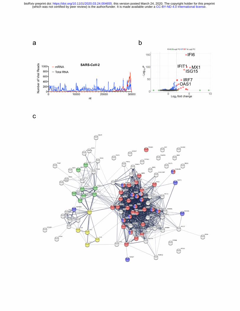
Figure 1. Host Transcriptional response to SARS-CoV-2 infection in A549 cells. (a)
read coverage along the SARS-CoV-2 genome. Number of viral reads per each position
of the virus genome. Blue graph indicate read coverage when NGS libraries were
prepared using Ilumina’s TruSeq Stranded Total RNA Gold kit. Red graph indicate read
coverage when NGS libraries were prepared using Ilumina’s TruSeq RNA Library Prep
Kit v2. (b) Volcano plot depicting differentially expressed genes in response to SARS-
CoV-2 infection. Red dots indicate genes with a |Log2(Fold Change)| > 2. The identity of
top induced genes is indicated. (c) Protein interaction network of significantly induced
genes in response to SARS-CoV-2 infection. Genes involved in enriched biological
processes and molecular functions are indicated in color. Red: genes involved in the
response to virus (GO:009615, FDR<<0.0001). Green: genes with cytokine activity
(GO:0005125, FDR<0.001). Yellow: genes involved in complement activation
(GO:0006956, FDR<0.0005). Blue: genes with RNA binding capability (GO: 0003723,
FDR<0.0005). Gene enrichment analyses were performed using STRING.
.CC-BY-ND 4.0 International license(which was not certified by peer review) is the author/funder. It is made available under aThe copyright holder for this preprintthis version posted March 24, 2020. . https://doi.org/10.1101/2020.03.24.004655doi: bioRxiv preprint
.CC-BY-ND 4.0 International license(which was not certified by peer review) is the author/funder. It is made available under aThe copyright holder for this preprintthis version posted March 24, 2020. . https://doi.org/10.1101/2020.03.24.004655doi: bioRxiv preprint
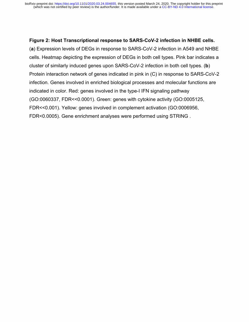
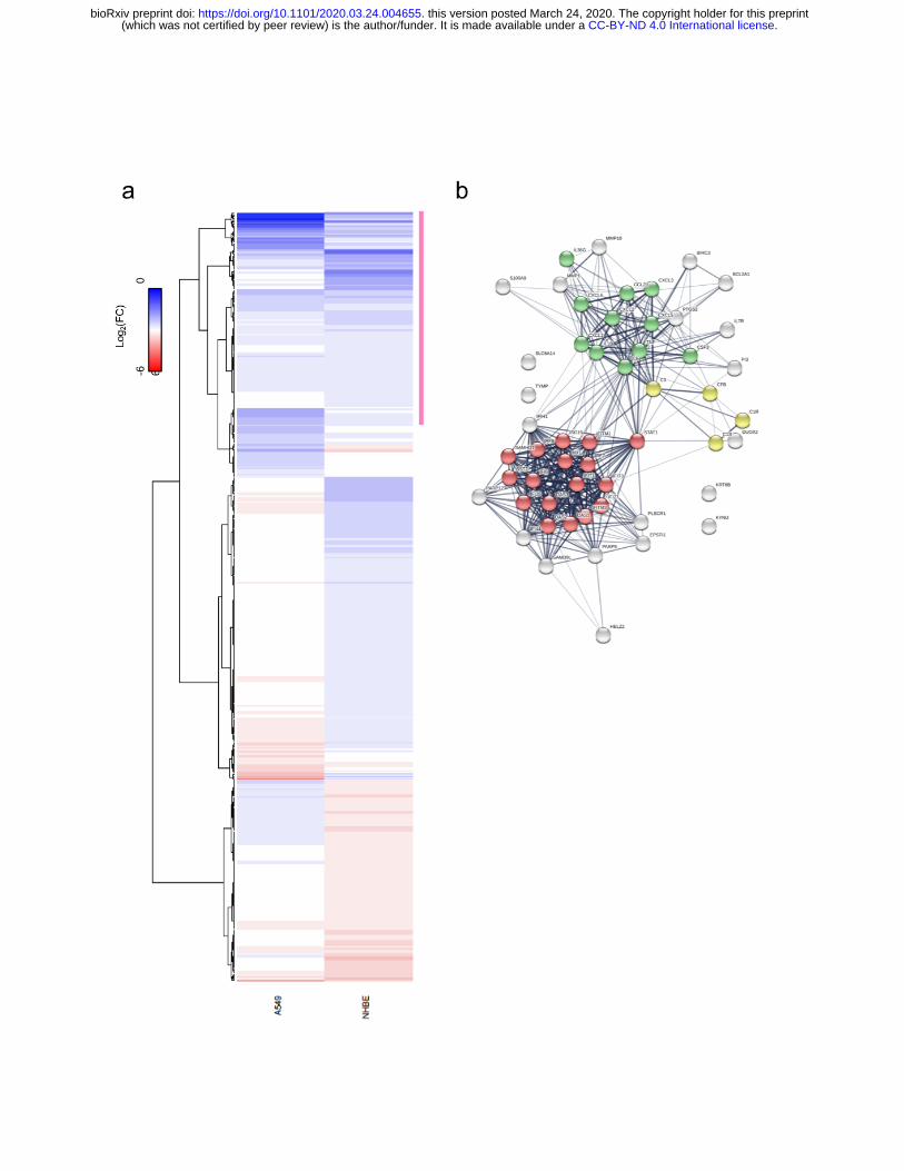
Figure 2: Host Transcriptional response to SARS-CoV-2 infection in NHBE cells. (a) Expression levels of DEGs in response to SARS-CoV-2 infection in A549 and NHBE
cells. Heatmap depicting the expression of DEGs in both cell types. Pink bar indicates a
cluster of similarly induced genes upon SARS-CoV-2 infection in both cell types. (b)
Protein interaction network of genes indicated in pink in (C) in response to SARS-CoV-2
infection. Genes involved in enriched biological processes and molecular functions are
indicated in color. Red: genes involved in the type-I IFN signaling pathway
(GO:0060337, FDR<<0.0001). Green: genes with cytokine activity (GO:0005125,
FDR<<0.001). Yellow: genes involved in complement activation (GO:0006956,
FDR<0.0005). Gene enrichment analyses were performed using STRING .
.CC-BY-ND 4.0 International license(which was not certified by peer review) is the author/funder. It is made available under aThe copyright holder for this preprintthis version posted March 24, 2020. . https://doi.org/10.1101/2020.03.24.004655doi: bioRxiv preprint
.CC-BY-ND 4.0 International license(which was not certified by peer review) is the author/funder. It is made available under aThe copyright holder for this preprintthis version posted March 24, 2020. . https://doi.org/10.1101/2020.03.24.004655doi: bioRxiv preprint
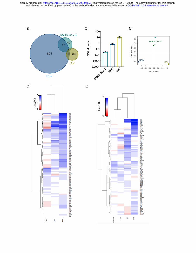
Figure 3: Unique host transcriptional profiles elicited differentially in response to diverse respiratory viruses. (a) Shared DEGs in SARS-CoV-2, RSV and IAV infected
A549 cells. Venn diagram of DEGs in each sample, genes shared/unique between each
sample are indicated. (b) Virus replication levels in A549 cells. Percentage of virus-
aligned reads (over total reads) for each infected sample. Data from 3 independent
biological replicates. (c) Principal component analysis (PCA) for the global
transcriptional response to respiratory viruses. Sparse PCA depicting global
transcriptome profiles between samples. All data from infections in A549 cells, except
for (*) that was performed in NHBE cells. (d-e) Heatmaps depicting the expression
levels of genes involved in the type-I IFN response (d) or with cytokine or chemokine
activity (e).
.CC-BY-ND 4.0 International license(which was not certified by peer review) is the author/funder. It is made available under aThe copyright holder for this preprintthis version posted March 24, 2020. . https://doi.org/10.1101/2020.03.24.004655doi: bioRxiv preprint
.CC-BY-ND 4.0 International license(which was not certified by peer review) is the author/funder. It is made available under aThe copyright holder for this preprintthis version posted March 24, 2020. . https://doi.org/10.1101/2020.03.24.004655doi: bioRxiv preprint

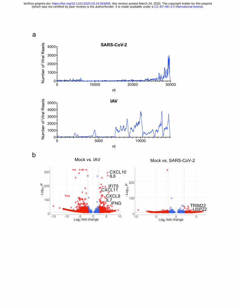
Figure 4: Host Transcriptional response to SARS-CoV-2 infection in vivo. (a) read
coverage along the SARS-CoV-2 and IAV (A/California/04) genomes. Number of viral reads per
each position of the virus genome. (b) Volcano plot depicting differentially expressed genes in
response to SARS-CoV-2 or IAV (A/California/04) infection. Red dots indicate genes with a
|Log2(Fold Change)| > 2. The identity of top induced genes is indicated.
.CC-BY-ND 4.0 International license(which was not certified by peer review) is the author/funder. It is made available under aThe copyright holder for this preprintthis version posted March 24, 2020. . https://doi.org/10.1101/2020.03.24.004655doi: bioRxiv preprint
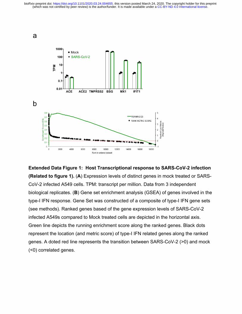
Extended Data Figure 1: Host Transcriptional response to SARS-CoV-2 infection (Related to figure 1). (A) Expression levels of distinct genes in mock treated or SARS-
CoV-2 infected A549 cells. TPM: transcript per million. Data from 3 independent
biological replicates. (B) Gene set enrichment analysis (GSEA) of genes involved in the
type-I IFN response. Gene Set was constructed of a composite of type-I IFN gene sets
(see methods). Ranked genes based of the gene expression levels of SARS-CoV-2
infected A549s compared to Mock treated cells are depicted in the horizontal axis.
Green line depicts the running enrichment score along the ranked genes. Black dots
represent the location (and metric score) of type-I IFN related genes along the ranked
genes. A doted red line represents the transition between SARS-CoV-2 (>0) and mock
(<0) correlated genes.
.CC-BY-ND 4.0 International license(which was not certified by peer review) is the author/funder. It is made available under aThe copyright holder for this preprintthis version posted March 24, 2020. . https://doi.org/10.1101/2020.03.24.004655doi: bioRxiv preprint
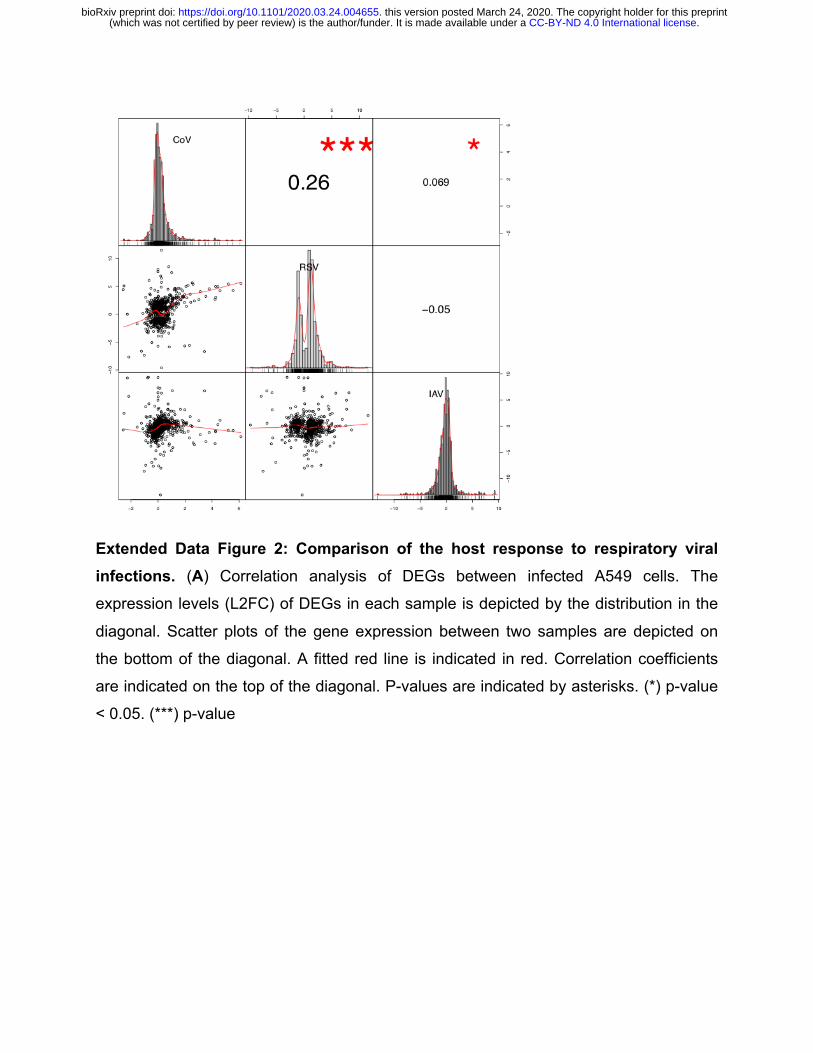
Extended Data Figure 2: Comparison of the host response to respiratory viral infections. (A) Correlation analysis of DEGs between infected A549 cells. The
expression levels (L2FC) of DEGs in each sample is depicted by the distribution in the
diagonal. Scatter plots of the gene expression between two samples are depicted on
the bottom of the diagonal. A fitted red line is indicated in red. Correlation coefficients
are indicated on the top of the diagonal. P-values are indicated by asterisks. (*) p-value
< 0.05. (***) p-value
.CC-BY-ND 4.0 International license(which was not certified by peer review) is the author/funder. It is made available under aThe copyright holder for this preprintthis version posted March 24, 2020. . https://doi.org/10.1101/2020.03.24.004655doi: bioRxiv preprint
Related Documents