Safety First: Banish Mycoplasma Take the Pink Link! www. .com detection treatment prevention

Welcome message from author
This document is posted to help you gain knowledge. Please leave a comment to let me know what you think about it! Share it to your friends and learn new things together.
Transcript
Safety First:Banish Mycoplasma
Take the Pink Link!
www. .com
There is another top address in Darmstadt:AppliChem GmbH Ottoweg 4 D - 64291 Darmstadt Phone +49 6151 9357-0 Fax +49 6151 9357-11
eMail [email protected] Internet www.applichem.com
detectiontreatment
prevention
Banish Mycoplasma • AppliChem © 2013
Firstly reported in 1956 (Robinson et al. 1956), the potential presence of myco-plasma in cell culture laboratories contin-ues to challenge scientists. The parasitic mycoplasmas represent a serious problem for all cell line-related fields in research as well as in industrial facilities for develop- ment or manufacture of cell-derived bio -lo gical and pharmaceutical products, inclu ding vaccines, monoclonal antibodies, drugs, and products for gene and cell ther-apy. Still, there is no perceivable re duction of cell culture infection rates (Ryan 2008),
Mysterious mycoplasmaA question of culture?!
even though risks and consequences caused by mycoplasma infections have been known for decades, and strategies for their prevention , detection and elimination are well established. Why are so many cell lines – while commonly well fostered by their cell cultur ists – still insufficiently pro-tected against the cell wall-free invader? Is this due to careless ness, or rather a lack of know ledge? Un for tunately we cannot pro-vide any data regarding this question – but a lot of facts demonstrating the importance of this un pop ular subject.

© 2013 AppliChem • Banish Mycoplasma 1
How do mycoplasmas commonly enter our labs and cultures? 2
What makes mycoplasma species worse than other bacterial contaminates –
and why is it a must to banish them from cell cultures? 2
Mycoplasma Prevention
How can I avoid contaminations?! 3
Disinfecting CO2 incubators and water baths 4
Preventing microbial growth in cell cultures 4
Mycoplasma Detection
How can I identify a mycoplasma contamination? 5
Using PCR for fast & reliable mycoplasma detection 5
Mycoplasma Elimination
What can I do to eliminate mycoplasma from an infected cell culture? 7
Treating mycoplasma-infected cells 7
The “mycoplasma problem” is known for decades – why does it still exist?! 10
References 11
Contents
Treating mycoplasma-infected cells 7
The “mycoplasma problem” is known for decades – why does it still exist?! 10
© 2013 AppliChem • Banish Mycoplasma
References 11 References 11 References 11
Introduction
2 Banish Mycoplasma • AppliChem © 2013
How do mycoplasmas commonly enter our labs and cultures?
What makes mycoplasma species worse than other bacterial contaminates – and why is it a must to banish them from cell cultures?
Mycoplasmas are omnipresent; their broad range of hosts includes humans and other mammals, birds, reptiles, fish, insects and plants (Razin et al. 1998). However, in cell culture laboratories, 95 % of all continuous cell line infections are caused by only six species from bovine (M. arginini & Acholeplasma laidlawii), swine (M. hyorhinis) – and human (M. orale, M. fermentans, M. hominis) origin (Drexler & Uphoff 2002). The main source of mycoplasma contaminations today are mycoplasma-infected cell cultures used in the same labora-tory (Rottem & Barile 1993, Drexler et al. 2002; Drexler & Uphoff 2002). The infection may be transferred by aerosols, particulates and inadequate cell culture technique directly – or indirectly via media, solutions and laboratory equipment contaminated by previous use in processing mycoplasma-infected cells. As a result, 15 – 35 % of all continuous cell lines are positive for mycoplasma, but only 1 % of the primary cell cultures (Drexler & Uphoff 2002). The second leading source is the laboratory personnel, explaining the fact that mycoplasma
species from human are the most common contaminates (responsible for 40 – 80 % of the infections) M. orale, colonizies the oral cavity and represents the primary species isolated from conta minated cell cultures.
Mycoplasma species from bovine origin or swine were traced back to contaminated sera and other animal-originating products, e.g. the prevalent presence of A. laidlawii and M. arginini indicating fetal or newborn bovine serum as the primary source of infection.
Nowadays, sera and media are rarely the source of mycoplasma conta mination (Lincoln & Lundin 1990; Armstrong et al. 2009) as long as they are purchased from reputable manufacturers that sterilize their products by several filtration steps using a 0.1 µm pore membrane filter and frequently control sterility.
In contrast to “common” bacteria, these tiny prokaryotes do not pos-sess a cell wall. Together with other cell wall-lacking bacteria – species of ureaplasma, acholeplasma, anaeroplasma, spiroplasma – they form the class of mollicutes. Nevertheless, the terms “mycoplasma” or formerly “pleuropneumonia-like organisms (PPLO)” and “molli cutes” are often used synonymously. Due to the absence of a cell wall, myco-plasmas are unaffected by antibiotics that interfere with peptido glycan formation, namely beta-lactam antibiotics. These include penicillin- derivatives, cephalosporins, and carbapenemes. Furthermore they are very flexible in shape which in addition to their small size (ranging from 0.1 to 0.8 µm in diameter, depending on the literature ) makes them difficult to filter from solutions. Mycoplasma species easily penetrate the membrane of 0.2 µm filters commonly used for sterilization of media, sera and other non-autoclavable reagents. Mycoplasma’s general dependence on complex enriched media (including host cell nutrients ) and defined environmental conditions – both perfectly realized in cell culture – and their very slow growth rates complicate identification of infected cells by common microbiological cultivation methods. Their small size and missing cell wall allows them to achieve high densities in cell cultures; often without being detectable by turbidity, cytopatho-genicity or even microscopic examination. However, the consequences of mycoplasma contaminations should not be underestimated; neither with regard to research (and the researcher’s career!), nor in terms of serious health risks for humans and animals. Please keep in mind that some members of the mycoplasma family are pathogenic organisms!
By growing covertly and undisturbed within a cell culture, mycoplasma can easily take over the control of reagents, equipment, and other cell lines within weeks (McGarrity 1976). Be aware that the lack of visible effects provides a false sense of security: While often behaving inconspicuous at first glance, the fastidious organisms are able to influence nearly every single cellular function, ranging from a decelerated growth rate to metabolic (including protein, RNA, DNA synthesis) and morphologic changes. All these effects are mainly based on a competition for essential nutrients (nucleosides, nucleotides, nucleo-bases, arginine and other amino acids, fatty acids, sugars, etc.) and the release of toxic, cytolytic or acidic metabolites. By up- and down-regu-lation of cytokines and growth factors, stress-response genes, transport proteins, receptors, ion channels, oxidases, tumor supressor and onco-genes, mycoplasmas significantly alter the gene expression profiles of cultured cells (Miller et al. 2003). Therefore they make any experiment carried out with infected cells questionable! Furthermore they are known to cause chromosomal aberrations in vitro, with chromosomal break-age, translocation events, and reduction or augmentation in chromosome number being the most frequent outcomes. Virus propagation might also be influenced in both directions, positively (by inhibiting interfer on induction and activity) as well as negatively (by competing for essential nutrients). Even though there are a large number of potential effects described in literature it is unpredictable which effect will occur. Possible effects depend on mycoplasma species and strain, the infected cell type, and certainly on environmental conditions (Rottem & Barile 1993).
© 2013 AppliChem • Banish Mycoplasma 3
Finally, besides biosafety concerns, the consequences of mycoplasma contamination on laboratory work are loss of time, efforts, money (regarding cells, media, materials, but also valuable biopharmaceuti-cals, if cultures were used for production of vaccines, antibodies or drugs) and good reputation. Research based on mycoplasma-contam-inated cell lines will produce inaccurate or erroneous results yielding misleading publications. Consider the personal embarrassment and maybe the loss of good reputation, if the published results are proven to be faulty due to a contamination problem. And how awkward will it be to get informed by a colleague that the cell line you provided is contaminated? All these factors should be reconsidered, when risking covert mycoplasma infections by NOT testing cell cultures and NOT actively fighting them by good laboratory practice.
a mycoplasma-free cell culture is a
PRECONDITION for safety and purity
of cell-derived products and reliable
results in scientific experiments.
To sum up
Prevention The good news: it is possible to minimize the risk of
general mycoplasma conta minations – and to exclude serious outbreaks.
How can I avoid contaminations?!
Probably, there will never be a point in time when mycoplasma conta m-i nations are completely banished from our labs – as long as humans are working there. But carrying out some general principles will mini-mize the risk of contaminations and prevent costly or embarrass-ing situations.• Strictly follow aseptic techniques and practices, including no
unnecessary talking, no mouth pipetting, no media supply by pouring, regular hand washing and disinfection! Do not use the laminar flow for storage of solutions and equipment! Only work with ONE cell line at a time and use separate materials for each cell line to avoid cross-contaminations! Make sure all media, solutions and materials are properly sterilized – the same holds true for any kind of occurring waste of course!
• Clean and disinfect surfaces, laminar flows, incubators, water baths and all other equipment frequently – before AND after the working procedure. Make sure the laboratory is cleaned up regu-larly and only authorized persons have access to the working area.
• Use antibiotics responsibly. For routine culture work antibiotic-free media should be employed. General usage of antibiotics to mask low hygiene levels, a lack of good aseptic techniques, or improper cell culture facilities are not a solution to the problem! Quite the con-trary, non-responsible use of antibiotics will make the situation even worse.
• Isolate incoming cell cultures (use a separate incubator or at least sealed flasks as well as separate culture media and materials) until the mycoplasma test results are proven to be negative.
• Test frequently for contamination – regardless of whether the cell culture contains any antibiotics or not! Routine testings for the presence of mycoplasma species are an absolute must for
the responsible scientist! Only by identification and treatment or elimination of the infected cell line the risk of further (cross-)conta-minations is banished and experiments yield stable and reliable results.
AppliChem offers a series of productspreventing microbial contaminations – in hoods, incubators, water baths and cell cultures. More information about this product line is available on page 4 and in the product table on page 12.
4 Banish Mycoplasma • AppliChem © 2013
Furthermore, it is highly recommended to freeze a cell stock as a backup for damaged or lost cell cultures. When dealing with cells of limited life span, cryopreservation is invaluable anyway. But also a stock of contin-uous cell lines should be stored properly below –130 °C to prevent in vitro cellular alteration (Hughes et al. 2007; Stacey & Masters 2008) and maintain reliable cultures of consistent quality for research and biopharmaceutical production. The advantages of a cryopreserved cell bank are a reduced risk of (cross-)contamination with microorganisms or other cell lines, prevention of phenotypic or genotypic drifts, and damages due to cell aging. But caution: Please be aware that mycoplasmas are able to survive freezing in liquid nitrogen – even without cryopreser-vation. For that reason, a contaminated liquid nitrogen container (e.g. due to an inadequately closed or contaminated sample of cells) might be a source of mycoplasma. But how do they enter the cell culture-containing cryo tube? Storage in the liquid phase together with a non-sufficient tube filling level might be the way in. Liquid nitrogen has the tendency to permeate the cryo tube, especially if it is filled insufficiently. To avoid any contamination risk, cryogenic vials should be properly stored in the vapor phase of the liquid nitrogen container.
Disinfecting CO2 incubators and water baths
Incubator-Clean™ (A5230) Incubator-Clean™ is a non-toxic and non-hazardous solution for disin-fection of incubators, sterile benches and other surfaces in cell culture and molecular biology laboratories. The problem of contamination in incubators and/or sterile work-benches is often a serious one, leading to extensive damage. Incubator-Clean™ prevents contamination with and growth of mycoplasma and other bacteria (and spores), fungi (and spores), and viruses (including HIV and Hepatitis B). The active ingre-dients are quaternary benzylammonium compounds, and the solution does not contain mercury, formaldehyde, phenol or alcohol. Further-more, Incubator-Clean™ is biodegradable and fully compatible with common work surfaces. Incubator-Clean™ is supplied in spray bottles.
Recommended use: Spray incubators every other week. It is not necessary to clear the incubator before spraying. Spray sterile benches once a day, or preferably before each laboratory worker begins to use the work area. The drying time is the reaction time!
Incuwater-Clean™ (A5219)The water required to create the humidity is a source of contamination which disperses in the incubator. In order to disinfect the water we re commend Incuwater-Clean™, which contains a disinfectant that does not cause damage to the stainless steel tray. Incuwater-Clean™ does not contain heavy metals, it is non-toxic, non-volatile, and extremely effective.
Incuwater-Clean™ is supplied as a 100X concentrated ready-to-use solution. The water should be replaced every two to four weeks.
Aquabator-Clean™, 100X (A9390)Aquabator-Clean™ is intended for disinfecting various kinds of water baths from bacteria and fungi. The active ingredient in Aquabator-Clean™ is safe to humans and does not cause any irritating effects to the skin when used at the recommended concentration. It is biodegrad-able. Use 10 ml Aquabator-Clean™ for each liter of water in the bath.
Caution: Aquabator-Clean™ is not suited for use in CO2 incubators!
Preventing microbial growth in cell cultures
CellCultureGuard (A8906)Our first choice cell culture reagent!The main measures to avoid contamination of cell cultures are sterile techniques, consistent compliance with good laboratory practice and sterilized equipment and vessels. However, for some cell lines like primary ones, hybridomas, or cell lines isolated from patients this might not be sufficient. The addition of an antibiotic is required to further reduce the risk of microbial contaminations. CellCultureGuard is a combination of novel antibiotics perfectly suited for protection of ani-mal and human cell cultures from contamination by a wide range of microorganisms: extra- and intracellular bacteria, mycoplas-ma, protozoa and fungi (yeast). The combination of innovative antibiotics is blocking the bacterial DNA synthesis and the protein bio-synthesis. Due to the combined activity there is a very low probability for the formation of resistants. Additionally, it is highly compatible with resistance markers and, at the recommended concentration, no side effects on cell cultures are observed.
Recommended use: CellCultureGuard is provided as a 100-fold concentrated sterile solution. Simply add 1 ml of CellCultureGuard to 100 ml of cell culture medium (or human or animal cell cultures). To ensure maximum protection at 37 °C, CellCultureGuard should be renewed after 7 days.
Prevention
© 2013 AppliChem • Banish Mycoplasma 5
Detection
The most sensitive method to detect mycoplasma is the direct culture method in suitable broth and agar media to obtain visible colonies. Theoretically, a single CFU (colony forming unit) per sample volume is detectable. Unfortunately this method is also the most time consuming (up to 28 days; due to the slow growth of mycoplasma species), and it requires experienced personnel conducting the experiments under controlled environmental conditions. Even if the difficult procedure is properly conducted (to start with the complex medium composition often requiring non-standard adjustments for individual species up to analysis of the results), the method is not 100 % effective, since some fastidious strains may not grow in pure culture. Therefore, an indi-rect detection method should be performed in addition. The most sensitive indirect mycoplasma tests are based on DNA fluorochrome staining (e.g. using DAPI) and PCR. Even if the detection limit of these methods is lower than for the direct culture method, they are absolutely sufficient for routine testings. Commonly, mycoplasma-conta minated cell cultures show high densities of mycoplasma (up to 107 – 108 CFU/ml) that are well suitable for the detection limits of these methods. In contrast to the PCR alternative, the traditional fluores-cence staining method requires more time and experience. In addition, the DNA-binding fluorescent stain is carcinogenic and needs to be handled carefully. Hence, for routine mycoplasma screenings, PCR analysis is recommended (Drexler & Uphoff 2002). This method is sensitive (depending on the kit almost as sensitive as the direct cul-ture method), very fast (results are obtained within hours) and detects cultivable as well as non-cultivable mycoplasma species. Furthermore, at least with commercially available kits, this method is very easy to perform and does not require a specific expertise.
Using PCR for fast & reliable mycoplasma detection
PCR Mycoplasma Test Kits: How do they work?! The principle is simple: A mycoplasma-specific DNA-fragment is ampli-fied exclusively by PCR, using a specific primer set. Only if mycoplasma DNA is present in the sample, a mycoplasma-specific PCR product of defined size (270 bp) is obtained – detectable on an agarose gel, or, in case of the qPCR Test Kit, using the FAM™ detection channel. The primer set binds selectively to the highly conserved 16S rRNA operon coding region of the mycoplasma genome, not detecting any other bacterial or eukaryotic DNA. The detection range includes the most frequently occurring contaminants M. fermentans, M. arginini, M. orale, M. hyorhinis, M. salivarium, M. hominis, but also less prevalent mycoplasma species, such as M. pneumoniae, Acholeplasma laid-lawii, M. synoviae, M. pulmonis, M. bovis – just to name a few.
How can I identify a mycoplasma contamination?
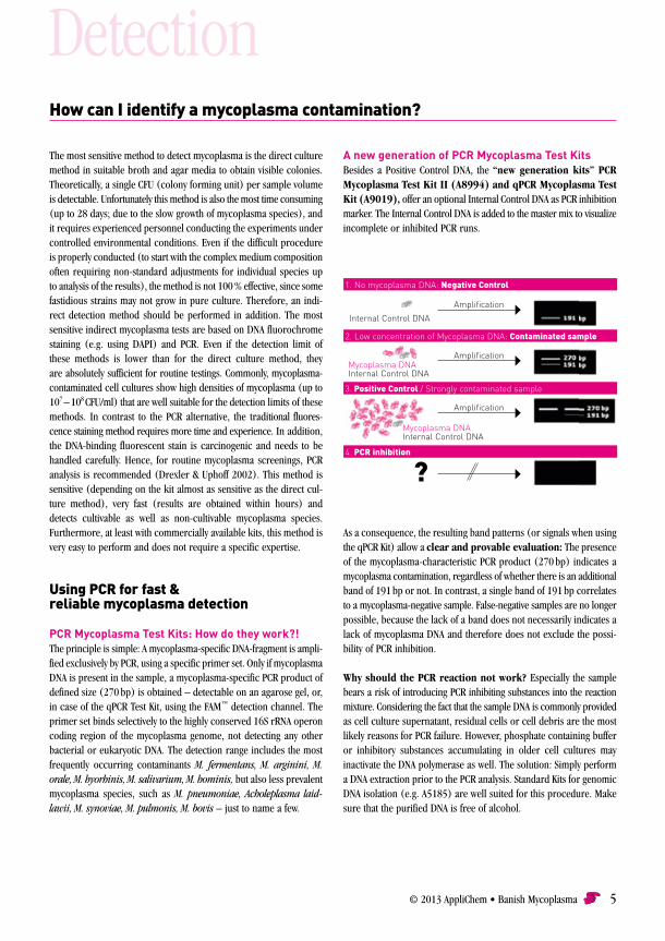
A new generation of PCR Mycoplasma Test KitsBesides a Positive Control DNA, the “new generation kits” PCR Mycoplasma Test Kit II (A8994) and qPCR Mycoplasma Test Kit (A9019), offer an optional Internal Control DNA as PCR inhibition marker. The Internal Control DNA is added to the master mix to visualize incomplete or inhibited PCR runs.
1. No mycoplasma DNA: Negative Control
Internal Control DNA
Mycoplasma DNAInternal Control DNA
Mycoplasma DNAInternal Control DNA
?
Amplification
Amplification
Amplification
2. Low concentration of Mycoplasma DNA: Contaminated sample
3. Positive Control / Strongly contaminated sample
4. PCR inhibition
As a consequence, the resulting band patterns (or signals when using the qPCR Kit) allow a clear and provable evaluation: The presence of the mycoplasma-characteristic PCR product (270 bp) indicates a mycoplasma contamination, regardless of whether there is an additi onal band of 191 bp or not. In contrast, a single band of 191 bp correlates to a mycoplasma-negative sample. False-negative samples are no longer possible, because the lack of a band does not necessarily indicates a lack of mycoplasma DNA and therefore does not exclude the possi-bility of PCR inhibition.
Why should the PCR reaction not work? Especially the sample bears a risk of introducing PCR inhibiting substances into the reaction mixture. Considering the fact that the sample DNA is commonly provided as cell culture supernatant, residual cells or cell debris are the most likely reasons for PCR failure. However, phosphate containing buffer or inhibitory substances accumulating in older cell cultures may inactivate the DNA polymerase as well. The solution: Simply perform a DNA extraction prior to the PCR analysis. Standard Kits for genomic DNA isolation (e.g. A5185) are well suited for this procedure. Make sure that the purified DNA is free of alcohol.
6 Banish Mycoplasma • AppliChem © 2013
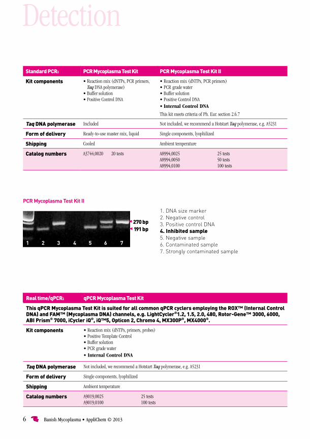
Standard PCR: PCR Mycoplasma Test Kit PCR Mycoplasma Test Kit II
Kit components • Reaction mix (dNTPs, PCR primers, Taq DNA polymerase)
• Buffer solution• Positive Control DNA
• Reaction mix (dNTPs, PCR primers)• PCR grade water• Buffer solution• Positive Control DNA• Internal Control DNA
This kit meets criteria of Ph. Eur. section 2.6.7
Taq DNA polymerase Included Not included, we recommend a Hotstart Taq polymerase, e.g. A5231
Form of delivery Ready-to-use master mix, liquid Single components, lyophilized
Shipping Cooled Ambient temperature
Catalog numbers A3744,0020 20 tests A8994,0025 25 testsA8994,0050 50 testsA8994,0100 100 tests
270 bp191 bp
1 2 3 4 5 6 7
Real time/qPCR: qPCR Mycoplasma Test Kit
This qPCR Mycoplasma Test Kit is suited for all common qPCR cyclers employing the ROX™ (Internal Control DNA) and FAM™ (Mycoplasma DNA) channels, e.g. LightCycler®1.2, 1.5, 2.0, 480, Rotor-Gene™ 3000, 6000, ABI Prism® 7000, iCycler iQ®, iQ™5, Opticon 2, Chromo 4, MX300P®, MX4000®.
Kit components • Reaction mix (dNTPs, primers, probes)• Positive Template Control • Buffer solution• PCR grade water• Internal Control DNA
Taq DNA polymerase Not included, we recommend a Hotstart Taq polymerase, e.g. A5231
Form of delivery Single components, lyophilized
Shipping Ambient temperature
Catalog numbers A9019,0025 25 testsA9019,0100 100 tests
PCR Mycoplasma Test Kit II
1. DNA size marker2. Negative control 3. Positive control DNA4. Inhibited sample5. Negative sample6. Contaminated sample7. Strongly contaminated sample
Detection
© 2013 AppliChem • Banish Mycoplasma 7
This answer is easy: Autoclave the contaminated cells, at best, together with any bottle of medium and reagent used with this relevant culture . Don’t forget subsequent cleaning and disinfection of surfaces, hoods, incubators, pipettors etc. – or better, the whole lab! Make sure that other cell lines are not infected as well! Cleaning up a contami-nated culture with an anti-mycoplasma treatment is recommended only for very valuable or irreplaceable cultures, and if the potential source of mycoplasma was previously banished from the laboratory. Efforts for the attempted rescue are high and, until now, no universal mycoplasma-eliminating reagent is available. Antibiotic resistance, cytotoxicity, and a reduced viability of chronically or multiply infected cells may be reasons to prevent curing (Fleckenstein & Drexler 1996).
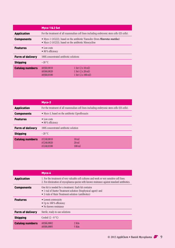
Despite existing resistances, the most reliable and efficient treatment of mycoplasma contaminations is the addition of suitable antibiotics, such as quinolones, tetracyclines and macrolides (Drexler & Uphoff 2002). In an experiment with 251 chronically mycoplasma-positive cell lines, treatment with ciprofloxacin provided recovery-levels of 78 %, with 15 % of the cell cultures remaining contaminated due to resistance and 7 % loss by cell death during the elimination procedure . The combination of tiamulin and minocycline even reached curing of 82 % of all treated cell cultures, showing a lower resistance level (7 %) but higher cytotoxicity (11 % of the cell cultures died during the treatment; data taken from Drexler & Uphoff 2002). Besides the traditional mycoplasma-eliminating agents Myco-1 & 2 (tiamulin and minocycline) and Myco-3 (ciprofloxacin), AppliChem now offers a new solution for effective and permanent removal of mycoplasma species from cell culture: Myco-4 provides a broad spectrum of activity (including any type of mycoplasma, acholeplasma, spiroplasma and entomoplasma) combined with very low cytotoxicity and a low resis-tance risk due to an initial biophysical mode of action.
AppliChem’s PCR Mycoplasma Test Kits enable identification of mycoplasma-contaminated
cell cultures – fast and effective! The PCR technique allows highly sensitive detection of both cultivable
and non-cultivable mycoplasma species. Repro-ducible results are provided within hours, making
PCR the method of choice for frequent routine testings.
Treating mycoplasma-infected cells
What can I do to eliminate mycoplasma from an infected cell culture?
Elimination
…for those cases when it happened:
Antibiotics are the most effective treatment for
mycoplasma contaminations!
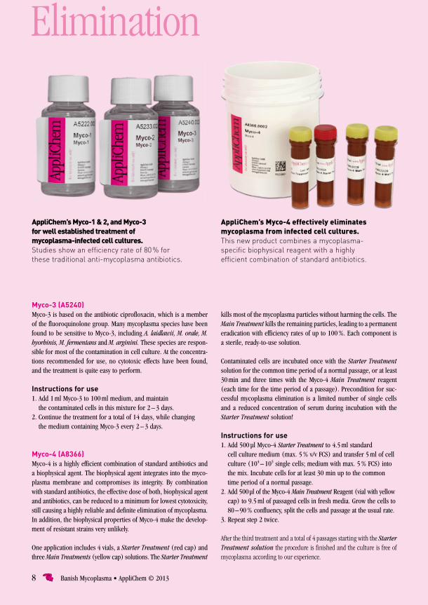
Myco-1 & 2 Set (A8360)Myco-1 is based on the antibiotic tiamulin, which is produced by the fungus Pleurotus mutilus. Myco-2 is based on minocycline, a tetra cy-cline derivative. Myco-1 (A5222) and Myco-2 (A5233) are generally used sequentially in combination.
Instructions for use1. Add 1 ml Myco-1 to 100 ml medium, and maintain
the contaminated cells in this mixture for 4 days. 2. After 4 days, add 1 ml Myco-2 to 100 ml fresh medium,
and maintain the cells in this second mixture for 3 days.3. The above, together, are considered as one treatment cycle.
It may be necessary to repeat this cycle 2 – 3 times.
8 Banish Mycoplasma • AppliChem © 2013
AppliChem’s Myco-1 & 2, and Myco-3 for well established treatment of mycoplasma-infected cell cultures. Studies show an efficiency rate of 80 % for these traditional anti-mycoplasma antibiotics.
kills most of the mycoplasma particles without harming the cells. The Main Treatment kills the remaining particles, leading to a permanent eradication with efficiency rates of up to 100 %. Each component is a sterile, ready-to-use solution.
Contaminated cells are incubated once with the Starter Treatment solution for the common time period of a normal passage, or at least 30 min and three times with the Myco-4 Main Treatment reagent (each time for the time period of a passage). Precondition for suc-cessful mycoplasma elimination is a limited number of single cells and a reduced concentration of serum during incubation with the Starter Treatment solution!
Instructions for use1. Add 500 µl Myco-4 Starter Treatment to 4.5 ml standard
cell culture medium (max. 5 % v/v FCS) and transfer 5 ml of cell culture (104 – 105 single cells; medium with max. 5 % FCS) into the mix. Incubate cells for at least 30 min up to the common time period of a normal passage.
2. Add 500 µl of the Myco-4 Main Treatment Reagent (vial with yellow cap) to 9.5 ml of passaged cells in fresh media. Grow the cells to 80 – 90 % confluency, split the cells and passage at the usual rate.
3. Repeat step 2 twice.
After the third treatment and a total of 4 passages starting with the Starter Treatment solution the procedure is finished and the culture is free of mycoplasma according to our experience.
AppliChem’s Myco-4 effectively eliminates mycoplasma from infected cell cultures. This new product combines a mycoplasma- specific biophysical reagent with a highly efficient combination of standard antibiotics.
Elimination
Myco-3 (A5240)Myco-3 is based on the antibiotic ciprofloxacin, which is a member of the fluoroquinolone group. Many mycoplasma species have been found to be sensitive to Myco-3, including A. laidlawii, M. orale, M. hyorhinis, M. fermentans and M. arginini. These species are respon-sible for most of the contamination in cell culture. At the concentra-tions recommended for use, no cytotoxic effects have been found, and the treatment is quite easy to perform.
Instructions for use1. Add 1 ml Myco-3 to 100 ml medium, and maintain
the contaminated cells in this mixture for 2 – 3 days.2. Continue the treatment for a total of 14 days, while changing
the medium containing Myco-3 every 2 – 3 days.
Myco-4 (A8366)Myco-4 is a highly efficient combination of standard antibiotics and a biophysical agent. The biophysical agent integrates into the myco-plasma membrane and compromises its integrity. By combination with standard antibiotics, the effective dose of both, biophysical agent and antibiotics, can be reduced to a minimum for lowest cytotoxicity, still causing a highly reliable and definite elimination of mycoplasma. In addition, the biophysical properties of Myco-4 make the develop-ment of resistant strains very unlikely.
One application includes 4 vials, a Starter Treatment (red cap) and three Main Treatments (yellow cap) solutions. The Starter Treat ment
© 2013 AppliChem • Banish Mycoplasma 9
Myco-3
Application For the treatment of all mammalian cell lines including embryonic stem cells (ES cells).
Components • Myco-3, based on the antibiotic Ciprofloxacin
Features • Low costs• 80 % efficiency
Form of delivery 100X concentrated antibiotic solution
Shipping –20 °C
Catalog numbers A5240,0010 10 mlA5240,0020 20 mlA5240,0100 100 ml
Myco-1 & 2 Set
Application For the treatment of all mammalian cell lines including embryonic stem cells (ES cells).
Components • Myco-1 (A5222), based on the antibiotic Tiamulin (from Pleurotus mutilus)• Myco-2 (A5233), based on the antibiotic Minocycline
Features • Low costs• 80 % efficiency
Form of delivery 100X concentrated antibiotic solutions
Shipping –20 °C
Catalog numbers A8360,0010 1 Set (2 x 10 ml)A8360,0020 1 Set (2 x 20 ml)A8360,0100 1 Set (2 x 100 ml)
Myco-4
Application 1. For the treatment of very valuable cell cultures and week or very sensitive cell lines. 2. For elimination of mycoplasma species with known resistance against standard antibiotics.
Components One kit is needed for a treatment. Each kit contains • 1 vial of Starter Treatment solution (biophysical agent) and • 3 vials of Main Treatment solution (antibiotics)
Features • Lowest cytotoxicity• Up to 100 % efficiency• No known resistance
Form of delivery Sterile, ready-to-use solutions
Shipping Cooled (2 – 8 °C)
Catalog numbers A8366,0002 2 KitsA8366,0005 5 Kits
10 Banish Mycoplasma • AppliChem © 2013
There are two main reasons why mycoplasma contaminations are not banished from cell culture laboratories yet: First, half of the re searchers still do not test their cell cultures for mycoplasma (Ryan 2008) and second, there is a tendency to rely on antibiotics instead of good aseptic practices.
Even though cell culture experts agree that general use of antibiotics can increase the severity of contamination problems, the routine use of antibiotics in cell culture laboratories is still prevalent. Particularly mycoplasma contamination rates are much higher in cell lines grown in antibiotic-containing medium than in antibiotic-free cultures (Barile 1973). If microorganisms, bacteria or fungi, are accidentally brought into antibiotic-free culture medium, they will replicate non-inhibited, soon leading to visible indicators of contamination: turbid-ity, filamen tary structures, color changes due to pH alteration. In
contrast, the presence of antibiotics will prevent the microbial growth – maybe. Unfortunately there is no absolute guarantee that the added antibiotics act against the intro-duced microorganisms (probably a mixture of different species), and sooner or later the user will
The “mycoplasma problem” has been known for decades – why does it still exist?!
face some kind of resistance phenomenon. If the introduced germ is fully resistant to the antibiotic, it will hopefully rapidly overgrow the culture and become visible within a short period of time. If the intro-duced microorga nism only shows a partial resistance the situation is worse. Due to the latent static level of partly resistant contamina tions, the risk of cross contaminations and usage of the affected culture in experiments or bio-pro duction should not be underestimated. This worst case is very likely, if the invader belongs to the species of mycoplasma (e.g. brought into the culture through aerosol droplets from the mouth of the cell culturist), since most common antibi-otics used in cell culture do not act on mycoplasma! Besides the beta-lactams being ineffective anyway, high resistance levels of mycoplasma against streptomycin (88 %), kanamycin (73 %), genta-mycin (80 %) and neomycin (86 %) were determined (Lundin & Lincoln 1994).
Apart from Barile's observation of strongly increased rates of myco-plasma contamination, morphological and functional changes are other disadvantages one has to take into account (Kuhlmann 1996), when using antibiotics on a routine basis. Anyhow, there exist useful appli-cations for antibiotics in cell culture, e.g. within the first two weeks of primary culture. In order not to create new resistances due to inacti-vation of the antibiotic, the antibiotic-containing medium should be refreshed frequently.
As an alternative to classical cell culture antibiotics like penicillin-streptomycin, AppliChem provides a new product to prevent microbial growth in cell cultures: CellCultureGuard. This combination of selected antibiotics (one being a fluoroquinolone) offers a wide range of anti-microbial activity, making it our first choice cell culture reagent: CellCultureGuard (A8906) is active against extra- and intra-cellular bacteria, mycoplasma, protozoa and fungi (yeast). Additionally, it is highly compatible with resistance markers and bears a low risk of resistance development.
© 2013 AppliChem • Banish Mycoplasma 11
• Armstrong SE, Mariano JA and Lundin DJ (2010) The Scope of Mycoplasma
Contamination within the Biopharmaceutical Industry. Biologicals 38:211-213.
• Barile MF, Hopps HE, Grabowski MW, Riggs DB and Del Giudice RA (1973) The
Identification and Sources of Mycoplasmas Isolated from Contaminated Cell
Cultures. Ann. NY Acad. Sci. 225:251-264.
• Drexler HG and Uphoff CC (2002) Mycoplasma Conta mi nation of Cell
Cultures: Incidence, Sources, Effects, Detection, Elimination, Prevention.
Cytotechnology 39:75-90.
• Drexler HG, Uphoff CC, DirksWG and MacLeod RAF (2002) Mixups and myco-
plasma: The enemies within. Leukemia Res 26:329–333.
• Fleckenstein E and Drexler HG (1996) Elimination of Mycoplasma Contami-
nation in Cell Cultures. Biochemica 1:48-51.
• Hughes P, Marshall D, Reid Y, Parkes H and Gelber C (2007) The Costs of Using
Unauthenticated, Over-passaged Cell Lines: How Much More Data Do We
Need? BioTechniques 43:575-586.
• Kuhlmann I (1996) The Prophylactic Use of Antibiotics in Cell Culture.
Cytotechnology 19:95-105.
• Lincoln CK and Lundin DJ (1990) Mycoplasma Detection and Control. United
States Federation for Culture Collection Newsletter 20 (4):1-3.
• Lundin DJ and Lincoln CK (1994) Mycoplasmal Contamination of Cell
Cultures within the Clinical Diagnostic Laboratory. Amer. Clin. Lab. (4):6.
For more information, please see the Mycoplasma Resource Centre at
www.bionique.com
References
• McGarrity GJ (1976) Spread and Control of Mycoplasmal Infection of Cell
Cultures. In Vitro 12:643-647.
• Miller CJ, Kassem HS, Pepper SD, Hey Y, Ward TH, Margison GP (2003)
Mycoplasma infection significantly alters microarray gene expression
profiles. BioTechniques 35:812-814.
• Razin S, Yogev D and Naot Y (1998) Molecular biology and pathogenicity
of mycoplasmas. Microbiol Mol Biol Rev 62:1094-1156.
• Robinson LB, Wichelhausen RH and Roizman B (1956) Conta mination
of Human Cell Cultures by Pleurop neumonia-like Organisms. Science
124:1147-1148.
• Rottem S and Barile MF (1993) Beware of Mycoplasmas. Trends in Bio-
technology 11:143-150.
• Ryan JA (2008) Understanding and Managing Cell Culture Contamination.
Corning, Inc. Technical Bulletin.
• Stacey GN and Masters JR (2008) Cryopreservation and Banking of Mamma-
lian Cell Lines. Nature Protocols 3: 1981-1989.
6.
Please see our website www.applichem.com
for products, protocols & more information.
12 Banish Mycoplasma • AppliChem © 2013
Prod. No Description Features
Detection
A3744 PCR Mycoplasma Test Kit Ready-to-use (Taq Polymerase included!); contains a positive control DNA.
A8994 PCR Mycoplasma Test Kit II Highest sensitivity of < 10 CFU/ml; according to Ph. Eur. (section 2.6.7). Besides a positive control DNA (non-infectious!) this kit provides an internal control DNA to visualize potential PCR inhibitions. The DNA Polymerase is not included, we recommend a Hotstart Taq Polymerase, e.g. SuperHot Taq DNA Polymerase (A5231).
A9019 qPCR Mycoplasma Test Kit Designed for qPCR applications. Provides a non-infectious positive control (FAM) and an internal con-trol (ROX) to visualize potential PCR inhibitions. The DNA Polymerase is not included, we recommend a Hotstart Taq Polymerase, e.g. SuperHot Taq DNA Polymerase (A5231).
A1001 DAPI Excellent fluorescent dye for mycoplasma detection via DNA-staining followed by microscopy.
Elimination
A8360 Myco-1 & 2 Set 2-step treatment with tiamulin and minocycline.
A5240 Myco-3 Single-component treatment with ciprofloxacin.
A8366 Myco-4 2-step treatment with a mycoplasma-specific biophysical agent followed by an appropriate antibiotic combination.
Prevention
A5230 Incubator-Clean™ Non-toxic and biodegradable disinfectant for incubators and sterile benches; prevents contamination with and growth of fungi, bacteria (including mycoplasma) and viruses (including HIV and Hepatits B). Fully compatible with common work surfaces (non-corrosive!).
A5219 Incuwater-Clean™ Non-toxic, non-volatile, and extremely effective disinfectant for CO2 incubator water baths.
A9390 Aquabator-Clean™ (100X) Disinfectant for prevention of microbial growth in common water baths.
A8906 CellCultureGuard Combination of especially selected antibiotics to prevent microbial growth (extra- and intracellular bacteria, mycoplasma, protozoa and fungi) in cell cultures; provides high compatibility with resistance markers and low risk of resistance development.
Please note that AppliChem also provides • transfection reagents • growth factors and cytokines • vitamins • amino acids • antibiotics • antimycotics and other reagents for cell culture applications.
Features
PCR Mycoplasma Test Kit Ready-to-use (Taq Polymerase included!); contains a positive control DNA.
PCR Mycoplasma Test Kit II Highest sensitivity of < 10 CFU/ml; according to Ph. Eur. (section 2.6.7). Besides a positive control DNA
Test your culture – try AppliChem!

© 2013 AppliChem • Banish Mycoplasma 13
Quality
All AplliChem products are carefully con-
trolled to guarantee that our customers
continuously receive reliable highest quality.
This is documented by certifications accord-
ing to ISO 9001. Our products will fulfil your
expectations and your individual, particular
requirements are our business.
AppliChem is continuously gaining new cus-
tomers, due to the exact and constant quality,
as well as to the advantageous prices of our
products and services. AppliChem is a reliable
partner. Our quality control department pro-
vides detailed documentation on request.
about usVision
AppliChem was founded with the aim of
supplying chemicals for chemical, biological,
phar ma ceutical and clinical research.
It was also intended that AppliChem's
products should be available worldwide.
Experience
Our chemists have many years of in-depth
experience and offer a sound partnership
in helping to solve your problems in the lab.
With you or for you – we want to develop
new products. As well as flexibility, we assure
you of strict confidentiality in all your projects.
Assortment
We prepare and provide you with chemicals
and reagents including even those not listed
in our current catalogs. When talking of
“chemicals” in the widest sense of the
word, we offer the service ’all products –
one supplier‘.

4t M
atth
es +
Tra
ut ·
Dar
mst
adt
There is another top address in Darmstadt:AppliChem GmbH Ottoweg 4 D - 64291 Darmstadt Phone +49 6151 9357-0 Fax +49 6151 9357-11
eMail [email protected] Internet www.applichem.com
A78,E;2013
Related Documents