ISSN : 2376-0249 Vol 5 • Iss 7• 1000620 Nov, 2018 DOI: 10.4172/2376-0249.1000620 Clinica-Medical Image I n t er n a t i o n a l J o u r n a l o f C l i n i c al & M e d i c a l I m a g in g ISSN: 2376-0249 International Journal of Clinical & Medical Images Aplasia Cutis Congenita Moustaide K*, Nassiri A, Aqil N, Gallouj S and Mernissi FZ Route Sidi Harazem, Hospital Universitaire Hassan II, Morocco Keywords: Syndrome; Chronic; Gastrointestinal; Skin aplasia Clinical Image Congenital Cutaneous Aplasia of the vertex is well known. However, there are other rarer forms. We present a case of a cutaneous aplasia of the 2 lower limbs. A female infant had a skin defect of the thighs and knees at birth. The cutaneous involvement was deep and wide with exposure of the fascia (10 × 15 cm). The mother had no significant antecedent. The clinical examination was normal and a report for another malformation was negative (transfontanellar ultrasound, medullary, abdominal, radiograph of the spine). Local treatment was performed until an artificial dermis graft could be done (Figures 1 and 2). Aplasia cutis Congenita (ACC) is an uncommon disorder presented at birth [1], which commonly involves the skin of the scalp *Corresponding author: Kaoutar Moustaide, Route Sidi Harazem, Hospital Universitaire Hassan II, Morocco, Tel No: + 212641796117; E-mail: [email protected] Citation: Moustaide K, Nassiri A, Aqil N, Gallouj S, Mernissi FZ (2018) Aplasia Cutis Congenita. Int J Clin Med Imaging 5: 620. doi:10.4172/2376-0249.1000620 Copyright: © 2018 Moustaide K. This is an open-access article distributed under the terms of the Creative Commons Attribution License, which permits unrestricted use, distribution, and reproduction in any medium, provided the original author and source are credited. (1) (2) Figure 1: Shows infant suffering from aplasia cutis congenita Figure 2: Cutaneous Aplasia involvement on thighs and knees

Welcome message from author
This document is posted to help you gain knowledge. Please leave a comment to let me know what you think about it! Share it to your friends and learn new things together.
Transcript
ISSN : 2376-0249
Vol 5 • Iss 7• 1000620 Nov, 2018DOI: 10.4172/2376-0249.1000620
Clinica-Medical Image
Inter
natio
nal J
ournal of Clinical & M
edical Imaging
ISSN: 2376-0249
International Journal of Clinical & Medical Images
Aplasia Cutis CongenitaMoustaide K*, Nassiri A, Aqil N, Gallouj S and Mernissi FZRoute Sidi Harazem, Hospital Universitaire Hassan II, Morocco
Keywords: Syndrome; Chronic; Gastrointestinal; Skin aplasia
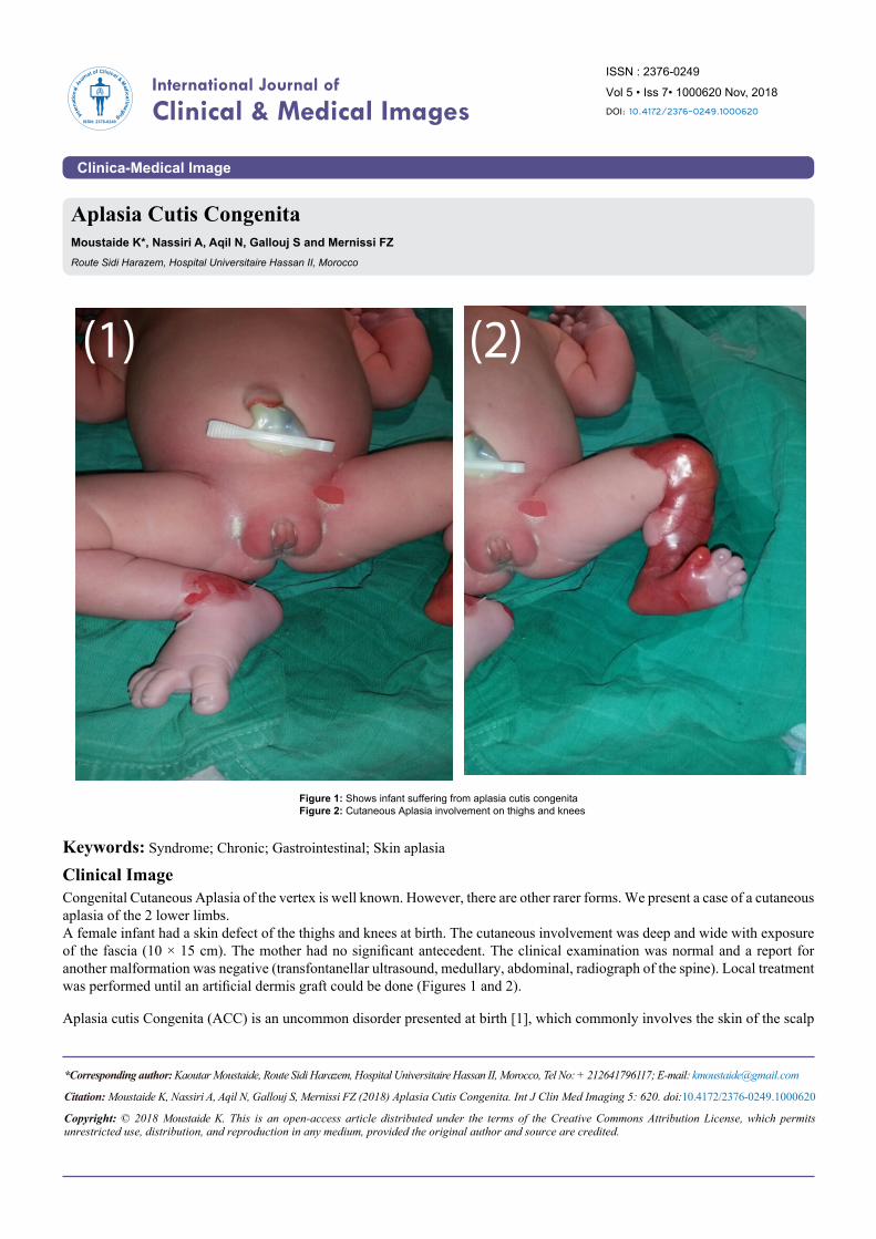
Clinical ImageCongenital Cutaneous Aplasia of the vertex is well known. However, there are other rarer forms. We present a case of a cutaneous aplasia of the 2 lower limbs.A female infant had a skin defect of the thighs and knees at birth. The cutaneous involvement was deep and wide with exposure of the fascia (10 × 15 cm). The mother had no significant antecedent. The clinical examination was normal and a report for another malformation was negative (transfontanellar ultrasound, medullary, abdominal, radiograph of the spine). Local treatment was performed until an artificial dermis graft could be done (Figures 1 and 2).
Aplasia cutis Congenita (ACC) is an uncommon disorder presented at birth [1], which commonly involves the skin of the scalp
*Corresponding author: Kaoutar Moustaide, Route Sidi Harazem, Hospital Universitaire Hassan II, Morocco, Tel No: + 212641796117; E-mail: [email protected]
Citation: Moustaide K, Nassiri A, Aqil N, Gallouj S, Mernissi FZ (2018) Aplasia Cutis Congenita. Int J Clin Med Imaging 5: 620. doi:10.4172/2376-0249.1000620
Copyright: © 2018 Moustaide K. This is an open-access article distributed under the terms of the Creative Commons Attribution License, which permits unrestricted use, distribution, and reproduction in any medium, provided the original author and source are credited.
(1) (2)
Figure 1: Shows infant suffering from aplasia cutis congenitaFigure 2: Cutaneous Aplasia involvement on thighs and knees
Volume 5 • Issue 7 • IJCMI
• Page 2 of 2 •
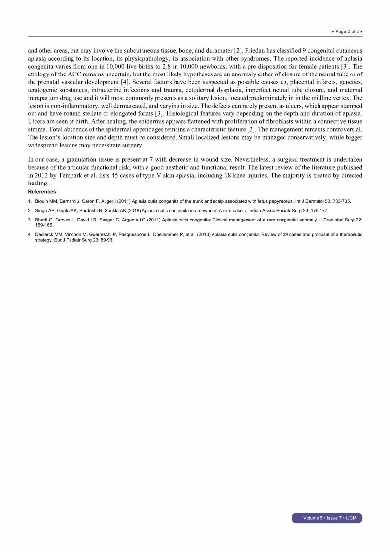
and other areas, but may involve the subcutaneous tissue, bone, and duramater [2]. Friedan has classified 9 congenital cutaneous aplasia according to its location, its physiopathology, its association with other syndromes. The reported incidence of aplasia congenita varies from one in 10,000 live births to 2.8 in 10,000 newborns, with a pre-disposition for female patients [3]. The etiology of the ACC remains uncertain, but the most likely hypotheses are an anormaly either of closure of the neural tube or of the prenatal vascular development [4]. Several factors have been suspected as possible causes eg, placental infarcts, genetics, teratogenic substances, intrauterine infections and trauma, ectodermal dysplasia, imperfect neural tube closure, and maternal intrapartum drug use and it will most commonly presents as a solitary lesion, located predominately in in the midline vertex. The lesion is non-inflammatory, well dermarcated, and varying in size. The defects can rarely present as ulcers, which appear stamped out and have rotund stellate or elongated forms [3]. Histological features vary depending on the depth and duration of aplasia. Ulcers are seen at birth. After healing, the epidermis appears flattened with proliferation of fibroblasts within a connective tissue stroma. Total abscence of the epidermal appendages remains a characteristic feature [2]. The management remains controversial. The lesion’s location size and depth must be considered. Small localized lesions may be managed conservatively, while bigger widespread lesions may necessitate surgery.
In our case, a granulation tissue is present at 7 with decrease in wound size. Nevertheless, a surgical treatment is undertaken because of the articular functional risk; with a good aesthetic and functional result. The latest review of the literature published in 2012 by Tempark et al. lists 45 cases of type V skin aplasia, including 18 knee injuries. The majority is treated by directed healing.References
1. Blouin MM, Bernard J, Caron F, Auger I (2011) Aplasia cutis congenita of the trunk and scalp associated with fetus papyraceus. Int J Dermatol 50: 733-735.
2. Singh AP, Gupta AK, Pardeshi R, Shukla AK (2018) Aplasia cutis congenita in a newborn: A rare case. J Indian Assoc Pediatr Surg 23: 175-177.
3. Bharti G, Groves L, David LR, Sanger C, Argenta LC (2011) Aplasia cutis congenita: Clinical management of a rare congenital anomaly. J Craniofac Surg 22: 159-165 .
4. Declerck MM, Vinchon M, Guerreschi P, Pasquesoone L, Dhellemmes P, et al. (2013) Aplasia cutis congenita: Review of 29 cases and proposal of a therapeutic strategy. Eur J Pediatr Surg 23: 89-93.
Related Documents