REVIEW published: 23 June 2015 doi: 10.3389/fnagi.2015.00117 Frontiers in Aging Neuroscience | www.frontiersin.org 1 June 2015 | Volume 7 | Article 117 Edited by: Merce Pallas, University of Barcelona, Spain Reviewed by: Marino M. Bianchin, Universidade Federal do Rio Grande do Sul, Brazil Stefano Delli Pizzi, University G. d’Annunzio" of Chieti-Pescara", Italy *Correspondence: Philip P. Foster, Pulmonary, Sleep and Critical Care Medicine, Department of Internal Medicine and Department of Nano Medicine and Biomedical Engineering, The University of Texas-Medical School at Houston, 6431 Fannin, MSB 1.434, Houston, TX 77030, USA [email protected] Received: 17 December 2014 Accepted: 01 June 2015 Published: 23 June 2015 Citation: Foster PP (2015) Role of physical and mental training in brain network configuration. Front. Aging Neurosci. 7:117. doi: 10.3389/fnagi.2015.00117 Role of physical and mental training in brain network configuration Philip P. Foster 1, 2 * 1 Department of Nano Medicine and Biomedical Engineering, The Brown Foundation, Institute of Molecular Medicine for the Prevention of Human Diseases, The University of Texas Health Science Center at Houston - Medical School, Houston, TX, USA, 2 Pulmonary, Sleep and Critical Care Medicine, Department of Internal Medicine, The University of Texas Health Science Center at Houston - Medical School, Houston, TX, USA It is hypothesized that the topology of brain networks is constructed by connecting nodes which may be continuously remodeled by appropriate training. Efficiency of physical and/or mental training on the brain relies on the flexibility of networks’ architecture molded by local remodeling of proteins and synapses of excitatory neurons producing transformations in network topology. Continuous remodeling of proteins of excitatory neurons is fine-tuning the scaling and strength of excitatory synapses up or down via regulation of intra-cellular metabolic and regulatory networks of the genome-transcriptome-proteome interface. Alzheimer’s disease is a model of “energy cost-driven small-world network disorder” with dysfunction of high-energy cost wiring as the network global efficiency is impaired by the deposition of an informed agent, the amyloid-β , selectively targeting high-degree nodes. In schizophrenia, the interconnectivity and density of rich-club networks are significantly reduced. Training-induced homeostatic synaptogenesis-enhancement, presumably via reconfiguration of brain networks into greater small-worldness, appears essential in learning, memory, and executive functions. A macroscopic cartography of creation-removal of synaptic connections in a macro-network, and at the intra-cellular scale, micro-networks regulate the physiological mechanisms for the preferential attachment of synapses. The strongest molecular relationship of exercise and functional connectivity was identified for brain-derived neurotrophic factor (BDNF). The allele variant, rs7294919, also shows a powerful relationship with the hippocampal volume. How the brain achieves this unique quest of reconfiguration remains a puzzle. What are the underlying mechanisms of synaptogenesis promoting communications brain ↔ muscle and brain ↔ brain in such trainings? What is the respective role of independent mental, physical, or combined-mental-physical trainings? Physical practice seems to be playing an instrumental role in the cognitive enhancement (brain ↔ muscle com.). However, mental training, meditation or virtual reality (films, games) require only minimal motor activity and cardio-respiratory stimulation. Therefore, other potential paths (brain ↔ brain com.) molding brain networks are nonetheless essential. Patients with motor neuron disease/injury (e.g., amyotrophic lateral sclerosis, traumatism) also achieve successful cognitive enhancement albeit they may only elicit mental practice. Keywords: Alzheimer’s disease, schizophrenia, depression, Yoga-meditation, video games, fluid intelligence, spatial memory, SNPs

Welcome message from author
This document is posted to help you gain knowledge. Please leave a comment to let me know what you think about it! Share it to your friends and learn new things together.
Transcript
REVIEWpublished: 23 June 2015
doi: 10.3389/fnagi.2015.00117
Frontiers in Aging Neuroscience | www.frontiersin.org 1 June 2015 | Volume 7 | Article 117
Edited by:
Merce Pallas,
University of Barcelona, Spain
Reviewed by:
Marino M. Bianchin,
Universidade Federal do Rio Grande
do Sul, Brazil
Stefano Delli Pizzi,
University G. d’Annunzio"
of Chieti-Pescara", Italy
*Correspondence:
Philip P. Foster,
Pulmonary, Sleep and Critical Care
Medicine, Department of Internal
Medicine and Department of Nano
Medicine and Biomedical Engineering,
The University of Texas-Medical
School at Houston, 6431 Fannin,
MSB 1.434, Houston, TX 77030, USA
Received: 17 December 2014
Accepted: 01 June 2015
Published: 23 June 2015
Citation:
Foster PP (2015) Role of physical and
mental training in brain network
configuration.
Front. Aging Neurosci. 7:117.
doi: 10.3389/fnagi.2015.00117
Role of physical and mental trainingin brain network configuration
Philip P. Foster 1, 2*
1Department of Nano Medicine and Biomedical Engineering, The Brown Foundation, Institute of Molecular Medicine for the
Prevention of Human Diseases, The University of Texas Health Science Center at Houston - Medical School, Houston, TX,
USA, 2 Pulmonary, Sleep and Critical Care Medicine, Department of Internal Medicine, The University of Texas Health Science
Center at Houston - Medical School, Houston, TX, USA
It is hypothesized that the topology of brain networks is constructed by connecting
nodes which may be continuously remodeled by appropriate training. Efficiency of
physical and/or mental training on the brain relies on the flexibility of networks’
architecture molded by local remodeling of proteins and synapses of excitatory
neurons producing transformations in network topology. Continuous remodeling of
proteins of excitatory neurons is fine-tuning the scaling and strength of excitatory
synapses up or down via regulation of intra-cellular metabolic and regulatory
networks of the genome-transcriptome-proteome interface. Alzheimer’s disease is
a model of “energy cost-driven small-world network disorder” with dysfunction
of high-energy cost wiring as the network global efficiency is impaired by the
deposition of an informed agent, the amyloid-β, selectively targeting high-degree
nodes. In schizophrenia, the interconnectivity and density of rich-club networks
are significantly reduced. Training-induced homeostatic synaptogenesis-enhancement,
presumably via reconfiguration of brain networks into greater small-worldness, appears
essential in learning, memory, and executive functions. A macroscopic cartography of
creation-removal of synaptic connections in a macro-network, and at the intra-cellular
scale, micro-networks regulate the physiological mechanisms for the preferential
attachment of synapses. The strongest molecular relationship of exercise and functional
connectivity was identified for brain-derived neurotrophic factor (BDNF). The allele variant,
rs7294919, also shows a powerful relationship with the hippocampal volume. How the
brain achieves this unique quest of reconfiguration remains a puzzle. What are the
underlying mechanisms of synaptogenesis promoting communications brain ↔ muscle
and brain ↔ brain in such trainings? What is the respective role of independent mental,
physical, or combined-mental-physical trainings? Physical practice seems to be playing
an instrumental role in the cognitive enhancement (brain ↔ muscle com.). However,
mental training, meditation or virtual reality (films, games) require only minimal motor
activity and cardio-respiratory stimulation. Therefore, other potential paths (brain↔ brain
com.) molding brain networks are nonetheless essential. Patients with motor neuron
disease/injury (e.g., amyotrophic lateral sclerosis, traumatism) also achieve successful
cognitive enhancement albeit they may only elicit mental practice.
Keywords: Alzheimer’s disease, schizophrenia, depression, Yoga-meditation, video games, fluid intelligence,
spatial memory, SNPs
Foster Brain network reconfiguration
A long-standing question in the neuroscience of aging hasbeen the interplay between physical activity andmental practices,especially to prevent the potential aging-related cognitivedecline in reasoning, executive function, processing speed,memory, and spatial ability. Intelligence spans unequally amongindividuals and follows approximately a normal distributionin the population (Deary et al., 2010). Converging lines ofevidence indicate that intelligence is not confined to a limitedbrain area. Rather, the “connectome” or structural networkconfiguration of brain regions correlates to individual differencesin intelligence (Bullmore and Sporns, 2009; Sporns and Bullmore,2014). Based on diffusion tensor imaging (DTI), white matterintegrity, particularly long-distance white matter associationtracts, such as arcuate and uncinate fasciculi, has been correlatedto increased cognitive ability (Turken et al., 2008; Dearyet al., 2010). Clearly, it seems that intelligence requires bothintegrity and high degree of organizational efficiency of whitematter networks. The existence of rich-club networks andresting-state networks (RSNs) became fundamental in cognitiveneuroscience. Modeling the topology of rich-club networks fromDTI data has been instrumental to determine the dysfunctionof their connectivity in patients with schizophrenia (van denHeuvel et al., 2013). However, the underlying mechanisms ofphysical and mental training on brain networks still remain,for the most part, elusive. Despite accumulation of growingevidence for brain networks’ remodeling, synaptogenesis, andneurogenesis resulting from extensive communications intra- orinter-organs, (brain ↔ brain com.) and (brain ↔ muscle com.),during motion, activities, exercise training, exergaming, exercisesimulation, spatial memory stimulation, mental training, socialinteractions, or meditation, the hierarchical relationship of thosecommunications has yet to be clarified.
Original Configuration
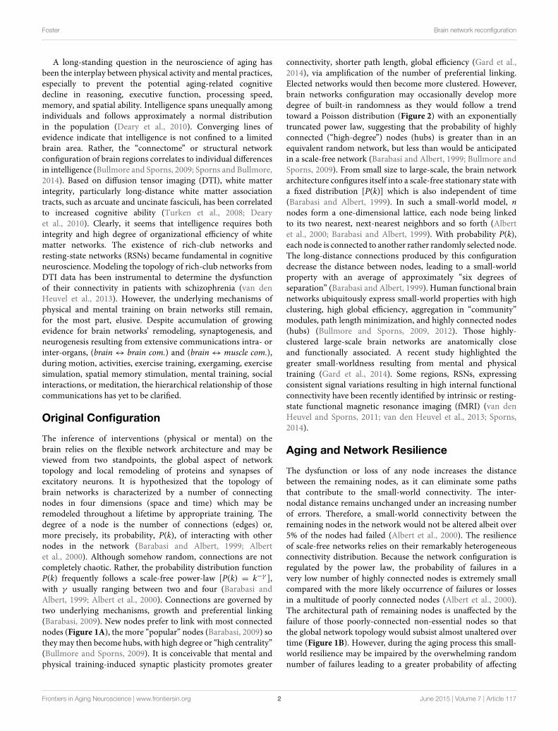
The inference of interventions (physical or mental) on thebrain relies on the flexible network architecture and may beviewed from two standpoints, the global aspect of networktopology and local remodeling of proteins and synapses ofexcitatory neurons. It is hypothesized that the topology ofbrain networks is characterized by a number of connectingnodes in four dimensions (space and time) which may beremodeled throughout a lifetime by appropriate training. Thedegree of a node is the number of connections (edges) or,more precisely, its probability, P(k), of interacting with othernodes in the network (Barabasi and Albert, 1999; Albertet al., 2000). Although somehow random, connections are notcompletely chaotic. Rather, the probability distribution functionP(k) frequently follows a scale-free power-law [P(k) = k−γ ],with γ usually ranging between two and four (Barabasi andAlbert, 1999; Albert et al., 2000). Connections are governed bytwo underlying mechanisms, growth and preferential linking(Barabasi, 2009). New nodes prefer to link with most connectednodes (Figure 1A), the more “popular” nodes (Barabasi, 2009) sotheymay then become hubs, with high degree or “high centrality”(Bullmore and Sporns, 2009). It is conceivable that mental andphysical training-induced synaptic plasticity promotes greater
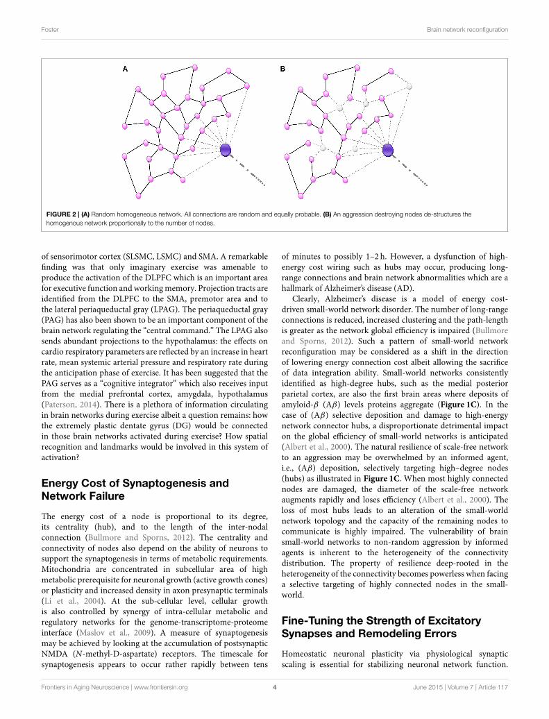
connectivity, shorter path length, global efficiency (Gard et al.,2014), via amplification of the number of preferential linking.Elected networks would then become more clustered. However,brain networks configuration may occasionally develop moredegree of built-in randomness as they would follow a trendtoward a Poisson distribution (Figure 2) with an exponentiallytruncated power law, suggesting that the probability of highlyconnected (“high-degree”) nodes (hubs) is greater than in anequivalent random network, but less than would be anticipatedin a scale-free network (Barabasi and Albert, 1999; Bullmore andSporns, 2009). From small size to large-scale, the brain networkarchitecture configures itself into a scale-free stationary state witha fixed distribution [P(k)] which is also independent of time(Barabasi and Albert, 1999). In such a small-world model, nnodes form a one-dimensional lattice, each node being linkedto its two nearest, next-nearest neighbors and so forth (Albertet al., 2000; Barabasi and Albert, 1999). With probability P(k),each node is connected to another rather randomly selected node.The long-distance connections produced by this configurationdecrease the distance between nodes, leading to a small-worldproperty with an average of approximately “six degrees ofseparation” (Barabasi and Albert, 1999). Human functional brainnetworks ubiquitously express small-world properties with highclustering, high global efficiency, aggregation in “community”modules, path length minimization, and highly connected nodes(hubs) (Bullmore and Sporns, 2009, 2012). Those highly-clustered large-scale brain networks are anatomically closeand functionally associated. A recent study highlighted thegreater small-worldness resulting from mental and physicaltraining (Gard et al., 2014). Some regions, RSNs, expressingconsistent signal variations resulting in high internal functionalconnectivity have been recently identified by intrinsic or resting-state functional magnetic resonance imaging (fMRI) (van denHeuvel and Sporns, 2011; van den Heuvel et al., 2013; Sporns,2014).
Aging and Network Resilience
The dysfunction or loss of any node increases the distancebetween the remaining nodes, as it can eliminate some pathsthat contribute to the small-world connectivity. The inter-nodal distance remains unchanged under an increasing numberof errors. Therefore, a small-world connectivity between theremaining nodes in the network would not be altered albeit over5% of the nodes had failed (Albert et al., 2000). The resilienceof scale-free networks relies on their remarkably heterogeneousconnectivity distribution. Because the network configuration isregulated by the power law, the probability of failures in avery low number of highly connected nodes is extremely smallcompared with the more likely occurrence of failures or lossesin a multitude of poorly connected nodes (Albert et al., 2000).The architectural path of remaining nodes is unaffected by thefailure of those poorly-connected non-essential nodes so thatthe global network topology would subsist almost unaltered overtime (Figure 1B). However, during the aging process this small-world resilience may be impaired by the overwhelming randomnumber of failures leading to a greater probability of affecting
Frontiers in Aging Neuroscience | www.frontiersin.org 2 June 2015 | Volume 7 | Article 117

Foster Brain network reconfiguration
FIGURE 1 | (A) Small-world network: A scale-free network. The size of a
node (vertex) is proportionally related to the number of connections (edges)
that links it to other nodes in the network. Highly-wired nodes with high
degree of centrality or high connectivity are illustrated by large blue spheres.
Small pink spheres represent low-degree nodes. Hubs (blue spheres), often
interact with other regions or “community” modules within the global
network; they are highly connected and have high centrality. The bold edges
depict connections with other modules or regions. (B) The scale-free
network (same as in A) is inhomogeneous thus resilient as it is relatively
minimally affected by a random and non-directed aggression toward a few
high-degree nodes mainly causing the loss of more frequent more profuse
low-degree nodes. The aggression is visually represented by the loss of
nodes (white) and missing edges (dotted lines). (C) An informed agent
aggressing high degree nodes and hubs. Visually, the network architecture is
completely de-structured with the detrimental loss of a major number of
connections (loss nodes in white, missing edges as dotted lines).
high-degree nodes. In addition, the repair process of failed nodesmay be impaired with aging. Therefore, the functional integrationbecomes reduced by lengthening of the characteristic path lengthof networks.
Instantaneous Brain Network Response toExercise
A hint on how patients with neuron disease/injury (e.g.,amyotrophic lateral sclerosis, traumatism) may stimulate specificexercise-sensitive brain networks without any motor activity isprovided by the following experiment. Hypnotic suggestion of
exercising “uphill” elicits an increased breathing response (rateand minute ventilation, V̇E) (Paterson, 2014). If the respiratorymuscles are not too severely affected by the neuro-musculardisease such patients may respond to the suggestion andpossibly to autosuggestion. The terminology “central command”has been used to express the physiological modulation ofcardiorespiratory response to a constant exercise or a simulationof exercise (Paterson, 2014). Imaginary exercise (imaginingcycling uphill) induced activation of the dorsal lateral prefrontalcortex (DLPFC), superolateral sensorimotor cortex (SLSMC),lateral sensorimotor cortex (LSMC), and supplementary motorarea (SMA) as seen by positron emission scanning (Paterson,2014). Voluntarily breathing alone produced solely an activation
Frontiers in Aging Neuroscience | www.frontiersin.org 3 June 2015 | Volume 7 | Article 117
Foster Brain network reconfiguration
FIGURE 2 | (A) Random homogeneous network. All connections are random and equally probable. (B) An aggression destroying nodes de-structures the
homogenous network proportionally to the number of nodes.
of sensorimotor cortex (SLSMC, LSMC) and SMA. A remarkablefinding was that only imaginary exercise was amenable toproduce the activation of the DLPFC which is an important areafor executive function and workingmemory. Projection tracts areidentified from the DLPFC to the SMA, premotor area and tothe lateral periaqueductal gray (LPAG). The periaqueductal gray(PAG) has also been shown to be an important component of thebrain network regulating the “central command.” The LPAG alsosends abundant projections to the hypothalamus: the effects oncardio respiratory parameters are reflected by an increase in heartrate, mean systemic arterial pressure and respiratory rate duringthe anticipation phase of exercise. It has been suggested that thePAG serves as a “cognitive integrator” which also receives inputfrom the medial prefrontal cortex, amygdala, hypothalamus(Paterson, 2014). There is a plethora of information circulatingin brain networks during exercise albeit a question remains: howthe extremely plastic dentate gyrus (DG) would be connectedin those brain networks activated during exercise? How spatialrecognition and landmarks would be involved in this system ofactivation?
Energy Cost of Synaptogenesis andNetwork Failure
The energy cost of a node is proportional to its degree,its centrality (hub), and to the length of the inter-nodalconnection (Bullmore and Sporns, 2012). The centrality andconnectivity of nodes also depend on the ability of neurons tosupport the synaptogenesis in terms of metabolic requirements.Mitochondria are concentrated in subcellular area of highmetabolic prerequisite for neuronal growth (active growth cones)or plasticity and increased density in axon presynaptic terminals(Li et al., 2004). At the sub-cellular level, cellular growthis also controlled by synergy of intra-cellular metabolic andregulatory networks for the genome-transcriptome-proteomeinterface (Maslov et al., 2009). A measure of synaptogenesismay be achieved by looking at the accumulation of postsynapticNMDA (N-methyl-D-aspartate) receptors. The timescale forsynaptogenesis appears to occur rather rapidly between tens
of minutes to possibly 1–2 h. However, a dysfunction of high-energy cost wiring such as hubs may occur, producing long-range connections and brain network abnormalities which are ahallmark of Alzheimer’s disease (AD).
Clearly, Alzheimer’s disease is a model of energy cost-driven small-world network disorder. The number of long-rangeconnections is reduced, increased clustering and the path-lengthis greater as the network global efficiency is impaired (Bullmoreand Sporns, 2012). Such a pattern of small-world networkreconfiguration may be considered as a shift in the directionof lowering energy connection cost albeit allowing the sacrificeof data integration ability. Small-world networks consistentlyidentified as high-degree hubs, such as the medial posteriorparietal cortex, are also the first brain areas where deposits ofamyloid-β (Aβ) levels proteins aggregate (Figure 1C). In thecase of (Aβ) selective deposition and damage to high-energynetwork connector hubs, a disproportionate detrimental impacton the global efficiency of small-world networks is anticipated(Albert et al., 2000). The natural resilience of scale-free networkto an aggression may be overwhelmed by an informed agent,i.e., (Aβ) deposition, selectively targeting high–degree nodes(hubs) as illustrated in Figure 1C. When most highly connectednodes are damaged, the diameter of the scale-free networkaugments rapidly and loses efficiency (Albert et al., 2000). Theloss of most hubs leads to an alteration of the small-worldnetwork topology and the capacity of the remaining nodes tocommunicate is highly impaired. The vulnerability of brainsmall-world networks to non-random aggression by informedagents is inherent to the heterogeneity of the connectivitydistribution. The property of resilience deep-rooted in theheterogeneity of the connectivity becomes powerless when facinga selective targeting of highly connected nodes in the small-world.
Fine-Tuning the Strength of ExcitatorySynapses and Remodeling Errors
Homeostatic neuronal plasticity via physiological synapticscaling is essential for stabilizing neuronal network function.
Frontiers in Aging Neuroscience | www.frontiersin.org 4 June 2015 | Volume 7 | Article 117
Foster Brain network reconfiguration
Overall remodeling of proteins of excitatory neurons is fine-tuning the scaling and strength of excitatory synapses upor down via activation of AMPA receptors, BDNF, guanylatekinase–associated protein (GKAP), TNF-α and all-trans retinoicacid (Shin et al., 2012). On a macroscopic scale of fourdimensions, the degrees of nodes are modified and the small-world networks’ architectures are continuously remodeled intonew configurations. External treatment such as physical ormental training may also infer with the homeostatic scaling andreconfigure brain networks.
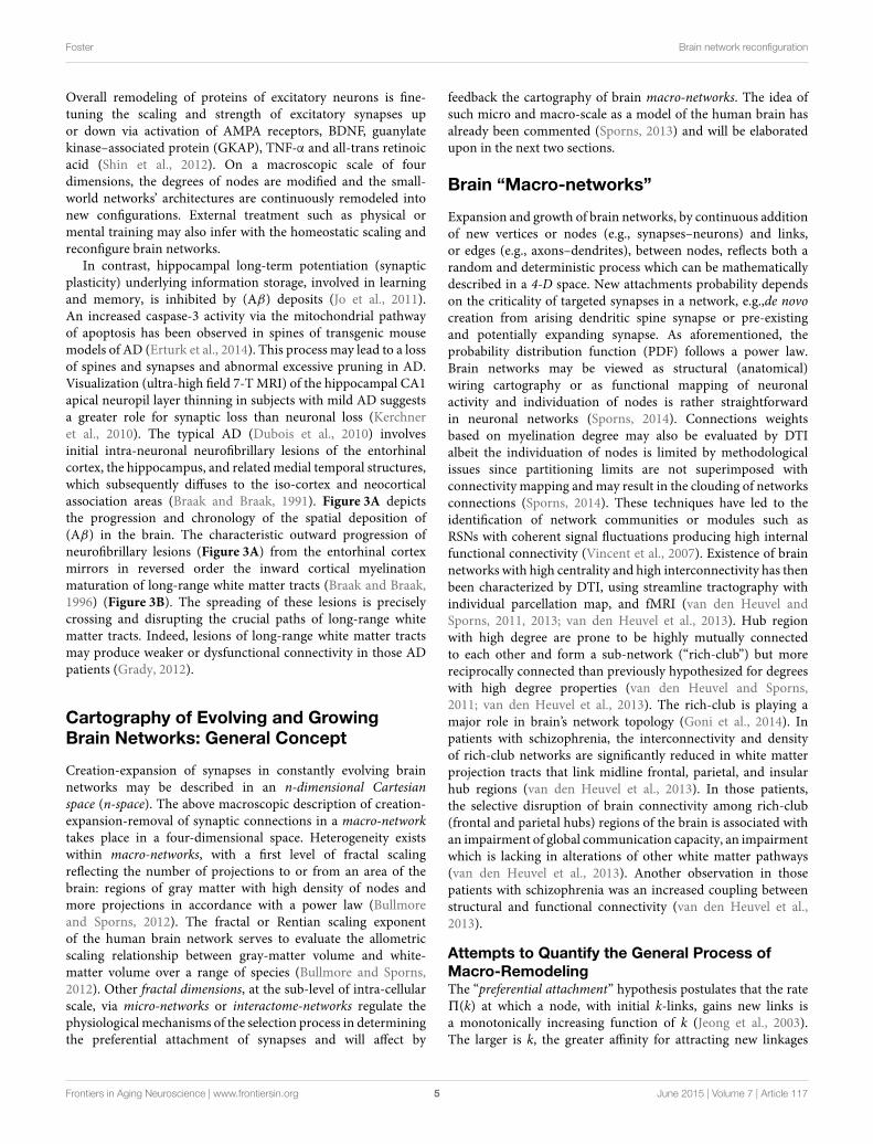
In contrast, hippocampal long-term potentiation (synapticplasticity) underlying information storage, involved in learningand memory, is inhibited by (Aβ) deposits (Jo et al., 2011).An increased caspase-3 activity via the mitochondrial pathwayof apoptosis has been observed in spines of transgenic mousemodels of AD (Erturk et al., 2014). This process may lead to a lossof spines and synapses and abnormal excessive pruning in AD.Visualization (ultra-high field 7-TMRI) of the hippocampal CA1apical neuropil layer thinning in subjects with mild AD suggestsa greater role for synaptic loss than neuronal loss (Kerchneret al., 2010). The typical AD (Dubois et al., 2010) involvesinitial intra-neuronal neurofibrillary lesions of the entorhinalcortex, the hippocampus, and related medial temporal structures,which subsequently diffuses to the iso-cortex and neocorticalassociation areas (Braak and Braak, 1991). Figure 3A depictsthe progression and chronology of the spatial deposition of(Aβ) in the brain. The characteristic outward progression ofneurofibrillary lesions (Figure 3A) from the entorhinal cortexmirrors in reversed order the inward cortical myelinationmaturation of long-range white matter tracts (Braak and Braak,1996) (Figure 3B). The spreading of these lesions is preciselycrossing and disrupting the crucial paths of long-range whitematter tracts. Indeed, lesions of long-range white matter tractsmay produce weaker or dysfunctional connectivity in those ADpatients (Grady, 2012).
Cartography of Evolving and GrowingBrain Networks: General Concept
Creation-expansion of synapses in constantly evolving brainnetworks may be described in an n-dimensional Cartesianspace (n-space). The above macroscopic description of creation-expansion-removal of synaptic connections in a macro-networktakes place in a four-dimensional space. Heterogeneity existswithin macro-networks, with a first level of fractal scalingreflecting the number of projections to or from an area of thebrain: regions of gray matter with high density of nodes andmore projections in accordance with a power law (Bullmoreand Sporns, 2012). The fractal or Rentian scaling exponentof the human brain network serves to evaluate the allometricscaling relationship between gray-matter volume and white-matter volume over a range of species (Bullmore and Sporns,2012). Other fractal dimensions, at the sub-level of intra-cellularscale, via micro-networks or interactome-networks regulate thephysiological mechanisms of the selection process in determiningthe preferential attachment of synapses and will affect by
feedback the cartography of brain macro-networks. The idea ofsuch micro and macro-scale as a model of the human brain hasalready been commented (Sporns, 2013) and will be elaboratedupon in the next two sections.
Brain “Macro-networks”
Expansion and growth of brain networks, by continuous additionof new vertices or nodes (e.g., synapses–neurons) and links,or edges (e.g., axons–dendrites), between nodes, reflects both arandom and deterministic process which can be mathematicallydescribed in a 4-D space. New attachments probability dependson the criticality of targeted synapses in a network, e.g.,de novocreation from arising dendritic spine synapse or pre-existingand potentially expanding synapse. As aforementioned, theprobability distribution function (PDF) follows a power law.Brain networks may be viewed as structural (anatomical)wiring cartography or as functional mapping of neuronalactivity and individuation of nodes is rather straightforwardin neuronal networks (Sporns, 2014). Connections weightsbased on myelination degree may also be evaluated by DTIalbeit the individuation of nodes is limited by methodologicalissues since partitioning limits are not superimposed withconnectivity mapping andmay result in the clouding of networksconnections (Sporns, 2014). These techniques have led to theidentification of network communities or modules such asRSNs with coherent signal fluctuations producing high internalfunctional connectivity (Vincent et al., 2007). Existence of brainnetworks with high centrality and high interconnectivity has thenbeen characterized by DTI, using streamline tractography withindividual parcellation map, and fMRI (van den Heuvel andSporns, 2011, 2013; van den Heuvel et al., 2013). Hub regionwith high degree are prone to be highly mutually connectedto each other and form a sub-network (“rich-club”) but morereciprocally connected than previously hypothesized for degreeswith high degree properties (van den Heuvel and Sporns,2011; van den Heuvel et al., 2013). The rich-club is playing amajor role in brain’s network topology (Goni et al., 2014). Inpatients with schizophrenia, the interconnectivity and densityof rich-club networks are significantly reduced in white matterprojection tracts that link midline frontal, parietal, and insularhub regions (van den Heuvel et al., 2013). In those patients,the selective disruption of brain connectivity among rich-club(frontal and parietal hubs) regions of the brain is associated withan impairment of global communication capacity, an impairmentwhich is lacking in alterations of other white matter pathways(van den Heuvel et al., 2013). Another observation in thosepatients with schizophrenia was an increased coupling betweenstructural and functional connectivity (van den Heuvel et al.,2013).
Attempts to Quantify the General Process ofMacro-RemodelingThe “preferential attachment” hypothesis postulates that the rate5(k) at which a node, with initial k-links, gains new links isa monotonically increasing function of k (Jeong et al., 2003).The larger is k, the greater affinity for attracting new linkages
Frontiers in Aging Neuroscience | www.frontiersin.org 5 June 2015 | Volume 7 | Article 117
Foster Brain network reconfiguration
FIGURE 3 | (A) In Alzheimer’s disease (AD), the outward progression of
neurofibrillary lesions spreads from the entorhinal cortex toward the
cortex (blue-purple spirals and arrows). Those lesions are crossing and
blocking the path of long-distance white matter tracts. Braak stages are
in Roman numerals. (B) The inward cortical myelination maturation of
long-range white matter tracts is directed toward the entorhinal cortex
(red spiral and purple arrows) and replicates in reversed order the
neurofibrillary lesions of AD.
by this “popular” node. It has been proposed that the k-links’distribution follows a power law (Barabasi and Albert, 1999;Albert et al., 2000). As a result, the time course of the k-links’distribution, or degree kiof the i
th node may be obtained by thedifferential equation (Jeong et al., 2003)
dki
dt= m5(k)i, (1)
wherem is a constant, and 5(k) may be expressed as follows
5(k)i =kγ
i∑
j kγ
j
, (2)
with γ > 0 an unknown scaling exponent. When γ = 1,Equation (2) becomes a scale-free model (Barabasi and Albert,1999), and the probability distribution function (PDF), P(k),frequently follows a scale-free power-law [P(k) = k−γ ], withusually γ = 3. As the network expands, the function 5(ki)provides the rate of acquisition of new links by a pre-existingnode with k-links. Knowing 5(k) requires evaluation of how oldnodes acquire new nodes, as a function of the degree of the oldnode.
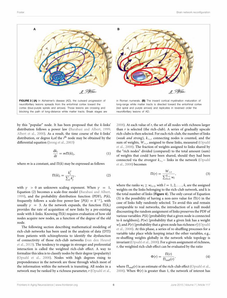
The following section describing mathematical modeling ofrich-club networks has been used in the analysis of data (DTI)from patients with schizophrenia to identify the dysfunctionof connectivity of those rich-club networks (van den Heuvelet al., 2013). The tendency to engage in stronger and preferentialinteraction is called the weighted rich-club effect. A way toformalize this idea is to classify nodes by their degree (popularity)(Opsahl et al., 2008). Nodes with high degrees rising topreponderance in the network are those through which most ofthe information within the network is transiting. All nodes in anetwork may be ranked by a richness parameter, r (Opsahl et al.,
2008). At each value of r, the set of all nodes with richness largerthan r is selected (the rich-club). A series of gradually upscalerich-clubs is then selected. For each rich-club, the number of links(weak and strong), k>r , connecting nodes is counted, and thesum of weights, W>r , assigned to these links, measured (Opsahlet al., 2008). The fraction of weights assigned to links shared bythe “rich nodes” divided (compared) to the total amount (sum)of weights that could have been shared, should they had beenconnected via the strongest k>r− links in the network (Opsahlet al., 2008) becomes
5(r) =W>r
∑k>r
l=1wl
, (3)
where the ranks wl ≥ wl+1, with l = 1, 2, . . . , k, are the assignedweights on the links belonging to the rich-club network, and k isthe total number of links (Figure 4). The only caveat of Equation(3) is the possibility of having a non-zero value for 5(r) in thecase of links fully randomly selected. To avoid this and remaincomparable to real networks, the introduction of a null modeldiscounting the random assignment of links preserves the PDF ofvarious variables: P(k) [probability that a given node is connectedto k neighbors]; P(w) [probability that a given link has a weightw]; and P(r) [probability that a given node has richness r] (Opsahlet al., 2008). At this phase, a series of re-shuffling processes for avariable take place while keeping intact the other variables, e.g.,re-shuffling weights globally in the network while topology isinvariant (Opsahl et al., 2008). For a given assignment of richness,r, the weighted rich-club effect can be evaluated by the ratio
8(r) =5(r)
5null(r), (4)
where 5null(r) is an estimate of the rich-club effect (Opsahl et al.,2008). When 8(r) is greater than 1, the network of interest has
Frontiers in Aging Neuroscience | www.frontiersin.org 6 June 2015 | Volume 7 | Article 117

Foster Brain network reconfiguration
FIGURE 4 | Illustration of a rich-club network. (A) The size of nodes is
proportional to their richness. High-degree nodes are shaded-blue,
lower-degree are shaded-pink. The weights (strength) assigned to the links
are indicated by Indo-Arabic numerals. Links (dark brown) are connecting
nodes (shaded-blue) within the rich-club network. Only six links are
connecting those high-degree nodes together, thus k>r = 6, and
W>r = 4+ 10+ 5+ 1+ 1+ 1 = 22. (B) Not all strongest k>r = 6−links (dark
brown) of the entire network are connecting rich-club network’s nodes
(shaded-blue). Some strong links are also connecting lower-degree nodes
(shaded-pink). Therefore, the denominator of Equation (3) becomes∑k>r
l=1 wl = 6+ 10+ 5+ 6+ 6+ 6 = 39 and the ratio is readily obtained as
5(r) = 22/
39. Modified and re-sampled from Opsahl et al. (2008).
a positive weighted rich-club effect. To the contrary, if 8(r), issmaller than 1, links in the network of interest are weaker thanrandomly predicted.
Macro-Remodeling as a Function of TimeAnother way to express Equation (1) is to observe the addition ofnew synapses (nodes) in a small time-interval1t, starting at timet, and ending at t +1t. Thereby, instead of5(k), it is also possibleto study F(k), the cumulative distribution function (CDF) of newnodes (e.g., synapses) creation, defined as (Jeong et al., 2003)
F(k) =
∫ k
05(ki)dk. (5)
The same treatment may be applied to the variation of the rich-club effect in time by studying 8(r) (Equation 4) so that the CDFof the rich-club effect becomes
F(r) =
∫ r
08(r)dr. (6)
However, the overarching question is not whether the CDF,5(k),of the overall growth of the brain network follows a fixedfunction. Such a function ignores the elementary processes,essential components, in the selection process of creating,expanding or removing nodes in a constantly evolving realnetwork (Ghoshal et al., 2013). Rather, how elementary processesunderlying preferential attachments, e.g., related to the aging-fitness of nodes, availability-functionality of genomic and
proteomic intra-cellular systems, and preferential paths of actionpotential would regulate the network evolution?
Decision, at the Elementary Level, ofMacro-RemodelingBased on several inputs from those elementary processes,received at time t, each ith synapse, inasmuch as one or moresynapses may represent one node (or vertex), makes its owndecision about its state and the selection of future connections. Away to assess the selective process by which a synapse follows itsown agenda is the introduction of Boolean networks (Albert andBarabasi, 2000) in the next phase of the decision tree. In brainnetworks, the Boolean model would be made up of N synapses(vertices) which are defined by Boolean variables (spins) thatmay take values σi = 0 (not connected) or 1 (connected). Eachsynapse may receive inputs from k synapses. Potential randomvalues of the spins selected from these neighboring synapsesare as much as 2k. The Boolean function, Bi, grants each ofthese 2k inputs (Albert and Barabasi, 2000), an output value of1 (e.g., synaptic connection established) or 0 (no further durableconnection). The outputs are randomly selected, assigning avalue 1 for a probability P (will be connected) and a value 0for a probability (1–P) (will not be connected). The Booleanmatrix determines, at any time t, the spin of the ith synapse, basedon the spin of its k neighboring synapses. Figure 5 illustratesthe case of k = 2, where Node i may receive four potentiallydifferent inputs from two neighboring synapses, j and l(Figure 5A).
Frontiers in Aging Neuroscience | www.frontiersin.org 7 June 2015 | Volume 7 | Article 117

Foster Brain network reconfiguration
FIGURE 5 | The Boolean matrix (C) defines Bi , or adjacency matrix (in
graph theory) whose rows and columns (right matrix M [Bi
(
σj, σl
)
] , C)
represent the nodes (e.g., synapses) and whose entries represent the
edges (e.g., axons) of the graph (left matrix Mj,l , B). The attribution of the
output Bi
(
σj, σl
)
= σi , e.g., to Synapse i, to each input(
σj
)
and(
σl
)
depends
on the values of those inputs (0 or 1) and the order of first edge
activation/stimulation(
σj, σl
)
or(
σl, σj
)
(initial propagation of an axonal action
potential) as illustrated on (A). The values of Bi
(
σj, σl
)
have been arbitrarily
selected for the example shown.
Intra-Cellular “Micro-networks”
In a fractal dimension of lower scale, micro-networks exhibitsimilar patterns as macro-networks. However, differentassignments are specific to the micro-scale so that verticesmay become macromolecules, while the edges are biophysical,biochemical, and functional interactions of DNA (genesequences), RNA or metabolites (Vidal et al., 2011). In atranscriptional regulatory network, nodes correspond totranscription factors or presumed DNA regulatory elements,and edges represent the chemical binding between the two.In systems biology, four types of micro-networks are used todescribe biological processes (Le Novere, 2015).
The interactome-networks are generally used to evaluatewhether X affects Y (physical or chemical interaction), e.g.,between genes and proteins. The ENIGMA Consortium lookedat the intergenic single nucleotide polymorphisms (SNPs)influencing the hippocampal volume (Stein et al., 2012). In thisparticular example, one SNP shows a powerful relationship withthe hippocampal volume normalized to intracranial volume.The SNP, locus rs7294919, of gene RPL 6P15, on chromosome12, was associated with hippocampal volume. In this case, X(allele variant rs7294919) is known, the protein Y not fullyidentified, and the phenotype, at macro-network fractal scale, isthe hippocampal volume. This SNP, rs7294919, was presumed toregulate the transcriptional activity of a neighboring gene, TESC,and its end-product tescalcin (Stein et al., 2012; Dannlowskiet al., 2015). Tescalcin (Y?) may enhance hippocampal cellproliferation and differentiation.
The activity flow networks provide additional informationabout whether Y is increased or decreased. As an example, mostsynaptic genes (X) are upregulated in mild cognitive impairment(MCI) compared to age-matched controls, particularly in thehippocampus, superior frontal gyrus, and post central gyruswhile being downregulated in AD (Cribbs et al., 2012; Berchtoldet al., 2014). The genes X may be involved in the encoding andincreased production of some proteins (Y) such as neurexins,integrins, or cadherins (Berchtold et al., 2014).
The process description networks detail and quantify howX affects Y (directed, sequential, mechanistic); they may beused to describe a mass transfer. An example is provided bya SNP of the Neuregulin-1 (NRG-1) gene (X); an imbalancein Neuregulin-1 (NRG-1β) may disrupt the dopaminergic andglutamatergic functions, neurotransmitter pathways associatedwith schizophrenia (Kwon et al., 2008). The NRG-1β protein(Y) exerts its effects on synaptic plasticity within minutes andincrease levels of extracellular dopamine levels dopamine in thehippocampus and striatum.
The entity relationship networks introduce the directionalityof the relationship, e.g., X stimulates Y, but Y does not influenceX; such networks are used for mapping the cell cycle or apoptosis(Le Novere, 2015). Those micro-networks usually express a scale-free topology and high clustering (Barabasi and Oltvai, 2004). Incells, scale-free networks are ultra-small and their path lengthsare short. A short path length implies that a local perturbation ofthe concentration, [X], could affect Y, Z and rapidly spread to theentire network—path of two or three reactions away.
Hierarchical or fractal modularity of network topology existsfor reconfiguration of connections between nodes (Bullmoreand Sporns, 2012). This hierarchical modularity and macro-networks homeostasis rely on the optimal functionality of anintact genomic and proteomic intra-cellular systemwithinmicro-networks. In contrast, the opposite is not true and optimaloperation of macro-networks does not seem necessary for propermicro-networks functionality. However, a hierarchy seems toexist, with a sovereignty of the macro-networks over micro-networks. In the synaptic macro-network, the inference byrepetitions of propagation of action potential (e.g., training), onthe aging–fitness of nodes (synapses) seems critical to increasethe synaptic density.
Synaptogenesis Induction and BrainNetworks
From a structural standpoint, brain plasticity entails the potentialof neurons to change their synaptic connections (Ashfordand Jarvik, 1985). The lengthening of axons, sprouting ofcollateral ramifications, and remodeling allow the dwelling ofnew synapses, new cognitive, and behavioral operations (Fosteret al., 2011). Skeletal muscle exercise affects brain plasticity andmay arrest, slow down or even reverse the pathophysiologicalevolution to MCI (Foster et al., 2011).
Aging affects three high-degree and large-scale brain networksmodulating interconnectivity between the frontal cortex andthe rest of the brain: (1) Default mode network (DMN)
Frontiers in Aging Neuroscience | www.frontiersin.org 8 June 2015 | Volume 7 | Article 117

Foster Brain network reconfiguration
(posterior cingulate, ventral–superior frontal medial cortices,and bilateral lateral occipital, middle frontal, hippocampal, andpara-hippocampal, and middle temporal cortices]; (2) Fronto-executive network (FE); and (3) Fronto-parietal (FP) network(Voss et al., 2010). Skeletal muscle activity mitigates the age-related deterioration of those networks depending on the qualityand length of training. One year of walking increased functionalconnectivity within the DMN and the FE Networks but non-significant at 6 months (Voss et al., 2010). A group, trainingin non-aerobic stretching, toning and balance, also showedincreased functional connectivity in the DMN after 6 monthsand in the FP Network after 12 months. It appears that anexercise-induced restoration of age-deteriorated brain networkstook place. Improved functional connectivity was associatedwith greater improvement in executive function and behavioralnormalization.
Activity-induced plasticity of spines and synapses models thenodes and the edges of small-world networks. Pruning of synapseexuberance or redundancy to shorter the path length and globalefficiency is also an aspect of the normal plasticity of the brain.Healthy brains, especially in the hippocampus, are the sites ofcontinual effervescence of molding new synapses or eliminatingothers (Sheng and Kim, 2011; Shin et al., 2012; Sporns, 2013).However, pruning may exceed the physiological homeostaticlevel. In AD, excess of synapse scaling has emerged as animportant factor of small-world network disorder (Erturk et al.,2014). Degradation of spatial memory is also an inaugurating signof cognitive impairment and a potential presentation of AD.
Exercise and Spatial Memory Activation:Simultaneously or Isolated?
Training-dependent (physical activity or mental stimulation)reconfiguration of brain networks is essential in learning,memory, and executive functions. Besides exercise, mentalpractice is known to mitigate the age-related cognitive decline,especially via regular exposures to virtual reality (Anguera et al.,2013; Robert et al., 2014). How the brain achieves this uniquequest remains a puzzle. Film directors and video game designersare immersing the audience in their virtual world by causinga widespread brain arousal (Hasson et al., 2004; Hasson andMalach, 2006). Virtual reality requires only minimal motoractivity and cardio-respiratory stimulation. Perception of thesurrounding environment or virtual world of a computer screenelicits spatial memory in order to localize the characteristicconfiguration of the scene on display. Perception, motion ornavigation in a three-dimensional space are closely related tospatial memory requiring permanent visual tracking of thedirection and distance from reference points, or landmarks,and their integration by hippocampal and entorhinal networkmechanisms underlying grid cells executive mapping (Haftinget al., 2005).
Skeletal muscle activity during childhoodmay produce greateradult-like recruitment of anterior prefrontal brain regions whichare critical for maintenance and goal-oriented cognitive control(Chaddock-Heyman et al., 2013). Regularly exercise-trained
children may learn to better maintain a sustained taskthat demands selective attention and distraction suppression,reflecting a more mature brain function with a post-trainingreduction of the anterior prefrontal cortex activation (Chaddock-Heyman et al., 2013). Exercise may also allow protection fromthe negative cognitive and emotional consequences of inevitablestress (van Praag et al., 2014). There is a growing body ofevidence that aerobic exercise training in older humans increases(serum) BDNF levels, and selectively increased the volume of theanterior hippocampus including the DG, where neurogenesis isprone to arise, as well as subiculum and CA1 subfields knownto encoding spatial memory (Erickson et al., 2011; Voss et al.,2013b). In humans, an age-related reduction of the number ofhippocampal neurons has been observed albeit the DG seemsless affected by the loss of neurons (Spalding et al., 2013). Theforming of memory representation and recognition (patternseparation) may originate from the DG, via recurrent axonsmossy terminals, within CA3, which might be “detonators” fortheir downstream neuronal targets in the CA3 network (Aimoneet al., 2011). Furthermore, DG neurogenesis relies on signalsfrom two separate neuronal populations (Aimone et al., 2011,2014): (a) “hyperexcitable” immature neurons, incompletelytuned neurons, with yet low connectivity, open to inputs; and(b) sharply tuned neurons. Exercise increases the proliferationof neuroprogenitor cells in the DG (Van et al., 1999, 2005; Denget al., 2010).
It appears that direct simultaneous stimulation of brainand muscle enhances the communication processes (brain ↔
muscle com.) and is necessary to produce a superior effect oncognitive benefits. Isolating the effects of the direct stimulationof the brain and muscle by trying to offset the transmission ofinformation between them provides insights into the relevanceof the communication process (brain ↔ muscle com.). Animalresearch has shown that enriched environment alone does notincrease hippocampal neurogenesis but running alone does;skeletal muscle exercise, rather than cognitive stimulation, isrequired for hippocampal neurogenesis (Mustroph et al., 2012;Voss et al., 2013b; Kobilo et al., 2014).
A study (Anderson-Hanley et al., 2012) brought aboutfurther insights into the role of virtual reality-enhanced sub-maximal skeletal muscle exercise training (3 months, 45min/5times/week at 60% heart rate reserve). Observing a 3-Dvirtual navigation on a computer screen while exercising ona stationary bicycle (“exergaming”) provided greater cognitive(executive) benefits than stationary bicycle alone. Therefore,another mechanism not directly related to muscle contractionsand cardiorespiratory stimulation may be responsible for theimprovement in cognition.
Molecular Basis for Induction of BrainNetwork Remodeling
By Skeletal Muscle ExerciseNeurotrophins are essential proteins for neurogenesis. Thephysiology of neurotrophins may be studied in a systematicway by first looking at their direct effect on the brain
Frontiers in Aging Neuroscience | www.frontiersin.org 9 June 2015 | Volume 7 | Article 117

Foster Brain network reconfiguration
and then examining the communication (brain ↔ musclecom.).
At the brain level, exercise increases BDNF levels in thehippocampus of young and aged brains (Neeper et al., 1995,1996; Berchtold et al., 2002). Specifically, BDNF mRNA levelswere elevated in the DG of running animals (Farmer et al.,2004) rather than in area CA1 (Voss et al., 2013b). The greatesteffects of exercise on BDNF seem to target highly plastic,or transformable areas, responsive to environmental stimuli(Volkmar and Greenough, 1972; Castren et al., 1992). Theenhancement of neurogenesis and learning in exercising animalsmay be increased via levels of BDNF (Berchtold et al., 2010;Adlard et al., 2011). Tropomyosin receptor kinase B (TrkB) isa receptor for BDNF, and their post-exercise training levels (7-days regimen) are elevated in the hippocampus of rats (Dinget al., 2011; Voss et al., 2013b). Synthesis of the mature formof BDNF (mBDNF) is produced from the proteolytic cleavageof a precursor protein, proBDNF (Ding et al., 2011). Serumprotease tissue-type plasminogen activator (tPA), ubiquitousin the CNS (present in hippocampus), converts plasminogenonto plasmin which in turns cleaves proBDNF (Ding et al.,2011). mBDNF is a major synaptogenesis inductor, acting onintracellular signaling micro-networks also involved in synaptictransmission (Ding et al., 2011). BDNF also regulates multipleneurotransmitters, including the dopaminergic, cholinergic, andGABAergic (gamma-aminobutyric acid) systems (Knusel et al.,1991). Evidence for a correlation of serum BDNF, insulin-like growth factor (IGF-1), and vascular endothelial growthfactor (VEGF) with an increased functional brain networks’connectivity in the medial and lateral temporal cortices has beendemonstrated (Voss et al., 2013a). The strongest relationshipwith functional connectivity was identified for BDNF and,BDNF activity may be modulated by IGF-1 and VEGF,both inducing the growth of endothelial cells, via expressingnitric oxide synthase (eNOS), also required for exercise-induced up-regulation of BDNF in the hippocampus (Vosset al., 2013a). Exercise-induced up-regulation of BDNF, IGF-1, and VEGF may neutralize some age-related diminution ofneurogenesis, synaptogenesis in DMN, FP, and FE networks.A growing line of evidence for underlying exercise-inducedneurogenesis in the DMN network is also inferred by IGF-1 being a key player in the prevention or annulment ofaging-induced cognitive impairment or AD (Carro et al., 2005,2006).
A way to understand the communication (brain ↔ musclecom.) is to study peripheral physiological molecular mechanisms.Brain plasticity and spatial memory performance were enhancedby intra-peritoneal administration of AMP-activated proteinkinase (AMPK) in wild-type mice (Kobilo et al., 2014). Micewith muscle-specific mutated AMPK α2-subunit (AMPK-DN)did not modify their behavior, further providing support for amuscle-mediated mechanism in a (brain↔muscle com.) (Kobiloet al., 2014). In skeletal muscle, the exercise-induced productionof peroxisome proliferator-activated receptor-gamma co-activator (PGC-1α1) modifies the tryptophan-kynureninemetabolism and protects from stress-induced depression(Agudelo et al., 2014; Moon and van Praag, 2014). This
mechanism is mediated by the actions of PGC-1α1 transcriptionfactor inducing overexpression of skeletal muscle kynurenineaminotransferase, shifting the reaction of muscle kynurenine(KYN) toward its metabolite kynurenic acid (KYNA). A majorfraction of brain KYN is produced in the muscle, and if notsufficiently offsets by local metabolization, KYN is able to crossthe blood-brain barrier and may cause neuroinflammationand neuronal cell death while KYNA does not. KYN maycontribute to clinical depression (Agudelo et al., 2014). Aging-related increased levels of IL-1β and TNF-α, pro-inflammatorycytokines, are associated with a reduced performance on aversivememory test (Lovatel et al., 2013) underlying their role on brainnetworks. A two-week exercise training protocol decreased IL-1βand TNF-α, and increased histone H4 acetylation while lowerlevels of histone H4 acetylation were observed in hippocampi ofolder sedentary rats (Lovatel et al., 2013). Alternatively, a singleexercise session reversed the decline in methylation of histoneH3, at K9, in aged mice, further suggesting that exercise maypositively influence transcriptional activity (Elsner et al., 2013)and pointing to the role of epigenetics in the communicationprocess (brain↔muscle com.).
Association with Common Genetic Variants ofHippocampus Molding, Regardless of TrainingThemulti-center ENIGMA (“Enhancing Neuroimaging Geneticsthrough Meta-Analysis”) Consortium conducted a meta-analysisof a large population sample of healthy individuals (N =
5775) and patients with anxiety, depression, bipolar disorder,AD or schizophrenia (N = 2020), computed the hippocampalvolume from 3-D anatomical T1-weighted MRI and a genome-wide association looking at the intergenic SNPs influencingthe hippocampal volume normalized to intracranial volume(Stein et al., 2012; Thompson et al., 2014). Strikingly, mostpolymorphisms known for synaptic molding and hippocampalplasticity-modifiers showed little association in the discoverysample [BDNF, NRG1, PICALM, TOMM40, DTNBP1, CLU,COMT]. Only one SNP, locus rs7294919, of gene RPL 6P15,on chromosome 12, shows a powerful relationship with thehippocampal volume with a decrease of about 47.6mm3 (Steinet al., 2012). This finding is also in line with another study (Biset al., 2012) showing the strongest association was for rs7294919,located at 12q24, betweenHRK and FBXW8 (Bis et al., 2012; Steinet al., 2012). HRK is expressed at high levels in the amygdala,entorhinal cortex and hippocampus and elsewhere in the brain.The HRK protein regulates neuronal apoptosis via a pathwayinvolved in aging and AD (Bis et al., 2012). FBXW8 encodes aprotein involved in the ubiquitin proteasome system suggestinga role for removal of aggregating toxic protein such as theTau protein. In another study, the hippocampal volume showedanother intergenic association near the HRK gene (rs77956314;12q24.22) (Hibar et al., 2015). The hippocampal volume isknown to be reduced in major depression, AD or schizophrenia(Wright et al., 2000; Videbech and Ravnkilde, 2004; van de Polet al., 2006, 2007; Jack et al., 2010). White matter projectiontracts which may be identified using FA (fractional anisotropy)measurements extracted by DTI, reflecting fiber density, axonaldiameter, and myelination in white matter, were found to be
Frontiers in Aging Neuroscience | www.frontiersin.org 10 June 2015 | Volume 7 | Article 117

Foster Brain network reconfiguration
highly heritable (approximately 70–80% of the total phenotypicvariance) (Kochunov et al., 2015).
These genome-wide associations did not evaluate anyinterventional inference (e.g., exercise training) and its epigeneticcomponent. It may be hypothesized that other genes (e.g.,BDNF),possibly upregulated by skeletal muscle exercise, are modifyingthe hippocampal phenotype.
Questions about Inference of Physical andMental Activities
Puzzling questions arise about how factors are intertwinedor independently promoting neuronal plasticity. What arethe underlying mechanisms molding brain networks andenhancing fluid intelligence as well as emotional and behavioralpatterns? Would mental practice alone be sufficient for synapticremodeling? Would skeletal muscle solicitation alone causesynaptogenesis? What are the underlying mechanisms ofsynaptogenesis promoting the communications (brain↔musclecom.) and (brain ↔ brain com.) in such trainings? A recentstudy provides clues on how aforementioned training factorsinterplay in neuronal plasticity and brain network configuration(Gard et al., 2014). Gard et al. (2014) designed a case-controlstudy comparing fluid intelligence and brain functional networkarchitecture in three groups (Yoga or meditation practitioners,and controls) matching for demographics data. This studymay suggest further insights on the respective role of mentalpractice and physical activity in neuronal plasticity. Associationof mental practice and physical activity was reflected into slowerage-related decline in fluid intelligence, a shorter normalizedcharacteristic path length, greater small-worldness, greater globalefficiency, lower clustering coefficient, greater resilience of
resting state networks. Mindfulness was correlated to fluidintelligence and brain functional resilience and integration. Forsome measures of functional integration and segregation, thetrend was slightly more statistically significant in Yogis thanmeditators. It is plausible that the greater network resiliencein Yogis practitioners may have been explained by a longerperiod of past training at the time of the measurements. Indeed,meditation certainly requires only minimal motor execution andcardio-respiratory activation while Yoga solicits both mentalpracticing associated to actual sustained sub-maximal skeletalmuscle activity. Key features from the specific training ofboth groups also highlights possibilities: would the combinationof both mental practice and physical activity be required?A 6-month period of intermittent exercise alone (dancing),without improvement of maximal aerobic capacity (VO2max),suffices for cognitive improvement (Kattenstroth et al., 2013)and plausible underlying neurogenesis. However, the 6-monthprotocol is not improving fluid intelligence. Alternatively,would further enhancement of maximal aerobic capacity benecessary to further improve cognitive abilities at an evenhigher level? Mental practice is also multi-faceted as well aswould its potential impact be on brain networks. Ultimately, theancillary question should rather be: would longer and greater
mental and/or physical training during one’s lifetime mattermore?
Acknowledgments
Helpful advice was provided by Dr. Roland Glowinski(Department of Mathematics, University of Houston, UniversityPierre and Marie Curie Paris VI) for correctness and format ofmathematical equations.
References
Adlard, P. A., Engesser-Cesar, C., and Cotman, C. W. (2011). Mild stress facilitates
learning and exercise improves retention in aged mice. Exp. Gerontol. 46,
53–59. doi: 10.1016/j.exger.2010.10.001
Agudelo, L. Z., Femenia, T., Orhan, F., Porsmyr-Palmertz, M., Goiny, M.,
Martinez-Redondo, V., et al. (2014). Skeletal muscle PGC-1alpha1 modulates
kynurenine metabolism and mediates resilience to stress-induced depression.
Cell 159, 33–45. doi: 10.1016/j.cell.2014.07.051
Aimone, J. B., Deng,W., and Gage, F. H. (2011). Resolving newmemories: a critical
look at the dentate gyrus, adult neurogenesis, and pattern separation. Neuron
70, 589–596. doi: 10.1016/j.neuron.2011.05.010
Aimone, J. B., Li, Y., Lee, S. W., Clemenson, G. D., Deng, W., and Gage, F. H.
(2014). Regulation and function of adult neurogenesis: from genes to cognition.
Physiol. Rev. 94, 991–1026. doi: 10.1152/physrev.00004.2014
Albert, R., and Barabasi, A. L. (2000). Dynamics of complex systems: scaling
laws for the period of boolean networks. Phys. Rev. Lett. 84, 5660–5663. doi:
10.1103/PhysRevLett.84.5660
Albert, R., Jeong, H., and Barabasi, A. L. (2000). Error and attack tolerance of
complex networks. Nature 406, 378–382. doi: 10.1038/35019019
Anderson-Hanley, C., Arciero, P. J., Brickman, A. M., Nimon, J. P., Okuma,
N., Westen, S. C., et al. (2012). Exergaming and older adult cognition:
a cluster randomized clinical trial. Am. J. Prev. Med. 42, 109–119. doi:
10.1016/j.amepre.2011.10.016
Anguera, J. A., Boccanfuso, J., Rintoul, J. L., Al-Hashimi, O., Faraji, F., Janowich,
J., et al. (2013). Video game training enhances cognitive control in older adults.
Nature 501, 97–101. doi: 10.1038/nature12486
Ashford, J. W., and Jarvik, L. (1985). Alzheimer’s disease: does
neuron plasticity predispose to axonal neurofibrillary degeneration?
N. Engl. J. Med. 313, 388–389. doi: 10.1056/NEJM19850808
3130616
Barabasi, A. L., and Albert, R. (1999). Emergence of scaling in
random networks. Science 286, 509–512. doi: 10.1126/science.286.
5439.509
Barabasi, A. L., and Oltvai, Z. N. (2004). Network biology: understanding the cell’s
functional organization. Nat. Rev. Genetics 5, 101–113. doi: 10.1038/nrg1272
Barabasi, A. L. (2009). Scale-free networks: a decade and beyond. Science 325,
412–413. doi: 10.1126/science.1173299
Berchtold, N. C., Castello, N., and Cotman, C. W. (2010). Exercise and time-
dependent benefits to learning and memory. Neuroscience 167, 588–597. doi:
10.1016/j.neuroscience.2010.02.050
Berchtold, N. C., Kesslak, J. P., and Cotman, C. W. (2002). Hippocampal brain-
derived neurotrophic factor gene regulation by exercise and the medial septum.
J. Neurosci. Res. 68, 511–521. doi: 10.1002/jnr.10256
Berchtold, N. C., Sabbagh, M. N., Beach, T. G., Kim, R. C., Cribbs, D. H.,
and Cotman, C. W. (2014). Brain gene expression patterns differentiate mild
cognitive impairment from normal aged and Alzheimer’s disease. Neurobiol.
Aging 35, 1961–1972. doi: 10.1016/j.neurobiolaging.2014.03.031
Bis, J. C., DeCarli, C., Smith, A. V., van der Lijn, F., Crivello, F., Fornage, M., et al.
(2012). Aging research in genomic epidemiology Common variants at 12q14
and 12q24 are associated with hippocampal volume. Nat. Genetics 44, 545–551.
doi: 10.1038/ng.2237
Braak, H., and Braak, E. (1991). Neuropathological stageing of Alzheimer-related
changes. Acta Neuropathol. 82, 239–259. doi: 10.1007/BF00308809
Frontiers in Aging Neuroscience | www.frontiersin.org 11 June 2015 | Volume 7 | Article 117

Foster Brain network reconfiguration
Braak, H., and Braak, E. (1996). Development of Alzheimer-related neurofibrillary
changes in the neocortex inversely recapitulates cortical myelogenesis. Acta
Neuropathol. 92, 197–201. doi: 10.1007/s004010050508
Bullmore, E., and Sporns, O. (2009). Complex brain networks: graph theoretical
analysis of structural and functional systems. Nat. Rev. Neurosci. 10, 186–198.
doi: 10.1038/nrn2575
Bullmore, E., and Sporns, O. (2012). The economy of brain network organization.
Nat. Rev. Neurosci. 13, 336–349. doi: 10.1038/nrn3214
Carro, E., Spuch, C., Trejo, J. L., Antequera, D., and Torres-Aleman, I. (2005).
Choroid plexus megalin is involved in neuroprotection by serum insulin-like
growth factor I. J. Neurosci. 25, 10884–10893. doi: 10.1523/JNEUROSCI.2909-
05.2005
Carro, E., Trejo, J. L., Gerber, A., Loetscher, H., Torrado, J., Metzger, F.,
et al. (2006). Therapeutic actions of insulin-like growth factor I on APP/PS2
mice with severe brain amyloidosis. Neurobiol. Aging 27, 1250–1257. doi:
10.1016/j.neurobiolaging.2005.06.015
Castren, E., Zafra, F., Thoenen, H., and Lindholm, D. (1992). Light regulates
expression of brain-derived neurotrophic factor mRNA in rat visual cortex.
Proc. Natl. Acad. Sci. U.S.A. 89, 9444–9448. doi: 10.1073/pnas.89.20.9444
Chaddock-Heyman, L., Erickson, K. I., Voss, M. W., Knecht, A. M., Pontifex,
M. B., Castelli, D. M., et al. (2013). The effects of physical activity on
functional MRI activation associated with cognitive control in children:
a randomized controlled intervention. Front. Hum. Neurosci. 7:72. doi:
10.3389/fnhum.2013.00072
Cribbs, D. H., Berchtold, N. C., Perreau, V., Coleman, P. D., Rogers, J., Tenner,
A. J., et al. (2012). Extensive innate immune gene activation accompanies brain
aging, increasing vulnerability to cognitive decline and neurodegeneration: a
microarray study. J. Neuroinflammation 9, 179. doi: 10.1186/1742-2094-9-179
Dannlowski, U., Grabe, H. J.,Wittfeld, K., Klaus, J., Konrad, C., Grotegerd, D., et al.
(2015). Multimodal imaging of a tescalcin (TESC)-regulating polymorphism
(rs7294919)-specific effects on hippocampal gray matter structure. Mol.
Psychiatry 20, 398–404. doi: 10.1038/mp.2014.39
Deary, I. J., Penke, L., and Johnson, W. (2010). The neuroscience of human
intelligence differences. Nat. Rev. Neurosci. 11, 201–211. doi: 10.1038/nrn2793
Deng, W., Aimone, J. B., and Gage, F. H. (2010). New neurons and new memories:
how does adult hippocampal neurogenesis affect learning and memory? Nat.
Rev. Neurosci. 11, 339–350. doi: 10.1038/nrn2822
Ding, Q., Ying, Z., and Gomez-Pinilla, F. (2011). Exercise influences hippocampal
plasticity by modulating brain-derived neurotrophic factor processing.
Neuroscience 192, 773–780. doi: 10.1016/j.neuroscience.2011.06.032
Dubois, B., Feldman, H. H., Jacova, C., Cummings, J. L., Dekosky, S. T., Barberger-
Gateau, P., et al. (2010). Revising the definition of Alzheimer’s disease: a new
lexicon. Lancet Neurol. 9, 1118–1127. doi: 10.1016/S1474-4422(10)70223-4
Elsner, V. R., Lovatel, G. A., Moyses, F., Bertoldi, K., Spindler, C., Cechinel, L. R.,
et al. (2013). Exercise induces age-dependent changes on epigenetic parameters
in rat hippocampus: a preliminary study. Exp. Gerontol. 48, 136–139. doi:
10.1016/j.exger.2012.11.011
Erickson, K. I., Voss, M.W., Prakash, R. S., Basak, C., Szabo, A., Chaddock, L., et al.
(2011). Exercise training increases size of hippocampus and improves memory.
Proc. Natl. Acad. Sci. U.S.A. 108, 3017–3022. doi: 10.1073/pnas.1015950108
Erturk, A., Wang, Y., and Sheng, M. (2014). Local pruning of dendrites and spines
by caspase-3-dependent and proteasome-limited mechanisms. J. Neurosci. 34,
1672–1688. doi: 10.1523/JNEUROSCI.3121-13.2014
Farmer, J., Zhao, X., Van, P. H., Wodtke, K., Gage, F. H., and Christie, B. R. (2004).
Effects of voluntary exercise on synaptic plasticity and gene expression in the
dentate gyrus of adult male Sprague-Dawley rats in vivo. Neuroscience 124,
71–79. doi: 10.1016/j.neuroscience.2003.09.029
Foster, P. P., Rosenblatt, K. P., and Kuljis, R. O. (2011). Exercise-induced cognitive
plasticity, implications for mild cognitive impairment and Alzheimer’s disease.
Front. Neurol. 2:28. doi: 10.3389/fneur.2011.00028
Gard, T., Taquet, M., Dixit, R., Holzel, B. K., de Montjoye, Y. A., Brach,
N., et al. (2014). Fluid intelligence and brain functional organization in
aging yoga and meditation practitioners. Front. Aging Neurosci. 6:76. doi:
10.3389/fnagi.2014.00076
Ghoshal, G., Chi, L., and Barabasi, A. L. (2013). Uncovering the role of elementary
processes in network evolution. Sci. Rep. 3:2920. doi: 10.1038/srep02920
Goni, J., van den Heuvel, M. P., Avena-Koenigsberger, A., Velez de Mendizabal,
N., Betzel, R. F., Griffa, A., et al. (2014). Resting-brain functional connectivity
predicted by analytic measures of network communication. Proc. Natl. Acad.
Sci. U.S.A. 111, 833–838. doi: 10.1073/pnas.1315529111
Grady, C. (2012). The cognitive neuroscience of ageing. Nat. Rev. Neurosci. 13,
491–505. doi: 10.1038/nrn3256
Hafting, T., Fyhn, M., Molden, S., Moser, M. B., and Moser, E. I. (2005).
Microstructure of a spatial map in the entorhinal cortex. Nature 436, 801–806.
doi: 10.1038/nature03721
Hasson, U., and Malach, R. (2006). Human brain activation during viewing
of dynamic natural scenes. Novartis Found. Symp. 270, 203–212. doi:
10.1002/9780470034989.ch16
Hasson, U., Nir, Y., Levy, I., Fuhrmann, G., and Malach, R. (2004). Intersubject
synchronization of cortical activity during natural vision. Science 303,
1634–1640. doi: 10.1126/science.1089506
Hibar, D. P., Stein, J. L., Renteria, M. E., Arias-Vasquez, A., Desrivieres, S.,
Jahanshad, N., et al. (2015). Common genetic variants influence human
subcortical brain structures. Nature 520, 224–229. doi: 10.1038/nature14101
Jack, C. R. Jr., Knopman, D. S., Jagust, W. J., Shaw, L. M., Aisen, P. S.,
Weiner, M. W., et al. (2010). Hypothetical model of dynamic biomarkers
of the Alzheimer’s pathological cascade. Lancet Neurol. 9, 119–128. doi:
10.1016/S1474-4422(09)70299-6
Jeong, H., Neda, Z., and Barabasi, A. L. (2003). Measuring preferential attachment
in evolving networks. Europhys. Lett. 61, 567–572. doi: 10.1209/epl/i2003-
00166-9
Jo, J., Whitcomb, D. J., Olsen, K. M., Kerrigan, T. L., Lo, S. C., Bru-Mercier, G.,
et al. (2011). Abeta(1-42) inhibition of LTP is mediated by a signaling pathway
involving caspase-3, Akt1 and GSK-3beta. Nat. Neurosci. 14, 545–547. doi:
10.1038/nn.2785
Kattenstroth, J. C., Kalisch, T., Holt, S., Tegenthoff,M., and Dinse, H. R. (2013). Six
months of dance intervention enhances postural, sensorimotor, and cognitive
performance in elderly without affecting cardio-respiratory functions. Front.
Aging Neurosci. 5:5. doi: 10.3389/fnagi.2013.00005
Kerchner, G. A., Hess, C. P., Hammond-Rosenbluth, K. E., Xu, D., Rabinovici, G.
D., Kelley, D. A., et al. (2010). Hippocampal CA1 apical neuropil atrophy in
mild Alzheimer disease visualized with 7-TMRI.Neurology 75, 1381–1387. doi:
10.1212/WNL.0b013e3181f736a1
Knusel, B., Winslow, J. W., Rosenthal, A., Burton, L. E., Seid, D. P., Nikolics,
K., et al. (1991). Promotion of central cholinergic and dopaminergic neuron
differentiation by brain-derived neurotrophic factor but not neurotrophin 3.
Proc. Natl. Acad. Sci. U.S.A. 88, 961–965. doi: 10.1073/pnas.88.3.961
Kobilo, T., Guerrieri, D., Zhang, Y., Collica, S. C., Becker, K. G., and van Praag, H.
(2014). AMPK agonist AICAR improves cognition and motor coordination in
young and aged mice. Learn. Mem. 21, 119–126. doi: 10.1101/lm.033332.113
Kochunov, P., Jahanshad, N., Marcus, D., Winkler, A., Sprooten, E., Nichols,
T. E., et al. (2015). Heritability of fractional anisotropy in human white
matter: a comparison of Human Connectome Project and ENIGMA-DTI data.
Neuroimage 111, 300–311. doi: 10.1016/j.neuroimage.2015.02.050
Kwon, O. B., Paredes, D., Gonzalez, C. M., Neddens, J., Hernandez, L., Vullhorst,
D., et al. (2008). Neuregulin-1 regulates LTP at CA1 hippocampal synapses
through activation of dopamine D4 receptors. Proc. Natl. Acad. Sci. U.S.A. 105,
15587–15592. doi: 10.1073/pnas.0805722105
Le Novere, N. (2015). Quantitative and logic modelling of molecular and gene
networks. Nat. Rev. Genet. 16, 146–158. doi: 10.1038/nrg3885
Li, Z., Okamoto, K., Hayashi, Y., and Sheng, M. (2004). The importance of
dendritic mitochondria in the morphogenesis and plasticity of spines and
synapses. Cell 119, 873–887. doi: 10.1016/j.cell.2004.11.003
Lovatel, G. A., Elsner, V. R., Bertoldi, K., Vanzella, C., Moyses Fdos, S.,
Vizuete, A., et al. (2013). Treadmill exercise induces age-related changes in
aversive memory, neuroinflammatory and epigenetic processes in the rat
hippocampus. Neurobiol. Learn. Mem. 101, 94–102. doi: 10.1016/j.nlm.2013.
01.007
Maslov, S., Krishna, S., Pang, T. Y., and Sneppen, K. (2009). Toolbox model of
evolution of prokaryotic metabolic networks and their regulation. Proc. Natl.
Acad. Sci. U.S.A. 106, 9743–9748. doi: 10.1073/pnas.0903206106
Moon, H. Y., and van Praag, H. (2014). Muscle over mind.Cell Metab. 20, 560–562.
doi: 10.1016/j.cmet.2014.09.012
Mustroph, M. L., Chen, S., Desai, S. C., Cay, E. B., DeYoung, E. K.,
and Rhodes, J. S. (2012). Aerobic exercise is the critical variable in an
enriched environment that increases hippocampal neurogenesis and water
Frontiers in Aging Neuroscience | www.frontiersin.org 12 June 2015 | Volume 7 | Article 117

Foster Brain network reconfiguration
maze learning in male C57BL/6J mice. Neuroscience 219, 62–71. doi:
10.1016/j.neuroscience.2012.06.007
Neeper, S. A., Gomez-Pinilla, F., Choi, J., and Cotman, C. (1995). Exercise and
brain neurotrophins. Nature 373, 109. doi: 10.1038/373109a0
Neeper, S. A., Gomez-Pinilla, F., Choi, J., and Cotman, C. W. (1996). Physical
activity increases mRNA for brain-derived neurotrophic factor and nerve
growth factor in rat brain. Brain Res. 726, 49–56. doi: 10.1016/0006-
8993(96)00273-9
Opsahl, T., Colizza, V., Panzarasa, P., and Ramasco, J. J. (2008). Prominence
and control: the weighted rich-club effect. Phys. Rev. Lett. 101:168702. doi:
10.1103/PhysRevLett.101.168702
Paterson, D. J. (2014). Defining the neurocircuitry of exercise hyperpnoea.
J. Physiol. 592, 433–444. doi: 10.1113/jphysiol.2013.261586
Robert, P. H., Konig, A., Amieva, H., Andrieu, S., Bremond, F., Bullock, R.,
et al. (2014). Recommendations for the use of Serious Games in people with
Alzheimer’s Disease, related disorders and frailty. Front. Aging Neurosci. 6:54.
doi: 10.3389/fnagi.2014.00054
Sheng, M., and Kim, E. (2011). The postsynaptic organization of synapses. Cold
Spring Harb. Perspect. Biol. 3:a005678. doi: 10.1101/cshperspect.a005678
Shin, S. M., Zhang, N., Hansen, J., Gerges, N. Z., Pak, D. T., Sheng, M., et al.
(2012). GKAP orchestrates activity-dependent postsynaptic protein remodeling
and homeostatic scaling.Nature Neurosci. 15, 1655–1666. doi: 10.1038/nn.3259
Spalding, K. L., Bergmann, O., Alkass, K., Bernard, S., Salehpour, M., Huttner, H.
B., et al. (2013). Dynamics of hippocampal neurogenesis in adult humans. Cell
153, 1219–1227. doi: 10.1016/j.cell.2013.05.002
Sporns, O., and Bullmore, E. T. (2014). From connections to function: the mouse
brain connectome atlas. Cell 157, 773–775. doi: 10.1016/j.cell.2014.04.023
Sporns, O. (2013). Making sense of brain network data. Nat. Methods 10, 491–493.
doi: 10.1038/nmeth.2485
Sporns, O. (2014). Contributions and challenges for network models in cognitive
neuroscience. Nat. Neurosci. 17, 652–660. doi: 10.1038/nn.3690
Stein, J. L., Medland, S. E., Vasquez, A. A., Hibar, D. P., Senstad, R. E., Winkler,
A. M., et al. (2012). Identification of common variants associated with
human hippocampal and intracranial volumes. Nat. Genet. 44, 552–561. doi:
10.1038/ng.2250
Thompson, P. M., Stein, J. L., Medland, S. E., Hibar, D. P., Vasquez, A. A., Renteria,
M. E., et al. (2014). The ENIGMA Consortium: large-scale collaborative
analyses of neuroimaging and genetic data. Brain Imaging Behav. 8, 153–182.
doi: 10.1007/s11682-013-9269-5
Turken, A., Whitfield-Gabrieli, S., Bammer, R., Baldo, J. V., Dronkers, N. F.,
and Gabrieli, J. D. (2008). Cognitive processing speed and the structure of
white matter pathways: convergent evidence from normal variation and lesion
studies. Neuroimage 42, 1032–1044. doi: 10.1016/j.neuroimage.2008.03.057
Van, P. H., Kempermann, G., and Gage, F. H. (1999). Running increases cell
proliferation and neurogenesis in the adult mouse dentate gyrus.Nat. Neurosci.
2, 266–270. doi: 10.1038/6368
Van, P. H., Shubert, T., Zhao, C., andGage, F. H. (2005). Exercise enhances learning
and hippocampal neurogenesis in aged mice. J. Neurosci. 25, 8680–8685. doi:
10.1523/JNEUROSCI.1731-05.2005
van de Pol, L. A., Hensel, A., Barkhof, F., Gertz, H. J., Scheltens, P., and van der
Flier, W. M. (2006). Hippocampal atrophy in Alzheimer disease: age matters.
Neurology 66, 236–238. doi: 10.1212/01.wnl.0000194240.47892.4d
van de Pol, L. A., van der Flier, W. M., Korf, E. S., Fox, N. C., Barkhof,
F., and Scheltens, P. (2007). Baseline predictors of rates of hippocampal
atrophy in mild cognitive impairment. Neurology 69, 1491–1497. doi:
10.1212/01.wnl.0000277458.26846.96
van denHeuvel, M. P., and Sporns, O. (2011). Rich-club organization of the human
connectome. J. Neurosci. 31, 15775–15786. doi: 10.1523/JNEUROSCI.3539-
11.2011
van den Heuvel, M. P., and Sporns, O. (2013). An anatomical substrate for
integration among functional networks in human cortex. J. Neurosci. 33,
14489–14500. doi: 10.1523/JNEUROSCI.2128-13.2013
van den Heuvel, M. P., Sporns, O., Collin, G., Scheewe, T., Mandl, R. C.,
Cahn, W., et al. (2013). Abnormal rich club organization and functional
brain dynamics in schizophrenia. JAMA Psychiatry 70, 783–792. doi:
10.1001/jamapsychiatry.2013.1328
van Praag, H., Fleshner, M., Schwartz, M. W., and Mattson, M. P. (2014). Exercise,
energy intake, glucose homeostasis, and the brain. J. Neurosci. 34, 15139–15149.
doi: 10.1523/JNEUROSCI.2814-14.2014
Vidal, M., Cusick, M. E., and Barabasi, A. L. (2011). Interactome networks and
human disease. Cell 144, 986–998. doi: 10.1016/j.cell.2011.02.016
Videbech, P., and Ravnkilde, B. (2004). Hippocampal volume and depression:
a meta-analysis of MRI studies. Am. J. Psychiatry 161, 1957–1966. doi:
10.1176/appi.ajp.161.11.1957
Vincent, J. L., Patel, G. H., Fox, M. D., Snyder, A. Z., Baker, J. T., Van Essen, D.
C., et al. (2007). Intrinsic functional architecture in the anaesthetized monkey
brain. Nature 447, 83–86. doi: 10.1038/nature05758
Volkmar, F. R., and Greenough, W. T. (1972). Rearing complexity affects
branching of dendrites in the visual cortex of the rat. Science 176, 1445–1447.
doi: 10.1126/science.176.4042.1445
Voss, M. W., Erickson, K. I., Prakash, R. S., Chaddock, L., Kim, J. S., Alves, H.,
et al. (2013a). Neurobiological markers of exercise-related brain plasticity in
older adults. Brain Behav. Immun. 28, 90–99. doi: 10.1016/j.bbi.2012.10.021
Voss, M. W., Prakash, R. S., Erickson, K. I., Basak, C., Chaddock, L., Kim, J.
S., et al. (2010). Plasticity of brain networks in a randomized intervention
trial of exercise training in older adults. Front. Aging Neurosci. 2:32. doi:
10.3389/fnagi.2010.00032
Voss, M. W., Vivar, C., Kramer, A. F., and van Praag, H. (2013b). Bridging animal
and human models of exercise-induced brain plasticity. Trends Cogn. Sci. 17,
525–544. doi: 10.1016/j.tics.2013.08.001
Wright, I. C., Rabe-Hesketh, S., Woodruff, P. W., David, A. S., Murray, R.
M., and Bullmore, E. T. (2000). Meta-analysis of regional brain volumes in
schizophrenia. Am. J. Psychiatry 157, 16–25. doi: 10.1176/ajp.157.1.16
Conflict of Interest Statement: The author declares that the research was
conducted in the absence of any commercial or financial relationships that could
be construed as a potential conflict of interest.
Copyright © 2015 Foster. This is an open-access article distributed under the terms
of the Creative Commons Attribution License (CC BY). The use, distribution or
reproduction in other forums is permitted, provided the original author(s) or licensor
are credited and that the original publication in this journal is cited, in accordance
with accepted academic practice. No use, distribution or reproduction is permitted
which does not comply with these terms.
Frontiers in Aging Neuroscience | www.frontiersin.org 13 June 2015 | Volume 7 | Article 117
Related Documents


![The Evolution of Network Configuration: A Tale of Two Campuses · C.2.3 [Computer-Communication Networks]: Network Opera-tions—Network management General Terms Management, Measurement](https://static.cupdf.com/doc/110x72/60857f478a03d8143935ead9/the-evolution-of-network-coniguration-a-tale-of-two-campuses-c23-computer-communication.jpg)