RNAi nanomaterials targeting immune cells as an anti- tumor therapy: the missing link in cancer treatment? The MIT Faculty has made this article openly available. Please share how this access benefits you. Your story matters. Citation Conde, Joao, Christina E. Arnold, Furong Tian, and Natalie Artzi. “RNAi Nanomaterials Targeting Immune Cells as an Anti-Tumor Therapy: The Missing Link in Cancer Treatment?” Materials Today (August 2015). As Published http://dx.doi.org/10.1016/j.mattod.2015.07.005 Publisher Elsevier Version Final published version Citable link http://hdl.handle.net/1721.1/100748 Terms of Use Creative Commons Attribution Detailed Terms http://creativecommons.org/licenses/by-nc-nd/4.0/

Welcome message from author
This document is posted to help you gain knowledge. Please leave a comment to let me know what you think about it! Share it to your friends and learn new things together.
Transcript
RNAi nanomaterials targeting immune cells as an anti-tumor therapy: the missing link in cancer treatment?
The MIT Faculty has made this article openly available. Please share how this access benefits you. Your story matters.
Citation Conde, Joao, Christina E. Arnold, Furong Tian, and Natalie Artzi.“RNAi Nanomaterials Targeting Immune Cells as an Anti-TumorTherapy: The Missing Link in Cancer Treatment?” Materials Today(August 2015).
As Published http://dx.doi.org/10.1016/j.mattod.2015.07.005
Publisher Elsevier
Version Final published version
Citable link http://hdl.handle.net/1721.1/100748
Terms of Use Creative Commons Attribution
Detailed Terms http://creativecommons.org/licenses/by-nc-nd/4.0/
Please cite this article in press as: J. Conde, et al., Mater. Today (2015), http://dx.doi.org/10.1016/j.mattod.2015.07.005
Materials Today � Volume 00, Number 00 �August 2015 RESEARCH
RNAi nanomaterials targeting immunecells as an anti-tumor therapy: themissing link in cancer treatment?Joao Conde1,2,*, Christina E. Arnold1, Furong Tian3 and Natalie Artzi1,4,*
1Massachusetts Institute of Technology, Institute for Medical Engineering and Science, Cambridge, MA, USA2 School of Engineering and Materials Science, Queen Mary University of London, London, UK3 Focas Research Institute, Dublin Institute of Technology, Camden Row, Dublin, Ireland4Department of Medicine, Brigham and Women’s Hospital, Harvard Medical School, Boston, MA, USA
siRNA delivery targeting tumor cells and cancer-associated immune cells has been gaining momentum
in the last few years. A combinatorial approach for silencing crucial factors essential for tumor
progression in cancer-associated immune cells and in cancer cells simultaneously can effectively shift
the tumor microenvironment from pro-oncogenic to anti-tumoral. Gene-therapy using RNAi
nanomaterials can help shift this balance; however, fully utilizing the potential of RNAi relies on
effective and specific delivery. RNAi nanomaterials can act as a Trojan horse which delivers siRNAs
against immunosuppressive factors and reverses the regulatory activity of tumor immune cells residing
in the tumor microenvironment. Here we review potential RNAi targets, means to activate and control
the immune response, as well as ways to design delivery nanovehicles for successful RNAi
immunotherapy.
IntroductionThe archetype for cancer treatment is slowly changing from rela-
tively nonspecific cytotoxic agents to selective mechanism-based
therapeutics. The combination of immune-targeted gene silencing
and other cancer therapeutics represents an untapped opportunity
in cancer therapy and requires a deeper understanding of specific
tumor mechanisms.
Tumor cells induce the infiltration of other cell types and
instruct them (fibroblasts, endothelial cells and immune cells)
in a cell-contact dependent (paracrine, receptor-mediated) as well
as contact independent manner (endocrine, cytokines and other
signaling molecules) to establish a self-promoting and mutually
self-reinforcing tumor microenvironment (TME) that promotes
tumor progression [1]. Tumor associated immune cells are major
contributors to the TME as well as tumor growth and develop-
ment, and their levels can be correlated to patient prognosis [2].
Modulation of this microenvironment represents the key for
controlling tumor growth, as well as the development of metasta-
sis. The enormous potential of targeting the immune system for
improved cancer therapies was recognized as the ‘Science break-
through of the Year 2013’ [3].
Several approaches to target tumor immunity are being ex-
plored, including (1) cancer vaccines [4,5], (2) immune cell
checkpoint inhibitors and (3) specific immune cell depletion
[6]. Cancer immunotherapy can be employed as a single therapy
or in combination with therapeutics directly targeting tumor
cells [7–9]. Targeting the immune system for anti-tumor
responses has several advantages over therapies targeting tumor
cells alone and especially over broad chemotherapeutic agents.
In contrast to chemotherapeutics with dose-limiting toxicities
and potential drug resistance in patients, re-programming can-
cer-associated immune cells to combat tumorigenesis is
highly specific and able to induce a long lasting memory re-
sponse [10].
RNAi technology, such as short interfering RNA (siRNA), has
already been shown to modulate specific gene expression in cancer
cells with subsequent tumor regression. Therefore, we believe
that this technology should be extended to target immune cells,
individually or as a combination therapy [11]. Despite their
high potential, using naked siRNA molecules presents several
RESEARCH:Review
*Corresponding authors:. Conde, J. ([email protected]), Artzi, N. ([email protected])
1369-7021/� 2015 The Authors. Published by Elsevier Ltd. This is an open access article under the CC BY-NC-ND license (http://creativecommons.org/licenses/by-nc-nd/4.0/). http://dx.doi.org/10.1016/
j.mattod.2015.07.005 1
challenges, such as extremely short half-lives (i.e. minutes), weak
protection against nucleases poor chemical stability, and dissoci-
ation from the vectors used [12]. Therefore it is imperative to
pursue appropriate design and construction of nanoparticles for
safe and efficient siRNA delivery.
Nanotechnology offers versatile, targeted delivery platforms
for RNAi therapeutics [13–16]. In the last decade the use of
inorganic (quantum dots, gold, silica and magnetic nanoparti-
cles) and organic nanoparticles (liposomes, lipids, dendrimers,
micelles) as siRNAs delivery agents has been extensively de-
scribed [17–23]. Therefore, using RNAi in conjunction with
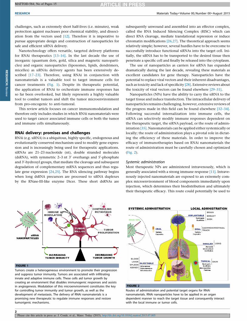
nanomaterials is a valuable tool to target immune cells for
cancer treatments (Fig. 1). Despite its therapeutic potential,
the application of RNAi to orchestrate immune responses has
so far been overlooked, but likely represents a highly valuable
tool to combat tumors and shift the tumor microenvironment
from pro-oncogenic to anti-tumoral.
This review article focuses on cancer immunomodulation and
therefore only includes studies in which RNAi nanomaterials were
used to target cancer associated immune cells or both the tumor
and immune cells simultaneously.
RNAi delivery: promises and challengesRNAi (e.g. siRNA) is a ubiquitous, highly specific, endogenous and
evolutionarily conserved mechanism used to modify gene expres-
sion and is increasingly being used for therapeutic applications.
siRNAs are 21–23 nucleotide (nt), double stranded molecules
(dsRNA), with symmetric 2–3 nt 30 overhangs and 50-phosphate
and 30-hydroxyl groups, that mediate the cleavage and subsequent
degradation of complementary mRNA sequences and thus regu-
late gene expression [24,25]. The RNA silencing pathway begins
when long dsRNA precursors are processed to siRNA duplexes
by the RNase-III-like enzyme Dicer. These short dsRNAs are
subsequently unwound and assembled into an effector complex,
called the RNA Induced Silencing Complex (RISC) which can
direct RNA cleavage, mediate translational repression or induce
chromatin modifications [26,27]. The theoretical approach seems
relatively simple; however, several hurdles have to be overcome to
successfully introduce functional siRNAs into the target cell. Ini-
tially, the siRNA has to be transported to the desired tissue then
penetrate a specific cell and finally be released into the cytoplasm.
The use of nanoparticles as carriers for siRNA has expanded
dramatically during the last decade, revealing these materials as
excellent candidates for gene therapy. Nanoparticles have the
potential to replace viral vectors and their inherent disadvantages,
such as toxicity and lack of specificity [28]. Extensive reviews about
the toxicity of viral vectors can be found elsewhere [29–31].
Nanoparticles (NPs) have the ability to carry the siRNA to the
target tissue and induce transfection. The intracellular delivery of
nanoparticles remains challenging, however, extensive reviews of
the progress made in this field can be found elsewhere [32–34].
Following successful internalization into immune cells, the
siRNA can selectively modify immune responses dependent on
the therapeutic target, the siRNA payload, or the route of admin-
istration [35]. Nanomaterials can be applied either systemically or
locally; the route of administration plays a pivotal role in dictat-
ing the efficiency of these materials. In order to improve the
efficacy of immunotherapies based on RNAi nanomaterials the
route of administration must be carefully chosen and optimized
(Fig. 2).
Systemic administrationMost therapeutic NPs are administered intravenously, which is
generally associated with a strong immune response [11]. Intrave-
nously injected nanomaterials are exposed to an extremely com-
plex microenvironment of blood components immediately upon
injection, which determines their biodistribution and ultimately
their therapeutic efficacy. This route could potentially be used to
RESEARCH Materials Today � Volume 00, Number 00 �August 2015
MATTOD-584; No of Pages 15
Please cite this article in press as: J. Conde, et al., Mater. Today (2015), http://dx.doi.org/10.1016/j.mattod.2015.07.005
FIGURE 1
Tumors create a heterogeneous environment to promote their progression
and suppress tumor immunity. Tumors are associated with infiltrating
innate and adaptive immune cells. These cells aid tumor growth by
creating an environment that disables immunogenic responses and assistsin angiogenesis. Modulation of this microenvironment constitutes the key
for controlling tumor immunity and tumor growth, as well as the
development of metastasis. The delivery of RNAi nanomaterials is a
promising new therapeutic to regulate immune responses and restoretumorigenic mechanisms.
FIGURE 2
Routes of administration and potential target organs for RNAi
nanomaterials. RNAi nanoparticles have to be applied in an organ
dependent manner to reach the target tissue and consequently interactwith the local immune or tumor cells.
2
RESEARCH:Review
deliver RNAi NPs to cells in the blood stream (e.g. monocytes, NK
cells, B and T lymphocytes) thereby directly targeting the immune
system, however due to the flow rate in blood the uptake of
nanoparticles is challenging. Systemically injected RNAi NPs are
rapidly cleared from the blood stream and accumulate in kidney,
spleen, liver and lymph nodes, where they are taken up by scav-
enging dendritic cells and macrophages [16]. This mechanism can
be exploited in case of kidney or liver pathologies to target these
immune cells and to regulate the local microenvironment. In
contrast, the delivery to other organs is more challenging. This
difficulty can be overcome by using a local delivery system,
reducing cellular barriers, or by developing new ways to exploit
nanoparticles’ extravasation into specific target sites [36]. Within
an intact vasculature the extravasation and diffusion of nanopar-
ticles to target tissues is difficult [37]. This however is an advantage
for the delivery to tumors, which present defective (leaky) vascu-
lature and poor lymphatic drainage, resulting in an enhanced
permeability and retention (EPR) effect that facilitates the delivery
of therapeutic particles to these regions [38,39]. In fact, it has been
shown that nanoparticles � 400 nm in diameter extravasate
through tumor vessels and specifically accumulate at tumor sites
[40–42]. Also, tumors with defective blood capillaries allow nano-
particles to passively accumulate. Alternatively, NPs can be active-
ly anchored to specific biomarkers (over-)expressed by tumor cells
through targeting moieties [37]. Nanoparticles for in vivo siRNA
delivery (�100 nm diameter) can be further modified to increase
steric stabilization and active targeting via ligands, such as anti-
bodies, peptides and aptamers [43–48]. Despite these strategies,
homing of systemically administered nanoparticles to specific
target organs followed by tissue extravasation remains challenging
[36].
Local administrationNanoparticles used in conjunction with local delivery system
encounter fewer cellular barriers. The local application of nano-
particles at or near the target sites might be the method of
choice for the multitude of pathologies. In fact, local delivery
can prevent most of the systemic toxicity while simultaneously
enabling effective concentration and retention of the therapeu-
tic agent. Continuous release of siRNA into the local cell/tissue
microenvironment can be accomplished by formulating siRNA
into biocompatible, biodegradable and immunologically inert
matrices, like hydrogels [49,50]. These hydrogels can be further
engineered to interact with specific tissues and to control the
release kinetics and stability of the nanoparticles. Intelligent
hydrogel-based matrix designs would enable the programming
of nanoparticles’ release kinetics to occur in response to extrin-
sic factors such as light, temperature or pH. This can be com-
bined with the ability to track the release using optical imaging,
making these hydrogels ideal candidates for the local delivery of
siRNAs [50–52].
Implantable or locally injectable NP-embedded hydrogels can
be exploited to deliver siRNAs to target tissues. As an example, the
skin is a very attractive organ to deliver engineered RNAi NPs.
Nanoparticles can be delivered subcutaneously by intradermal
injection [53], epidermal electroporation [54] or via microneedles
[55] but also as topical applications using hydrogel scaffolds or
patches [50].
Inhalation and ingestionNasal and oral routes can be exploited for future RNAi applications
in cancer immunotherapy for lung or gastric delivery, although
oral administration has a lower bioavailability in contrast to the
systemic route. The delivery of NPs via nasal routes can target the
mucosa-associated lymphoid tissues and residing macrophages or
lymphocytes, with a concomitant passage of immune cells/pro-
ducts to the circulation and other body tissues. In fact, the delivery
by intratracheal instillation is very efficient as the therapeutic
material is directly delivered to bronchial airways, where they
can efficiently target immune cells and tumor cells [22]. This
non-surgical technique is simpler than inhalation exposure pro-
cedures, permitting the introduction of a wide dose range to the
lungs within a short time [56].
Taken together, nanomaterial-based delivery of siRNA provides
a strategy for innovative targeting of specific cell populations for
therapeutic purposes [57]. In fact, in the last years nanoparticles
have been gaining momentum as robust system for the effective
delivery of therapeutic siRNA owing to their inherent properties,
chemical stability and physical constancy, high purity via repro-
ducible synthetic protocols allowing for adjustable size and mor-
phology control, ease of surface modification for improved siRNA
binding and targeted delivery [58]. In the past decade, the use of
inorganic and organic nanoparticles as siRNAs delivery agents has
been extensively investigated and described especially for novel
cancer therapeutics [13,16]. Recent advances in the development
of nanomaterials (liposomes and lipid-based [59–61], metallic
nanoparticles [22,23], RNA nanoparticles [62–66], and poly-
mers/dendrimers [50,67–69]) for siRNA delivery in cancer thera-
pies enabled the production of highly potent, specific and
biocompatible nanoparticle delivery vehicles. These nanomater-
ials have already shown promising results in tumor targeting and
may represent a valuable role in targeting immune cells in the
tumor microenvironment. Although such nanomaterials were
successfully used for targeting tumor cells, siRNA platforms tar-
geting the immune system for cancer therapy are still under
development.
Immune cells and cancer: knocking down barriers intherapyThe immune system is paramount for homeostatic functions,
clearance of debris, tissue repair and defense during infections.
In particular, immune cells have the unique ability to recognize
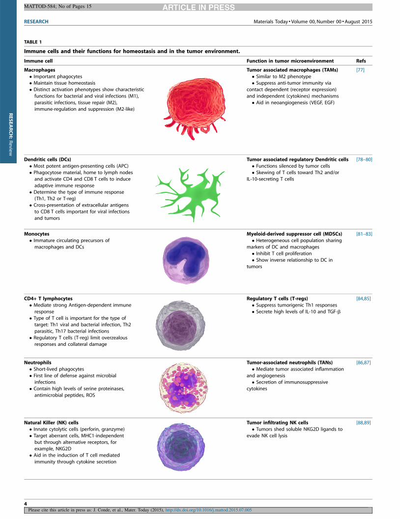
aberrant/cancerous cells and induce their elimination (Table 1).
This task is mainly performed by macrophages, neutrophils and
natural killer (NK) cells (innate cells) as a first line of defense and is
mediated through their cytotoxic mechanisms [70]. As a second
line of defense, macrophages and dendritic cells present tumor-
associated antigens (TAAs) to T cells and induce a potent adaptive
immune response, a mechanism exploited for cancer vaccines
[71]. Despite these powerful immune mechanisms, tumors devel-
op mechanisms to evade the immune system on several levels,
such as changing the expression of surface molecules to become
invisible to immune cells as well as actively suppressing tumor-
directed immunogenic responses to induce immunotolerance, a
process referred to as immunoediting [72]. Hence, immune cells
associated with tumors are modified from tumoricidal toward
Materials Today � Volume 00, Number 00 �August 2015 RESEARCH
MATTOD-584; No of Pages 15
Please cite this article in press as: J. Conde, et al., Mater. Today (2015), http://dx.doi.org/10.1016/j.mattod.2015.07.005
3
RESEARCH:Review
RESEARCH Materials Today � Volume 00, Number 00 �August 2015
MATTOD-584; No of Pages 15
Please cite this article in press as: J. Conde, et al., Mater. Today (2015), http://dx.doi.org/10.1016/j.mattod.2015.07.005
TABLE 1
Immune cells and their functions for homeostasis and in the tumor environment.
Immune cell Function in tumor microenvironment Refs
Macrophages
� Important phagocytes� Maintain tissue homeostasis
� Distinct activation phenotypes show characteristic
functions for bacterial and viral infections (M1),
parasitic infections, tissue repair (M2),immune-regulation and suppression (M2-like)
Tumor associated macrophages (TAMs)
� Similar to M2 phenotype� Suppress anti-tumor immunity via
contact dependent (receptor expression)
and independent (cytokines) mechanisms
� Aid in neoangiogenesis (VEGF, EGF)
[77]
Dendritic cells (DCs)
� Most potent antigen-presenting cells (APC)� Phagocytose material, home to lymph nodes
and activate CD4 and CD8 T cells to induce
adaptive immune response
� Determine the type of immune response(Th1, Th2 or T-reg)
� Cross-presentation of extracellular antigens
to CD8 T cells important for viral infections
and tumors
Tumor associated regulatory Dendritic cells
� Functions silenced by tumor cells� Skewing of T cells toward Th2 and/or
IL-10-secreting T cells
[78–80]
Monocytes
� Immature circulating precursors of
macrophages and DCs
Myeloid-derived suppressor cell (MDSCs)
� Heterogeneous cell population sharing
markers of DC and macrophages
� Inhibit T cell proliferation� Show inverse relationship to DC in
tumors
[81–83]
CD4+ T lymphocytes
� Mediate strong Antigen-dependent immune
response� Type of T cell is important for the type of
target: Th1 viral and bacterial infection, Th2
parasitic, Th17 bacterial infections
� Regulatory T cells (T-reg) limit overzealousresponses and collateral damage
Regulatory T cells (T-regs)
� Suppress tumorigenic Th1 responses
� Secrete high levels of IL-10 and TGF-b
[84,85]
Neutrophils
� Short-lived phagocytes
� First line of defense against microbial
infections� Contain high levels of serine proteinases,
antimicrobial peptides, ROS
Tumor-associated neutrophils (TANs)
� Mediate tumor associated inflammation
and angiogenesis
� Secretion of immunosuppressivecytokines
[86,87]
Natural Killer (NK) cells
� Innate cytolytic cells (perforin, granzyme)� Target aberrant cells, MHC1-independent
but through alternative receptors, for
example, NKG2D� Aid in the induction of T cell mediated
immunity through cytokine secretion
Tumor infiltrating NK cells
� Tumors shed soluble NKG2D ligands toevade NK cell lysis
[88,89]
4
RESEARCH:Review
tumorigenic and further implement the immunosuppressive en-
vironment through the secretion of cytokines, chemokines and
metabolic mediators as well as through cell-contact dependent
signaling mechanisms. The role of tumor-associated immune cells
and their contribution to tumor progression have been extensively
reported elsewhere [6,73–76]. A deeper understanding of the
changes of immune cell phenotype and their signaling networks
is important in order to identify targets to reset and modulate the
immune response.
How to activate an anti-tumoral immune response andsensitize to immune-mediated destructionThe immune system comprises multiple distinct cell types that
work in concert to maintain tissue homeostasis and to orchestrate
the immune response. Their activation is dependent on local
environmental stimuli that trigger specific signaling loops thus
determining the type of immune response generated. The activa-
tion factors for immune cells for activation of immune cells can be
classified into 4 categories; chemokine and cytokines, metabolites,
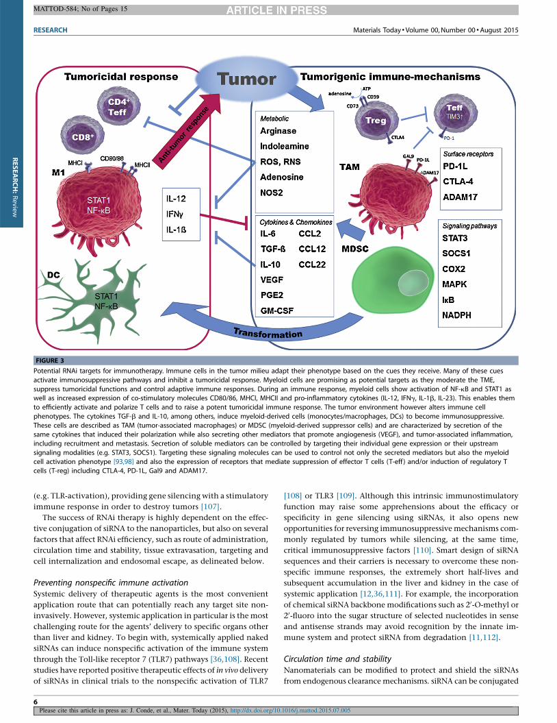
cell surface receptors and intracellular signaling mediators (Fig. 3).
In a simplified point-of-view, the cytokines IL-12, IFNg, IL-1b
and IL-23 are key pro-inflammatory factors that promote tumor-
icidal immune functions; these are associated with M1-type
macrophages, mature DCs, CD4+-Th1 and cytotoxic CD8+ T cells.
Activated macrophages and DCs present increased levels of NF-kB
and STAT1 as well as the co-stimulatory molecules CD80/86,
MHCI and MHCII. Together, these factors are essential during
infections, while in cancer they indicate a potent anti-tumor
response [91–93].
The tumor microenvironment (TME) however alters immune
cell phenotypes, which typically leads to elevated levels of IL-6,
TGF-b, PGE2, COX2, MCP-1, M-CSF, IL-4 and IL-10. These factors
are involved in immunosuppression (tumor promoting inflamma-
tion and angiogenesis) and are linked to tumor-associated macro-
phages (TAMs), myeloid-derived suppressor cells (MDSCs) and
regulatory T cells (T-reg) (Fig. 3) [74,94]. The TME is further
characterized by metabolites that play an important role in im-
mune cell functions; these are, for example, specific amino acids
important for tumoricidal T cell activation (e.g. Tryptophan,
depleted by Indoleamine (IDO); Arginine, depleted by Arginase),
while their alternate degradation products (L-Kynurenine, orni-
thine) favor immunosuppressive regulatory T cells [95]. Surface
receptors, such as CTLA-4 and PD-1 mediate suppression through
silencing tumor-specific T cells. Intracellular signaling molecules
including transcription factors (e.g. STATs, NF-kB, HIF) and their
accessory molecules (e.g. SOCS, ikB, IKKb) are paramount in
determining the activation state of immune cells [96–99]. They
dictate transcriptional programs that drive the immune response
and, in the case of cancer, reinforce the immunosuppressive tumor
environment, while simultaneously inhibiting a tumoricidal re-
sponse (Fig. 3) [6].
Due to the concomitant regulatory mechanisms within the
TME, many factors have redundant functions, therefore targeting
specific combinations rather than a single target might be neces-
sary to tip the balance from immunosuppression to immunoge-
nicity.
Tumor-associated myeloid cells (TAMs, DCs and MDSCs) are
potent mediators of the suppressive TME through the above-
mentioned factors and are able to either activate or suppress
immune responses which make them a prime target for immuno-
therapy [100]. Their intracellular signaling pathways are crucial to
these functions; therefore, the regulation of these factors is one
potential approach to convert their phenotype. Additionally, new
therapeutics that target immunosuppressive T cells show promis-
ing results for cancer. As an example, antibodies blocking the
CTLA-4 (ipilimumab) or PD-1 (nivolumab) receptor on T cells
show improved tumor responses [101]. The best approach to target
individual or multiple factors still needs to be carefully evaluated.
RNAi is ideally suited to target immune cells; it allows the
targeting of individual or multiple targets and can be tailored to
target a specific cell type. In contrast to antibody blockade [102],
RNAi can be used to directly down-modulate gene expression in
immune cells in order to regulate signaling molecules (e.g. CTLA-
4, PD-1, STATs, NF-kB) and ultimately their phenotype, to drive
tumoricidal responses. In the past decades a multitude of factors
that are responsible for immunosuppression in tumor-associated
immune cells have been identified and can be targeted to control
immunosuppression. Some of these factors are already being
successfully targeted, in particular, STAT3 (via RNAi or small
molecule inhibitors) showing a shift toward a potent anti-tumor
response [103–105]. Other potential targets remain to be investi-
gated (e.g. non-canonical NF-kB pathway to inhibit MDSC-sup-
pression) [106]. RNAi offers the possibility to control the
expression of any desired mediator, independent on the availabil-
ity of pharmacologically active inhibitors. Consequently, combin-
ing RNAi immunotherapy with new advances in nanomaterial
technology is a great opportunity to improve cancer treatments.
RNAi nanomaterials design for the modulation of animmune response in cancerModulating in vivo immune response using RNAi nanomaterials
can be divided into two categories: inhibition of immune sup-
pression or enhancement of the immune response. To achieve
these goals proper design of RNAi nanomaterials must be fulfilled
in order to attain a successful immune response. The antitumoral
effect of specific RNAi treatment should not be dependent exclu-
sively on the inhibitory effect of siRNA, but should also be
combined with RNAi inducing immunostimulatory effects
Materials Today � Volume 00, Number 00 �August 2015 RESEARCH
MATTOD-584; No of Pages 15
Please cite this article in press as: J. Conde, et al., Mater. Today (2015), http://dx.doi.org/10.1016/j.mattod.2015.07.005
TABLE 1 (Continued )
Immune cell Function in tumor microenvironment Refs
Cytotoxic T cell (CD8)
� Can kill virus infected and cancer cells� MHC-1 dependent
Tumor-specific cytotoxic T cell
� Insufficient activation by DCs leadsto T cell exhaustion
[85,90]
5
RESEARCH:Review
(e.g. TLR-activation), providing gene silencing with a stimulatory
immune response in order to destroy tumors [107].
The success of RNAi therapy is highly dependent on the effec-
tive conjugation of siRNA to the nanoparticles, but also on several
factors that affect RNAi efficiency, such as route of administration,
circulation time and stability, tissue extravasation, targeting and
cell internalization and endosomal escape, as delineated below.
Preventing nonspecific immune activationSystemic delivery of therapeutic agents is the most convenient
application route that can potentially reach any target site non-
invasively. However, systemic application in particular is the most
challenging route for the agents’ delivery to specific organs other
than liver and kidney. To begin with, systemically applied naked
siRNAs can induce nonspecific activation of the immune system
through the Toll-like receptor 7 (TLR7) pathways [36,108]. Recent
studies have reported positive therapeutic effects of in vivo delivery
of siRNAs in clinical trials to the nonspecific activation of TLR7
[108] or TLR3 [109]. Although this intrinsic immunostimulatory
function may raise some apprehensions about the efficacy or
specificity in gene silencing using siRNAs, it also opens new
opportunities for reversing immunosuppressive mechanisms com-
monly regulated by tumors while silencing, at the same time,
critical immunosuppressive factors [110]. Smart design of siRNA
sequences and their carriers is necessary to overcome these non-
specific immune responses, the extremely short half-lives and
subsequent accumulation in the liver and kidney in the case of
systemic application [12,36,111]. For example, the incorporation
of chemical siRNA backbone modifications such as 20-O-methyl or
20-fluoro into the sugar structure of selected nucleotides in sense
and antisense strands may avoid recognition by the innate im-
mune system and protect siRNA from degradation [11,112].
Circulation time and stabilityNanomaterials can be modified to protect and shield the siRNAs
from endogenous clearance mechanisms. siRNA can be conjugated
RESEARCH Materials Today � Volume 00, Number 00 �August 2015
MATTOD-584; No of Pages 15
Please cite this article in press as: J. Conde, et al., Mater. Today (2015), http://dx.doi.org/10.1016/j.mattod.2015.07.005
FIGURE 3
Potential RNAi targets for immunotherapy. Immune cells in the tumor milieu adapt their phenotype based on the cues they receive. Many of these cues
activate immunosuppressive pathways and inhibit a tumoricidal response. Myeloid cells are promising as potential targets as they moderate the TME,
suppress tumoricidal functions and control adaptive immune responses. During an immune response, myeloid cells show activation of NF-kB and STAT1 aswell as increased expression of co-stimulatory molecules CD80/86, MHCI, MHCII and pro-inflammatory cytokines (IL-12, IFNg, IL-1b, IL-23). This enables them
to efficiently activate and polarize T cells and to raise a potent tumoricidal immune response. The tumor environment however alters immune cell
phenotypes. The cytokines TGF-b and IL-10, among others, induce myeloid-derived cells (monocytes/macrophages, DCs) to become immunosuppressive.
These cells are described as TAM (tumor-associated macrophages) or MDSC (myeloid-derived suppressor cells) and are characterized by secretion of thesame cytokines that induced their polarization while also secreting other mediators that promote angiogenesis (VEGF), and tumor-associated inflammation,
including recruitment and metastasis. Secretion of soluble mediators can be controlled by targeting their individual gene expression or their upstream
signaling modalities (e.g. STAT3, SOCS1). Targeting these signaling molecules can be used to control not only the secreted mediators but also the myeloidcell activation phenotype [93,98] and also the expression of receptors that mediate suppression of effector T cells (T-eff ) and/or induction of regulatory T
cells (T-reg) including CTLA-4, PD-1L, Gal9 and ADAM17.
6
RESEARCH:Review
to the surface of nanoparticles or entrapped in nanovesicles, which
act to protect the siRNA from serum nucleases and to increase their
chemical stability. Compared to conventional transfection agents,
nanoparticle-conjugated siRNAs have been shown to be less suscep-
tible to degradation by nuclease activity, to exhibit greater cellular
uptake and to have a higher siRNA effective concentration, all of
which have accelerated siRNA research into this delivery method
over the past few years. Unfortunately, linking the siRNA to a
nanoparticle alone does not protect it from clearance. Blood serum
components interact with siRNA/nanomaterials and mark them for
uptake via the mononuclear phagocyte system (MPS), especially by
Kupffer cells in the liver [12,111]. In fact, the stimulatory or anti-
stimulatory action is typically due to binding of proteins in the
blood, which influence the uptake of nanoparticles by cells [12]. The
use of nanomaterials composed of hydrophilic polymers including
acrylic acid, acrylamide, and maleic anhydride polymers and copo-
lymers, as well as allylamine, ethyleneimine, oxazoline (for exam-
ple, Polyethylene glycol, Polyethylenimine, Poly(acrylic acid),
Poly(vinyl alcohol), Poly(N-isopropylacrylamide)) inhibits serum
protein binding and clearance by immune cells which thereby
drastically increases their circulation time [12,113].
Tissue extravasationParticle size is critical for efficient tissue delivery. Nanoparticles in
the size range of 10–100 nm are generally accepted as efficient
delivery agents, determined by in vivo clearance, biodistribution
and toxicity. Particles of less than 10 nm are subject to renal
clearance, while larger particles >15 mm are removed from the
circulation by the reticulo-endothelial system (RES) in the liver
and spleen [58,59]. The RES (also known as macrophage or mono-
nuclear phagocyte systems), is a network of cells located through-
out the body that support the elimination of small particles, also
involved in the identification of foreign substances in blood and
tissues [114].
Owing to the effective elimination of NPs by the RES, the
optimal delivery of NPs to target sites through intravascular deliv-
ery constitutes a challenge. NPs’ uptake, mainly by macrophages,
is dependent on size, charge and other surface modifications,
which influence the lifetime and the diffusion of nanoparticles
to certain cells/tissues/organs. Therefore, the size of nanoparticles
and their payload should be large enough to prevent rapid leakage
in blood capillaries but at the same time small enough to escape
from the scavenge of macrophages in the RES, such as the liver and
spleen or being cleared out by the kidneys. Appropriately sized
nanoparticles can be chemically modified to increase their reten-
tion time in the circulatory system, using cationic polymers as
described [115,116] or directly engineered to target phagocytic
cells to increase uptake and antigen presentation [117].
Targeting and cell internalizationOnce the siRNA is inside the target tissue it has to reach the target
cells while excluding healthy cells. Nanoparticles can be function-
alized with cell-specific ligands that allow receptor-mediated up-
take into target cells, for example markers which are overexpressed
on tumor or immune cells. Additionally, the surface charge on the
nanoparticle is a crucial factor that affects cellular internalization
(by both normal/cancer cells and immune cells) and also deter-
mines potential in vivo circulation. Positively charged particles
have been shown to exhibit increased internalization not only by
tumor cells but also by macrophages and DCs compared to neutral
or negatively charged nanoparticles. In general, positively charged
particles are also phagocytosed by macrophages but are more
efficiently taken up because of electrostatic interactions between
the positively charged particle surface and the negatively charged
cellular membrane. Conversely, nanoparticles with negative sur-
face charges typically exhibit low cellular internalization; however
these nanoparticles can circulate longer in vivo and thus, better
accumulate at tumor sites [118,119].
Endosomal escapeIn order to activate the RNAi pathway, siRNAs must be successfully
delivered into the cytoplasm, a process challenged by the large size
and hydrophilicity of the nanoparticles, limiting their ability to
cross the cell membrane in the absence of a transfection agent.
Moreover, the cellular uptake of nanoparticles (endocytosis) as
well as their subsequent discharge (exocytosis) is affected by their
shape, size and charge. Generally, small (<200 nm) positively
charged nanoparticles adsorb to the negatively charged plasma
membrane, followed by clathrin-dependent endocytosis. In con-
trast, larger particles (>200 nm) enter the cell by receptor- and
clathrin-independent endocytosis [120]. The uptake pathway can
greatly influence the interaction/effect of particles on cellular
responses. The cell types that have been studied most extensively
for distinct uptake mechanisms are DCs and macrophages, where
several studies show that the route of uptake as well as the particle
size significantly changes their responses. While DCs generally
internalize (pinocytosis) smaller particles and show the highest
antigen-presentation capacity; macrophages efficiently phagocy-
tize larger particles (<10 mm) but are less potent antigen-presenters
[121,122].
Once the nanoparticle is taken up by the target cells via endo-
cytosis, its release from the endosome into the cytoplasm is the
next challenge. Many of the described systems/vehicles get
trapped in the endosome, which fuses with lysosomes (i.e. endo-
lysosome) thereby destroying the siRNAs. Specific targeting via
fusogenic peptides (i.e. Influenza hemagglutinin – HA) [123],
lysosomotropic compounds/surfactants (i.e. Chloroquine, Quina-
crine, Tilorone, Suramine) [124] or PEI polymer [125] is necessary
to enable the endosomal/endolysosomal escape and allow the
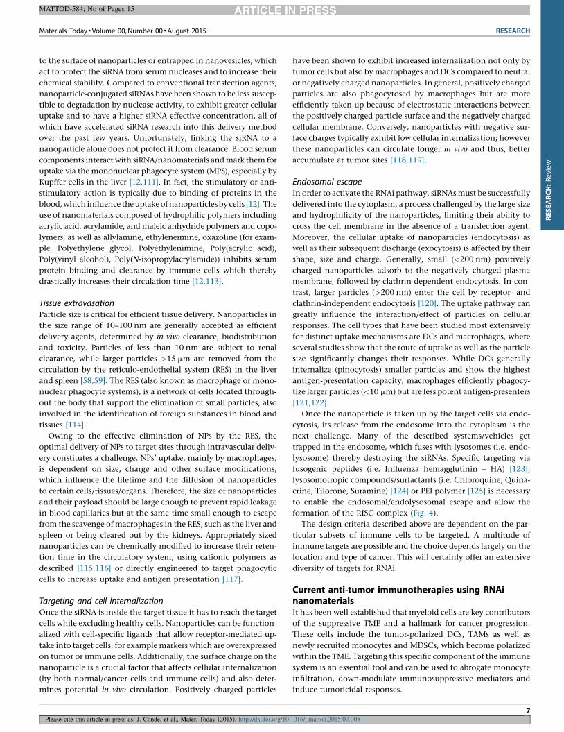
formation of the RISC complex (Fig. 4).
The design criteria described above are dependent on the par-
ticular subsets of immune cells to be targeted. A multitude of
immune targets are possible and the choice depends largely on the
location and type of cancer. This will certainly offer an extensive
diversity of targets for RNAi.
Current anti-tumor immunotherapies using RNAinanomaterialsIt has been well established that myeloid cells are key contributors
of the suppressive TME and a hallmark for cancer progression.
These cells include the tumor-polarized DCs, TAMs as well as
newly recruited monocytes and MDSCs, which become polarized
within the TME. Targeting this specific component of the immune
system is an essential tool and can be used to abrogate monocyte
infiltration, down-modulate immunosuppressive mediators and
induce tumoricidal responses.
Materials Today � Volume 00, Number 00 �August 2015 RESEARCH
MATTOD-584; No of Pages 15
Please cite this article in press as: J. Conde, et al., Mater. Today (2015), http://dx.doi.org/10.1016/j.mattod.2015.07.005
7
RESEARCH:Review
Interestingly, only few studies have been performed using RNAi
nanomaterials for immunotherapy in cancer. The targeting of an
important sub-set of the populations of immune cells in combi-
nation with cancer cells could shift the tumor microenvironment
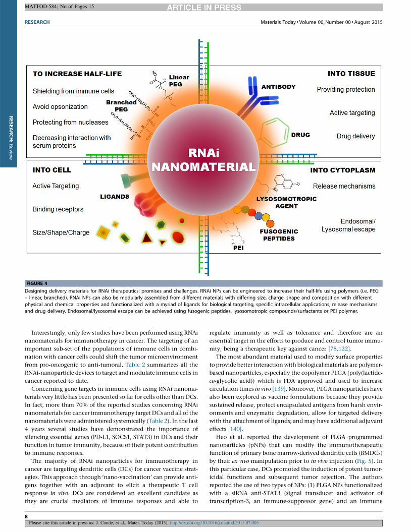
from pro-oncogenic to anti-tumoral. Table 2 summarizes all the
RNAi-nanoparticle devices to target and modulate immune cells in
cancer reported to date.
Concerning gene targets in immune cells using RNAi nanoma-
terials very little has been presented so far for cells other than DCs.
In fact, more than 70% of the reported studies concerning RNAi
nanomaterials for cancer immunotherapy target DCs and all of the
nanomaterials were administered systemically (Table 2). In the last
4 years several studies have demonstrated the importance of
silencing essential genes (PD-L1, SOCS1, STAT3) in DCs and their
function in tumor immunity, because of their potent contribution
to immune responses.
The majority of RNAi nanoparticles for immunotherapy in
cancer are targeting dendritic cells (DCs) for cancer vaccine strat-
egies. This approach through ‘nano-vaccination’ can provide anti-
gens together with an adjuvant to elicit a therapeutic T cell
response in vivo. DCs are considered an excellent candidate as
they are crucial mediators of immune responses and able to
regulate immunity as well as tolerance and therefore are an
essential target in the efforts to produce and control tumor immu-
nity, being a therapeutic key against cancer [78,122].
The most abundant material used to modify surface properties
to provide better interaction with biological materials are polymer-
based nanoparticles, especially the copolymer PLGA (poly(lactide-
co-glycolic acid)) which is FDA approved and used to increase
circulation times in vivo [139]. Moreover, PLGA nanoparticles have
also been explored as vaccine formulations because they provide
sustained release, protect encapsulated antigens from harsh envir-
onments and enzymatic degradation, allow for targeted delivery
with the attachment of ligands; and may have additional adjuvant
effects [140].
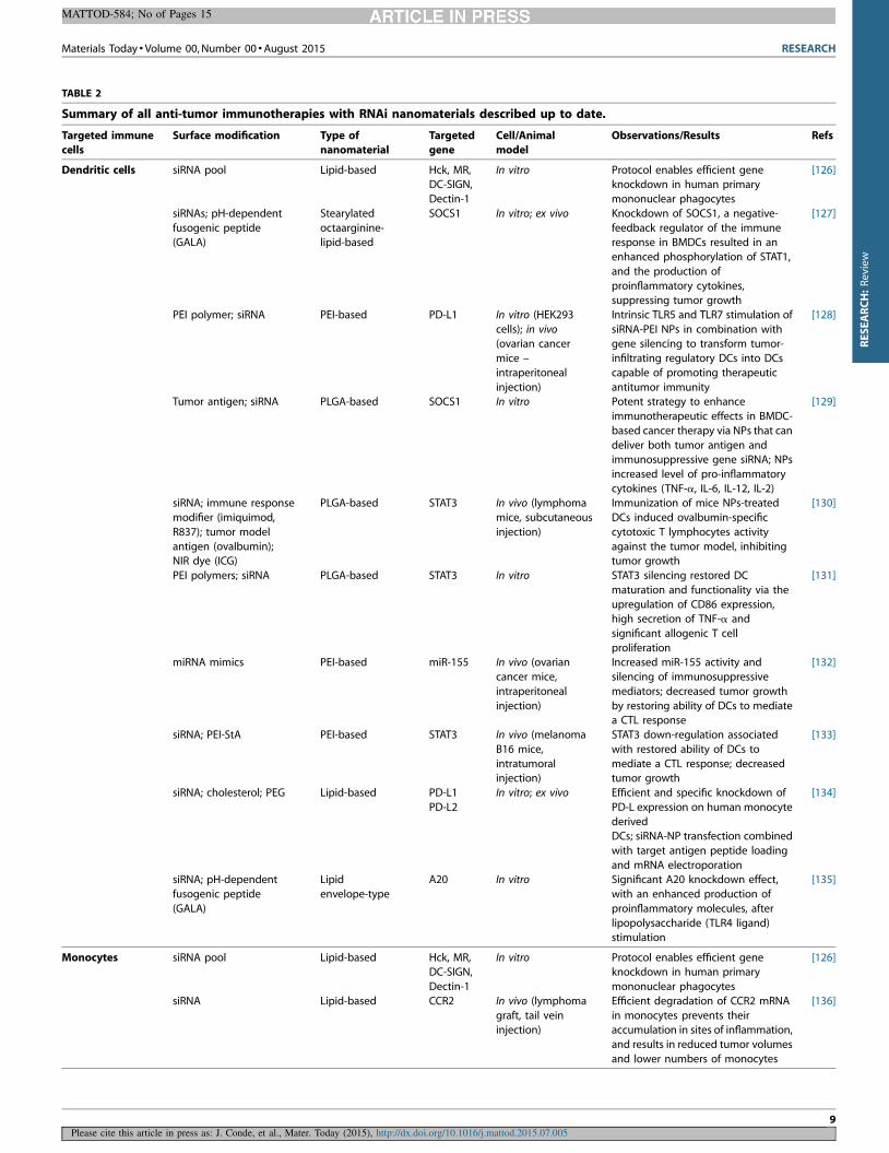
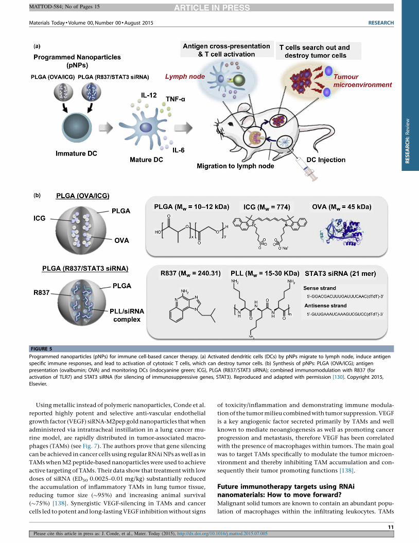
Heo et al. reported the development of PLGA programmed
nanoparticles (pNPs) that can modify the immunotherapeutic
function of primary bone marrow-derived dendritic cells (BMDCs)
by their ex vivo manipulation prior to in vivo injection (Fig. 5). In
this particular case, DCs promoted the induction of potent tumor-
icidal functions and subsequent tumor rejection. The authors
reported the use of two types of NPs: (1) PLGA NPs functionalized
with a siRNA anti-STAT3 (signal transducer and activator of
transcription-3, an immune-suppressor gene) and an immune
RESEARCH Materials Today � Volume 00, Number 00 �August 2015
MATTOD-584; No of Pages 15
Please cite this article in press as: J. Conde, et al., Mater. Today (2015), http://dx.doi.org/10.1016/j.mattod.2015.07.005
FIGURE 4
Designing delivery materials for RNAi therapeutics: promises and challenges. RNAi NPs can be engineered to increase their half-life using polymers (i.e. PEG
– linear, branched). RNAi NPs can also be modularly assembled from different materials with differing size, charge, shape and composition with different
physical and chemical properties and functionalized with a myriad of ligands for biological targeting, specific intracellular applications, release mechanisms
and drug delivery. Endosomal/lysosomal escape can be achieved using fusogenic peptides, lysosomotropic compounds/surfactants or PEI polymer.
8
RESEARCH:Review
Materials Today � Volume 00, Number 00 �August 2015 RESEARCH
MATTOD-584; No of Pages 15
Please cite this article in press as: J. Conde, et al., Mater. Today (2015), http://dx.doi.org/10.1016/j.mattod.2015.07.005
TABLE 2
Summary of all anti-tumor immunotherapies with RNAi nanomaterials described up to date.
Targeted immunecells
Surface modification Type ofnanomaterial
Targetedgene
Cell/Animalmodel
Observations/Results Refs
Dendritic cells siRNA pool Lipid-based Hck, MR,DC-SIGN,
Dectin-1
In vitro Protocol enables efficient geneknockdown in human primary
mononuclear phagocytes
[126]
siRNAs; pH-dependent
fusogenic peptide(GALA)
Stearylated
octaarginine-lipid-based
SOCS1 In vitro; ex vivo Knockdown of SOCS1, a negative-
feedback regulator of the immuneresponse in BMDCs resulted in an
enhanced phosphorylation of STAT1,
and the production of
proinflammatory cytokines,suppressing tumor growth
[127]
PEI polymer; siRNA PEI-based PD-L1 In vitro (HEK293
cells); in vivo(ovarian cancer
mice –
intraperitoneal
injection)
Intrinsic TLR5 and TLR7 stimulation of
siRNA-PEI NPs in combination withgene silencing to transform tumor-
infiltrating regulatory DCs into DCs
capable of promoting therapeutic
antitumor immunity
[128]
Tumor antigen; siRNA PLGA-based SOCS1 In vitro Potent strategy to enhance
immunotherapeutic effects in BMDC-
based cancer therapy via NPs that can
deliver both tumor antigen andimmunosuppressive gene siRNA; NPs
increased level of pro-inflammatory
cytokines (TNF-a, IL-6, IL-12, IL-2)
[129]
siRNA; immune response
modifier (imiquimod,
R837); tumor model
antigen (ovalbumin);NIR dye (ICG)
PLGA-based STAT3 In vivo (lymphoma
mice, subcutaneous
injection)
Immunization of mice NPs-treated
DCs induced ovalbumin-specific
cytotoxic T lymphocytes activity
against the tumor model, inhibitingtumor growth
[130]
PEI polymers; siRNA PLGA-based STAT3 In vitro STAT3 silencing restored DC
maturation and functionality via the
upregulation of CD86 expression,high secretion of TNF-a and
significant allogenic T cell
proliferation
[131]
miRNA mimics PEI-based miR-155 In vivo (ovarian
cancer mice,
intraperitoneal
injection)
Increased miR-155 activity and
silencing of immunosuppressive
mediators; decreased tumor growth
by restoring ability of DCs to mediatea CTL response
[132]
siRNA; PEI-StA PEI-based STAT3 In vivo (melanoma
B16 mice,
intratumoralinjection)
STAT3 down-regulation associated
with restored ability of DCs to
mediate a CTL response; decreasedtumor growth
[133]
siRNA; cholesterol; PEG Lipid-based PD-L1
PD-L2
In vitro; ex vivo Efficient and specific knockdown of
PD-L expression on human monocyte
derivedDCs; siRNA-NP transfection combined
with target antigen peptide loading
and mRNA electroporation
[134]
siRNA; pH-dependent
fusogenic peptide
(GALA)
Lipid
envelope-type
A20 In vitro Significant A20 knockdown effect,
with an enhanced production of
proinflammatory molecules, after
lipopolysaccharide (TLR4 ligand)stimulation
[135]
Monocytes siRNA pool Lipid-based Hck, MR,DC-SIGN,
Dectin-1
In vitro Protocol enables efficient geneknockdown in human primary
mononuclear phagocytes
[126]
siRNA Lipid-based CCR2 In vivo (lymphoma
graft, tail veininjection)
Efficient degradation of CCR2 mRNA
in monocytes prevents theiraccumulation in sites of inflammation,
and results in reduced tumor volumes
and lower numbers of monocytes
[136]
9
RESEARCH:Review
response modifier (imiquimod, R837) for the activation of DCs via
toll-like receptor 7 (TLR7) and (2) PLGA NPs containing tumor
model antigen (ovalbumin, OVA) and near-infrared (NIR) dyes
(indocyanine green, ICG), to deliver tumor-antigen-specific infor-
mation to DCs ex vivo and track the migration of DCs in vivo. These
innovative pNPs were internalized into DCs, induced TLR activa-
tion, silenced immunosuppressive genes, led to cross-presenta-
tion, and inhibited tumor growth significantly. With a huge
potential for efficient DC-based cancer therapy, these pNPs can
tailor the function of immunotherapeutic cells and monitor their
migration in vivo [130].
In another key report, Cubillos-Ruiz et al. described a new system
to activate TLRs using polyethylenimine (PEI)-based nanoparti-
cles. PEI nanoparticles may be very useful as carriers to target
immune cells. Every macromolecule of PEI can be protonated and
catalyzed into a polymer with high-density cationic potential and
a highly branched network [141], which allows PEI to disrupt the
endosomal membrane by osmotic imbalance, also known as the
‘proton-sponge effect’. This effect occurs when unprotonated
species can absorb protons as they are pumped into the lysosome.
This results in an increased influx of Cl� ions and water that causes
swelling and rupture of the lysosomal membrane with subsequent
release of its contents into the cytoplasm [142]. Moreover, PEI by
itself is able to trigger robust TLR5 activation in wild-type mice, but
not in Tlr5�/� littermates, and can further stimulate APCs within
the tumor microenvironment when combined with the encapsu-
lated immunostimulatory siRNA [128].
The authors also showed that in the absence of targeting, PEI-
complexed siRNA oligonucleotides elicit the stimulation of TLR3
and TLR7 and that the nonspecific activation of multiple TLRs
(specifically, TLR5 and TLR7) reversed the immunosuppressive
phenotype of human and mouse ovarian tumor-associated DCs.
Further, linear PEI-based nanoparticles encapsulating siRNA were
preferentially and enthusiastically scavenged by regulatory DCs
expressing CD11c and programmed cell death 1-ligand 1 (PD-L1)
in ovarian cancer mice. These NPs modified the immunosuppres-
sive phenotype of ovarian tumor-associated DCs (human and
mouse) to an immunostimulatory phenotype which resulted in
increased antigen presentation and increased numbers of tumor-
specific cytotoxic CD8+ T cells in the tumor microenvironment.
Mice treated with PEI-siRNA NPs showed improved survival and
superior anti-tumor immunity as compared to non-targeting
siRNA-PEI nanocomplexes. These NPs have the capacity to trans-
form tumor-infiltrating regulatory DCs into DCs able to enhance
anti-tumor immunity. Taking advantage of the intrinsic immu-
nostimulatory capacity of siRNA may activate TLR pathways. The
authors demonstrate that the inherent capacity of siRNA-PEI
nanocomplexes to activate innate immune mechanisms offers a
major opportunity to reverse tumor-induced immunosuppression
while simultaneously silencing specific TLR genes [128].
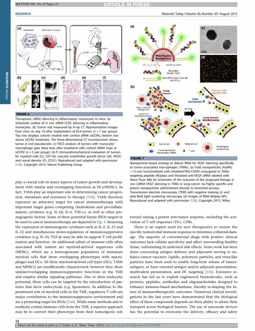
In contrast to targeting dendritic cells (or other tumor-associat-
ed immune cells), a promising approach by Anderson and co-
workers investigates the inhibition of inflammatory monocyte
infiltration by blocking CCR2, an essential monocyte homing
factor. The authors focused on CCR2 because the recruitment of
inflammatory monocytes intimately depends on the chemokine/
chemokine receptor pair MCP-1/CCR2. This study reports the
optimization of lipid nanoparticles containing CCR2-silencing
short interfering RNA are able to localize with monocytes, with
considerable accumulation in spleen and bone marrow with rapid
clearance from the blood, when administered systemically in mice
[136]. Due to the physical properties of cationic lipids used, in the
efficient encapsulation of oligonucleotides, this study shows a
lipid nanoparticle system can be used for in vivo delivery of siRNA
to immune cells, Efficient degradation of CCR2 mRNA in mono-
cytes prevents their accumulation in sites of inflammation, spe-
cifically reducing tumor volumes and accumulation of
inflammatory monocytes in the tumor (Fig. 6). This study may
also be extended to induce tumoricidal functions in monocytes
prior to their migration to tumor sites but the primary focus was on
inflammatory diseases where infiltration of inflammatory mono-
cytes is part of the pathophysiology. This study definitely opens a
new translational avenue to approach the many diseases driven by
recruitment of inflammatory monocytes [136]. However, no tar-
geting moiety was used to achieve active targeting of the studied
immune cells.
RESEARCH Materials Today � Volume 00, Number 00 �August 2015
MATTOD-584; No of Pages 15
Please cite this article in press as: J. Conde, et al., Mater. Today (2015), http://dx.doi.org/10.1016/j.mattod.2015.07.005
TABLE 2 (Continued )
Targeted immune
cells
Surface modification Type of
nanomaterial
Targeted
gene
Cell/Animal
model
Observations/Results Refs
MacrophagesTAMs
siRNA pool Lipid-based Hck, MR,DC-SIGN,
Dectin-1
In vitro Protocol enables efficient geneknockdown in human primary
mononuclear phagocytes
[126]
Copolymers (AzEMA,
DMAEMA,BMA-co-PAA-co-DMAEMA);
siRNAs
Mannosylated
Polymeric Micelles
CD206
(mannosereceptor)
In vitro pH responsive micelles improved the
delivery of siRNA into primarymacrophages by fourfold and induce
�90% knockdown
[137]
siRNA; PEG; M2 peptide Gold NPs VEGF In vitro; in vivo(lung cancer mice,
intratracheal
instillation)
Treatment with low doses of siRNA(ED50 0.0025–0.01 mg/kg) in a multi
and long-term dosing system
substantially reduces the recruitment
of TAMs in lung tumor tissue, reducestumor size (�95%), and increases
animal survival (�75%) in mice
[138]
A20, negative regulator of the toll-like receptor and TNF receptor signaling pathways; AzEMA, 2-azidoethyl methacrylate; DMAEMA, 2-dimethylaminoethyl methacrylate; BMA, butyl
methacrylate; BMDCs, bone marrow-derived dendritic cells; CCR2, Chemokine (C–C motif ) receptor 2; DC-SIGN, dendritic cell-specific intercellular adhesion molecule-3-grabbing non-
integrin, also known as CD209 cluster of differentiation 209; Dectin-1, natural killer-cell-receptor-like C-type lectin; Hck, hemopoietic cell kinase; ICG, indocyanine green; MR, major
histocompatibility complex; PAA, 2-propylacrylic acid; PEI, polyethylenimine; PEI-StA, stearic acid-modified polyethylenimine; PD-L1, programmed cell death 1-ligand 1; PLGA, poly(lactide-
co-glycolic acid); SOCS1, suppressor of cytokine signaling 1; STAT3, signal transducer and activator of transcription-3; VEGF, vascular endothelial growth factor.
10
RESEARCH:Review
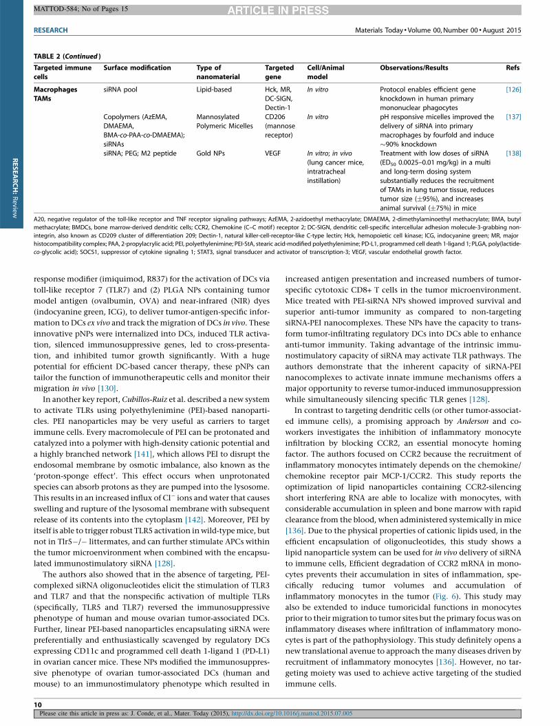
Using metallic instead of polymeric nanoparticles, Conde et al.
reported highly potent and selective anti-vascular endothelial
growth factor (VEGF) siRNA-M2pep gold nanoparticles that when
administered via intratracheal instillation in a lung cancer mu-
rine model, are rapidly distributed in tumor-associated macro-
phages (TAMs) (see Fig. 7). The authors prove that gene silencing
can be achieved in cancer cells using regular RNAi NPs as well as in
TAMs when M2 peptide-based nanoparticles were used to achieve
active targeting of TAMs. Their data show that treatment with low
doses of siRNA (ED50 0.0025–0.01 mg/kg) substantially reduced
the accumulation of inflammatory TAMs in lung tumor tissue,
reducing tumor size (�95%) and increasing animal survival
(�75%) [138]. Synergistic VEGF-silencing in TAMs and cancer
cells led to potent and long-lasting VEGF inhibition without signs
of toxicity/inflammation and demonstrating immune modula-
tion of the tumor milieu combined with tumor suppression. VEGF
is a key angiogenic factor secreted primarily by TAMs and well
known to mediate neoangiogenesis as well as promoting cancer
progression and metastasis, therefore VEGF has been correlated
with the presence of macrophages within tumors. The main goal
was to target TAMs specifically to modulate the tumor microen-
vironment and thereby inhibiting TAM accumulation and con-
sequently their tumor promoting functions [138].
Future immunotherapy targets using RNAinanomaterials: How to move forward?Malignant solid tumors are known to contain an abundant popu-
lation of macrophages within the infiltrating leukocytes. TAMs
Materials Today � Volume 00, Number 00 �August 2015 RESEARCH
MATTOD-584; No of Pages 15
Please cite this article in press as: J. Conde, et al., Mater. Today (2015), http://dx.doi.org/10.1016/j.mattod.2015.07.005
FIGURE 5
Programmed nanoparticles (pNPs) for immune cell-based cancer therapy. (a) Activated dendritic cells (DCs) by pNPs migrate to lymph node, induce antigenspecific immune responses, and lead to activation of cytotoxic T cells, which can destroy tumor cells. (b) Synthesis of pNPs: PLGA (OVA/ICG); antigen
presentation (ovalbumin; OVA) and monitoring DCs (indocyanine green; ICG), PLGA (R837/STAT3 siRNA); combined immunomodulation with R837 (for
activation of TLR7) and STAT3 siRNA (for silencing of immunosuppressive genes, STAT3). Reproduced and adapted with permission [130]. Copyright 2015,
Elsevier.
11
RESEARCH:Review
play a crucial role in many aspects of tumor growth and develop-
ment with similar and overlapping functions as DCs/MDSCs. In
fact, TAMs play an important role in determining cancer progres-
sion, metastasis and resistance to therapy [143]. TAMs therefore
represent an attractive target for cancer immunotherapy with
important target genes comprising chemokines and pro-inflam-
matory cytokines (e.g. IL-1b, IL-6, TNF-a), as well as other pro-
angiogenic factors. Some of these potential future RNAi targets to
be used in cancer immunotherapy are depicted in Fig. 3. Restoring
the expression of immunogenic cytokines such as IL-2, IL-15 and
IL-12 and simultaneous down-regulation of immunosuppressive
cytokines (e.g. IL-10, TGF-b) may be able to support T cell prolif-
eration and function. An additional subset of immune cells often
associated with tumors are myeloid-derived suppressor cells
(MDSC), which are a heterogeneous population of immature
myeloid cells that show overlapping phenotypes with macro-
phages and DCs. All three myeloid-derived cell types (DCs, TAMs
and MDSCs) are insufficiently characterized; however they show
similar/overlapping immunosuppressive functions in the TME
and employ similar signaling pathways. Due to their endocytic
potential, these cells can be targeted by the introduction of par-
ticles that favor endocytosis (e.g. liposomes). In addition to the
prominent role of myeloid cells in the TME, regulatory T cells are
major contributors to the immunosuppressive environment and
are a promising target for RNAi [144]. While some methods aim to
eradicate certain immune cells from the TME, a superior approach
may be to convert their phenotype from their tumorigenic role
toward raising a potent anti-tumor response, including the acti-
vation of T cell responses (Th1, CD8).
There is an urgent need for new therapeutics to restore the
specific tumoricidal immune response to minimize collateral dam-
age. The majority of conventional drugs with positive clinical
outcomes lack cellular specificity and affect surrounding healthy
tissue, culminating in undesired side effects. Some work has been
done concerning antigen delivery and adjuvants serving to en-
hance cancer vaccines. Lipidic, polymeric particles, and virus-like
particles have been used to enable long-term release of tumor-
antigens, as have oriented antigen and/or adjuvant presentation,
multivalent presentation, and DC targeting [145]. Extensive re-
search has led us to exploit engineered biomolecules, such as
proteins, peptides, antibodies and oligonucleotides designed to
enhance immune-based mechanisms, thereby re-shaping the fu-
ture of immunotherapeutic outcomes. Numerous clinical investi-
gations in the last years have demonstrated that the biological
effect of these compounds depends on their ability to attain their
transport across biological barriers. The use of nanoscale devices
has the potential to overcome the delivery, efficacy and safety
RESEARCH Materials Today � Volume 00, Number 00 �August 2015
MATTOD-584; No of Pages 15
Please cite this article in press as: J. Conde, et al., Mater. Today (2015), http://dx.doi.org/10.1016/j.mattod.2015.07.005
FIGURE 6
Therapeutic siRNA silencing in inflammatory monocytes in mice. (a)Schematic outline of in vivo siRNA CCR2 silencing in inflammatory
monocytes. (b) Tumor size measured by X-ray CT. Representative images
from mice on day 10 after implantation of EL4-tumors (n = 7 per group).
Top row displays cohorts treated with control siRNA (siCON), bottom rowshows siCCR2 treatment. The three-dimensional CT reconstruction shows
tumor in red pseudocolor. (c) FACS analysis of tumors with monocyte/
macrophage gate (blue box) after treatment with control siRNA (top) or
siCCR2 (n = 5 per group). (d–f ) Immunohistochemical evaluation of tumorsfor myeloid cells ((c), CD11b), vascular endothelial growth factor ((d), VEGF)
and vessel density ((f ), CD31). Reproduced and adapted with permission
[136]. Copyright 2015, Nature Publishing Group.
FIGURE 7
Nanoparticle-based strategy to deliver RNAi for VEGF silencing specifically
to tumor-associated macrophages (TAMs). (a) Gold nanoparticles (AuNPs,�15 nm) functionalized with thiolated-PEG-COOH conjugated to TAMs-
targeting peptide (M2pep) and thiolated anti-VEGF siRNA labeled with
Alexa Fluor 488 (b) Schematic of the outcome of the proposed therapy in
vivo (siRNA VEGF silencing in TAMs in lung cancer via highly specific andpotent nanoparticles administered directly to bronchial airways.
Transmission electron microscopy (TEM) with negative staining (c) and
dark-field light scattering microscopy (d) images of RNAi-M2pep NPs.
Reproduced and adapted with permission [138]. Copyright 2015, Wiley.
12
RESEARCH:Review
issues associated with these biological blockades. Nanoparticles
offer further advantages for efficient targeting of immune cells,
such as pathogen-like size/appearance beneficial for increasing
cellular uptake by phagocytic cells, and a capacity to carry high
levels of therapeutic payloads, such as siRNAs. However, the
development of clinical nanoformulations capable of selectively
delivering siRNA to all immune cells remains challenging but not
impossible [146]. Some of the most promising modifications ex-
tensively used in the past to only target tumor cells can now also be
applied to target immune cells this includes specific functionalities
and payloads (Table 3).
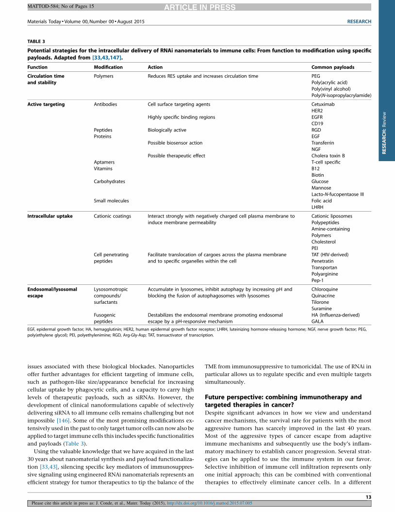
Using the valuable knowledge that we have acquired in the last
30 years about nanomaterial synthesis and payload functionaliza-
tion [33,43], silencing specific key mediators of immunosuppres-
sive signaling using engineered RNAi nanomaterials represents an
efficient strategy for tumor therapeutics to tip the balance of the
TME from immunosuppressive to tumoricidal. The use of RNAi in
particular allows us to regulate specific and even multiple targets
simultaneously.
Future perspective: combining immunotherapy andtargeted therapies in cancer?Despite significant advances in how we view and understand
cancer mechanisms, the survival rate for patients with the most
aggressive tumors has scarcely improved in the last 40 years.
Most of the aggressive types of cancer escape from adaptive
immune mechanisms and subsequently use the body’s inflam-
matory machinery to establish cancer progression. Several strat-
egies can be applied to use the immune system in our favor.
Selective inhibition of immune cell infiltration represents only
one initial approach; this can be combined with conventional
therapies to effectively eliminate cancer cells. In a different
Materials Today � Volume 00, Number 00 �August 2015 RESEARCH
MATTOD-584; No of Pages 15
Please cite this article in press as: J. Conde, et al., Mater. Today (2015), http://dx.doi.org/10.1016/j.mattod.2015.07.005
TABLE 3
Potential strategies for the intracellular delivery of RNAi nanomaterials to immune cells: From function to modification using specificpayloads. Adapted from [33,43,147].
Function Modification Action Common payloads
Circulation time
and stability
Polymers Reduces RES uptake and increases circulation time PEG
Poly(acrylic acid)
Poly(vinyl alcohol)Poly(N-isopropylacrylamide)
Active targeting Antibodies Cell surface targeting agents
Highly specific binding regions
Biologically active
Possible biosensor action
Possible therapeutic effect
Cetuximab
HER2EGFR
CD19
Peptides RGDProteins EGF
Transferrin
NGF
Cholera toxin BAptamers T-cell specific
Vitamins B12
Biotin
Carbohydrates GlucoseMannose
Lacto-N-fucopentaose III
Small molecules Folic acid
LHRH
Intracellular uptake Cationic coatings Interact strongly with negatively charged cell plasma membrane to
induce membrane permeability
Cationic liposomes
PolypeptidesAmine-containing
Polymers
Cholesterol
PEICell penetrating
peptides
Facilitate translocation of cargoes across the plasma membrane
and to specific organelles within the cell
TAT (HIV-derived)
Penetratin
TransportanPolyarginine
Pep-1
Endosomal/lysosomalescape
Lysosomotropiccompounds/
surfactants
Accumulate in lysosomes, inhibit autophagy by increasing pH andblocking the fusion of autophagosomes with lysosomes
ChloroquineQuinacrine
Tilorone
Suramine
Fusogenicpeptides
Destabilizes the endosomal membrane promoting endosomalescape by a pH-responsive mechanism
HA (Influenza-derived)GALA
EGF, epidermal growth factor; HA, hemagglutinin; HER2, human epidermal growth factor receptor; LHRH, luteinizing hormone-releasing hormone; NGF, nerve growth factor; PEG,
poly(ethylene glycol); PEI, polyethylenimine; RGD, Arg-Gly-Asp; TAT, transactivator of transcription.
13
RESEARCH:Review
approach, endogenous cells can be modified with RNAi nano-
particles ex vivo and then be used as a Trojan horse to reverse
the regulatory activity of tumor immune cells while targeting
cancer cells.
Importantly, tumors are frequently associated with the over-
expression of immunosuppressive genes in order to evade immu-
nocytotoxicity. The use of RNAi nanomaterials in vivo to silence
specific genes will be an invaluable tool for future approaches in
cancer therapies. We propose using specific RNAi nanomaterials
for cancer immunotherapy to eliminate the immunosuppressive
function of tumor-associated immune cells and at the same time to
raise potent anti-tumor immune responses. Despite the discussed
difficulties, RNAi nanoparticles are ideally suited to pursue new
avenues of cancer immunotherapies due to their versatility and
specificity. Further, the use of intelligent nanoparticles can be
efficiently combined with available successful therapies to increase
their efficacy. Cell-targeted therapies have the capacity to inhibit
molecular pathways that are crucial for tumor growth and main-
tenance and minimize collateral damage in healthy cells. Some
targeted therapies such as drug-coated nanoparticles elicit dramat-
ic tumor regressions, but generally only demonstrate a short-lived
response thus limiting their overall clinical benefit. Immunother-
apy stimulates host immune responses that potentially results in
long-lived tumor inhibition, anti-tumor immune memory and,
consequently, improved clinical outcomes. This suggests that the
two approaches might have complementary roles and that com-
binatorial therapy can have an important synergistic effect in
cancer treatment [8,9]. RNAi nanomaterials used to target and
inhibit the immunosuppressive nature of the tumor microenvi-
ronment are slowly gaining attention. However, the development
of efficient delivery vehicles for in vivo applications, especially
when systemic delivery to immune cells is sought, has remained a
challenge due to lack of specificity, selectivity, and targeting
[12,36]. We believe that nanomaterials used for RNAi immuno-
therapy have a huge potential to overcome some of these draw-
backs and provide a ray of hope for more efficacious future cancer
therapies [36].
References
[1] H.A. Goubran, et al. Cancer Growth Metastasis 7 (2014) 9.
[2] B.Z. Qian, J.W. Pollard, Cell 141 (2010) 39.
[3] J. Couzin-Frankel, Science 342 (2014) 1432.
[4] E. Shahar, et al. Vaccine 28 (2010) 7279.
[5] N.H. Cho, et al. Nat. Nanotechnol. 6 (2011) 675.
[6] C. Devaud, et al. Oncoimmunology 2 (2013).
[7] I. Mellman, et al. Nature 480 (2011) 480.
[8] M. Vanneman, G. Dranoff, Nat. Rev. Cancer 12 (2012) 237.
[9] E. Sharon, et al. Radiat. Res. 182 (2014) 252.
[10] J.J. Kim, I.F. Tannock, Nat. Rev. Cancer 5 (2005) 516.
[11] K.A. Whitehead, et al. Annu. Rev. Chem. Biomol. Eng. 2 (2011) 77.
[12] R. Kanasty, et al. Nat. Mater. 12 (2013) 967.
[13] J. Conde, et al. Adv. Drug Deliv. Rev. 81 (2015) 169.
[14] J. Conde, N. Artzi, Trends Biotechnol. 33 (2015) 141.
[15] P. Kesharwani, et al. Biomaterials 33 (2012) 7138.
[16] A. Daka, D. Peer, Adv. Drug Deliv. Rev. 64 (2012) 1508.
[17] D. Zheng, et al. Proc. Natl. Acad. Sci. U. S. A. 109 (2012) 11975.
[18] M.V. Yezhelyev, et al. J. Am. Chem. Soc. 130 (2008) 9006.
[19] H. Mok, et al. Mol. Pharm. 7 (2010) 1930.
[20] Y.C. Chen, et al. Mol. Therapy 18 (2010) 1650.
[21] M. Chen, et al. J. Phys. Chem. Lett. 1 (2010) 3167.
[22] J. Conde, et al. Biomaterials 34 (2013) 7744.
[23] J. Conde, et al. ACS Nano 6 (2012) 8316.
[24] M.R. Lares, et al. Trends Biotechnol. 28 (2010) 570.
[25] W.Q. Yang, Y. Zhang, Expert Opin. Biol. Therapy 12 (2012) 1495.
[26] R.W. Carthew, E.J. Sontheimer, Cell 136 (2009) 642.
[27] J. Soutschek, et al. Nature 432 (2004) 173.
[28] E. Marshall, Science 286 (1999) 2244.
[29] N. Lewinski, et al. Small 4 (2008) 26.
[30] S. Sharifi, et al. Chem. Soc. Rev. 41 (2012) 2323.
[31] F. Joris, et al. Chem. Soc. Rev. 42 (2013) 8339.
[32] S. Nimesh, et al. Nanomedicine 6 (2011) 729.
[33] L.Y.T. Chou, et al. Chem. Soc. Rev. 40 (2011) 233.
[34] B. Yameen, et al. J. Control. Release 190 (2014) 485.
[35] D. Mohanan, et al. J. Control. Release 147 (2010) 342.
[36] K.A. Whitehead, et al. Nat. Rev. Drug Discov. 8 (2009) 129.
[37] P.R. Gil, W.J. Parak, ACS Nano 2 (2008) 2200.
[38] J. Conde, et al. J. Drug Deliv. 2012 (2012) 751075.
[39] J. Conde, et al. Front. Pharmacol. 4 (2013) 134.
[40] C. Minelli, et al. Small 6 (2010) 2336.
[41] J. Zhou, et al. Pharmaceuticals (Basel) 6 (2013) 85.
[42] S.M. Moghimi, et al. Pharmacol. Rev. 53 (2001) 283.
[43] J. Conde, et al. Front. Chem. 2 (2014) 48.
[44] M.E. Davis, et al. Nature 464 (2010) 1067.
[45] C. Wong, et al. Proc. Natl. Acad. Sci. U. S. A. 108 (2011) 2426.
[46] D.W. Bartlett, et al. Proc. Natl. Acad. Sci. U. S. A. 104 (2007) 15549.
[47] S.D. Perrault, W.C.W. Chan, Proc. Natl. Acad. Sci. U. S. A. 107 (2010) 11194.
[48] S.E. Leucuta, Curr. Drug Deliv. 10 (2013) 208.
[49] S. Ramakrishnan, Cancer Biol. Ther. 11 (2011) 849.
[50] N. Segovia, et al. Adv. Healthc. Mater. 4 (2015) 271.
[51] N. Artzi, et al. Nat. Mater. 10 (2011) 704.
[52] N. Artzi, et al. Adv. Mater. 21 (2009) 3399.
[53] I.C. Kourtis, et al. PLOS ONE 8 (2013).
[54] S. Buchan, et al. J. Immunol. 174 (2005) 6292.
[55] M. Zaric, et al. ACS Nano 7 (2013) 2042.
[56] K.E. Driscoll, et al. Toxicol. Sci. 55 (2000) 24.
[57] D. Peer, et al. Nat. Nanotechnol. 2 (2007) 751.
[58] J. Conde, et al. Nano Today (2015), http://dx.doi.org/10.1016/j.nan-
tod.2015.06.008 (in press).
[59] J. Li, et al. J. Control. Release 158 (2012) 108.
[60] C.E. Ashley, et al. Nat. Mater. 10 (2011) 389.
[61] W. Hasan, et al. Nano Lett. 12 (2012) 287.
[62] Y. Shu, et al. Mol. Therapy 19 (2011) 1304.
[63] P. Guo, Nat. Nanotechnol. 5 (2010) 833.
[64] D. Shu, et al. Nat. Nanotechnol. 6 (2011) 658.
[65] F. Haque, et al. Nano Today 7 (2012) 245.
[66] Y. Shu, et al. Adv. Drug Deliv. Rev. 66 (2014) 74.
[67] R.B. Restani, et al. RSC Adv. 4 (2014) 54872.
[68] A. Agrawal, et al. ACS Nano 3 (2009) 2495.
[69] O. Taratula, et al. Curr. Drug Deliv. 8 (2011) 59.
[70] M.D. Vesely, et al. Annu. Rev. Immunol. 29 (29) (2011) 235.
[71] F. Avogadri, J.D. Wolchok, Nat. Biotechnol. 30 (2012) 328.
[72] D. Mittal, et al. Curr. Opin. Immunol. 27 (2014) 16.
[73] D.I. Gabrilovich, et al. Nat. Rev. Immunol. 12 (2012) 253.
[74] B. Burkholder, et al. Biochim. Biophys. Acta: Rev. Cancer 1845 (2014) 182.
[75] M. Vitale, et al. Eur. J. Immunol. 44 (2014) 1582.
[76] R. Noy, J.W. Pollard, Immunity 41 (2014) 49.
[77] M. Jinushi, Y. Komohara, Biochim. Biophys. Acta 1855 (2015) 123.
[78] K. Palucka, J. Banchereau, Nat. Rev. Cancer 12 (2012) 265.
[79] Y. Ma, et al. Semin. Cancer Biol. 22 (2012) 298.
[80] L. Wayteck, et al. Cancer Lett. 352 (2014) 113.
[81] O. Draghiciu, et al. Oncoimmunology 4 (2015).
[82] F. Mattei, et al. Neoplasia 14 (2012) 1223.
[83] N. Obermajer, et al. Blood 118 (2011) 5498.
[84] R. Roychoudhuri, et al. Curr. Opin. Immunol. 33 (2015) 101.
[85] S. Karimi, et al. Immunology 144 (2015) 186.
[86] A.M. Houghton, Cell Cycle 9 (2010) 1732.
[87] C. Tecchio, et al. Semin. Cancer Biol. 23 (2013) 159.
[88] A. Bruno, et al. J. Natl. Cancer Inst. 106 (2014).
[89] L.N. Bodduluru, et al. Cancer Lett. 357 (2015) 454.
[90] J. Maher, E.T. Davies, Br. J. Cancer 91 (2004) 817.
[91] M.L. Xu, et al. Clin. Dev. Immunol. 2010 (2010) 832454.
[92] P. Jeannin, et al. Immunotherapy 3 (2011) 23.
[93] X.Q. Tang, et al. Immunology 138 (2013) 93.
[94] W.H. Fridman, et al. Nat. Rev. Cancer 12 (2012) 298.
[95] T. Wang, et al. Front. Immunol. 5 (2014) 358.
RESEARCH Materials Today � Volume 00, Number 00 �August 2015
MATTOD-584; No of Pages 15
Please cite this article in press as: J. Conde, et al., Mater. Today (2015), http://dx.doi.org/10.1016/j.mattod.2015.07.005
14
RESEARCH:Review
[96] H. Yu, et al. Nat. Rev. Cancer 14 (2014) 736.
[97] S.I. Grivennikov, M. Karin, Cytokine Growth Factor Rev. 21 (2010) 11.
[98] H. Yu, et al. Nat. Rev. Cancer 9 (2009) 798.
[99] C. Rebe, et al. JAKSTAT 2 (2013) e23010.
[100] L.A. Haile, et al. Immunol. Invest. 41 (2012) 581.
[101] C.U. Blank, A. Enk, Int. Immunol. (2014), http://dx.doi.org/10.1093/intimm/
dxu076.
[102] R. Lameris, et al. Crit Rev. Oncol. Hematol. 92 (2014) 153.
[103] M. Kortylewski, et al. Nat. Biotechnol. 27 (2009) 925-U88.
[104] S.F. Hussain, et al. Cancer Res. 67 (2007) 9630.
[105] Z.C. Luo, et al. Biomaterials 38 (2015) 50.
[106] J.P. Yu, et al. J. Immunol. 193 (2014) 2574.
[107] M. Goldsmith, et al. Nanomedicine (Lond.) 6 (2011) 1771.
[108] V. Hornung, et al. Nat. Med. 11 (2005) 263.
[109] M.E. Kleinman, et al. Nature 452 (2008) 591.
[110] G.A. Rabinovich, et al. Annu. Rev. Immunol. 25 (2007) 267.
[111] J.C. Burnett, J.J. Rossi, Chem. Biol. 19 (2012) 60.
[112] A.D. Judge, et al. Nat. Biotechnol. 23 (2005) 457.
[113] J. Wang, et al. AAPS J. 12 (2010) 492.
[114] S.D. Li, L. Huang, Biochim. Biophys. Acta: Biomembr. 1788 (2009) 2259.
[115] S. Gao, et al. Mol. Therapy 17 (2009) 1225.
[116] J.E. Zuckerman, et al. Proc. Natl. Acad. Sci. U. S. A. 109 (2012) 3137.
[117] A. Franca, et al. Nanomedicine 6 (2011) 1175.
[118] F. Alexis, et al. Mol. Pharm. 5 (2008) 505.
[119] L. Thiele, et al. Pharm. Res. 20 (2003) 221.
[120] S.D. Conner, S.L. Schmid, Nature 422 (2003) 37.
[121] C. Foged, et al. Int. J. Pharm. 298 (2005) 315.
[122] Z. Amoozgar, M.S. Goldberg, Adv. Drug Deliv. Rev. (2014), http://dx.doi.org/
10.1016/j.addr.2014.09.007 (Epub ahead of print).
[123] S. Oliveira, et al. Int. J. Pharm. 331 (2007) 211.
[124] A.M. Villamil Giraldo, et al. Biochem. Soc. Trans. 42 (2014) 1460.
[125] Y.J. Kwon, Acc. Chem. Res. 45 (2012) 1077.
[126] A. Troegeler, et al. Immunol. Cell Biol. 92 (2014) 699.
[127] H. Akita, et al. J. Control. Release 143 (2010) 311.
[128] J.R. Cubillos-Ruiz, et al. J. Clin. Invest. 119 (2009) 2231.
[129] M.B. Heo, et al. Acta Biomater. 10 (2014) 2169.
[130] M.B. Heo, Y.T. Lim, Biomaterials 35 (2014) 590.
[131] A. Alshamsan, et al. Mol. Pharm. 7 (2010) 1643.
[132] J.R. Cubillos-Ruiz, et al. Cancer Res. 72 (2012) 1683.
[133] A. Alshamsan, et al. Transl. Oncol. 4 (2011) 178.
[134] W. Hobo, et al. Cancer Immunol. Immunother. 62 (2013) 285.
[135] S. Warashina, et al. Biol. Pharm. Bull. 34 (2011) 1348.
[136] F. Leuschner, et al. Nat. Biotechnol. 29 (2011) 1005.
[137] S.S. Yu, et al. Mol. Pharm. 10 (2013) 975.
[138] J. Conde, et al. Adv. Funct. Mater. 25 (2015) 4183.
[139] H.K. Makadia, S.J. Siegel, Polymers (Basel) 3 (2011) 1377.
[140] B.S. Zolnik, et al. Endocrinology 151 (2010) 458.
[141] X. Huang, et al. Int. J. Nanomed. 9 (2014) 4785.
[142] A. Akinc, et al. J. Gene Med. 7 (2005) 657.
[143] J.G. Quatromoni, E. Eruslanov, Am. J. Transl. Res. 4 (2012) 376.
[144] A. Herrmann, et al. J. Clin. Invest. 124 (2014) 2977.
[145] R.E. Serda, Int. J. Nanomed. 8 (2013) 1683.
[146] S. Weinstein, D. Peer, Nanotechnology 21 (2010).
[147] R.A. Sperling, W.J. Parak, Philos. Trans. A: Math. Phys. Eng. Sci. 368 (2010) 1333.
Materials Today � Volume 00, Number 00 �August 2015 RESEARCH
MATTOD-584; No of Pages 15
Please cite this article in press as: J. Conde, et al., Mater. Today (2015), http://dx.doi.org/10.1016/j.mattod.2015.07.005
15
RESEARCH:Review
Related Documents


![CANCER TARGETING STRATEGIES OF NANOMATERIALSiapc-obp.com/assets/files/349331_09_NMRM_19.pdf · cancer targeting [8-10]. Non-viral nanomaterials have several advantages such as low](https://static.cupdf.com/doc/110x72/60719b790cbce024cc3df0dc/cancer-targeting-strategies-of-nanomaterialsiapc-obpcomassetsfiles34933109nmrm19pdf.jpg)