Relationship between oxidative stress, glutathione S-transferase polymorphisms and hydroxyurea treatment in sickle cell anemia Danilo Grünig Humberto Silva a,d , Edis Belini Junior a , Lidiane de Souza Torres a , Octávio Ricci Júnior b , Clarisse de Castro Lobo c , Claudia Regina Bonini-Domingos a , Eduardo Alves de Almeida d, ⁎ a UNESP—Sao Paulo State University, Department of Biology, Hemoglobin and Hematologic Genetic Diseases Laboratory, Sao Paulo, Brazil b Sao Jose do Rio Preto Medical School—FAMERP, Department of Medicine, Sao Paulo, Brazil c Hematological State Institute “Arthur de Siqueira Cavalcanti”—HEMORIO, Rio de Janeiro, Brazil d UNESP—Sao Paulo State University, Department of Chemistry and Environmental Sciences, Sao Paulo, Brazil abstract article info Article history: Submitted 26 January 2011 Revised 10 March 2011 Available online 12 April 2011 (Communicated by Sir D. Weatherall, F.R.S., 15 March 2011) Keywords: Sickle cell anemia Oxidative stress Hydroxyurea GST polymorphisms This study evaluated the oxidative stress and antioxidant capacity markers in sickle cell anemia (SCA) patients with and without treatment with hydroxyurea. We assessed GSTT1, GSTM1 and GSTP1 polymorphisms in patients and a control group. The study groups were composed of 48 subjects without hemoglobinopathies and 28 SCA patients, 13 treated with HU [SCA (+HU)], and 15 SCA patients not treated with HU [SCA (-HU)]. We observed a significant difference for GSTP1 polymorphisms in SCA patients with the V/V genotype that showed higher glutathione (GSH) and Trolox equivalent antioxidant capacity (TEAC) (p = 0.0445 and p = 0.0360), respectively, compared with the I/I genotype. HU use was associated with a 35.2% decrease in the lipid peroxidation levels of the SCA (+ HU) group (p b 0.0001). Moreover, the SCA (+HU) group showed higher TEAC as compared to the control group (p = 0.002). We did not find any significant difference in glutathione-S-transferase (GST) activity between the groups (p = 0.76), but the catalase (CAT) activity was about 17% and 30% decreased in the SCA (+ HU) and SCA (-HU) groups, respectively (p b 0.00001). Whereas the plasma GSH levels were ~ 2 times higher in the SCA patients than the control group (p = 0.0005). HU use has contributed to higher CAT activity and TEAC, and lower lipid peroxidation in patients under treatment. These findings may explain the influence of HU in ameliorating oxidative stress on SCA subjects. © 2011 Elsevier Inc. All rights reserved. Introduction Sickle cell anemia (SCA), an inherited monogenic disease, is caused by a transversion in the codon of the sixth amino acid of the beta globin chain, on chromosome 11 [1], leading to formation of a defective he- moglobin form, hemoglobin S (Hb S). In the deoxygenated state, the Hb S tends to aggregate into rodlike polymers, resulting in the deformed sickle shape and rigidity of red blood cells (RBCs) characteristic of this condition [2]. Normal RBCs are usually subjected to oxidative stress as a result of continuous reactive oxygen species (ROS) production that accom- panies Hb autoxidation, a condition that increases in SCA [3–5], leading to a continuous inflammatory response and oxidative stress [6]. Among the SCA-derived oxidative stress consequences are an increase in membrane lipid peroxidation levels and alterations in antioxidant defense systems. However, the few studies that have examined antioxidant enzymes in SCA patients have found contradictory results [7]. Glutathione S-transferases (GSTs) constitute multifunctional en- zymes that are coded by at least eight distinct loci: α (GSTA), μ (GSTM), θ (GSTT), π (GSTP), σ (GSTS), κ (GSTK), ω (GSTO), and ζ (GSTZ), each one composed by one or more homodimeric or heterodimeric isoforms [8,9]. These enzymes are involved in the conjugation reactions during phase II of the xenobiotic metabolism, catalyzing reactions between glutathione (GSH) and a variety of potentially toxic and carcinogenic electrophilic compounds [10,11], besides, GSTs also display peroxidase activity and can thus protect from oxidative damage [12]. The defi- ciency in the activity of this enzyme can be derived from the inherited GSTs polymorphisms, e.g., GSTT1 (22q11.23), GSTM1 (1q13.3) and GSTP1 (11q13) [13]. Hydroxyurea (HU) administration is one of the most important therapies for SCA patients [14,15]. The pharmacologic effects that may contribute to the efficacy of HU in SCA treatment includes the induction of Hb F production and increase in RBC water content (which reduces the polymerization of Hb S). HU also increases microvascular Blood Cells, Molecules, and Diseases 47 (2011) 23–28 Abbreviations: CAT, catalase; GSH, glutathione; GST, glutathione-S-transferase; HU, hydroxyurea; HPLC, high performance liquid chromatography; RBC, red blood cells; ROS, reactive oxygen species; SCA, sickle cell anemia; TBARS, thiobarbituric-acid- reacting substances; TEAC, Trolox equivalent antioxidant capacity. ⁎ Corresponding author at: Rua Cristóvão Colombo, 2265, Jardim Nazareth, CEP: 15054–000 Sao Jose do Rio Preto, Sao Paulo, Brazil. E-mail address: [email protected] (E.A. de Almeida). 1079-9796/$ – see front matter © 2011 Elsevier Inc. All rights reserved. doi:10.1016/j.bcmd.2011.03.004 Contents lists available at ScienceDirect Blood Cells, Molecules, and Diseases journal homepage: www.elsevier.com/locate/ybcmd

Welcome message from author
This document is posted to help you gain knowledge. Please leave a comment to let me know what you think about it! Share it to your friends and learn new things together.
Transcript
Blood Cells, Molecules, and Diseases 47 (2011) 23–28
Contents lists available at ScienceDirect
Blood Cells, Molecules, and Diseases
j ourna l homepage: www.e lsev ie r.com/ locate /ybcmd
Relationship between oxidative stress, glutathione S-transferase polymorphisms andhydroxyurea treatment in sickle cell anemia
Danilo Grünig Humberto Silva a,d, Edis Belini Junior a, Lidiane de Souza Torres a, Octávio Ricci Júnior b,Clarisse de Castro Lobo c, Claudia Regina Bonini-Domingos a, Eduardo Alves de Almeida d,⁎a UNESP—Sao Paulo State University, Department of Biology, Hemoglobin and Hematologic Genetic Diseases Laboratory, Sao Paulo, Brazilb Sao Jose do Rio Preto Medical School—FAMERP, Department of Medicine, Sao Paulo, Brazilc Hematological State Institute “Arthur de Siqueira Cavalcanti”—HEMORIO, Rio de Janeiro, Brazild UNESP—Sao Paulo State University, Department of Chemistry and Environmental Sciences, Sao Paulo, Brazil
Abbreviations: CAT, catalase; GSH, glutathione; GST,hydroxyurea; HPLC, high performance liquid chromatoROS, reactive oxygen species; SCA, sickle cell anemireacting substances; TEAC, Trolox equivalent antioxidan⁎ Corresponding author at: Rua Cristóvão Colombo
15054–000 Sao Jose do Rio Preto, Sao Paulo, Brazil.E-mail address: [email protected] (E.A. de A
1079-9796/$ – see front matter © 2011 Elsevier Inc. Aldoi:10.1016/j.bcmd.2011.03.004
a b s t r a c t
a r t i c l e i n f oArticle history:Submitted 26 January 2011Revised 10 March 2011Available online 12 April 2011
(Communicated by Sir D. Weatherall, F.R.S.,15 March 2011)
Keywords:Sickle cell anemiaOxidative stressHydroxyureaGST polymorphisms
This study evaluated the oxidative stress and antioxidant capacity markers in sickle cell anemia (SCA) patientswith andwithout treatmentwith hydroxyurea.WeassessedGSTT1,GSTM1 andGSTP1polymorphisms inpatientsand a control group. The study groups were composed of 48 subjects without hemoglobinopathies and 28 SCApatients, 13 treatedwithHU [SCA (+HU)], and 15 SCApatients not treatedwithHU [SCA (−HU)].Weobserved asignificant difference for GSTP1 polymorphisms in SCA patients with the V/V genotype that showed higherglutathione (GSH) and Trolox equivalent antioxidant capacity (TEAC) (p=0.0445 and p=0.0360), respectively,comparedwith the I/I genotype. HU usewas associatedwith a 35.2% decrease in the lipid peroxidation levels of theSCA (+HU) group (pb0.0001). Moreover, the SCA (+HU) group showed higher TEAC as compared to the controlgroup (p=0.002). We did not find any significant difference in glutathione-S-transferase (GST) activity betweenthe groups (p=0.76), but the catalase (CAT) activitywas about 17% and 30% decreased in the SCA (+HU) and SCA(−HU)groups, respectively (pb0.00001).Whereas theplasmaGSHlevelswere~2 timeshigher in theSCApatientsthan the control group (p=0.0005). HU use has contributed to higher CAT activity and TEAC, and lower lipidperoxidation in patients under treatment. Thesefindingsmay explain the influence ofHU inameliorating oxidativestress on SCA subjects.
glutathione-S-transferase; HU,graphy; RBC, red blood cells;a; TBARS, thiobarbituric-acid-t capacity., 2265, Jardim Nazareth, CEP:
lmeida).
l rights reserved.
© 2011 Elsevier Inc. All rights reserved.
Introduction
Sickle cell anemia (SCA), an inheritedmonogenic disease, is caused bya transversion in the codon of the sixth amino acid of the beta globinchain, on chromosome 11 [1], leading to formation of a defective he-moglobin form, hemoglobin S (Hb S). In the deoxygenated state, the HbS tends to aggregate into rodlike polymers, resulting in the deformedsickle shape and rigidity of red blood cells (RBCs) characteristic of thiscondition [2].
Normal RBCs are usually subjected to oxidative stress as a resultof continuous reactive oxygen species (ROS) production that accom-panies Hb autoxidation, a condition that increases in SCA [3–5], leadingto a continuous inflammatory response and oxidative stress [6]. Among
the SCA-derived oxidative stress consequences are an increase inmembrane lipid peroxidation levels and alterations in antioxidantdefense systems. However, the few studies that have examinedantioxidant enzymes in SCA patients have found contradictory results[7].
Glutathione S-transferases (GSTs) constitute multifunctional en-zymes that are coded by at least eight distinct loci:α (GSTA), μ (GSTM),θ (GSTT),π (GSTP),σ (GSTS), κ (GSTK),ω (GSTO), and ζ (GSTZ), each onecomposed by one or more homodimeric or heterodimeric isoforms[8,9]. These enzymes are involved in the conjugation reactions duringphase II of the xenobiotic metabolism, catalyzing reactions betweenglutathione (GSH) and a variety of potentially toxic and carcinogenicelectrophilic compounds [10,11], besides, GSTs also display peroxidaseactivity and can thus protect from oxidative damage [12]. The defi-ciency in the activity of this enzyme can be derived from the inheritedGSTspolymorphisms, e.g.,GSTT1 (22q11.23),GSTM1 (1q13.3) andGSTP1(11q13) [13].
Hydroxyurea (HU) administration is one of the most importanttherapies for SCA patients [14,15]. The pharmacologic effects that maycontribute to the efficacy of HU in SCA treatment includes the inductionof Hb F production and increase in RBCwater content (which reducesthe polymerization of Hb S). HU also increases microvascular
Table 1Hemoglobin fraction concentration in SCA patients and control group.
CG (n=48) SCA (+HU) (n=13) SCA (−HU) (n=15)
Hb A (%)⁎ 94.88±2.12a 12.58±16.39b 8.06±14.2b
Hb A2 (%)⁎ 2.75±0.39a 3.99±0.66b 3.75±1.02b
Hb F (%)⁎ 1.08±1.13a 10.93±7.07b 6.16±4.54b
Hb S (%)# 0.0 70.40±16.07a 80.39±16.08b
CG: control group; SCA (+HU): patients treated with HU; SCA (−HU): patients nottreated with HU. Data were expressed as mean±standard deviation.⁎ Different letters indicate statistical difference (Kruskal–Wallis test followed by post
hoc Dunn test, pb0.05).# Different letters indicate statistical difference (Mann–Whitney test, pb0.05).
24 D.G.H. Silva et al. / Blood Cells, Molecules, and Diseases 47 (2011) 23–28
navigation by sickled cells, and decreases RBC adhesion to endothe-lium by diminishing the expression of endothelial adhesion molecules[15,16].
There are no reports about the influences of GSTT1, GSTM1 andGSTP1 polymorphisms on oxidative stress parameters in SCA patients.Therefore, the aim of this study was to evaluate, in SCA patients, theoxidative stress and antioxidant capacity markers, correlating themwith HU treatment and GSTT1, GSTM1 and GSTP1 polymorphismscompared with a control group.
Methods
Subjects
Seventy-six Brazilian subjects were included in the study (30males and 46 females; mean age: 23.8 years old; range: 9–65 yearsold). They were from the northwest region of Sao Paulo state andsouthwest region of Rio de Janeiro state in Brazil. The test group wascomposed by 28 SCA patients (11 males and 17 females; mean age:27.7 years old) in a clinical follow-up in São Jose do Rio Preto (SP) andRio de Janeiro (RJ). The control group was composed of 48 subjectswithout hemoglobinopathies living also in the northwest region ofSao Paulo state (19 males and 29 females; mean age: 21.9 years old).
All the patients were screened using a questionnaire, and wereexcluded if they were smokers or drinkers. They were also asked aboutpain crisis, hemolytic crisis, and if they received blood transfusion or hadhad stroke in the last two months. The medications used were alsoaccessed, and those taking medication known to affect the analyzedparameters were excluded from the study. All subjects gave theirinformed consent, and the study was approved by the Data SafetyMonitoring Board (DSMB) according to Brazilian regulations.
All SCApatients had access to the samemedicationprotocol and thenwere separated into two groups: SCA patients treated with HU [SCA(+HU); n=13] and SCA patients not treated with HU [SCA (−HU);n=15].
Biological samples
Blood samples (11 mL) were collected through venipuncture inheparinized and EDTA tubes. The heparinized blood (7 mL) wasincubated for 20 min at 37 °C and then centrifuged at 206g for 20 minto separate the plasma for the thiobarbituric-acid-reacting substances(TBARS) and TEAC. The EDTA sample fraction (4 mL) was aliquoted,being 2 mL for the hemoglobinopathies tests, genotypic determina-tion, and enzymatic activities analysis. The other 2 mL was submittedto centrifugation at 825g for 10 min to obtain the plasma and thenwere frozen at −80 °C for GSH determination.
Hemoglobin phenotypes and genotypes
Hb identificationwas performed using electrophoresis on celluloseacetate pH 8.4, and agar electrophoresis at pH 6.2. The Hb fractionquantification was obtained using high performance liquid chroma-tography (HPLC) by the automated VARIANTTM equipment (Bio-RadLaboratories, CA, USA) [17]. Cell morphologymicroscopic analysis wasperformed on the stained blood using May-Grünwald–Giemsa. In allpatient samples, the Hb genotype was developed by molecularanalysis using PCR–RFLP. The segment amplification that encodesHb S was accomplished by specific primers, and the amplicon wascleaved by the DdeI restriction endonuclease (New England BioLabs,MA, USA) [18].
Glutathione S-transferase polymorphism genotyping
The polymorphism analysis for the GSTM1 and the GSTT1 geneswas determined simultaneously in a single assay using a multiplex
PCR approach with a housekeeping CYP1A gene co-amplification as aninternal control [19].
GSTP1 polymorphism was performed using PCR–RFLP and thespecific primers for exon 5 (Ile105Val). Following PCR, the productwas digested with BsmA1 restriction endonuclease (New EnglandBioLabs, MA, USA). The digest was submitted to an electrophoresis ona 3.5% agarose gel containing ethidium bromide [20].
Biochemical analysis
Lipid peroxidation levels were assessed in heparinized plasma usingthe thiobarbituric-acid-reactive substances assay [21]. Antioxidantcapacity was also determined in heparinized plasma samples accordingto their equivalence to Trolox (6-hydroxy-2,5,7,8-tetramethychroman-2-carboxylic acid) [22]. For the total GST activity, the blood sampleswere diluted in a 3.5 μM 2-mercaptoethanol, 10 μMNADP, and 2.7 mMEDTA hemolyzing solution (1:20, v/v) and then assayed using 1-chloro-2,4-dinitrobenzene (CDNB) as substrate at 340 nm. The assay wascarried out in 0.2 MK-phosphate buffer, pH 6.5, 1 mMCDNB, 1 mMGSH(ε=9.6 mM−1 cm−1) [23]. For the analysis of CAT activity, bloodsamples were diluted in ultrapure water (1:50, v/v) and then 10 uLwasused to measure CAT activity, by the decrease in absorbance at 240 nm(ε=0.04 mM−1 cm−1) due to the consumption of H2O2 (10 mM H2O2
in 1 M Tris–HCl buffer pH 8.0 containing 5 mM EDTA) [24].GSH concentration was determined in EDTA plasma samples using
HPLC coupled to a coulometric electrochemical detector (CoulochemIII ESA, Bedford, MA) [25]. Under these conditions, GSH clearly elutedat ~6 min. GSH was extracted from the plasma samples by addingperchloric acid to the plasma sample (10% final concentration). Aftervigorous stirring and 10 min on ice, the mixture was centrifuged at825 g for 10 min at 4 °C. The extract was then filtered through Millexsyringe filter units (0.22 μm) and directly injected into the HPLCsystem. The calculations were based on a calibration curve previouslyconstructed by injecting authentic GSH standards into the HPLCsystem.
Statistical analysis
Statistical analysis was performed using the Statistica 9.0 software(Statsoft Inc.). Data were expressed as mean±standard deviation andwere tested for normal distribution using the Shapiro Wilk test. Datawere also tested for homogeneity of variances using the Levene test.Means between the groups were compared by applying the t test orone-way ANOVA followed by the post hoc Tukey test for parametricdata, and the Mann–Whitney test or Kruskal–Wallis followed by thepost hoc Dunn test for non-parametric data. To assess the degree ofassociation between the studied variables, we used the Pearsoncorrelation for parametric data and the Spearman correlation for non-parametric data and linear regression. To compare the GST genotypesbetween the studied groups Pearson Chi-square test supplemented byFisher's exact test was used. The level of significance was set atpb0.05.
Table 2Prevalence of glutathione S-transferase polymorphisms in the control group and SCApatients.
Control group N (%) SCA patients N (%) p valuea
GSTM1[+] 21 (43.7) 20 (71.4) 0.0195[−] 27 (56.3) 8 (21.6) 0.0195
GSTT1[+] 27 (56.3) 9 (32.2) 0.0423[−] 21 (43.7) 19 (67.8) 0.0423
GSTM1/T1[+]/[−] 13 (27.0) 14 (50.0) 0.0440[+]/[+] 8 (16.7) 6 (21.4) 0.6054[−]/[+] 19 (39.6) 3 (10.7) 0.0074[−]/[−] 8 (16.7) 5 (17.9) 0.8994
GSTP1I/I 34 (70.8) 5 (17.9) 0.0001I/V 12 (25.0) 16 (57.1) 0.0050V/V 2 (4.2) 7 (25.0) 0.0066
Wild [+] and null [−] genotypes, I/I wild homozygous; I/V heterozygate; V/V mutanthomozygous.
a Comparisons were made by Pearson Chi square test supplemented by Fisher's exacttest.
25D.G.H. Silva et al. / Blood Cells, Molecules, and Diseases 47 (2011) 23–28
Results and discussion
It has been shown that HU treatment improves SCA patient's clinicalconditions. In our study, all hemoglobin parameters were significantlydifferent between the groups (Table 1), as expected for the SCA, and thelevels of Hb Fwas increased in SCA patients treatedwithHU, confirmingthe previouslywell-described HU effect [15]. It is interesting to note thehigh Hb F levels in the SCA (−HU) group which may have beeninfluenced by non-target factors in this study, such as the three majorloci – Xmn1-HBG2 single nucleotide polymorphism, HBS1L-MYBintergenic region on chromosome 6q, and BCL11A – that contribute20%–50% of the trait Hb F variance in SCA patients [26].
It should bementioned that theHbA2 levelswere slightly higher thanexpected for SCA, and this is due to the automatedHPLC characteristics inwhich Hb S acetylated subfractions elute at the Hb A2window increasingHb A2 levels [27], and the high Hb A levels observed in both SCA groupsare transfusion remnants, because the blood transfusions are indispens-able and very frequent in SCA treatment [28]. The transfusion remnantHb A levels could have masked the well-characterized chronic oxidativestress in SCA widely reported in the literature [4–7], since the increasedHb A levels foster a dilution of erythrocyte Hb S levels and a subsequent
Table 3Mean values and standard desviation of measured biochemical parameters in the control g
GSTM1/T1
[+]/[−] [+]/[+] [−]/[+] [−]/[−]
TBARS (ng/mL)Control group 290.03±209.15 220.12±65.95 191.77±139.21 252.87±18SCA patients 1501.00±472.72 1532.40±775.47 1466.78±558.57 1634.19±83
TEAC (mM)Control group 1.93±0.15 1.91±0.14 1.87±0.13 1.87±0.1SCA patients 2.00±0.18 2.02±0.14 2.13±0.08 1.96±0.2
GST (U/mL)Control group 1.72±0.81 1.26±0.54 1.63±0.70 1.32±0.7SCA patients 1.62±0.51 1.97±0.96 1.13±0.33 1.52±0.5
CAT (U/mL)Control group 2339.65±466.35 2332.74±298.25 2427.23±314.53 2301.05±16SCA patients 1804.98±445.88 1828.63±587.26 1685.44±763.56 1921.12±69
GSH (μM)Control group 0.37±0.31 0.29±0.16 0.37±0.39 0.33±0.2SCA patients 0.57±0.37 0.73±0.07 0.99±0.53 0.87±0.6
Wild [+] and null [−] genotype. I/I wild homozygous; I/V mutant homozygous. Data were⁎ Comparisons were made by Kruskal–Wallis test followed by post hoc Dunn test.# Different letters indicate statistical difference.
decline in theHbSpolymerization [29] that is aprimarypathophysiologicevent in this genetic disorder [30]. However, these biased results werenot observed in the obtained data.
The co-inheritance of polymorphisms associated to the SCA patientsmay modulate phenotypic expression [31], therefore we investigatedthe frequency of GST polymorphisms (GSTM1, GSTT1 and GSTP1) and itsrelationship with oxidative stress markers in SCA and in the controlgroup. Human GSTs have been well characterized as ethnic-dependentpolymorphism frequencies [32] and largely divergent among popula-tions around the world [33]. The gene distribution of GST poly-morphisms in this study is summarized in Table 2.
The frequency data obtained for the control group are similar tothose described in the literature, whereas the SCA patients grouppresented lower frequencies for the null GSTM1 (21.6%) and I/I(17.9%) genotypes when compared with the frequencies obtained forBrazilian population [32,34–37]. However, this is the first studyreporting the GSTM1, GSTT1 and GSTP1 polymorphisms' frequency inSCA patients and the significant difference when compared with thecontrol group.
The association of GSTM1, GSTT1 and GSTP1 polymorphisms withvarious diseases such as cancer and inflammatory diseases, andresponses in the metabolism, efficacy and toxicity of certain drugshave been widely investigated [38–40] but few studies have investi-gated their relationship with oxidative stress parameters. Bessa et al.[41] showed that subjects with null GSTM1 and GSTT1 or null genotypefor both genes had significantly lower erythrocyte GST activity thanthose with GSTM1+/GSTT1+, and also higher lipid peroxidation levels,lower erythrocyte GSH levels and decreased activities of CAT,superoxide dismutase (SOD) and GPx, whereas there was no significantdifference in these oxidative stress-related parameters between theGSTM1 and GSTT1 genotypes.
The influences of GSTM1, GSTT1 and GSTP1 polymorphisms on theoxidative stress parameters (TBARS, TEAC and GSH plasma levels, andactivities of CAT and GST) in the control group and SCA patients areshown in Table 3. Our results showed that the GST polymorphisms didnot influence significantly on any of the biochemical markers in thecontrol group, whereas in SCA patients, we observed significantdifference only for GSTP1 polymorphisms. SCA patients with the V/Vgenotype showed higher GSH and TEAC levels (p=0.0445 andp=0.0360, respectively) compared with the I/I genotype, indicatinga possible correlation between these polymorphisms with oxidativestress in SCA patients. However, further researches with larger samplesize and association with SCA clinical manifestations are needed, to
roup and patients in the presence of different GST polymorphisms found.
p value⁎ GSTP1 pvalue⁎
I/I I/V V/V
7.29 0.4166 218.20±158.26 245.91±164.59 331.00±169.70 0.59167.66 0.9867 1624.80±535.62 1535.28±629.53 1478.89±590.81 0.8505
4 0.4166 1.88±0.02 1.93±0.12 1.98±0.12 0.38111 0.5733 1.86±0.11a 2.02±0.18 2.08±0.11b 0.0445#
6 0.4166 1.51±0.64 1.51±0.92 1.78±0.61 0.70152 0.3454 1.58±0.61 1.54±0.57 1.70±0.92 0.9576
7.25 0.5623 2387.81±388.69 2439.55±256.80 2334.50±219.10 0.87511.05 0.9600 1823.23±689.74 1769.58±492.94 1978.37±526.04 0.7425
7 0.9960 0.33±0.31 0.34±0.26 0.71±0.43 0.23334 0.5681 0.38±0.28a 0.71±0.44 0.96±0.24b 0.0360#
expressed as mean±standard deviation.
26 D.G.H. Silva et al. / Blood Cells, Molecules, and Diseases 47 (2011) 23–28
better predict the relative risk generated by the co-inheritance of GSTpolymorphisms andHbSmutation in the pathophysiology development.
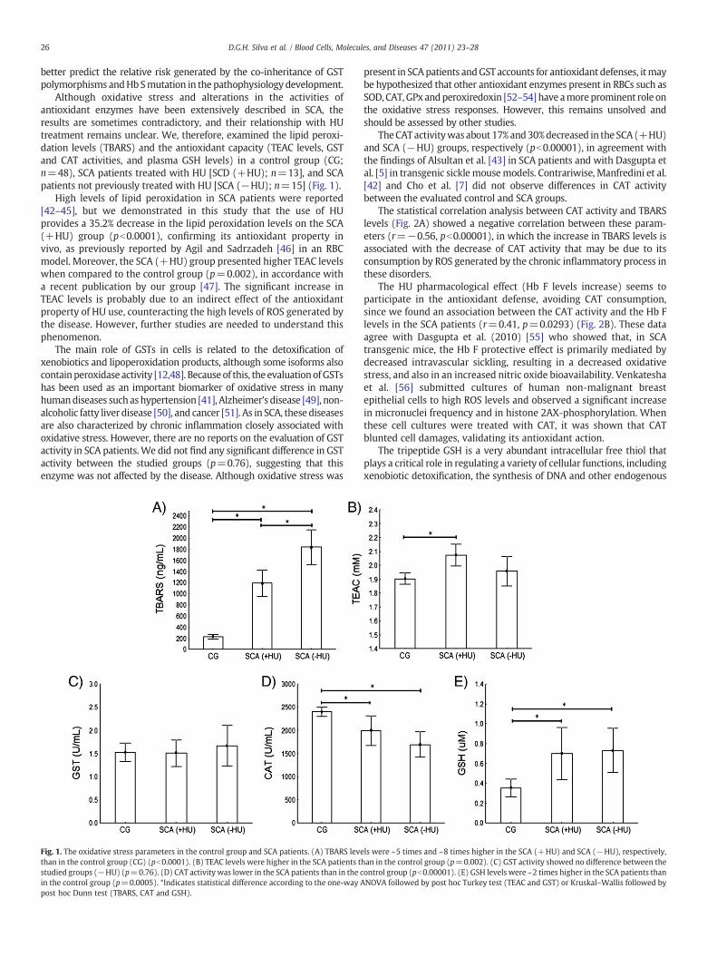
Although oxidative stress and alterations in the activities ofantioxidant enzymes have been extensively described in SCA, theresults are sometimes contradictory, and their relationship with HUtreatment remains unclear. We, therefore, examined the lipid peroxi-dation levels (TBARS) and the antioxidant capacity (TEAC levels, GSTand CAT activities, and plasma GSH levels) in a control group (CG;n=48), SCA patients treated with HU [SCD (+HU); n=13], and SCApatients not previously treated with HU [SCA (−HU); n=15] (Fig. 1).
High levels of lipid peroxidation in SCA patients were reported[42–45], but we demonstrated in this study that the use of HUprovides a 35.2% decrease in the lipid peroxidation levels on the SCA(+HU) group (pb0.0001), confirming its antioxidant property invivo, as previously reported by Agil and Sadrzadeh [46] in an RBCmodel. Moreover, the SCA (+HU) group presented higher TEAC levelswhen compared to the control group (p=0.002), in accordance witha recent publication by our group [47]. The significant increase inTEAC levels is probably due to an indirect effect of the antioxidantproperty of HU use, counteracting the high levels of ROS generated bythe disease. However, further studies are needed to understand thisphenomenon.
The main role of GSTs in cells is related to the detoxification ofxenobiotics and lipoperoxidation products, although some isoforms alsocontainperoxidase activity [12,48]. Becauseof this, the evaluationofGSTshas been used as an important biomarker of oxidative stress in manyhumandiseases such ashypertension [41], Alzheimer's disease [49], non-alcoholic fatty liver disease [50], and cancer [51]. As in SCA, these diseasesare also characterized by chronic inflammation closely associated withoxidative stress. However, there are no reports on the evaluation of GSTactivity in SCA patients.We did not find any significant difference in GSTactivity between the studied groups (p=0.76), suggesting that thisenzyme was not affected by the disease. Although oxidative stress was
Fig. 1. The oxidative stress parameters in the control group and SCA patients. (A) TBARS levethan in the control group (CG) (pb0.0001). (B) TEAC levels were higher in the SCA patients tstudied groups (−HU) (p=0.76). (D) CAT activity was lower in the SCA patients than in the cin the control group (p=0.0005). *Indicates statistical difference according to the one-way Apost hoc Dunn test (TBARS, CAT and GSH).
present in SCApatients andGST accounts for antioxidant defenses, itmaybe hypothesized that other antioxidant enzymes present in RBCs such asSOD, CAT,GPxandperoxiredoxin [52–54] have amoreprominent role onthe oxidative stress responses. However, this remains unsolved andshould be assessed by other studies.
TheCATactivitywasabout 17%and30%decreased in theSCA (+HU)and SCA (−HU) groups, respectively (pb0.00001), in agreement withthe findings of Alsultan et al. [43] in SCA patients and with Dasgupta etal. [5] in transgenic sicklemousemodels. Contrariwise, Manfredini et al.[42] and Cho et al. [7] did not observe differences in CAT activitybetween the evaluated control and SCA groups.
The statistical correlation analysis between CAT activity and TBARSlevels (Fig. 2A) showed a negative correlation between these param-eters (r=−0.56, pb0.00001), in which the increase in TBARS levels isassociated with the decrease of CAT activity that may be due to itsconsumption by ROS generated by the chronic inflammatory process inthese disorders.
The HU pharmacological effect (Hb F levels increase) seems toparticipate in the antioxidant defense, avoiding CAT consumption,since we found an association between the CAT activity and the Hb Flevels in the SCA patients (r=0.41, p=0.0293) (Fig. 2B). These dataagree with Dasgupta et al. (2010) [55] who showed that, in SCAtransgenic mice, the Hb F protective effect is primarily mediated bydecreased intravascular sickling, resulting in a decreased oxidativestress, and also in an increased nitric oxide bioavailability. Venkateshaet al. [56] submitted cultures of human non-malignant breastepithelial cells to high ROS levels and observed a significant increasein micronuclei frequency and in histone 2AX-phosphorylation. Whenthese cell cultures were treated with CAT, it was shown that CATblunted cell damages, validating its antioxidant action.
The tripeptide GSH is a very abundant intracellular free thiol thatplays a critical role in regulating a variety of cellular functions, includingxenobiotic detoxification, the synthesis of DNA and other endogenous
ls were ~5 times and ~8 times higher in the SCA (+HU) and SCA (−HU), respectively,han in the control group (p=0.002). (C) GST activity showed no difference between theontrol group (pb0.00001). (E) GSH levels were ~2 times higher in the SCA patients thanNOVA followed by post hoc Turkey test (TEAC and GST) or Kruskal–Wallis followed by
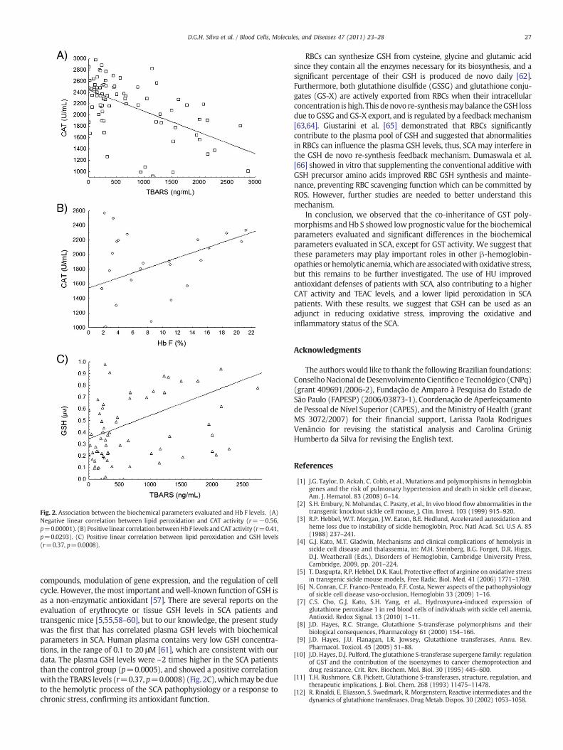
Fig. 2. Association between the biochemical parameters evaluated and Hb F levels. (A)Negative linear correlation between lipid peroxidation and CAT activity (r=−0.56,p=0.00001). (B) Positive linear correlationbetweenHbF levels andCAT activity (r=0.41,p=0.0293). (C) Positive linear correlation between lipid peroxidation and GSH levels(r=0.37, p=0.0008).
27D.G.H. Silva et al. / Blood Cells, Molecules, and Diseases 47 (2011) 23–28
compounds, modulation of gene expression, and the regulation of cellcycle. However, the most important and well-known function of GSH isas a non-enzymatic antioxidant [57]. There are several reports on theevaluation of erythrocyte or tissue GSH levels in SCA patients andtransgenic mice [5,55,58–60], but to our knowledge, the present studywas the first that has correlated plasma GSH levels with biochemicalparameters in SCA. Human plasma contains very low GSH concentra-tions, in the range of 0.1 to 20 μM [61], which are consistent with ourdata. The plasma GSH levels were ~2 times higher in the SCA patientsthan the control group (p=0.0005), and showed a positive correlationwith the TBARS levels (r=0.37, p=0.0008) (Fig. 2C),whichmay be dueto the hemolytic process of the SCA pathophysiology or a response tochronic stress, confirming its antioxidant function.
RBCs can synthesize GSH from cysteine, glycine and glutamic acidsince they contain all the enzymes necessary for its biosynthesis, and asignificant percentage of their GSH is produced de novo daily [62].Furthermore, both glutathione disulfide (GSSG) and glutathione conju-gates (GS-X) are actively exported from RBCs when their intracellularconcentration is high. This denovo re-synthesismaybalance theGSH lossdue to GSSG and GS-X export, and is regulated by a feedbackmechanism[63,64]. Giustarini et al. [65] demonstrated that RBCs significantlycontribute to the plasma pool of GSH and suggested that abnormalitiesin RBCs can influence the plasma GSH levels, thus, SCA may interfere inthe GSH de novo re-synthesis feedback mechanism. Dumaswala et al.[66] showed in vitro that supplementing the conventional additive withGSH precursor amino acids improved RBC GSH synthesis and mainte-nance, preventing RBC scavenging function which can be committed byROS. However, further studies are needed to better understand thismechanism.
In conclusion, we observed that the co-inheritance of GST poly-morphisms andHb S showed low prognostic value for the biochemicalparameters evaluated and significant differences in the biochemicalparameters evaluated in SCA, except for GST activity. We suggest thatthese parameters may play important roles in other β-hemoglobin-opathies or hemolytic anemia,which are associatedwith oxidative stress,but this remains to be further investigated. The use of HU improvedantioxidant defenses of patients with SCA, also contributing to a higherCAT activity and TEAC levels, and a lower lipid peroxidation in SCApatients. With these results, we suggest that GSH can be used as anadjunct in reducing oxidative stress, improving the oxidative andinflammatory status of the SCA.
Acknowledgments
The authorswould like to thank the following Brazilian foundations:ConselhoNacional deDesenvolvimento Científico e Tecnológico (CNPq)(grant 409691/2006-2), Fundação de Amparo à Pesquisa do Estado deSão Paulo (FAPESP) (2006/03873-1), Coordenação de Aperfeiçoamentode Pessoal de Nível Superior (CAPES), and theMinistry of Health (grantMS 3072/2007) for their financial support, Larissa Paola RodriguesVenâncio for revising the statistical analysis and Carolina GrünigHumberto da Silva for revising the English text.
References
[1] J.G. Taylor, D. Ackah, C. Cobb, et al., Mutations and polymorphisms in hemoglobingenes and the risk of pulmonary hypertension and death in sickle cell disease,Am. J. Hematol. 83 (2008) 6–14.
[2] S.H. Embury, N. Mohandas, C. Paszty, et al., In vivo blood flow abnormalities in thetransgenic knockout sickle cell mouse, J. Clin. Invest. 103 (1999) 915–920.
[3] R.P. Hebbel, W.T. Morgan, J.W. Eaton, B.E. Hedlund, Accelerated autoxidation andheme loss due to instability of sickle hemoglobin, Proc. Natl Acad. Sci. U.S A. 85(1988) 237–241.
[4] G.J. Kato, M.T. Gladwin, Mechanisms and clinical complications of hemolysis insickle cell disease and thalassemia, in: M.H. Steinberg, B.G. Forget, D.R. Higgs,D.J. Weatherall (Eds.), Disorders of Hemoglobin, Cambridge University Press,Cambridge, 2009, pp. 201–224.
[5] T. Dasgupta, R.P. Hebbel, D.K. Kaul, Protective effect of arginine on oxidative stressin transgenic sickle mouse models, Free Radic. Biol. Med. 41 (2006) 1771–1780.
[6] N. Conran, C.F. Franco-Penteado, F.F. Costa, Newer aspects of the pathophysiologyof sickle cell disease vaso-occlusion, Hemoglobin 33 (2009) 1–16.
[7] C.S. Cho, G.J. Kato, S.H. Yang, et al., Hydroxyurea-induced expression ofglutathione peroxidase 1 in red blood cells of individuals with sickle cell anemia,Antioxid. Redox Signal. 13 (2010) 1–11.
[8] J.D. Hayes, R.C. Strange, Glutathione S-transferase polymorphisms and theirbiological consequences, Pharmacology 61 (2000) 154–166.
[9] J.D. Hayes, J.U. Flanagan, I.R. Jowsey, Glutathione transferases, Annu. Rev.Pharmacol. Toxicol. 45 (2005) 51–88.
[10] J.D. Hayes, D.J. Pulford, The glutathione S-transferase supergene family: regulationof GST and the contribution of the isoenzymes to cancer chemoprotection anddrug resistance, Crit. Rev. Biochem. Mol. Biol. 30 (1995) 445–600.
[11] T.H. Rushmore, C.B. Pickett, Glutathione S-transferases, structure, regulation, andtherapeutic implications, J. Biol. Chem. 268 (1993) 11475–11478.
[12] R. Rinaldi, E. Eliasson, S. Swedmark, R. Morgenstern, Reactive intermediates and thedynamics of glutathione transferases, Drug Metab. Dispos. 30 (2002) 1053–1058.
28 D.G.H. Silva et al. / Blood Cells, Molecules, and Diseases 47 (2011) 23–28
[13] V. Sharma,B.Kumar,R. Saxena,GlutathioneS-transferasegenedeletionsand their effecton iron status in HbE/beta thalassemia patients, Ann. Hematol. 89 (2010) 411–414.
[14] R.E. Ware, B. Aygun, Advances in the use of hydroxyurea, Hematology Am. Soc.Hematol. Educ. Program. (2009) 62–69.
[15] Z.Y. Aliyu, A.R. Tumblin, G.J. Kato, Current therapy of sickle cell disease,Haematologica 91 (2006) 7–11.
[16] C. Lanaro, C.F. Franco-Penteado, D.M. Albuqueque, et al., Altered levels of cytokinesand inflammatorymediators in plasma and leukocytes of sickle cell anemia patientsand effects of hydroxyurea therapy, J. Leukoc. Biol. 85 (2009) 235–242.
[17] C.R. Bonini-Domingos, Metodologias Laboratoriais Para o Diagnóstico de Hemo-globinopatias e Talassemias, NH, São José do Rio Preto, 2006.
[18] R.K. Saiki, S. Scharf, F. Faloona, et al., Enzymatic amplification of beta-globingenomic sequences and restriction site analysis for diagnosis of sickle-cell anemia,Science 230 (1985) 1350–1354.
[19] S.Z. AbdelRahman, R.A. Elzein, W.A. Anwar, W.W. Au, A multiplex PCR procedurefor polymorphic analysis of GSTM1 and GSTT1 genes in population studies, CancerLett. 107 (1996) 229–233.
[20] N.L. Nock, C. Bock, C. Neslund-Dudas, et al., Polymorphisms in glutathione S-transferase genes increase risk of prostate cancer biochemical recurrencedifferentially by ethnicity and disease severity, Cancer Causes Control 20 (2009)1915–1926.
[21] M. Uchiyama, M. Mihara, Determination of malonaldehyde precursor in tissues bythiobarbituric acid test, Anal. Biochem. 86 (1978) 271–278.
[22] R. Re, N. Pellegrini, A. Proteggente, et al., Antioxidant activity applying animproved ABTS radical cation decolorization assay, Free Radic. Biol. Med. 26(1999) 1231–1237.
[23] J.H. Keen,W.H. Habig, W.B. Jakoby, Mechanism for several activities of glutathioneS-transferases, J. Biol. Chem. 251 (1976) 6183–6188.
[24] E. Beutler, Red cell metabolism. A manual of biochemical methods, Ann. Intern.Med. 83 (1975) 919.
[25] A. Rodriguez-Ariza, F. Toribio, J. Lopezbarea, Rapid-determination of glutathionestatus in fish liver using high-performance liquid-chromatography and electro-chemical detection, J. Chromatogr. B Biomed. Appl. 656 (1994) 311–318.
[26] S.L. Thein, S. Menzel, Discovering the genetics underlying foetal haemoglobinproduction in adults, Br. J. Haematol. 145 (2009) 455–467.
[27] P. Van Delft, E. Lenters, M. Bakker-Verweij, et al., Evaluating five dedicatedautomatic devices for haemoglobinopathy diagnostics in multi-ethnic popula-tions, Int. J. Lab. Hematol. 31 (2009) 484–495.
[28] J.L. Kwiatkowski, E. Yim, S. Miller, R.J. Adams, Effect of transfusion therapy ontranscranial doppler ultrasonography velocities in children with sickle cell disease,Pediatr. Blood Cancer (2010 Dec 23)8 -ediatr.
[29] S.L. Thein,Geneticmodifiersof the â-haemoglobinopathies, Br. J. Haematol. 141 (2008)357–366.
[30] P.G. Vekilov, Sickle-cell haemoglobin polymerization: is it the primary pathogenicevent of sickle-cell anaemia? Br. J. Haematol. 139 (2007) 173–184.
[31] K.Y. Fertrin, F.F. Costa, Genomic polymorphisms in sickle cell disease: implicationsfor clinical diversity and treatment, Expert Rev. Hematol. 3 (2010) 443–458.
[32] A. Rossini, D.C. Rapozo, L.M. Amorim, et al., Frequencies of GSTM1, GSTT1, andGSTP1polymorphisms in a Brazilian population, Genet. Mol. Res. 1 (2002) 233–240.
[33] Z.Mo, Y. Gao, Y. Cao, et al., An updatingmeta-analysis of theGSTM1,GSTT1, andGSTP1polymorphisms and prostate cancer: a HuGE review, Prostate 69 (2009) 662–688.
[34] M.M. Lima Jr., M.N. Oliveira, F. Granja, et al., Lack of association of GSTT1, GSTM1,GSTO1, GSTP1 and CYP1A1 polymorphisms for susceptibility and outcome inBrazilian prostate cancer patients, Folia Biol. (Praha) 54 (2008) 102–108.
[35] M.A.S. Pinhel, M.A. Nakazone, J.C. Cacao, et al., Glutathione S-transferase variantsincrease susceptibility for late-onset Alzheimer's disease: association study andrelationship with apolipoprotein E epsilon4 allele, Clin. Chem. Lab. Med. 46(2008) 439–445.
[36] L.S. Ondei. Estresse oxidativo em pacientes beta talassêmicos e com deficiência deglicose-6-fosfato desidrogenase. 2009. Sao Jose do Rio Preto: Instituto deBiociências, Letras e Ciências Exatas, Universidade Estadual Paulista. Ref Type:Thesis/Dissertation.
[37] L.A.V. Magno, J. Talbot, T. Talbot, et al., Glutathione S-transferase variants in aBrazilian population, Pharmacology 83 (2009) 231–236.
[38] L. Tamer, M. Calikoglu, N.A. Ates, et al., Glutathione-S-transferase gene poly-morphisms (GSTT1, GSTM1, GSTP1) as increased risk factors for asthma,Respirology 9 (2004) 493–498.
[39] S.L. Zhong, S.F. Zhou, X. Chen, et al., Relationship between genotype and enzymeactivity of glutathione S-transferases M1 and P1 in Chinese, Eur. J. Pharm. Sci. 28(2006) 77–85.
[40] L. Kilburn, M.F. Okcu, T.Wang, et al., Glutathione S-transferase polymorphisms areassociated with survival in anaplastic glioma patients, Cancer 116 (2010)2242–2249.
[41] S.S. Bessa, E.M.M. Ali, S.M. Hamdy, The role of glutathione S-transferase M1 and T1gene polymorphisms and oxidative stress-related parameters in Egyptianpatients with essential hypertension, Eur. J. Intern. Med. 20 (2009) 625–630.
[42] V. Manfredini, L.L. Lazzaretti, I.H. Griebeler, et al., Blood antioxidant parameters insickle cell anemia patients in steady state, J. Natl Med. Assoc. 100 (2008) 897–902.
[43] A.I. Alsultan, M.A. Seif, T.T. Amin, et al., Relationship between oxidative stress,ferritin and insulin resistance in sickle cell disease, Eur. Rev. Med. Pharmacol. Sci.14 (2010) 527–538.
[44] T. Repka, R.P. Hebbel, Hydroxyl radical formation by sickle erythrocyte-membranes—role of pathological iron deposits and cytoplasmic reducing agents,Blood 78 (1991) 2753–2758.
[45] E.D. Sess, M.A. Carbonneau, M. Meite, et al., Markers of lipid peroxidation,inflammatory proteins and plasma tocopherols in homozygotic and heterozygoticsickle cell anemia, Bull. Soc. Pathol. Exot. 91 (1998) 238–241.
[46] A. Agil, S.M.H. Sadrzadeh, Hydroxy-urea protects erythrocytes against oxidativedamage, Redox Rep. 5 (2000) 29–34.
[47] E.L. Shimauti, D.G. Silva, E.A. de Almeida, et al., Serum melatonin level andoxidative stress in sickle cell anemia, Blood Cells Mol. Dis. 45 (2010) 297–301.
[48] P. Maher, Redox control of neural function: background, mechanisms, andsignificance, Antioxid. Redox Signal. 8 (2006) 1941–1970.
[49] D.T. Nam, M. Arseneault, V. Murthy, C. Ramassamy, Potential role of acrolein inneurodegeneration and inAlzheimer's disease, Curr.Mol. Pharmacol. 3 (2010)66–78.
[50] R.N. Hardwick, C.D. Fisher, M.J. Canet et al. Diversity in antioxidant responseenzymes in progressive stages of human non-alcoholic fatty liver disease. DrugMetab Dispos. In press. 30–8–2010.Ref Type: In Press.
[51] E.B. Burlakova, G.P. Zhizhina, S.M. Gurevich, et al., Biomarkers of oxidative stressand smoking in cancer patients, J. Cancer Res. Ther. 6 (2010) 47–53.
[52] R.M. Johnson, G. Goyette, Y. Ravindranath, Y.S. Ho, Hemoglobin autoxidation andregulation of endogenous H2O2 levels in erythrocytes, Free Radic. Biol. Med. 39(2005) 1407–1417.
[53] S.G. Rhee, H.Z. Chae, K. Kim, Peroxiredoxins: a historical overview and speculativepreview of novel mechanisms and emerging concepts in cell signaling, Free Radic.Biol. Med. 38 (2005) 1543–1552.
[54] F.M. Low, M.B. Hampton, A.V. Peskin, C.C. Winterbourn, Peroxiredoxin 2 functionsas a noncatalytic scavenger of low-level hydrogen peroxide in the erythrocyte,Blood 109 (2007) 2611–2617.
[55] T. Dasgupta, M.E. Fabry, D.K. Kaul, Antisickling property of fetal hemoglobinenhances nitric oxide bioavailability and ameliorates organ oxidative stress intransgenic-knockout sickle mice, Am. J. Physiol. Regul. Integr. Comp. Physiol. 298(2010) R394–R402.
[56] V.A. Venkatesha, S. Venkataraman, E.H. Sarsour, et al., Catalase amelioratespolychlorinated biphenyl-induced cytotoxicity in nonmalignant human breastepithelial cells, Free Radic. Biol. Med. 45 (2008) 1094–1102.
[57] R.M. Liu, K.A.G. Pravia, Oxidative stress and glutathione in TGF-beta-mediatedfibrogenesis, Free Radic. Biol. Med. 48 (2010) 1–15.
[58] V.L. Tatum, C.K. Chow, Antioxidant status and susceptibility of sickle erythrocytesto oxidative and osmotic stress, Free Radic. Res. 25 (1996) 133–139.
[59] M.A.F. Chaves, M.S.S. Leonart, A.J. do Nascimento, Oxidative process inerythrocytes of individuals with hemoglobin S, Hematology 13 (2008) 187–192.
[60] S.S. Somjee, R.P. Warrier, J.L. Thomson, et al., Advanced glycation end-products insickle cell anaemia, Br. J. Haematol. 128 (2005) 112–118.
[61] M.A. Mansoor, A.M. Svardal, P.M. Ueland, Determination of the in vivo redoxstatus of cysteine, cysteinylglycine, homocysteine, and glutathione in humanplasma, Anal. Biochem. 200 (1992) 218–229.
[62] P.D. Dass, E.W. Bermes, E.W. Holmes, Renal and hepatic output of glutathione inplasma and whole blood, Biochim. Biophys. Acta 1156 (1992) 99–102.
[63] R. Rossi, A. Milzani, I. le-Donne, et al., Different metabolizing ability of thiolreactants in human and rat blood—biochemical and pharmacological implica-tions, J. Biol. Chem. 276 (2001) 7004–7010.
[64] S.R. Thom, M. Kang, D. Fisher, H. Ischiropoulos, Release of glutathione fromerythrocytes and other markers of oxidative stress in carbonmonoxide poisoning,J. Appl. Physiol. 82 (1997) 1424–1432.
[65] D.Giustarini, A.Milzani, I. le-Donne, R. Rossi, Red blood cells as a physiological sourceof glutathione for extracellular fluids, Blood Cells Mol. Dis. 40 (2008) 174–179.
[66] U.J. Dumaswala, L. Zhuo, S. Mahajan, et al., Glutathione protects chemokine-scavenging and antioxidative defense functions in human RBCs, Am. J. Physiol.Cell Physiol. 280 (2001) C867–C873.
Related Documents