PROSTHETIC HEART VALVE : Types, Selection, and Functional assessment Dr SHAJUDEEN 2 nd Year DM Resident

PROSTHETIC HEART VALVE : Types, Selection, and Functional assessment Dr SHAJUDEEN 2 nd Year DM Resident.
Dec 30, 2015
Welcome message from author
This document is posted to help you gain knowledge. Please leave a comment to let me know what you think about it! Share it to your friends and learn new things together.
Transcript
PROSTHETIC HEART VALVE : Types, Selection, and Functional assessment Dr SHAJUDEEN 2nd Year DM Resident
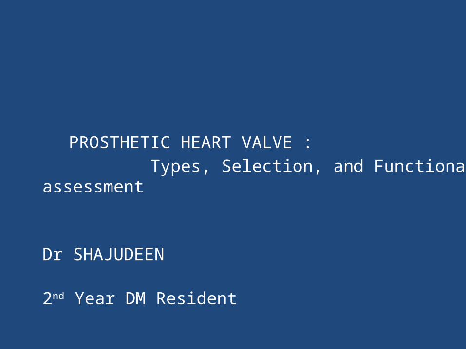
HISTORY OF PROSTHETIC HEART VALVE
First Mechanical valve was designed by Charles Hufnagel in
1954 ( Implanted in descending thoracic aorta for AR)
Plexiglass (Methyl Methacrylate)cage &Silicone-coated nylon poppet
• Dwight Harken perfomed the first aortic valve
replacement in 1960
• Nina Braunwald implanted the first Ball and
cage valve in Mitral position in 1960
• Homograft was first developed by Donald Ross
• Porcine valve was first implanted by Binet et al.
• Alian Carpenter discovered Gluteraldehyde Fixation
• The name “Bioprosthetic “valve was coined by Carpentier.
• David implanted the first stentless porcine bioprosthesis in 1988• Ross & Boyes performed the first successful allograft replacement from cadaver in 1962
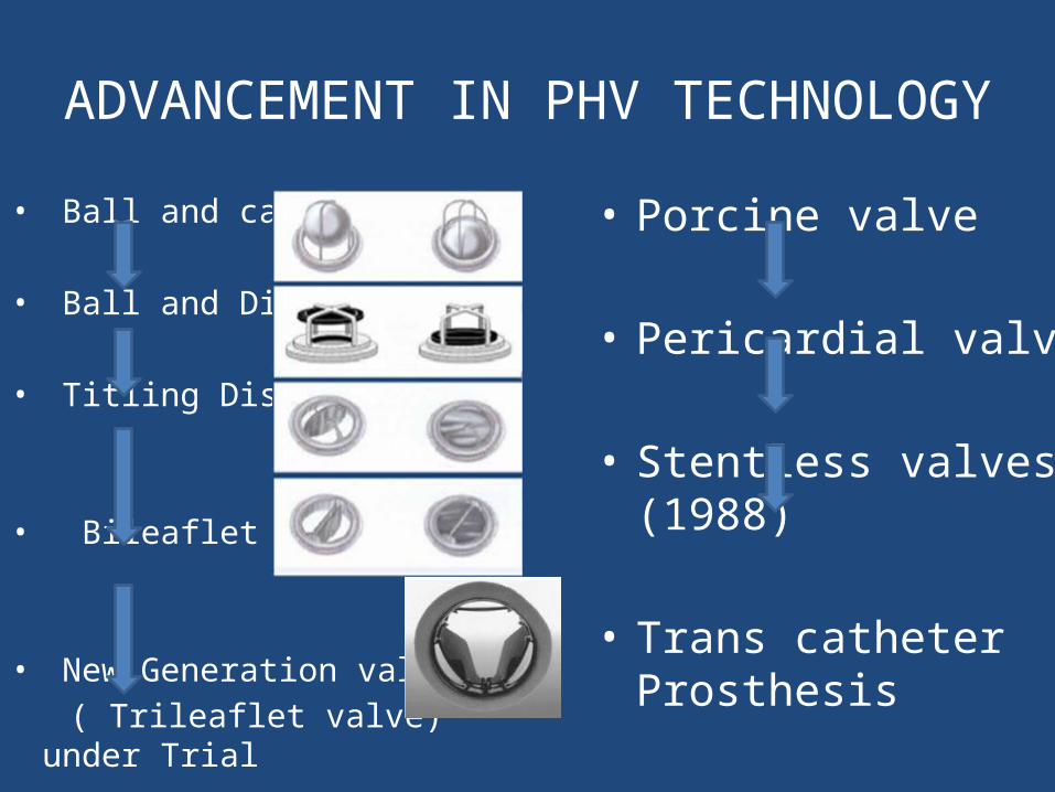
ADVANCEMENT IN PHV TECHNOLOGY
• Ball and cage
• Ball and Disc
• Titling Disc
• Bileaflet
• New Generation valve ( Trileaflet valve) under Trial
• Porcine valve
• Pericardial valve
• Stentless valves (1988)
• Trans catheter Prosthesis
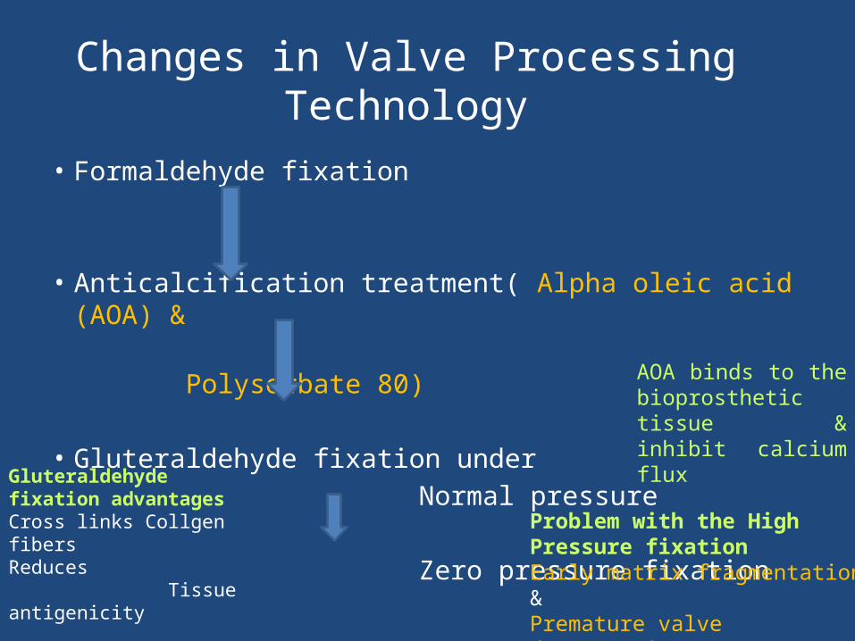
Changes in Valve Processing Technology
• Formaldehyde fixation
• Anticalcification treatment( Alpha oleic acid (AOA) & Polysorbate 80)
• Gluteraldehyde fixation under Normal pressure
Zero pressure fixation
AOA binds to the bioprosthetic tissue & inhibit calcium flux
Problem with the High Pressure fixationEarly matrix fragmentation & Premature valve degeneration
Gluteraldehyde fixation advantagesCross links Collgen fibersReduces Tissue antigenicity Enzymatic degradation Cell viability
Prosthetic Heart valve
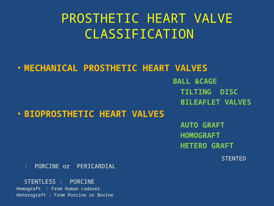
PROSTHETIC HEART VALVE CLASSIFICATION
• MECHANICAL PROSTHETIC HEART VALVES BALL &CAGE TILTING DISC BILEAFLET VALVES
• BIOPROSTHETIC HEART VALVES AUTO GRAFT HOMOGRAFT HETERO GRAFT
STENTED : PORCINE or PERICARDIAL STENTLESS : PORCINEHomograft : From Human cadaverHeterograft : From Porcine or Bovine
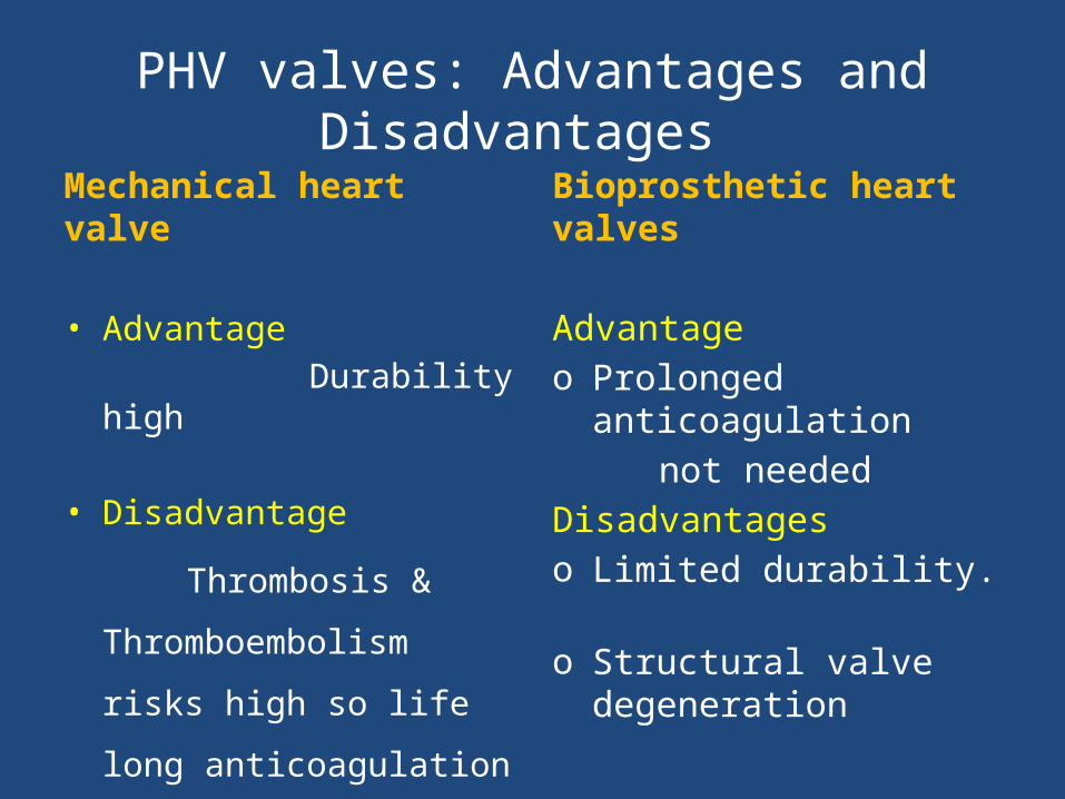
PHV valves: Advantages and Disadvantages
Mechanical heart valve
• Advantage Durability high
• Disadvantage
Thrombosis &
Thromboembolism risks
high so life long
anticoagulation
Bioprosthetic heart valves
Advantageo Prolonged anticoagulation not neededDisadvantageso Limited durability. o Structural valve degeneration
Mechanical PHV Basic structure
The ball is a silicone rubber
polymer, impregnated with
barium sulfate for radiopacity,
which oscillates in a cage of
cobalt-chromium alloy
TTK-CHITRA
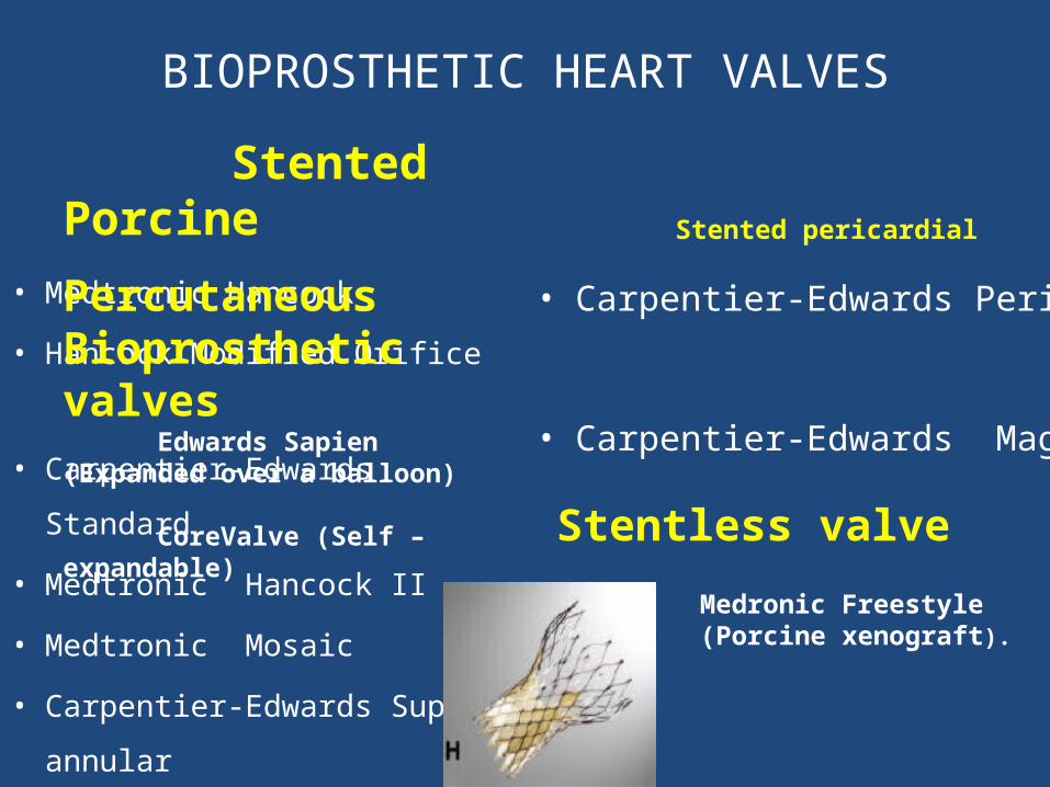
BIOPROSTHETIC HEART VALVES
Stented Porcine
• Medtronic Hancock
• Hancock Modified Orifice
• Carpentier-Edwards Standard
• Medtronic Hancock II
• Medtronic Mosaic
• Carpentier-Edwards Supra-
annular
Stented pericardial
• Carpentier-Edwards Perimount
• Carpentier-Edwards Magna
Stentless valve
Medronic Freestyle (Porcine xenograft).
Percutaneous Bioprosthetic valves Edwards Sapien (Expanded over a balloon) CoreValve (Self –expandable)
Mechanical valve types merits and demerits
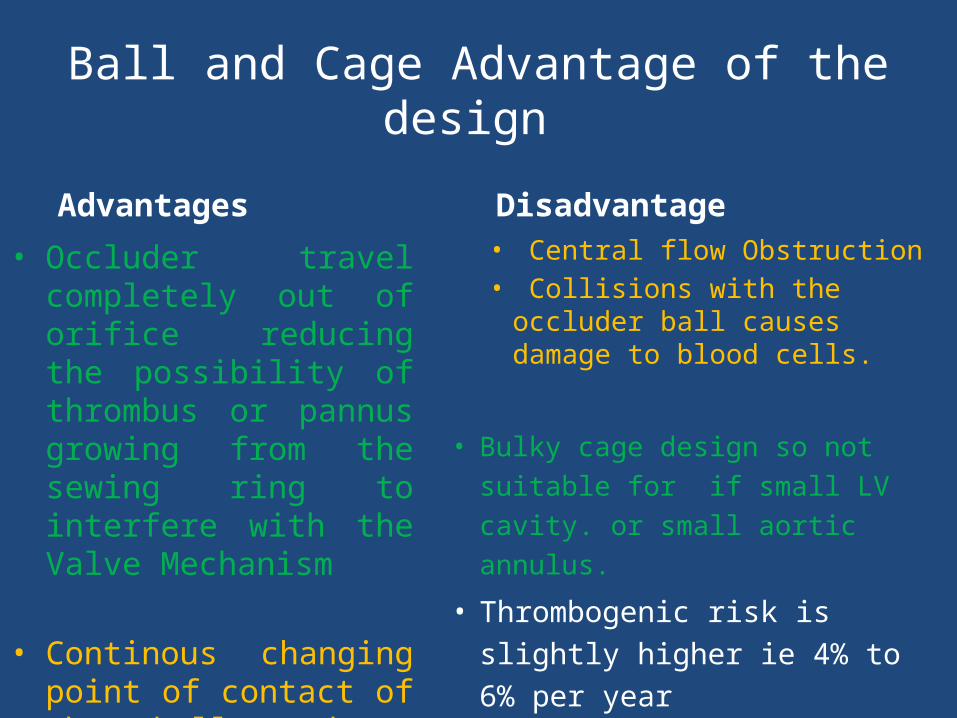
Ball and Cage Advantage of the design
Advantages
• Occluder travel completely out of orifice reducing the possibility of thrombus or pannus growing from the sewing ring to interfere with the Valve Mechanism
• Continous changing point of contact of the ball reduces the wear and tear in any one area
Disadvantage
• Central flow Obstruction• Collisions with the occluder
ball causes damage to blood cells.
• Bulky cage design so not suitable for if small LV cavity. or small aortic annulus.
• Thrombogenic risk is slightly higher ie 4% to 6% per year

TILTING DISC : KEY FEATURES
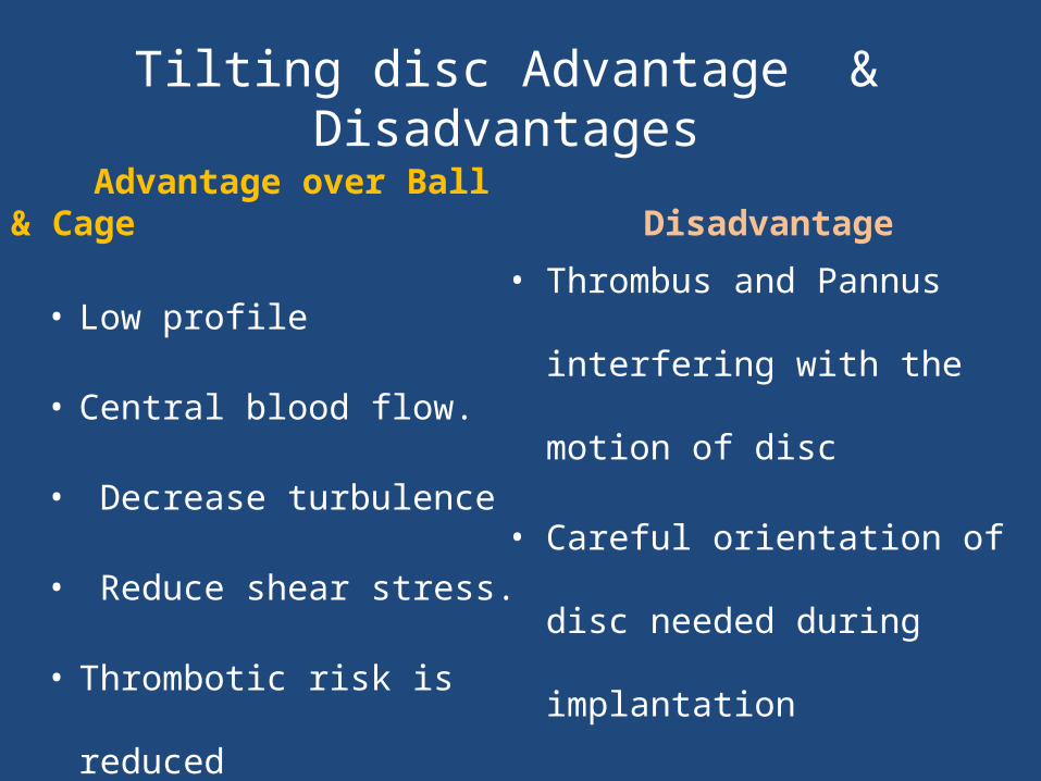
Tilting disc Advantage & Disadvantages
Advantage over Ball & Cage
• Low profile
• Central blood flow.
• Decrease turbulence
• Reduce shear stress.
• Thrombotic risk is reduced
Disadvantage
• Thrombus and Pannus interfering
with the motion of disc
• Careful orientation of disc needed
during implantation
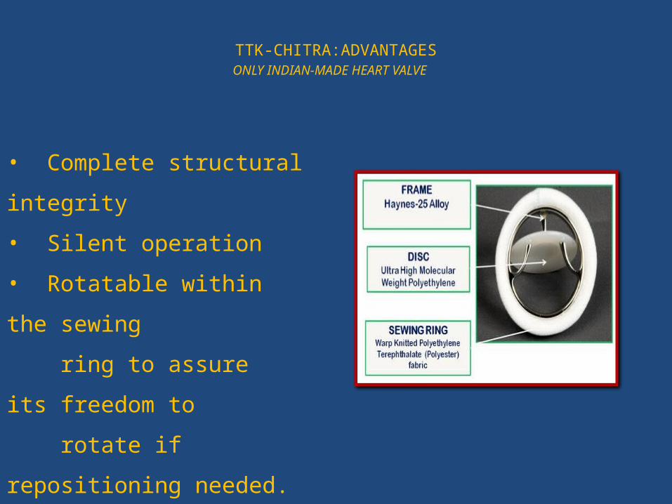
TTK-CHITRA:ADVANTAGESONLY INDIAN-MADE HEART VALVE
• Complete structural integrity
• Silent operation
• Rotatable within the sewing
ring to assure its freedom to
rotate if repositioning needed.
• Low profile, most price-friendly
• Low thromboembolism even if
poor anticoagulant compliance
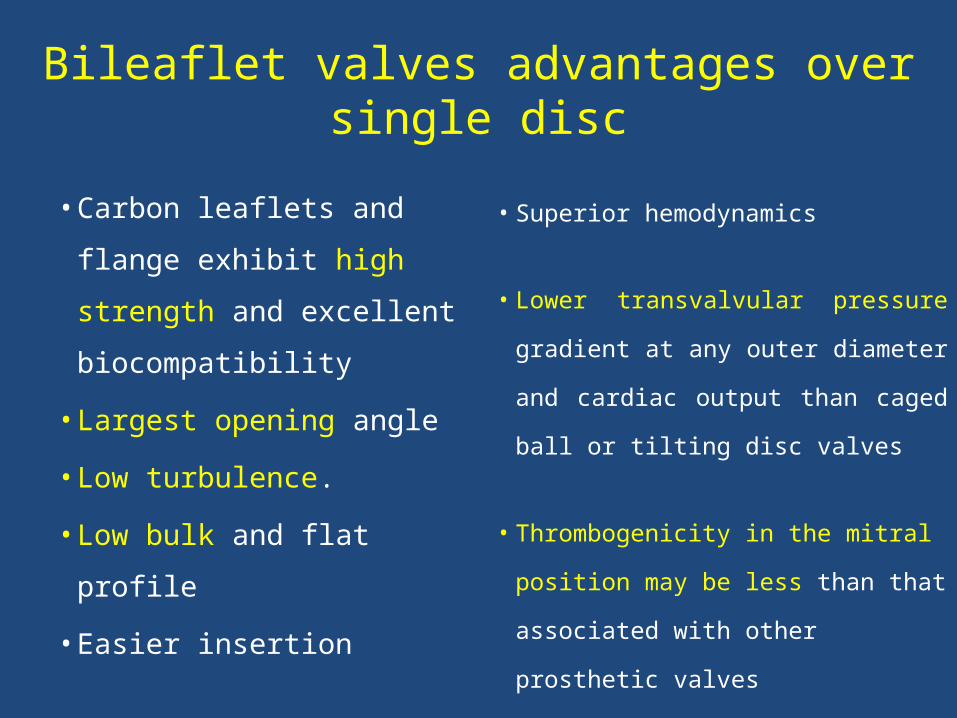
Bileaflet valves advantages over single disc
• Carbon leaflets and
flange exhibit high
strength and excellent
biocompatibility
• Largest opening angle
• Low turbulence.
• Low bulk and flat profile
• Easier insertion
• Superior hemodynamics
• Lower transvalvular pressure gradient
at any outer diameter and cardiac
output than caged ball or tilting disc
valves
• Thrombogenicity in the mitral position
may be less than that associated with
other prosthetic valves
Bioprosthetic valves : Key features

Pericardial valve:
• Made from Bovine pericardium mainly but from Porcine
or Equine also.
• Pericardial valve are invariably stented
• Increased durability due to increased amount of collagen
• More symmetrical function of leaflet so better
hemodynamics
Homograft (Allograft) Aortic Valves
• Harvested from cadavers usually within 24 hours of donor death
• Insertion: usually in the Aortic position without a prosthetic
stent and implanted in the subcoronary position with valve
alone or the valve and a portion of aorta are implanted as a
root replacement, with reimplantation of the coronary arteries
into the graft

Pulmonary Autografts
• Ross procedure : Patient's own pulmonary valve and adjacent main
pulmonary artery are removed and used to replace the diseased
aortic valve with reimplantation of the coronary arteries in to the
graft.
• The autograft is non thrombogenic and no anticoagulation is
needed
• Risk of Endocarditis is very low.
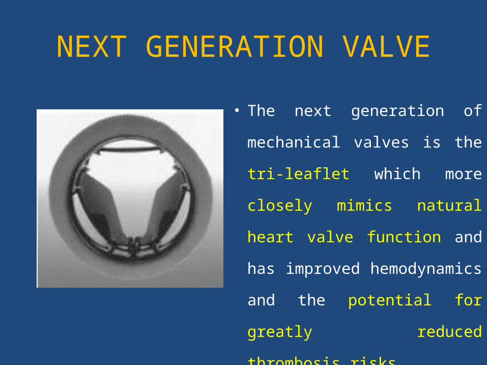
NEXT GENERATION VALVE
• The next generation of
mechanical valves is the tri-
leaflet which more closely
mimics natural heart valve
function and has improved
hemodynamics and the
potential for greatly reduced
thrombosis risks.
SELECTION OF PROSTHETIC HEART VALVE
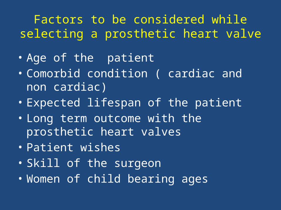
Factors to be considered while selecting a prosthetic heart valve
• Age of the patient• Comorbid condition ( cardiac and non cardiac)• Expected lifespan of the patient• Long term outcome with the prosthetic heart
valves• Patient wishes• Skill of the surgeon• Women of child bearing ages
• For valve replacement for IE: Homograft
preffered
• For Narrow aortic root if root
enlargement/replacement not possible choice
is Bileaflet valves.
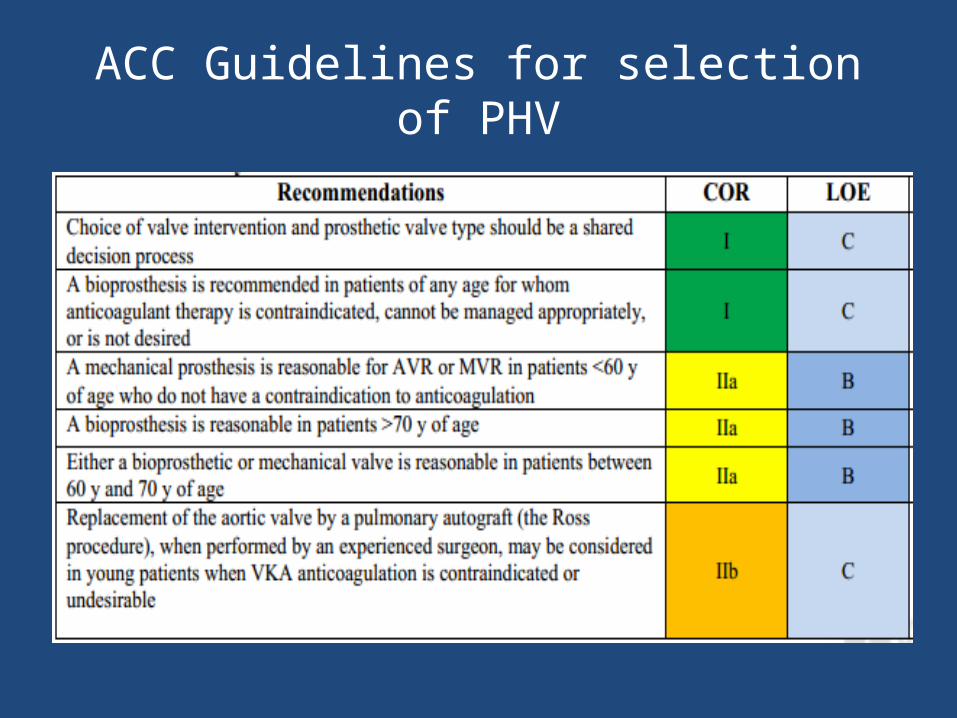
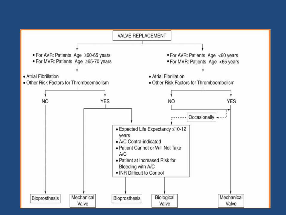
ACC Guidelines for selection of PHV
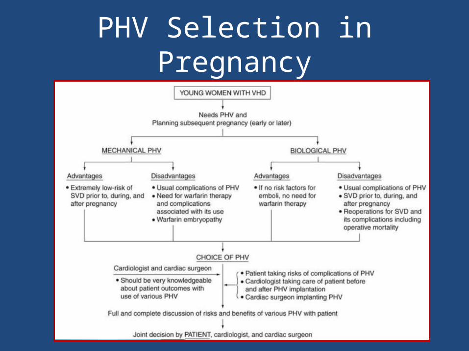
PHV Selection in Pregnancy
Evaluation of prosthetic valves
• CLINICAL INFORMATION &CLINICAL EXAMINATION• IMAGING OF THE VALVES
CXR2D echocardiographyTEE 3D echo CineFluoroCTCardiac catheterisation
Evaluation of prosthetic valves
CLINICAL INFORMATION
• Clinical data : Reason for the study & the patient’s
symptoms• Type & size of replaced valve.• Date of surgery.• Patient’s height, weight, and BSA should be recorded
to assess whether prosthesis-patient mismatch (PPM) is present
• BP & HR– HR particularly important in mitral and tricuspid
evaluations because the mean gradient is dependent on the diastolic filling period
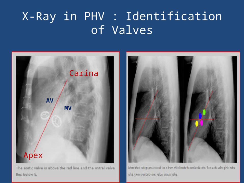
X-Ray in PHV : Identification of Valves
Carina
Apex
AVMV
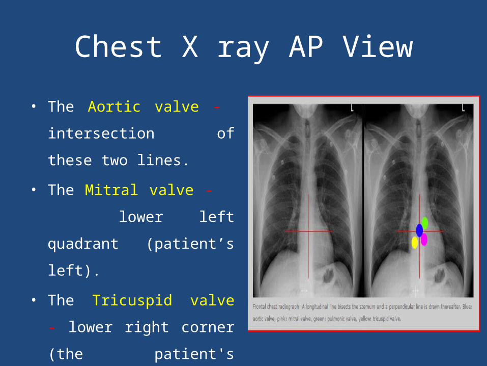
Chest X ray AP View
• The Aortic valve - intersection
of these two lines.
• The Mitral valve - lower left
quadrant (patient’s left).
• The Tricuspid valve - lower
right corner (the patient's
right)
• The Pulmonic valve- upper left
corner (the patient's left).
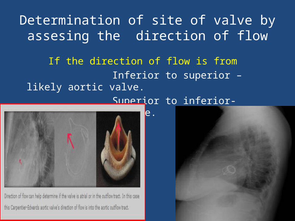
Determination of site of valve by assesing the direction of flow
If the direction of flow is from Inferior to superior – likely aortic valve. Superior to inferior- likely a mitral valve.
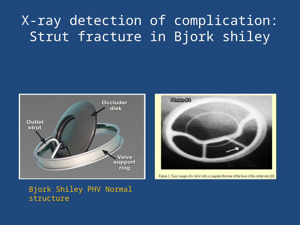
X-ray detection of complication:Strut fracture in Bjork shiley
Bjork Shiley PHV Normal structure
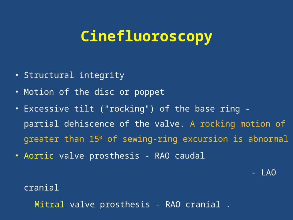
Cinefluoroscopy
• Structural integrity
• Motion of the disc or poppet
• Excessive tilt ("rocking") of the base ring - partial dehiscence of the
valve. A rocking motion of greater than 150 of sewing-ring
excursion is abnormal
• Aortic valve prosthesis - RAO caudal
- LAO cranial
Mitral valve prosthesis - RAO cranial .
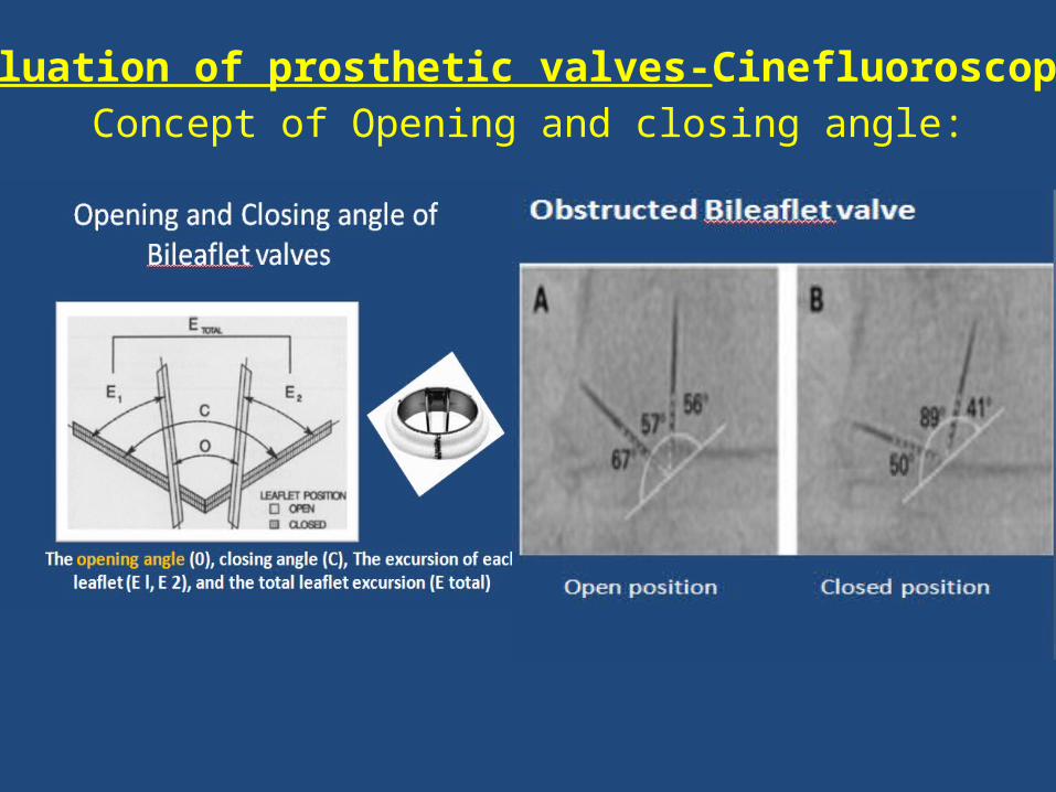
Evaluation of prosthetic valves-Cinefluoroscopy
Concept of Opening and closing angle:
Opening angle
Medronic hall 750
St Jude Medical standard &, Reagent,On X
850
CarboMedics standard 780
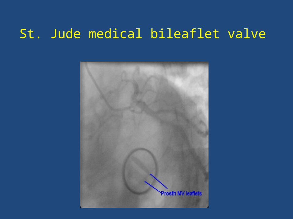
St. Jude medical bileaflet valve
ECHO ASSESSMENT OF PROSTHETIC HEART VALVES
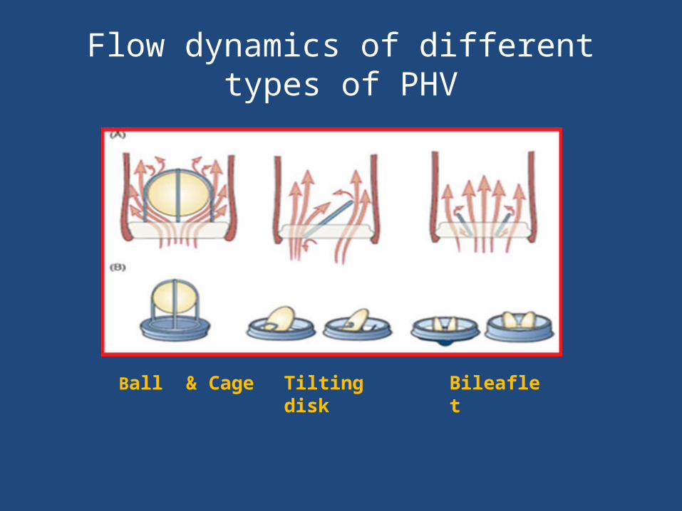
Flow dynamics of different types of PHV
Ball & Cage Tilting disk Bileaflet
Echo in PHV Evaluation : General consideration
• Compared with a native valve the prosthetic valves
are inherently stenotic.
• The type and size of prosthesis determines what is
considered normal function for that valve. So
gradients, EOA, and degree of physiologic
regurgitation will vary based on valve type,
manufacturer, and valve size.
Echo in PHV Evaluation : General consideration
• Always use multiple views during echo
evaluation of PHV.
• For Stented valves-ultrasound beam aligned
parallel to flow to avoid the shadowing effects
of the stents and sewing ring
Echo in PHV Evaluation : General consideration
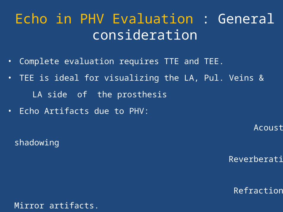
• Complete evaluation requires TTE and TEE.
• TEE is ideal for visualizing the LA, Pul. Veins &
LA side of the prosthesis
• Echo Artifacts due to PHV:
Acoustic shadowing
Reverberation
Refraction & Mirror artifacts.
Doppler assessment:General consideration
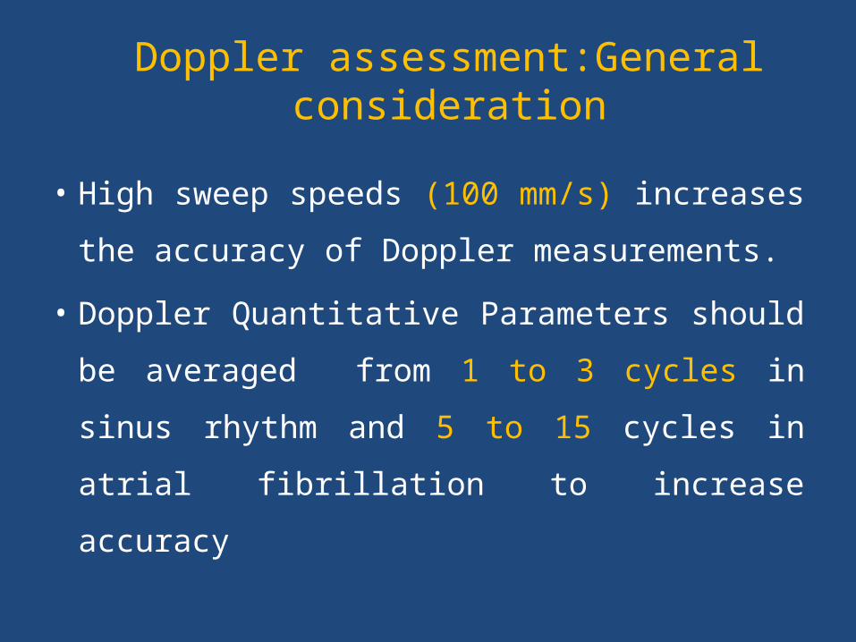
• High sweep speeds (100 mm/s) increases the
accuracy of Doppler measurements.
• Doppler Quantitative Parameters should be
averaged from 1 to 3 cycles in sinus rhythm
and 5 to 15 cycles in atrial fibrillation to
increase accuracy
TEE Views
• The most useful views include the mid
esophageal 4-chamber, 2-chamber, & 3-
chamber views.
• Transgastric views may be useful when
assessing LV size and function or papillary
muscle and chordal anatomy.
ECHO FEATURES OF BILEAFLET VALVE
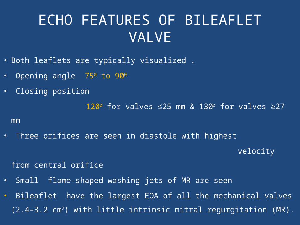
• Both leaflets are typically visualized .
• Opening angle 750 to 900
• Closing position
1200 for valves ≤25 mm & 1300 for valves ≥27 mm
• Three orifices are seen in diastole with highest
velocity from central orifice
• Small flame-shaped washing jets of MR are seen
• Bileaflet have the largest EOA of all the mechanical valves (2.4–3.2
cm2) with little intrinsic mitral regurgitation (MR).
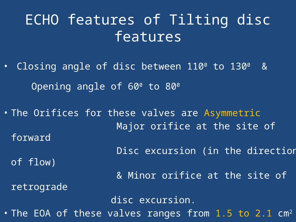
ECHO features of Tilting disc features
• Closing angle of disc between 1100 to 1300 &
Opening angle of 600 to 800
• The Orifices for these valves are Asymmetric Major orifice at the site of forward Disc excursion (in the direction of flow) & Minor orifice at the site of retrograde disc excursion.• The EOA of these valves ranges from 1.5 to 2.1 cm2
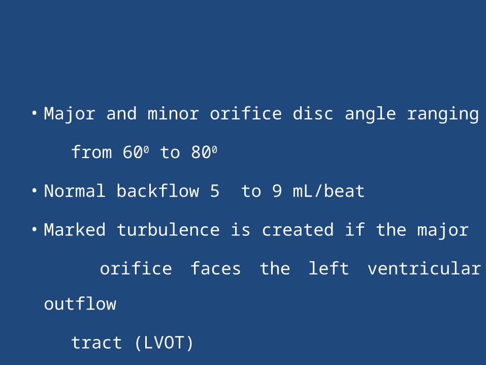
• Major and minor orifice disc angle ranging
from 600 to 800
• Normal backflow 5 to 9 mL/beat
• Marked turbulence is created if the major
orifice faces the left ventricular outflow
tract (LVOT)
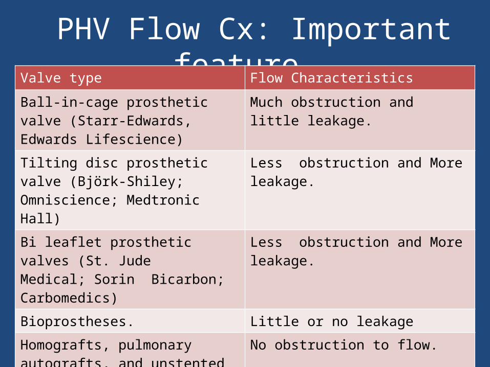
PHV Flow Cx: Important feature.Valve type Flow Characteristics
Ball-in-cage prosthetic valve (Starr-Edwards, Edwards Lifescience)
Much obstruction and little leakage.
Tilting disc prosthetic valve (Björk-Shiley; Omniscience; Medtronic Hall)
Less obstruction and More leakage.
Bi leaflet prosthetic valves (St. JudeMedical; Sorin Bicarbon; Carbomedics)
Less obstruction and More leakage.
Bioprostheses. Little or no leakage
Homografts, pulmonary autografts, and unstented bioprosthetic valves (Medtronic Freestyle,Toronto, Ontario, Canada)
No obstruction to flow.
Stented bioprostheses (leaflets suspended within a frame)
Obstructive to flow.
2D ECHO assessment of PHV
TIMING OF ECHO CARDIOGRAPHIC FOLLOW-UP
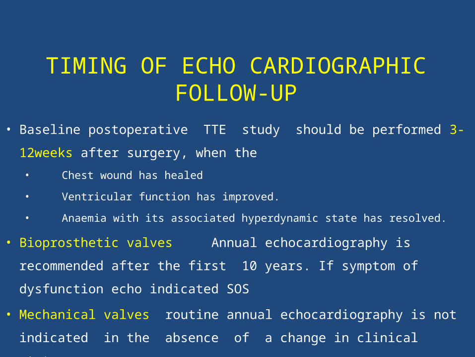
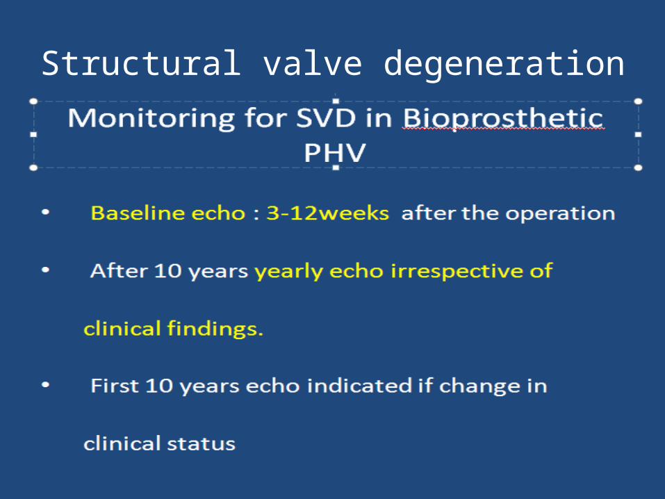
• Baseline postoperative TTE study should be performed 3-12weeks
after surgery, when the
• Chest wound has healed
• Ventricular function has improved.
• Anaemia with its associated hyperdynamic state has resolved.
• Bioprosthetic valves Annual echocardiography is recommended
after the first 10 years. If symptom of dysfunction echo indicated SOS
• Mechanical valves routine annual echocardiography is not indicated
in the absence of a change in clinical status.
2D ECHO ASSESMENT
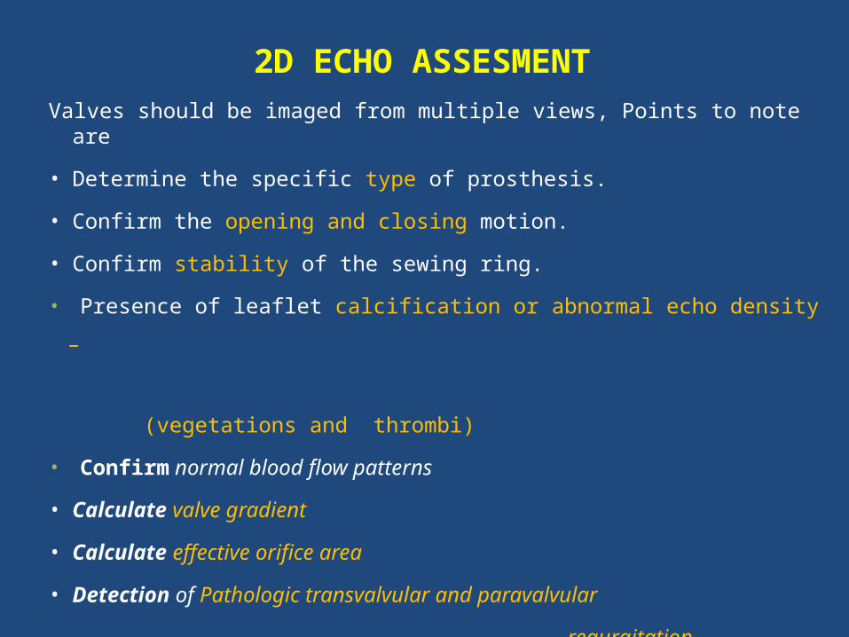
Valves should be imaged from multiple views, Points to note are
• Determine the specific type of prosthesis.
• Confirm the opening and closing motion.
• Confirm stability of the sewing ring.
• Presence of leaflet calcification or abnormal echo density –
(vegetations and thrombi)
• Confirm normal blood flow patterns
• Calculate valve gradient
• Calculate effective orifice area
• Detection of Pathologic transvalvular and paravalvular
regurgitation.
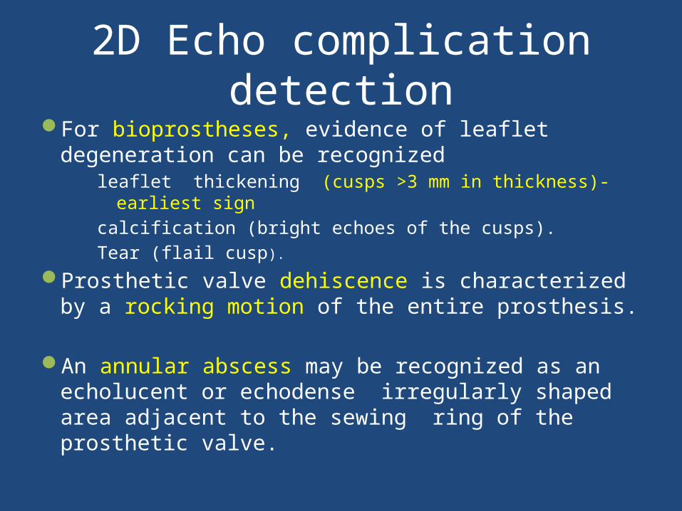
2D Echo complication detectionFor bioprostheses, evidence of leaflet degeneration can be
recognized leaflet thickening (cusps >3 mm in thickness)-earliest signcalcification (bright echoes of the cusps). Tear (flail cusp).
Prosthetic valve dehiscence is characterized by a rocking motion of the entire prosthesis.
An annular abscess may be recognized as an echolucent or echodense irregularly shaped area adjacent to the sewing ring of the prosthetic valve.
• Doppler Echocardiography in PHV evaluation
PRIMARY GOALS OF DOPPLER INTERROGATION
• Assesment of prosthetic valve obstruction
• Detection and quantification of prosthetic valve
regurgitation
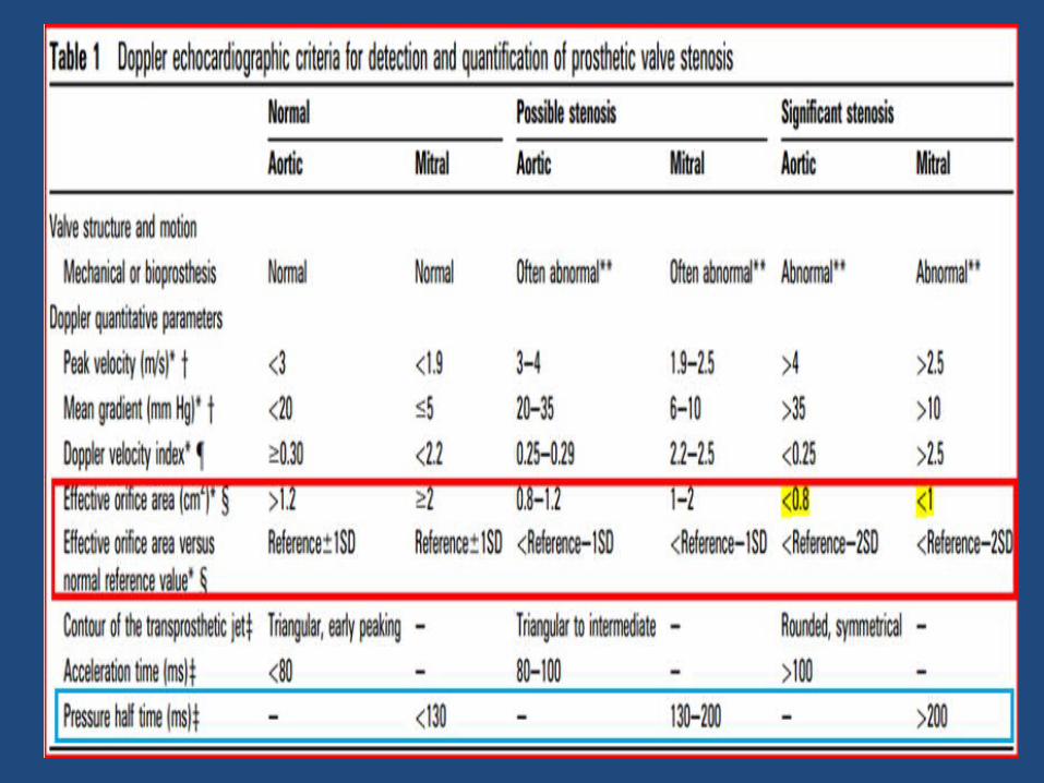
Doppler Assessment of Obstruction of Prosthetic Valves Stenosis
• Quantitative parameters of Prosthetic valve Stenosis
Trans prosthetic flow velocity & Pressure gradients.
Valve EOA.
Doppler velocity index(DVI). Contour of trans prosthetic jet and acceleration time (AT)¥
¥ For Prosthetic Aortic valve Stenosis
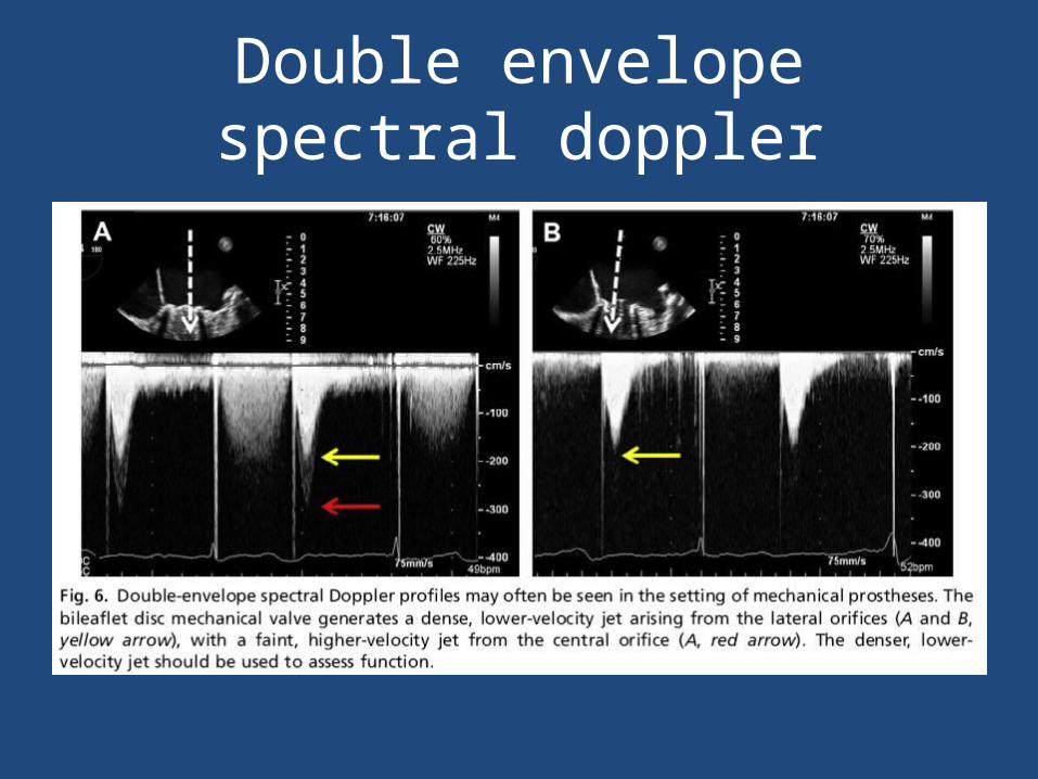
Double envelope spectral doppler
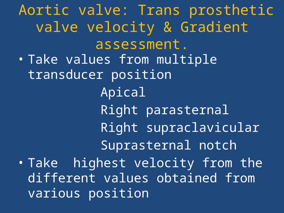
Aortic valve: Trans prosthetic valve velocity & Gradient assessment.
• Take values from multiple transducer position Apical Right parasternal Right supraclavicular Suprasternal notch• Take highest velocity from the different values
obtained from various position
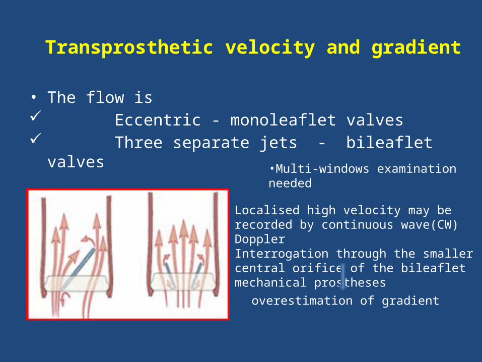
Transprosthetic velocity and gradient
• The flow is Eccentric - monoleaflet valves Three separate jets - bileaflet valves
•Multi-windows examination needed
Localised high velocity may be recorded by continuous wave(CW) DopplerInterrogation through the smaller central orifice of the bileaflet mechanical prostheses
overestimation of gradient
Prosthetic Heart valve Gradient calculation.
Equation • Δ P = 4V2
or• If LVOT velocity more than 1.5 cm2
Δ P =4 (VPRAV2 - VLVOT
2)
Limitation of doppler transvalvular Gradient
measurement is that it is FLOW DEPENDENT
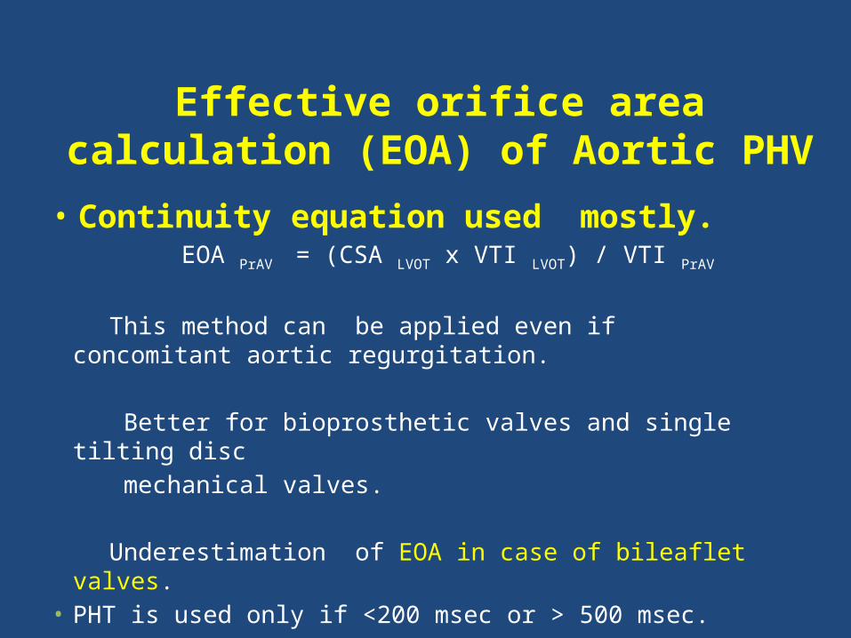
Effective orifice area calculation (EOA) of Aortic PHV
• Continuity equation used mostly. EOA PrAV = (CSA LVOT x VTI LVOT) / VTI PrAV
This method can be applied even if concomitant aortic
regurgitation.
Better for bioprosthetic valves and single tilting disc mechanical valves.
Underestimation of EOA in case of bileaflet valves.• PHT is used only if <200 msec or > 500 msec.
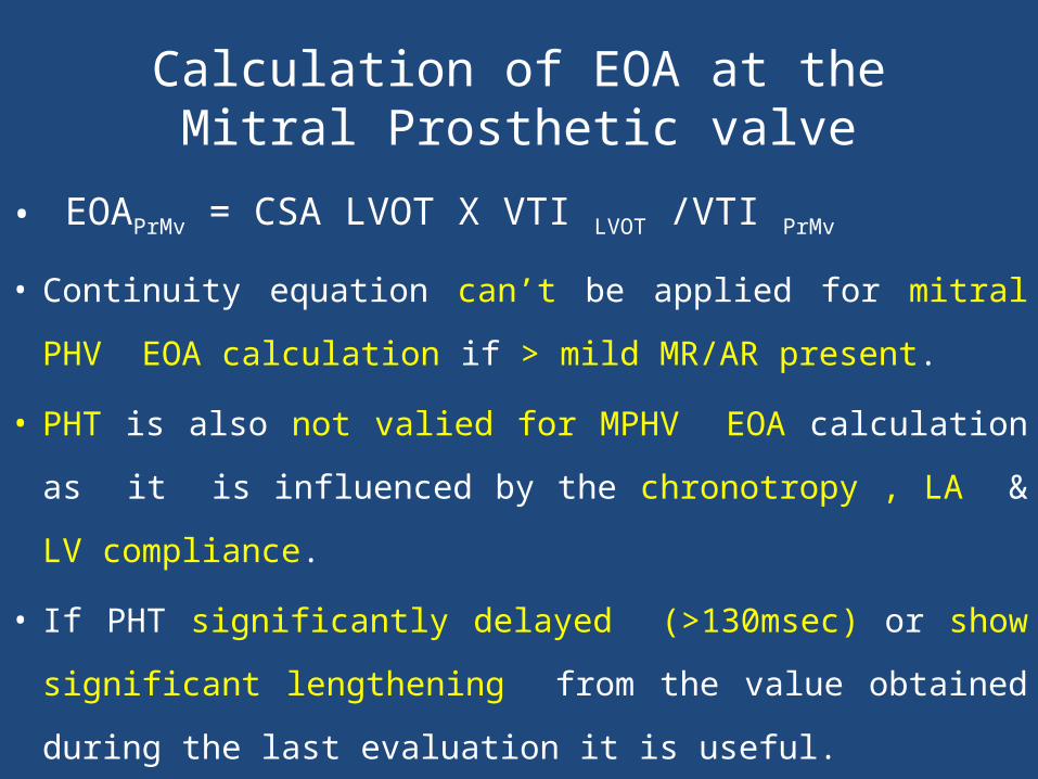
Calculation of EOA at the Mitral Prosthetic valve
• EOAPrMv = CSA LVOT X VTI LVOT /VTI PrMv
• Continuity equation can’t be applied for mitral PHV EOA
calculation if > mild MR/AR present.
• PHT is also not valied for MPHV EOA calculation as it is
influenced by the chronotropy , LA & LV compliance.
• If PHT significantly delayed (>130msec) or show significant
lengthening from the value obtained during the last
evaluation it is useful.
How to take measurement for continuity equation
• VTI LVOT by PW doppler at LVOT at the same location
at which LVOT diameter taken ie 0.5 -1 cm distal to
the LVOT.
• VTI PRAV : CW at the Aortic prosthesis
• CSA LVOT: PLAX zoomed view ie 0.5 -1 cm distal to
the LVOT.
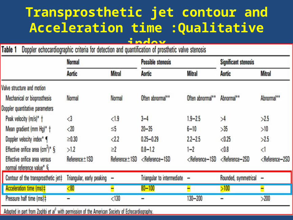
Transprosthetic jet contour and Acceleration time :Qualitative index
• Normal Contour: Triangular & short AT
• PHVObstruction: Rounded contour with peaking at mid ejection time & prolonged AT(>100msec)
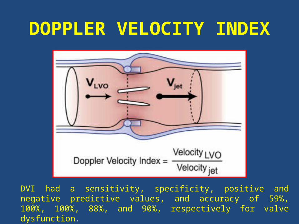
DOPPLER VELOCITY INDEX
DVI had a sensitivity, specificity, positive and negative predictive values, and accuracy of 59%, 100%, 100%, 88%, and 90%, respectively for valve dysfunction.
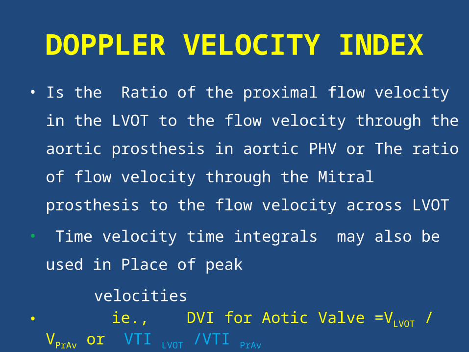
DOPPLER VELOCITY INDEX
• Is the Ratio of the proximal flow velocity in the LVOT to the flow
velocity through the aortic prosthesis in aortic PHV or The ratio of
flow velocity through the Mitral prosthesis to the flow velocity
across LVOT
• Time velocity time integrals may also be used in Place of peak
velocities • ie., DVI for Aotic Valve =VLVOT / VPrAv or VTI LVOT /VTI PrAv
• DVI for Mitral Valve = VPr Mv /V LVOT or VTI PrMv/ VTI PrAV
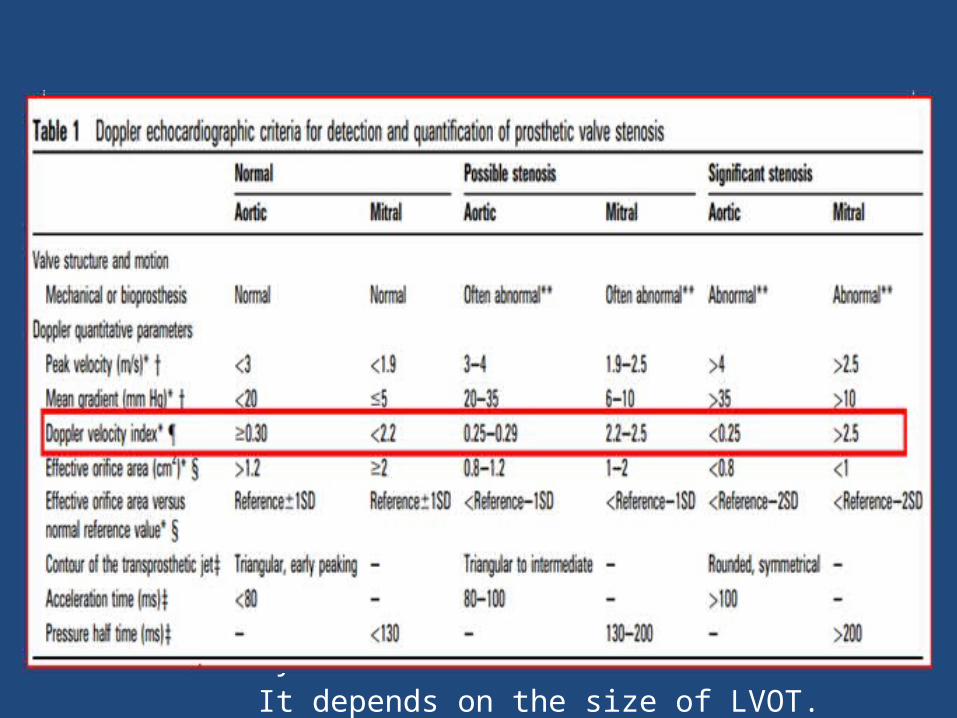
• DVI can be helpful to screen for valve stenosis, particularly when
the
– Crosssectional area of the LVOT cannot be obtained
• DVI is always less than one, because velocity will always
accelerate through the prosthesis.
• DVI is not affected by high flow conditionsDisadvantage Does not distinguish obstruction due to PPM or intrinsic dysfunction It depends on the size of LVOT.
PROSTHETIC TRICUSPID & PULMONARY VALVE STENOSIS
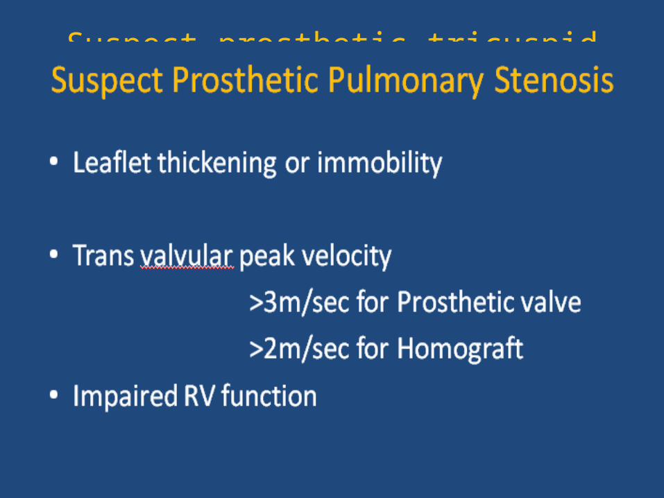
Suspect prosthetic tricuspid stenosis if
• Prosthetic valve leaflet morphology and moblity
abnormal
• Peak velocity >1.7 m/sec
• Mean Gradient ≥ 6mm of Hg
• PHT at least 230msec
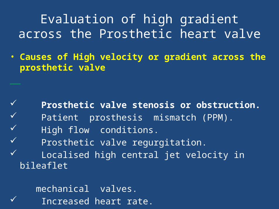
Evaluation of high gradient across the Prosthetic heart valve
• Causes of High velocity or gradient across the prosthetic valve Prosthetic valve stenosis or obstruction. Patient prosthesis mismatch (PPM). High flow conditions. Prosthetic valve regurgitation. Localised high central jet velocity in bileaflet mechanical valves. Increased heart rate.
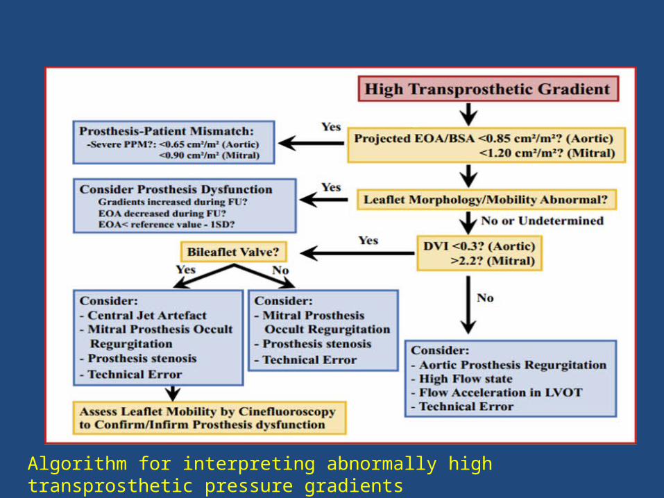
Algorithm for interpreting abnormally high transprosthetic pressure gradients
DETECTION AND
QUANTIFICATION OFPROSTHETIC VALVE REGURGITATION
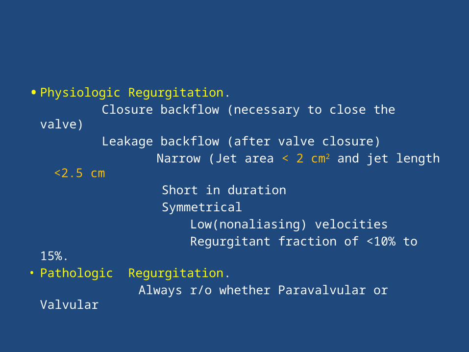
• Physiologic Regurgitation. Closure backflow (necessary to close the valve) Leakage backflow (after valve closure)
Narrow (Jet area < 2 cm2 and jet length <2.5 cm Short in duration Symmetrical
Low(nonaliasing) velocities Regurgitant fraction of <10% to 15%.• Pathologic Regurgitation. Always r/o whether Paravalvular or Valvular
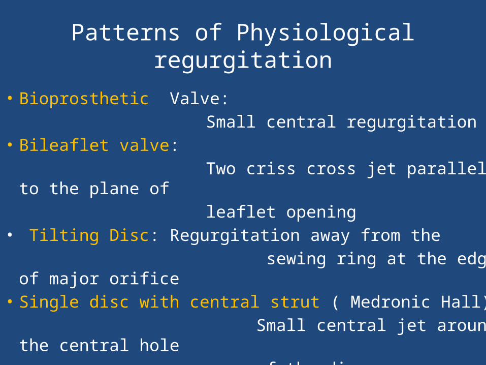
Patterns of Physiological regurgitation
• Bioprosthetic Valve: Small central regurgitation• Bileaflet valve: Two criss cross jet parallel to the plane of leaflet opening• Tilting Disc: Regurgitation away from the sewing ring at the edge of major orifice• Single disc with central strut ( Medronic Hall) Small central jet around the central hole of the disc
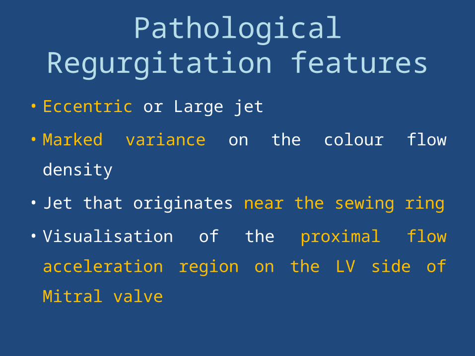
Pathological Regurgitation features
• Eccentric or Large jet
• Marked variance on the colour flow density
• Jet that originates near the sewing ring
• Visualisation of the proximal flow acceleration
region on the LV side of Mitral valve
Prosthetic Mitral regurgitation Echo Evaluation
Prosthetic Mitral regurgitation Echo Evaluation
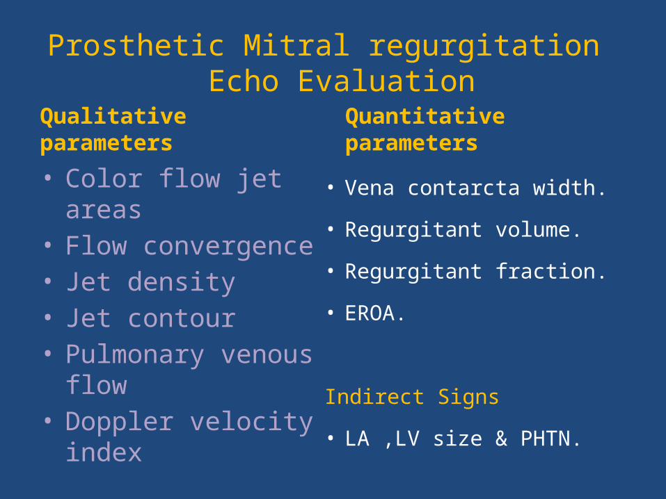
Qualitative parameters
• Color flow jet areas• Flow convergence• Jet density • Jet contour• Pulmonary venous flow• Doppler velocity index
Quantitative parameters
• Vena contarcta width.
• Regurgitant volume.
• Regurgitant fraction.
• EROA.
Indirect Signs
• LA ,LV size & PHTN.
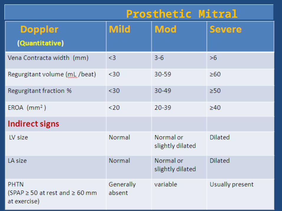
Prosthetic Mitral regurgitation severityValve structure and motion
Mild Mod Severe
Mechnaical or bioprosthetic
Usually normal Usually abnormal Usually abnormal
Doppler parameters ( Qualitative or Semiquantitative )
Color flow jet areas •Small central jet <4 cm2 or <20% of LA area
•Large central jet >8cm2 or >40% of LA area or variable size wall impinging jet swirling in LA
Flow convergence None Intermediate Large
Jet density(CW) Incomplete or faint Dense Dense
Jet contour(CW) Parabolic Usually parabolic Early Peaking triangular
Pulmonary venous flow(PW)
Systolic dominance Systolic blunting Systolic flow reversal
Doppler velocity index(PW)
<2.2 2.2-2.5 >2.5

Aortic regurgitation
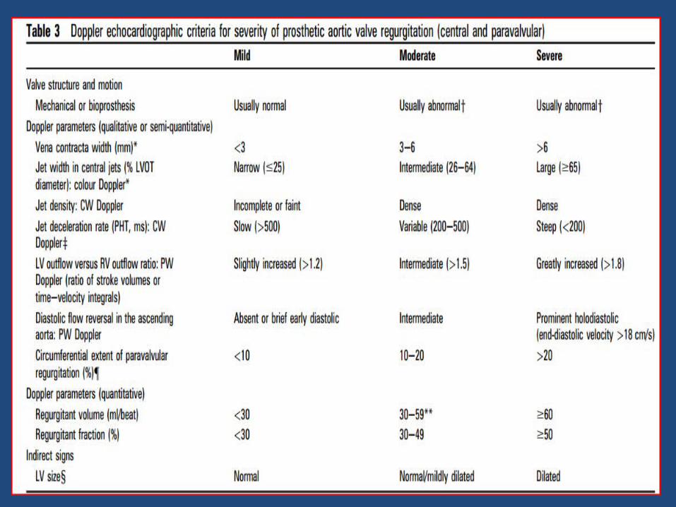
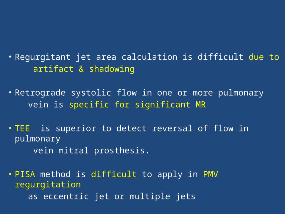
• Regurgitant jet area calculation is difficult due to artifact & shadowing
• Retrograde systolic flow in one or more pulmonary vein is specific for significant MR
• TEE is superior to detect reversal of flow in pulmonary vein mitral prosthesis.
• PISA method is difficult to apply in PMV regurgitation as eccentric jet or multiple jets
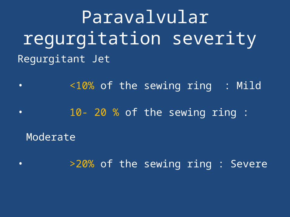
Paravalvular regurgitation severity
Regurgitant Jet
• <10% of the sewing ring : Mild
• 10- 20 % of the sewing ring : Moderate
• >20% of the sewing ring : Severe

Limitation of Echo in PHV assessment
• IN QUANTIFICATION OF PHV REGURGITATION
Para valvular jet, Eccentric jet & Multijet difficult to quantify• EVALUATION OF PROSTHETIC LEAFLET MOBILTY
TTE and TEE has limited sensitivity for the detection of abnormalities of leaflet morphology and mobility• EVALUATION OF PVE
TTE and TEE are less sensitive in detection of PVE
COMPLICATION OF PROSTHETIC HEART VALVES
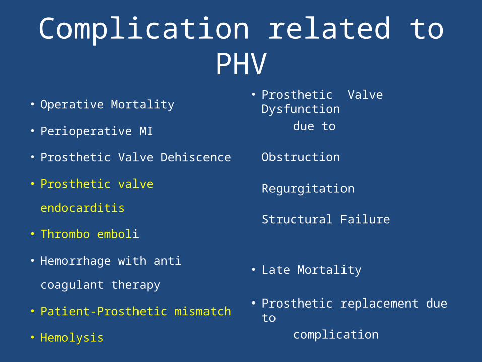
Complication related to PHV
• Operative Mortality
• Perioperative MI
• Prosthetic Valve Dehiscence
• Prosthetic valve endocarditis
• Thrombo emboli
• Hemorrhage with anti coagulant
therapy
• Patient-Prosthetic mismatch
• Hemolysis
• Prosthetic Valve Dysfunction due to Obstruction Regurgitation Structural Failure
• Late Mortality
• Prosthetic replacement due to
complication
Patient prosthesis mismatch
Patient Prosthesis Mismatch
• Valve prosthesis–patient mismatch (VP–PM)
described in 1978 by Dr. Rahimtoola.
• PPM occurs when EOA of a normally functioning
prosthetic valve is too small in relation to the body
size resulting in abnormal gradient across the valve.
• Indexed EOA (EOA/BSA) is the parameter widely
used to identify and predict PPM
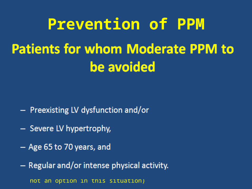
Prevention of PPM
• Avoided by systematically
– Calculating the projected indexed EOA of the prosthesis
– Model with better hemodynamic performance eg Stentless valve
– Aortic root enlargement to accommodate a larger size of the same
prosthesis model.
– Supra annular placement: Prevents PPM IN 98% of AVR
(The prevention of PPM in the mitral position difficult than in the
aortic position because valve annulus enlargement or stentless
valve implantation is not an option in this situation)
Prosthetic valve thrombus and Pannus
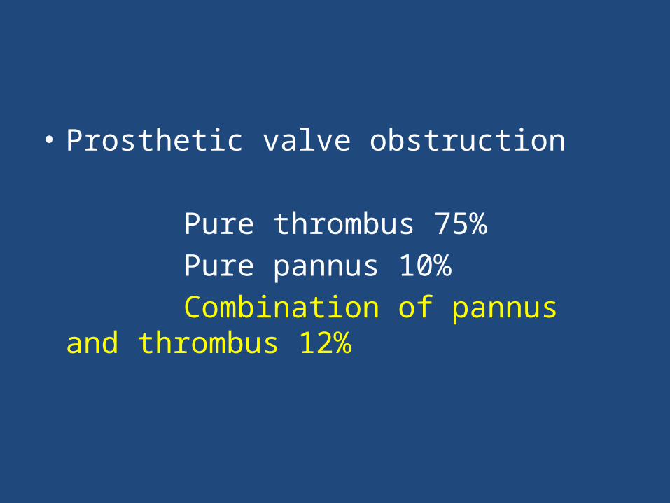
• Prosthetic valve obstruction
Pure thrombus 75% Pure pannus 10% Combination of pannus and thrombus 12%
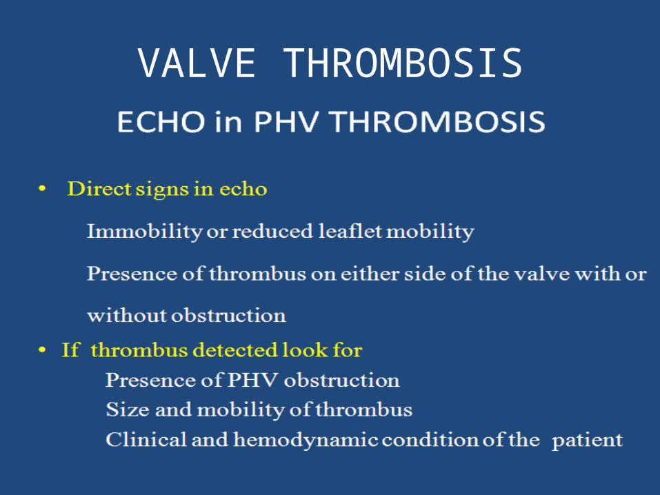
VALVE THROMBOSIS
• Definition
Any thrombus in the absence of infection attached
to or near the operated valve that occclude the
path of blood flow or impede the operation of the
valves
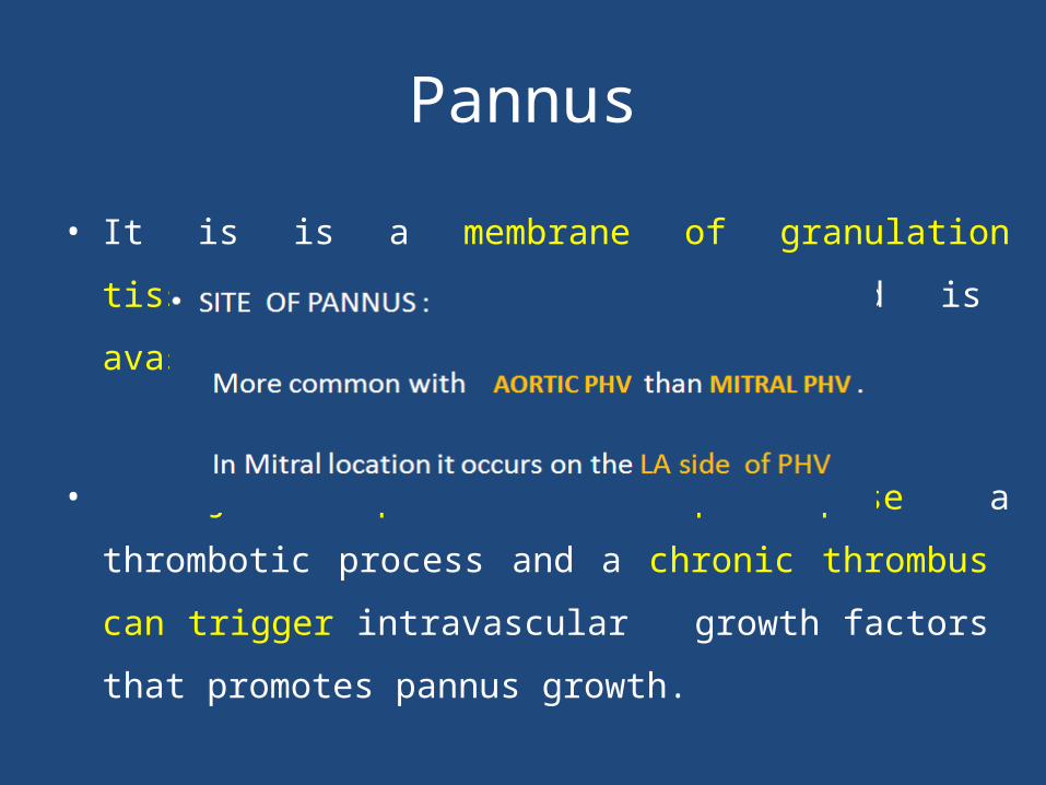
Pannus
• It is is a membrane of granulation tissue as an response
to healing and is avascular in nature
• Injured pannus can predispose a thrombotic process and a
chronic thrombus can trigger intravascular growth factors that
promotes pannus growth.
• This is more common with tilting disc on the side of minor
orifice.
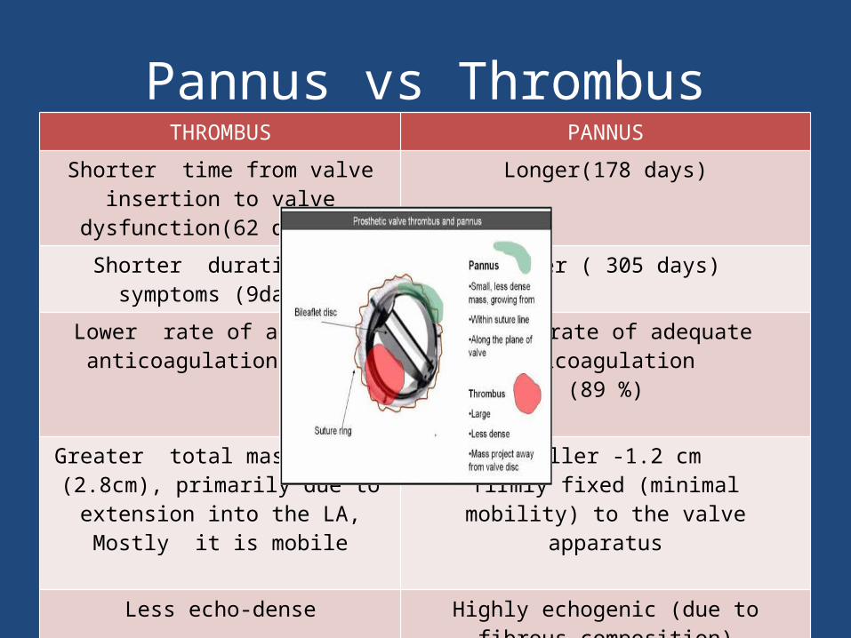
Pannus vs ThrombusTHROMBUS PANNUS
Shorter time from valve insertion to valve dysfunction(62 days )
Longer(178 days)
Shorter duration of symptoms (9days) Longer ( 305 days)
Lower rate of adequate anticoagulation (21%)
Higher rate of adequate anticoagulation (89 %)
Greater total mass length (2.8cm), primarily due to extension into the LA,
Mostly it is mobile
Smaller -1.2 cmfirmly fixed (minimal mobility) to the valve
apparatus
Less echo-dense Highly echogenic (due to fibrous composition)
Associated with spontaneous contrast,Common in mitral and tricuspid
position
Common in aortic position Para valve jet suggests pannus
Structural valve degeneration
Structural valve degeneration
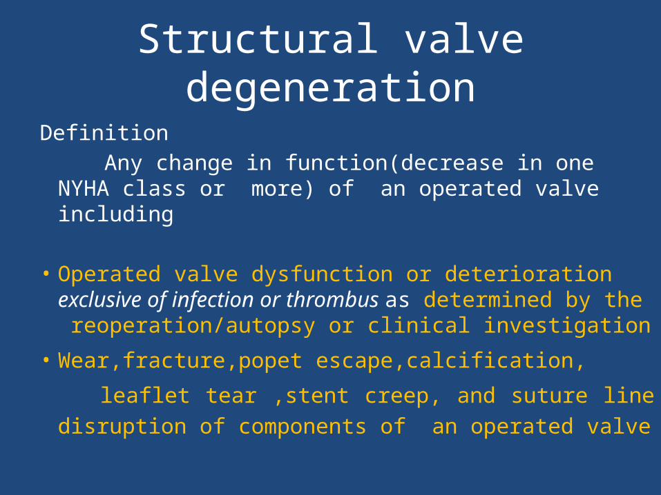
Definition Any change in function(decrease in one NYHA class
or more) of an operated valve including
• Operated valve dysfunction or deterioration exclusive of infection or thrombus as determined by the reoperation/autopsy or clinical investigation
• Wear,fracture,popet escape,calcification,
leaflet tear ,stent creep, and suture line disruption of components of an operated valve
Structural valve degeneration
• SVD is the most common cause of Bio PHV failure
• Freedom from structural valve degeneration
Stented porcine valves : 30- 60% at 15
years Pericardial valves : 86% at 12 years
• Mortality for reoperation for SVD is 2-
3times than first operation.
Types of degeneration
• CALCIFIC DEGERATION• NON CALCIFIC DEGERATION ( 30 %)
Sequele of degeneration
PHV Stenosis
PHV Regurgitation
or Both
Mechanical PHV Structural failure
• Strut fracture
• Leaflet escape
• Occluder dysfunction due to lipid adsoption
Prosthetic valve infective endocarditis
• Early endocarditis < 60 days P.O.D- perioperative bacteremia
from skin/wound infections/contaminated intravascular
devices.
Organisms: Staphylococcus epidermidis, S. aureus, gram-
negative bacteria, diphtheroids, and fungi.
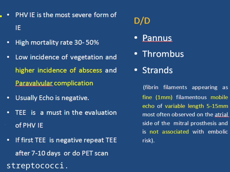
Late prosthetic-valve endocarditis (>60 days POD) is usually
caused by the organisms responsible for native-valve
endocarditis, most often streptococci.
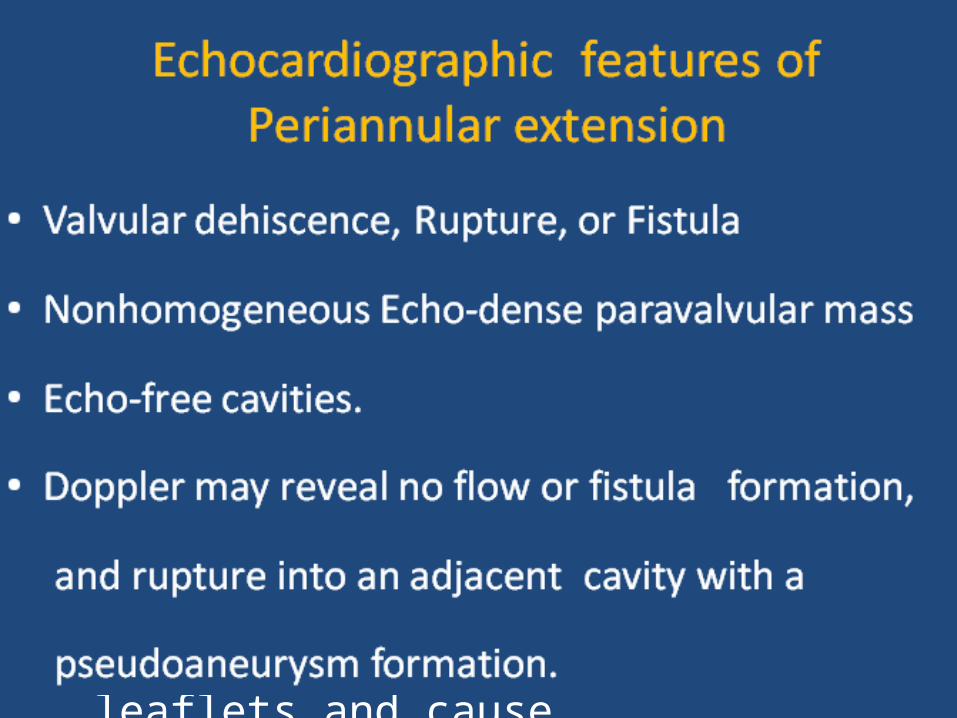
Site of vegetation
• Mechanical valves: Between the sewing ring & annulus and cause paravalvular abscess, dehiscence, peudonaeurysm, fistulas • Bioprosthetic valve: IE effects the valve leaflets and cause cusp tear perforation, and vegetation
Paravalvular regurgitation
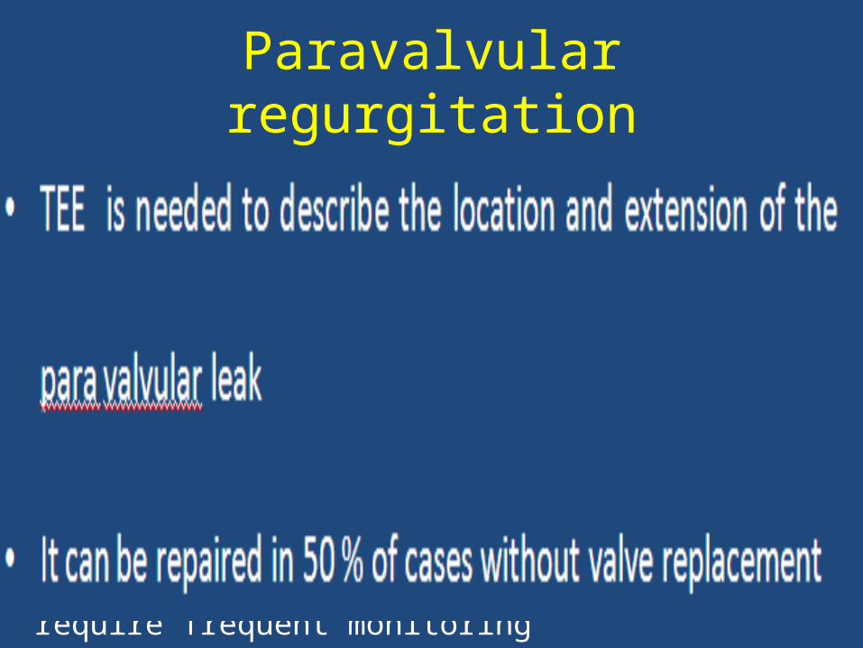
Paravalvular regurgitation
Causes• Infection• Suture dehiscence or fibrosis• Calcification of native annulus causing inadequate contact between the sewing ring and annulus
• More common with the trans catheter aortic valve implantation
• Mild Paravalvular Regurgitation good prognosis require frequent monitoring

Thank u

QUIZ

• Q No .1 Name of this person?
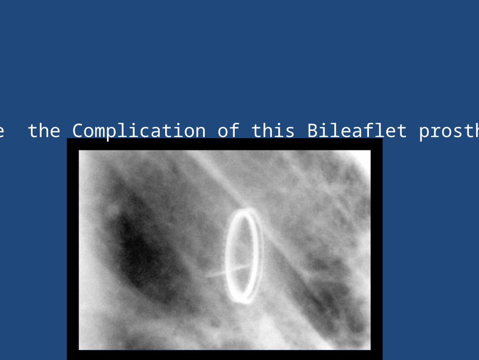
Q.No 2 Name the Complication of this Bileaflet prosthetic valve.
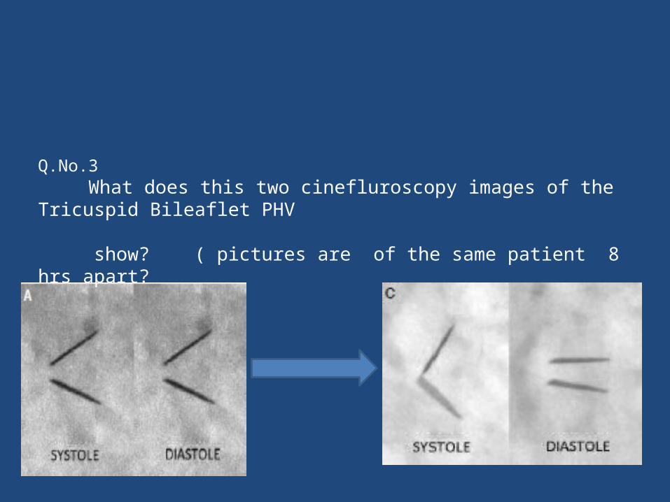
Q.No.3 What does this two cinefluroscopy images of the Tricuspid Bileaflet PHV
show? ( pictures are of the same patient 8 hrs apart?
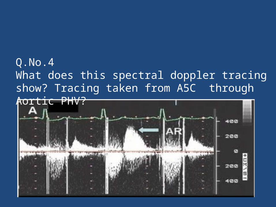
Q.No.4What does this spectral doppler tracing show? Tracing taken from A5C through Aortic PHV?
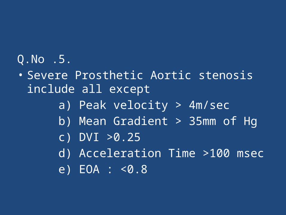
Q.No .5.• Severe Prosthetic Aortic stenosis include all
except a) Peak velocity > 4m/sec b) Mean Gradient > 35mm of Hg c) DVI >0.25 d) Acceleration Time >100 msec e) EOA : <0.8
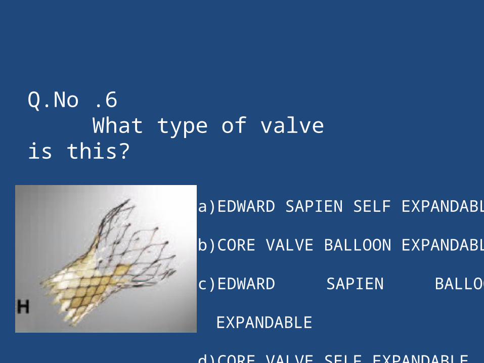
Q.No .6 What type of valve is this?
a) EDWARD SAPIEN SELF EXPANDABLE
b) CORE VALVE BALLOON EXPANDABLE
c) EDWARD SAPIEN BALLOON EXPANDABLE
d) CORE VALVE SELF EXPANDABLE
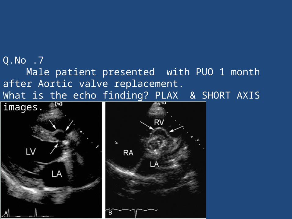
Q.No .7 Male patient presented with PUO 1 month after Aortic valve replacement. What is the echo finding? PLAX & SHORT AXIS images.
Q.No .8
Para valvular regurgitation is said to be severe if regurgitation is _____% of the sewing ring circumferance?
a) >20%
b) >30%
c) >40%
d) >50%
• Q.No .8 Features of Physiological regurgitation?
A) Jet area < 2 cm2 and jet length <2.5 cm
B) Jet area > 2cm2 but < 5cm2 and jet length
<2.5 cm
C) Jet area >2cm2 and jet length >2.5cm
D) jet area of >5cm2 and jet Length of 2.5 cm
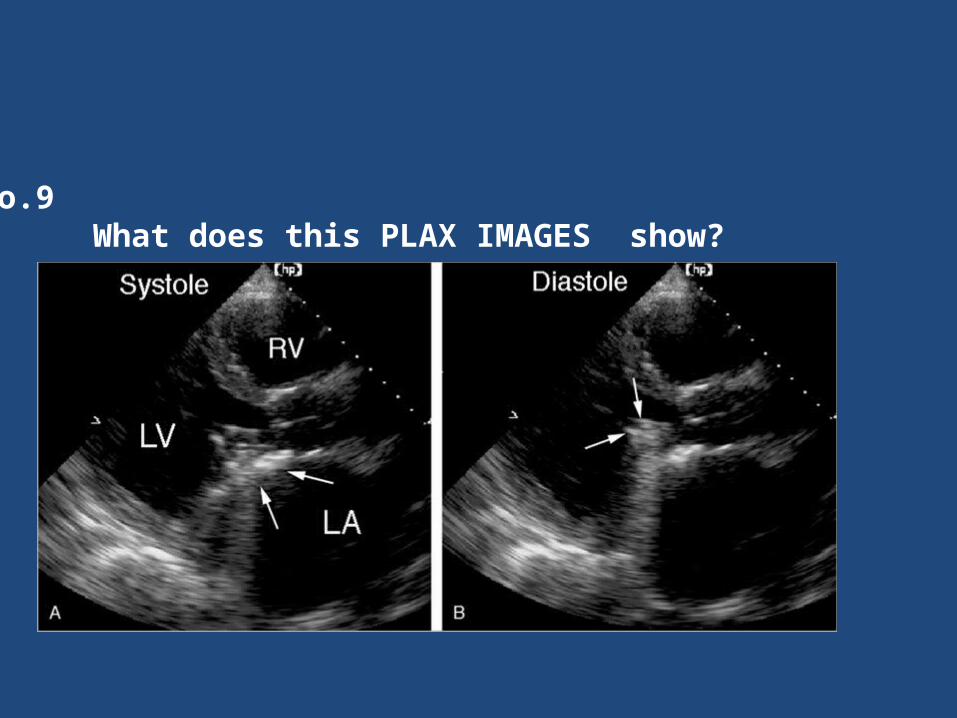
Q.No.9 What does this PLAX IMAGES show?
• Q.No 10
Severity Aortic PPM is defined as a) Indexed EOA ≤0.65 cm2 /m2
b) Indexed EOA ≤0.85 cm2 /m2
c) Indexed EOA ≤1.2 cm2 /m2
d) Indexed EOA ≤0.9 cm2 /m2
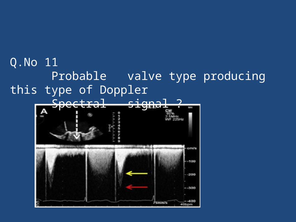
Q.No 11 Probable valve type producing this type of Doppler Spectral signal ?
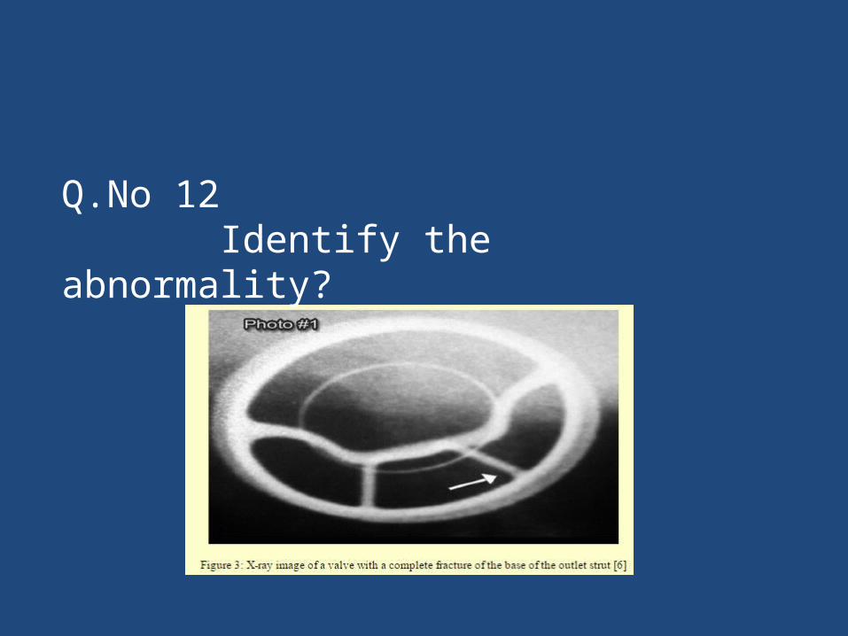
Q.No 12 Identify the abnormality?
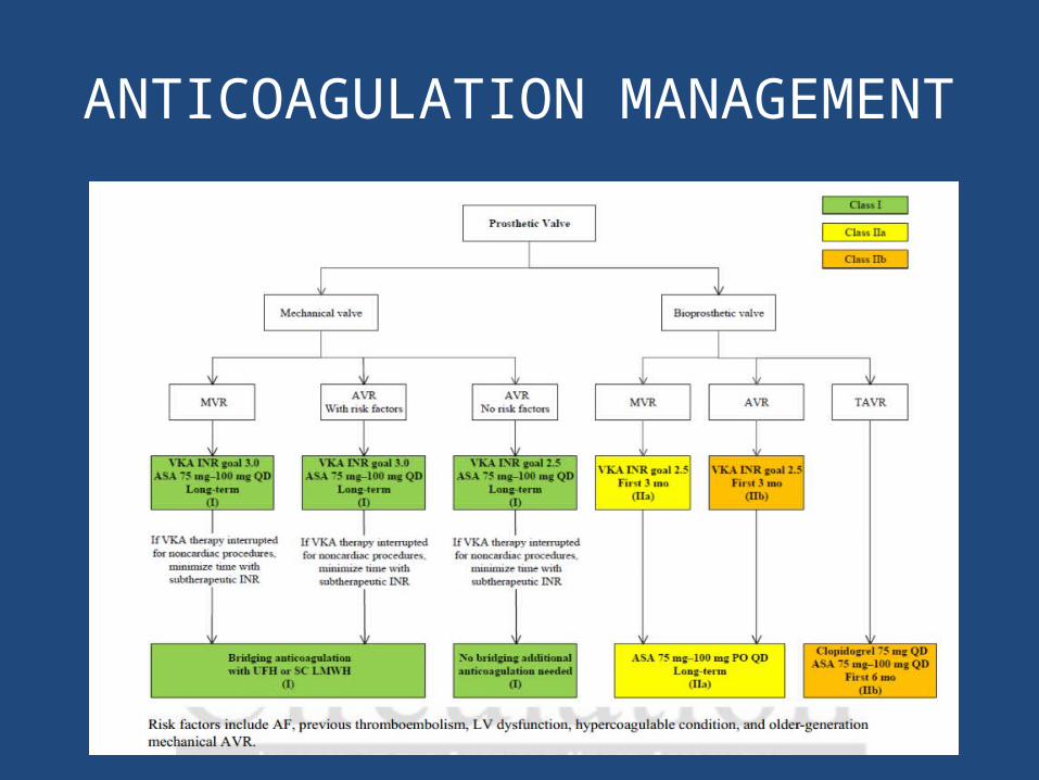
ANTICOAGULATION MANAGEMENT
Related Documents