Production of a composite hyaluronic acid/gelatin blood plasma gel for hydrogel-based adipose tissue engineering applications Esra Korurer, 1 Halime Kenar, 1 Emek Doger, 2 Erdal Karaoz 1 1 Center for Stem Cell and Gene Therapies Research and Practice, Kocaeli University, Izmit Kocaeli, Turkey 41380 2 Department of Gynecology and Obstetrics, Kocaeli University, Izmit Kocaeli, Turkey 41380 Received 14 July 2013; accepted 22 July 2013 Published online 10 August 2013 in Wiley Online Library (wileyonlinelibrary.com). DOI: 10.1002/jbm.a.34901 Abstract: Standard approaches to soft-tissue reconstruction include autologous adipose tissue transplantation, but most of the transferred adipose tissue is generally reabsorbed in a short time. To overcome this problem, long lasting implant- able hydrogel materials that can support tissue regeneration must be produced. The purpose of this study was to evaluate the suitability of composite 3D natural origin scaffolds for reconstructive surgery applications through in vitro tests. The Young’s modulus of the glutaraldehyde crosslinked hyal- uronic acid/gelatin (HA/G) plasma gels, composed of human platelet-poor plasma, gelatin and human umbilical cord hyal- uronic acid, was determined as 3.5 kPa, close to that of soft tissues. The composite HA/G plasma gels had higher porosity than plain plasma gels (72.5% vs. 63.86%). Human adipose tissue derived stem cells (AD-MSCs) were isolated from human lipoaspirates and characterized with flow cytometry, and osteogenic and adipogenic differentiation. Cell prolifera- tion assay of AD-MSCs on the HA/G plasma gels revealed the nontoxic nature of these constructs. Adipogenic differentia- tion was distinctly better on HA/G plasma gels than on plain plasma gels. The results showed that the HA/G plasma gel with its suitable pore size, mechanical properties and excel- lent cell growth and adipogenesis supporting properties can serve as a useful scaffold for adipose tissue engineering applications. V C 2013 Wiley Periodicals, Inc. J Biomed Mater Res Part A: 102A: 2220–2229, 2014. Key Words: adipose tissue, tissue engineering, plasma gel, hyaluronic acid, stem cells How to cite this article: Koruer E, Kenar H, Doger E, Karaoz E. 2014. Production of a composite hyaluronic acid/gelatin blood plasma gel for hydrogel-based adipose tissue engineering applications. J Biomed Mater Res Part A 2014:102A:2220–2229. INTRODUCTION Adipose tissue substitutes are required in reconstructive and plastic surgery. Adipose tissue is used for tissue recon- struction following a mastectomy and other tumor resec- tions, posttraumatic defect reconstruction (especially burns), treatment of congenital abnormalities and augmen- tation of breast, cheek and chin. 1,2 A source of excess adi- pose tissue is available from almost every individual. Therefore, autologous fat grafts would appear to be optimal for the restoration of soft tissue volume. 3 However, autolo- gous adipose tissue remains minimally effective due to insufficient neovascularization. In the long term, this leads to necrosis and apoptosis in free fat grafts and resultant tis- sue resorption over time. 4 The attenuation of the fat graft requires repeated surgery. 1 In tissue engineering applications, cell seeding with growth factors and proteins is accomplished on 3D porous scaffold and after that scaffolds are transplanted to recipient in order to repair functions of tissues or organs. 5 Because lots of cells are needed for production of a tissue equivalent, stem cells have drawn attention due to their ability to pro- liferate easily in the laboratory and to differentiate into many cell types. The MSCs can decrease and prevent cell death and speed up the angiogenesis in the damaged tissues by secreting growth factors and cytokines, can differentiate into cells of the target tissue and can suppress immune rejection even in their allogeneic use. 6,7 Adipose tissue- derived stem cells (AD-MSCs) are used in many soft tissue engineering applications. The most important advantage of AD-MSCs is that they can be obtained easily from standard lipoaspirate materials; they are found in high frequency in human adipose tissue. 8 The niche (microenvironment) has a crucial effect on commitment of stem cells into the differentiation or self- renewal pathways. The scaffolds are mimicking the cell niche, so various biomaterial types are being tested in adi- pose tissue engineering. The natural polymers such as gela- tin, collagen, silk, fibrin and agarose have been used as biomaterials for long time. 9 These materials are biocompati- ble and available commercially for tissue engineering appli- cations. 10 The artificial extracellular matrix (ECM), in other words the scaffold, should be suitable for cell attachment, migration, proliferation and contribute to regeneration of the target tissue. 11 The physical properties of the hydrogels Correspondence to: E. Korurer; e-mail: [email protected] 2220 V C 2013 WILEY PERIODICALS, INC.

Welcome message from author
This document is posted to help you gain knowledge. Please leave a comment to let me know what you think about it! Share it to your friends and learn new things together.
Transcript
Production of a composite hyaluronic acid/gelatin blood plasma gel forhydrogel-based adipose tissue engineering applications
Esra Korurer,1 Halime Kenar,1 Emek Doger,2 Erdal Karaoz1
1Center for Stem Cell and Gene Therapies Research and Practice, Kocaeli University,
Izmit Kocaeli, Turkey 413802Department of Gynecology and Obstetrics, Kocaeli University, Izmit Kocaeli, Turkey 41380
Received 14 July 2013; accepted 22 July 2013
Published online 10 August 2013 in Wiley Online Library (wileyonlinelibrary.com). DOI: 10.1002/jbm.a.34901
Abstract: Standard approaches to soft-tissue reconstruction
include autologous adipose tissue transplantation, but most
of the transferred adipose tissue is generally reabsorbed in a
short time. To overcome this problem, long lasting implant-
able hydrogel materials that can support tissue regeneration
must be produced. The purpose of this study was to evaluate
the suitability of composite 3D natural origin scaffolds for
reconstructive surgery applications through in vitro tests. The
Young’s modulus of the glutaraldehyde crosslinked hyal-
uronic acid/gelatin (HA/G) plasma gels, composed of human
platelet-poor plasma, gelatin and human umbilical cord hyal-
uronic acid, was determined as 3.5 kPa, close to that of soft
tissues. The composite HA/G plasma gels had higher porosity
than plain plasma gels (72.5% vs. 63.86%). Human adipose
tissue derived stem cells (AD-MSCs) were isolated from
human lipoaspirates and characterized with flow cytometry,
and osteogenic and adipogenic differentiation. Cell prolifera-
tion assay of AD-MSCs on the HA/G plasma gels revealed the
nontoxic nature of these constructs. Adipogenic differentia-
tion was distinctly better on HA/G plasma gels than on plain
plasma gels. The results showed that the HA/G plasma gel
with its suitable pore size, mechanical properties and excel-
lent cell growth and adipogenesis supporting properties can
serve as a useful scaffold for adipose tissue engineering
applications. VC 2013 Wiley Periodicals, Inc. J Biomed Mater Res Part
A: 102A: 2220–2229, 2014.
Key Words: adipose tissue, tissue engineering, plasma gel,
hyaluronic acid, stem cells
How to cite this article: Koruer E, Kenar H, Doger E, Karaoz E. 2014. Production of a composite hyaluronic acid/gelatin bloodplasma gel for hydrogel-based adipose tissue engineering applications. J Biomed Mater Res Part A 2014:102A:2220–2229.
INTRODUCTION
Adipose tissue substitutes are required in reconstructiveand plastic surgery. Adipose tissue is used for tissue recon-struction following a mastectomy and other tumor resec-tions, posttraumatic defect reconstruction (especiallyburns), treatment of congenital abnormalities and augmen-tation of breast, cheek and chin.1,2 A source of excess adi-pose tissue is available from almost every individual.Therefore, autologous fat grafts would appear to be optimalfor the restoration of soft tissue volume.3 However, autolo-gous adipose tissue remains minimally effective due toinsufficient neovascularization. In the long term, this leadsto necrosis and apoptosis in free fat grafts and resultant tis-sue resorption over time.4 The attenuation of the fat graftrequires repeated surgery.1
In tissue engineering applications, cell seeding withgrowth factors and proteins is accomplished on 3D porousscaffold and after that scaffolds are transplanted to recipientin order to repair functions of tissues or organs.5 Becauselots of cells are needed for production of a tissue equivalent,stem cells have drawn attention due to their ability to pro-liferate easily in the laboratory and to differentiate into
many cell types. The MSCs can decrease and prevent celldeath and speed up the angiogenesis in the damaged tissuesby secreting growth factors and cytokines, can differentiateinto cells of the target tissue and can suppress immunerejection even in their allogeneic use.6,7 Adipose tissue-derived stem cells (AD-MSCs) are used in many soft tissueengineering applications. The most important advantage ofAD-MSCs is that they can be obtained easily from standardlipoaspirate materials; they are found in high frequency inhuman adipose tissue.8
The niche (microenvironment) has a crucial effect oncommitment of stem cells into the differentiation or self-renewal pathways. The scaffolds are mimicking the cellniche, so various biomaterial types are being tested in adi-pose tissue engineering. The natural polymers such as gela-tin, collagen, silk, fibrin and agarose have been used asbiomaterials for long time.9 These materials are biocompati-ble and available commercially for tissue engineering appli-cations.10 The artificial extracellular matrix (ECM), in otherwords the scaffold, should be suitable for cell attachment,migration, proliferation and contribute to regeneration ofthe target tissue.11 The physical properties of the hydrogels
Correspondence to: E. Korurer; e-mail: [email protected]
2220 VC 2013 WILEY PERIODICALS, INC.
are largely similar to living tissues. The similarities includehigh water content, softness, flexible structure and lowinterfacial tension with water or biological fluids.12
Hyaluronic acid-based hydrogels support cell attach-ment, proliferation, and expansion.13 Fibrin is widely usedwith other polymers to form composites because of its bio-compatibility and easiness of manufacturing with desiredproperties.13 In comparison to other materials used for tis-sue engineering purposes, fibrin gel has advantages such aspossibility of autologous production and controllable degra-dation rate.14 However, in use of allogeneic plasma to manu-facture the fibrin gel, immunogenic reactions against foreignmaterials shall be reduced.14
In the present study we have explored the effect of scaf-folds from naturally derived materials, namely, HA/G bloodplasma gels and plain plasma gels on AD-MSC viability, pro-liferation, and adipogenic differentiation. Fibrinogen wasprecipitated from human platelet-poor plasma and hyal-uronic acid isolated from human umbilical cords. Compositegels of fibrinogen with gelatin and hyaluronic acid (HA/Gplasma gels) were produced afterwards. HA/G plasma gelcharacterization was made in comparison to plain plasmagels. Gels with higher porosity and pore size were obtainedupon addition of gelatin and hyaluronic acid to the fibrino-gen solution and young modulus of the gels was improved.The HA/G plasma gels supported AD-MSC attachment andproliferation and their porous structure was preserved atthe end of 21-day culture. Adipogenic differentiation poten-tial of AD-MSCs on HA/G plasma gels and plain plasma gelswas investigated; lipid vesicle accumulation was more pro-nounced on the HA/G plasma gels, even in the absence ofdifferentiation factors.
MATERIALS AND METHODS
AD-MSC isolation and expansionAdipose tissue was obtained by lipoaspiration of subcutane-ous fat of the abdominal region from female donors. Thedonors consented with the procedure and agreed with fur-ther research of aspirated fat in accordance with the ethicalguidelines of the Kocaeli University Medical Ethics Commit-tee. Fatty portions of lipoaspirate samples were collectedwith pipette and digested with 0.1% (w/v) collagenase(Gibco) at 37�C for 60 min in a shaking water bath. Afterthe digestion process, cell suspension was centrifuged at1500 rpm for 10 min. The pellet was resuspended in thephosphate-buffered saline (PBS, Gibco) for washing and cen-trifuged at 1800 rpm for 10 min. This washing process wasrepeated twice. The pellet was resuspended in 5 mL basalmedium (DMEM/F12 (Gibco) containing 10% fetal bovineserum (FBS; Invitrogen/ GIBCO), 1% L-Glutamine, 1 ngmL21 bFGF (Gibco) and 0.1% primocin (Invivogen) and fil-tered through 100-lm nylon mesh (BD Biosciences). The iso-lated adipose derived cells were plated in a 25 cm2 tissueculture flask (BD Biosciences) and were cultured in a humidi-fied atmosphere of 5% CO2 at 37�C. The cells were detachedby using 0.25% trypsin-EDTA solution (Sigma) when90% confluence was obtained. First-passage (P1) AD-MSCswere plated in tissue culture flasks at 1 3 103 cells cm22
and cultured in the basal medium. When the cells were 80%confluent, they were passaged (P2). Medium was changedtwice per week.
Characterization of AD-MSCsThe adipose tissue derived stem cells were characterizedwith flow cytometry at the third passage and also were dif-ferentiated into osteogenic and adipogenic lineages. Flowcytometry was performed using a FACS Calibur (BD Bio-sciences, San Diego, USA). The data were analyzed with CellQuest software (BD Biosciences) and the forward and sidescatter profiles gated out of debris and dead cells. The sur-face antigens (CD13, CD29, CD44, CD90, CD146, CD166,HLA ABC, CD3, CD8, CD11b, CD14, CD15, CD19, CD33,CD34, CD45, CD117, and HLA-DR) of the cells were charac-terized with flow cytometry, and osteogenic and adipogenicdifferentiation was performed as standard protocols.
In vitro osteogenic differentiationCells from passage three were seeded onto six-well plates(3000 cells cm22) in which type I collagen-coated coverslipswere placed. The osteogenic medium was composed ofMEM (Invitrogen/GIBCO) supplemented with 10 nM dexa-methasone (Sigma–Aldrich), 50 mg mL21 ascorbate-2-phosphate (Wako Chemicals), 10 mM b-glycerophosphate(Sigma–Aldrich), 0.1% primocin (Invivogen) and 10% FBS(Invitrogen/GIBCO). The cells were cultured for 2 weeks inthe osteogenic medium; the medium in the wells wasreplaced twice a week. At the end of the second week,osteogenic differentiation was assessed by staining withAlizarin Red (Sigma–Aldrich).
In vitro adipogenic differentiationTo induce adipogenic differentiation cells from passagethree were seeded (3000 cells cm22) on fibronectin coatedcoverslips (BD Biosciences) in six-well plates. The adipo-genic medium composed of MEM (Invitrogen/GIBCO) sup-plemented with 10% FBS (Invitrogen/GIBCO), 0.5 mMisobutyl-methyl xanthine (IBMX-Sigma–Aldrich), 1026 Mdexamethasone (Sigma–Aldrich), 10 mg mL21 insulin (Invi-trogen/GIBCO), 200 mM indomethacin (Sigma–Aldrich), and0.1% primocin (Invivogen) was added on the cells for a cul-ture period of 2 weeks. The medium was replaced twice aweek. Intracellular lipid vesicles indicating adipogenic differ-entiation were confirmed by Oil Red O (Sigma–Aldrich)staining.
Hyaluronic acid isolation from umbilical cordsHyaluronic acid isolation from umbilical cords was per-formed according to Lago et al.14 Briefly, human umbilicalcord residues (1 kg) were divided into small pieces (4 mm3 4 mm) and treated with PBS at 4�C overnight. The dayafter, the tissue pieces were treated with 4L 0.2% sodiumchloride solution. The liquid part was collected and treatedwith 300–600 mL of 1% cetyltrimethylammonium bromide(CTAB) solution and hyaluronic acid aggregations wereformed. The obtained precipitate was transferred into 0.9Mcalcium chloride solution to dissociate the HA-CTAB
ORIGINAL ARTICLE
JOURNAL OF BIOMEDICAL MATERIALS RESEARCH A | JUL 2014 VOL 102A, ISSUE 7 2221
complex. The suspension was treated with 25% v/v aque-ous ethanol; the nucleic acids were precipitated and sepa-rated by centrifugation (1000 g, 10 min). The resultingsuspension was deproteinized four times through chloro-form solvent extraction (10% of the total volume) per-formed every day. HA was precipitated by adding 75% v/vaqueous ethanol and acetone.
Characterization of umbilical cord derived hyaluronicacidNearly 1% umbilical cord derived HA solution was preparedand the percentage of nucleic acid and protein contamina-tion was determined by measurement of absorbance at 260and 280 nm with a pico-drop spectrophotometer. The hyal-uronic acid sodium salt from rooster comb (>98% purity,protein content of <2%, Sigma) was used as a referencestandard. The protein content of the hyaluronic acid solu-tions was determined with BCA test (Sigma–Aldrich) too,for confirmation. The umbilical cord derived HA and thecommercial hyaluronic acid sodium salt from rooster combwere dried on glass slides and stained with alcian blue.About 1% alcian blue solution in 3% acetic acid (Riedel-deHaen) was prepared and the hyaluronic acid samples werestained in this solution for 30 min. After extensive washingthe images were taken under a light microscope.
Fibrinogen isolation from human blood plasmaThe human blood plasma of healthy donors that was out ofdate for transfusion was collected from Kocaeli UniversityBlood Bank. The precipitation of fibrinogen was carried outusing a combination of ethanol and cold treatment previ-ously described by Park et al.15 Briefly, absolute ethanolwas added to blood plasma and this mixture of ethanol-blood plasma was stored at 220�C for 20 min. Later, sam-ples were centrifuged at 3200 rpm at 4�C for 8 min. Thepellet was resuspended in 0.9% NaCl and samples werestored at 37�C for 15 min to dissolve the fibrinogen. Theconcentrated fibrinogen solution was aliquoted and storedat 220�C.
Preparation of hyaluronic acid/gelatin plasma gels andplain plasma gelsHyaluronic acid and gelatin were cross-linked using ethyl-3-(3-dimethyl aminopropyl) carbodiimide hydrochloride/N-hydroxyl succinimide (EDC/NHS). 1% EDC and 0.2 M NHSwere prepared in MES buffer. Nearly 500 mL of EDC/NHSsolution was added to the 500 mL 1% hyaluronic acid solu-tion and incubated for 30 min at room temperature. The 2-mercaptoethanol (Merck) was added to the sample solutionto a final concentration of 0.14% to stop EDC binding. The6% gelatin solution prepared in PBS with 150 mM NaClwas added to sample solution and incubated overnight(Final hyaluronic acid/gelatin ratio: 2/98 w/w). HA/Gplasma gel was formed by mixing 120 mL concentratedhuman fibrinogen (480 mg mL21), 30 mL crosslinked hyal-uronic acid/gelatin solution and 30 mL 40 mM CaCl2 solu-tion in each well of a 48-well plate. Later, 50 mL thrombin(40 U mL21) was added to each well and mixed quickly by
moving the pipette tip in the well. The mixture was storedat 37�C for 2 h for polymerization. The plain plasma gelwas prepared with concentrated human fibrinogen, water,40 mM CaCl2 and thrombin (40 U mL21). HA/G plasma gelswere maintained in 1% (v/v) solution of gluteraldehyde(GA) at room temperature for 30 min for crosslinking. Thescaffolds were washed in HBSS three times a day for 4days. The crosslinked plain plasma gels were prepared thesame way but water was used instead of the hyaluronicacid/gelatin solution.
Scanning electron microscopyThe porous and fibrous morphologies of HA/G plasma gelsand plain plasma gels were observed under a Scanning Elec-tron Microscope (SEM) model QUANTA 400F Field Emission.AD-MSC seeded HA/G plasma gels were fixed with 4% para-formaldehyde and washed with PBS. Later, the gels werefrozen in 280�C and lyophilized using Cool safe 110-4 (Lab-ogene), and then grossly cut with a blade to observe withSEM. The gels were coated with 10 nm thick Au-Pd andwere observed under low-vacuum conditions.
Mechanical testCompression test of the gels (9.2 mm in diameter) was con-ducted with LR 30 K mechanical tester (Lloyd instruments)which can apply a force up to 2N in the compression rangeof %10 and 1 mm min21 speed. Load was applied to gelsuntil destruction. Three samples were tested from each gelgroup.
Measurement of gel porosity and pore sizeA 10-mm-thick cryosection was obtained from HA/G plasmaand plain plasma gels under the same freezing conditions.The cryosections were incubated with 4% paraformalde-hyde in PBS at room temperature for 15 min to stabilizegels’ morphological features. The gels were stained withcoomassie brilliant blue (Merck) to determine their porosityand pore size through microscopy. For preparation of thedye, 1:1 (v/v) methanol and distilled water were mixed. Gla-cial acetic acid (Riedel-de Haen) up to 10% of the solutionand coomassie brilliant blue were mixed to obtain 0.25%dye solution and the final solution was filtered through a0.45-lm membrane filter. The dye solution was applied onthe crosslinked cryosections and incubated for 30 min. Thecryosections were stored in mixture of water, methanol andacetic acid without dye for 30 min for destaining. Imageswere taken under a light microscope, and gel porosity andpore size were determined from these images by using theScion Image program.
Cell viability and proliferation assayThe viability, adhesion and proliferation rate of AD-MSCs onHA/G plasma gels was determined by tetrazolium salt, 2-(4-iodophenyl)-3-(4-nitrophenyl)-5-(2,4-dis-ulfophenyl)-2H tet-razolium, monosodium salt (WST-1) test (Roche). AD-MSCsuspensions were seeded at 7.5 3 103 cells cm22 on HA/Gplasma gels and coverslips in 48-well plates and were incu-bated in 5% CO2 atmosphere at 37�C for 1, 5, 7, 14, and
2222 KORURER ET AL. PRODUCTION OF A COMPOSITE HYALURONIC ACID/GELATIN BLOOD PLASMA GEL
21 days. The 1st day, culture medium was removed, thesamples were washed twice with PBS and transferredinto10% WST-1 reagent containing plate. The plate wasincubated for 2 h at 37�C in dark. The gels and coverslipswithout cells were used as blanks. Absorbance of the solu-tion from the wells was measured at 480 nm with a UV-visible spectrophotometer (VersaMax, Molecular Device,USA). For each group, experiments were repeated threetimes and measurements were completed in triplicate. Inthe other days (5th, 7th, 14th, and 21st day) the abovementioned protocol was repeated.
Adipogenic differentiation of AD-MSCs on the gelsAD- MSCs were seeded at 2.5 3 104 cell cm22 density onthe HA/G plasma and plain plasma gels and when the cellsreached 70% confluence, adipogenic differentiation was ini-tiated with the adipogenic medium: MEM (Invitrogen/GIBCO) supplemented with 10% FBS (Invitrogen/GIBCO),0.5 mM isobutyl-methylxanthine (IBMX-Sigma–Aldrich),1026 M dexamethasone (Sigma–Aldrich, Fluka Chemie AG,Buchs, Switzerland), 10 mg mL21 insulin (Invitrogen/GIBCO), 200 mM indomethacin (Sigma–Aldrich) and 0.1%primocin (Invivogen). The medium was replaced twice aweek. At the end of 3-week culture, the presence of intracel-lular lipid vesicles, which indicate adipogenic differentiationwas confirmed by Oil Red O (Sigma–Aldrich) staining with0.5% Oil red O in methanol.
Statistical analysisAll experiments were repeated a minimum of three times.Data are reported as mean6 standard deviation (SD) andall statistical analyses were performed using SPSS 14.0. Inaddition, data were analyzed using the paired t test. Differ-ences between the experimental and control groups wereregarded as statistically significant when p< 0.05.
RESULTS
Isolation and characterization of human AD-MSCsThe mesenchymal stem cells isolated from human adiposetissue were used in this study. After 5 days following enzy-matic isolation, the attached cells at the flask surfacebecame fibroblast-like in morphology. The cells were cul-tured until 70% confluence and subcultured and cryopre-served for further characterization and use in the study.Defined markers exist that specifically identify MSCs. Theflow cytometry data indicated that the adipose tissuederived third passage cells expressed CD13, CD29, CD44,CD90, CD146, and CD166, but not CD3, CD8, CD11b, CD14,CD15, CD19, CD33, CD34, CD45, CD117, or HLA-DR. Thesefindings are consistent with the undifferentiated mesenchy-mal stem cell surface antigen profile, i.e., the cells possessedimmunophenotypic characteristics of MSCs (Fig. 1).
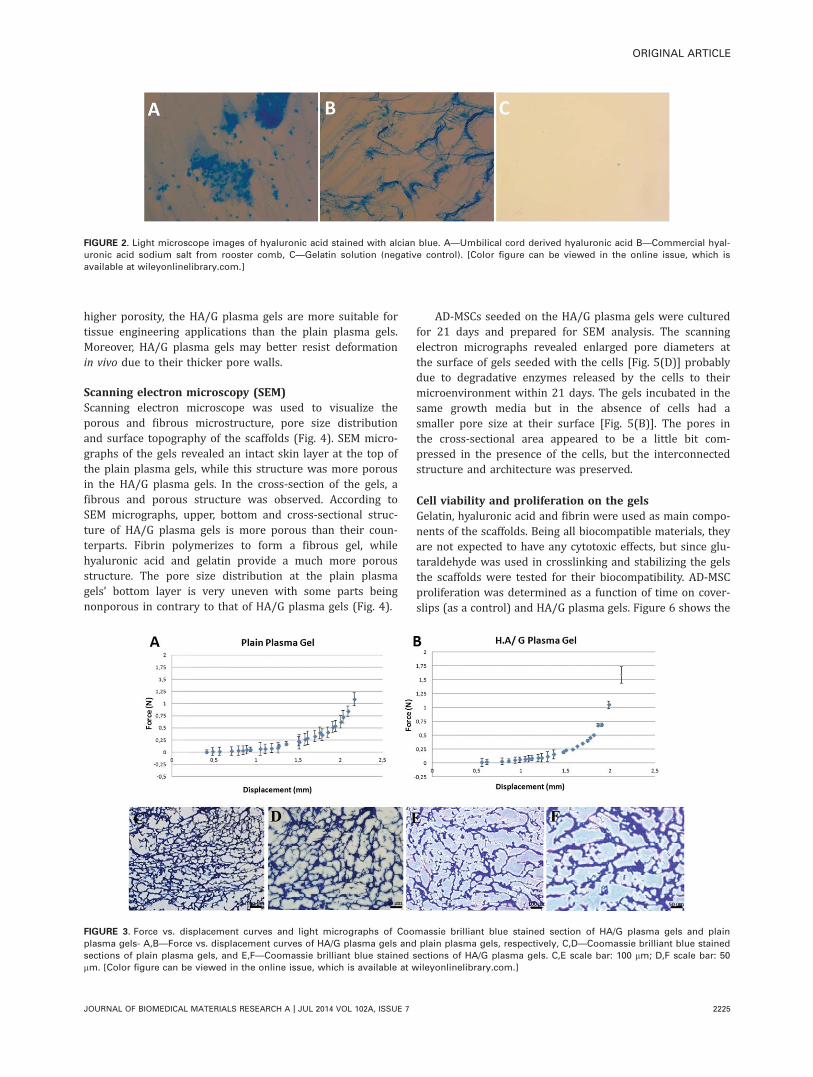
Characterization of umbilical cord derived hyaluronicacidAbout 640 mg hyaluronic acid was obtained from about 1kg of human umbilical cord residues. A 1% umbilical cordderived HA solution was analyzed for nucleic acid and pro-
tein contamination through measurement of absorbance at260 and 280 nm, respectively, with a pico-drop spectropho-tometer. The hyaluronic acid sodium salt from rooster comb(> 98% purity, protein content of <2%, Sigma) was used asa reference standard. The rooster comb derived hyaluronicacid contained 16.7 ng mL21 double-stranded DNA and 0.2mg mL21 direct protein according to the data obtained fromthe pico-drop spectrophotometer. On the other hand, theumbilical cord derived HA solution contained 39.4 ng mL21
double-stranded DNA and 0.6 mg mL21 direct protein.Considering the amount of direct protein, protein con-
tent of umbilical cord derived HA was 6% and the percent-age of purity was determined as 94%, accordingly. Inaddition, DNA content of umbilical cord derived HA wasdetermined as 2.4 times of the DNA content of the roostercomb derived HA. Umbilical cord derived HA, hyaluronicacid sodium salt from rooster comb, and gelatin solution (asa negative control) were stained with alcian blue (Fig. 2).The blue coloration of the umbilical cord derived materialproves its glycosaminoglycan (GAG) content; hyaluronic acidis the most abundant GAG of the umbilical cord matrix.
Characterization studies of HA/G plasma gels and plainplasma gelsThe scaffolds were 9.2 mm in diameter and acquired theirfinal form at about an hour after mixing the components.HA/G plasma gels and plain plasma gels were strong, com-pact and easy to handle.
Mechanical testThe compression test was applied to investigate the elasticmoduli of HA/G plasma gels and plain plasma gels. Thecompressive strengths of the scaffolds were determinedfrom stress–strain curve by applying the load until the scaf-folds were destroyed. Elastic modules of plain plasma gelsand HA/G plasma gels were calculated as 2.146 0.045 kPaand 3.536 0.23 kPa, respectively, from the slope of the lin-ear portion of the stress–strain curve generated from theDisplacement (mm) vs. Force (N) graphs [Fig. 3(A,B)]. Addi-tion of hyaluronic acid and gelatin to the fibrin gel causedan increase in elastic modulus.
Gel porosity and pore sizeThe gels were stained with coomassie brilliant blue todetermine their porosity and pore size. The porosity of HA/G plasma and plain plasma gels were calculated as72.05%6 5.85% and 63.86%64.27%, respectively, byusing the Scion Image program. The transverse length ofthe pores was measured to get an idea about the pore sizeof the scaffolds (Fig. 3). The average pore diameter of theHA/G plasma gels and plain plasma gels were calculated as1336 20 mm and 586 9 mm, respectively. Therefore, addi-tion of hyaluronic acid and gelatin to the fibrin gel causedan improvement in both pore size and porosity. The poresize of a scaffold has an effect on various aspects of the tis-sue culture such as intercellular communication, cell expan-sion on and within the scaffold, transportation of nutrientsand waste material.16,17 Having a larger pore size and
ORIGINAL ARTICLE
JOURNAL OF BIOMEDICAL MATERIALS RESEARCH A | JUL 2014 VOL 102A, ISSUE 7 2223
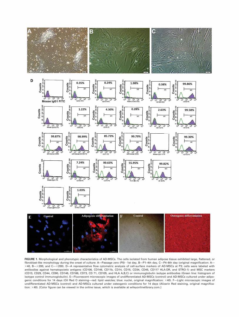
FIGURE 1. Morphological and phenotypic characteristics of AD-MSCs. The cells isolated from human adipose tissue exhibited large, flattened, or
fibroblast-like morphology during the onset of culture: A—Passage zero (P0)21st day, B—P1–4th day, C—P4–6th day (original magnification: A—
340, B—3200, and C—3200). D—A representative flow cytometric analysis of cell-surface markers of AD-MSCs at P3; cells were labeled with
antibodies against hematopoietic antigens (CD106, CD146, CD11b, CD14, CD15, CD34, CD45, CD117 HLA-DR, and STRO-1) and MSC markers
(CD13, CD29, CD44, CD90, CD146, CD166, CD73, CD 71, CD105, and HLA A,B,C) or immunoglobulin isotype antibodies (Green line: histogram of
isotype control immunoglobulin). E—Fluorescent microscopic images of undifferentiated AD-MSCs (control) and AD-MSCs cultured under adipo-
genic conditions for 14 days (Oil Red O staining—red: lipid vesicles; blue: nuclei, original magnification: 340). F—Light microscopic images of
undifferentiated AD-MSCs (control) and AD-MSCs cultured under osteogenic conditions for 14 days (Alizarin Red staining, original magnifica-
tion: 340). [Color figure can be viewed in the online issue, which is available at wileyonlinelibrary.com.]
higher porosity, the HA/G plasma gels are more suitable fortissue engineering applications than the plain plasma gels.Moreover, HA/G plasma gels may better resist deformationin vivo due to their thicker pore walls.
Scanning electron microscopy (SEM)Scanning electron microscope was used to visualize theporous and fibrous microstructure, pore size distributionand surface topography of the scaffolds (Fig. 4). SEM micro-graphs of the gels revealed an intact skin layer at the top ofthe plain plasma gels, while this structure was more porousin the HA/G plasma gels. In the cross-section of the gels, afibrous and porous structure was observed. According toSEM micrographs, upper, bottom and cross-sectional struc-ture of HA/G plasma gels is more porous than their coun-terparts. Fibrin polymerizes to form a fibrous gel, whilehyaluronic acid and gelatin provide a much more porousstructure. The pore size distribution at the plain plasmagels’ bottom layer is very uneven with some parts beingnonporous in contrary to that of HA/G plasma gels (Fig. 4).
AD-MSCs seeded on the HA/G plasma gels were culturedfor 21 days and prepared for SEM analysis. The scanningelectron micrographs revealed enlarged pore diameters atthe surface of gels seeded with the cells [Fig. 5(D)] probablydue to degradative enzymes released by the cells to theirmicroenvironment within 21 days. The gels incubated in thesame growth media but in the absence of cells had asmaller pore size at their surface [Fig. 5(B)]. The pores inthe cross-sectional area appeared to be a little bit com-pressed in the presence of the cells, but the interconnectedstructure and architecture was preserved.
Cell viability and proliferation on the gelsGelatin, hyaluronic acid and fibrin were used as main compo-nents of the scaffolds. Being all biocompatible materials, theyare not expected to have any cytotoxic effects, but since glu-taraldehyde was used in crosslinking and stabilizing the gelsthe scaffolds were tested for their biocompatibility. AD-MSCproliferation was determined as a function of time on cover-slips (as a control) and HA/G plasma gels. Figure 6 shows the
FIGURE 3. Force vs. displacement curves and light micrographs of Coomassie brilliant blue stained section of HA/G plasma gels and plain
plasma gels- A,B—Force vs. displacement curves of HA/G plasma gels and plain plasma gels, respectively, C,D—Coomassie brilliant blue stained
sections of plain plasma gels, and E,F—Coomassie brilliant blue stained sections of HA/G plasma gels. C,E scale bar: 100 mm; D,F scale bar: 50
mm. [Color figure can be viewed in the online issue, which is available at wileyonlinelibrary.com.]
FIGURE 2. Light microscope images of hyaluronic acid stained with alcian blue. A—Umbilical cord derived hyaluronic acid B—Commercial hyal-
uronic acid sodium salt from rooster comb, C—Gelatin solution (negative control). [Color figure can be viewed in the online issue, which is
available at wileyonlinelibrary.com.]
ORIGINAL ARTICLE
JOURNAL OF BIOMEDICAL MATERIALS RESEARCH A | JUL 2014 VOL 102A, ISSUE 7 2225
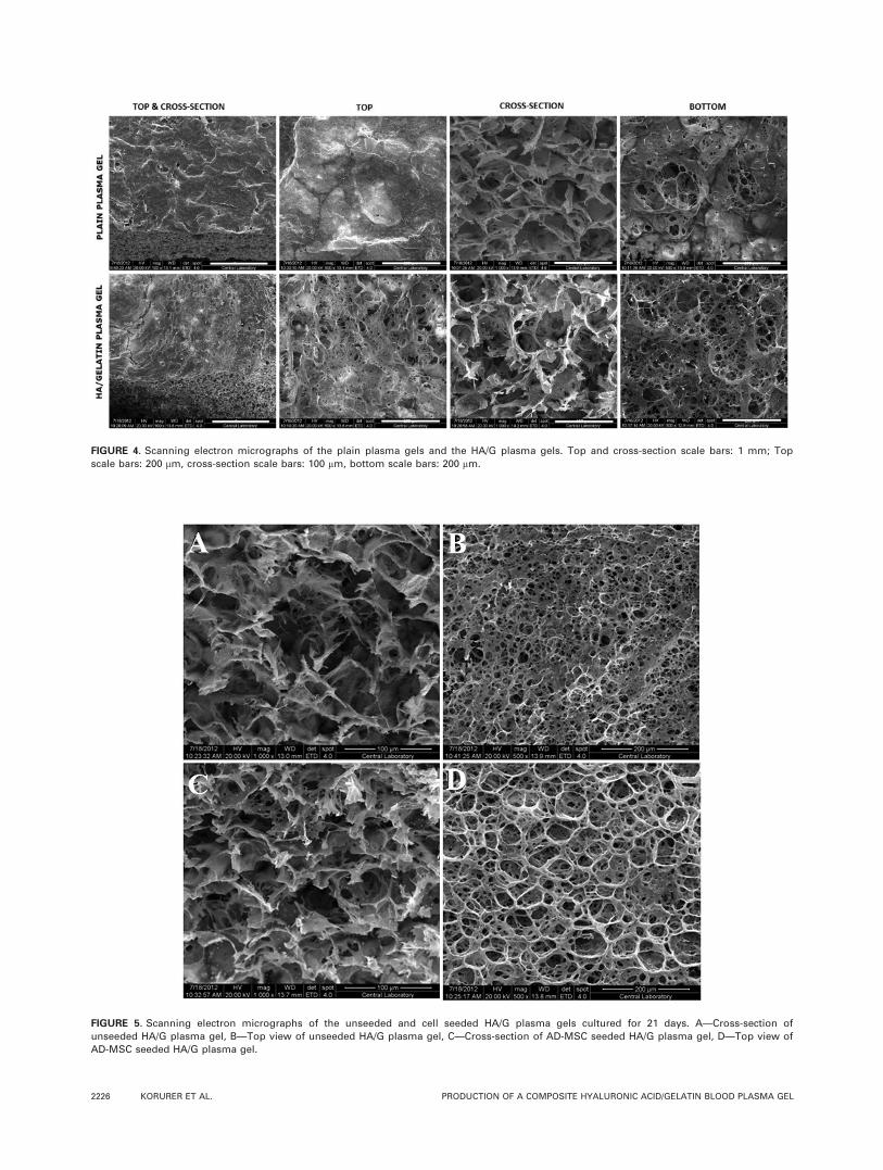
FIGURE 4. Scanning electron micrographs of the plain plasma gels and the HA/G plasma gels. Top and cross-section scale bars: 1 mm; Top
scale bars: 200 mm, cross-section scale bars: 100 mm, bottom scale bars: 200 mm.
FIGURE 5. Scanning electron micrographs of the unseeded and cell seeded HA/G plasma gels cultured for 21 days. A—Cross-section of
unseeded HA/G plasma gel, B—Top view of unseeded HA/G plasma gel, C—Cross-section of AD-MSC seeded HA/G plasma gel, D—Top view of
AD-MSC seeded HA/G plasma gel.
2226 KORURER ET AL. PRODUCTION OF A COMPOSITE HYALURONIC ACID/GELATIN BLOOD PLASMA GEL
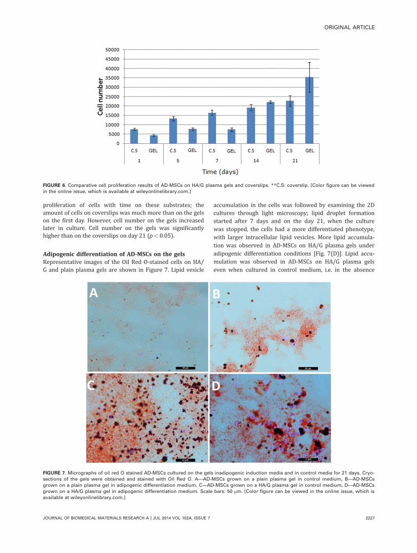
proliferation of cells with time on these substrates; theamount of cells on coverslips was much more than on the gelson the first day. However, cell number on the gels increasedlater in culture. Cell number on the gels was significantlyhigher than on the coverslips on day 21 (p< 0.05).
Adipogenic differentiation of AD-MSCs on the gelsRepresentative images of the Oil Red O-stained cells on HA/G and plain plasma gels are shown in Figure 7. Lipid vesicle
accumulation in the cells was followed by examining the 2Dcultures through light microscopy; lipid droplet formationstarted after 7 days and on the day 21, when the culturewas stopped, the cells had a more differentiated phenotype,with larger intracellular lipid vesicles. More lipid accumula-tion was observed in AD-MSCs on HA/G plasma gels underadipogenic differentiation conditions [Fig. 7(D)]. Lipid accu-mulation was observed in AD-MSCs on HA/G plasma gelseven when cultured in control medium, i.e. in the absence
FIGURE 6. Comparative cell proliferation results of AD-MSCs on HA/G plasma gels and coverslips. **C.S: coverslip. [Color figure can be viewed
in the online issue, which is available at wileyonlinelibrary.com.]
FIGURE 7. Micrographs of oil red O stained AD-MSCs cultured on the gels inadipogenic induction media and in control media for 21 days. Cryo-
sections of the gels were obtained and stained with Oil Red O. A—AD-MSCs grown on a plain plasma gel in control medium, B—AD-MSCs
grown on a plain plasma gel in adipogenic differentiation medium, C—AD-MSCs grown on a HA/G plasma gel in control medium, D—AD-MSCs
grown on a HA/G plasma gel in adipogenic differentiation medium. Scale bars: 50 mm. [Color figure can be viewed in the online issue, which is
available at wileyonlinelibrary.com.]
ORIGINAL ARTICLE
JOURNAL OF BIOMEDICAL MATERIALS RESEARCH A | JUL 2014 VOL 102A, ISSUE 7 2227
of differentiation factors [Fig. 7(C)]. In the plain plasma gels,lipid accumulation could be visualized only in a small num-ber of cells [Fig. 7(B)]. AD-MSCs did not show any adipo-genic differentiation on the plain plasma gels when culturedin the control medium [Fig. 7(A)]. Therefore, the compositeplasma gel with hyaluronic acid and gelatin is able alone toinduce adipogenic differentiation of AD-MSCs.
DISCUSSION
In tissue engineering applications potential cell sourcesinclude autologous cells from the patient, allogeneic cellsfrom a donor, and xenogeneic cells.18 Autologous cells seemto be most promising due to the exclusion of immune rejec-tion and ethical issues.19 AD-MSCs are easily isolated forautologous use in plastic or aesthetic surgeries nowadaysand were used in this study. AD-MSCs can be isolated bycollagenase digestion following liposuction.20 MSCs havebeen reported to be negative for the hematopoietic markersCD34 and CD45, whereas they are positive for Stro-1, CD29,CD44, CD90, and CD105.21,22 Most of the surface proteinsexpressed by MSCs have been demonstrated to also beexpressed by AD-MSCs, with the exception of Stro-1.20 Stro-1 is supposed to represent the nearest approximation toidentify the “pure” MSCs, although a few hematopoietic cellsweakly express Stro-1.23
It is very advantageous to use natural biomaterials suchas fibrinogen, hyaluronic acid, and gelatin as a scaffoldmaterial in tissue engineering; they are bioactive, biocom-patible and they possess mechanical properties similar tothat of soft tissues.24,25 On the other hand, it is difficult tocontrol their physicochemical properties and their rate ofdissolution. Many biological materials have been used in acontrolled manner in recent studies.11,26 Films composed ofalginate and gelatin mixed in different ratios were shown toinduce cell proliferation and hydrogels composed of hyal-uronic acid and gelatin were successfully used in the fieldof cartilage tissue engineering.27,28 In a study by Lei et al.,expansion of mouse MSCs on hyaluronic acid gels wasinvestigated; here the number of cross-links and concentra-tion of RGD (arginine-glycine-aspartic acid) directed cellularbehavior. It was revealed by this study that cells on morerigid hydrogels spread more, migrate less, and as a resulttheir growth rate falls down.29 All these studies point outthe importance of scaffold properties and provision of thetarget tissue microenvironment in the culture in obtaining adesired cellular behavior. Accordingly, natural ECM materialswere used in this study to produce scaffolds in the form ofgels with elastic moduli very close to that of fat tissue.Addition of hyaluronic acid and gelatin to the fibrinincreased the elastic modulus of the gels, but it was still inan acceptable range.
Long term stability of scaffolds is necessary to provideenough time for cell proliferation and matrix productionwithout causing any implant collapse. Fibrin gels are poten-tially attractive scaffolds for use in tissue engineering; how-ever their use is often impaired by fast dissolution and lackof shape stability. Commercially available fibrin sealants
tend to shrink and disintegrate in vitro and in vivo due tofibrinolysis after a few days and almost completely dissolvewithin 3–4 weeks.30,31 Therefore, in this study after the pro-duction of HA/G plasma gels, they were crosslinked withglutaraldehyde to obtain a long-term scaffold shape stabilityfor adipose tissue engineering applications.
Fiber based scaffolds induce tissue regeneration by pro-viding structural communication. In a study by Mol et al.,32
less loss of soluble collagen to the medium was observedwhen the cells were loaded to PCL coated fibrous PLGAscaffolds in a fibrin gel carrier and a more mature ECM wasdeposited in shorter period of time.33 The scaffolds withfiber structure have high similarity to ECM. However, highporosity scaffolds are preferred since they provide bettercell communication, nutrient and gas transportation. In thisstudy, as a result of hyaluronic acid and gelatin addition tothe fibrin gel, both porous and fibrous scaffolds were pro-duced. HA/G plasma gels had larger pore size and higherporosity in comparison to plain plasma gels. This alsomakes them more suitable for use in soft tissue engineering.HA/G plasma gel macro and microstructure was preservedwhen incubated in medium for 21 days; in presence of AD-MSCs the gel material was metabolized which led toincrease in surface pore size and a little bit downfall ininner pores, but still the structure was preserved. After cellseeding the gels were cultured under static conditions andthis may explain the accumulation and growth of the cellsbeing mainly on the top portion where they can have anaccess to nutrients and oxygen. The cell penetration wouldbe better in a perfused culture. On the other hand, accord-ing to the WST-1 assay results, cell proliferation rate on thegels increased after day 14 (Fig. 6) when compared to cellproliferation rate on the glass slides. The AD-MSCs on thecoverslips had a higher proliferation rate between day 1and 7. Although the gels contain natural extracellular matrixmaterial that is expected to be very much suitable forattachment and growth of AD-MSCs, the glutaraldehydeused to crosslink the gels in order to have a stable structuremay have a growth retarding effect. We presume that AD-MSCs need a week to adapt to the crosslinked material andcan proliferate further after synthesis of their own ECM.Cell proliferation on the coverslips slowed down very muchafter day 14 due to contact inhibition while the cells on thegels continued to proliferate probably by migrating towardsand occupying the inner parts of the 3D structure. Gel sur-face and glass slide surface area are similar, so increase incell number on the gels can only be explained with AD-MSCpenetration and growth within the pores. Therefore HA/Gplasma gels did not have any negative effects on cell prolif-eration in a long run.
The cell shape, cytoskeletal components34 and ECMstructure and composition35,36 have been found to stronglyinfluence adipocyte differentiation and function. Further-more, adipocyte precursor cell adhesion, proliferation, anddifferentiation can strongly be influenced by components ofthe ECM which play a pivotal role in the adipose tissuedevelopment.35,37 Both the biological material and themechanical properties of a scaffold may affect MSC
2228 KORURER ET AL. PRODUCTION OF A COMPOSITE HYALURONIC ACID/GELATIN BLOOD PLASMA GEL
differentiation. Young modulus of breast adipose tissue wasdetermined as close to 1.9 kPa by Samani et al.38 AD-MSCson the HA/G plasma gels differentiated into adipocytes, asevidenced by intracellular lipid vesicle deposition, in pres-ence and even absence of soluble differentiation factors.Adipogenic differentiation observed in control medium maybe either due to mechanical properties of the gels beingclose to that of adipose tissue, or due to presence of hyal-uronic acid or gelatin in the structure of the gels. Adipo-genic differentiation of AD-MSCs was not observed on theplain plasma gels, which had mechanical properties closerto that of native adipose tissue, when cultured in controlmedium. This phenomenon emphasizes more the impor-tance of biological composition of the scaffolds.
As a conclusion, the results of this study showed that acomposite HA/G plasma gel with suitable pore size andmechanical properties can support both AD-MSC growthand adipogenic differentiation. Therefore the HA/G plasmagel developed in this study can serve as a useful scaffold foradipose tissue engineering applications.
REFERENCES1. Patrick CW. Adipose Tissue Engineering: The Future of Breast and
Soft Tissue Reconstruction Following Tumor Resection. Semin.
Surg. Oncol. 2000;19:302–311.
2. Patrick C. Tissue engineering strategies for adipose tissue repair.
Anat Rec 2001;263:361–366.
3. Katz AJ, Llull R, Hedrick MH, Futrell J. Emerging approaches to
the tissue engineering of fat. Clin Plast Surg 1999;26:587.
4. Nishimura T, Hashimoto H, Nakanishi I, Furukawa M. Microvascu-
lar angiogenesis and apoptosis in the survival of free fat grafts.
Laryngoscope 2000;110:1333–1338.
5. Vacanti CA. The history of tissue engineering. J Cell Mol Med
2007;10:569–576.
6. Goldstein LSB, Schneider M. Stem Cells for Dummies: For Dum-
mies; Published Online: 10 JAN 2012, DOI: 10.1002/
9781118269244.ch18 Copyright VC 2010 Wiley Publishing, Inc., Indi-
anapolis, Indiana.
7. Vunjak-Novakovic G. In Culture of Cells for Tissue Engineering;
Vunjak-Novakovic, G.; Freshney, R. I., Eds.; Wiley: Hoboken, NJ,
USA. 2006; p 132.
8. Guilak F, Awad HA, Fermor B, Leddy HA, Gimble JM. Adipose-
derived adult stem cells for cartilage tissue engineering. Biorheol-
ogy 2004;41:389–400.
9. Freyman T, Yannas I, Yokoo R, Gibson L. Fibroblast contraction of
a collagen–GAG matrix. Biomaterials 2001;22:2883–2891.
10. Park JB, Lakes RS. Biomaterials: An Introduction. New York:
Springer; 2007.
11. Vats A, Tolley N, Polak J, Gough J. Scaffolds and biomaterials for
tissue engineering: A review of clinical applications. Clin Otolar-
yngol Allied Sci 2003;28:165–172.
12. Satish C, Satish K, Shivakumar H. Hydrogels as controlled drug
delivery systems: Synthesis, crosslinking, water and drug trans-
port mechanism. Indian J Pharm Sci 2006;68:133.
13. Ducheyne P, Healy KE, Hutmacher DW, Grainger DW, Kirkpatrick
CJ. editors. Comprehensive Biomaterials, Vol. 2. Elsevier, Amster-
dam, 2011. p 239–259.
14. Lago G, Oru~na L, Cremata JA, P�erez C, Coto G, Lauzan E,
Kennedy JF. Isolation, purification and characterization of hyalur-
onan from human umbilical cord residues. Carbohydr Polym
2005;62:321–326.
15. Park JJ, Cintron JR, Siedentop KH, Orsay CP, Pearl RK, Nelson
RL, Abcarian H. Technical manual for manufacturing autologous
fibrin tissue adhesive. Dis Colon Rectum 1999;42:1334–1338.
16. O’Brien FJ, Harley B, Yannas IV, Gibson LJ. The effect of pore
size on cell adhesion in collagen-GAG scaffolds. Biomaterials
2005;26:433–441.
17. Van Tienen TG, Heijkants RGJC, Buma P, de Groot JH, Pennings
AJ, Veth RPH. Tissue ingrowth and degradation of two biode-
gradable porous polymers with different porosities and pore
sizes. Biomaterials 2002;23:1731–1738.
18. Griffith LG, Naughton G. Tissue engineering—Current challenges
and expanding opportunities. Science 2002;295:1009–1014.
19. Curtis A, Riehle M. Tissue engineering: The biophysical back-
ground. Phys Med Biol 2001;46:R47.
20. Gronthos S, Franklin DM, Leddy HA, Robey PG, Storms RW,
Gimble JM. Surface protein characterization of human adipose
tissue-derived stromal cells. J Cell Physiol 2001;189:54–63.
21. Pittenger MF, Mackay AM, Beck SC, Jaiswal RK, Douglas R,
Mosca JD, Moorman MA, Simonetti DW, Craig S, Marshak DR.
Multilineage potential of adult human mesenchymal stem cells.
Science 1999;284:143–147.
22. Devine SM. Mesenchymal stem cells: Will they have a role in the
clinic? J Cell Biochem 2002;85:73–79.
23. Gronthos S, Graves S, Ohta S, Simmons P. The STRO-11 fraction
of adult human bone marrow contains the osteogenic precursors.
Blood 1994;84:4164–4173.
24. Chou CH, Cheng WTK, Kuo TF, Sun JS, Lin FH, Tsai JC. Fibrin
glue mixed with gelatin/hyaluronic acid/chondroitin-6-sulfate tri-
copolymer for articular cartilage tissue engineering: The results of
real-time polymerase chain reaction. J Biomed Mater Res A 2007;
82:757–767.
25. Weigel PH, Fuller GM, LeBoeuf RD. A model for the role of hyal-
uronic acid and fibrin in the early events during the inflammatory
response and wound healing. J Theor Biol 1986;119:219–234.
26. Dawson E, Mapili G, Erickson K, Taqvi S, Roy K. Biomaterials for
stem cell differentiation. Adv Drug Deliv Rev 2008;60:215–228.
27. Hu X, Li D, Zhou F, Gao C. Biological hydrogel synthesized from
hyaluronic acid, gelatin and chondroitin sulfate by click chemistry.
Acta Biomater 2011;7:1618–1626.
28. Rosellini E, Cristallini C, Barbani N, Vozzi G, Giusti P. Preparation
and characterization of alginate/gelatin blend films for cardiac tis-
sue engineering. J Biomed Mater Res A 2008;91:447–453.
29. Lei Y, Gojgini S, Lam J, Segura T. The spreading, migration and
proliferation of mouse mesenchymal stem cells cultured inside
hyaluronic acid hydrogels. Biomaterials 2011;32:39–47.
30. Weisel JW. Fibrinogen and fibrin. Adv Protein Chem 2005;70:247–
299.
31. Sidelmann JJ, Gram J, Jespersen J, Kluft C. Fibrin clot formation
and lysis: Basic mechanisms. Semin Thromb Hemost 2000;26:
605–618.
32. Hirsh J, Johnston M, Teoh K. Autologous fibrin glue and methods
for its preparation and use. Google Patents; Patent Number:
5,643,192 , 1997.
33. Mol A, van Lieshout MI, Dam-de Veen CG, Neuenschwander S,
Hoerstrup SP, Baaijens F, Bouten CVC. Fibrin as a cell carrier in
cardiovascular tissue engineering applications. Biomaterials 2005;
26:3113–3121.
34. McBeath R, Pirone DM, Nelson CM, Bhadriraju K, Chen CS. Cell
shape, cytoskeletal tension, and RhoA regulate stem cell lineage
commitment. Dev Cell 2004;6:483–495.
35. Nakajima I, Yamaguchi T, Ozutsumi K, Aso H. Adipose tissue
extracellular matrix: Newly organized by adipocytes during differ-
entiation. Differentiation 2004;63:193–200.
36. Smas CM, Sul HS. Control of adipocyte differentiation. Biochem J
1995;309 (Part 3):697.
37. Patrick CW, Wu X. Integrin-mediated preadipocyte adhesion and
migration on laminin-1. Ann Biomed Eng 2003;31:505–514.
38. Samani A, Bishop J, Luginbuhl C, Plewes DB. Measuring the elas-
tic modulus of ex vivo small tissue samples. Phys Med Biol 2003;
48:2183.
ORIGINAL ARTICLE
JOURNAL OF BIOMEDICAL MATERIALS RESEARCH A | JUL 2014 VOL 102A, ISSUE 7 2229
Related Documents