Archives of Cardiovascular Disease (2015) 108, 197—205 Available online at ScienceDirect www.sciencedirect.com REVIEW Pocket-sized focused cardiac ultrasound: Strengths and limitations Échoscopie à l’aide d’échographe de poche : avantages et inconvénients Mariana Mirabel a,b,c,∗ , David Celermajer d , Anne-Sophie Beraud e,f , Xavier Jouven a,b,c , Eloi Marijon a,b,c , Albert A. Hagège a,b,c a Assistance publique—Hôpitaux de Paris, hôpital Européen-Georges-Pompidou, 75015 Paris, France b Université Paris Descartes, Sorbonne Paris Cité, 75006 Paris, France c Inserm U970, Paris Cardiovascular Research Centre—PARCC, 75737 Paris Cedex 15, France d Sydney Medical School, Sydney, Australia e Stanford University Hospital, Stanford, CA, USA f Clinique Pasteur, 31300 Toulouse, France Received 27 November 2014; received in revised form 9 January 2015; accepted 13 January 2015 Available online 5 March 2015 KEYWORDS Echocardiography; Focused cardiac ultrasound; Education; Heart failure; Emergency medicine Summary Focused cardiac ultrasound (FCU) has emerged in recent years and has created new possibilities in the clinical assessment of patients both in and out of hospital. The increasing portability of echocardiographic devices, with some now only the size of a smartphone, has widened the spectrum of potential indications and users, from the senior cardiologist to the medical student. However, many issues still need to be addressed, especially the acknowledg- ment of the advantages and limitations of using such devices for FCU, and the extent of training required in this rapidly evolving field. In recent years, an increasing number of studies involving FCU have been published with variable results. This review outlines the evidence for the use of FCU with pocket-echo to address specific questions in daily clinical practice. © 2015 Elsevier Masson SAS. All rights reserved. Abbreviations: ESC, European Society of Cardiology; FCU, Focused cardiac ultrasound. ∗ Corresponding author. Paris Cardiovascular Research Centre, Inserm U970, hôpital Européen-Georges-Pompidou, 56, rue Leblanc, 75737 Paris cedex 15, France. E-mail address: [email protected] (M. Mirabel). http://dx.doi.org/10.1016/j.acvd.2015.01.002 1875-2136/© 2015 Elsevier Masson SAS. All rights reserved.

Welcome message from author
This document is posted to help you gain knowledge. Please leave a comment to let me know what you think about it! Share it to your friends and learn new things together.
Transcript
Archives of Cardiovascular Disease (2015) 108, 197—205
Available online at
ScienceDirectwww.sciencedirect.com
REVIEW
Pocket-sized focused cardiac ultrasound:Strengths and limitations
Échoscopie à l’aide d’échographe de poche : avantages et inconvénients
Mariana Mirabela,b,c,∗, David Celermajerd,Anne-Sophie Beraude,f, Xavier Jouvena,b,c,Eloi Marijona,b,c, Albert A. Hagègea,b,c
a Assistance publique—Hôpitaux de Paris, hôpital Européen-Georges-Pompidou, 75015 Paris,Franceb Université Paris Descartes, Sorbonne Paris Cité, 75006 Paris, Francec Inserm U970, Paris Cardiovascular Research Centre—PARCC, 75737 Paris Cedex 15, Franced Sydney Medical School, Sydney, Australiae Stanford University Hospital, Stanford, CA, USAf Clinique Pasteur, 31300 Toulouse, France
Received 27 November 2014; received in revised form 9 January 2015; accepted 13 January 2015Available online 5 March 2015
KEYWORDSEchocardiography;Focused cardiacultrasound;Education;Heart failure;Emergency medicine
Summary Focused cardiac ultrasound (FCU) has emerged in recent years and has created newpossibilities in the clinical assessment of patients both in and out of hospital. The increasingportability of echocardiographic devices, with some now only the size of a smartphone, haswidened the spectrum of potential indications and users, from the senior cardiologist to themedical student. However, many issues still need to be addressed, especially the acknowledg-ment of the advantages and limitations of using such devices for FCU, and the extent of trainingrequired in this rapidly evolving field. In recent years, an increasing number of studies involvingFCU have been published with variable results. This review outlines the evidence for the use ofFCU with pocket-echo to address specific questions in daily clinical practice.
© 2015 Elsevier Masson SAS. All rights reserved.Abbreviations: ESC, European Society of Cardiology; FCU, Focused cardiac ultrasound.∗ Corresponding author. Paris Cardiovascular Research Centre, Inserm U970, hôpital Européen-Georges-Pompidou, 56, rue Leblanc, 75737
Paris cedex 15, France.E-mail address: [email protected] (M. Mirabel).
http://dx.doi.org/10.1016/j.acvd.2015.01.0021875-2136/© 2015 Elsevier Masson SAS. All rights reserved.
198 M. Mirabel et al.
MOTS CLÉSÉchocardiographiecardiaque ;Échoscopie ;Éducation ;Médecine d’urgence
Résumé L’avènement de l’échoscopie a ouvert des possibilités diagnostiques nouvelles cesdernières années, à la fois en pré-hospitalier et à l’hôpital. Les échographes de pochesont des appareils de la taille d’un téléphone portable. Ils permettent une grande mobilitéouvrant l’éventail d’utilisateurs potentiels, allant du cardiologue expérimenté à l’étudiant enmédecine. De nombreuses questions demeurent néanmoins sans réponse à ce jour. Une atten-tion particulière doit être portée aux avantages et aux limites de l’utilisation de ces appareilspour l’échoscopie afin d’en assurer une bonne utilisation. Les modalités de formation restentaussi à préciser. De nombreuses études ont été publiées ces dernières années avec des résul-tats variables. Cette revue résume le niveau de connaissances actuelles de l’échoscopie à l’aided’échographes de poche.
. Tou
IHaganiafswabc
tpssnddefudpp
el•••
ord
RpcIs
pton
L
Fs(aficLwrmtFhl
Ll
EbTgcnbLsc
F
Icfrm
© 2015 Elsevier Masson SAS
ntroductionistory taking and clinical examination alone do not alwaysllow precise cardiac diagnoses. Point-of-care echocardio-raphy ultrasounds, using portable devices, has emergeds a bedside tool in many specialities, from trauma toeonatology [1]. However, a particularly challenging areas likely to be focused cardiac ultrasound (FCU) because
wide variety of patients need assessment of ventricularunction and estimation of filling pressures in differentettings. Several studies have shown incremental benefithen FCU is added to the general physical examination,nd investigators have suggested that FCU will somedayecome an integral part of the physical examination andould even replace the stethoscope [2—8].
Technological advances have led to the miniaturiza-ion of echocardiographic machines, with gradually smallerortable devices. Pocket-echocardiography is defined by theize of the devices: small enough to fit into a coat pocket,lightly larger than a smartphone. The use of pocket-echoecessarily implies FCU due to the limited functions of theseevices. The relatively low cost and high portability of theseevices make them accessible to a wide range of practition-rs in different settings: at the bedside, in or out of hospital,rom the outpatients’ cardiology clinic to the intensive carenit. Before rapid and uncontrolled proliferation of theseevices, there is an urgent need to address indications,rotocols, limitations and training for the optimal use ofocket-echocardiography [9].
When assessing the accuracy and feasibility of pocket-chocardiography for FCU, three components are particu-arly important:
image quality and technical limitations due to the device;the expectations practitioners have of FCU;expertise of the user/reader.
Some studies have addressed these issues separately andthers in combination. In this review, we explore the cur-ent level of evidence for the use of different pocket-echoevices for FCU in daily clinical practice.
eliability of FCU withocket-echocardiography in different
linical scenariosn experienced hands, pocket-echocardiography may be ofignificant diagnostic value when used in conjunction with
sc(
s droits réservés.
hysical examination [2—8]. Systematic use of FCU may leado a change of management and/or diagnosis in up to 20%f patients for whom comprehensive echocardiography wasot requested after traditional clinical assessment [3,5,8].
eft ventricular size and function
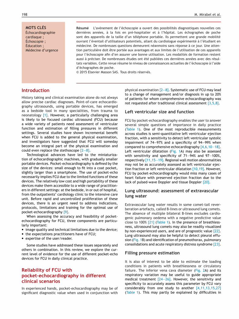
CU by pocket-echocardiography enables the user to answereveral simple questions of importance in daily practiceTable 1). One of the most reproducible measurementscross studies is semi-quantitative left ventricular ejectionraction, with a sensitivity to detect left ventricular systolicmpairment of 74—97% and a specificity of 94—99% whenompared to comprehensive echocardiography [4,6,10—18].eft ventricular dilatation (Fig. 1A) may also be assessedith sensitivity and specificity of 71—94% and 97—100%,
espectively [11,15—19]. Regional wall motion abnormalitiesay not be as accurately assessed as left ventricular ejec-
ion fraction or left ventricular dilatation [10,19]. However,CU by pocket-echocardiography would miss many cases ofeart failure with preserved ejection fraction due to theack of pulsed-wave Doppler and tissue Doppler [20].
ung ultrasound: assessment of extravascularung water
xtravascular lung water results in some comet-tail rever-eration artefacts, called B-lines or ultrasound lung comets.he absence of multiple bilateral B-lines excludes cardio-enic pulmonary oedema with a negative predictive valuelose to 100% [21] (Table 1). In the presence of breathless-ess, ultrasound lung comets may also be readily visualizedy non-experienced users, and are of prognostic value [22].ung ultrasound may also be helpful to detect pleural effu-ion (Fig. 1B) and identification of pneumothorax, pulmonaryonsolidations and acute respiratory distress syndrome [23].
illing pressure estimation
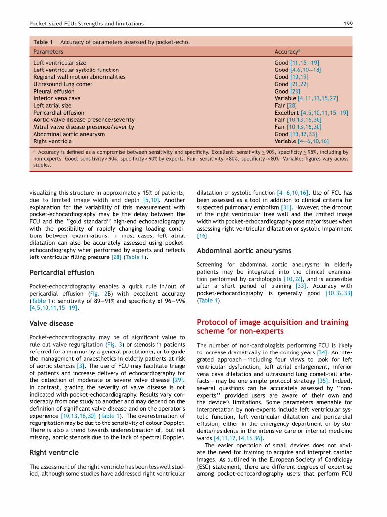
t is also of interest to be able to estimate the loadingonditions in patients with breathlessness or circulatoryailure. The inferior vena cava diameter (Fig. 2A) and itsespiratory variation may be useful to guide appropriateedical treatment [24—26]. However, the sensitivity and
pecificity to accurately assess this parameter by FCU varyonsiderably from one study to another [4,11,13,15,27]Table 1). This may partly be explained by difficulties in
Pocket-sized FCU: Strengths and limitations 199
Table 1 Accuracy of parameters assessed by pocket-echo.
Parameters Accuracya
Left ventricular size Good [11,15—19]Left ventricular systolic function Good [4,6,10—18]Regional wall motion abnormalities Good [10,19]Ultrasound lung comet Good [21,22]Pleural effusion Good [23]Inferior vena cava Variable [4,11,13,15,27]Left atrial size Fair [28]Pericardial effusion Excellent [4,5,10,11,15—19]Aortic valve disease presence/severity Fair [10,13,16,30]Mitral valve disease presence/severity Fair [10,13,16,30]Abdominal aortic aneurysm Good [10,32,33]Right ventricle Variable [4—6,10,16]
a Accuracy is defined as a compromise between sensitivity and specificity. Excellent: sensitivity ≥ 90%, specificity ≥ 95%, including bynon-experts. Good: sensitivity > 90%, specificity > 90% by experts. Fair: sensitivity ≈ 80%, specificity ≈ 80%. Variable: figures vary across
dbsowa[
A
Sptap(
Ps
Ttgvvfsetitedw
studies.
visualizing this structure in approximately 15% of patients,due to limited image width and depth [5,10]. Anotherexplanation for the variability of this measurement withpocket-echocardiography may be the delay between theFCU and the ‘‘gold standard’’ high-end echocardiographywith the possibility of rapidly changing loading condi-tions between examinations. In most cases, left atrialdilatation can also be accurately assessed using pocket-echocardiography when performed by experts and reflectsleft ventricular filling pressure [28] (Table 1).
Pericardial effusion
Pocket-echocardiography enables a quick rule in/out ofpericardial effusion (Fig. 2B) with excellent accuracy(Table 1): sensitivity of 89—91% and specificity of 96—99%[4,5,10,11,15—19].
Valve disease
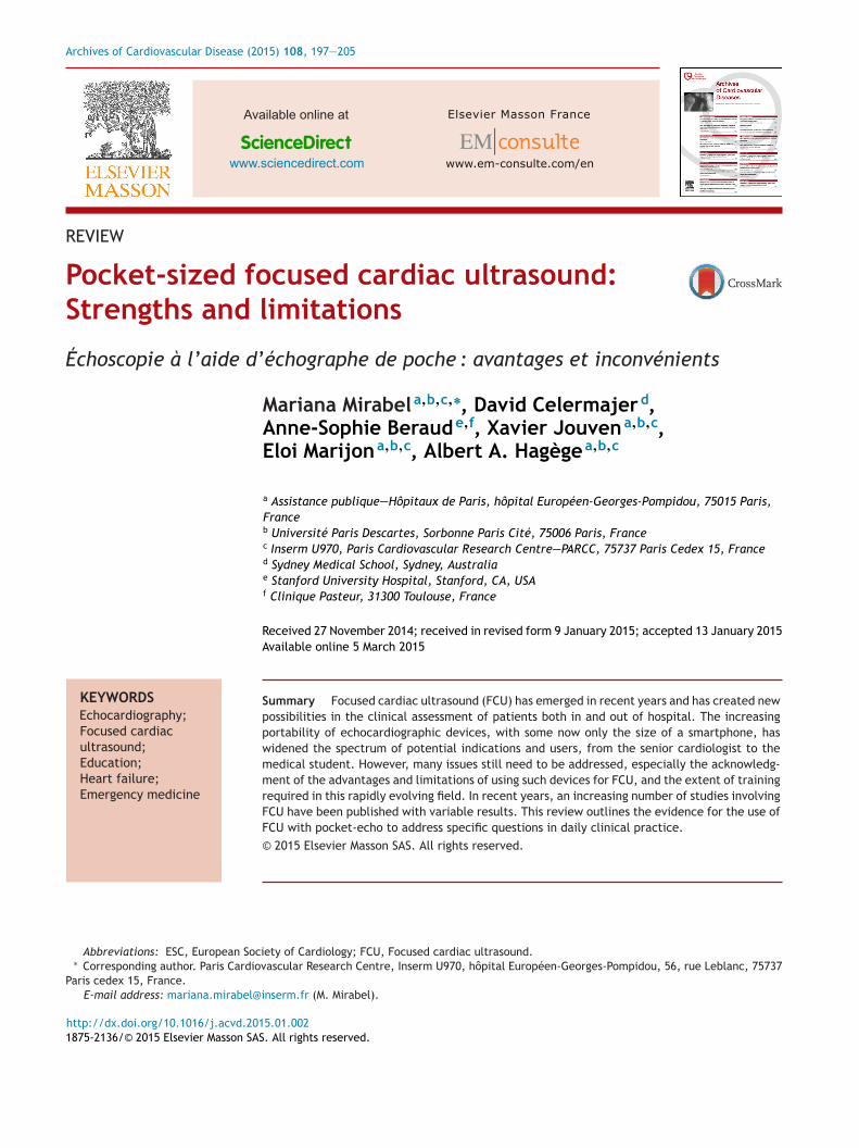
Pocket-echocardiography may be of significant value torule out valve regurgitation (Fig. 3) or stenosis in patientsreferred for a murmur by a general practitioner, or to guidethe management of anaesthetics in elderly patients at riskof aortic stenosis [3]. The use of FCU may facilitate triageof patients and increase delivery of echocardiography forthe detection of moderate or severe valve disease [29].In contrast, grading the severity of valve disease is notindicated with pocket-echocardiography. Results vary con-siderably from one study to another and may depend on thedefinition of significant valve disease and on the operator’sexperience [10,13,16,30] (Table 1). The overestimation ofregurgitation may be due to the sensitivity of colour Doppler.There is also a trend towards underestimation of, but notmissing, aortic stenosis due to the lack of spectral Doppler.
Right ventricle
The assessment of the right ventricle has been less well stud-ied, although some studies have addressed right ventricular
ai(a
ilatation or systolic function [4—6,10,16]. Use of FCU haseen assessed as a tool in addition to clinical criteria foruspected pulmonary embolism [31]. However, the dropoutf the right ventricular free wall and the limited imageidth with pocket-echocardiography pose major issues whenssessing right ventricular dilatation or systolic impairment16].
bdominal aortic aneurysms
creening for abdominal aortic aneurysms in elderlyatients may be integrated into the clinical examina-ion performed by cardiologists [10,32], and is accessiblefter a short period of training [33]. Accuracy withocket-echocardiography is generally good [10,32,33]Table 1).
rotocol of image acquisition and trainingcheme for non-experts
he number of non-cardiologists performing FCU is likelyo increase dramatically in the coming years [34]. An inte-rated approach — including four views to look for leftentricular dysfunction, left atrial enlargement, inferiorena cava dilatation and ultrasound lung comet-tail arte-acts — may be one simple protocol strategy [35]. Indeed,everal questions can be accurately assessed by ‘‘non-xperts’’ provided users are aware of their own andhe device’s limitations. Some parameters amenable fornterpretation by non-experts include left ventricular sys-olic function, left ventricular dilatation and pericardialffusion, either in the emergency department or by stu-ents/residents in the intensive care or internal medicineards [4,11,12,14,15,36].
The easier operation of small devices does not obvi-
te the need for training to acquire and interpret cardiacmages. As outlined in the European Society of CardiologyESC) statement, there are different degrees of expertisemong pocket-echocardiography users that perform FCU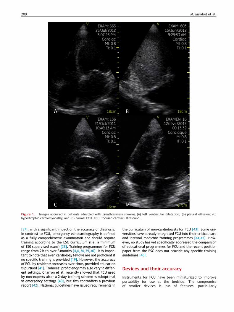
200 M. Mirabel et al.
F snessh rdiac
[Iatortnoiebir
tvaeopg
D
igure 1. Images acquired in patients admitted with breathlesypertrophic cardiomyopathy, and (D) normal FCU. FCU: focused ca
37], with a significant impact on the accuracy of diagnosis.n contrast to FCU, emergency echocardiography is defineds a fully comprehensive examination and should requireraining according to the ESC curriculum (i.e. a minimumf 150 supervised scans) [38]. Training programmes for FCUange from 2 h to over 3 months [4,6,36,39,40]. It is impor-ant to note that even cardiology fellows are not proficient ifo specific training is provided [19]. However, the accuracyf FCU by residents increases over time, provided educations pursued [41]. Trainees’ proficiency may also vary in differ-
nt settings. Charron et al. recently showed that FCU usedy non-experts after a 2-day training scheme is suboptimaln emergency settings [40], but this contradicts a previouseport [42]. National guidelines have issued requirements inIpo
showing (A) left ventricular dilatation, (B) pleural effusion, (C) ultrasound.
he curriculum of non-cardiologists for FCU [43]. Some uni-ersities have already integrated FCU into their critical carend internal medicine training programmes [44,45]. How-ver, no study has yet specifically addressed the comparisonf educational programmes for FCU and the recent positionaper from the ESC does not provide any specific traininguidelines [46].
evices and their accuracy
nstruments for FCU have been miniaturized to improveortability for use at the bedside. The compromisef smaller devices is loss of features, particularly
Pocket-sized FCU: Strengths and limitations 201
ubcos
VbtbaD
Figure 2. Images acquired in patients with circulatory failure — sand (B) pericardial effusion (arrow).
cardiac-applicable spectral Doppler, tissue Doppler, strainand three-dimensional imaging.
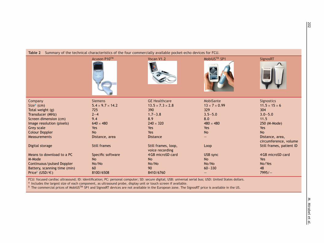
There are currently four pocket-echo machines avail-able on the market: Acuson P10TM (Siemens, Mountain View,CA, USA); Vscan V1.2 (GE Healthcare, Milwaukee, WI, USA);MobiUSTM SP1 (MobiSante, Redmond, WA, USA); and SignosRT
(Signostics, Thebarton, Australia). Characteristics and capa-bilities of these devices are summarized in Table 2. To date,all published studies have tested either Acuson P10TM oria
Figure 3. Images acquired in patients referred for a murmur in the outpand (B) normal FCU with no valve disease on colour Doppler. FCU: focuse
tal view — showing (A) normal inferior vena cava diameter (arrow)
scan V1.2 machines. Briefly, both devices are switched ony lifting the hinged display and include depth and gain con-rols. The Acuson P10 has tissue greyscale harmonic imagingut provides no colour Doppler. The Vscan V1.2 device has
dual probe that enables cardiac imaging and vascularoppler.
Although pocket-echocardiography cannot yield the samemage quality as more sophisticated machines, it has beenssessed as ‘‘good’’ in most patients [13,17,47]. Left heart
atient’s clinic showing (A) pathological mitral regurgitation (arrow)d cardiac ultrasound.
202
M.
Mirabel
et al.
Table 2 Summary of the technical characteristics of the four commercially available pocket-echo devices for FCU.
Acuson P10TM Vscan V1.2 MobiUSTM SP1 SignosRT
Company Siemens GE Healthcare MobiSante SignosticsSizea (cm) 5.4 × 9.7 × 14.2 13.5 × 7.3 × 2.8 13 × 7 × 0.99 11.5 × 15 × 6Total weight (g) 725 390 329 304Transducer (MHz) 2—4 1.7—3.8 3.5—5.0 3.0—5.0Screen dimension (cm) 9.4 8.9 8.0 11.5Image resolution (pixels) 640 × 480 240 × 320 480 × 480 250 (M-Mode)Grey scale Yes Yes Yes YesColour Doppler No Yes No NoMeasurements Distance, area Distance — Distance, area,
circumference, volumeDigital storage Still frames Still frames, loop,
voice recordingLoop Still frames, patient ID
Means to download to a PC Specific software 4 GB microSD card USB sync 4 GB microSD cardM-Mode No No No YesContinuous/pulsed Doppler No/No No/No No/No No/YesBattery, scanning time (min) 60 90 60—330 48Priceb (USD/D ) 8100/6508 8410/6760 — 7995/—
FCU: focused cardiac ultrasound; ID: identification; PC: personal computer; SD: secure digital; USB: universal serial bus; USD: United States dollars.a Includes the largest size of each component, as ultrasound probe, display unit or touch screen if available.b The commercial prices of MobiUSTM SP1 and SignosRT devices are not available in the European zone. The SignosRT price is available in the US.
R
[
[
[
[
[
[
[
[
Pocket-sized FCU: Strengths and limitations
dimensions measured with Acuson P10TM and Vscan V1.2devices are accurate when compared to high-end echocar-diography performed by experienced cardiologists [13,48].Exportation of images to a full-size screen does not improvethe accuracy of measurements or diagnosis, and real-timeinterpretation increases efficiency [16]. There has been nopeer-reviewed published evidence to date assessing theimage quality of MobiUSTM SP1 or SignosRT (according to asystematic search of PubMed and Embase).
Advantages, limitations and perspectivesof FCU by pocket-echocardiography
Pocket-echocardiography is convenient for daily practice.It is readily available and protocols of image acquisitiononly take 2—5 min [3,8,11,49]. Pocket-echocardiographyallows for immediate diagnosis as the same person acquiresand interprets the images. The systematic use of pocket-echocardiography for FCU may be cost effective [50]; andthe workflow of sonographers and cardiologists could beimproved by the use of pocket-echocardiography [18,51].Use of FCU may also reduce length of stay in patients admit-ted for heart failure [52]. However, there are two mainissues that have limited the widespread use of pocket-echocardiography. Firstly, the lack of reimbursement bysocial security systems in different countries may haveplayed a role. Secondly, legal responsibility of FCU hasnot been clearly defined, as FCU is not 100% accurate andwill not replace comprehensive echocardiography nor will itdetect incidental abnormalities that could impact on patientprognosis. Setting training requirements and certification inFCU would probably — at least partly — address these issues.
Pocket-echocardiography may also emerge as a potentialscreening tool for population-based surveillance pro-grammes or in specific target groups [53]. Rheumatic heartdisease is a potentially preventable disease in low-incomecountries and echocardiography based active surveillancehas emerged as a potential alternative to cardiac ausculta-tion [54—58]. Also, systematic screening for hypertrophiccardiomyopathy (Fig. 1C) in young athletes by FCU per-formed by sports medicine physicians may be an attractivesolution for the possible prevention of sudden death [59].
Conclusions
FCU is an emerging field that may be enhanced by the use ofpocket-echocardiography. There is now sufficient evidenceto support its use by experts, provided the questions arecircumscribed and within the technical capacities of thedevice. Non-experts may also accurately answer a numberof simple questions that may impact on patient care. Aware-ness of the technical limitations of these devices, validationof standardized training schemes, evaluation of accuracy ofdiagnoses and cost-effectiveness are pivotal issues in deter-mining the future of FCU by pocket-echocardiography.
Disclosure of interest
The authors declare that they have no conflicts of interestconcerning this article.
[
203
eferences[1] Moore CL, Copel JA. Point-of-care ultrasonography. New Engl J
Med 2011;364:749—57.[2] Kimura BJ, Demaria AN. Empowering physical examination:
the ‘‘laying on’’ of ultrasound. JACC Cardiovasc Imaging2008;1:602—4.
[3] Cardim N, Fernandez Golfin C, Ferreira D, et al. Usefulness of anew miniaturized echocardiographic system in outpatient car-diology consultations as an extension of physical examination.J Am Soc Echocardiogr 2011;24:117—24.
[4] Galderisi M, Santoro A, Versiero M, et al. Improved cardio-vascular diagnostic accuracy by pocket-size imaging devicein non-cardiologic outpatients: the NaUSiCa (Naples Ultra-sound Stethoscope in Cardiology) study. Cardiovasc Ultrasound2010;8:51.
[5] Mjolstad OC, Dalen H, Graven T, et al. Routinely addingultrasound examinations by pocket-sized ultrasound devicesimproves inpatient diagnostics in a medical department. EurJ Intern Med 2012;23:185—91.
[6] Panoulas VF, Daigeler AL, Malaweera AS, et al. Pocket-sizehand-held cardiac ultrasound as an adjunct to clinical exami-nation in the hands of medical students and junior doctors. EurHeart J Cardiovasc Imaging 2013;14:323—30.
[7] Shmueli H, Burstein Y, Sagy I, et al. Briefly trained medi-cal students can effectively identify rheumatic mitral valveinjury using a hand-carried ultrasound. Echocardiography2013;30:621—6.
[8] Skjetne K, Graven T, Haugen BO, et al. Diagnostic influence ofcardiovascular screening by pocket-size ultrasound in a cardiacunit. Eur J Echocardiogr 2011;12:737—43.
[9] Spencer KT, Kimura BJ, Korcarz CE, et al. Focused cardiacultrasound: recommendations from the American Society ofEchocardiography. J Am Soc Echocardiogr 2013;26:567—81.
10] Andersen GN, Haugen BO, Graven T, et al. Feasibility and reli-ability of point-of-care pocket-sized echocardiography. Eur JEchocardiogr 2011;12:665—70.
11] Biais M, Carrie C, Delaunay F, et al. Evaluation of a new pocket-echoscopic device for focused cardiac ultrasonography in anemergency setting. Crit Care 2012;16:R82.
12] Razi R, Estrada JR, Doll J, Spencer KT. Bedside hand-carriedultrasound by internal medicine residents versus traditionalclinical assessment for the identification of systolic dysfunc-tion in patients admitted with decompensated heart failure. JAm Soc Echocardiogr 2011;24:1319—24.
13] Prinz C, Dohrmann J, van Buuren F, et al. Diagnostic per-formance of hand-held echocardiography for the assessmentof basic cardiac morphology and function: a validationstudy in routine cardiac patients. Echocardiography 2012;29:887—94.
14] Mjolstad OC, Andersen GN, Dalen H, et al. Feasibility andreliability of point-of-care pocket-size echocardiography per-formed by medical residents. Eur Heart J Cardiovasc Imaging2013;14:1195—202.
15] Liebo MJ, Israel RL, Lillie EO, et al. Is pocket mobile echocar-diography the next-generation stethoscope? A cross-sectionalcomparison of rapidly acquired images with standard transtho-racic echocardiography. Ann Intern Med 2011;155:33—8.
16] Testuz A, Muller H, Keller PF, et al. Diagnostic accuracy ofpocket-size hand-held echocardiographs used by cardiologistsin the acute care setting. Eur Heart J Cardiovasc Imaging2013;14:38—42.
17] Prinz C, Voigt JU. Diagnostic accuracy of a hand-held ultrasoundscanner in routine patients referred for echocardiography. J Am
Soc Echocardiogr 2011;24:111—6.18] Reant P, Dijos M, Arsac F, et al. Validation of a new bedsideechoscopic heart examination resulting in an improvement inecho-lab workflow. Arch Cardiovasc Dis 2011;104:171—7.
2
[
[
[
[
[
[
[
[
[
[
[
[
[
[
[
[
[
[
[
[
[
[
[
[
[
[
[
[
[
[
[
[
[
[
[
[
[
04
19] Giusca S, Jurcut R, Ticulescu R, et al. Accuracy of hand-held echocardiography for bedside diagnostic evaluation in atertiary cardiology center: comparison with standard echocar-diography. Echocardiography 2011;28:136—41.
20] Penicka M, Vanderheyden M, Bartunek J. Diagnosis of heart fail-ure with preserved ejection fraction: role of clinical Dopplerechocardiography. Heart 2014;100:68—76.
21] Gargani L. Lung ultrasound: a new tool for the cardiologist.Cardiovasc Ultrasound 2011;9:6.
22] Mai TV, Shaw DJ, Amundson SA, Agan DL, Kimura BJ. Learningto apply the pocket ultrasound device on the critically ill: com-paring six ‘‘quick-look’’ signs for quality and prognostic valuesduring initial use by novices. Crit Care 2013;17:448.
23] Volpicelli G, Elbarbary M, Blaivas M, et al. Internationalevidence-based recommendations for point-of-care lung ultra-sound. Intensive Care Med 2012;38:577—91.
24] Goonewardena SN, Gemignani A, Ronan A, et al. Compar-ison of hand-carried ultrasound assessment of the inferiorvena cava and N-terminal pro-brain natriuretic peptide forpredicting readmission after hospitalization for acute decom-pensated heart failure. JACC Cardiovasc Imaging 2008;1:595—601.
25] Yavasi O, Unluer EE, Kayayurt K, et al. Monitoring the responseto treatment of acute heart failure patients by ultrasono-graphic inferior vena cava collapsibility index. Am J Emerg Med2014;32:403—7.
26] Tchernodrinski S, Lucas BP, Athavale A, et al. Inferior venacava diameter change after intravenous furosemide in patientsdiagnosed with acute decompensated heart failure. J ClinUltrasound 2014, http://dx.doi.org/10.1002/jcu.22173 [Epubahead of print].
27] Lafitte S, Alimazighi N, Reant P, et al. Validation of the small-est pocket-echoscopic device’s diagnostic capabilities in heartinvestigation. Ultrasound Med Biol 2011;37:798—804.
28] Gianstefani S, Catibog N, Whittaker AR, et al. Pocket-size imag-ing device: effectiveness for ward-based transthoracic studies.Eur Heart J Cardiovasc Imaging 2013;14:1132—9.
29] Arden C, Chambers JB, Sandoe J, et al. Can we improve thedetection of heart valve disease? Heart 2014;100:271—3.
30] Abe Y, Ito M, Tanaka C, et al. A novel and simple method usingpocket-sized echocardiography to screen for aortic stenosis. JAm Soc Echocardiogr 2013;26:589—96.
31] Mansencal N, Vieillard-Baron A, Beauchet A, et al. Triagepatients with suspected pulmonary embolism in the emergencydepartment using a portable ultrasound device. Echocardiog-raphy 2008;25:451—6.
32] Dijos M, Pucheux Y, Lafitte M, et al. Fast track echo of abdom-inal aortic aneurysm using a real pocket ultrasound device atbedside. Echocardiography 2012;29:285—90.
33] Bonnafy T, Lacroix P, Desormais I, et al. Reliability of the mea-surement of the abdominal aortic diameter by novice operatorsusing a pocket-sized ultrasound system. Arch Cardiovasc Dis2013;106:644—50.
34] Haji DL, Royse A, Royse CF. Review article: clinical impact ofnon-cardiologist-performed transthoracic echocardiography inemergency medicine, intensive care medicine and anaesthesia.Emerg Med Australas 2013;25:4—12.
35] Kimura BJ, Yogo N, O’Connell CW, et al. Cardiopulmonarylimited ultrasound examination for ‘‘quick-look’’ bedsideapplication. Am J Cardiol 2011;108:586—90.
36] Vignon P, Dugard A, Abraham J, et al. Focused training forgoal-oriented hand-held echocardiography performed by non-cardiologist residents in the intensive care unit. Intensive CareMed 2007;33:1795—9.
37] Sicari R, Galderisi M, Voigt JU, et al. The use of pocket-size imaging devices: a position statement of the EuropeanAssociation of Echocardiography. Eur J Echocardiogr 2011;12:85—7.
[
M. Mirabel et al.
38] Neskovic AN, Hagendorff A, Lancellotti P, et al. Emergencyechocardiography: the European Association of Cardiovascu-lar Imaging recommendations. Eur Heart J Cardiovasc Imaging2013;14:1—11.
39] Stokke TM, Ruddox V, Sarvari SI, et al. Brief group training ofmedical students in focused cardiac ultrasound may improvediagnostic accuracy of physical examination. J Am Soc Echocar-diogr 2014;27:1238—46.
40] Charron C, Templier F, Goddet NS, Baer M, Vieillard-Baron A.Difficulties encountered by physicians in interpreting focusedechocardiography using a pocket ultrasound machine in pre-hospital emergencies. Eur J Emerg Med 2015;22:17—22.
41] Prinz C, Dohrmann J, van Buuren F, et al. The impor-tance of training in echocardiography: a validation studyusing pocket-echocardiography. J Cardiovasc Med (Hager-stown) 2012;13:700—7.
42] Jensen MB, Sloth E, Larsen KM, Schmidt MB. Transthoracicechocardiography for cardiopulmonary monitoring in intensivecare. Eur J Anaesthesiol 2004;21:700—7.
43] Pelliccia F, Palmiero P, Maiello M, Losi MA. Italian Chap-ter of the International Society of Cardiovascular U. Italianchapter of the International Society of cardiovascular ultra-sound expert consensus document on training requirementsfor non-cardiologists using hand-carried ultrasound devices.Echocardiography 2012;29:745—50.
44] Beraud AS, Rizk NW, Pearl RG, Liang DH, Patterson AJ. Focusedtransthoracic echocardiography during critical care medicinetraining: curriculum implementation and evaluation of profi-ciency*. Crit Care Med 2013;41:e179—E181.
45] Solomon SD, Saldana F. Point-of-care ultrasound in medi-cal education — stop listening and look. N Engl J Med2014;370:1083—5.
46] Neskovic AN, Edvardsen T, Galderisi M, et al. Focus car-diac ultrasound: the European Association of CardiovascularImaging viewpoint. Eur Heart J Cardiovasc Imaging 2014;15:956—60.
47] Culp BC, Mock JD, Chiles CD, Culp Jr WC. The pocket-echocardiograph: validation and feasibility. Echocardiography2010;27:759—64.
48] Fukuda S, Shimada K, Kawasaki T, et al. Pocket-sized transtho-racic echocardiography device for the measurement of cardiacchamber size and function. Circ J 2009;73:1092—6.
49] Frederiksen CA, Juhl-Olsen P, Larsen UT, et al. New pocket-echocardiography device is interchangeable with high-endportable system when performed by experienced examiners.Acta Anaesthesiol Scand 2010;54:1217—23.
50] Kitada R, Fukuda S, Watanabe H, et al. Diagnostic accuracyand cost-effectiveness of a pocket-sized transthoracic echo-cardiographic imaging device. Clin Cardiol 2013;36:603—10.
51] Badano LP, Nucifora G, Stacul S, et al. Improved workflow,sonographer productivity, and cost-effectiveness of echocardi-ographic service for inpatients by using miniaturized systems.Eur J Echocardiogr 2009;10:537—42.
52] Lucas BP, Candotti C, Margeta B, et al. Hand-carried echocar-diography by hospitalists: a randomized trial. Am J Med2011;124:766—74.
53] Celermajer DS, Chow CK, Marijon E, Anstey NM, Woo KS.Cardiovascular disease in the developing world: prevalences,patterns, and the potential of early disease detection. J AmColl Cardiol 2012;60:1207—16.
54] Marijon E, Mirabel M, Celermajer DS, Jouven X. Rheumaticheart disease. Lancet 2012;379:953—64.
55] Marijon E, Ou P, Celermajer DS, et al. Prevalence of rheumaticheart disease detected by echocardiographic screening. NewEngl J Med 2007;357:470—6.
56] Beaton A, Aliku T, Okello E, et al. The utility of hand-heldechocardiography for early diagnosis of rheumatic heart dis-ease. J Am Soc Echocardiogr 2014;27(1):42—9.

Pocket-sized FCU: Strengths and limitations
[57] Mirabel M, Bacquelin R, Tafflet M, et al. Screening forrheumatic heart disease: evaluation of a focused cardiac ultra-
sound approach. Circ Cardiovasc Imaging 2015;8(1), http://dx.doi.org/10.1161/CIRCIMAGING.114.002324 [pii: e002324].[58] Beaton A, Lu JC, Aliku T, et al. The utility of hand-held echocar-diography for early rheumatic heart disease diagnosis: a field
[
205
study. Eur Heart J Cardiovasc Imaging 2015 [pii: jeu296 (Epubahead of print)].
59] Yim ES, Gillis EF, Ojala K, et al. Focused transthoracicechocardiography by sports medicine physicians: measure-ments relevant to hypertrophic cardiomyopathy. J UltrasoundMed 2013;32:333—8.
Related Documents