DISEASES OF AQUATIC ORGANISMS Dis Aquat Org Vol. 67: 93–104, 2005 Published November 9 INTRODUCTION Flavobacterium psychrophilum is the causative agent of bacterial cold water disease (CWD) and has been recognized as a serious problem in freshwater aquaculture worldwide, particularly for salmonid fishes (Nematollahi et al. 2003). In Japan, CWD in ayu Plecoglossus altivelis has been occurring since 1987 (Wakabayashi et al. 1994). Ayu are highly prized in Japan not only as a delicacy but also as a game fish. According to the statistics by the Ministry of Agricul- ture, Forestry and Fisheries, the amount of money yielded by ayu fisheries and farming comprises one- third of the total value of the production of freshwater fisheries in Japan. Ayu are osmerid fish having a life span of only 1 yr. Many are amphidromous, but there are also some landlocked populations. Many rivers are stocked extensively with juvenile ayu every year for recreational angling. The landlocked population of Lake Biwa had been the single dominant source of juvenile ayu for stocking and farming until the mid 1990s. Therefore, once the ayu in Lake Biwa were infested with F. psychrophilum, the disease spread rapidly throughout Japan, although it is not known when the bacteria entered the lake. Since then, CWD has been a serious problem in fishing and farming of ayu. CWD of ayu has some different characteristics from those of CWD in salmonid fishes. Local fisheries research stations throughout Japan report that the dis- ease generally occurs in early summer when water temperature is 16 to 18°C. This is in contrast with © Inter-Research 2005 · www.int-res.com *Email: [email protected] Pathogenesis of experimentally induced bacterial cold water disease in ayu Plecoglossus altivelis Satoshi Miwa*, Chihaya Nakayasu Inland Station, National Research Institute of Aquaculture, Fisheries Research Agency, Tamaki, Mie 519-0423, Japan ABSTRACT: Ayu Plecoglossus altivelis were experimentally infected with Flavobacterium psy- chrophilum, which is the causative agent of bacterial cold water disease (CWD). The fish infected by immersion usually died within an hour after they became moribund. The blood volume and haema- tocrit values of moribund fish were low, while those values of many infected fish that were not mori- bund were in the range of controls. Most of the affected fish in the immersion-infected groups had ulcerative lesions on their lower jaw. No histological evidence of haemolysis was observed. These results suggest that rapid bleeding occurred through ulcerative lesions, probably causing hypoxia which killed the fish. Ulcerative lesions developed on the dorsal skin when this area had been slightly abraded artificially prior to immersion challenge. Histologically, F. psychrophilum was initially found on the skin that had microscopic injuries, but not on normal skin. The bacterium then entered the der- mis and migrated through connective tissues. The lesions subsequently expanded into the underlying musculature through the myosepta, developed necrotic myositis and formed externally open ulcers. Only in later stages of infection did mild lesions develop in the internal organs and the gill, probably caused by the bacterium migrating through blood vessels. This suggests that infection with CWD through the gill or digestive tract is unlikely. Virtually no open lesions were found in ayu challenged by intramuscular injections except at the injection sites. The results suggest that skin injuries are major portals of entry for F. psychrophilum in ayu, and the bacterium has affinity for collagenous connective tissues. KEY WORDS: Cold water disease · Flavobacterium psychrophilum · Pathogenesis · Ayu · Plecoglossus altivelis Resale or republication not permitted without written consent of the publisher

Welcome message from author
This document is posted to help you gain knowledge. Please leave a comment to let me know what you think about it! Share it to your friends and learn new things together.
Transcript
DISEASES OF AQUATIC ORGANISMSDis Aquat Org
Vol. 67: 93–104, 2005 Published November 9
INTRODUCTION
Flavobacterium psychrophilum is the causativeagent of bacterial cold water disease (CWD) and hasbeen recognized as a serious problem in freshwateraquaculture worldwide, particularly for salmonidfishes (Nematollahi et al. 2003). In Japan, CWD in ayuPlecoglossus altivelis has been occurring since 1987(Wakabayashi et al. 1994). Ayu are highly prized inJapan not only as a delicacy but also as a game fish.According to the statistics by the Ministry of Agricul-ture, Forestry and Fisheries, the amount of moneyyielded by ayu fisheries and farming comprises one-third of the total value of the production of freshwaterfisheries in Japan. Ayu are osmerid fish having a lifespan of only 1 yr. Many are amphidromous, but there
are also some landlocked populations. Many rivers arestocked extensively with juvenile ayu every year forrecreational angling. The landlocked population ofLake Biwa had been the single dominant source ofjuvenile ayu for stocking and farming until the mid1990s. Therefore, once the ayu in Lake Biwa wereinfested with F. psychrophilum, the disease spreadrapidly throughout Japan, although it is not knownwhen the bacteria entered the lake. Since then, CWDhas been a serious problem in fishing and farmingof ayu.
CWD of ayu has some different characteristics fromthose of CWD in salmonid fishes. Local fisheriesresearch stations throughout Japan report that the dis-ease generally occurs in early summer when watertemperature is 16 to 18°C. This is in contrast with
© Inter-Research 2005 · www.int-res.com*Email: [email protected]
Pathogenesis of experimentally induced bacterialcold water disease in ayu Plecoglossus altivelis
Satoshi Miwa*, Chihaya Nakayasu
Inland Station, National Research Institute of Aquaculture, Fisheries Research Agency, Tamaki, Mie 519-0423, Japan
ABSTRACT: Ayu Plecoglossus altivelis were experimentally infected with Flavobacterium psy-chrophilum, which is the causative agent of bacterial cold water disease (CWD). The fish infected byimmersion usually died within an hour after they became moribund. The blood volume and haema-tocrit values of moribund fish were low, while those values of many infected fish that were not mori-bund were in the range of controls. Most of the affected fish in the immersion-infected groups hadulcerative lesions on their lower jaw. No histological evidence of haemolysis was observed. Theseresults suggest that rapid bleeding occurred through ulcerative lesions, probably causing hypoxiawhich killed the fish. Ulcerative lesions developed on the dorsal skin when this area had been slightlyabraded artificially prior to immersion challenge. Histologically, F. psychrophilum was initially foundon the skin that had microscopic injuries, but not on normal skin. The bacterium then entered the der-mis and migrated through connective tissues. The lesions subsequently expanded into the underlyingmusculature through the myosepta, developed necrotic myositis and formed externally open ulcers.Only in later stages of infection did mild lesions develop in the internal organs and the gill, probablycaused by the bacterium migrating through blood vessels. This suggests that infection with CWDthrough the gill or digestive tract is unlikely. Virtually no open lesions were found in ayu challengedby intramuscular injections except at the injection sites. The results suggest that skin injuries aremajor portals of entry for F. psychrophilum in ayu, and the bacterium has affinity for collagenousconnective tissues.
KEY WORDS: Cold water disease · Flavobacterium psychrophilum · Pathogenesis · Ayu · Plecoglossus altivelis
Resale or republication not permitted without written consent of the publisher
Dis Aquat Org 67: 93–104, 2005
salmonids in which the disease occurs most frequentlywhen the water temperature is between 4 and 10°C(Shotts & Starliper 1999). The most notable clinical signof CWD in ayu is anaemia as revealed by pale gills(Iida & Mizokami 1996). Ulcerative lesions are oftenfound on the caudal peduncle of affected wild ayu,including stocked fish (Iida & Mizokami 1996). How-ever, when CWD occurs in cultured ayu, ulcerativelesions on the surface of the body trunk are often notobserved, although haemorrhagic lesions or ulcersdevelop on the lower jaw of many of the affected fish.The ulcerative lesions found on the caudal peduncle ofaffected wild ayu might develop following infection bythe bacteria into skin wounds caused by fishing (snag-ging) or by territorial aggression by other ayu. Erosionof fins (fin rot) or tail loss, which are typical clinicalsigns for CWD in salmonids (Nematollahi et al. 2003),are not observed in ayu. Furthermore, the F. psy-chrophilum strain(s) that causes CWD in ayu is distinctserologically and genetically from those in other fishspecies including salmonids (Izumi & Wakabayashi1999, Izumi et al. 2003, Tabata 2004). However, thepathogenesis of the disease in ayu has not beenstudied so far.
In the present study, we infected cultured ayu with F.psychrophilum experimentally, recorded clinical signsand confirmed the development of anaemia by mea-suring haematocrit and blood volumes. In a secondexperiment, we produced the ulcerative lesions on thedorsal skin by making a slight abrasion prior to immer-sion infection. The progression of the disease was thenstudied histopathologically. In a third experiment, toprove that the lesions on the body surface were causedby the bacteria attaching to the skin directly from theoutside of the body, we infected fish with either immer-sion or intramuscular injection and confirmed thatthe lesions on the body surface do not develop in theinjection-challenged fish.
MATERIALS AND METHODS
Fish and experimental conditions. Juvenile ayuwere obtained from a private hatchery (Nisshin MarineTech), where no CWD incidence had occurred, andtransported to the Inland Station of National ResearchInstitute of Aquaculture (NRIA; Tamaki, Mie prefec-ture, Japan). The first batch of 1000 fish was obtainedin February 2003 and their average body weight was1.1 g. The second batch of 500 fish was obtained inApril 2004 and the average body weight was 1.0 g. Thefish were fed commercial diet and reared in a 500 ltank, aerated and supplied with running freshwater at15 to 17°C until they grew to the size at which theexperiments were possible (>2 g). After transfer to the
NRIA, ayu were reared for 104 d before Expt 1, for 77 dbefore Expt 2, and for 100 d before Expt 3. In all theexperimental infections in the present study, the fishwere kept in round tanks that were 34 cm in diameter.Each tank contained about 15 l of aerated, runningfresh water at 16 to 17°C during the experiments.
Bacteria. The Flavobacterium psychrophilum strainFPSG011227 was obtained from Shiga PrefecturalFisheries Research Station and used for all experimen-tal infections. This strain was originally isolated fromayu in Shiga prefecture and the identity was confirmedby PCR for small subunit ribosomal RNA (Toyama et al.1994). The genotype of the bacterium was B-S (Izumiet al. 2003). The bacterium was cultured with modifiedcytophaga (MCY) broth (Wakabayashi & Egusa 1974;0.2% tryptone, 0.05% yeast extract, 0.02% beefextract, 0.02% sodium acetate, 0.02% sodium chlo-ride) at 15°C, centrifuged at 15 000 × g for 10 min, andthe supernatant was replaced with fresh broth. Glyc-erol was then added to the culture at 10%, and 500 µlaliquots were stored at –80°C. For each experimentalinfection, an aliquot of the stored bacteria was newlythawed, and 10 µl of the aliquot was mixed in 3 ml ofMCY broth and cultured for 24 h at 15°C with shaking.The culture was then added to 1 l of MCY broth andfurther cultured for 24 h at 15°C with shaking and thenused for the challenges. The number of bacteria wasestimated from the broth before dilution into theimmersion bath by counting colonies on MCY agar(MCY broth + 1.2% agar) plates inoculated with10-fold serial dilutions.
Expt 1. For the bacterial challenge, 80 fish were putinto a bucket containing 9 l of aerated water to whichthe culture of Flavobacterium psychrophilum in 1 l ofMCY broth was added. The estimated number of bac-teria in the challenge bath was 1.6 × 10⁸ CFU (colonyforming units) ml–1. The fish were thus exposed to thebacterium for 2 h and subsequently divided into 4experimental tanks, each of which contained 20 fish.For the control, 40 fish were immersed in water con-taining MCY broth at 10% without F. psychrophilumfor 2 h. These were divided into 2 experimental tankscontaining each 20 fish. Thus, a total of 120 fish wereused for this experiment. The body weight of the fishused in this experiment was 3.30 ± 0.74 g (average andstandard deviation). Fish in both groups were rearedby feeding commercial diet ad libitum. The fish in theexperimental tanks were observed at 1 h intervals dur-ing daytime every day, and moribund or lethargic fishwere sampled. The first moribund fish was sampled onDay 2, and the last one was sampled on Day 6. Thecaudal peduncle of the sampled fish was cut with ascalpel and as much blood as possible was immedi-ately withdrawn into heparinized capillaries. The cap-illaries were centrifuged for 5 min at 15 000 × g and the
94
Miwa & Nakayasu: Pathogenesis of cold water disease
volume of sampled blood and haematocrit values weremeasured. The external clinical signs were recordedfor all dead or moribund fish. All of the surviving fish inthe challenged groups and 10 fish from each of thecontrol groups were sampled on Day 6, and their bloodvolume and haematocrit values were measured simi-larly. The fish were anaesthetized with 2-phe-noxyethanol before blood sampling except for themoribund fish.
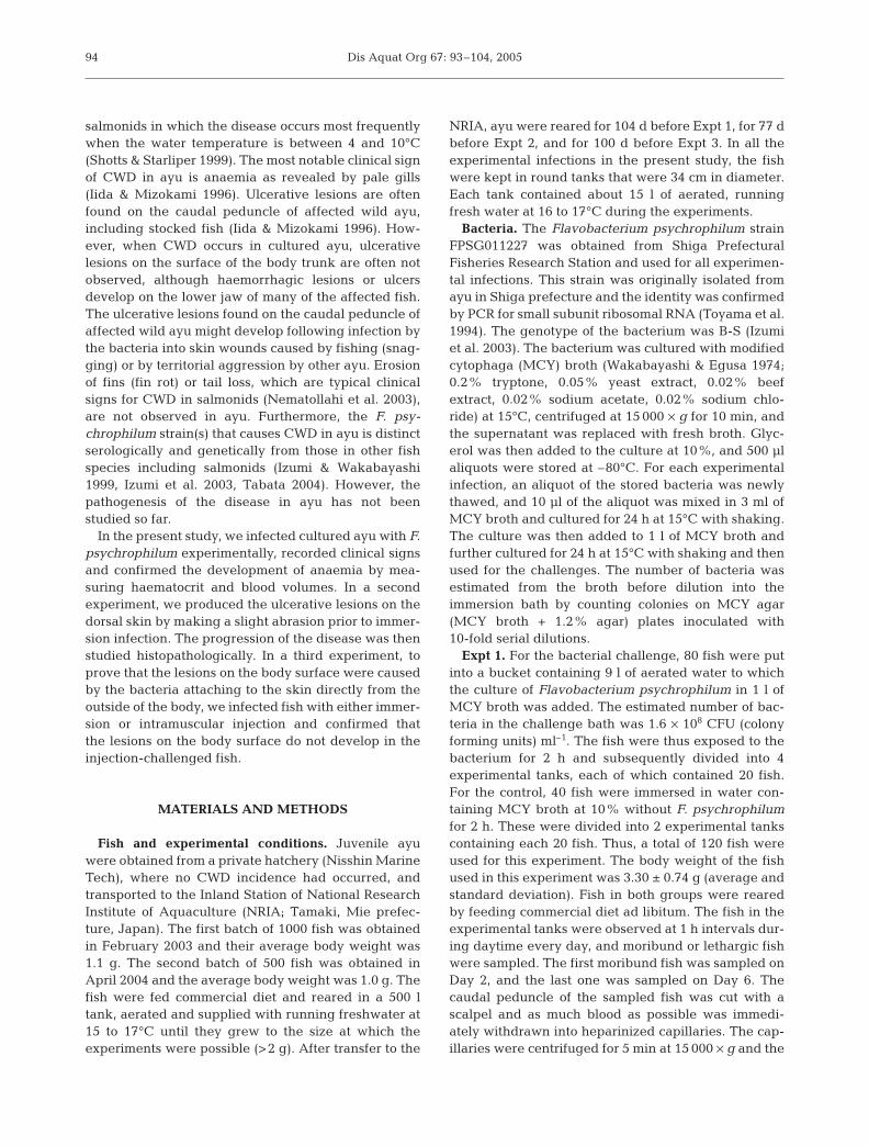
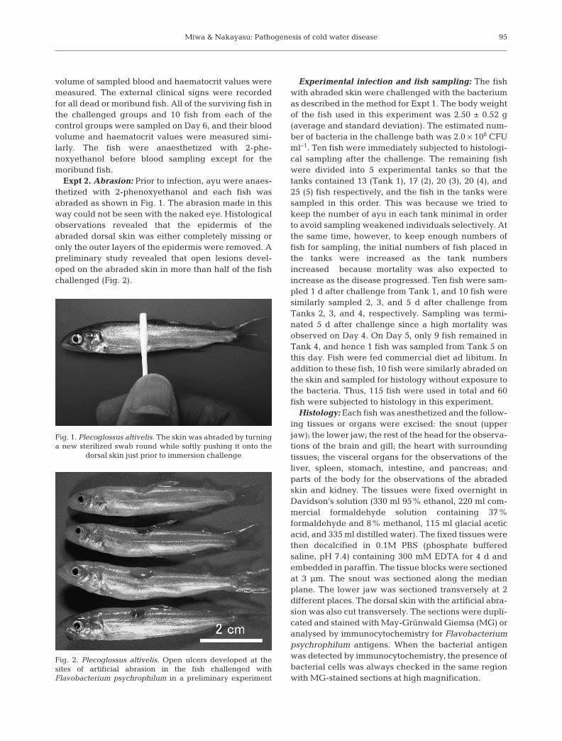
Expt 2. Abrasion: Prior to infection, ayu were anaes-thetized with 2-phenoxyethanol and each fish wasabraded as shown in Fig. 1. The abrasion made in thisway could not be seen with the naked eye. Histologicalobservations revealed that the epidermis of theabraded dorsal skin was either completely missing oronly the outer layers of the epidermis were removed. Apreliminary study revealed that open lesions devel-oped on the abraded skin in more than half of the fishchallenged (Fig. 2).
Experimental infection and fish sampling: The fishwith abraded skin were challenged with the bacteriumas described in the method for Expt 1. The body weightof the fish used in this experiment was 2.50 ± 0.52 g(average and standard deviation). The estimated num-ber of bacteria in the challenge bath was 2.0 × 10⁸ CFUml–1. Ten fish were immediately subjected to histologi-cal sampling after the challenge. The remaining fishwere divided into 5 experimental tanks so that thetanks contained 13 (Tank 1), 17 (2), 20 (3), 20 (4), and25 (5) fish respectively, and the fish in the tanks weresampled in this order. This was because we tried tokeep the number of ayu in each tank minimal in orderto avoid sampling weakened individuals selectively. Atthe same time, however, to keep enough numbers offish for sampling, the initial numbers of fish placed inthe tanks were increased as the tank numbersincreased because mortality was also expected toincrease as the disease progressed. Ten fish were sam-pled 1 d after challenge from Tank 1, and 10 fish weresimilarly sampled 2, 3, and 5 d after challenge fromTanks 2, 3, and 4, respectively. Sampling was termi-nated 5 d after challenge since a high mortality wasobserved on Day 4. On Day 5, only 9 fish remained inTank 4, and hence 1 fish was sampled from Tank 5 onthis day. Fish were fed commercial diet ad libitum. Inaddition to these fish, 10 fish were similarly abraded onthe skin and sampled for histology without exposure tothe bacteria. Thus, 115 fish were used in total and 60fish were subjected to histology in this experiment.
Histology: Each fish was anesthetized and the follow-ing tissues or organs were excised: the snout (upperjaw); the lower jaw; the rest of the head for the observa-tions of the brain and gill; the heart with surroundingtissues; the visceral organs for the observations of theliver, spleen, stomach, intestine, and pancreas; andparts of the body for the observations of the abradedskin and kidney. The tissues were fixed overnight inDavidson’s solution (330 ml 95% ethanol, 220 ml com-mercial formaldehyde solution containing 37%formaldehyde and 8% methanol, 115 ml glacial aceticacid, and 335 ml distilled water). The fixed tissues werethen decalcified in 0.1M PBS (phosphate bufferedsaline, pH 7.4) containing 300 mM EDTA for 4 d andembedded in paraffin. The tissue blocks were sectionedat 3 µm. The snout was sectioned along the medianplane. The lower jaw was sectioned transversely at 2different places. The dorsal skin with the artificial abra-sion was also cut transversely. The sections were dupli-cated and stained with May-Grünwald Giemsa (MG) oranalysed by immunocytochemistry for Flavobacteriumpsychrophilum antigens. When the bacterial antigenwas detected by immunocytochemistry, the presence ofbacterial cells was always checked in the same regionwith MG-stained sections at high magnification.
95
Fig. 1. Plecoglossus altivelis. The skin was abraded by turninga new sterilized swab round while softly pushing it onto the
dorsal skin just prior to immersion challenge
Fig. 2. Plecoglossus altivelis. Open ulcers developed at thesites of artificial abrasion in the fish challenged withFlavobacterium psychrophilum in a preliminary experiment
Dis Aquat Org 67: 93–104, 2005
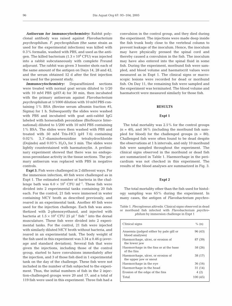
Antiserum for immunocytochemistry: Rabbit poly-clonal antibody was raised against Flavobacteriumpsychrophilum. F. psychrophilum (the same strain asused for the experimental infections) was killed with0.3% formalin, washed with PBS, and used as the anti-gen. The killed bacterium (1.3 × 10⁸ CFU) was injectedinto a rabbit subcutaneously with complete Freundadjuvant. The rabbit was given 3 booster shots each ofthe same amount of the antigen on Days 14, 28 and 42,and the serum obtained 52 d after the first injectionwas used for the present study.
Immunocytochemistry: Deparaffinized sectionswere treated with normal goat serum diluted to 1/20with 10 mM PBS (pH7.4) for 30 min, then incubatedwith the primary antiserum against Flavobacteriumpsychrophilum at 1/1000 dilution with 10 mM PBS con-taining 1% BSA (Bovine serum albumin fraction #5,Sigma) for 1 h. Subsequently the slides were washedwith PBS and incubated with goat anti-rabbit IgGlabeled with horseradish peroxidase (BioSource Inter-national) diluted to 1/200 with 10 mM PBS containing1% BSA. The slides were then washed with PBS andtreated with 50 mM Tris-HCl (pH 7.6) containing0.02% 3,3’-diaminobenzidine tetrahydrochloride(Dojindo) and 0.03% H2O2 for 5 min. The slides werelightly counterstained with haematoxylin. A prelimi-nary experiment showed that there was no endoge-nous peroxidase activity in the tissue sections. The pri-mary antiserum was replaced with PBS in negativecontrols.
Expt 3. Fish were challenged in 2 different ways. Forthe immersion infection, 40 fish were challenged as inExpt 1. The estimated number of bacteria in the chal-lenge bath was 6.0 × 10⁷ CFU ml–1. These fish weredivided into 2 experimental tanks containing 20 fisheach. For the control, 21 fish were immersed in watercontaining MCY broth as described previously, andreared in an experimental tank. Another 40 fish wereused for the injection challenge. Each fish was anes-thetized with 2-phenoxyethanol, and injected withbacteria at 1.5 × 10⁵ CFU 25 µl–1 fish–1 into the dorsalmusculature. These fish were divided into 2 experi-mental tanks. For the control, 21 fish were injectedwith similarly diluted MCY broth without bacteria, andreared in an experimental tank. The body weight ofthe fish used in this experiment was 3.34 ± 0.46 g (aver-age and standard deviation). Several fish that weregiven the injections, including those of the controlgroup, started to have convulsions immediately afterthe injection, and 3 of those fish died in 1 experimentaltank on the day of the challenge. These fish were notincluded in the number of fish subjected to the experi-ment. Thus, the initial numbers of fish in the 2 injec-tion-challenged groups were 20 and 17, and a total of119 fish were used in this experiment. Three fish had a
convulsion in the control group, and they died duringthe experiment. The injections were made deep insidethe fish trunk body close to the vertebral column toprevent leakage of the inoculum. Hence, the inoculummay have physically pressed the spinal cord andthereby caused a convulsion in the fish. The inoculummay have also entered into the spinal fluid in somefish. During the experiment, moribund fish were sam-pled, and blood volume and haematocrit values weremeasured as in Expt 1. The clinical signs or macro-scopic lesions were recorded for dead or moribundfish. On Day 11, the remaining fish were sampled andthe experiment was terminated. The blood volume andhaematocrit were measured similarly for those fish.
RESULTS
Expt 1
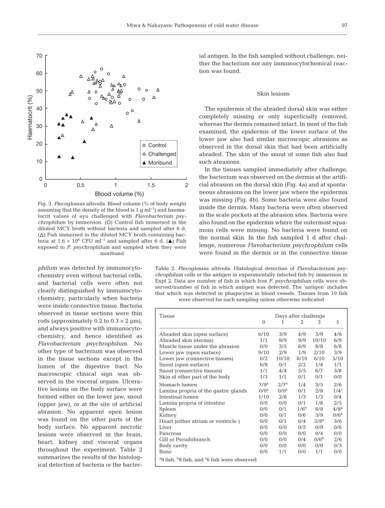
The total mortality was 2.5% for the control groups(n = 40), and 56% (including the moribund fish sam-pled for blood) for the challenged groups (n = 80).Challenged fish were often found to be dead despitethe observations at 1 h intervals, and only 10 moribundfish were sampled throughout the experiment. Theclinical signs observed in the moribund or dead fishare summarized in Table 1. Haemorrhage in the peri-cardium was not checked in this experiment. Theresults of the blood analyses are summarized in Fig. 3.
Expt 2
The total mortality other than the fish used for histol-ogy sampling was 65% during the experiment. Inmany cases, the antigen of Flavobacterium psychro-
96
Clinical signs % (n)
Anaemia (judged either by pale gill or 96 (43)blood analyses)
Haemorrhage, ulcer, or erosion of 87 (39)the lower jaw
Haemorrhage in the fins or at the base 58 (26)of the fins
Haemorrhage, ulcer, or erosion of 38 (17)the upper jaw or snout
Haemorrhage in the eye 36 (16)Haemorrhage in the head 31 (14)Erosion of the edge of the fins 4 (2)
Total 100 (45)
Table 1. Plecoglossus altivelis. Clinical signs observed in deador moribund fish infected with Flavobacterium psychro-
philum by immersion challenge in Expt 1
Miwa & Nakayasu: Pathogenesis of cold water disease
philum was detected by immunocyto-chemistry even without bacterial cells,and bacterial cells were often notclearly distinguished by immunocyto-chemistry, particularly when bacteriawere inside connective tissue. Bacteriaobserved in tissue sections were thinrods (approximately 0.2 to 0.3 × 2 µm),and always positive with immunocyto-chemistry, and hence identified asFlavobacterium psychrophilum. Noother type of bacterium was observedin the tissue sections except in thelumen of the digestive tract. Nomacroscopic clinical sign was ob-served in the visceral organs. Ulcera-tive lesions on the body surface wereformed either on the lower jaw, snout(upper jaw), or at the site of artificialabrasion. No apparent open lesionwas found on the other parts of thebody surface. No apparent necroticlesions were observed in the brain,heart, kidney and visceral organsthroughout the experiment. Table 2summarizes the results of the histolog-ical detection of bacteria or the bacter-
ial antigen. In the fish sampled without challenge, nei-ther the bacterium nor any immunocytochemical reac-tion was found.
Skin lesions
The epidermis of the abraded dorsal skin was eithercompletely missing or only superficially removed,whereas the dermis remained intact. In most of the fishexamined, the epidermis of the lower surface of thelower jaw also had similar microscopic abrasions asobserved in the dorsal skin that had been artificiallyabraded. The skin of the snout of some fish also hadsuch abrasions.
In the tissues sampled immediately after challenge,the bacterium was observed on the dermis at the artifi-cial abrasion on the dorsal skin (Fig. 4a) and at sponta-neous abrasions on the lower jaw where the epidermiswas missing (Fig. 4b). Some bacteria were also foundinside the dermis. Many bacteria were often observedin the scale pockets at the abrasion sites. Bacteria werealso found on the epidermis where the outermost squa-mous cells were missing. No bacteria were found onthe normal skin. In the fish sampled 1 d after chal-lenge, numerous Flavobacterium psychrophilum cellswere found in the dermis or in the connective tissue
97
Control
Challenged
Moribund
Blood volume (%)
Hae
mat
ocrit
(%)
0 0.5 1.51 2
70
60
50
40
30
20
10
0
Fig. 3. Plecoglossus altivelis. Blood volume (% of body weightassuming that the density of the blood is 1 g ml–1) and haema-tocrit values of ayu challenged with Flavobacterium psy-chrophilum by immersion. (s) Control fish immersed in thediluted MCY broth without bacteria and sampled after 6 d.(n) Fish immersed in the diluted MCY broth containing bac-teria at 1.6 × 10⁸ CFU ml–1 and sampled after 6 d. (m) Fish exposed to F. psychrophilum and sampled when they were
moribund
Tissue Days after challenge0 1 2 3 5
Abraded skin (open surface) 6/10 3/9 4/9 3/9 4/8Abraded skin (dermis) 1/1 9/9 9/9 10/10 6/9Muscle tissue under the abrasion 0/0 3/5 8/9 8/8 6/8Lower jaw (open surface) 9/10 2/9 1/9 2/10 3/9Lower jaw (connective tissues) 0/2 10/10 8/10 6/10 5/10Snout (open surface) 6/8 0/1 2/2 1/4 1/1Snout (connective tissues) 1/1 4/4 5/5 6/7 3/8Skin of other part of the body 1/3 1/1 0/1 0/1 0/0
Stomach lumen 7/9a 2/7a 1/4 3/5 2/6Lamina propria of the gastric glands 0/0a 0/0a 0/1 2/8 1/4c
Intestinal lumen 1/10 2/8 1/3 1/3 0/4Lamina propria of intestine 0/0 0/0 0/1 1/8 2/5Spleen 0/0 0/1 1/6b 8/8 4/8a
Kidney 0/0 0/1 0/6 3/9 0/6a
Heart (either atrium or ventricle ) 0/0 0/1 0/4 3/8a 3/6Liver 0/0 0/0 0/5 0/9 0/6Pancreas 0/0 0/0 0/0 0/4 0/0Gill or Pseudobranch 0/0 0/0 0/4 0/6b 2/6Body cavity 0/0 0/0 0/0 0/0 0/3Bone 0/0 1/1 0/0 1/1 0/0a9 fish, b8 fish, and c6 fish were observed
Table 2. Plecoglossus altivelis. Histological detection of Flavobacterium psy-chrophilum cells or the antigen in experimentally infected fish by immersion inExpt 2. Data are number of fish in which free F. psychrophilum cells were ob-served/number of fish in which antigen was detected. The ‘antigen’ includesthat which was detected in phagocytes in blood vessels. Tissues from 10 fish
were observed for each sampling unless otherwise indicated
Dis Aquat Org 67: 93–104, 200598
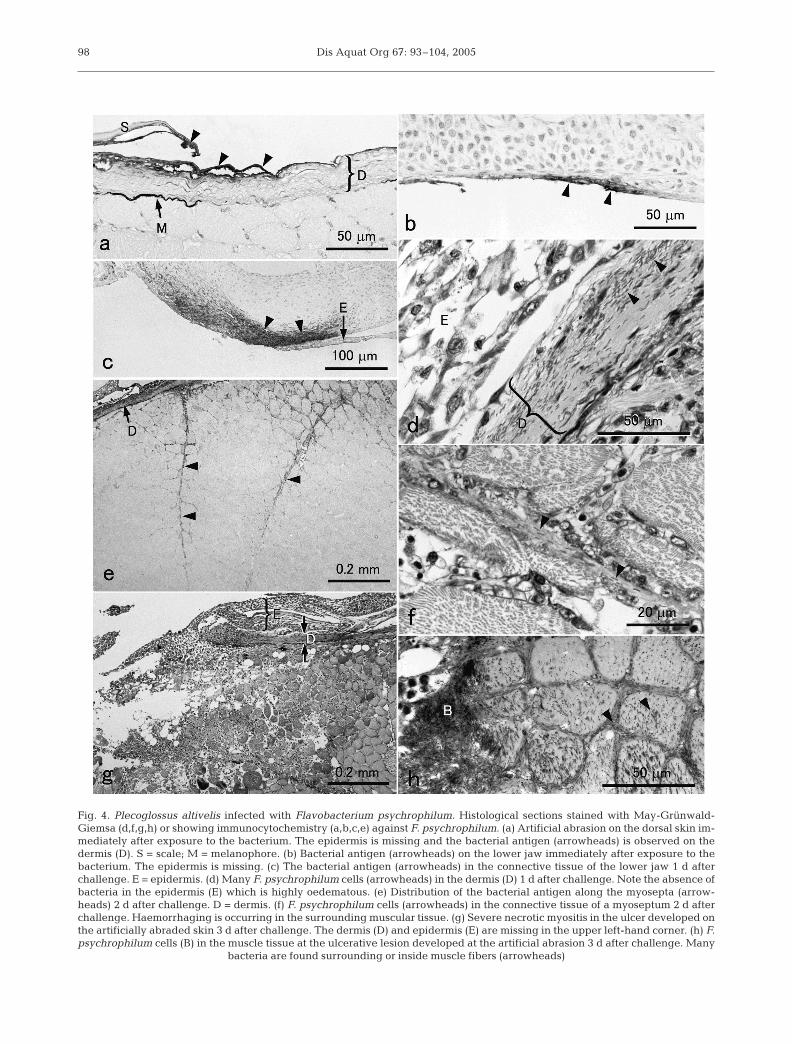
Fig. 4. Plecoglossus altivelis infected with Flavobacterium psychrophilum. Histological sections stained with May-Grünwald-Giemsa (d,f,g,h) or showing immunocytochemistry (a,b,c,e) against F. psychrophilum. (a) Artificial abrasion on the dorsal skin im-mediately after exposure to the bacterium. The epidermis is missing and the bacterial antigen (arrowheads) is observed on thedermis (D). S = scale; M = melanophore. (b) Bacterial antigen (arrowheads) on the lower jaw immediately after exposure to thebacterium. The epidermis is missing. (c) The bacterial antigen (arrowheads) in the connective tissue of the lower jaw 1 d afterchallenge. E = epidermis. (d) Many F. psychrophilum cells (arrowheads) in the dermis (D) 1 d after challenge. Note the absence ofbacteria in the epidermis (E) which is highly oedematous. (e) Distribution of the bacterial antigen along the myosepta (arrow-heads) 2 d after challenge. D = dermis. (f) F. psychrophilum cells (arrowheads) in the connective tissue of a myoseptum 2 d afterchallenge. Haemorrhaging is occurring in the surrounding muscular tissue. (g) Severe necrotic myositis in the ulcer developed onthe artificially abraded skin 3 d after challenge. The dermis (D) and epidermis (E) are missing in the upper left-hand corner. (h) F.psychrophilum cells (B) in the muscle tissue at the ulcerative lesion developed at the artificial abrasion 3 d after challenge. Many
bacteria are found surrounding or inside muscle fibers (arrowheads)
Miwa & Nakayasu: Pathogenesis of cold water disease
under the epidermis (Fig. 4c,d). Inflammatory cellsoften accumulated around the infection sites, althoughin most of the cases the bacterial antigen was notobserved in those cells.
On Day 2, lesions such as erosion of a small area ofthe skin, haemorrhage, or slight bulging wereobserved at the sites of artificial abrasion in 7 fish. Thedermis of the infection sites was often swollen, con-taining numerous bacteria, whereas virtually no freebacteria were observed in the epidermis, although theepidermis at the lesions was always oedematous. Bac-teria were also found in the muscle layer under theabraded skin, particularly along the myosepta(Fig. 4e,f). Numerous inflammatory cells accumulatedat the infection sites where haemorrhage was oftenobserved. The bacterial antigen or bacterial cells wereoften found in many of these inflammatory cells whichwere presumably phagocytes such as neutrophils andmacrophages.
On Day 3, necrotizing myositis was often observed inthe centre of the lesion (Fig. 4g). In some of the lesions,large numbers of bacteria were seen surrounding orinside the muscle fibres (Fig. 4h). In severe cases, thedermis and myosepta were virtually replaced by bac-terial masses and the dermis in the centre of the lesionswas often lost. Extensive haemorrhage was alsoobserved in these lesions. The bacterial invasion andinflammation also proceeded deeply into the connec-tive tissues of the lower jaw or the snout of several fishand the tissues were often sloughed in these lesions.
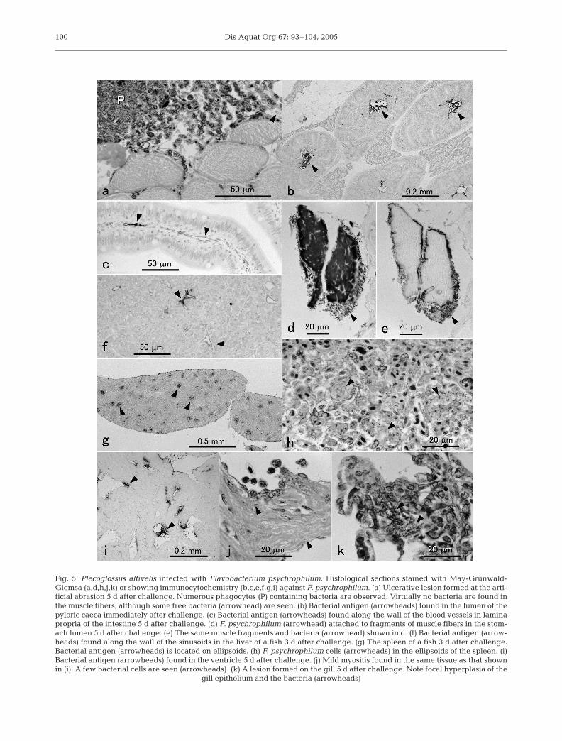
On Day 5, ulcers were found at the sites of artificialabrasion in 7 fish. Either haemorrhage or sloughing oftissue was observed in the lower jaw of 6 fish. The ero-sion of the tissues generally became more severe inthese lesions. In 2 fish, however, only a small numberof free bacteria were observed in the ulcerativelesions, although numerous leucocytes with bacteria orthe bacterial antigen in their cytoplasm were seen(Fig. 5a).
Digestive tract
Many Flavobacterium psychrophilum cells werefound in the stomach lumen immediately after immer-sion. However, the bacterium was detected in theintestinal lumen in only 1 fish, although the bacterialantigen was found in the lumen of the intestine andpyloric caeca in all fish examined (Fig. 5b). Bacterialcells or the bacterial antigen were always observed inthe stomach or intestinal lumen of some fish. Inside theorgans, the bacterial antigen was usually first detectedalong the blood vessel wall in lamina propria (Fig. 5c).Bacterial cells were also found in lamina propria insome fish although no clear morphological lesions
were observed. No F. psychrophilum directly attachedto the epithelial cells, and no bacteria were found inthe epithelium throughout the experiment. In thestomach lumen of 2 fish, fragments of muscle fibersinfested with many F. psychrophilum cells wereobserved on Day 5 (Fig. 5d,e).
Other organs
Neither bacteria nor bacterial antigen wereobserved in the brain. Bones and cartilage were freefrom bacteria even when severe lesions developed inthe surrounding connective tissues except for 2 casesin which small numbers of bacteria were found in theperiphery of bones. In the other organs, as in the diges-tive tract, the bacterial antigen began to appear alongthe wall of blood vessels on Day 2, and bacterial cellswere most frequently observed on Day 3 (Table 2). Insome fish sampled on Days 2 and 3, the bacterial anti-gen was diffusely localized in the lumen of blood ves-sels. Small numbers of leucocytes containing the bac-terial antigen in their cytoplasm were first observed insome fish 1 d after the challenge. On Day 2, many ofthese cells appeared in the kidney and spleen, and inthe blood vessels of the other internal organs includingheart and liver. These cells were continually observedon Days 3 and 5 in most of the fish. The bacterial anti-gen in the liver parenchyma was first found on Day 3.The antigen was found along the endothelium of thesinusoids, or in unidentified cells along the sinusoids inmost of the fish (Fig. 5f). In some fish observed on Days3 and 5, hepatocytes themselves contained the bacter-ial antigen which was confined to a certain compart-ment in the cytoplasm. In the spleen, the bacterial anti-gen, and bacteria were observed in the connectivetissue of ellipsoids (Fig. 5g,h). Many free bacteria wereobserved in the ellipsoids on Day 3, although no appar-ent necrotic lesion was observed. Many leucocyteswith strong reaction of the bacterial antigen werefound in the kidney of 6 fish on Day 2, and small num-bers of free bacteria were observed on Day 3. Exceptfor migrating leucocytes in the lumen, the bacterialantigen was first detected in the tissues of the heart onDay 3. Mild inflammation of the cardiac muscle andthe atrial wall was observed in some fish on Days 3and 5 (Fig. 5i,j). However, the numbers of bacteriaobserved in these lesions were very small and noapparent necrotic lesion was found. In the gill orpseudobranch, bacteria were found around the bloodvessels or around the cartilage in the gill of some fishon Days 3 and 5. Focal hyperplasia of the gill epithe-lium was found in these tissues (Fig. 5k). In most of theorgans, the intensity of the antigen reaction and thenumbers of bacteria generally decreased on Day 5.
99
Dis Aquat Org 67: 93–104, 2005100
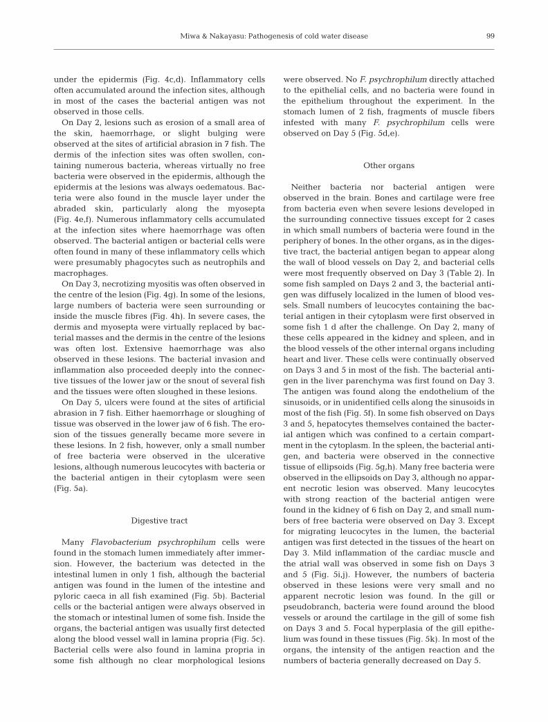
Fig. 5. Plecoglossus altivelis infected with Flavobacterium psychrophilum. Histological sections stained with May-Grünwald-Giemsa (a,d,h,j,k) or showing immunocytochemistry (b,c,e,f,g,i) against F. psychrophilum. (a) Ulcerative lesion formed at the arti-ficial abrasion 5 d after challenge. Numerous phagocytes (P) containing bacteria are observed. Virtually no bacteria are found inthe muscle fibers, although some free bacteria (arrowhead) are seen. (b) Bacterial antigen (arrowheads) found in the lumen of thepyloric caeca immediately after challenge. (c) Bacterial antigen (arrowheads) found along the wall of the blood vessels in laminapropria of the intestine 5 d after challenge. (d) F. psychrophilum (arrowhead) attached to fragments of muscle fibers in the stom-ach lumen 5 d after challenge. (e) The same muscle fragments and bacteria (arrowhead) shown in d. (f) Bacterial antigen (arrow-heads) found along the wall of the sinusoids in the liver of a fish 3 d after challenge. (g) The spleen of a fish 3 d after challenge.Bacterial antigen (arrowheads) is located on ellipsoids. (h) F. psychrophilum cells (arrowheads) in the ellipsoids of the spleen. (i)Bacterial antigen (arrowheads) found in the ventricle 5 d after challenge. (j) Mild myositis found in the same tissue as that shownin (i). A few bacterial cells are seen (arrowheads). (k) A lesion formed on the gill 5 d after challenge. Note focal hyperplasia of the
gill epithelium and the bacteria (arrowheads)
Miwa & Nakayasu: Pathogenesis of cold water disease
Expt 3
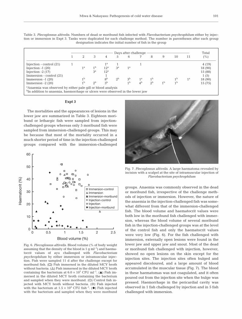
The mortalities and the appearances of lesions in thelower jaw are summarized in Table 3. Eighteen mori-bund or lethargic fish were sampled from injection-challenged groups whereas only 5 moribund fish weresampled from immersion-challenged groups. This maybe because that most of the mortality occurred in amuch shorter period of time in the injection-challengedgroups compared with the immersion-challenged
groups. Anaemia was commonly observed in the deador moribund fish, irrespective of the challenge meth-ods of injection or immersion. However, the nature ofthe anaemia in the injection-challenged fish was some-what different from that of the immersion-challengedfish. The blood volume and haematocrit values wereboth low in the moribund fish challenged with immer-sion, whereas the blood volume of several moribundfish in the injection-challenged groups was at the levelof the control fish and only the haematocrit valueswere very low (Fig. 6). For the fish challenged withimmersion, externally open lesions were found in thelower jaw and upper jaw and snout. Most of the deador moribund fish challenged with injection, however,showed no open lesions on the skin except for theinjection sites. The injection sites often bulged andappeared discoloured, and a large amount of bloodaccumulated in the muscular tissue (Fig. 7). The bloodin these haematomas was not coagulated, and it oftenpoured out from the injection site when the bulge waspressed. Haemorrhage in the pericardial cavity wasobserved in 1 fish challenged by injection and in 5 fishchallenged with immersion.
101
Days after challenge Total1 2 3 4 5 6 7 8 9 10 11 (%)
Injection - control (21) 1 1a 1 1 4 (19)Injection -1 (20) 1a 1b 12a 3a 1a 18 (90)Injection -2 (17) 3a 12a 15 (88)Immersion - control (21) 1 1 (5)Immersion -1 (20) 1b 8b 2b 3b 1a 1b 1b 1a 18 (90)Immersion -2 (20) 1b 2b 3b 1b 4b 2b 1b 1b 15 (75)aAnaemia was observed by either pale gill or blood analysisbIn addition to anaemia, haemorrhage or ulcers were observed in the lower jaw
Table 3. Plecoglossus altivelis. Numbers of dead or moribund fish infected with Flavobacterium psychrophilum either by injec-tion or immersion in Expt 3. Tanks were duplicated for each challenge method. The number in parentheses after each group
designation indicates the initial number of fish in the group
Blood volume (%)
0
60
50
40
30
20
10
01 20.5 1.5 2.5
Immersion-controlImmersionImmersion-moribundInjection-controlInjectionInjection-moribund
Hae
mat
ocrit
(%)
Fig. 6. Plecoglossus altivelis. Blood volume (% of body weightassuming that the density of the blood is 1 g ml–1) and haema-tocrit values of ayu challenged with Flavobacteriumpsychrophilum by either immersion or intramuscular injec-tion. Fish were sampled 11 d after the challenge except formoribund fish. (h) Fish immersed in the diluted MCY brothwithout bacteria. (n) Fish immersed in the diluted MCY brothcontaining the bacterium at 6.0 × 10⁷ CFU ml–1. (m) Fish im-mersed in the diluted MCY broth containing the bacteriumand sampled when they were moribund. (s) Control fish in-jected with MCY broth without bacteria. (e) Fish injectedwith the bacterium at 1.5 × 10⁵ CFU fish–1. (f) Fish injectedwith the bacterium and sampled when they were moribund
Fig. 7. Plecoglossus altivelis. A large haematoma revealed byincision with a scalpel at the site of intramuscular injection of
Flavobacterium psychrophilum
Dis Aquat Org 67: 93–104, 2005
DISCUSSION
Portals of entry
The results of the present study suggest thatFlavobacterium psychrophilum enters the body of ayuthrough injured skin. The bacterial entry into the bodythrough healthy skin seems improbable since no bac-terium or bacterial antigen was observed in the intactskin. The external lesions of CWD-affected ayu aremost likely due to F. psychrophilum infection throughalready existing skin injuries. This is also supported bythe virtual absence of ulcers or haemorrhagic lesionson the body surface of fish challenged with intramus-cular injection except at the site of injection. This fur-ther suggests that ayu are unlikely to be infected withF. psychrophilum if the fish has no external injuries.Kondo et al. (2002) also reported the adherence of F.psychrophilum to the lower jaw and caudal pedunclewhere the epidermis had collapsed. The importance ofmechanical injuries as the portals of entry of F. psy-chrophilum has already been suggested in salmonids.Although the experimental infection of F. psy-chrophilum through immersion is often difficult(Nematollahi et al. 2003), incision or removal of themucus layer from the skin markedly enhances the sus-ceptibility of rainbow trout to water-borne bacteria(Madetoja et al. 2000). Martinez et al. (2004) alsoreported that the ray axis of fins needs to be exposedby previous damage for the invasion of F. psy-chrophilum. Skin injuries are also important portals forsome other bacterial infections of fish such asAeromonas hydrophila infection in the channel catfishIctalurus punctatus (Ventura & Grizzle 1987). Smallparticles like bacteria can be passively attached toinjured skin surface (Kiryu & Wakabayashi 1999, Suk-enda & Wakabayashi 2001). The histological observa-tions of the present study suggest that, once the bac-terium adheres to the dermis, it enters the tissuequickly. However, it is not certain whether the bacteriaattached to superficially injured epidermis can reachthe dermis, since almost no free bacteria wereobserved in the epidermal tissue of the skin.
Most ayu used for the present experiments alreadyhad microscopic abrasions on the lower jaw, althoughthese lesions could not be observed with the nakedeye. The skin of the lower jaw was probably abradedwhen the fish scraped the wall or the bottom of therearing tank with their teeth, which is a characteristicbehavior of ayu. In spontaneously occurring CWD incultured ayu, one of the clinical signs most often notedis lesions on the lower jaw. In the present study, noapparent lesions developed on the lower jaw when thefish were injected with the bacterium, and hencelesions on the lower jaw are most likely caused by
direct infection with the bacterium on abraded skin, asdiscussed above. Thus, it is likely that cultured ayutypically have similar abrasions on the lower jaw, andthat these abrasions are the major portals of F. psy-chrophilum infection for these fish.
The absence of bacteria in the gill or in the tissues ofdigestive tract until later stages of infection suggeststhat the bacterium does not infect ayu through theseorgans. This is in agreement with previous studiesin which orally administered Flavobacterium psy-chrophilum could not reproduce the disease (Decostereet al. 2000, Madetoja et al. 2000). Although bacteria orthe bacterial antigen were detected in these organs, itseems clear that these infections occurred from bacteriamigrating through blood vessels, since the bacterialantigen or bacterial cells were first detected on the wallof blood vessels of these organs a few days afterchallenge. Many F. psychrophilum cells were observedin the stomach lumen immediately after the immersionchallenge. In the intestinal lumen, however, bacterialcells were observed in only 1 fish, although the bacter-ial antigen was clearly detected in all fish examined.This suggests that most of the ingested bacteria wereprobably killed and digested in the stomach or intes-tine. In the stomach lumen of 2 fish sampled on Day 5,fragments of muscle fibres with a large number ofimmunoreactive bacterial cells were observed. Thisclearly suggests that these fish had bitten other fish ontheir lesions. Although infection through the digestivetract seems improbable, such fish can be infected withbacteria through injuries on the lower jaw.
Affinity of the bacterium for tissues
In the early stage of infection, Flavobacterium psy-chrophilum proliferated in the dermal connective tis-sue and seemed to migrate through the dermis andmyosepta into the fish’s body. In later stages, manybacteria were observed in the skeletal muscle fibresand some were found in cardiac muscle fibres. Freebacteria were seldom observed in epithelial tissues.These observations suggest not only the affinity of thebacterium for the collagenous connective tissues, butalso the importance of direct access of the bacterium tosuch tissues through mechanical injuries to establishinfection. Salmonid fishes affected with CWD alsosometimes develop lesions similar to those found inayu, such as ulcers on the skin or on the mouth (Dals-gaard 1993). For salmonid CWD, there is evidence thatF. psychrophilum has affinity for hard tissues such ascartilage, bone or fin rays (Ostland et al. 1997). Ekman& Norrgren (2003) also reported accumulation ofF. psychrophilum on the vertebrae in 3 species ofsalmonids experimentally infected with CWD. CWD in
102
Miwa & Nakayasu: Pathogenesis of cold water disease
salmonids is sometimes called ‘fin rot’ and one of theearly signs of the disease is loss of fin-tip integrity(Shotts & Starliper 1999). Martinez et al. (2004)reported that F. psychrophilum was present on theossified axis of fin rays but not in the epidermis of theexperimentally infected Atlantic salmon Salmo salar.They showed that the bacterium penetrated directlyinto the ray axis probably by secreting protease(s) thateroded the tissue matrix. Since the major organicmatrix of bone is collagen and collagen is also one ofthe major organic components of the cartilage matrix,it seems probable that F. psychrophilum generally hasan affinity for collagen. In fact, F. psychrophilum pro-duces enzymes that degrade collagen (Ostland et al.2000). Martinez et al. (2004) argues that the require-ment for calcium for the activation of an extracellularprotease of the bacterium is the reason for the prefer-ence of the bacterium for the fin rays in early phases ofsalmonid CWD. In the present study, small numbers ofthe bacterium were found in the periphery of bones in2 cases. These were, however, rather exceptional, andbones or cartilages were usually intact even whenthere were severe lesions in surrounding connectivetissues. Hence, there may be a difference in therequirement for calcium or in the preference for thecollagen molecules between the proteases of F. psy-chrophilum strains causing CWD in ayu and thosecausing CWD in salmonids. In addition, F. psy-chrophilum seems to have a certain affinity for striatedmuscular tissues since mild myocarditis and severenecrotic myositis were found in the present study.Necrotic myositis was also reported in CWD-infectedsalmonids (Lumsden et al. 1996).
Cause of death
In the present study, anaemia was the most fre-quently observed clinical sign of spontaneous CWD inayu. Anaemia is also one of the most notable clinicalsigns for CWD in salmonids (Nematollahi et al. 2003).In Expt 1, all moribund fish showed smaller blood vol-ume and lower haematocrit values than those of thecontrols, while the blood volume and haematocrit val-ues of many of the infected fish other than the mori-bund fish were in the range of the control group. Fur-thermore, fish died very quickly, less than 1 h afterthey became moribund. These observations suggestthat at a certain stage of infection, the anaemiaadvanced very swiftly and killed ayu probably byhypoxia. This anaemia resulted most likely from bleed-ing through lesions on the fish body for the followingreasons. First, it is improbable for aplastic anaemia tooccur in such a short period of time. Besides, no appar-ent necrotic lesion was observed in the haematopoietic
tissues in the present study. Second, no evidence sug-gests that haemolysis had occurred. If haemolysis wasthe major cause for the anaemia in such a short periodof time as in the present experiments, jaundice shouldhave been observed. For example, jaundice is clearlyobserved in bacterial haemolytic jaundice of yellowtailSeriola quinqueradiata in which the causative bacterialyse erythrocytes in the peripheral blood (Sorimachi etal. 1993, Maeno et al. 1995). Hence, haemolysis byFlavobacterium psychrophilum does not seem to con-tribute substantially to the anaemia of CWD-affectedayu although F. psychrophilum seems to be able to lysefish erythrocytes to a certain degree (Lorenzen et al.1997). Third, the blood volume was decreased in themoribund or lethargic ayu. F. psychrophilum is knownto produce an enzyme which degrades fibrinogen(Nematollahi et al. 2003), and this may inhibit bloodcoagulation in the ulcerative lesions. Inflammation ofthe atrial wall and haemorrhage observed in the peri-cardial cavity suggest that the heart also can be a siteof bleeding in CWD-affected ayu. Considering thatanaemia and external lesions are the most commonclinical signs for spontaneous CWD in ayu, anaemia(and probably hypoxia) caused by bleeding from openlesions is also likely to be the cause of death for ayuaffected spontaneously by CWD.
Anaemia was also observed in the injection-challenged fish, although no apparent open lesion wasformed. Unlike immersion-challenged fish, however,several moribund fish in the injection-challengedgroups exhibited blood volumes comparable to thoseof the control fish, although their haematocrit valueswere very low. In many of the injection-challengedfish, large haematomas had developed at the site of theinjection. These suggest that erythrocytes had some-how accumulated in this lesion and did not return tothe circulation, thereby causing low haematocrit val-ues, although the mechanism of this is uncertain. Thiscondition was apparently caused by the injection of alarge amount of bacteria deep inside the body of thefish and would never occur in spontaneous infectionsof CWD.
Infection of internal organs and recovery fromthe disease
Although many leucocytes containing bacterial anti-gen appeared in the internal organs on Day 2, the anti-gen or bacteria were not detected substantially untilDays 3 and 5 in the spleen, heart, kidney and gill. Formost of the internal organs, the bacterial antigen wasfirst detected along the wall of blood vessels except forthat in the migrating leucocytes. This suggests thatthese organs were infected with bacteria transported
103
Dis Aquat Org 67: 93–104, 2005
from skin lesions through the vascular system. Manyfree bacteria were observed in the ellipsoids of thespleen on Day 3, although no severe lesion wasobserved. Splenomegaly is one of the major clinicalsigns in spontaneous CWD in ayu (Iida & Mizokami1996) as well as in salmonids (Rangdale et al. 1999,Decostere et al. 2001, Ekman & Norrgren 2003). Insalmonid CWD, necrotic changes and the presence ofmany free bacteria in the spleen have been reported inaffected fish (Rangdale et al. 1999, Ekman & Norrgren2003). Hence, severe histopathological changes mayalso appear in the spleen of ayu if the disease becomesmore chronic.
On Day 5, some fish showed evidence of recoveryfrom the disease. In these fish, many leucocytes withbacteria or the bacterial antigen were observed in skinulcers, but only small numbers of free bacteria werefound in the lesions and virtually no bacteria werefound in the adjacent muscle fibres, suggesting thatfurther expansion of the ulcers had ceased. In thepresent study, numerous leucocytes ingested Flavo-bacterium psychrophilum or its antigen. This seems tosuggest the importance of phagocytosis by immunecells for defense against F. psychrophilum. Hepato-cytes are probably involved in the processing of sub-stances from broken bacterial cells, since the bacterialantigen was detected in the hepatocytes in later stagesof infection. Thus, ayu may survive CWD if theimmune system is well activated before severe bleed-ing occurs.
Acknowledgements. This study was supported in part by theJapanese Ministry of Agriculture, Forestry, and Fisheries.
LITERATURE CITED
Dalsgaard I (1993) Virulence mechanisms in Cytophaga psy-chrophila and other Cytophaga-like bacteria pathogenicfor fish. Annu Rev Fish Dis 3:127–144
Decostere A, Lammens M, Haesebrouck F (2000) Difficultiesin experimental infection studies with Flavobacteriumpsychrophilum in rainbow trout (Oncorhynchus mykiss)using immersion, oral and anal challenges. Res Vet Sci 69:165–169
Decostere A, D’Haese M, Lammens M, Nelis H, HaesebrouckF (2001) In vivo study of phagocytosis, intracellular sur-vival and multiplication of Flavobacterium psychrophilumin rainbow trout, Oncorhynchus mykiss (Walbaum),spleen phagocytes. J Fish Dis 24:481–487
Ekman E, Norrgren L (2003) Pathology and immunohisto-chemistry in 3 species of salmonids after experimentalinfection with Flavobacterium psychrophilum. J Fish Dis26:529–538
Iida Y, Mizokami A (1996) Outbreaks of coldwater disease inwild ayu and pale chub. Fish Pathol 31:157–164
Izumi S, Wakabayashi H (1999) Further study on serotyping ofFlavobacterium psychrophilum. Fish Pathol 34:89–90
Izumi S, Aranishi F, Wakabayashi H (2003) Genotyping ofFlavobacterium psychrophilum using PCR-RFLP analysis.
Dis Aquat Org 56:207–214Kiryu I, Wakabayashi H (1999) Adherence of suspended par-
ticles to the body surface of rainbow trout. Fish Pathol 34:177–182
Kondo M, Kawai K, Kurohara K, Oshima S (2002) Adherenceof Flavobacterium psychrophilum on the body surface ofthe ayu Plecoglossus altivelis. Microbes Infect 4:279–283
Lorenzen E, Dalsgaard I, Bernardet JF (1997) Characterizationof isolates of Flavobacterium psychrophilum associated withcoldwater disease or rainbow trout fry syndrome I: pheno-typic and genomic studies. Dis Aquat Org 31:197–208
Lumsden JS, Ostland VE, Ferguson HW (1996) Necroticmyositis in cage cultured rainbow trout, Oncorhynchusmykiss (Walbaum), caused by Flexibacter psychrophilus.J Fish Dis 19:113–119
Madetoja J, Nyman P, Wiklund T (2000) Flavobacterium psy-chrophilum, invasion into and shedding by rainbow troutOncorhynchus mykiss. Dis Aquat Org 43:27–38
Maeno Y, Nakajima K, Sorimachi M, Inui Y (1995) Pathologi-cal studies of jaundiced yellowtail, Seriola quinqueradi-ata. Fish Pathol 30:7–14
Martinez JL, Casado A, Enriquez R (2004) Experimentalinfection of Flavobacterium psychrophilum in fins ofAtlantic salmon Salmo salar revealed by scanning electronmicroscopy. Dis Aquat Org 59:79–84
Nematollahi A, Decostere A, Pasmans F, Haesebrouck F(2003) Flavobacterium psychrophilum infections insalmonid fish. J Fish Dis 26:563–574
Ostland VE, McGrogan DG, Ferguson HW (1997) Cephalicosteochondritis and necrotic scleritis in intensively rearedsalmonids associated with Flexibacter psychrophilus.J Fish Dis 20:443–451
Ostland VE, Byrne PJ, Hoover G, Ferguson HW (2000)Necrotic myositis of rainbow trout, Oncorhynchus mykiss(Walbaum): proteolytic characteristics of a crude extracel-lular preparation from Flavobacterium psychrophilum.J Fish Dis 23:329–336
Rangdale RE, Richards RH, Alderman DJ (1999) Histopatho-logical and electron microscopical observations on rain-bow trout fry syndrome. Vet Rec 144:251–254
Shotts EB, Starliper CE (1999) Flavobacterial diseases: colum-naris disease, cold-water disease and bacterial gill dis-ease. In: Woo PTK, Bruno DW (eds) Fish diseases and dis-orders, Vol 3: Viral, bacterial and fungal infections. CABIPublishing, New York, p 559–576
Sorimachi M, Maeno Y, Nakajima K, Inouye K, Inui Y (1993)Causative agent of jaundice of yellowtail, Seriola quin-queradiata. Fish Pathol 28:119–124
Sukenda, Wakabayashi H (2001) Adherence and infectivity ofgreen fluorescent protein-labeled Pseudomonas pleco-glossicida to ayu Plecoglossus altivelis. Fish Pathol 36:161–167
Tabata K (2004) Relationships of the infectivity of Flavobac-terium psychrophilum between native fishes and releasedayu Plecoglossus altivelis in a river. Nippon SuisanGakkaishi 70:318–323 (in Japanese with English abstract)
Toyama T, Kita-Tsukamoto K, Wakabayashi H (1994) Identifi-cation of Cytophaga psychrophila by PCR targeted 16Sribosomal RNA. Fish Pathol 29:271–275
Ventura MT, Grizzle JM (1987) Evaluation of portals of entryof Aeromonas hydrophila in channel catfish. Aquaculture65:205–214
Wakabayashi H, Egusa S (1974) Characteristic of myxobacte-ria associated with some freshwater fish diseases in Japan.Bull Jpn Soc Sci Fish 40:751–757
Wakabayashi H, Toyama T, Iida T (1994) A study on serotyp-ing of Cytophaga psychrophila isolated from fishes inJapan. Fish Pathol 29:101–104
104
Editorial responsibility: David Bruno,Aberdeen, UK
Submitted: December 13, 2004; Accepted: June 28, 2005Proofs received from author(s): November 1, 2005
Related Documents