*Corresponding Author Address: Dr Abu-Hussein Muhamad.Email: [email protected] International Journal of Dental and Health Sciences Volume 02, Issue 05 Case Report ORTHODONTIC TREATMENT OF AN IMPACTED MAXILLARY CENTRAL INCISOR COMBINED WITH SURGICAL EXPOSURE Péter Borbély 1 , Nezar Watted 2 , Ivana Dubovská 3 Viktória Hegedűs 4 Abu-Hussein Muhamad 5 1.Fogszabályozási Stúdió, Budapest, Hungary 2.Clinics and Policlinics for Dental, Oral and Maxillofacial Diseases of the Bavarian Julius-Maximilian- University Wuerzburg, Germany 3.Institute of Dentistry and Oral Sciences, Faculty of Medicine and Dentistry, Palacký University, Olomouc, Czech Republic 4.Department of Pediatric Dentistry and Orthodontics, University of Debrecen, Debrecen, Hungary 5.University of Naples Federic II, Naples, Italy, Department of Pediatric Dentistry, University of Athens,Athens, Greece ABSTRACT: Impaction of maxillary permanent incisors is not a frequent case in dental practice, but its treatment is challenging because of these teeth importance to facial esthetics Management by a combination of orthodontics and surgery produces a satisfactory result. The surgical exposure and orthodontic traction of impacted central incisor after surgical exposure of impacted maxillary central incisor teeth is presented in this case report. Key words: Impacted tooth, Maxillary incisors orthodontics, tooth movement INTRODUCTION: The impact of maxillary permanent incisor development may possess important problems in terms of esthetics and occlusion in the early mixed dentition[1] . Children with a marked delayed eruption of permanent incisors are usually at the age of 8-10 years old. The maxillary incisor can be considered impacted if the following conditions exist: 1.) No historical record of previous extraction, 2.) Eruption of contra-lateral incisor which occur 6 months earlier, 3.) Both incisors are un-erupted and the lower incisors have erupted one year previously or deviate from the normal sequence of eruption e.g., lateral incisors erupt before the central incisor, 4.) Maxillary incisors have delayed eruption 6 months after the normal eruption date[2,3] . The prevalence of unerupted maxillary incisors in the 5-12 year age group has been reported as 0.13%. In a referred population to regional hospitals the prevalence has been estimated at 2.6%. Unerupted incisors are more common in males than females with a ratio of 2.7:1. Almost half (47%) of all unerupted maxillary incisors are due to supernumeraries. The mesiodont variety has more eruptive disturbances compared to the palatodont[4].

ORTHODONTIC TREATMENT OF AN IMPACTED MAXILLARY CENTRAL INCISOR COMBINED WITH SURGICAL EXPOSURE
Jan 22, 2018
Welcome message from author
This document is posted to help you gain knowledge. Please leave a comment to let me know what you think about it! Share it to your friends and learn new things together.
Transcript
*Corresponding Author Address: Dr Abu-Hussein Muhamad.Email: [email protected]
International Journal of Dental and Health Sciences
Volume 02, Issue 05
Case Report
ORTHODONTIC TREATMENT OF AN
IMPACTED MAXILLARY CENTRAL INCISOR
COMBINED WITH SURGICAL EXPOSURE
Péter Borbély1 , Nezar Watted
2, Ivana Dubovská
3 Viktória Hegedűs
4 Abu-Hussein Muhamad
5
1.Fogszabályozási Stúdió, Budapest, Hungary 2.Clinics and Policlinics for Dental, Oral and Maxillofacial Diseases of the Bavarian Julius-Maximilian- University Wuerzburg, Germany 3.Institute of Dentistry and Oral Sciences, Faculty of Medicine and Dentistry, Palacký University, Olomouc, Czech Republic 4.Department of Pediatric Dentistry and Orthodontics, University of Debrecen, Debrecen, Hungary 5.University of Naples Federic II, Naples, Italy, Department of Pediatric Dentistry, University of Athens,Athens, Greece
ABSTRACT:
Impaction of maxillary permanent incisors is not a frequent case in dental practice, but its treatment is challenging because of these teeth importance to facial esthetics Management by a combination of orthodontics and surgery produces a satisfactory result. The surgical exposure and orthodontic traction of impacted central incisor after surgical exposure of impacted maxillary central incisor teeth is presented in this case report. Key words: Impacted tooth, Maxillary incisors orthodontics, tooth movement
INTRODUCTION:
The impact of maxillary permanent incisor
development may possess important
problems in terms of esthetics and
occlusion in the early mixed dentition[1] .
Children with a marked delayed eruption
of permanent incisors are usually at the
age of 8-10 years old. The maxillary incisor
can be considered impacted if the
following conditions exist:
1.) No historical record of previous
extraction,
2.) Eruption of contra-lateral incisor which
occur 6 months earlier,
3.) Both incisors are un-erupted and the
lower incisors have erupted one year
previously or deviate from the normal
sequence of eruption e.g., lateral incisors
erupt before the central incisor,
4.) Maxillary incisors have delayed
eruption 6 months after the normal
eruption date[2,3] .
The prevalence of unerupted maxillary
incisors in the 5-12 year age group has
been reported as 0.13%. In a referred
population to regional hospitals the
prevalence has been estimated at 2.6%.
Unerupted incisors are more common in
males than females with a ratio of 2.7:1.
Almost half (47%) of all unerupted
maxillary incisors are due to
supernumeraries. The mesiodont variety
has more eruptive disturbances compared
to the palatodont[4].
Borbely P. et al., Int J Dent Health Sci 2015; 2(5): XXXX-XXXX
703
The occurrence of erupted lateral incisors
associated with the non-appearance of
one or both of the central incisors should
always be deemed abnormal when a child
is between 8 and 10 years of age[5].
Although impaction of a permanent tooth
is rarely diagnosed during the mixed
dentition period, an impacted central
incisor is usually diagnosed accurately
when there is delay in the eruption of the
tooth. However, the abnormality in the
appearance can also be due to other
clinical features and malformation of
other elements of the craniofacial
complex. Tooth impaction may result
from a number of local causes. The
principal local factors involved in this
anomaly are supernumerary teeth,
odontomas, and trauma.[6]Table.1
The following radiographs need to be
taken to assist in the diagnosis and
management[7,8]:
- an anterior occlusal radiograph for
general assessment purposes.
- two periapical radiographs should be
taken using the parallax technique for
detailed assessment of the position, root
and crown morphology. It has been
shown that the use of horizontal parallax
technique is better than vertical parallax.
- if an anterior occlusal and a panoramic
radiograph are already available, the
vertical tube shift (VTS) technique can also
be used for assessment
In recent years CBCT has been introduced
as a technique for imaging of dental and
maxillofacial structures. CBCT is a medical
image acquisition technique based on a
cone-shaped X-ray beam centred on a
two-dimensional (2D) detector. The
source-detector system performs one
rotation around the object producing a
series of 2D images. The images are
reconstructed in a three-dimensional (3D)
data set using a modification of the
original cone-beam algorithm[9].
CBCT imaging provides orthodontists with
an excellent tool to improve diagnosis,
treatment planning and outcome
assessment in appropriate malocclusion
.Studies have shown that CBCT is more
sensitive than conventional radiography
for both impacted teeth localization and
identification of root resorption of
adjacent teeth.
The comprehensive images in 3 planes
provided by CBCT can assist surgeons in
choosing the appropriate surgical
approach, identifying the tooth that
should be extracted, and reducing the
amount of surgical trauma on the
adjacent hard and soft tissues[10,11].
Adequate space (9mm for central incisor
and 7mm for lateral incisor) should be
created prior to any surgical intervention
to enhance spontaneous eruption. Almost
half of the impacted incisors erupt
spontaneously following the removal of
obstruction and creation of space[12].
Maintenance of space throughout the
treatment is crucial to prevent the lost of
space which can lead to secondary
inhibition of spontaneous eruption of the
impacted tooth[13].
Borbely P. et al., Int J Dent Health Sci 2015; 2(5): XXXX-XXXX
704
The impacted tooth is left open to the oral
environment following the surgery and
surrounded by the incised palatal or labial
mucosa[1,2]. An attachment may be
placed during or after the procedure. This
technique is termed open eruption
technique or exposure and can be
performed in two ways[14-17]:
a) The window technique
This direct technique involved removal of
overlying mucosa and the finally erupted
tooth will have a non-keratinized labial
gingival mucosa.
b) The apically repositioned flap
The procedure involves apically
repositioning the raised flap that
incorporates attached gingiva overlying
the impacted tooth and is expected to
provide adequate width of attached
gingiva.
The closed eruption technique has been
favoured by many clinicians who claimed
that the aesthetic and periodontal
outcome is far more superior when
compared with the apically positioned
flap. With this method, a labial or palatal
flap is raised and an attachment with gold
chain or a bracket/eyelet with ligature
wire is bonded to the enamel surface of
the tooth using acid-etch technique,
preferably with a light cured adhesive,
before the flap is replaced. Orthodontic
traction is then applied[1].
There are a few criteria to evaluate when
considering the best method for
uncovering unerupted tooth.
-Labio-lingual position of the unerupted
tooth
For labially positioned tooth any
technique is possible. However, if the
unerupted tooth is in centre of the
alveolus, it may be difficult to approach by
the two open eruption technique. A
simple window technique may be
preferred if the impacted tooth is low
down in the alveolus and bucco lingually
close to its place in the arch.
For palatally positioned tooth where there
is presence of thick palatal mucosa tissue
the window technique usually requires
placement of a periodontal pack to
prevent regrowth of tissue over the
exposed tooth.
-Vertical position of the unerupted tooth
relative to the mucogingival junction
If the crown of the unerupted tooth is
positioned coronal to mucogingival
junction any one of the three techniques
can be used to uncover the tooth. If it is
positioned apical to the mucogingival
junction the window technique may not
be appropriate and the apically position
technique is appropriate. To uncover the
tooth that is significantly apical to the
mucogingival junction a close eruption
technique is preferred.
- The amount of gingiva in the area of
the unerupted tooth
In the presence of insufficient gingiva in
the area of the unerupted tooth, an
apically positioned flap is preferred. A 2 -
3 mm of attached gingiva over the crown
Borbely P. et al., Int J Dent Health Sci 2015; 2(5): XXXX-XXXX
705
of the tooth allows any of the three
technique to be used.
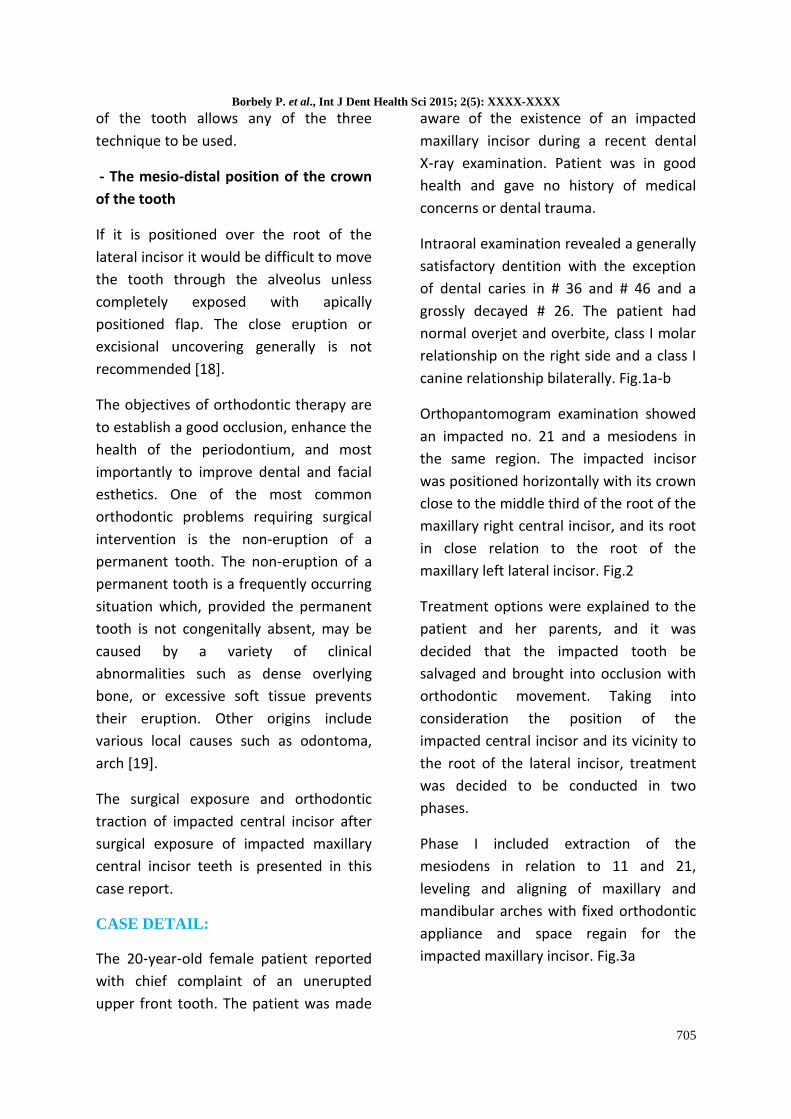
- The mesio-distal position of the crown
of the tooth
If it is positioned over the root of the
lateral incisor it would be difficult to move
the tooth through the alveolus unless
completely exposed with apically
positioned flap. The close eruption or
excisional uncovering generally is not
recommended [18].
The objectives of orthodontic therapy are
to establish a good occlusion, enhance the
health of the periodontium, and most
importantly to improve dental and facial
esthetics. One of the most common
orthodontic problems requiring surgical
intervention is the non-eruption of a
permanent tooth. The non-eruption of a
permanent tooth is a frequently occurring
situation which, provided the permanent
tooth is not congenitally absent, may be
caused by a variety of clinical
abnormalities such as dense overlying
bone, or excessive soft tissue prevents
their eruption. Other origins include
various local causes such as odontoma,
arch [19].
The surgical exposure and orthodontic
traction of impacted central incisor after
surgical exposure of impacted maxillary
central incisor teeth is presented in this
case report.
CASE DETAIL:
The 20‑year‑old female patient reported
with chief complaint of an unerupted
upper front tooth. The patient was made
aware of the existence of an impacted
maxillary incisor during a recent dental
X‑ray examination. Patient was in good
health and gave no history of medical
concerns or dental trauma.
Intraoral examination revealed a generally
satisfactory dentition with the exception
of dental caries in # 36 and # 46 and a
grossly decayed # 26. The patient had
normal overjet and overbite, class I molar
relationship on the right side and a class I
canine relationship bilaterally. Fig.1a-b
Orthopantomogram examination showed
an impacted no. 21 and a mesiodens in
the same region. The impacted incisor
was positioned horizontally with its crown
close to the middle third of the root of the
maxillary right central incisor, and its root
in close relation to the root of the
maxillary left lateral incisor. Fig.2
Treatment options were explained to the
patient and her parents, and it was
decided that the impacted tooth be
salvaged and brought into occlusion with
orthodontic movement. Taking into
consideration the position of the
impacted central incisor and its vicinity to
the root of the lateral incisor, treatment
was decided to be conducted in two
phases.
Phase I included extraction of the
mesiodens in relation to 11 and 21,
leveling and aligning of maxillary and
mandibular arches with fixed orthodontic
appliance and space regain for the
impacted maxillary incisor. Fig.3a
Borbely P. et al., Int J Dent Health Sci 2015; 2(5): XXXX-XXXX
706
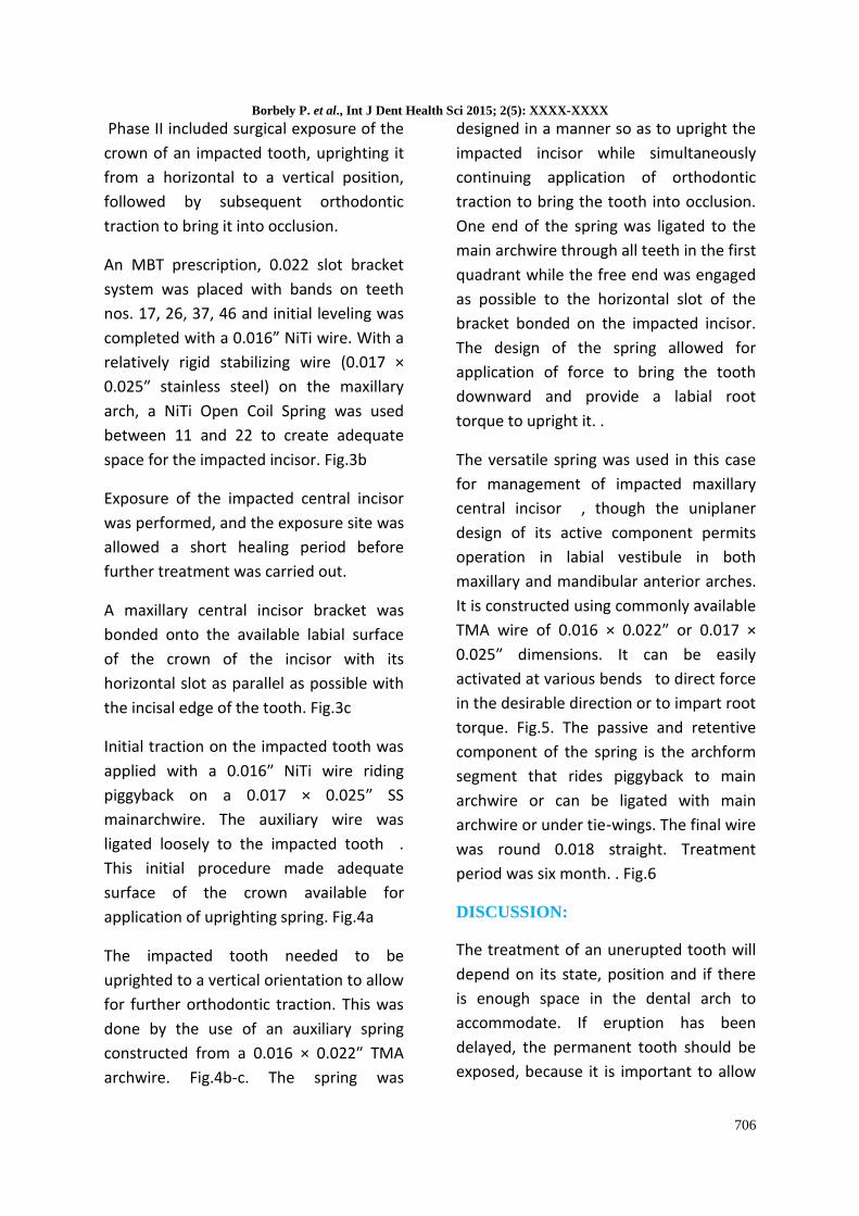
Phase II included surgical exposure of the
crown of an impacted tooth, uprighting it
from a horizontal to a vertical position,
followed by subsequent orthodontic
traction to bring it into occlusion.
An MBT prescription, 0.022 slot bracket
system was placed with bands on teeth
nos. 17, 26, 37, 46 and initial leveling was
completed with a 0.016” NiTi wire. With a
relatively rigid stabilizing wire (0.017 ×
0.025″ stainless steel) on the maxillary
arch, a NiTi Open Coil Spring was used
between 11 and 22 to create adequate
space for the impacted incisor. Fig.3b
Exposure of the impacted central incisor
was performed, and the exposure site was
allowed a short healing period before
further treatment was carried out.
A maxillary central incisor bracket was
bonded onto the available labial surface
of the crown of the incisor with its
horizontal slot as parallel as possible with
the incisal edge of the tooth. Fig.3c
Initial traction on the impacted tooth was
applied with a 0.016″ NiTi wire riding
piggyback on a 0.017 × 0.025″ SS
mainarchwire. The auxiliary wire was
ligated loosely to the impacted tooth .
This initial procedure made adequate
surface of the crown available for
application of uprighting spring. Fig.4a
The impacted tooth needed to be
uprighted to a vertical orientation to allow
for further orthodontic traction. This was
done by the use of an auxiliary spring
constructed from a 0.016 × 0.022″ TMA
archwire. Fig.4b-c. The spring was
designed in a manner so as to upright the
impacted incisor while simultaneously
continuing application of orthodontic
traction to bring the tooth into occlusion.
One end of the spring was ligated to the
main archwire through all teeth in the first
quadrant while the free end was engaged
as possible to the horizontal slot of the
bracket bonded on the impacted incisor.
The design of the spring allowed for
application of force to bring the tooth
downward and provide a labial root
torque to upright it. .
The versatile spring was used in this case
for management of impacted maxillary
central incisor , though the uniplaner
design of its active component permits
operation in labial vestibule in both
maxillary and mandibular anterior arches.
It is constructed using commonly available
TMA wire of 0.016 × 0.022″ or 0.017 ×
0.025″ dimensions. It can be easily
activated at various bends to direct force
in the desirable direction or to impart root
torque. Fig.5. The passive and retentive
component of the spring is the archform
segment that rides piggyback to main
archwire or can be ligated with main
archwire or under tie‑wings. The final wire
was round 0.018 straight. Treatment
period was six month. . Fig.6
DISCUSSION:
The treatment of an unerupted tooth will
depend on its state, position and if there
is enough space in the dental arch to
accommodate. If eruption has been
delayed, the permanent tooth should be
exposed, because it is important to allow
Borbely P. et al., Int J Dent Health Sci 2015; 2(5): XXXX-XXXX
707
the tooth to erupt into correct position as
soon as possible[1,2,7,8].
Proffit has considered problems in
treating impacted teeth in three distinct
areas: Surgical exposure for access,
placement of a utilitarian attachment and
orthodontic force application [2]. The first
two areas have common solutions.
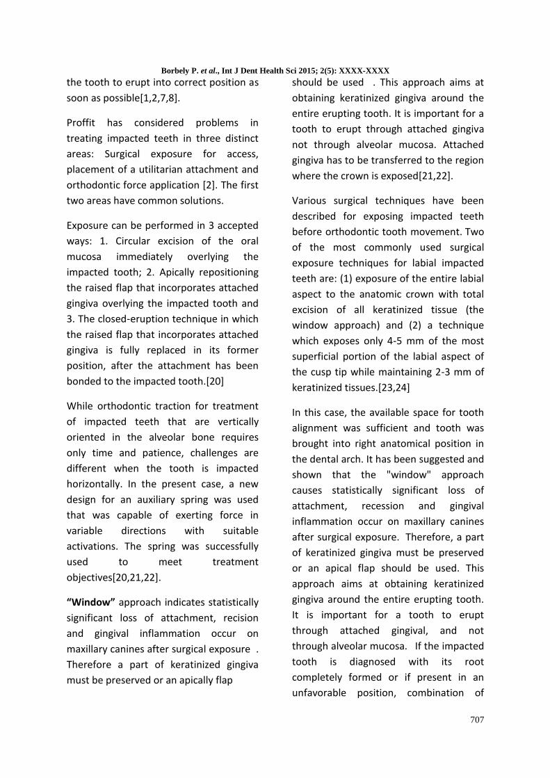
Exposure can be performed in 3 accepted
ways: 1. Circular excision of the oral
mucosa immediately overlying the
impacted tooth; 2. Apically repositioning
the raised flap that incorporates attached
gingiva overlying the impacted tooth and
3. The closed‑eruption technique in which
the raised flap that incorporates attached
gingiva is fully replaced in its former
position, after the attachment has been
bonded to the impacted tooth.[20]
While orthodontic traction for treatment
of impacted teeth that are vertically
oriented in the alveolar bone requires
only time and patience, challenges are
different when the tooth is impacted
horizontally. In the present case, a new
design for an auxiliary spring was used
that was capable of exerting force in
variable directions with suitable
activations. The spring was successfully
used to meet treatment
objectives[20,21,22].
“Window” approach indicates statistically
significant loss of attachment, recision
and gingival inflammation occur on
maxillary canines after surgical exposure .
Therefore a part of keratinized gingiva
must be preserved or an apically flap
should be used . This approach aims at
obtaining keratinized gingiva around the
entire erupting tooth. It is important for a
tooth to erupt through attached gingiva
not through alveolar mucosa. Attached
gingiva has to be transferred to the region
where the crown is exposed[21,22].
Various surgical techniques have been
described for exposing impacted teeth
before orthodontic tooth movement. Two
of the most commonly used surgical
exposure techniques for labial impacted
teeth are: (1) exposure of the entire labial
aspect to the anatomic crown with total
excision of all keratinized tissue (the
window approach) and (2) a technique
which exposes only 4-5 mm of the most
superficial portion of the labial aspect of
the cusp tip while maintaining 2-3 mm of
keratinized tissues.[23,24]
In this case, the available space for tooth
alignment was sufficient and tooth was
brought into right anatomical position in
the dental arch. It has been suggested and
shown that the "window" approach
causes statistically significant loss of
attachment, recession and gingival
inflammation occur on maxillary canines
after surgical exposure. Therefore, a part
of keratinized gingiva must be preserved
or an apical flap should be used. This
approach aims at obtaining keratinized
gingiva around the entire erupting tooth.
It is important for a tooth to erupt
through attached gingival, and not
through alveolar mucosa. If the impacted
tooth is diagnosed with its root
completely formed or if present in an
unfavorable position, combination of
Borbely P. et al., Int J Dent Health Sci 2015; 2(5): XXXX-XXXX
708
surgical and orthodontic treatment has to
be carried out.
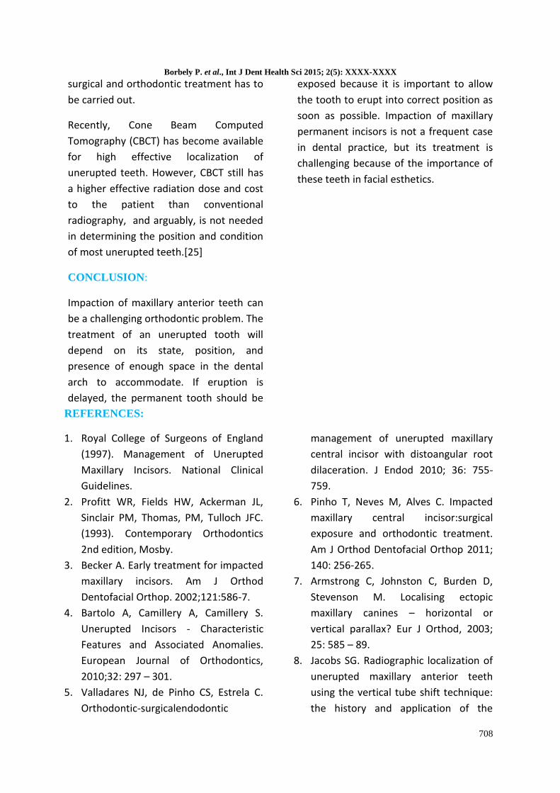
Recently, Cone Beam Computed
Tomography (CBCT) has become available
for high effective localization of
unerupted teeth. However, CBCT still has
a higher effective radiation dose and cost
to the patient than conventional
radiography, and arguably, is not needed
in determining the position and condition
of most unerupted teeth.[25]
CONCLUSION:
Impaction of maxillary anterior teeth can
be a challenging orthodontic problem. The
treatment of an unerupted tooth will
depend on its state, position, and
presence of enough space in the dental
arch to accommodate. If eruption is
delayed, the permanent tooth should be
exposed because it is important to allow
the tooth to erupt into correct position as
soon as possible. Impaction of maxillary
permanent incisors is not a frequent case
in dental practice, but its treatment is
challenging because of the importance of
these teeth in facial esthetics.
REFERENCES:
1. Royal College of Surgeons of England
(1997). Management of Unerupted
Maxillary Incisors. National Clinical
Guidelines.
2. Profitt WR, Fields HW, Ackerman JL,
Sinclair PM, Thomas, PM, Tulloch JFC.
(1993). Contemporary Orthodontics
2nd edition, Mosby.
3. Becker A. Early treatment for impacted
maxillary incisors. Am J Orthod
Dentofacial Orthop. 2002;121:586-7.
4. Bartolo A, Camillery A, Camillery S.
Unerupted Incisors - Characteristic
Features and Associated Anomalies.
European Journal of Orthodontics,
2010;32: 297 – 301.
5. Valladares NJ, de Pinho CS, Estrela C.
Orthodontic-surgicalendodontic
management of unerupted maxillary
central incisor with distoangular root
dilaceration. J Endod 2010; 36: 755-
759.
6. Pinho T, Neves M, Alves C. Impacted
maxillary central incisor:surgical
exposure and orthodontic treatment.
Am J Orthod Dentofacial Orthop 2011;
140: 256-265.
7. Armstrong C, Johnston C, Burden D,
Stevenson M. Localising ectopic
maxillary canines – horizontal or
vertical parallax? Eur J Orthod, 2003;
25: 585 – 89.
8. Jacobs SG. Radiographic localization of
unerupted maxillary anterior teeth
using the vertical tube shift technique:
the history and application of the
Borbely P. et al., Int J Dent Health Sci 2015; 2(5): XXXX-XXXX
703
method with some case reports. Am J
Orthod Dentofacial Orthop, 1999
Oct;116(4):415-423.
9. JM Nervina Cone beam computed
tomography use in orthodontics.
Australian Dental Journal 2012; 57: (1
Suppl): 95-102.
10. Ali Alqerban, Reinhilde Jacobs, Steffen
Fieuws, Guy Willems Comparison of
two cone beam computed tomographic
systems versus panoramic imaging for
localization of impacted maxillary
canines and detection of root
resorption. European Journal of
Orthodontics, 2011; 33:93–102.
11. K Horner, M Islam, L Flygare, K
Tsiklakis, E Whaites. Basic principles for
use of dental cone beam computed
tomography: consensus guidelines of
the European Academy of Dental and
Maxillofacial Radiology
Dentomaxillofacial Radiology, 2009;38:
187-195.
12. Ashkenazi M, Greenberg BP, Chodik G,
Rakocz M. Postoperative prognosis of
unerupted teeth after removal of
supernumerary teeth or odontomas,
2007 May;131(5):614-9.
13. Dalia Smailience, Antanas Sidlauskas,
Jevgenija Bucinskiene. Impaction of the
central maxillary incisor associated
with supernumerary teeth: initial
position and spontaneous eruption
timing. Stomatologija, Baltic Dental
And Maxillofacial Journal, 2006;8:103-
107
14. McNamara T, Woolfe SN, McNamara
CM. Orthodontic management of a
dilacerated maxillary central incisor
with an unusual sequel. J Clin
Orthod,1998;32:293–7.
15. S.Chaushu, I. Brin, Y. Ben-Bassat,
Y.Zilberman and A.Becker. Periodontal
status following surgical –orthodontic
alignment of impacted central incisors
with an open –eruption technique.
European journal of Orthodontics,
2003;25:579-584.
16. Nilesh V. Joshi. Periodontal status
following treatment of impacted
maxillary canines by closed eruption
technique: An overview and case
report. Compendium of continuing
education in Dentistry March 2014,
Vol35, Issue3.
17. Lin Tng-Tzen. Treatment of an
impacted dilacerated maxillary central
incisor. Am. J Orthod Dentofacial
Orthop. 1999;115:406-409.
18. Kuftinec. MM. Stom. D and Shapira.
The impacted maxillary canine; I
Review of concepts. 62; 317 - 325,
1955.
19. Becker A, editor. The orthodontic
treatment of impacted teeth. 1 st ed.
Mosby: Company; 1998. p. 53-85
20. Lundberg M, Wennström JL.
Development of gingiva following
surgical exposure of a facially
positioned unerupted incisor. J
Periodontol 1988;59:652‑5.
21. Vibhute PV. Versatile auxilliary
orthodontic spring for orthodontic
correction of impacted teeth. J Indian
Orthod Soc 2011;45:40‑7.
22. Xubair A, Graber TM, Vanarsdall R, Vig
KW. Orthodontics: Current Principles
Borbely P. et al., Int J Dent Health Sci 2015; 2(5): XXXX-XXXX
704
and Techniques. 5th ed. Philadelphia,
PA, USA: Mosby; 2012.
23. Lin YT. Treatment of an impacted
dilacerated maxillary central incisor.
Am J Orthod Dentofacial Orthop
1999;115:406-9
24. Spear FM, Kokich VG, Matthews DP.
Interdisciplinary management of
anterior dental esthetics. J Am Dent
Assoc. 2006;137(2):160-169.
25. Kang BC, Yoon SJ, Lee JS, Al-Rawi W,
Palomo JM. The use of cone beam
computed tomography for the
evaluation of pathology,
developmental anomalies and
traumatic injuries relevant to
orthodontics. Semin Orthod 2011;17:
20-33.
FIGURES:
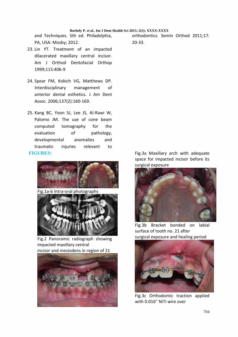
Fig.1a-b Intra-oral photographs
Fig.2 Panoramic radiograph showing impacted maxillary central incisor and mesiodens in region of 21
Fig.3a Maxillary arch with adequate space for impacted incisor before its surgical exposure
Fig.3b Bracket bonded on labial surface of tooth no. 21 after surgical exposure and healing period
Fig.3c Orthodontic traction applied with 0.016” NiTi wire over
Borbely P. et al., Int J Dent Health Sci 2015; 2(5): XXXX-XXXX
703
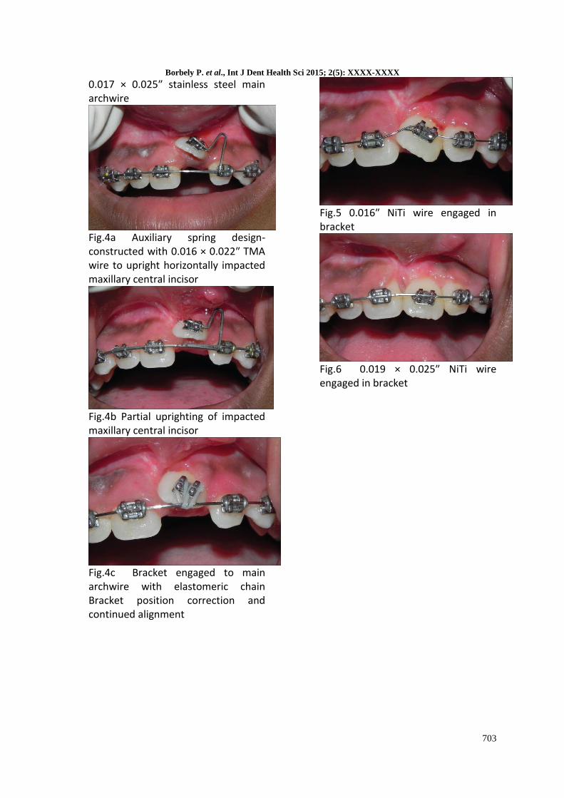
0.017 × 0.025″ stainless steel main archwire
Fig.4a Auxiliary spring design-constructed with 0.016 × 0.022″ TMA wire to upright horizontally impacted maxillary central incisor
Fig.4b Partial uprighting of impacted maxillary central incisor
Fig.4c Bracket engaged to main archwire with elastomeric chain Bracket position correction and continued alignment
Fig.5 0.016″ NiTi wire engaged in bracket
Fig.6 0.019 × 0.025″ NiTi wire engaged in bracket
Related Documents