iMedPub Journals ht tp://www.imedpub.com Medical Case Reports 2015 Vol. 1 No. 1:1 1 © Copyright iMedPub | This article is available in: http://medical-case-reports.imedpub.com Case report Vanessa Castro 1 , Lucas SE 2 , Daniel SC 3 , Jesus do PC 4 , Antonio Irineu TN 5 and Henrique MV 5 1 Graduate student, Postgraduate Program in Denstry, Dental School, Federal University of Uberlândia, Uberlândia, MG, Brazil 2 Graduate student, Postgraduate Program in Denstry, Dental School, Federal University of Bahia, Salvador, BA, Brazil 3 Formerly Fellow in Oral and Maxillofacial Surgery, Baylor College of Denstry, Texas A&M University System, Baylor University Medical Center, Dallas, TX – USA; currently in Private Pracce, Araraquara, SP, Brazil 4 Postgraduate Program in Denstry, Dental School, Federal University of Uberlândia, Uberlândia, MG, Brazil 5 Private Pracce, Salvador, BA, Brazil Corresponding author: Vanessa Castro DDS, MS. Graduate student, Postgraduate Program in Dentistry, Dental School, Federal University of Uberlândia, Uberlândia, Pará Avenue, 1720, Umuarama, Block 2U, room 07, 38400-902, Uberlândia, MG, Brazil [email protected] Tel: +55 34 3218-2550 +55 71 3451-4247 Introducon Changes or disrupons in the process of mandibular development can happen in the early stages of formaon and the malformaons may be isolated or part of a syndrome [1]. Several eologies are associated with growth disorders and abnormalies in the formaon of the mandibular condyle and related structures. These disorders may occur in the intrauterine stage at the end of the first trimester of pregnancy and result in diseases such as condylar aplasia or hypoplasia. Moreover, hyperplasia of the condyle is not visible at birth and appears to be gradually acquired during growth [2]. Classically, congenital disorders and altered development of the temporomandibular joint (TMJ) that affect the mandibular condyle have been classified according to four condions: aplasia, hypoplasia, hyperplasia and neoplasia [3]. Kaneyama et al. [2] considered the clinical and morphological aspects of TMJ, and classified the congenital deformies and developmental abnormalies of the mandibular condyle into three major groups and two subgroups: (1) hypoplasia or aplasia, including (I) primary condylar aplasia and hypoplasia; (II) secondary condylar hypoplasia (2) hyperplasia and (3) bifidity. Hypoplasia or aplasia of the mandibular condyle indicates underdevelopment or non-development and is mainly associated with various craniofacial abnormalies. Congenital condylar hypoplasia is characterized by unilateral or bilateral underdevelopment of the mandibular condyle, and it usually occurs as part of a systemic condion of the original first and second branchial arches, i.e., mandibulofacial dysostosis. Acquired (secondary) condylar hypoplasia can be caused by local factors (e.g., trauma, infecon of the mandibular bone or middle ear or radiaon) or systemic factors, such as toxic agents, rheumatoid arthris and mucopolysaccharoidosis [2,4]. Paents with diseases and TMJ-specific condions, such as missing or deformed anatomical structures (e.g., a lack of porons Orthodonc-Surgical Treatment of a Rare Case of Aplasia of the Mandibular Condyles Abstract Changes or disrupons in the process of mandibular development can happen in the early stages of formaon, and the malformaons may be isolated or part of a syndrome. Hypoplasia or aplasia of the mandibular condyles indicates underdevelopment or non-development and is associated with mulple craniofacial abnormalies. Paents who experience these congenital deformies may benefit from reconstrucon using total joint prostheses. In addion, orthognathic surgery may be required to treat severe respiratory syndromes and to correct dentofacial deformies to obtain funconal and aesthec results. Therefore, this paper presents a rare case of aplasia of the mandibular condyles and treatment of dentofacial deformies associated with severe obstrucve sleep apnea and the reconstrucon of the temporomandibular joint. Keywords: Aplasia; Mandibular condyles; Orthognathic surgery; TMJ surgery; Obstrucve sleep apnea Received: Sep 15, 2015, Accepted: Oct 15, 2015, Published: Oct 17, 2015

Welcome message from author
This document is posted to help you gain knowledge. Please leave a comment to let me know what you think about it! Share it to your friends and learn new things together.
Transcript
iMedPub Journalshttp://www.imedpub.com
Medical Case Reports2015
Vol. 1 No. 1:1
1© Copyright iMedPub | This article is available in: http://medical-case-reports.imedpub.com
Case report
Vanessa Castro1, Lucas SE2, Daniel SC3, Jesus do PC4, Antonio Irineu TN5 and Henrique MV5
1 Graduate student, Postgraduate Program in Dentistry, Dental School, Federal University of Uberlândia, Uberlândia, MG, Brazil
2 Graduate student, Postgraduate Program in Dentistry, Dental School, Federal University of Bahia, Salvador, BA, Brazil
3 Formerly Fellow in Oral and Maxillofacial Surgery, Baylor College of Dentistry, Texas A&M University System, Baylor University Medical Center, Dallas, TX – USA; currently in Private Practice, Araraquara, SP, Brazil
4 Postgraduate Program in Dentistry, Dental School, Federal University of Uberlândia, Uberlândia, MG, Brazil
5 Private Practice, Salvador, BA, Brazil
Corresponding author: Vanessa Castro
DDS, MS. Graduate student, Postgraduate Program in Dentistry, Dental School, Federal University of Uberlândia, Uberlândia, Pará Avenue, 1720, Umuarama, Block 2U, room 07, 38400-902, Uberlândia, MG, Brazil
Tel: +55 34 3218-2550 +55 71 3451-4247
IntroductionChanges or disruptions in the process of mandibular development can happen in the early stages of formation and the malformations may be isolated or part of a syndrome [1].
Several etiologies are associated with growth disorders and abnormalities in the formation of the mandibular condyle and related structures. These disorders may occur in the intrauterine stage at the end of the first trimester of pregnancy and result in diseases such as condylar aplasia or hypoplasia. Moreover, hyperplasia of the condyle is not visible at birth and appears to be gradually acquired during growth [2].
Classically, congenital disorders and altered development of the temporomandibular joint (TMJ) that affect the mandibular condyle have been classified according to four conditions: aplasia, hypoplasia, hyperplasia and neoplasia [3]. Kaneyama et al. [2] considered the clinical and morphological aspects of TMJ, and classified the congenital deformities and developmental abnormalities of the mandibular condyle into three major groups and two subgroups: (1) hypoplasia or aplasia, including (I) primary condylar aplasia and hypoplasia; (II) secondary condylar hypoplasia (2) hyperplasia and (3) bifidity.
Hypoplasia or aplasia of the mandibular condyle indicates underdevelopment or non-development and is mainly associated with various craniofacial abnormalities. Congenital condylar hypoplasia is characterized by unilateral or bilateral underdevelopment of the mandibular condyle, and it usually occurs as part of a systemic condition of the original first and second branchial arches, i.e., mandibulofacial dysostosis. Acquired (secondary) condylar hypoplasia can be caused by local factors (e.g., trauma, infection of the mandibular bone or middle ear or radiation) or systemic factors, such as toxic agents, rheumatoid arthritis and mucopolysaccharoidosis [2,4].
Patients with diseases and TMJ-specific conditions, such as missing or deformed anatomical structures (e.g., a lack of portions
Orthodontic-Surgical Treatment of a Rare Case of Aplasia of the Mandibular Condyles
AbstractChanges or disruptions in the process of mandibular development can happen in the early stages of formation, and the malformations may be isolated or part of a syndrome. Hypoplasia or aplasia of the mandibular condyles indicates underdevelopment or non-development and is associated with multiple craniofacial abnormalities. Patients who experience these congenital deformities may benefit from reconstruction using total joint prostheses. In addition, orthognathic surgery may be required to treat severe respiratory syndromes and to correct dentofacial deformities to obtain functional and aesthetic results. Therefore, this paper presents a rare case of aplasia of the mandibular condyles and treatment of dentofacial deformities associated with severe obstructive sleep apnea and the reconstruction of the temporomandibular joint.
Keywords: Aplasia; Mandibular condyles; Orthognathic surgery; TMJ surgery; Obstructive sleep apnea
Received: Sep 15, 2015, Accepted: Oct 15, 2015, Published: Oct 17, 2015
2This article is available in: http://medical-case-reports.imedpub.com
ARCHIVOS DE MEDICINAISSN 1698-9465
2015Vol. 1 No. 1:1
Medical Case Reports
of the condyle and ramus in cases of congenital deformities), can benefit from reconstruction using a total joint prosthesis [5]. The alloplastic provides a biomechanical solution for the treatment of severe arthritic disease, efficiently and effectively addresses the distorted and mutilated anatomy, eliminates the need for a second surgical site and reduces the associated morbidity [6].
In addition, mandibular advancement and/or counter-clockwise rotation of the maxillo-mandibular complex may be necessary to correct dentofacial deformities and to achieve optimal function and esthetics. In some circumstances, these repositioning movements can create a large gap between the fossa and mandibular structures. In such cases, the custom prosthesis can promote appropriate and individual adaptation to the anatomy of each patient [5].
In this report, a rare case of aplasia of the mandibular condyle is presented along with a discussion regarding the treatment of associated dentofacial deformities.
Case ReportPatient was 35 years of age, male and leucoderm. He attended the clinical maxillo-facial surgery department for evaluation of his facial deformity and dental malocclusion. He presented, as the main complaint, malocclusion, lip incompetence, severe obstructive sleep apnea (OSA), snoring and daytime sleepiness. The patient reported a history of orthodontic allowance for twelve years, including the extraction of the four first premolars. He also reported that he used CPAP (continuous positive airway pressure) for treatment of severe OSA but discontinued CPAP because of discomfort. He claimed to have hypertension and to have undergone cranioplasty for the treatment of craniosynostosis when he was seven and nine months old. Ten years ago, he underwent bilateral corneal transplantation.
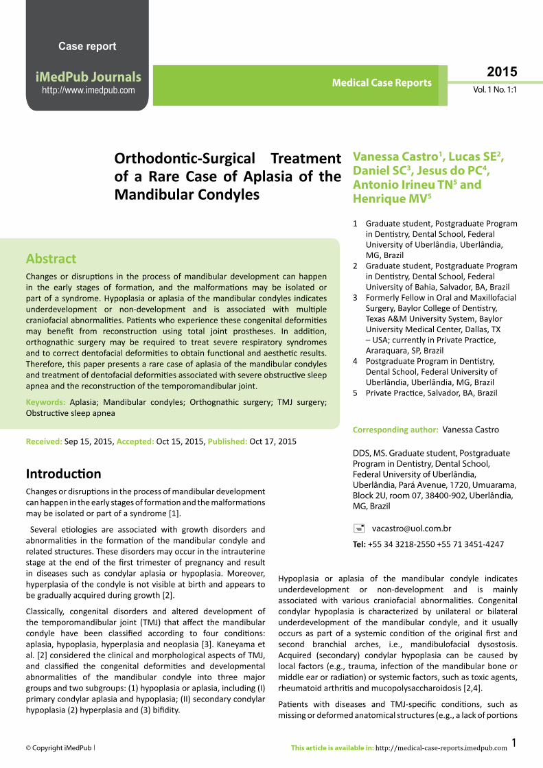
A physical facial examination revealed a severely convex profile, severe micrognathia, anteroposterior deficiency in the zigomatic and paranasal zone, vertical maxillary excess with overexposure of the upper incisors at rest (11 mm), lack of projection of the chin, lip incompetence and shortening of the chin-neck line (Figure 1). The intraoral examination revealed Angle Class II malocclusion and an overjet of 4.5 mm, horizontal bone loss and gingival recession in the maxilla and mandible (Figure 2).
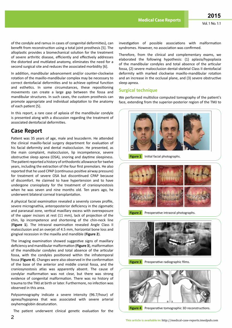
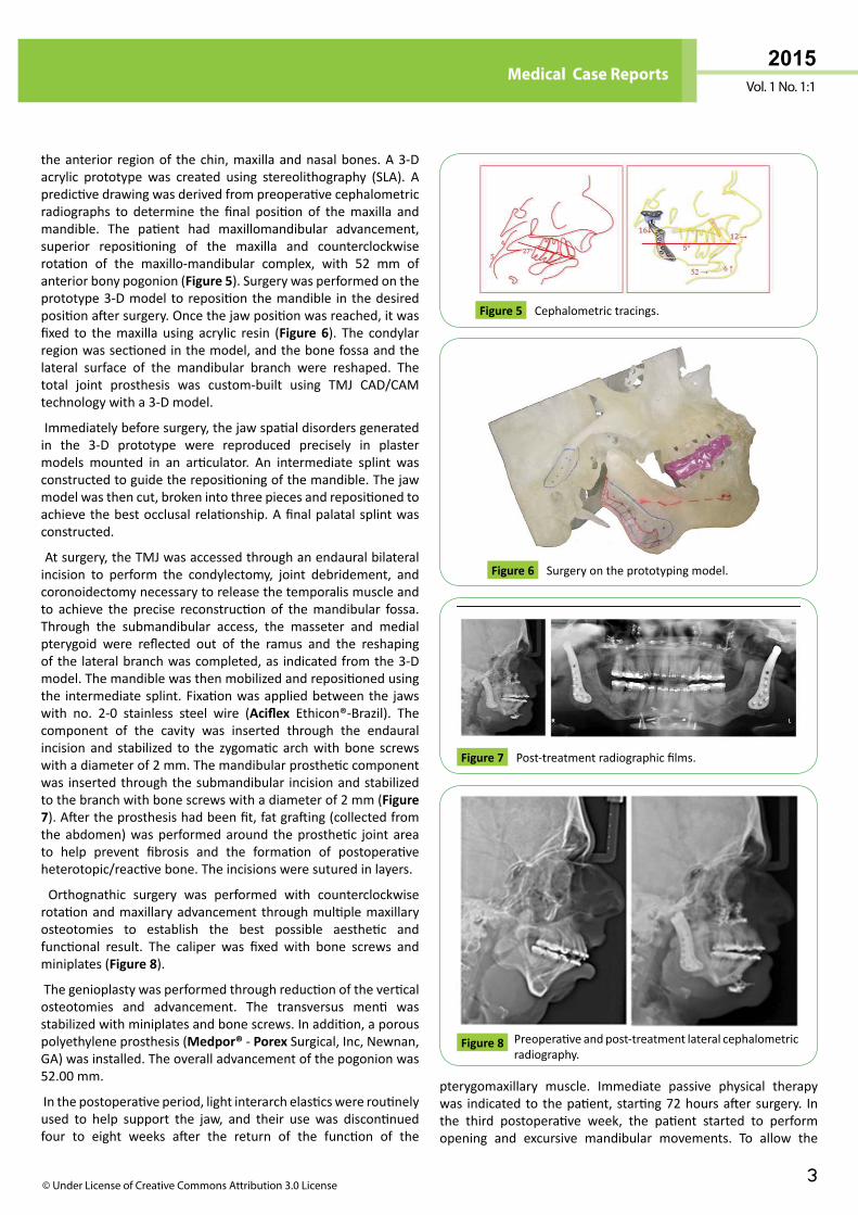
The imaging examination showed suggestive signs of maxillary deficiency and mandibular malformation (Figure 3), malformation of the mandibular condyles and total absence of the articular fossa, with the condyles positioned within the infratemporal fossa (Figure 4). Changes were also observed in the conformation of the base of the anterior and middle cranial fossa, and the craniosynostosis atlas was apparently absent. The cause of condylar malformation was not clear, but there was strong evidence of congenital malformation. There was no history of trauma to the TMJ at birth or later. Furthermore, no infection was observed in this area.
Polysomnography indicate a severe intensity (96.7/hour) of apnea/hypopnea that was associated with severe arterial oxyhemoglobin desaturation.
The patient underwent clinical genetic evaluation for the
investigation of possible associations with malformation syndromes. However, no association was confirmed.
Therefore, from the clinical and complementary exams, we elaborated the following hypothesis: (1) aplasia/hypoplasia of the mandibular condyles and total absence of the articular fossa, (2) severe malocclusion dental-skeletal Class II dentofacial deformity with marked clockwise maxillo-mandibular rotation and an increase in the occlusal plane, and (3) severe obstructive sleep apnea.
Surgical techniqueWe performed multislice computed tomography of the patient's face, extending from the superior-posterior region of the TMJ to
Initial facial photographs.Figure 1
Preoperative intraoral photographs.Figure 2
Preoperative radiographic films.Figure 3
Preoperative tomographic 3D reconstructions.Figure 4
3
ARCHIVOS DE MEDICINAISSN 1698-9465
2015Vol. 1 No. 1:1
Medical Case Reports
© Under License of Creative Commons Attribution 3.0 License
the anterior region of the chin, maxilla and nasal bones. A 3-D acrylic prototype was created using stereolithography (SLA). A predictive drawing was derived from preoperative cephalometric radiographs to determine the final position of the maxilla and mandible. The patient had maxillomandibular advancement, superior repositioning of the maxilla and counterclockwise rotation of the maxillo-mandibular complex, with 52 mm of anterior bony pogonion (Figure 5). Surgery was performed on the prototype 3-D model to reposition the mandible in the desired position after surgery. Once the jaw position was reached, it was fixed to the maxilla using acrylic resin (Figure 6). The condylar region was sectioned in the model, and the bone fossa and the lateral surface of the mandibular branch were reshaped. The total joint prosthesis was custom-built using TMJ CAD/CAM technology with a 3-D model.
Immediately before surgery, the jaw spatial disorders generated in the 3-D prototype were reproduced precisely in plaster models mounted in an articulator. An intermediate splint was constructed to guide the repositioning of the mandible. The jaw model was then cut, broken into three pieces and repositioned to achieve the best occlusal relationship. A final palatal splint was constructed.
At surgery, the TMJ was accessed through an endaural bilateral incision to perform the condylectomy, joint debridement, and coronoidectomy necessary to release the temporalis muscle and to achieve the precise reconstruction of the mandibular fossa. Through the submandibular access, the masseter and medial pterygoid were reflected out of the ramus and the reshaping of the lateral branch was completed, as indicated from the 3-D model. The mandible was then mobilized and repositioned using the intermediate splint. Fixation was applied between the jaws with no. 2-0 stainless steel wire (Aciflex Ethicon®-Brazil). The component of the cavity was inserted through the endaural incision and stabilized to the zygomatic arch with bone screws with a diameter of 2 mm. The mandibular prosthetic component was inserted through the submandibular incision and stabilized to the branch with bone screws with a diameter of 2 mm (Figure 7). After the prosthesis had been fit, fat grafting (collected from the abdomen) was performed around the prosthetic joint area to help prevent fibrosis and the formation of postoperative heterotopic/reactive bone. The incisions were sutured in layers.
Orthognathic surgery was performed with counterclockwise rotation and maxillary advancement through multiple maxillary osteotomies to establish the best possible aesthetic and functional result. The caliper was fixed with bone screws and miniplates (Figure 8).
The genioplasty was performed through reduction of the vertical osteotomies and advancement. The transversus menti was stabilized with miniplates and bone screws. In addition, a porous polyethylene prosthesis (Medpor® - Porex Surgical, Inc, Newnan, GA) was installed. The overall advancement of the pogonion was 52.00 mm.
In the postoperative period, light interarch elastics were routinely used to help support the jaw, and their use was discontinued four to eight weeks after the return of the function of the
pterygomaxillary muscle. Immediate passive physical therapy was indicated to the patient, starting 72 hours after surgery. In the third postoperative week, the patient started to perform opening and excursive mandibular movements. To allow the
Cephalometric tracings.Figure 5
Surgery on the prototyping model.Figure 6
Post-treatment radiographic films.Figure 7
Preoperative and post-treatment lateral cephalometric radiography.
Figure 8
4This article is available in: http://medical-case-reports.imedpub.com
ARCHIVOS DE MEDICINAISSN 1698-9465
2015Vol. 1 No. 1:1
Medical Case Reports
complete initial healing of the jaw bone, a liquid diet was followed for four months. The orthodontic treatment was completed to provide a final intercuspal interarch with adequate masticatory function. The orthodontic appliance was kept for eight months postoperatively and then removed by the orthodontist. Retainers were then installed in the maxilla and mandible.
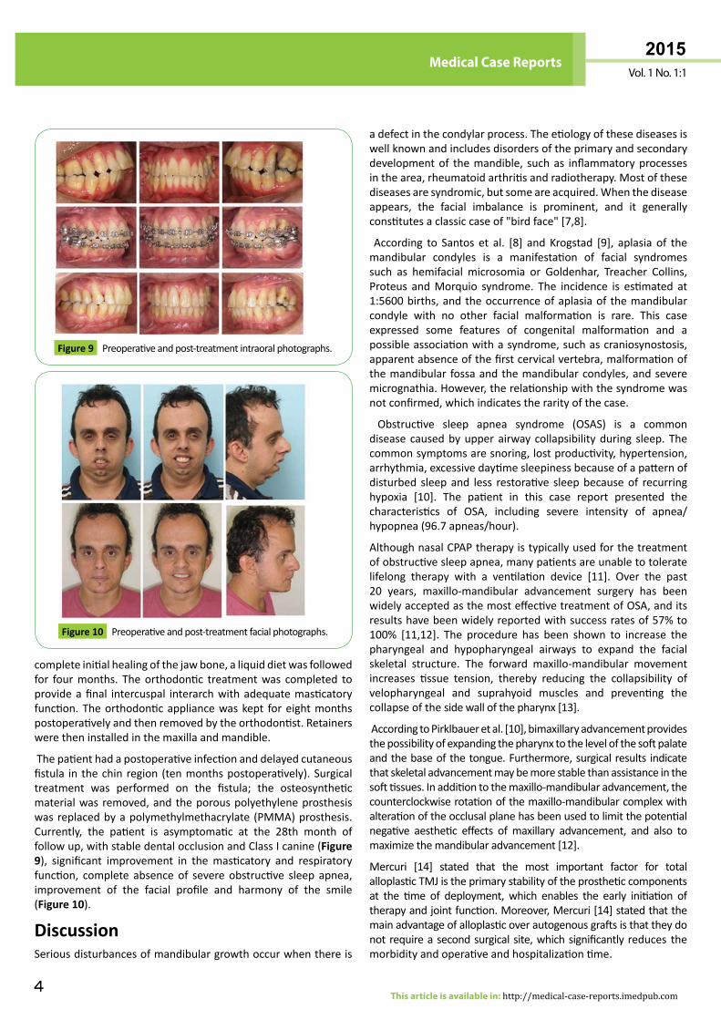
The patient had a postoperative infection and delayed cutaneous fistula in the chin region (ten months postoperatively). Surgical treatment was performed on the fistula; the osteosynthetic material was removed, and the porous polyethylene prosthesis was replaced by a polymethylmethacrylate (PMMA) prosthesis. Currently, the patient is asymptomatic at the 28th month of follow up, with stable dental occlusion and Class I canine (Figure 9), significant improvement in the masticatory and respiratory function, complete absence of severe obstructive sleep apnea, improvement of the facial profile and harmony of the smile (Figure 10).
DiscussionSerious disturbances of mandibular growth occur when there is
Preoperative and post-treatment intraoral photographs.Figure 9
Preoperative and post-treatment facial photographs.Figure 10
a defect in the condylar process. The etiology of these diseases is well known and includes disorders of the primary and secondary development of the mandible, such as inflammatory processes in the area, rheumatoid arthritis and radiotherapy. Most of these diseases are syndromic, but some are acquired. When the disease appears, the facial imbalance is prominent, and it generally constitutes a classic case of "bird face" [7,8].
According to Santos et al. [8] and Krogstad [9], aplasia of the mandibular condyles is a manifestation of facial syndromes such as hemifacial microsomia or Goldenhar, Treacher Collins, Proteus and Morquio syndrome. The incidence is estimated at 1:5600 births, and the occurrence of aplasia of the mandibular condyle with no other facial malformation is rare. This case expressed some features of congenital malformation and a possible association with a syndrome, such as craniosynostosis, apparent absence of the first cervical vertebra, malformation of the mandibular fossa and the mandibular condyles, and severe micrognathia. However, the relationship with the syndrome was not confirmed, which indicates the rarity of the case.
Obstructive sleep apnea syndrome (OSAS) is a common disease caused by upper airway collapsibility during sleep. The common symptoms are snoring, lost productivity, hypertension, arrhythmia, excessive daytime sleepiness because of a pattern of disturbed sleep and less restorative sleep because of recurring hypoxia [10]. The patient in this case report presented the characteristics of OSA, including severe intensity of apnea/hypopnea (96.7 apneas/hour).
Although nasal CPAP therapy is typically used for the treatment of obstructive sleep apnea, many patients are unable to tolerate lifelong therapy with a ventilation device [11]. Over the past 20 years, maxillo-mandibular advancement surgery has been widely accepted as the most effective treatment of OSA, and its results have been widely reported with success rates of 57% to 100% [11,12]. The procedure has been shown to increase the pharyngeal and hypopharyngeal airways to expand the facial skeletal structure. The forward maxillo-mandibular movement increases tissue tension, thereby reducing the collapsibility of velopharyngeal and suprahyoid muscles and preventing the collapse of the side wall of the pharynx [13].
According to Pirklbauer et al. [10], bimaxillary advancement provides the possibility of expanding the pharynx to the level of the soft palate and the base of the tongue. Furthermore, surgical results indicate that skeletal advancement may be more stable than assistance in the soft tissues. In addition to the maxillo-mandibular advancement, the counterclockwise rotation of the maxillo-mandibular complex with alteration of the occlusal plane has been used to limit the potential negative aesthetic effects of maxillary advancement, and also to maximize the mandibular advancement [12].
Mercuri [14] stated that the most important factor for total alloplastic TMJ is the primary stability of the prosthetic components at the time of deployment, which enables the early initiation of therapy and joint function. Moreover, Mercuri [14] stated that the main advantage of alloplastic over autogenous grafts is that they do not require a second surgical site, which significantly reduces the morbidity and operative and hospitalization time.

5
ARCHIVOS DE MEDICINAISSN 1698-9465
2015Vol. 1 No. 1:1
Medical Case Reports
© Under License of Creative Commons Attribution 3.0 License
The option to use dentures in this case was substantiated by the characteristics of the articular components arising from the aplasia of the mandibular condyles and the total absence of the articular fossa, which was associated with the need to perform a surgical breakthrough (52 mm) and counterclockwise rotation of the maxillo-mandibular complex for the treatment of sleep apnea and skeletal deformity class II.
Conclusions In cases of jaw malformations, in which the anatomy of the condyles is totally changed or malformed, joint reconstruction with custom dentures is indicated. These prostheses permit large surgical maxillo-mandibular movements, thus enabling the correction of skeletal deformities and permitting early physical therapy and joint function.
Conflicts of Interest and Source of Funding: none declared.

6This article is available in: http://medical-case-reports.imedpub.com
ARCHIVOS DE MEDICINAISSN 1698-9465
2015Vol. 1 No. 1:1
Medical Case Reports
References1 Venturin JS, Shintaku WH, Shigeta Y, Ogawa T, Le B, et al. (2010)
Temporomandibular joint condylar abnormality: evaluation, treatment planning, and surgical approach. J Oral Maxillofac Surg 68: 1189-1196.
2 Kaneyama K, Segami N, Hatta T (2008) Congenital deformities and developmental abnormalities of the mandibular condyle in the temporomandibular joint. Congenital Anomalies 48: 118-125.
3 Sala PS, Vázquez DE, Rodríguez BA, Gay EC (2010) Bifid mandibular condyle: a disorder in itsown right? JADA. 2010; 141: 1076-1085.
4 Tank W, Wright D, Iizuka T (1998) Unilateral dysplasia of the mandibular condyle: report of a case. J Oral Maxillofac Surg 56: 765-769.
5 Wolford LM, Rodrigues DB, McPhillips Am (2010) Management of the infected temporomandibular joint total joint prosthesis. J Oral Maxillofac Surg 68: 2810-2823.
6 Wolford LM (2003) Concomitant temporomandibular joint and orthognathic surgery. J Oral Maxillofac Surg 61: 1198-1204.
7 Ferri J, Carneiro JM, Lemiere E, Vereecke F, Baralle MM (2006) Severe congenital hypoplasia of the mandibular condyle-diagnosis and treatment: a report of 2 cases. J Oral Maxillofac Surg 64: 972-980.
8 Santos KC, Campos Jr AF, Kanajia JF, Lascala CA, Costa C, et al. (2009) Mandibular condyle aplasia: case report. Rev Odontol UNESP 38: 375-378.
9 Krogstad O (1997) Aplasia of the mandibular condyle. Eur J Orthod 19: 483-489.
10 Pirklbauer K, Russmueller G, Stiebellehner L, Nell C, Sinko K, et al. (2011) Maxillomandibular advancement for treatment of obstructive sleep apnea syndrome: a systematic review. J Oral Maxillofac Surg 69: 165-176.
11 Hendler BH, Costello BJ, Silverstein K (2001) A protocol for uvulopalatopharyngoplasty, mortised genioplasty, and maxillomandibular advancement in patients with obstructive sleep apnea: an analysis of 40 cases. J Oral Maxillofac Surg 59: 892.
12 Li KK, Riley RW, Powell NB (2001) Patient’s perception of the facial appearance after maxillomandibular advancement for obstructive sleep apnea syndrome. J Oral Maxillofac Surg 59: 377.
13 Li KK (2011) Obstructive sleep apnea. J Oral Maxillofac Surg 69: 687-694.
14 Louis GM (2007) A rationale for total alloplastic temporomandibular joint reconstruction in the management of idiopathic/progressive condylar resorption. J Oral Maxillofac Surg 65: 1600-1609.
Related Documents