J Bagh College Dentistry Vol. 27(3), September 2015 Orthodontic considerations Pedodontics, Orthodontics and Preventive Dentistry130 Orthodontic considerations of functional occlusion in Class І normal occlusion Doaa B. Al-Nassar, B.D.S. (1) Hadeel A. Al-Hashimi, B.D.S., M.Sc. (2) ABSTRACT Background: The therapeutic goal of orthodontic treatment is to establish ideal occlusion which includes both static and functional aspects. The objective of this study was to clarify functional occlusal treatment goals by analyzing functional occlusion in subjects with established normal occlusion and identify the differences between canine protected occlusion and group function occlusion. Materials and Methods: The sample consisted of 62 subjects with normal occlusion and with an age range of (18-25 years).Functional occlusal contacts during lateral excursion were identified on a fully adjustable articulator, and then the samples were classified according to: 1) Type of functional occlusion: A) Canine protected occlusion group (canine protected occlusion on both working sides). B) Group function occlusion group (group function occlusion on both working sides). C) Mixed functional occlusion group (canine protected occlusion on one side and group function occlusion on the other side ) . 2) Working side contact into: Canine protected occlusion (62 sides) and group function occlusion (62 sides). Then different variables were analyzed. Results: Results revealed that vertical canine overlap, position of maxillary canine and 1 st molar mesiobuccal cusp tip to the center of opposing embrasure/groove, and arch form analysis had significant difference between groups. Adding to that, some of the maxillary and mandibular teeth were significantly differed between groups in regard to crown angulation and inclination. Conclusions: It was concluded that the vertical canine overlap, position of maxillary canine and 1 st molar mesiobuccal cusp tip to the center of the opposing embrasure/groove, arch form harmony were important functional aspects of orthodontic treatment goals. Key words: Orthodontic considerations, functional occlusion, normal occlusion. (J Bagh Coll Dentistry 2015; 27(3):130- 139). INTRODUCTION The therapeutic goal of orthodontic treatment is to create an occlusion that has a close resemblance to established normal occlusal features. This orthodontic picture of "ideal occlusion" is largely the result of work by Angle (1900) and Andrews (1972). In assessing the quality of completed orthodontic treatment, considerable emphasis is placed on the static occlusal relationship with less emphasis on the importance of the functional occlusion (1) . Furthermore; orthodontic classifications are related more to anatomic and esthetic standards than to neuromuscular harmony and functional stability (2) . However, the optimal functional occlusion type has not been so easily identified (3) . As widely known, the specialist needs to be expertise in all aspects of functional occlusion because a very close relationship has been found between orthodontic treatment and all components of the masticatory system. The Angle’s classification is a good way to start with, but no way offers adequate parameters for defining treatment goals. Many occlusions pres- (1)Master student. Department of Orthodontics. College of Dentistry, University of Baghdad. (2)Professor. Department of Orthodontics. College of Dentistry, University of Baghdad. senting solid class I relationship may present several pathological aspects (4) . The gentle lateral and protrusive shift is not only necessary from the standpoint of mandibular movement, but also necessary from the orthodontist's point of view in terms of post- treatment stability of the tooth positions (5) . The achievement of the ideal functional occlusion provided a satisfactory stability after orthodontic treatment. Thus, the establishment of the static and functional criteria leads to the success of the orthodontic treatment by minimizing orthodontic relapse and prevents the appearance of occlusal pathologies (6) . After reviewing the related studies and articles, no previous study was found to identify the occlusal features that related to each type of functional occlusion. Normal occlusion is of great importance since it is the aim of orthodontic treatment. So this present study formulated to highlight on determinants of functional occlusion as an orthodontic treatment goals. MATERIALS AND METHOD Sample The sample of the present study consisted of 62 subjects (20 males and 42 females), which

Welcome message from author
This document is posted to help you gain knowledge. Please leave a comment to let me know what you think about it! Share it to your friends and learn new things together.
Transcript
J Bagh College Dentistry Vol. 27(3), September 2015 Orthodontic considerations
Pedodontics, Orthodontics and Preventive Dentistry130
Orthodontic considerations of functional occlusion in Class І normal occlusion
Doaa B. Al-Nassar, B.D.S. (1)
Hadeel A. Al-Hashimi, B.D.S., M.Sc. (2)
ABSTRACT Background: The therapeutic goal of orthodontic treatment is to establish ideal occlusion which includes both static and functional aspects. The objective of this study was to clarify functional occlusal treatment goals by analyzing functional occlusion in subjects with established normal occlusion and identify the differences between canine protected occlusion and group function occlusion. Materials and Methods: The sample consisted of 62 subjects with normal occlusion and with an age range of (18-25 years).Functional occlusal contacts during lateral excursion were identified on a fully adjustable articulator, and then the samples were classified according to: 1) Type of functional occlusion: A) Canine protected occlusion group (canine protected occlusion on both working sides). B) Group function occlusion group (group function occlusion on both working sides). C) Mixed functional occlusion group (canine protected occlusion on one side and group function occlusion on the other side ). 2) Working side contact into: Canine protected occlusion (62 sides) and group function occlusion (62 sides). Then different variables were analyzed. Results: Results revealed that vertical canine overlap, position of maxillary canine and 1st molar mesiobuccal cusp tip to the center of opposing embrasure/groove, and arch form analysis had significant difference between groups. Adding to that, some of the maxillary and mandibular teeth were significantly differed between groups in regard to crown angulation and inclination. Conclusions: It was concluded that the vertical canine overlap, position of maxillary canine and 1st molar mesiobuccal cusp tip to the center of the opposing embrasure/groove, arch form harmony were important functional aspects of orthodontic treatment goals. Key words: Orthodontic considerations, functional occlusion, normal occlusion. (J Bagh Coll Dentistry 2015; 27(3):130-139).
INTRODUCTION The therapeutic goal of orthodontic treatment
is to create an occlusion that has a close resemblance to established normal occlusal features. This orthodontic picture of "ideal occlusion" is largely the result of work by Angle (1900) and Andrews (1972). In assessing the quality of completed orthodontic treatment, considerable emphasis is placed on the static occlusal relationship with less emphasis on the importance of the functional occlusion (1). Furthermore; orthodontic classifications are related more to anatomic and esthetic standards than to neuromuscular harmony and functional stability (2).
However, the optimal functional occlusion type has not been so easily identified (3). As widely known, the specialist needs to be expertise in all aspects of functional occlusion because a very close relationship has been found between orthodontic treatment and all components of the masticatory system. The Angle’s classification is a good way to start with, but no way offers adequate parameters for defining treatment goals. Many occlusions pres- (1)Master student. Department of Orthodontics. College of Dentistry, University of Baghdad. (2)Professor. Department of Orthodontics. College of Dentistry, University of Baghdad.
senting solid class I relationship may present several pathological aspects (4).
The gentle lateral and protrusive shift is not only necessary from the standpoint of mandibular movement, but also necessary from the orthodontist's point of view in terms of post-treatment stability of the tooth positions (5). The achievement of the ideal functional occlusion provided a satisfactory stability after orthodontic treatment. Thus, the establishment of the static and functional criteria leads to the success of the orthodontic treatment by minimizing orthodontic relapse and prevents the appearance of occlusal pathologies (6).
After reviewing the related studies and articles, no previous study was found to identify the occlusal features that related to each type of functional occlusion. Normal occlusion is of great importance since it is the aim of orthodontic treatment. So this present study formulated to highlight on determinants of functional occlusion as an orthodontic treatment goals. MATERIALS AND METHOD Sample
The sample of the present study consisted of 62 subjects (20 males and 42 females), which

J Bagh J Bagh College Dentistry
Pedodontics, Orthodontics and Preventive Dentistry
were selected from the students of College of Dentistry/Baghdad Universitycriteria were considered in sample 1. Iraqi Arab adult subjects with age from 18
years.2. Clinically skeletal class I.3. No clear asymmetries in the dental arches
and face and no history of previous facial trauma assessed by visual examination.
4. They have full set of permanent teeth in both jaws "excluding the 3
5. They have bilateral class I molar and canine relationwith normaland well
6. No previous occlusal adjustments and/or teeth attrition.
7. No restorations or carious lesions that extended to cusps inclines or incisal edges and no artificial prosthesis.
8. No pathologic periodontal problems according to the gingival index.
9. No TMJ problem according to research diagnostic criteria and clinical exa
Method For each participant informed consent was obtained before the start of examination.occlusion of each subject was assessed by intraoral examination and the fwas identified by clinical inspecleft side and then confirmed from the articulator. The subjects included in the sample were distributed into three main groups1. Canine protected occlusion
subjects)occlusion during lateral mandibular movement, contacts occur only between upper and lower canine on the working side.
2. Group function occlusionBoth sides are group function occlusion during lateral mandibular movement, more than contact
3. Mixed functional occlusionsubjects)occlusion and the otocclusion.After completion of clinical examination
pairs of dental casts (2 upper and 2 lower)producedother for measurements. Dental casts for measurementthe parallelism of the upper and lower cast bases to the occlusal plane
The relation of the maxillary arch to the temporomandibular joint (hinge axis) was
College Dentistry
Pedodontics, Orthodontics and Preventive Dentistry
were selected from the students of College of Dentistry/Baghdad Universitycriteria were considered in sample
Iraqi Arab adult subjects with age from 18years. Clinically skeletal class I.No clear asymmetries in the dental arches
and face and no history of previous facial trauma assessed by visual examination.They have full set of permanent teeth in both jaws "excluding the 3They have bilateral class I molar and canine relationships, class I incisor classification with normal over
well-aligned archesNo previous occlusal adjustments and/or teeth attrition. No restorations or carious lesions that extended to cusps inclines or incisal edges and no artificial prosthesis. No pathologic periodontal problems according to the gingival index.No TMJ problem according to research diagnostic criteria and clinical exa
For each participant informed consent was obtained before the start of examination.occlusion of each subject was assessed by intraoral examination and the fwas identified by clinical inspecleft side and then confirmed from the articulator. The subjects included in the sample were distributed into three main groups
Canine protected occlusionsubjects): Both sides are canine protected occlusion during lateral mandibular movement, contacts occur only between upper and lower canine on the working side.Group function occlusionBoth sides are group function occlusion during lateral mandibular movement, more than one tooth on the working side acontact. Mixed functional occlusionsubjects): One side is canine protected occlusion and the otocclusion. After completion of clinical examination
pairs of dental casts (2 upper and 2 lower)produced; one for mounother for measurements. Dental casts for measurements were prepared in a way to ensure the parallelism of the upper and lower cast bases to the occlusal plane (Figure 1, A and B)
The relation of the maxillary arch to the temporomandibular joint (hinge axis) was
College Dentistry
Pedodontics, Orthodontics and Preventive Dentistry
were selected from the students of College of Dentistry/Baghdad University. The following criteria were considered in sample
Iraqi Arab adult subjects with age from 18
Clinically skeletal class I. No clear asymmetries in the dental arches
and face and no history of previous facial trauma assessed by visual examination.They have full set of permanent teeth in both jaws "excluding the 3rd molar".They have bilateral class I molar and canine
ships, class I incisor classification -jet and overbite (2
aligned arches No previous occlusal adjustments and/or teeth
No restorations or carious lesions that extended to cusps inclines or incisal edges and no artificial prosthesis. No pathologic periodontal problems according to the gingival index.No TMJ problem according to research diagnostic criteria and clinical exa
For each participant informed consent was obtained before the start of examination.occlusion of each subject was assessed by intraoral examination and the functional occlusion was identified by clinical inspection left side and then confirmed from the articulator. The subjects included in the sample were distributed into three main groups:
Canine protected occlusion: Both sides are canine protected
occlusion during lateral mandibular movement, contacts occur only between upper and lower canine on the working side.Group function occlusion'GFO'Both sides are group function occlusion during lateral mandibular movement, more
one tooth on the working side a
Mixed functional occlusion: One side is canine protected
occlusion and the other is group function
After completion of clinical examinationpairs of dental casts (2 upper and 2 lower)
; one for mounting in articulator and the other for measurements. Dental casts for
were prepared in a way to ensure the parallelism of the upper and lower cast bases
(Figure 1, A and B)The relation of the maxillary arch to the
temporomandibular joint (hinge axis) was
College Dentistry Vol. 27(3), September 2015
Pedodontics, Orthodontics and Preventive Dentistry
were selected from the students of College of The following
criteria were considered in sample selection: Iraqi Arab adult subjects with age from 18-
No clear asymmetries in the dental arches and face and no history of previous facial trauma assessed by visual examination. They have full set of permanent teeth in both
molar". They have bilateral class I molar and canine
ships, class I incisor classification jet and overbite (2-4 mm)
No previous occlusal adjustments and/or teeth
No restorations or carious lesions that extended to cusps inclines or incisal edges
No pathologic periodontal problems according to the gingival index. No TMJ problem according to research diagnostic criteria and clinical examination.
For each participant informed consent was obtained before the start of examination. Static occlusion of each subject was assessed by intra
unctional occlusion tion for right and
left side and then confirmed from the articulator. The subjects included in the sample were
: Canine protected occlusion'CPO'(22
: Both sides are canine protected occlusion during lateral mandibular movement, contacts occur only between upper and lower canine on the working side.
'GFO' (22 subjects)Both sides are group function occlusion during lateral mandibular movement, more
one tooth on the working side are in
Mixed functional occlusion 'MFO' (18 : One side is canine protected
her is group function
After completion of clinical examination, two pairs of dental casts (2 upper and 2 lower) were
ting in articulator and the other for measurements. Dental casts for
were prepared in a way to ensure the parallelism of the upper and lower cast bases
(Figure 1, A and B). The relation of the maxillary arch to the
temporomandibular joint (hinge axis) was
7(3), September 2015
Pedodontics, Orthodontics and Preventive Dentistry131
were selected from the students of College of The following
-25
No clear asymmetries in the dental arches and face and no history of previous facial
They have full set of permanent teeth in both
They have bilateral class I molar and canine ships, class I incisor classification
4 mm)
No previous occlusal adjustments and/or teeth
No restorations or carious lesions that extended to cusps inclines or incisal edges
No pathologic periodontal problems
No TMJ problem according to research mination.
For each participant informed consent was Static
occlusion of each subject was assessed by intra-unctional occlusion
for right and left side and then confirmed from the articulator. The subjects included in the sample were
(22 : Both sides are canine protected
occlusion during lateral mandibular movement, contacts occur only between upper and lower canine on the working side.
(22 subjects): Both sides are group function occlusion during lateral mandibular movement, more
re in
(18 : One side is canine protected
her is group function
two were
ting in articulator and the other for measurements. Dental casts for
were prepared in a way to ensure the parallelism of the upper and lower cast bases
The relation of the maxillary arch to the temporomandibular joint (hinge axis) was
transferred to fully adjustable articulator (Stratos300, Ivoclar Vivadent, Austria) by the mean of Universal TransferVivadent, Austria)
Figure
registration: (A) Frontal view; (B) Lateral
modeling wax (of 4 sheets thickness) softened in a hot water at 55on the bite fork of UTS3D. bow to the articulator was done according to the ''personalized model transfer with the UTS3D'' instruction (instruction supplied with the articulator), previously prepared for mounting. During mountingcentric fixation lock to keep the models in centric occlufixation lock was opened, and the adjustable incisal plate was mounted, at this time the articulator was ready (Figure 3
Mounted models in centric position; (B) articulator ready for eccentric movement.
Then, working side and noncontacts were recorded by manipulating the articulator to the lat
7(3), September 2015
131
transferred to fully adjustable articulator (Stratos300, Ivoclar Vivadent, Austria) by the mean of Universal TransferVivadent, Austria)
Figure 1: Preparation of to be parallel to the occlusal plane: (A)
Figure 2: Transfer bow during bite registration: (A) Frontal view; (B) Lateral
The subject was asked to bite onmodeling wax (of 4 sheets thickness) softened in a hot water at 55on the bite fork of UTS3D. bow to the articulator was done according to the ''personalized model transfer with the UTS3D'' instruction (instruction supplied with the articulator), using the models which were previously prepared for mounting. During mounting, the articulator was held in centric by centric fixation lock to keep the models in centric occlusion (Figure fixation lock was opened, and the adjustable incisal plate was mounted, at this time the articulator was ready (Figure 3, B).
Figure 3: Articulator adjustment: (A) Mounted models in centric position; (B) articulator ready for eccentric movement.
Then, working side and noncontacts were recorded by manipulating the articulator to the lat
7(3), September 2015 Orthodontic
transferred to fully adjustable articulator (Stratos300, Ivoclar Vivadent, Austria) by the mean of Universal Transfer bow System (UTS3D, Ivoclar Vivadent, Austria) (Figure 2,
1: Preparation of to be parallel to the occlusal plane: (A)
upper; (B) lower.
2: Transfer bow during bite registration: (A) Frontal view; (B) Lateral
view.
subject was asked to bite onmodeling wax (of 4 sheets thickness) softened in a hot water at 55on the bite fork of UTS3D. bow to the articulator was done according to the ''personalized model transfer with the UTS3D'' instruction (instruction supplied with the
using the models which were previously prepared for mounting. During
the articulator was held in centric by centric fixation lock to keep the models in centric
(Figure 3, A). Then, the centric fixation lock was opened, and the adjustable incisal plate was mounted, at this time the articulator was ready for eccentric movement
3: Articulator adjustment: (A) Mounted models in centric position; (B) articulator ready for eccentric movement.
Then, working side and noncontacts were recorded by manipulating the articulator to the lateral cusp to cusp position.
Orthodontic
transferred to fully adjustable articulator (Stratos300, Ivoclar Vivadent, Austria) by the mean of
bow System (UTS3D, Ivoclar (Figure 2, A and B).
1: Preparation of dental casts bases to be parallel to the occlusal plane: (A)
upper; (B) lower.
2: Transfer bow during bite registration: (A) Frontal view; (B) Lateral
view. subject was asked to bite on a
modeling wax (of 4 sheets thickness) which softened in a hot water at 55ºC (7) and was pon the bite fork of UTS3D. Transfer of the face bow to the articulator was done according to the ''personalized model transfer with the UTS3D'' instruction (instruction supplied with the
using the models which were previously prepared for mounting. During
the articulator was held in centric by centric fixation lock to keep the models in centric
, A). Then, the centric fixation lock was opened, and the adjustable incisal plate was mounted, at this time the
for eccentric movement
3: Articulator adjustment: (A) Mounted models in centric position; (B) articulator ready for eccentric movement.
Then, working side and non-working side contacts were recorded by manipulating the
eral cusp to cusp position.
Orthodontic considerations
transferred to fully adjustable articulator (Stratos-300, Ivoclar Vivadent, Austria) by the mean of
bow System (UTS3D, Ivoclar
dental casts bases
to be parallel to the occlusal plane: (A)
2: Transfer bow during bite
registration: (A) Frontal view; (B) Lateral
a piece of which was was placed
Transfer of the face bow to the articulator was done according to the ''personalized model transfer with the UTS3D'' instruction (instruction supplied with the
using the models which were previously prepared for mounting. During
the articulator was held in centric by centric fixation lock to keep the models in centric
, A). Then, the centric fixation lock was opened, and the adjustable incisal plate was mounted, at this time the
for eccentric movement
3: Articulator adjustment: (A)
Mounted models in centric position; (B) the articulator ready for eccentric movement.
working side contacts were recorded by manipulating the
eral cusp to cusp position.
considerations
J Bagh J Bagh College Dentistry
Pedodontics, Orthodontics and Preventive Dentistry
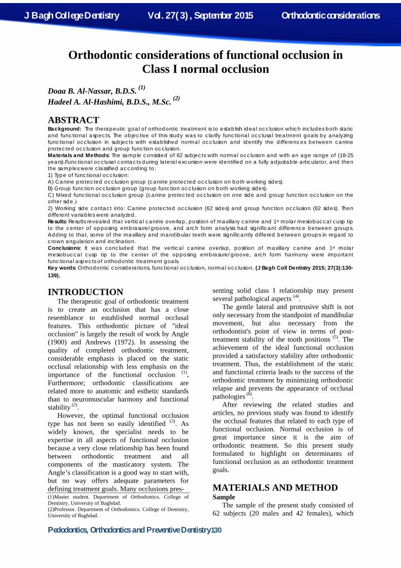
The groups were identified according to the type of functional occlusion (Fi
Figure 4: Working side contact: (A) Canine protected occlusion; (B) Group function
On the model prepared for measurements; the
facial axis of the clinical crown and its midpoint (facial axis point), was marked with a pencil each crown of all teeth except third molars. The facial axis of the clinical crown represents the most prominent portion of the central lobe on the facial surface of each crown; for molars, it represents the buccal groove that separatwo large famarked as the midpoint of the faclinical crown
Figure 5
The facial axis of the clinical
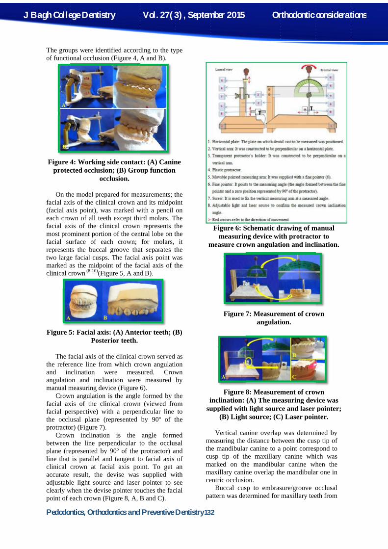
the reference line from which crown angulationand inclination were measured. angulation and inclination were measured by manual measuring device (Figure
Crown angulation is the angle formed by the facial axis of the clinical crown (viewed from facial perspective) with a perpendicular line to the occlusal plane (represented by protractor) (Figure 7
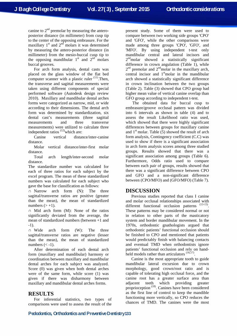
Crown inclination is the angle formed between the line perpendicular to the occlusal plane (represented by 90º of the protractor) and line that is parallel and tangent to facial axis of clinical crown at facial axis point. To get an accurate result, the devise was suppliedadjustable light source and laser pointer to see clearly when the devise pointer touches the facipoint of each crown
College Dentistry
Pedodontics, Orthodontics and Preventive Dentistry
groups were identified according to the type f functional occlusion (Fi
4: Working side contact: (A) Canine protected occlusion; (B) Group function
occlusion.
On the model prepared for measurements; the facial axis of the clinical crown and its midpoint (facial axis point), was marked with a pencil each crown of all teeth except third molars. The facial axis of the clinical crown represents the most prominent portion of the central lobe on the facial surface of each crown; for molars, it represents the buccal groove that separatwo large facial cusps. marked as the midpoint of the faclinical crown (8-10)(Figure 5, A and B)
5: Facial axis: (A) Anterior teeth; (B) Posterior teeth.
The facial axis of the clinical reference line from which crown angulation
and inclination were measured. angulation and inclination were measured by manual measuring device (Figure
Crown angulation is the angle formed by the facial axis of the clinical crown (viewed from
l perspective) with a perpendicular line to the occlusal plane (represented by protractor) (Figure 7).
Crown inclination is the angle formed between the line perpendicular to the occlusal plane (represented by 90º of the protractor) and
t is parallel and tangent to facial axis of clinical crown at facial axis point. To get an accurate result, the devise was suppliedadjustable light source and laser pointer to see clearly when the devise pointer touches the facipoint of each crown (Figure 8
College Dentistry
Pedodontics, Orthodontics and Preventive Dentistry
groups were identified according to the type f functional occlusion (Figure 4, A and B).
4: Working side contact: (A) Canine protected occlusion; (B) Group function
occlusion.
On the model prepared for measurements; the facial axis of the clinical crown and its midpoint (facial axis point), was marked with a pencil each crown of all teeth except third molars. The facial axis of the clinical crown represents the most prominent portion of the central lobe on the facial surface of each crown; for molars, it represents the buccal groove that separa
cial cusps. The facial axis point was marked as the midpoint of the fac
(Figure 5, A and B)
: Facial axis: (A) Anterior teeth; (B) Posterior teeth.
The facial axis of the clinical crown served as reference line from which crown angulation
and inclination were measured. angulation and inclination were measured by manual measuring device (Figure 6
Crown angulation is the angle formed by the facial axis of the clinical crown (viewed from
l perspective) with a perpendicular line to the occlusal plane (represented by
). Crown inclination is the angle formed
between the line perpendicular to the occlusal plane (represented by 90º of the protractor) and
t is parallel and tangent to facial axis of clinical crown at facial axis point. To get an accurate result, the devise was suppliedadjustable light source and laser pointer to see clearly when the devise pointer touches the faci
(Figure 8, A, B and C)
College Dentistry Vol. 27(3), September 2015
Pedodontics, Orthodontics and Preventive Dentistry
groups were identified according to the type , A and B).
4: Working side contact: (A) Canine
protected occlusion; (B) Group function
On the model prepared for measurements; the facial axis of the clinical crown and its midpoint (facial axis point), was marked with a pencil each crown of all teeth except third molars. The facial axis of the clinical crown represents the most prominent portion of the central lobe on the facial surface of each crown; for molars, it represents the buccal groove that separates the
The facial axis point was cial axis of the
(Figure 5, A and B).
: Facial axis: (A) Anterior teeth; (B)
crown served as reference line from which crown angulation
and inclination were measured. Crown angulation and inclination were measured by
6). Crown angulation is the angle formed by the
facial axis of the clinical crown (viewed from l perspective) with a perpendicular line to
the occlusal plane (represented by 90º of the
Crown inclination is the angle formed between the line perpendicular to the occlusal plane (represented by 90º of the protractor) and
t is parallel and tangent to facial axis of clinical crown at facial axis point. To get an accurate result, the devise was supplied with adjustable light source and laser pointer to see clearly when the devise pointer touches the faci
, A, B and C).
7(3), September 2015
Pedodontics, Orthodontics and Preventive Dentistry132
groups were identified according to the type
4: Working side contact: (A) Canine
protected occlusion; (B) Group function
On the model prepared for measurements; the facial axis of the clinical crown and its midpoint (facial axis point), was marked with a pencil on each crown of all teeth except third molars. The facial axis of the clinical crown represents the most prominent portion of the central lobe on the facial surface of each crown; for molars, it
tes the The facial axis point was
ial axis of the
: Facial axis: (A) Anterior teeth; (B)
crown served as reference line from which crown angulation
Crown angulation and inclination were measured by
Crown angulation is the angle formed by the facial axis of the clinical crown (viewed from
l perspective) with a perpendicular line to 90º of the
Crown inclination is the angle formed between the line perpendicular to the occlusal plane (represented by 90º of the protractor) and
t is parallel and tangent to facial axis of clinical crown at facial axis point. To get an
with adjustable light source and laser pointer to see clearly when the devise pointer touches the facial
measure crown angulation and inclination.
inclination: (A) The measuringsupplied with light
measuring the distance between the cusp tip of the mandibular canine to a point correspond to cusp tip of the maxillarmarked on the mandibular canine when the maxillary canine overlap the mandibular one in centric occlusion.
pattern was determined for maxillary teeth from
7(3), September 2015
132
Figure 6: Schematic drawing of manual measuring device with protractor to
measure crown angulation and inclination.
Figure
Figure inclination: (A) The measuring
supplied with light (B) Light source; (C) Laser pointer.
Vertical canine overlap was determined by measuring the distance between the cusp tip of the mandibular canine to a point correspond to cusp tip of the maxillarmarked on the mandibular canine when the maxillary canine overlap the mandibular one in centric occlusion.
Buccal cusp to embrasure/groove occlusal pattern was determined for maxillary teeth from
7(3), September 2015 Orthodontic
6: Schematic drawing of manual measuring device with protractor to
measure crown angulation and inclination.
Figure 7: Measurement of crown angulation.
Figure 8: Measurement of crown inclination: (A) The measuring
supplied with light source and laser pointer; (B) Light source; (C) Laser pointer.
Vertical canine overlap was determined by measuring the distance between the cusp tip of the mandibular canine to a point correspond to cusp tip of the maxillary canine which was marked on the mandibular canine when the maxillary canine overlap the mandibular one in centric occlusion.
Buccal cusp to embrasure/groove occlusal pattern was determined for maxillary teeth from
Orthodontic
6: Schematic drawing of manual measuring device with protractor to
measure crown angulation and inclination.
7: Measurement of crown angulation.
: Measurement of crown inclination: (A) The measuring device was
source and laser pointer; (B) Light source; (C) Laser pointer.
Vertical canine overlap was determined by measuring the distance between the cusp tip of the mandibular canine to a point correspond to
y canine which was marked on the mandibular canine when the maxillary canine overlap the mandibular one in
Buccal cusp to embrasure/groove occlusal pattern was determined for maxillary teeth from
Orthodontic considerations
6: Schematic drawing of manual
measuring device with protractor to measure crown angulation and inclination.
7: Measurement of crown
: Measurement of crown
device was source and laser pointer;
(B) Light source; (C) Laser pointer.
Vertical canine overlap was determined by measuring the distance between the cusp tip of the mandibular canine to a point correspond to
y canine which was marked on the mandibular canine when the maxillary canine overlap the mandibular one in
Buccal cusp to embrasure/groove occlusal pattern was determined for maxillary teeth from
considerations
J Bagh College Dentistry Vol. 27(3), September 2015 Orthodontic considerations
Pedodontics, Orthodontics and Preventive Dentistry133
canine to 2nd premolar by measuring the antero-posterior distance (in millimeter) from cusp tip to the center of the opposing embrasure. For the maxillary 1st and 2nd molars it was determined by measuring the antero-posterior distance (in millimeter) from the mesio-buccal cusp tip to the opposing mandibular 1st and 2nd molars buccal grooves.
For arch form analysis, dental casts was placed on the glass window of the flat bed computer scanner with a plastic ruler (11).Then, the transverse and sagittal measurements were taken using different components of special performed software (Autodesk design review 2010). Maxillary and mandibular dental arches forms were categorized as narrow, mid, or wide according to their dimensions. The dental arch form was determined by standardization, six dental cast’s measurements (three sagittal measurements and three transverse measurements) were utilized to calculate three independent ratios (12)which are: Canine vertical distance/inter-canine distance. Molar vertical distance/inter-first molar distance. Total arch length/inter-second molar distance. The standardize number was calculated for each of three ratios for each subject by the excel program. The mean of these standardized numbers was calculated for each subject who gave the base for classification as follows: ∩ Narrow arch form (N): The three sagittal/transverse ratios are positive (greater than the mean), the mean of standardized numbers (> +1). ∩ Mid arch form (M): None of the ratios significantly deviated from the average, the mean of standardized numbers (between +1 and -1). ∩ Wide arch form (W): The three sagittal/transverse ratios are negative (lesser than the mean), the mean of standardized numbers (< -1).
After determination of each dental arch form (maxillary and mandibular) harmony or coordination between maxillary and mandibular dental arches for each subject was analyzed. Score (0) was given when both dental arches were of the same form, while score (1) was given if there was disharmony between maxillary and mandibular dental arches forms. RESULTS
For inferential statistics, two types of comparisons were used to assess the result of the
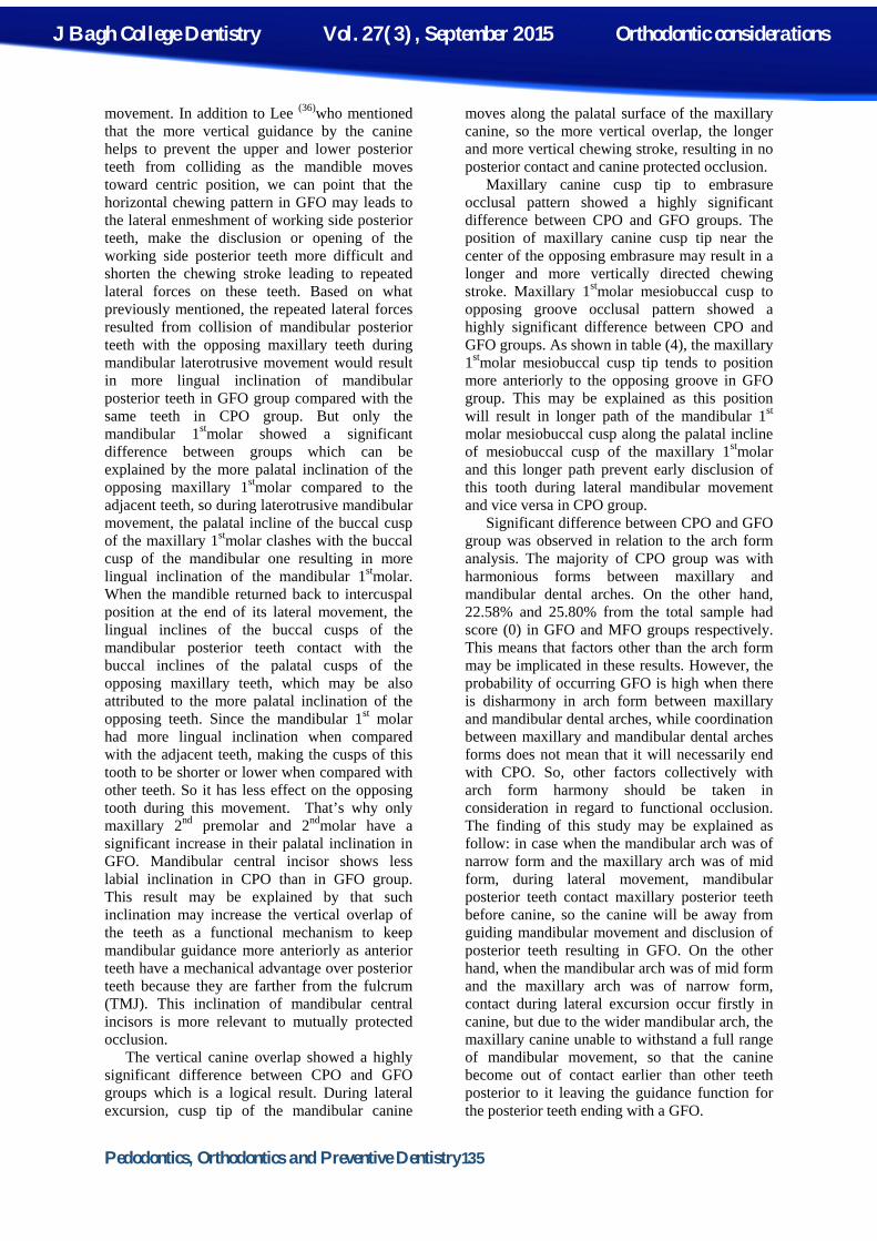
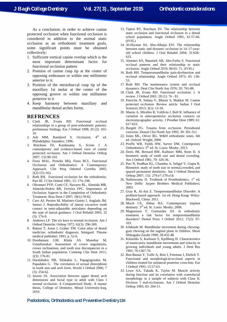
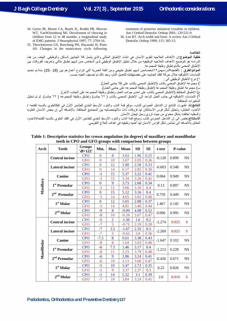
present study. Some of them were used to compare between two working side groups 'CPO' and 'GFO', while the other comparisons were made among three groups 'CPO', 'GFO', and 'MFO'. By using independent t-test only mandibular central and lateral incisors and 2ndmolar showed a statistically significant difference in crown angulation (Table 1), while 2nd premolar and 2ndmolar in the maxillary arch, central incisor and 1stmolar in the mandibular arch showed a statistically significant difference in crown inclination between the two groups (Table 2). Table (3) showed that CPO group had higher mean value of vertical canine overlap than GFO group according to independent t-test.
The obtained data for buccal cusp to embrasure/groove occlusal pattern was divided into 6 intervals as shown in table (4) and to assess the result Likelihood ratio was used, which showed that there were highly significant differences between groups for maxillary canine and 1st molar. Table (5) showed the result of arch form analysis, Contingency coefficient (C.C) was used to show if there is a significant association in arch form analysis scores among three studied groups. Results showed that there was a significant association among groups (Table 6). Furthermore, Odds ratio used to compare between each pair of groups, results showed that there was a significant difference between CPO and GFO and a non-significant difference between (CPO/MFO) and (GFO/MFO) (Table 7). DISCUSSION
Previous studies reported that class І canine and molar occlusal relationships associated with different functional occlusion patterns (13-15). These patterns may be considered normal or not in relation to other parts of the masticatory system and border mandibular movement. In the 1970s, orthodontic gnathologists argued that orthodontic patients’ functional occlusion should be finished to CPO and mentioned that patients would predictably finish with balancing contacts and eventual TMD when orthodontists ignore patients’ functional occlusion and rely on hand-held models rather than articulators (16,17).
Canine is the most appropriate tooth to guide mandibular lateral excursion due to crown morphology, good crown/root ratio and is capable of tolerating high occlusal force, and the canine root has a greater surface area than adjacent teeth which providing greater proprioception (18). Canines have been considered as the first line of control to keep the mandible functioning more vertically, so CPO reduces the chances of TMD. The canines were the most

J Bagh College Dentistry Vol. 27(3), September 2015 Orthodontic considerations
Pedodontics, Orthodontics and Preventive Dentistry134
likely candidates for mandibular guidance function as they prevent the lateral enmeshment of working side posterior teeth due to their strategic location from fulcrum and stress-breaking capabilities (19). Moreover, the forces directed out of the long axis of the posterior teeth in GFO associated with the presence of bacterial biofilm, can cause pathologies of occlusal origin (20,21). It has been observed that when canine protection is taken away, muscles stay active leading to clenching, grinding of teeth, abfraction, and gum recession (19).
Selection of sample in this study based on Class І normal occlusion, because the identification of the type of functional occlusion associated with normal static occlusion is of particular interest for orthodontists, since they direct their patients' treatment plan to achieve this type of static occlusion. In this study the age of the subjects was between 18-25 years, because the majority of facial growth is usually complete by 16-17 years of age (22). The late mandibular growth which occurs in late teens as well as earlier may influence the position of dentition relative to maxilla and mandible (23). So the changes which occurring during occlusal development could influence the occlusal contact pattern (15). Also, the subjects who were more than 25 years of age were excluded in the present study, due to more mutilation and attrition of dentition in those subjects (24,25).
Examining functional occlusion on articulated casts valid and reproducible method for occlusal diagnosis (1). It was established that fully-adjustable articulators have a large range of adjustability in three dimensions (18,26,27),So recording tooth contact by using a fully adjustable articulator with a correct transfer technique provide more accurate and reproducible results.
The CPO group showed higher mesial angulation for the mandibular second molar and higher distal angulation for the mandibular central and lateral incisors compared to GFO group. While, non-significant differences were found between the groups for all maxillary teeth (Table 1). This may be due to the fact that the mandible is the moving (functional) jaw, so it is more affected by other parts of the masticatory system. Misch and Abbas (28) considered maxilla as a force distribution unit and mandible as a force absorption unit. In the mandible only central, lateral incisors and second molar had significantly different angulations between groups; this may be related to the chewing kinematics and bio-functional anatomy of occlusion. The question here; is the chewing
kinematic cause such a difference, or this difference in crown angulation results in different chewing patterns between groups? The effect of occlusal factors on the function of the masticatory system is controversial topic for years (3,29). Some investigators hypothesized that occlusion and facial form contribute to functional variation (30-32), On the contrary, others (33) suggested that functional parameters and muscle activity patterns are significant factors contributing to occlusal schemes. Green et al. (34) mentioned that in the second year of life when the deciduous dentition reaches full occlusion, the characteristics of the shape of the masticatory cycle are finalized. Furthermore, Throckmorton et al. (35) added that the masticatory cycle do not vary much throughout life. It is possible that subjects with more vertical chewing patterns would best fit a canine protected functional occlusion scheme and those with horizontal chewing patterns would prefer more lateral freedom that would be consistent with group function (3). In conclusion there is a cause and effect relationship between occlusal parameter and type of chewing cycle or kinematic. The more horizontal chewing cycle in GFO may be a causative factor for the difference in crown angulations between groups in regard to mandibular anterior segment. This may be explained by that, the horizontal chewing cycle is associated with more horizontal (lateral) forces that transmitted from canine to mandibular anterior segment. Thus these teeth are in angulation more toward midline when compared with the same teeth of CPO group in which chewing cycle is more vertical. The vertical chewing kinematic in CPO and the direction of masseter muscle fibers, may result in more mesial angulation of the mandibular 2ndmolar to be parallel with this muscle fibers (2nd molar is near the masseter muscle).
Inclination of the posterior teeth may be of great importance in functional occlusion because of their direct relationship with lateral mandibular movement. The results of this study showed that there is a general increase in the palatal inclination for maxillary posterior teeth, and lingual inclination of mandibular 2nd premolar, 1st molar, and 2ndmolar in GFO group when compared with the same teeth in CPO group. Only maxillary 2nd premolar, 2nd molar and mandibular 1stmolar, showed statistical significant differences between groups (Table 2). These results may be explained by that; in CPO, the vertical chewing pattern helps to prevent the repeated lateral forces, and provide disclusion of the posterior teeth early during lateral mandibular
J Bagh College Dentistry Vol. 27(3), September 2015 Orthodontic considerations
Pedodontics, Orthodontics and Preventive Dentistry135
movement. In addition to Lee (36)who mentioned that the more vertical guidance by the canine helps to prevent the upper and lower posterior teeth from colliding as the mandible moves toward centric position, we can point that the horizontal chewing pattern in GFO may leads to the lateral enmeshment of working side posterior teeth, make the disclusion or opening of the working side posterior teeth more difficult and shorten the chewing stroke leading to repeated lateral forces on these teeth. Based on what previously mentioned, the repeated lateral forces resulted from collision of mandibular posterior teeth with the opposing maxillary teeth during mandibular laterotrusive movement would result in more lingual inclination of mandibular posterior teeth in GFO group compared with the same teeth in CPO group. But only the mandibular 1stmolar showed a significant difference between groups which can be explained by the more palatal inclination of the opposing maxillary 1stmolar compared to the adjacent teeth, so during laterotrusive mandibular movement, the palatal incline of the buccal cusp of the maxillary 1stmolar clashes with the buccal cusp of the mandibular one resulting in more lingual inclination of the mandibular 1stmolar. When the mandible returned back to intercuspal position at the end of its lateral movement, the lingual inclines of the buccal cusps of the mandibular posterior teeth contact with the buccal inclines of the palatal cusps of the opposing maxillary teeth, which may be also attributed to the more palatal inclination of the opposing teeth. Since the mandibular 1st molar had more lingual inclination when compared with the adjacent teeth, making the cusps of this tooth to be shorter or lower when compared with other teeth. So it has less effect on the opposing tooth during this movement. That’s why only maxillary 2nd premolar and 2ndmolar have a significant increase in their palatal inclination in GFO. Mandibular central incisor shows less labial inclination in CPO than in GFO group. This result may be explained by that such inclination may increase the vertical overlap of the teeth as a functional mechanism to keep mandibular guidance more anteriorly as anterior teeth have a mechanical advantage over posterior teeth because they are farther from the fulcrum (TMJ). This inclination of mandibular central incisors is more relevant to mutually protected occlusion.
The vertical canine overlap showed a highly significant difference between CPO and GFO groups which is a logical result. During lateral excursion, cusp tip of the mandibular canine
moves along the palatal surface of the maxillary canine, so the more vertical overlap, the longer and more vertical chewing stroke, resulting in no posterior contact and canine protected occlusion.
Maxillary canine cusp tip to embrasure occlusal pattern showed a highly significant difference between CPO and GFO groups. The position of maxillary canine cusp tip near the center of the opposing embrasure may result in a longer and more vertically directed chewing stroke. Maxillary 1stmolar mesiobuccal cusp to opposing groove occlusal pattern showed a highly significant difference between CPO and GFO groups. As shown in table (4), the maxillary 1stmolar mesiobuccal cusp tip tends to position more anteriorly to the opposing groove in GFO group. This may be explained as this position will result in longer path of the mandibular 1st molar mesiobuccal cusp along the palatal incline of mesiobuccal cusp of the maxillary 1stmolar and this longer path prevent early disclusion of this tooth during lateral mandibular movement and vice versa in CPO group.
Significant difference between CPO and GFO group was observed in relation to the arch form analysis. The majority of CPO group was with harmonious forms between maxillary and mandibular dental arches. On the other hand, 22.58% and 25.80% from the total sample had score (0) in GFO and MFO groups respectively. This means that factors other than the arch form may be implicated in these results. However, the probability of occurring GFO is high when there is disharmony in arch form between maxillary and mandibular dental arches, while coordination between maxillary and mandibular dental arches forms does not mean that it will necessarily end with CPO. So, other factors collectively with arch form harmony should be taken in consideration in regard to functional occlusion. The finding of this study may be explained as follow: in case when the mandibular arch was of narrow form and the maxillary arch was of mid form, during lateral movement, mandibular posterior teeth contact maxillary posterior teeth before canine, so the canine will be away from guiding mandibular movement and disclusion of posterior teeth resulting in GFO. On the other hand, when the mandibular arch was of mid form and the maxillary arch was of narrow form, contact during lateral excursion occur firstly in canine, but due to the wider mandibular arch, the maxillary canine unable to withstand a full range of mandibular movement, so that the canine become out of contact earlier than other teeth posterior to it leaving the guidance function for the posterior teeth ending with a GFO.
J Bagh College Dentistry Vol. 27(3), September 2015 Orthodontic considerations
Pedodontics, Orthodontics and Preventive Dentistry136
As a conclusion; in order to achieve canine protected occlusion when functional occlusion is considered in addition to the normal static occlusion as an orthodontic treatment goals, some significant points must be obtained collectively: 1. Sufficient vertical canine overlap which is the
most important determinant factor for functional occlusion pattern
2. Position of canine cusp tip at the center of opposing embrasure or within one millimeter anterior to it.
3. Position of the mesiobuccal cusp tip of the maxillary 1st molar at the center of the opposing groove or within one millimeter posterior to it
4. Keep harmony between maxillary and mandibular dental arches forms.
REFRENCES 1. Clark JR, Evans RD. Functional occlusal
relationships in a group of post-orthodontic patients: preliminary findings. Eur J Orthod 1998; 20 (2): 103-10.
2. Ash MM, Ramfjord S. Occlusion. 4th ed. Philadelphia: Saunders; 1996.
3. Rinchuse DJ, Kandasamy S, Sciote J. A contemporary and evidence-based view of canine protected occlusion. Am J Orthod Dentofac Orthop 2007; 132:90-102.
4. Feres MAL, Portella MQ, Feres RCL. Functional Occlusion and Orthodontics: A Contemporary Approach. Clin Pesq Odontol Curitiba 2005; 2(2):155-163.
5. Roth RH. Functional occlusion for the orthodontist. Part III. J Clin Orthod 1981; 15: 174–198.
6. Oltramari PVP, Conti CF, Navarro RL, Almeida MR, Almeida-Pedrin RR, Ferreira FPC. Importance of Occlusion Aspects in the Completion of Orthodontic Treatment. Braz Dent J 2007; 18(1):78-82.
7. Caro AJ, Peraire M, Martinez-Gomis J, Anglada JM, Samso J. Reproducibility of lateral excursive tooth contact in semi-adjustable articulator depending on the type of lateral guidance. J Oral Rehabil 2005; 32 (3): 174-9.
8. Andrews LF. The six keys to normal occlusion. Am J Orthod Dentofac Orthop 1972; 62(3): 296-309.
9. Rakosi T, Jonas I, Graber TM. Color atlas of dental medicine: orthodontic diagnosis. Sttutgard: Thieme medical publisher; 1993. p. 52-6.
10. Doodamani GM, Khala AS, Manohar M, Umashanakar. Assessment of crown angulations, crown inclinations, and tooth size discrepancies in a South Indian population. Contemp Clin Dent 2011; 2(3): 176-81.
11. Haralabakis NB, Sifakakis L, Papagrigorakis M, Papadakis G. The correlation of sexual dimorphism in tooth size and arch form. World J Orthod 2006; 7 (3): 254-62.
12. Jassim JA. Association between upper dental arch dimensions and facial type in adult with Class I normal occlusion: A Computerized Study. A master thesis, College of Dentistry, Mosul University-Iraq, 2010.
13. Tipton RT, Rinchuse DJ. The relationship between static occlusion and functional occlusion in a dental school population. Angle Orthod 1991; 61:57-66. (IVSL)
14. Al-Hiyasat AS, Abu-Alhaija ESJ. The relationship between static and dynamic occlusion in 14–17-year-old school children. J Oral Rehabil 2004; 31:628–633.
15. Alnimiri KS, Bataineh AB, Abo-Farha S. Functional occlusal patterns and their relationship to static occlusion. Angle Orthod 2010; 80:65–71. (IVSL)
16. Roth RH. Temporomandibular pain-dysfunction and occlusal relationship. Angle Orthod 1973; 43: 136-53.
17. Roth RH. The maintenance system and occlusal dynamics. Dent Clin North Am 1976; 20: 761-88.
18. Clark JR, Evans RD. Functional occlusion: I. A review. J Orthod 2001; 28 (1): 76 - 81.
19. Pasricha N, Sidana V, Bhasin S, Makkar M. Canine protected occlusion: Review article. Indian J Oral Sciences 2013; 3(1): 13-18.
20. Manns A, Miralles R, Valdivia J, Bull R. Influence of variation in anteroposterior occlusion contacts on electromyographic activity. J Prosthet Dent 1989; 61: 617-623.
21. Burgett FG. Trauma from occlusion. Periodontal concerns. Dental Clin North Am 1995; 39: 301-311.
22. Jones ML, Oliver RG. W&H orthodontic notes. 6th ed. Oxford: Wright; 2000.
23. Proffit WR, Fields HW, Sarver DM. Cotemporary Orthodontics. 5th ed. St. Louis: Mosby; 2013.
24. Doris JM, Bernard BW, Kuftinec MM, Stom D. A biometric study of tooth size and dental crowding. Am J Orthod 1981; 79: 326-36.
25. Puri N, Pradhan KL, Chandna A, Sehgal V, Gupta R. Biometric study of tooth size in normal,crowded, and spaced permanent dentitions. Am J Orthod Dentofac Orthop 2007; 132: 279.e7-279.e14.
26. Nallaswamy D. Textbook of Prosthodontics. 1st ed. New Delhi: Jaypee Brothers Medical Publishers; 2003.
27. Gray R, Al-Ani Z. Temporomandibular Disorder: A problem-based approach. 1st ed. Hong Kong: Wiley-Blackwell, China: 2011.
28. Misch CE, Abbas HA. Contemporary implant dentistry. 3rd ed. St. Louis: Mosby; 2008.
29. Magnusson T, Guimarães AS. Is orthodontic treatment a risk factor for temporomandibular disorders? Dental Press J Orthod 2012; 17(2): 97-103.
30. Ichihashi M. Mandibular movement during chewing-gum chewing on the sagittal plane in children. Shoni Shikagaku Zasshi 1990; 28:432-48.
31. Kiliaridis S, Karlsson S, Kjellberg H. Characteristics of masticatory mandibular movements and velocity in growing individuals and young adults. J Dent Res 1991; 70:1367-70.
32. Ben-Bassat Y, Yaffe A, Brin I, Freeman J, Ehrlich Y. Functional and morphological-occlusal aspects in children treated for unilateral posterior cross-bite. Eur J Orthod 1993; 15:57-63.
33. Lowe AA, Takada K, Taylor M. Muscle activity during function and its correlation with craniofacial morphology in a sample of subjects with Class II, Division 1 mal-occlusions. Am J Orthod Dentofac Orthop 1983; 83: 204-11.
J Bagh College Dentistry Vol. 27(3), September 2015 Orthodontic considerations
Pedodontics, Orthodontics and Preventive Dentistry137
34. Green JR, Moore CA, Ruark JL, Rodda PR, Morvee WT, VanWitzenburg MJ. Develoment of chewing in children from 12 to 48 months: a longitudinal study of EMG patterns. J Neurophysiol 1997; 77: 2704-16.
35. Throckmorton GS, Buschang PH, Hayasaki H, Pinto AS. Changes in the masticatory cycle following
treatment of posterior unilateral crossbite in children. Am J Orthod Dentofac Orthop 2001; 120:521-9.
36. Lee RT. Arch width and form: A review Am J Orthod Dentofac Orthop 1999; 115: 305-13.
الخلاصھالھدف من ھذه .نشاء الإطباق المثالي والذي یشمل كلا الجانبین الساكن والوظیفيإسنان ھي الأھداف العلاجیھ لتقویم الأن إ:خلفیة الموضوع
بین الإطباق الوظیفي لدى أشخاص ممن لدیھم إطباق مثالي وتعریف الفروقاتالوظیفیھ من خلال تحلیل الدراسھ ھو لتوضیح الأھداف العلاجیھ .إطباق وظیفة المجموعھوالإطباق المحمي بالناب
م تحدیدت.سنة) 25- 18(ومن الفئة العمریة التي تتراوح أعمارھم بین طبیعي إطباق لدیھم شخصاًممن٦٢منھتألفتعینةالدراس : الموادوالطرق :حسب العینھللتعدیل التام، وبعد ذلك تم تصنیف الجانبیھ على مفصلھقابلھ خلال حركة الفك الأطباقیھالتماسات
:نوع الاطباق الوظیفي الى ) ١ .)طباق المحمي بالناب على كلا جانبي العملالإ(طباق المحمي بالناب الإمجموعة ) أ
.)العملاطباق وظیفة المجموعھ على جانبي (مجموعة اطباق وظیفة المجموعھ ) ب ).طباق وظیفة المجموعھ على الجانب الآخرإطباق المحمي بالناب على احدى جوانب العمل والإ(الاطباق المختلط ) جثم تم تحلیل ).جانب ٦٢( طباق وظیفة المجموعھ إو) جانب ٦٢( طباق المحمي بالناب الإ: في جانب العمل الواحد الى الاطباقیھالتماسات ) ٢
. ةالمتغیرات المختلف/ ول في الفكالعلوي بالنسبھ للفتحھ الأوسط الخدي للطاحن الألقد اظھرت النتائج ان التداخل العمودي للناب، موقع قمة الناب والنتوء :النتائج
سنان العلویھ الأ ضافھ الى إن بعضبالإ. المختلفة حصائیھ بین المجامیعإذات دلالھ خدود المقابل، وتحلیل شكل قوس الأسنانكان لھا فروقاتالأ .سنانالأوالسفلیھ اختلفت بشكل معنوي من حیث تزوي ومیل تیجان
الاخدود /بالنسبھ للفتحھول في الفك العلوي الأوسط الخدي للطاحن الأالتداخل العمودي للناب وموقع قمة الناب والنتوء خلص الى إن:الأستنتاج .ھ وظیفیھ في اھداف العلاج التقویميسنان لھا أھمیالأاقواس شكل الى تجانس بالاضافھالمقابل
Table 1: Descriptive statistics for crown angulation (in degree) of maxillary and mandibular
teeth in CPO and GFO groups with comparison between groups
Arch Teeth Groups 'df=122' Min. Max. Mean SD SE t-test P-value
Max
illar
y
Central incisor CPO 0 8 3.63 1.96 0.25 -0.128 0.899 NS GFO 0 10 3.67 2.05 0.26
Lateral incisor CPO 0 12 5.88 2.58 0.33 -0.603 0.548 NS GFO 0 14 6.17 2.82 0.36
Canine CPO -1 15 5.37 3.22 0.41 0.064 0.949 NS GFO -1 15 5.34 3.26 0.41
1st Premolar CPO 0 9 3.73 2.68 0.34 0.13 0.897 NS GFO -2 11 3.66 3.16 0.4
2nd Premolar CPO 0 15 5.12 3.16 0.4 0.759 0.449 NS GFO -1 14 4.65 3.65 0.46
1st Molar CPO 0 12 5.65 2.88 0.37 1.467 0.145 NS GFO -3 14 4.81 3.46 0.44
2nd Molar CPO -9 8 -0.09 4.08 0.52 0.006 0.995 NS GFO -8 10 -0.10 3.67 0.47
Man
dibu
lar
Central incisor CPO -5 2 -1.50 1.6 0.2 -2.274 0.025 S GFO -7 5 -0.71 2.19 0.28
Lateral incisor CPO -7 3.5 -1.67 2.35 0.3 -2.269 0.025 S GFO -7 5 -0.62 2.8 0.36
Canine CPO -7.5 8 0.61 3.38 0.43 -1.647 0.102 NS GFO -8 8 1.64 3.63 0.46
1st Premolar CPO -6 7.5 1.46 3.17 0.4 -1.213 0.228 NS GFO -8 11 2.23 3.79 0.48
2nd Premolar CPO -6 9 2.86 3.24 0.41 -0.426 0.671 NS GFO -6 10 3.13 3.66 0.47
1st Molar CPO -5 10 3.47 2.72 0.35 0.22 0.826 NS GFO -1 9 3.37 2.37 0.3
2nd Molar CPO -1 14 5.32 3.1 0.39 2.6 0.010 S GFO -7 10 3.84 3.24 0.41
J Bagh College Dentistry Vol. 27(3), September 2015 Orthodontic considerations
Pedodontics, Orthodontics and Preventive Dentistry138
Table 2: Descriptive statistics for crown inclination (in degree) of maxillary and mandibular teeth in CPO and GFO groups with comparison between groups
Arch Teeth Groups 'df=122' Min. Max. Mean SD SE t-test P-value
Max
illar
y
Central incisor CPO -1 16 7.48 4.32 0.55 0.415 0.679 NS GFO -7 15 7.14 4.76 0.60
Lateral incisor CPO -7 18 5.34 5.53 0.70 -1 0.319 NS GFO -6 18 6.33 5.54 0.70
Canine CPO -11 19 -1.23 5.97 0.76 -0.802 0.424 NS GFO -11 13 -0.39 5.73 0.73
1st Premolar CPO -14 12 -1.46 5.19 0.66 1.261 0.21 NS GFO -14 10 -2.66 5.40 0.69
2nd Premolar CPO -17.5 8.5 -1.90 5.44 0.69 2.439 0.016 S GFO -18 7 -4.36 5.78 0.73
1st Molar CPO -18 6 -5.79 5.40 0.69 1.664 0.0.99 NS GFO -19 8 -7.41 5.45 0.69
2nd Molar CPO -19 10 -2.38 5.46 0.69 2.023 0.045 S GFO -17.5 16 -4.34 5.35 0.68
Man
dibu
lar
Central incisor CPO -9 12 3.01 6.18 0.79 -2.255 0.026 S GFO -8.5 14 5.4 5.57 0.71
Lateral incisor CPO -14 11 0.28 6.4 0.81 -1.098 0.274 NS GFO -13 11 1.43 5.21 0.66
Canine CPO -22 3 -5.46 5.56 0.71 -0.538 0.561 NS GFO -16.5 7 -4.89 5.35 0.68
1st Premolar CPO -22 5 -9.67 5.75 0.73 0.27 0.788 NS GFO -21 3 -9.95 5.46 0.69
2nd Premolar CPO -32 4 -14.5 6.54 0.83 1.351 0.179 NS GFO -33 0 -16.04 6.12 0.78
1st Molar CPO -35 19 -23.15 7.92 1.01 2.158 0.033 S GFO -35 -15 -25.63 4.41 0.56
2nd Molar CPO -38 -12 -26.97 5.85 0.74 0.375 0.708 NS GFO -38 24 -27.46 8.52 1.08
Table 3: Descriptive statistics for vertical canine overlap (mm) in CPO and GFO groups with comparison between groups.
Variable Groups 'df=122' Extreme values Mean SD SE t-test P-value
verti
cal
Can
ine
over
lap CPO Min. 2.06 3.203 0.626 0.080
4.048 0.000 HS
Max. 4.55
GFO Min. 1.46 2.730 0.675 0.086 Max. 3.99
Table 4: Intervals (mm) of buccal cusp to opposing embrasure/groove occlusal pattern in CPO and GFO with comparison between groups
Tooth Groups
Intervals Comparison
-2~-1 -1~0 0 0~1 1~2 2~2.5 Likelihood ratio df p-value
Canine CPO Freq. 0 0 31 13 14 4
14.406 4 0.006 (HS)
% 0 0 50 20.97 22.58 6.45
GFO Freq. 0 1 13 17 28 3 % 0 1.61 20.97 27.42 45.16 4.84
1st Premolar
CPO Freq. 0 5 30 13 12 2
4.247 4 0.375 (NS)
% 0 8.06 48.39 20.97 19.35 3.23
GFO Freq. 0 3 21 19 15 4 % 0 4.84 33.87 30.65 24.19 6.45
2nd Premolar
CPO Freq. 1 1 18 15 23 4 8.843 5 0.115
(NS) % 1.61 1.61 29.03 24.19 37.10 6.45 GFO Freq. 0 0 8 18 28 8

J Bagh College Dentistry Vol. 27(3), September 2015 Orthodontic considerations
Pedodontics, Orthodontics and Preventive Dentistry139
% 0 0 12.90 29.03 45.16 12.90
1st Molar
CPO Freq. 2 11 42 5 2 0
18.462 5 0.002 (HS)
% 3.23 17.74 67.74 8.06 3.23 0
GFO Freq. 2 1 38 11 9 1 % 3.23 1.61 61.29 17.74 14.52 1.61
2nd Molar
CPO Freq. 2 4 42 7 7 0
5.410 5 0.368 (NS)
% 3.23 6.45 67.74 11.29 11.29 0
GFO Freq. 2 1 39 8 10 2 % 3.23 1.61 62.90 12.90 16.13 3.23
Table 5: Arch form for maxillary and mandibular arches in CPO, GFO, and MFO groups.
Groups Score Arch form C
PO
0 No.=20
18 (M) 1 (W) 1 (N)
1 No.=2
1 'W' Maxillary arch 'M' Mandibular arch
1 'M' Maxillary arch 'N' Mandibular arch
GFO
0 No.=14
12 (M) 2 (W)
1 No.=8
4 'M' Maxillary arch 'N' Mandibular arch
4 'N' Maxillary arch 'M' Mandibular arch
MFO
0 No.=16
12 (M) 3 (N) 1 (W)
1 No.=2
'M' Maxillary arch 'W' Mandibular arch
Table 6: Arch form analysis in CPO, GFO, and MFO groups with comparison among groups.
Groups Freq. and Percentages Arch form analysis Total C.C Group difference
0 1 P- value
CPO Freq. 20 2 22
0.305 0.042 (S)
% Groups 90.90% 9.10% 100% % Total 32.25% 3.22% 35.47%
GFO Freq. 14 8 22
% Groups 63.60% 36.40% 100% %Total 22.58% 12.90% 35.48%
MFO Freq. 16 2 18
% Groups 88.90% 11.10% 100% % Total 25.80% 3.22% 29.02%
Table 7: Arch form analysis (comparison between each couple of groups by Odds Ratio).
Groups Odds Ratio P-value CPO/GFO 5.714 P=0.031 (S) CPO/MO 1.250 P=0.832 (NS)
MFO/GFO 4.566 P=0.067 (NS)
Related Documents